rna isolation – basic practical knowledge isolation.pdfrna extraction – basic practical...
TRANSCRIPT

Kis Enikő OKK-OSSKI
PCR training course June 13-17, 2016
RNA Extraction – Basic Practical Knowledge

• Only one strand – more vulnerable than two-stranded DNA.
• Ribose-phosphate backbone
• Uracil in place of thymine
• Different RNA pool in the nucleolus, the nucleus or in the cytoplasm.
• Complicated secondary and terciary structures for free energy minimalization.
RNA

Nature of information one can obtain through cell RNA content investigation:
• Gene expression-regulation information
• Alternative spliced transcripts investigation
• Gene fusion
• Epigenetic regulation
• Transpozone regulation

Types of RNA in the Cell
• Micro-RNA - miRNA: „ identification code” denotes mRNAs which are to be decoyed for the ribonuclease protein complex (22-24b)
• Small interferring RNA – siRNA: post-transcriptional silencing and methylation of DNA target sites; heterochromatin formation (22-24b)
• PIWI-interacting RNA – piRNA: in the cytoplasm they are RNA decomposers, while in the nucleus they draw DNA or histone methylation; they have a role in transposon mRNA breakdown (22-24b)
• Transfer RNA – tRNA: amino acid transport to the ribosomes during protein biosynthesis (76-90b)
• Small nucleolar RNA – snoRNA: rRNA maturation (around 100b)
• Small nuclear ribonucleic acid – snRNA: mRNA maturation (around 150b)
• Long non-coding RNA - lncRNA: transcription regulation (around200b)
• Messenger RNA – mRNA: template for protein biosynthesis (1900-2200b)
• Ribosomal RNA – rRNA: translation/protein synthesis (t.l. 7216b)

mRNA : facts that must be taken into consideration:
• Procaryotes: polycistronic – more proteins from the same mRNA: at the same time more ribosomal complexes work on the same, just maturated mRNA – which on the other end might simultaneously be decayed by a protein complex
• Eucharyotes: the majority is monocistronic mRNA – one protein – one mRNA
• mRNA presence in cells – gene expression analysis
• Transcription is time-dependent and abundance of mRNA in a cell depends on protein necessity and stimulus.
• Very variable between different cells in nature and quantity from fewer than 10 copies to several hundreds of copies – good quality RNA isolate is needed for further investigations.

Choosing a well working method…
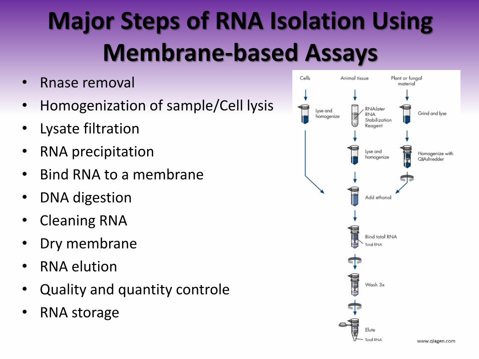
Major Steps of RNA Isolation Using Membrane-based Assays
• Rnase removal
• Homogenization of sample/Cell lysis
• Lysate filtration
• RNA precipitation
• Bind RNA to a membrane
• DNA digestion
• Cleaning RNA
• Dry membrane
• RNA elution
• Quality and quantity controle
• RNA storage

The Dreadful RNases
• Endoribonucleases or exoribonucleases.
• They are everywhere. RNase7 is secreted by human skin as antipathogene defense.
• Some tissues and cells contain them in abundant measures (eg. pancreas, some fruits)
• Reactivate after boiling or autoclaving.

How to protect one’s RNA from them?

Protection Against RNases
• We usually never isolate DNA and RNA in the same areas/labs.
• Equipment used for RNA extraction is cleaned thoroughly and kept separate from common lab equipment.
• We treat tables, racks and platforms with various harsh chemicals (eg. DEP, formamide, chloroform, NaOH and SDS combined solution) that destroy RNases.
• Always use and often change gloves. Even for preparation of labware and chemicals.
• At the beginning of the extraction RNA is exposed to RNases released from the sample. Use of different Rnase inhibitor reagents like β-mercapto-etanol, guanidine isothiocyanate, phenol or SDS in our solutions insures intrinsic protection of RNA.
• At the end of the procedure special care is required because in pure water RNA is very vulnerable. RNA is immediately placed on ice and frozen to -20°C or /especially in case of important samples/ -70°C.

Determining the Correct Amount of Starting Material
Depends:
• RNA content of sample
• Lysis buffer capacity
• The capacity of the method (eg: phenol-chloroform method vs. membrane based method) – kits usually emphasize this information
• Usually around 1*107 cells or 10-30mg tissue sample would work.
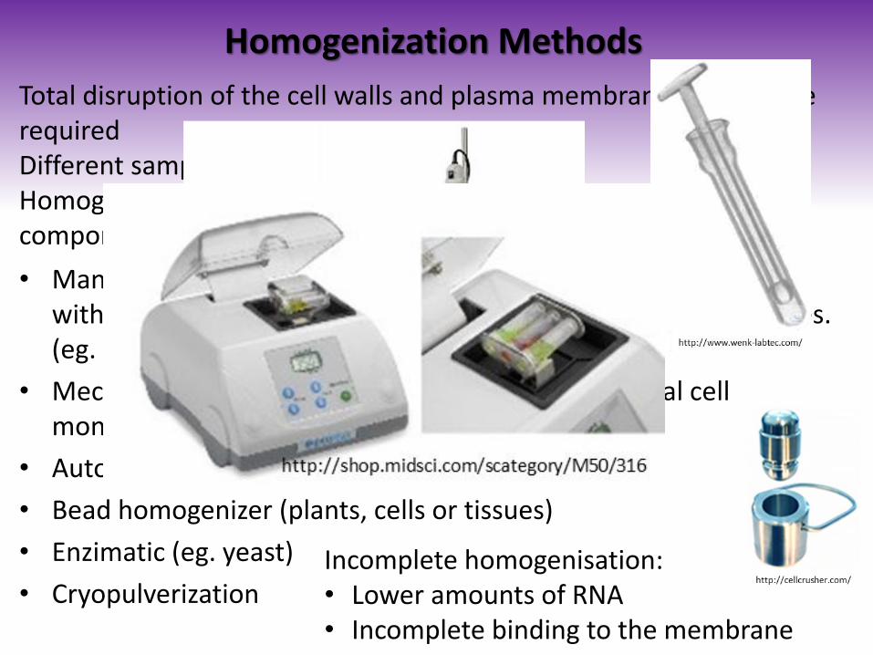
Homogenization Methods
• Manual: douncer (Alexander Dounce 1909-1997) – glass mortar with ceramic pestle - 13µm – cells are wrecked but not organelles. (eg. plants, filametous funghi)
• Mechanical: scalpel or syringe-and needle (eg. animal cell monolayers)
• Automatic homogenizator (eg. animal tissues)
• Bead homogenizer (plants, cells or tissues)
• Enzimatic (eg. yeast)
• Cryopulverization
Total disruption of the cell walls and plasma membranes of cells are required Different samples – different methods Homogenisation shears high molecular weight DNA and cellular components, reduces wiscosity of the sample
Incomplete homogenisation: • Lower amounts of RNA • Incomplete binding to the membrane

Homogenization of Sample and Cell Lysis - Considerations
Cultured cells:
• Easier to lyse – no/little homogenization required
• Needle and syringe
• Homogeneous RNA pool
Tissue sections
• Different lysis difficulties due to ECM composition
• Homogenizers: manual, mechanincal or automated
• Directly homogenise frozen samples
• Smaller sample requisite
• Heterogeneous RNA pool
• Hard to reproduce

Cell Lysis
Strong denaturating agents:
• Total RNA extraction
• Releases all nucleic acid content
• Proteins are also denaturated and inactivated. Rnases, too.
• Dnase treatment required
• Tissue lysis also feasible.
Mild lysis buffer:
• Keeping cell compartements intact
• Separated isolation of different RNA pools of different compartments
• Attention to RNases! Intrinsic inactivators are inevitable to use.

Purifying RNA
• Sample filtration might be needed in case of insufficient homogenization.
• RNA precipitation is usually performed by etanol or isopropanol.
• Binding RNA to a membrane facilitates purification/ otherwise it is imperial not to loose the precipitate.
• RNA binds strongly to the silica membrane in presence of high salt concentration, and strong denaturing agents (eg .: guanidium salts).
• DNA digestion by DNase I – very sensible to physical denaturation and functionates at room temperature.
• After centrifugation and washing steps, the nucleic acid can be washed off with low salt aqueous solution!

RNA Concentration and Quality Control • Quantity might be determined by:
• spectrophotometric quantification: 260nm: 1U corresponds to 40µg/ml
• Multi-Mode Microplate Reader: at 260/280nm using the Gene5. program / Nucleic Acid Quantification
• RNA quality control:
• 260/280nm: <1.8 protein contamination. Optimal: 2.00 (1.8- 2.3 )
• 260/230nm: <1.8 other organic contaminants. Optimal: 2.0-2.4
• 260/240nm: <1.4 ionic contamination. Optimal: 1.4
• Gel electrophoresis:
• agarose gel at a 1.2% concentration. For mammalian rRNA, a 28S:18S rRNA ratio of 2:1 is generally representative of good-quality RNA.
• Special RNase free and denaturating formaldehyde agarose gel electrophoresis.
• Bioanalyser: software-based electrophoresis analysis

RNA Quality Control 2.
Phenol-chloroform method
Membrane-based on-column isolation

Storage
• Short term (<1 week) dissolved in nuclease-free water or alcoholic precipitate at -20°C.
• Long-term storage (2-6 months): dissolved in nuclease-free water at -80°C.
• In pure formamide (100%), FORMAzol® or RNAlater® up to two years at -20°C, BUT: it must be cleaned prior to reverse transcription!
• Aliquoting samples is very useful in case of multiple downstream use – to avoid thaw-freez cycles

Thank You for Your attention!