review there is nothing permanent except … microbiology elsevier veterinary microbiology 43 ( i995...
TRANSCRIPT
veterinary microbiology
Veterinary Microbiology 43 ( I995 ) 103-122 ELSEVIER
Review
There is nothing permanent except change. The emergence of new virus diseases
Uwe Truyen a,*, Colin R. Parrish b, Timm C. Harder ‘, Oskar- Riiger Kaaden a
” Institute for Medical Microbiology, Infectious and Epidemic Diseases, Ludwig Maximilians University,
Veterinaer Str. I3? D-80539 Munich, Germany
b James A. Baker Institute for Animal Health, New York State College of Veterinary Medicine, Cornell University, Ithaca, New York 14853, USA
’ Department of Virology, Erasmus University Rotterdam, P. 0. Box 1738, 3000 DR Rotterdam, Netherlands
Received 14 March 1994; accepted 21 June 1994
Abstract
The sudden appearance of apparently new viruses with pathogenic potential is of fundamental
importance in medical microbiology and a constant threat to humans and animals. The emergence of
a “new” pathogen is not an isolated event, as for instance the frequent appearance of new influenza
virus strains demonstrates. Often the new virus strains co-circulate with the older strains in a suscep-
tible population, but a replacement of the older strains has been also observed. In rare instances the
new viruses can cause dramatic epidemics or pandemics, such as those observed with the human
immunodeficiency virus, canine parvovirus, or most recently, with the agent of bovine spongiform encephalopathy in the United Kingdom. The mechanisms of the emergence are not always clearly
understood, but an altered host range appears to be a common event. Whether a true change in host range occurs, or whether the virus adapted to the host and replicated more efficiently, is often unknown.
This review tries to summarize the facts that are known about a wide variety of “new” viruses of mammals. such as the simian, human and feline Ientiviruses, the feline coronaviruses, the feline
parvovintses, the carnivore morbilliviruses, the influenza A viruses, and the transmissible spongiform encephalopathies. A particular emphasis will be put on the genetic mechanisms that might have taken place and that might have been responsible for their sudden appearance.
Kqvwords: Virus. evolution
0378-l 135/95/$09.50 0 1995 Elsevier Science B.V. All rights reserved SSDIO378- I 115
104 Uwe Truyen et al. /Veterinary Microbiology 43 (1995) 103-122
1. Introduction
“There is nothing permanent except change”, this citation from Heraclitus, taken from Webster et al., 1992, nicely introduces the topic of this review. With the sudden appearance
of HIV as the cause of AIDS in the early 198Os, the study of virus evolution has gained
broad attention. During this devastating pandemic it became very obvious that “new”
infectious diseases are an ever present threat to humans and that we cannot feel secure in
having combatted the classical plagues.
The often high virulence and mortality of “newly” emerged viruses is thought to be the
expression of an yet incomplete adaption to the host. As with other parasites natural selection
should favor mutualism, the co-existence of host and parasite, and the new virus should
eventually loose its virulence and become well adapted to the new host. However, this
adaption can take a long period of time, so that many people or animals will have suffered
and died before the equilibrium between host and parasite is reached.
This review will only cover those viruses that have a considerable importance for man
or animal, and only examples of apparently “new” viruses, such as the feline parvoviruses,
the carnivore morbilliviruses, influenza A viruses, simian, human and feline lentiviruses, feline coronaviruses, and the transmissible spongiform encephalopathies will be discussed
in greater detail. “Emerging diseases”, caused by agents that had been known to be
pathogenic for the particular host and have only gained a broader distribution within the
susceptible species by for instance migration of the host, as seen in smallpox, are excluded.
The discussion of the mechanisms for the generation of “new” pathogens has to consider
two main developments: First, the genetic alteration of the agent, which might result in a
different phenotype of the agent (e.g., an altered host range or an altered pathogenic
potential) or secondly, a change in the ecology of the host, which might have given the
agent a higher efficiency of transmission within a susceptible population. Both factors may
be equally important and are, alone or combined, involved in the natural history of any
“new” pathogen examined so far.
1.1. Mechanisms of the genetic evolution
Mutation: Any organism containing DNA or RNA will show mutations of its genome
over time. The number of mutations and the mutation rate vary greatly between viruses, between genes, and even between gene regions. The mutation rate of an organism, i.e. the frequency of mutations occurring in a given time, is inversely proportional to its size. This
implies that any virus has a higher mutation rate than a procaryotic or eucaryotic organism. RNA viruses generally mutate more rapidly than DNA viruses. This is believed to be due to the lack of an error-correcting activity in RNA-dependent RNA polymerases during virus replication. Because of immunological pressure the mutation rate of surface protein genes is generally higher than that of the genes encoding internal structural proteins or of non- structural proteins.
A mutation can result in an amino acid substitution or deletion in the protein, referred to as “coding” or “non-synonymous” change, or may not result in an amino acid change,
* Corresponding author. Tel.: (0049) 89 2180-2535, Fax: (0049) 89 2180-2597, E-Mail:
uSal [email protected].

Uwe Truyen et al. /Veterinary Microbiology 43 (1995) 103-122 105
referred to as “silent” or “synonymous” change. Non-synonymous changes can further
be classified as “missense” (coding for a different amino acid) or “nonsense” (leading
to the termination of the protein) mutations. Missense changes are the most important changes evolutionarily since nonsense changes usually generate replication-defective
viruses. The importance, if any, of synonymous changes in most genes is often unknown.
The frequency of silent and non-silent changes in a given protein can give an indication of
possible selection pressures. Surface protein genes often show a high percentage of non-
silent changes due to immunological pressure, whereas internal proteins generally show a
greater accumulation of silent over non-silent changes.
Recombination: Genetic intratypic or intertypic recombinations - the exchange of whole
stretches of the genome between organisms - are other mechanisms of evolution. The co-
infection of a cell with two distinct viruses or virus strains is a prerequisite for that mech-
anism. The exchange of whole parts of the genome can result in sudden and profound
changes of the phenotype of the agent. Reassortment: For viruses with a segmented genome, such as influenza or reoviruses, a
third mechanism of evolution is of great importance. After co-infection of a cell genome
segments of different strains can be mixed and rearranged in the progeny viruses, and these
can generate new viruses with very distinct biological properties.
With all of these mechanisms, the majority of mutations and recombinations most likely
lead to viruses which are either replication-defective or which do not have any advantage
in the population compared to the parental viruses, and which therefore will not be selected.
1.2. Ecologicalfactors in the evolution of emerging viruses
Social circumstances: Other factors contributing to the emergence of viruses or virus
diseases are circumstantial and involve altered patterns of migration of the host or changes in the behavioral pattern. Altered food sources could also crucially influence the epidemi-
ology of “new viruses”, as seen with the agent of the bovine spongiform encephalopathy
(BSE) or with that of Kuru. Exposure of susceptible hosts to an agent can lead to the
emergence of a new disease by introducing a new virus resource.
Selective pressure and constraints: The fate of the “new” viruses in the host population
depends upon several factors, and is not easily predicted. The contagiousness of the virus
and the route of transmission may be important factors, and also the resistance and stability
of the virus. The more contagious and stable a virus is the more likely is that there will be an epi- or pandemic spread of the disease. Whether a “newly” evolved virus will co-exist with the parental or ancestral viruses in the host population depends upon whether the new virus acquired properties that give it significant epidemiological advantages.
Immunological pressure. Immunological pressure is one obvious possibility since a change and subsequent loss of an epitope critical for neutralization can give an epidemio- logical advantage for the virus in a susceptible population. This is certainly true for influenza viruses, where the reassortment of HA segments can generate a virus with very distinct antigenic features, that evades the immune response, and is able to spread without being affected by preexisting virus-specific immunological defense mechanisms for a certain period of time. This mechanism might have also contributed to the possible evolution of canine parvovirus from a virus of cats. The change in the capsid protein that allowed the
106 Uwe Truyen et al. / Veterinary Microbiology 43 (1995) 103-122
virus to replicate efficiently in the dog, were paralleled by the disappearance of a cat virus- specific neutralizing epitope.
Also mutations resulting in a change of biological properties, e.g. efficacy of virus
shedding or stability of the virion might influence the fate of the “new ” virus.
2. Feline parvoviruses
The emergence of canine parvovirus appears to constitute an example of the emergence
of a “new” pathogen based almost exclusively on genetic mutation.
Feline panleukopenia virus (FPV) has been known for decades. It can cause an acute
systemic disease in cats resulting in a profound leukopenia and enteritis. The virus was
known only from cats or raccoons until in the mid- 1940s when a very similar disease with
a high mortality was observed in mink. The mink agent named mink enteritis virus (MEV) , was indistinguishable from FPV by conventional serological and biochemical tests, and
was subsequently classified as a virulence variant of FPV. In 1978, another virus emerged
in yet another carnivore species, the dog. Again the symptoms resembled the well known
clinical picture seen for FPV infection of cats, and the virus isolated from diseased dogs
was initially indistinguishable from FPV. This new virus, canine parvovirus type 2 (CPV-
2), was responsible for a devastating pandemic affecting virtually all dog populations
worldwide. The reasons for the rapid and global spread of this virus were its high conta-
giousness and its remarkable tenacity. It hit an unprotected dog population and killed initially
thousands of dogs. The origin and the mechanisms of the virus evolution have been subject of great interest, and it is now generally believed that CPV was derived from feline panleu-
kopenia virus or a closely related carnivore parvovirus that gained the canine host range-
although its true origin still remains to be discovered. This virus system offers a nearly
unique opportunity to study virus evolution on the molecular level for several reasons. First,
parvoviruses are relatively simply organized viruses with a small DNA genome of about 5
kb, making genetic manipulations feasible. Secondly, the new virus CPV emerged during
the mid 1970s when techniques in virus and tissue conservation were well developed. This renders the preservation of potential ancestor viruses in some diagnostic laboratories or histopathologic collections highly likely.
Various hypotheses about the mechanisms of the evolution of the virus group have been developed. The most widely accepted hypothesis is the emergence of CPV from a virus of cats, most likely from FPV, by natural mutation (Appel et al., 1979; Parrish, 1990; Parrish, 1993). Other hypotheses have been that it could have been derived from a virus of a different carnivore such as the Arctic (blue) fox (Veijaleinen, 1988; U. Truyen and C.R. Parrish, unpublished results), or that it could have been generated from canine cells contaminated with FPV and then subsequent adaption of that virus to canine cells in vitro and distribution
via contaminated vaccines (Siegl, 1984). If the evolution was indeed from a cat virus to a new dog virus, the virus had to gain the
ability to infect, replicate in, and then shed from dogs. Previous studies have shown that FPV is capable of replicating in dogs, albeit in a restricted fashion (Truyen and Parrish, 1992). FPV replication could only be seen in the thymus and bone marrow, but not in the gut or mesenteric lymph nodes which are main target tissues for CPV and which are most
Uwe Truyen et al. /Veterinary Microbiology 43 (1995) 103-122 107
likely the prerequisite for efficient virus shedding. Therefore, during the evolution the CPV
ancestor had only to gain the ability to infect the gut in order to be shed and spread in the
dog population. By examining viruses recombinant between CPV and FPV it has been shown that the
capsid protein determines the host range of both viruses in vitro and in vivo (Parrish, 1991,
Chang et al., 1992, Truyen et al., 1994, U. Truyen and CR. Parrish, unpublished results).
Further analyses revealed that amino acids 80,564 and/or 568 of the feline sequence were
essential to give the virus the full replication ability in cats. If only one of those changes is
present, the resulting virus does replicate, although in a restricted fashion. When both regions
are the canine sequence the virus behaves like wild type CPV and does not replicate in cats
(Truyen et al., 1994). The mechanisms of the in vivo host range restriction are unknown, but do not appear to
involve interactions of the virus with feline serum constituents or red blood cells as observed
in other virus systems, like influenza virus. This has been examined under in vitro conditions
by incubating FPV or CPV with feline serum or erythrocytes at pH 7.4 and 37°C and
subsequent titration of the remaining infectivity. It could be shown that there was no
significant drop in infectivity compared to virus suspensions that were incubated with PBS
(Truyen et al., 1994). Three amino acids, amino acids 93, 103, and 323 all apparently determine the canine
host range of CPV - the ability to replicate in canine cells in vitro and in the canine gut in
vivo (Chang et al., 1992, U. Truyen and C.R. Parrish, unpublished data). Changing only those three amino acids in the FPV sequence generated a virus that infected the canine gut
and mesenteric lymph nodes, and which was shed in the feces to titers comparable to those
of wild type CPV. Furthermore, amino acid 93 created an CPV-specific epitope, generating
a virus that would have been serologically characterized as CPV.
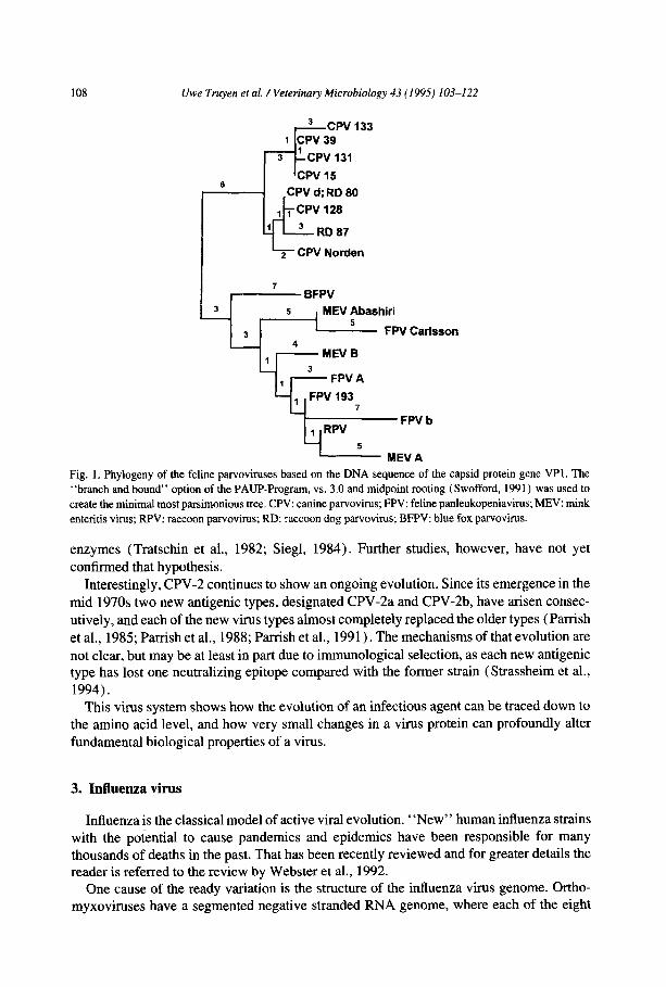
Sequence analysis of the capsid protein gene (which determines the canine and feline
host range) of numerous parvovirus isolates revealed that the feline parvoviruses form two
distinct clusters represented by FPV-type viruses from cats, raccoons, and mink, and by
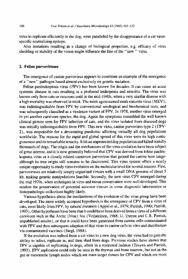
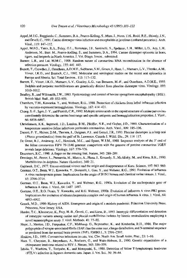
CPV-type viruses, isolated from dogs and raccoon dogs (Fig. 1) . Those clusters are sepa- rated by twelve conserved nucleotide changes, six coding and six non-coding changes
(Parrish, 199 1) . All six non-synonymous (coding) changes have been functionally defined,
as three are important for the feline in vivo host range and the three others for the canine host range in vitro and in vivo. Furthermore, amino acid 80 defines at least a part of an
FPV-specific epitope, while amino acid 93 defines a CPV-specific epitope (Parrish, 199 1, Chang et al., 1992; Truyen et al., 1994).
Interestingly, a virus isolated from an Arctic fox from Finland (blue fox parvovirus, BFPV) appears to be an intermediate between the FPV- and CPV cluster (U. Truyen and C.R. Parrish, unpublished results). Although the non-synonymous changes are all the feline sequence, three of the six synonymous changes were the canine sequence. The examination of further fox isolates is necessary to understand the role of the fox in the evolution of the feline parvoviruses.
Shortly after the first appearance of CPV the hypothesis was put forward that it has been derived from and distributed in an accidently or deliberately contaminated vaccine. This was primarily suggested by the fact that CPV replicated efficiently in feline cells, and that some FPV vaccine strains particularly resembled CPV in the pattern of single restriction
108 Uwe Truyen et al. /Veterinary Microbiology 43 (1995) 103-122
I ,L BFPV
q 3 , 5 , MEV ~bash;wCatisson
‘1 , +FPVA
Fig. 1. Phylogeny of the feline parvoviruses based on the DNA sequence of the capsid protein gene VPI. The “branch and bound” option of the PAUP-Program, vs. 3.0 and midpoint rooting (Swofford, 1991) was used to create the minimal most parsimonious tree. CPV: canine parvovirus; FPV: feline panleukopeniavirus; MEV: mink enteritis virus; RPV: raccoon parvovirus; RD: raccoon dog parvovirus; BFPV: blue fox parvovirus.
enzymes (Tratschin et al., 1982; Siegl, 1984). Further studies, however, have not yet
confirmed that hypothesis.
Interestingly, CPV-2 continues to show an ongoing evolution. Since its emergence in the
mid 1970s two new antigenic types, designated CPV-2a and CPV-2b, have arisen consec-
utively, and each of the new virus types almost completely replaced the older types (Parrish
et al., 1985; Parrish et al., 1988; Parrish et al., 1991). The mechanisms of that evolution are
not clear, but may be at least in part due to immunological selection, as each new antigenic type has lost one neutralizing epitope compared with the former strain (Strassheim et al., 1994).
This virus system shows how the evolution of an infectious agent can be traced down to the amino acid level, and how very small changes in a virus protein can profoundly alter
fundamental biological properties of a virus.
3. Influenza virus
Influenza is the classical model of active viral evolution. “New” human influenza strains with the potential to cause pandemics and epidemics have been responsible for many thousands of deaths in the past. That has been recently reviewed and for greater details the reader is referred to the review by Webster et al., 1992.
One cause of the ready variation is the structure of the influenza virus genome. Ortho- myxoviruses have a segmented negative stranded RNA genome, where each of the eight
Uwe Truyen et al. /Veterinary Microbiology 43 (1995) 103-122 109
segments codes for one or two structural or non-structural viral proteins, and each is
replicated independently. If a co-infection of a cell occurs, a reassortment of gene segments
can take place and can give rise to a new virus with an altered gene complement. Evolutionary studies of influenza viruses have to consider the viral genome organization,
and a phylogeny of each genome segment has to be determined and investigated separately.
In terms of evolution the focus has to be put on those proteins which determine the host
range. Although the final role of the virus proteins remains to be elucidated and other
proteins also influence host range (e.g., the PB2 protein (Subbarao et al., 1993)) two
structural proteins of particular importance for the pathogenesis and host range of these
viruses are the hemagglutinin (HA) and the neuraminidase (NA) (Schulman and Palese,
1977; Sugiura and Ueda, 1980). Hernagglutinin and neuraminidase. The HA is a membrane bound protein which is
glycosylated and acylated. It is the major structural protein of the virion and it mediates
receptor binding and also the fusion between the viral envelope and the endosome membrane. For those functions it has to undergo a proteolytic cleavage which is mediated
by host specific trypsin-like proteases which cleave the precursor HA0 into the active
subunits HA1 and HA2. These subunits are linked by disulfide bridges, and are organized
as homotrimers. The cleavability of the HA is a determinant of virulence but probably not
of host range, as there are cells which do cleave the HA but which still do not replicate the
virus (Chambers et al., 1988). Indirectly, however, it might have some influence on host
range, in that the cleaved HA is less stable at low pHs than the uncleaved HAO, which may
be important when transmission modes are used that involve fluids of low PH. More likely
to influence the host range is the receptor specificity of the various HA subtypes as influenza
strains from different hosts have different receptor specificities. The receptor for influenza
viruses has been shown to involve sialic acid. Different HA subtypes bind specifically to
certain sialyloligosaccharides. For example the human HA3 binds almost exclusively SA
2, 6Ga1, whereas the avian and equine HA3 binds to SA 2,3Gal (Rogers and Paulson, 1983). This receptor specificity appears to be reflected in part by a difference of amino acid
266 between those viruses, although that is not the sole amino acid change involved (Rogers
and D’Souza, 1989). All 14 HA antigenic subtypes (HI-H14) have been detected in avian
species, whereas only HI, H2, and H3 have been detected in humans, Hl and H3 in swine,
and H3 and H7 in horses, respectively. The reasons for these restrictions are unknown and
cannot be explained solely on the basis of different receptor specificities. The viral neura-
minidase (NA) is also involved in determination of host range, but the mechanisms are not defined. Interestingly, the distribution of different NA subtypes appears to be restricted to certain hosts, and seems to be linked to particular HAS.
Erlolution of in$uenza A viruses and the emergence of new pandemic strains: Although each genome segment and hence each viral protein may have evolved separately, the HA and NA appear to be of particular importance for the host range and pathogenicity of new strains and are therefore interesting structures in terms of the emergence of pandemic human strains.
The evolution of influenza viruses is well studied. The main divergence of the lineages influenza A, B, and C is thought to have occurred quite early, most probably before the 20th century (Smith and Palese, 1989). Assuming a constant molecular clock the earliest divergence among the mammalian strains probably occurred around 1800 when the equine
110
“RlJssian”
1977
HlNl
,-pJ
“Spanish” “Asian” “Hong-Kong”
1389 1900 1918 1957 1968
H2N2 H3N8 HlNl H2N2 H3N2 1
=: L1 E L_J
? = I
= Et% c
ZZ ZZ I I -
A 8? A 3 A 2
Uwe Truyen et al. /Veterinary Microbiology 43 (1995) X03-122
HlNl H2N2 H3N?
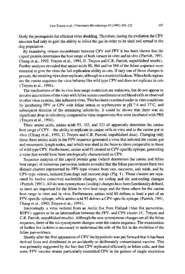
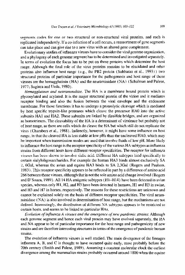
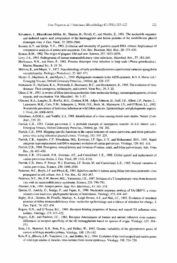
Fig. 2. Proposed evolution of the pandemic human influenza strains (modified from Webster et al., 1992).
Phylogenetic evidence suggests that an interspecies transmission from a duck virus to humans occurred before
1918, replacing the then prevalent human H3N8 strains. This HlNl virus most likely emerged in North America
and was carried to Europe by American world war I troops. This virus acquired three gene segments (HA, NA,
PB) from a H2N2 duck virus by reassortment, generating a virus that caused the “Asian flu” pandemic in 1957. The HA and PB gene segments were replaced in 1968 by an H3N? duck virus, creating the “Hong-Kong flu”
pandemic virus. This virus is still prevalent today. All new strains replaced the older strains. Surprisingly, the
HlNl known from ‘Spanish flu”, which disappeared in 1950, reappeared in 1977. It is suggested that this virus
escaped from a laboratory where it was stored during those years.
influenza 1 virus emerged (Gorman et al., 1990a, Gorman et al., 1990b). The current human viruses apparently first appeared in the early 19OOs, and these also probably gave rise to the classical swine influenza viruses prior to 1918 (Gorman et al., 1991).
Early experiments with RNA-RNA hybridization and oligonucleotide mapping showed that the HA and NA genes of the human pandemic strains were most closely related to those of avian strains, and that the only human HA types from this century - H 1, H2, H3 - have
an avian counterpart (Gorman et al., 1990a, Gorman et al., 1990b; Gorman et al., 1991). Interestingly, there appears to be a cyclical alternation of the Hl, H2, and H3 viruses as the predominant strains in humans. The mechanisms of those emergences were shown to be reassortment events of single genome segments between human and avian viruses or as
Uwe Truyen et al. / Veterinary Microbiology 43 ( 1995) 103-122 111
with HlNl virus of the Spanish influenza exchanges of all eight segments (Gorman et al.,
1990a, Gorman et al., 1990b). A schematic summary of the evolution of the pandemic
human strains is shown in Fig. 2. But how can those rearrangements occur when those viruses have such narrow host
ranges? From epidemiological data it appears that the pig may be the “reassortment vessel”, and that this occurs most frequently in Asia. This is suggested by several facts:
(I) Recent human pandemics (Asian influenza, 1957; Hong-Kong influenza, 1968;
Russian influenza, 1977) first occurred in China (an exception might have been the Spanish
influenza, which may have arisen in military camps in Kansas and was then taken to Europe
by U.S. troops in 1918).
(II) Human strains are confined to the Hl, H2, and H3 subtypes, those of swine to H2 and H3.
(III) Pigs are susceptible to HlNl and H3N2 influenza strains of both human and avian
origin.
( IV) Humans have accidentally contracted influenza viruses from pigs.
(V) There is no evidence for a susceptibility of man to true avian influenza viruses,
making an intermediate host necessary.
Circumstantial evidence suggests that southern China might be an influenza epicenter. In
these tropical and subtropical regions influenza strains in ducks, pigs and humans are
prevalent the whole year. Also the populations of humans, pigs, and ducks are the largest in the world, making an efficient interspecies transmission feasible. But although this
scenario might explain the emergence of pandemic and epidemic influenza strains, it is still
only speculation, and needs to be further evaluated by close examinations of Chinese influenza strains.
4. Transmissible Spongiform Encephalopathies (TSE)
This class of pathogen might not be directly analogous to emerging viruses, since the
nature of the agents is still only partially understood and current evidence suggests a proteinaceous nature which is different from viruses (Prusiner, 1989).
The agents of TSE have some unique features which are important for their epidemiology.
First they appear to be devoid of nucleic acids, and may constitute infectious proteins
(Prusiner, 1989). Secondly, they are extremely heat resistant, staying infectious even after boiling for several hours or after extensive X-ray radiation, and thirdly, they do not elicit an immune response, which might be due to their possible nature as a posttranslational modification of a host protein (Hope et al., 1986). They all cause after a long incubation
period a central nervous disease with a characteristic (spongiform) histopathology in defined regions of the CNS.
These agents, in particular Kuru and the bovine spongiform encephalopathy (BSE) appear to be examples where changes in particular cultural patterns or food sources were the most important factors leading to the emergence of the new disease.
In Km-u the mode of transmission was shown to be the ritual cannibalism among the Fore tribe in Papua-New Guinea. Generally only women and children acquired the disease and only very rarely men. Women were responsible for the dissection of the dead kinsmen and
112 Uwe Truyen et al. /Veterinary Microbiology 43 (1995) 103-122
the preparation of the meal. During the mourning ritual all flesh of dead kinsmen was
prepared on hot stones and cooked in bamboo cylinders, and those materials were rarely
washed or changed. During the preparation of the meal the soiled hands of the women were
cleaned by rubbing them in their armpits or elsewhere on their bodies. Any abrasions of the skin were potential locations for efficient inoculation and the transmission via local wounds
is generally thought to have been the main route of transmission (Gajdusek, 1977). After
learning about the epidemiology and uncovering the cannibalism as the main source of
infection the Fore people have stopped the ritual cannibalism and no more new infections
have been reported. The origin of the first case of Kuru is unknown, but a source from a
spontaneous case of Creutzfeldt-Jakob disease is suspected.
A very similar general epidemiology was seen with BSE in the United Kingdom. In 1985
an apparently new central nervous disease was observed in British cattle. It strongly resem-
bled the known symptoms of scrapie in sheep and it was soon obvious that both diseases
and agents were very similar, if not identical. The appearance of BSE is thought to be due
to the feeding of carcasses of scrapie-infected sheep via meat meal preparations to cattle. The agent has a remarkable resistance against heating, and even temperatures around 100°C
for hours do not significantly reduce infectivity. A change in the rendering processing of
meat meal and a peak in the sheep population in the UK most likely led to the appearance
of the disease in 1985 (Wilesmith et al., 1988). The processing of the carcasses, heating of
the carcasses, and treatment with organic solvents was reduced. This apparently enabled
the agent to survive the remaining harsh rendering processing procedures and to stay
infectious in the final product. Subsequently, the agent may have infected cattle and the first
animals which succumbed to clinical disease may have been included in the bone meal
production, thereby amplifying transmission and spread among the cattle population of the UK. After elucidation of this epidemiology, the feeding of meat meal to ruminants was
banned and with a delay corresponding to an incubation period of 5-7 years the incidence
of BSE appears to have reached a plateau during 1993, and is expected to decrease rapidly
and to finally vanish in the very near future. In May 1990 the first naturally occurring case of a transmissible spongiform encephalop-
athy in a cat was diagnosed (Wyatt et al., 1993). Since then more than 50 cases have been reported in domestic cats in the UK. It was known that cats were susceptible to experimental infection with the Creutzfeldt-Jakob disease agent (Mitrova and Mayer, 1977)) and exper-
imental inoculation of BSE material into cats reproduced the disease. Although not formerly proven from plain epidemiological data, the emergence of FSE (feline spongiform enceph- alopathy (as it was soon termed) ) is most likely due to the feeding of BSE contaminated bovine tissues to cats. Two wild felid species, the cheetah and the puma were diagnosed with FSE in 1992 in zoos in the UK and Australia. The two cheetahs were born in the same zoo developed the disease after feeding tissues including spinal cord from fallen cattle before the general awareness of BSE in the UK. One of the cheetahs was exported to Australia during the incubation period (summarized by Bradley and Wilesmith, 1993). The emergence of FSE is a further example of the emergence of a new foodbome infectious disease by introducing an until then unknown or very uncommon infectious agent into the foodchain.
Vwe Truyen et al. /Veterinary Microbiology 43 (1995) 103-122 113
5. Human and simian lentiviruses (UT)
The discussion of the sudden appearance and origin of the human immunodeficiency virus (HIV), the causative agent of AIDS, is one of the hot topics in medical virology since
the first isolation of HIV in 1983. The “new” disease first appeared in the early 1980s and
is already responsible for thousands of deaths among humans in USA and Europe, and it is expected to depopulate whole regions in Africa and Asia in the very near future. It is
estimated that in some African countries more than 40% of the people are infected with the
deadly virus. The natural history of AIDS has been reviewed recently, and the reader is
referred to Grmek, 1990, Nathanson et al., 1993, or Myers et al., 1993 for greater details.
The origin of HIV is still a matter of dispute, but it appears that the virus was around in
the human population for at least hundred years, but that it was confined to some isolated
African villages. It was due to social circumstances (e.g., urbanization and intercontinental
traffic) that it became initially distributed to the African cities and eventually to America, Europe and Asia. Here the virus spread by the well known routes mainly as a blood-borne
and sexually transmitted disease. A similar history has been described for the bubonic plague which was almost dormant in Asia in the Burma-Unnan region but which spread
readily after cultural changes due to the Mongol invasion (Krause, 1992).
Up to now two distinct human lentiviruses have been described, HIV-l and HIV-2. Both
have the ability to infect humans, to cause AIDS, and to spread in the population, although
it appears that HIV-2 is not as virulent as HIV- 1 (Levy, 1993).
A number of simian immunodeficiency viruses (SIV) have been isolated from non- human primates, such as SIV,,c from macaques, SIV,, from sooty mangabeys, SIV,,,
from African green monkeys, and SIV,,, from mandrills (summarized in)Desroisers,
1990. More recently another SIV isolate that is only distantly related to other SIV isolates but which is very closely related to HIV-l, named SIV,,,, was described in a captive
chimpanzee in Gabon (Huet et al., 1990). All known SIV isolates appear to be non- pathogenic for their respective hosts, and only New World and Asian Old World monkeys
develop disease, most likely because the virus is not yet adapted to those hosts. Natural
infections with SIV have not been described for Asian Old World or New World primates and SIV,,, is believed to have arisen by infection of Asian macaques with SIV,,, in a
primate center in the US where Asian and African Old World monkeys were housed together
(Allan, 1992).
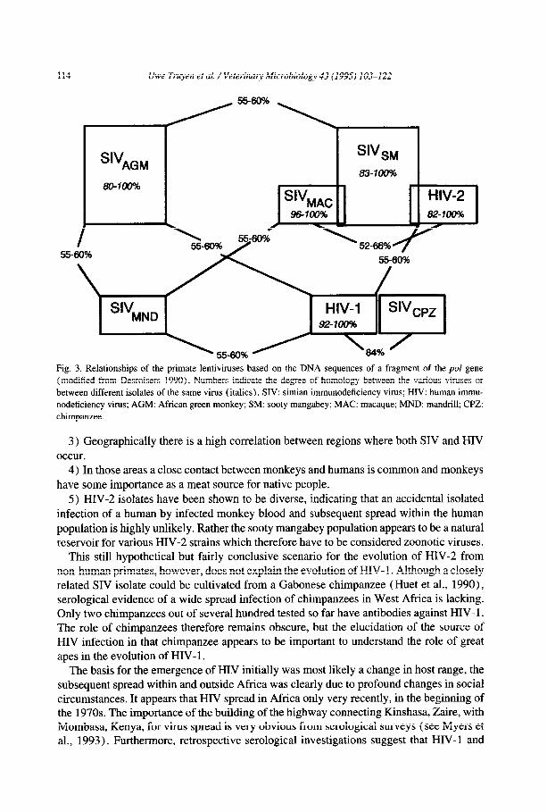
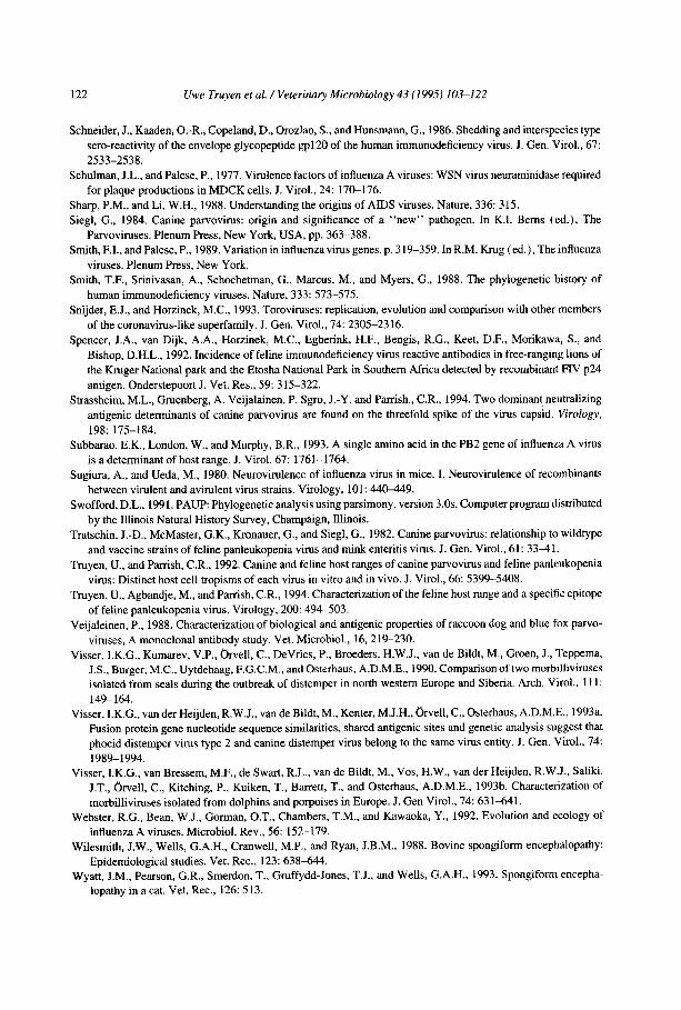
The HIV and SIV isolates cluster in complexes which show different degrees of similarity (Figure 3). Interestingly, both subtypes of HIV fall together with different SIV isolates into distinct clusters, HIV-2 together with SIV,, and SIV,,c, and HIV-l clusters with SIV,,. These relationships are the basis for the current hypothesis about the emergence of HIV as the result of an interspecies transmission from African non-human primates to man. This holds up nicely at least for HIV-2 for several reasons.
1) African monkeys have been shown to be enzootically infected with SIV. About 10% of wild-caught sooty mangabeys (Cercocebus atys) and about 20-50% of sub-saharan African green monkeys (Cercopithecus aerhiops) were shown to be infected with SIV,,
or SIV,,,, respectively (see Nathanson et al., 1993).
2) SIV and HIV-2 are albeit some diversity genetically highly related, and it is impossible to discriminate SIV and HIV-2 isolates (but not HIV-l isolates!) from the plain nucleotide sequences.
114 Uwe Truyen et al. /Veterinary Microbiology 43 (1995) 103-122
s'vAGM s'"SM
83-10056 80-100% c
%AC HIV-2 96100% 82-100%
S-60% 5560%
Fig. 3. Relationships of the primate lentiviruses based on the DNA sequences of a fragment of the pol gene (modified from Desroisers 1990). Numbers indicate the degree of homology between the various viruses or
between different isolates of the same virus (italics). SIV: simian immunodeficiency virus; HIV: human immu-
nodeficiency virus; AGM: African green monkey; SM: sooty mangabey; MAC: macaque; MND: mandrill; CPZ:
chimpanzee.
3) Geographically there is a high correlation between regions where both SIV and HIV
occur. 4) In those areas a close contact between monkeys and humans is common and monkeys
have some importance as a meat source for native people.
5) HIV-2 isolates have been shown to be diverse, indicating that an accidental isolated
infection of a human by infected monkey blood and subsequent spread within the human
population is highly unlikely. Rather the sooty mangabey population appears to be a natural reservoir for various HIV-2 strains which therefore have to be considered zoonotic viruses.
This still hypothetical but fairly conclusive scenario for the evolution of HIV-2 from non-human primates, however, does not explain the evolution of HIV- 1. Although a closely related SIV isolate could be cultivated from a Gabonese chimpanzee (Huet et al., 1990)) serological evidence of a wide spread infection of chimpanzees in West Africa is lacking. Only two chimpanzees out of several hundred tested so far have antibodies against HIV- 1. The role of chimpanzees therefore remains obscure, but the elucidation of the source of HIV infection in that chimpanzee appears to be important to understand the role of great apes in the evolution of HIV- 1.
The basis for the emergence of HIV initially was most likely a change in host range, the subsequent spread within and outside Africa was clearly due to profound changes in social circumstances. It appears that HIV spread in Africa only very recently, in the beginning of the 1970s. The importance of the building of the highway connecting Kinshasa, Zaire, with Mombasa, Kenya, for virus spread is very obvious from serological surveys (see Myers et al., 1993). Furthermore, retrospective serological investigations suggest that HIV-l and
Uwe Truyen et al. /Veterinary Microbiology 43 (1995) 103-122 115
HIV-2 emerged separately at about the same time in geographically separated regions of
Africa. HIV- 1 most likely emerged in southern and eastern Africa, whereas HIV-2 clearly
emerged in West-Africa. Assuming the emergence of both HIV 1 and HIV-2 from different ancestors in non-
human primates at about the same time the sudden parallel appearance seems highly
unlikely, unless there is a common reason for that. Two factors may have been responsible or contributed to the sudden spread of those
viruses. First, in the 1960s there was a peak in the export of monkeys from Africa to Europe
and into the USA. This entailed extensive handling of the animals. Secondly, there was a push in vaccination in ‘Third World’ countries which involved the intensive use of needles,
which did not get disinfected after use. Furthermore, the widely used polio vaccine was
produced in Vero cells of simian origin, and it was suggested that some batches could have been contaminated with simian lentiviruses, as was shown to have happened with the
polyoma virus SV40 (as summarized by Myers et al., 1993).
But for both of these theories there is not yet any proof. They can give, however, a
possible scenario which makes the highly improbable parallel appearance of two distinct human lentiviruses look no longer impossible.
At which time point the human viruses emerged from a common ancestor or when exactly
the simian and the human lineages separated is still very contradictory. The most accepted
estimates based on sequence analyses of different lentivirus genes suggest that the lineages
split about 40 to 200 years ago (Smith et al., 1988; Sharp and Li, 1988; Querat et al., 1990). But regardless of which estimate will prove to be the closest, the crucial step in HIV evolution
appeared to be the initial spread within Africa. It would be a great tragedy if beneficial and life saving vaccines have contributed to the pandemic character of this new devastating
disease.
6. Feline lentiviruses
Feline immunodeficiency virus (FIV), a member of the genus Lentivirus within the family Retroviridae was firstly isolated by Pedersen et al. ( 1987) from cats with clinical
signs of immunodeficiency. Although the virus is a pathogen for cats, it is especially regarded
as an animal model for the human acquired immunodeficiency syndrome (AIDS) mainly
for antiviral chemotherapy. FIV has been isolated from cats around the world and the FIV antibody prevalence varies between 1 and 30% (Ishida et al., 1988). Several isolates have been obtained independently from American, European and Asian countries and compari- sons of the nucleotide sequence of FIV proviral DNA indicated that the FIV genome organization is similar to that of HIV and other lentiviruses (Spencer et al., 1992). It is interesting to note that there is also an antigenic cross-reactivity between the major FIV core protein encoded by the gag gene and that of other lentiviruses such as maedi-visna virus, caprine arthritis-encephalitis virus, and equine infectious anaemia viruses (Schneider et al., 1986). Although all these viruses exhibit a strong species-specificity and do not create a hygienic risk for humans, the antigenic relationship suggests a common evolutionary ancestor. Whereas the gag gene of different FIV isolates appears to be highly conserved, sequence analyses of the enu gene from different American, European and Japanese isolates
116 Uwe Truyen et al. /Veterinary Microbiology 43 (1995) 103-122
revealed a clustered variability. Beside that variation in the sequence, however, the structural
comparison of the envelope glycoprotein also showed conserved regions between the iso- lates. Rigby et al. ( 1993) determined 16 fully conserved cysteine residues in the envelope
glycoprotein and six in the transmembrane protein. A phylogenetic study of the Ed and
gag sequences indicated that the FIV RNA sequences clearly form geographical clusters and that the Japanese FIV isolate represents a different subgroup of feline lentivirnses with
calculated amino acid homologies of 77% for the em and 92% for the gag genes (Rigby et
al., 1993). This variety of phylogenetic groups may be related to an intensive world-wide
trade in pet cats. Recently, multiple FIV-like lentivirus isolates designated PLV (puma lentivirus) were
obtained from Florida panthers (Fe& concoZor covi) . Genomic analysis of the PLV pal
genes revealed a high degree of sequence homologies with other known lentiviruses includ-
ing FIV. By immunoblots, a high prevalence of cross-reactive antibodies to an FIV-like
lentivirus was found in serum samples obtained from 12 species of free-ranging felids
including lions, cheetahs and pumas from Africa and Northern America (Olmsted et al.,
1992). Although the phylogenetic analysis of FIV and PLV sequences from disparate geographic locations showed a significant divergence between the domestic and non-domes-
tic feline immunodeficiency isolates, both viruses appear to have a common ancestor and are widely distributed in felides around the world. Despite serious efforts in many labora-
tories, the evolutionary origin of FIV and PLV remains obscure and sequence comparisons
of more virus isolates are necessary to answer this question.
7. Morbilliviruses of marine mammals and terrestrial carnivores
In 1988 more than 15 000 harbor seals (Phoca uitulina) in north western European
waters and during 1990-l 99 1 several hundreds of striped dolphins ( StenelZu coeruloalbu) in the Mediterranean Sea succumbed to epizootic diseases attracting remarkable public attention. Acute infections by previously unknown morbilliviruses (order Mononegavirales,
family Paramyxoviridae) in these populations have been identified as the primary cause (Osterhaus and Vedder, 1988; Domingo et al., 1990). Another outbreak of a morbillivirus- related plague in marine mammals was reported from Lake Baikal, Siberia, where large numbers of Baikal seals (Phocu sibiricu) were affected (see Barrett et al., 1992).
Molecular studies on the European harbor seal morbillivirus isolates yielded convincing evidence that a separate, so far unknown species in the morbillivirus genus was encountered, which is referred to as phocid distemper virus (PDV- 1) (Visser et al., 1990; Barrett et al., 1992). Canine distemper virus (CDV), a well known morbillivirus infecting terrestrial carnivores is the closest relative of PDV-1, yet easily distinguishable even by classical serological techniques (Harder et al., 1993). The morbillivims isolates obtained from Phocidae during the Lake Baikal outbreak, in contrast, appeared to be almost identical to prototype CDV strains and were termed PDV-2 (Visser et al., 1990).
The deduced amino acid sequence of the PDV-1 morbillivirus membrane glycoprotein H, mediating the attachment process of virions to host cells, revealed only 74% identity to CDV (see e.g. Kovamees et al., 1991). Due to the high divergence (which was also shown for further genes) the origin of PDV- 1 can not be simply explained as a recently emerged
I/we Truyen et al. / Veterinary Microbiology 43 (1995) 103-122 117
host variant of CDV although both viruses are likely to have originated from a common
ancestor. However, a similarity of 97% of the deduced amino acid sequence between the
fusion ( FO) membrane glycoprotein of CDV and PDV-2 contrasting to 86% between PDV-
1 and PDV-2 prompted the suggestion that PDV-2 may have evolved from prototype CDV
by recent interspecies transmission from terrestrial carnivores (Visser et al., 1993a).
Whether a similar situation is faced with antigenetically CDV-like morbilliviruses provok-
ing clinically overt and occasionally lethal disease in free-ranging collared peccaries (jav-
elinas, Tayassuidae) and old world large cats (tigers, lions, leopards) kept in North
American zoos remains to be resolved (Appel et al., 1991, Appel et al., 1994).
The morbilliviruses isolated from diseased cetacean species such as porpoises (Pho-
coena) of the North Sea and striped dolphins of the Mediterranean clustered into another
“new” and separate entity in the morbillivirus genus, this time being more closely related
to the peste de petit ruminants morbillivirus (PPRV) than to the PDV/CDV group (Barrett
et al., 1993; Visser et al., 1993b). The high degree of divergence within a P- (phosphopro-
tein) gene fragment of the dolphin and porpoise morbilliviruses to PDV and other morbil-
liviruses again supported the assumption that these viruses had circulated separately in some
cetacean populations already for a long period rather than a recent species jump from PDV-
l-infected seals is reflected (Barrett et al., 1993).
Consequently, the question arose from what sources -if CDV-infected terrestrial cami-
vores were ruled out- two different morbilliviruses might have been introduced into Eur-
opean populations of marine mammals which had no history of significantly increased
mortality due to infectious (morbillivirus-related) diseases at least during the last decades.
In the case of PDV- 1 -infected harbor seals of the North Sea the attention fixed on vagrant
Arctic harp seals (Phoca groenlundica) for several reasons: PDV-l-specific antibodies are
highly prevalent in this species (Markussen and Have, 1992), PDV antigen has recently
been detected in a diseased harp seal (Daousi et al., 1993)) and the population size is large
enough to perpetuate endemic morbillivirus infections. In addition, harp seals are known to
have migrated in small groups over unusually large distances into European waters in 1987,
possibly forced by changes in their natural environment. Whether the 1988 European seal
epizootic can be explained solely by introduction of PDV-1 by migrating Arctic seals is
still under discussion. Data that would elucidate the situation in cetaceans are still too scarce
to allow reliable conclusions.
In addition to the description of further “new” and separate morbillivirus species, more
data have to be collected to prove recent emergences of morbillivirus biotype-variants being
based on mutations and to further elucidate the molecular base of host selection and viru- lence. An effect of increased public awareness of diseases in wild life combined with
enhanced viral diagnostics still should be considered when searching for explanation of an apparently expanded host range of CDV-like viruses ( Appel et al., 1994).
More complete data are available for the human measles virus. Recent wild-type isolates displayed differences being most pronounced in the genes encoding the hemagglutinin (H)
and nucleocapsid (N) proteins compared to widely used vaccine strains. Some non-syn-
onymous mutations resulted even in antigenically altered variants (Rota et al., 1992, Rota
et al., 1994). However, measles virus is still considered a monotypic virus and the vaccines, when properly used, should elicit protective immunity.
118 Uwe Truyen et al. /Veterinary Microbiology 43 (1995) 103-122
8. Feline coronaviruses
Feline enteric coronavirus (FECV) and feline infectious peritonitis virus (FIPV) are
members of the coronavirus-like superfamily (CVL) and they exhibit the typical features
of other coronaviruses such as a single-stranded RNA genome of about 30 kb and positive
polarity. The envelope glycoproteins occur as petal-shaped projections, or peplomers, and are involved in the virus attachment to receptor sites of susceptible cells (Olsen, 1993).
Coronavirus infections are widely distributed in the cat population around the world. It
was reported that as many as 85% of cats in individual catteries or multi-cat households were found to have coronavirus-specific antibodies (Hoskins, 1993). FECV and FIPV are
antigenically closely related and generally induce an asymptomatic carrier state in adult cats. Virulent strains of FIPV, such as FIPV-79-1146 and FIPV-DF2, however, can cause
a disease with gastrointestinal signs, fibrinous serositis and disseminated vasculitis known
as feline infectious peritonitis. Strains of low virulence (e.g. FIPV-UCD2 and FIPV-UCD3)
generally induce persistent infections without clinical manifestations. Although the clinical
symptoms of the effusive form of FIP are rather obvious, FIP was firstly diagnosed only in
the mid 1960s. These observations suggest that FIPV may have emerged as a new virus
most likely by recombination with another coronavirus. The most likely candidate for such a genetic event is TGEV, the agent of transmissible gastroenteritis of swine. A genetic
relationship between TGEV and FIPV was already suggested from transmission experiments whereby the cat coronavirus induced mild gastrointestinal disorders in piglets whereas
TGEV although capable of replicating in cats proved to be non-pathogenic. Sequence
comparisons between the pig and cat viruses revealed that the homology at the 3’ terminus
between amino acids 275 and 1447 of the major peplomer gene was greater than 90%. The homology between ammo acids 1 and 274, however, was less than 40% (De Groat et al.,
1988). These data suggest that FIPV emerged by intertypic recombination between TGEV
and a yet unknown coronavirus, may be FECV (Pedersen et al., 1981). Homologous
recombination between highly conserved RNA sequences at predicted genes (‘hot spots’)
is well documented for other coronaviruses and was especially described for the murine
hepatitis virus (MHV) (Banner and Lai, 1988). The general mutation frequency for RNA viruses is assumed to be about 10e4 mutations per base. Considering that the genome of coronaviruses such as TGEV and FIPV is 30 kb, a single replication step of these viruses may involve a substitution of three ‘false’ bases, The existence of different virulent FIPV strains indicates that genetic variation is also a common characteristic of this virus (Hoskins,
1993). Recently, a modified live FIPV vaccine was developed as a temperature-sensitive mutant
by attenuation of the FIPV DF2 strain. This vaccine virus was derived after continuous passages in cell cultures followed by ultraviolet irradiation (Christianson et al., 1989). The vaccine was found to be protective against challenge infection with moderate doses of challenge virus whereas with higher doses ( > lo5 TCIDSO) the vaccine even increased the severity of the clinical signs. With regard to the high mutation frequency and the possible risk of recombination events known for coronaviruses, possible recombinations with field strains of FIPV have to be monitored.
Recent analysis of the genomic organization and replication strategy clearly indicate that the genera corona-, toro- and arteriviruses diverged from a common ancestor (Snijder and
I/we Truyen et al. /Veterinary Microbiology 43 (1995) 103-122 119
Horzinek, 1993). The most convincing evidence for this virus evolution is the primary
amino acid sequence homology in the replicase gene of all three genera of CVL.
9. Epilogue
The brief description of various “new” viruses showed that the phenotypic appearance
of newly emerged virus epidemics is the result of interactions of at least three different
factors. First, genetic mechanisms, such as mutation and selection are continuous events occurring
during the replication of pathogenic microorganisms thus changing their biological prop- erties (e.g. virulence, antigenicity) and creating the possibility for new diseases. In the case
of some positive-strand RNA viruses like toga-, pesti-, flavi- and arteriviruses (which are
not described in detail in this review) the evolutionary scenario is well understood and the
putative events leading to alterations of the genome organization and thus expression have
been described in concise terms. Such evolutionary events include 1) gene duplication, 2) capture and integration of new (cellular) genes, 3) recombination with distantly related
viruses, 4) gene rearrangement (reassortment) amongst others (Koonin and Dolja, 1993).
Second, environment-related factors, such as movement of large populations, e.g. by tourism, war events or natural catastrophes can contribute to the spread of pathogenic agents
from endemic areas to formerly free parts of the world. Also changes in the management
of animals have been shown to be important sources of “new” viral diseases. The emergence
of bovine spongiform encephalopathy in British dairy herds after feeding of scrapie-con- taminated meat meal thus leading to the crossing of the species barrier from sheep to cattle
is the most recent and convincing finding in the field of infectious disease evolution.
Third, the genotypic out@ of men and animals with special emphasis on MHC genes can
influence the locations and pattern of virus epidemics. All three factors have been discussed in this review. However, it is quite obvious and
generally accepted that the most important achievements in the understanding of the molec-
ular epidemiology of newly emerging diseases were elaborated by genetic approaches such
as molecular cloning and sequencing, restriction analysis, mapping of viral epitopes and computer analysis of nucleotide and amino acid sequences. The use of these techniques will
further elucidate the mechanisms of the emergence of “new” pathogens and may even help to predict the outcome of some new viruses.
Acknowledgements
TCH is grateful to A. Osterhaus for constructive discussions on morbillivirus infections.
References
Allan, J.S., 1992. Viral evolution and AIDS. J. NIH Res., 4: 51-54.
Appel. M.J.G., Scott, F.W., and Carmichael, L.E., 1979. Isolation and immunization stuhies of a canine parve-
like virus from dogs with haemorrhagic enteritis. Vet. Rec., 105: 156-159.
120 Uwe Truyen et al. / Veterinav Microbiology 43 (1995) 103-122
Appel, M.J.G., Reggiardo,C., Summers, B.A., Pearce-Kelling, S., Mare, J., Noon,T.H., Redd, R.E., Shively, J.N.,
and Grveli, C., 1991. Canine distemper virus infection and encephalitis in javelinas (collared peccaries). Arch.
Virol., 119: 147-152.
Appel, M.J.G., Yates, R.A., Foley, G.L., Bernstein, J.J., Santinelli, S., Spelman, L.H., Miller, L.D., Arp. L.H.,
Anderson, M., Barr, M., Pearce-Kelling, S., and Summers, B.A., 1994. Canine distemper epizootic in lions,
tigers, and leopards in North America. J. Vet. Diagn. Invest., submitted.
Banner, L.R., and Lai, M.M.C., 1988. Random nature of coronavims RNA recombination in the absence of
selection pressure. Virology, 155: 441445.
Barrett, T., Crowther, J., Osterhaus, A.D.M.E., Subbarao, S.M., Groen, J., Haas, L., Mamaev, L.V., Titenko, A.M.,
Visser, I.K.G., and Bostock, C.J., 1992. Molecular and serological studies on the recent seal epizootics in
Europe and Siberia. Sci. Total Environ., 115: 117-132.
Barrett. T., Visser, I.K.G., Mamaev, L.V., Goatley, L.G., van Bressem, M.-F., and Osterhaus, A.D.M.E., 1993.
Dolphin and porpoise morbilliviruses are genetically distinct from phocine distemper virus. Virology, 193:
1010-1012.
Bradley, R., and Wilesmith, J.W., 1993. Epidemiology and control of bovine spongiform encephalopathy (BSE).
British Med. Bull., 49: 932-959.
Chambers, T.M., Kawaoka, Y., and, Webster, R.G., 1988. Protection of chickens from lethal influenza infection
by vaccinia-expressed hemagglutinin. Virology, 167: 4 14-42 1. Chang, S.-F., Sgro, J.-Y., and Parrish, C.R., 1992. Multiple amino acids in the capsid structure of canine parvovirus
coordinately determine the canine host range and specific antigenic and hemagglutination properties. J. Virol.,
66: 68586867.
Christianson, K.K., Ingersoll, J.D., Landon, R.M., Pfeiffer, N.E., and Gerber, J.D., 1989. Characterization of a
temperature sensitive feline infectious peritonitis coronavirus. Arch. Virol., 109: 185-196.
Daousi, P.-Y., Haines, D.M., Thorsen, J., Duignan, P.J., and Geraci, J.R., 1993. Phocine distemper in a harp seal
(Phoca groenlundica) from the gulf of St. Lawrence. Canada .I. Wildl. Dis., 29: 114-l 17.
De Groot, R.J., Andeweg, A.C., Horzinek, M.C., and Spaan, W.J.M., 1988. Sequence analysis of the 3’ end of
the feline coronavims FIPV 79-1146 genome: comparison with the genome of porcine coronavirus TGEV
reveals large deletions. Virology, 167: 370-376.
Desroisers, R.C., 1990. A finger on the missing link. Nature, 345: 288-289.
Domingo, M., Ferrer, L., Pumarola, M., Marco, A., Plana, J., Kennedy, S., McAlisky, M., and Rima, B.K., 1990.
Morbillivirus in dolphins. Nature (London), 348: 21.
Gajdusek, DC., 1977. Unconventional viruses and the origin and disappearance of Kuru. Science, 197: 943-960.
Gorman. O.T., Bean, W.J., Kawaoka, Y., Donatelli, I., Guo, Y., and Webster, R.G., 1991. Evolution of influenza
A virus nucleoprotein genes: Implications for the origin of HlNl human and classical swine viruses. J. Virol.,
65: 3704-37 14.
Gorman. O.T., Bean, W.J., Kawaoka, Y., and Webster, R.G., 1990a. Evolution of the nucleoprotein gene of influenza A virus. J. Virol., 64: 1487-1497.
Gorman, O.T., R.O. Donis, Y. Kawaoka, and R.G. Webster, 1990b. Evolution of influenza A virus PB2 genes:
Implications for evolution of ribonucleoprotein complex and origin of human influenza A virus. J. Virol., 64:
48934902.
Grmek, M.D., 1990: History of AIDS. Emergence and origin of a modem pandemic. Princeton University Press,
Princeton, New Jersey, USA.
Harder, T.C., Klusmeyer, K., Frey, H.-R., Grvell, C., and Liess, B., 1993. Intertypic differentiation and detection
of intratypic variants among canine and phocid morbillivirus isolates by kinetic neutralization employing a
novel immunoplague assay. J. virol. Methods, 41: 77-92.
Hope, J., Morton, J.D., Farquahar, C.F., Multhaup, G., Beyreuther, K., and Kimberlin, R.H., 1986. The major
polypeptide of scrapie-associated fibrils (SAF) has the same size, charge distribution, and N-terminal sequence
as predicted from the normal brain protein ( PrP) EMBO J.. 5: 259 l-2597.
Hoskins, J.D., 1993. Coronavirus infections in cats. Vet. Clin. North Am. Small Anim. Prac., 23: I-16.
Huet, T., Cheynier, R., Meyerhans, A., Roelants, G., and Wain-Hobson, S., 1990. Genetic organization of a
chimpanzee lentivims related to HIV- 1. Nature, 345: 356-359.
Ishida, T., Washizu, T., Toriyabe, K., and Motoyoshi, S., 1988. Detection of feline T-Iymphotropic Ientivitus
(FTLV) infection in Japanes domestic cats. Japan. J. Vet. Sci., 50: 3944.
C/we Truyen et al. /Veterinary Microbiology 43 (1995) 103-122 121
Kovamees, .I., Blixenkrone-Moller, M., Sharma, B., Grvell, C.. and Norrby, E., 1991. The nucleotide sequence
and deduced amino acid composition of the haemagglutin and fusion proteins of the morbillivirus phocid
distemper virus. J. Gen. Virol., 72: 2959-2966.
Koonin, E.V. and Dolja, V.V., 1993. Evolution and taxonomy of positive-strand RNA viruses: Implications of
comparative analysis of amino acid sequences. Crit. Rev. Biochem. Mol. Biol., 28: 375-430.
Krause, R.M., 1992. The origin of plagues: Old and new. Science, 257: 1073-1078.
Levy, J.A., 1993. Pathogenesis of human immunodeficiency virus infections. Microbial. Rev., 57: 183-289.
Markussen, N.H., and Have, P., 1992. Phocine distemper virus infection in harp seals (Phoca groenlandica).
Marine Mammal Sci., 8: 19-24.
Mitrova, E., and Mayer, V., 1977. Neurohistology of early preclinicallesionsin experimental subacute spongiform
encephalopathy. Biologia (Bratislava) , 32: 663-67 1.
Myers. G.. MacInnes, K., and Myers, L., 1993. Phylogenetic moments in the AIDS epidemic. In S. S. Morse (ed.)
Emerging Viruses, Oxford University Press Inc., Oxford, pp. 120-137.
Nathanson, N., McGann, K.A., Wilesmith, J., Desrosiers, R.C., and Brookmeyer, R., 1993. The evolution of virus
diseases: Their emergence, epidemicity, and control. Virus Res., 29: 3-20.
Olsen, C.W., 1993. A review of feline infectious peritonitis virus: molecular biology, immunopathogenesis, clinical
aspects, and vaccination. Vet. Microbial., 36: l-37.
Olmsted, R.A., Langley, R., Roelke, M.E., Goeken, R.M., Adger-Johnson, D., Goff, J.P., Albert, J.P., Packer, C.,
Laurenson, M.K., Care, T.M.. Scheepers, L, Wildt, D.E., Bush, M., Martenson. J.S., and O’Brien, S.J., 1992.
Worldwide prevalence of lentivims infection in wild feline species: Epidemiologic and phylogenetic aspects.
J. Viral., 66: 6008-6018.
Osterhaus. A.D.M.E., and Vedder, E.J.. 1988. Identification of a virus causing recent seals deaths. Nature (Lon-
don), 335: 20.
Parrish, C.R., 1993. Canine parvovims 2: A probable example of interspecies transfer. In S.S. Morse (ed.)
Emerging Viruses, Oxford University Press Inc., Oxford. pp. 194-202.
Parrish, C.R., 1991. Mapping specific functions in the capsid structure of canine parvovirus and feline panleuko-
penia virus using infectious plasmid clones. Virology, 183: 195-205.
Parrish, C.R., Aquadro, C.F. Strassheim, M.L. Everman, J.F. Sgro, J.-Y. and Mohammed, H.O.. 1991. Rapid
antigenic-type replacement and DNA sequence evolution of canine parvovirus. Virology, 129: 401414.
Parrish, C.R., 1990. Emergence, natural history and variation of canine, mink, and feline parvoviruses. Adv. Virus
Res.. 38: 403-450.
Parrish, C.R., O’Connell, P.H., Everman, J.F., and Carmichael, L.E., 1988. Global spread and replacement of
canine parvovirus strains. J. Gen. Virol., 69: 111 l-l 116.
Parrish, C.R., Have, P. Foreyt, W.J. Everman, J.F. Senda, M. and Carmichael, L.E., 1985. Natural variation of
canine parvovirus. Science, 230: 1046-1048.
Pedersen, N.C., Boyle. J.F. and Floyd, K., 1981. Infection studies in kittens using feline infectious peritonitis virus
propagated in cell culture. Am. J. Vet. Res., 42: 363-367.
Pedersen, N.C., Ho, E.W, Brown, M.L., Yamamoto, J.K., 1987. Isolation of a T-lymphotropic virus from domestic
cats with an immunodeficiency-syndrome. Science, 235: 790-793.
Prusiner. S.B., 1989. Scrapie prions. Ann. Rev. Microbial., 43: 345-374.
Querat, G., Audoly, G., Sonigo. P., and Vigne, R., 1990. Nucleotide sequence analysis of SA-OMVV, a visna- related ovine lentivirus: phylogenetic history of lentiviruses. Virology, 175: 434-447.
Rigby, M.A.. Holmes, M. Pistello. Mackay, A., Leigh Brown. A.J., and Neil, J.C., 1993. Evolution of structural
proteins of feline immunodeficiency virus: molecular epidemiology and evidence of selection for change. J. Gen. Virol., 74: 425436.
Rogers, G.N., and D’Souza, B.L., 1989. Receptor binding properties of human and animal Hl influenza virus
isolates. Virology, 173: 317-322.
Rogers, G.N., and Paulson, J.C., 1983. Receptor determinants of human and animal influenza virus isolates:
differences in receptor specificity of the H3 hemagglutinin based on species of origin. Virology, 127: 361-
373.
Rota, J.S., Hummel, K.B., Rota. P.A., and Bellini, W., 1992. Genetic variability of the glycoprotein genes of current wild-type measles isolates. Virology, 188: 135-142.
Rota, P.A., Bloom, A.E.. Vanchiere, J.A., and Bellini, W.J., 1994. Evolution of the nucleocapsid and matrix genes
of wild-type strains of measles virus isolates from recent epidemics. Virology, 198: 724-730.
122 Uwe Truyen et al. / Veterinary Microbiology 43 (1995) 103-122
Schneider, J., Kaaden, O.-R., Copeland, D., Orozlan, S., and Hunsmann, G., 1986. Shedding and interspecies type
sero-reactivity of the envelope glycopeptide gp120 of the human immunodeficiency virus. J. Gen. Virol., 67:
2533-2538.
Schulman, J.L., and Palese, P., 1977. Virulence factors of influenza A viruses: WSN virus neuraminidase required
for plaque productions in MDCK cells. J. Viral., 24: 170-176.
Sharp, P.M., and Li, W.H., 1988. Understanding the origins of AIDS viruses. Nature, 336: 315.
Siegl, G., 1984. Canine parvovirus: origin and significance of a “new” pathogen. In K.I. Bems (ed.), The
Parvoviruses. Plenum Press, New York, USA, pp. 363-388.
Smith, FL, and Palese, P., 1989. Variation in influenza virus genes. p. 3 19-359. In R.M. Krug (ed.), The influenza
viruses. Plenum Press, New York.
Smith, T.F., Srinivasan, A., Schochetman, G., Marcus, M., and Myers, G., 1988. The phylogenetic history of
human immunodeficiency viruses. Nature, 333: 573-575.
Snijder, E.J., and Horzinek, M.C., 1993. Toroviruses: replication, evolution and comparison with other members
of the coronavirus-like superfamily. J. Gen. Virol., 74: 2305-2316.
Spencer, J.A., van Dijk, A.A., Horzinek, M.C., Egberink, H.F., Bengis, R.G., Keet, D.F., Morikawa, S., and
Bishop, D.H.L., 1992. Incidence of feline immunodeficiency virus reactive antibodies in free-ranging lions of
the Kruger National park and the Etosha National Park in Southern Africa detected by recombinant FIV p24
antigen. Onderstepoort J. Vet. Res., 59: 315-322.
Strassheim, M.L., Gruenberg, A. Veijalainen, P. Sgro, J.-Y. and Parrish., CR., 1994. Two dominant neutralizing
antigenic determinants of canine parvovirus are found on the threefold spike of the virus capsid. Virology, 198: 175-184.
Subbarao, E.K., London, W.. and Murphy, B.R., 1993. A single amino acid in the PB2 gene of influenza A virus
is a determinant of host range. J. Viral. 67: 1761-1764.
Sugiura, A., and Ueda, M., 1980. Neurovirulence of influenza virus in mice. I. Neurovirulence of recombinants
between virulent and avirulent virus strains. Virology, 101: 440-449.
Swofford, D.L., 1991. PAUP: Phylogenetic analysis using parsimony, version 3.0s. Computer program distributed
by the Illinois Natural History Survey, Champaign, Illinois.
Tratschin, J.-D., McMaster, G.K., Kronauer, G., and Siegl, G.. 1982. Canine parvovirus: relationship to wildtype
and vaccine strains of feline panleukopenia virus and mink enteritis virus. J. Gen. Viral., 61: 33-41.
Truyen, U., and Parrish, C.R., 1992. Canine and feline host ranges of canine parvovirus and feline panleukopenia
virus: Distinct host cell tropisms of each virus in vitro and in vivo. J. Virol., 66: 5399-5408.
Truyen, U., Agbandje, M., and Parrish, C.R., 1994. Characterization of the feline host range and a specific epitope
of feline panleukopenia virus. Virology, 200: 494-503.
Veijaleinen, P., 1988. Characterization of biological and antigenic properties of raccoon dog and blue fox parvo-
viruses, A monoclonal antibody study. Vet. Microbial., 16,219-230.
Visser, I.K.G., Kumarev. V.P., &vell, C., DeVries, P., Broeders, H.W.J., van de Bildt, M., Groen, J., Teppema, J.S., Burger, M.C., Uytdehaag, F.G.C.M., and Osterhaus, A.D.M.E., 1990. Comparisonof two morbilliviruses
isolated from seals during the outbreak of distemper in north western Europe and Siberia. Arch. Virol.. 111:
149-164.
Visser, I.K.G., van der Heijden, R.W.J., vande Bildt, M., Kenter, M.J.H., Grvell, C., Osterhaus, A.D.M.E., 1993a.
Fusion protein gene nucleotide sequence similarities, shared antigenic sites and genetic analysis suggest that
phocid distemper virus type 2 and canine distemper virus belong to the same virus entity. J. Gen. Virol., 74:
1989-1994.
Visser, I.K.G., van Bressem, M.F., de Swart, R.L., van de Bildt, M., Vos, H.W., van der Heijden, R.W.J., Saliki,
J.T., &vell, C., Kitching, P., Kuiken, T., Barrett, T., and Osterhaus, A.D.M.E., 1993b. Characterization of
morbilliviruses isolated from dolphins and porpoises in Europe. J. Gen Virol., 74: 631641.
Webster, R.G., Bean, W.J., Gonnan, O.T., Chambers, T.M., and Kawaoka, Y., 1992. Evolution and ecology of
influenza A viruses. Microbial. Rev., 56: 152-179.
Wilesmith, J.W., Wells, G.A.H., Cranwell, M.P., and Ryan, J.B.M., 1988. Bovine spongiform encephalopathy:
Epidemiological studies. Vet. Rec., 123: 6388644.
Wyatt, J.M., Pearson, G.R., Smerdon, T., Gruffydd-Jones, T.J., and Wells, G.A.H., 1993. Spongifottn encepha- lopathy in a cat. Vet. Rec., 126: 513.