review review leg ulcer management in … · review review 50 wound essentials figure 5.toe...
TRANSCRIPT
ReviewReview ReviewReview
Chronic oedema is a long-term condition as its title suggests. It causes disruption to patients’ lives and can be costly to treat if managed inappropriately. Leg ulcers in patients with chronic oedema occur more frequently than acknowledged by many healthcare professionals. This client group requires a specifi c management plan if the oedema is to be controlled and the ulceration healed.
LEG ULCER MANAGEMENT IN PATIENTS WITH CHRONIC OEDEMA
Lymphoedema is a chronic (long-term) condition, that is also referred to as chronic oedema. It is characterised by swelling of any part of the body resulting from failure of the lymphatic system. It is often thought of as a sequel to cancer treatment, predominantly in breast cancer patients (Armer et al, 2004; Stanton et al, 2006). However, a study carried out in a London primary care trust (PCT) concluded that chronic oedema is as common as leg ulceration, with a crude prevalence of 1.33 per 100,000 people, rising to one per 200 people over the age of 65 years (Moffatt et al, 2003). Furthermore, the study identifi ed that more than half of all patients were suffering from chronic oedema associated with poor venous function (also known as lymphovenous oedema) and primary lymphoedema.
The study also highlighted that a number of patients were not receiving adequate treatment. This appeared to be due to the provision of services for cancer-related lymphoedema but poorly
Tracy Green is a Macmillan Lymphoedema Clinical Nurse Specialist, Calderdale & Huddersfi eld NHS Foundation Trust
provided care for patients with non-cancer related lymphoedema (Moffatt et al, 2003).
A further study by Morgan et al (2005) assessed community nurses’ knowledge and skill in the care and management of patients with lymphoedema as adequate or poor. It was found that community nurses were uncertain of their role in conjunction with other professions caring for this patient group.
Although chronic oedema is not directly responsible for ulcer development, it will affect
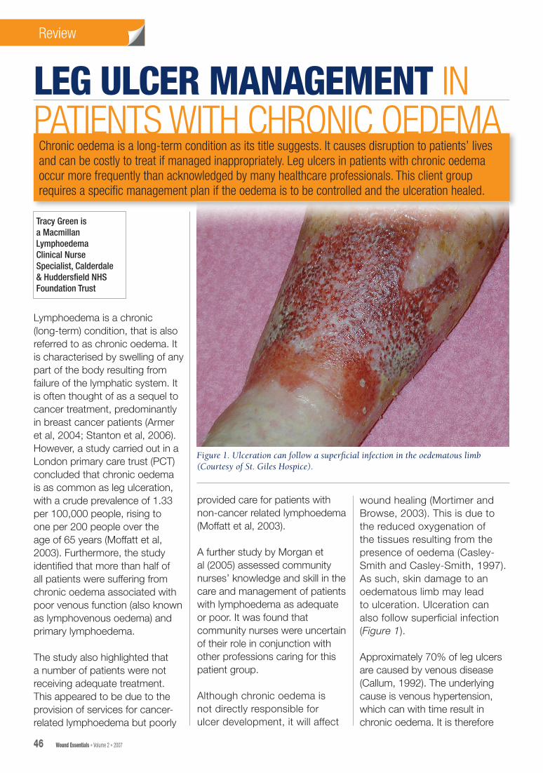
wound healing (Mortimer and Browse, 2003). This is due to the reduced oxygenation of the tissues resulting from the presence of oedema (Casley-Smith and Casley-Smith, 1997). As such, skin damage to an oedematous limb may lead to ulceration. Ulceration can also follow superfi cial infection (Figure 1).
Approximately 70% of leg ulcers are caused by venous disease (Callum, 1992). The underlying cause is venous hypertension, which can with time result in chronic oedema. It is therefore
46 Wound Essentials • Volume 2 • 2007
Figure 1. Ulceration can follow a superfi cial infection in the oedematous limb (Courtesy of St. Giles Hospice).
46-58lymphoedema.indd 2 3/6/07 10:52:23 pm
ReviewReview ReviewReview
likely that patients with co-existing oedema and leg ulcers, either as a result of trauma or venous disease, will be encountered frequently in practice.
This article aims to promote recognition of chronic oedema and lymphoedema in patients with leg ulceration, and will discuss what implications the diagnosis of chronic oedema will have on the management of the patient with leg ulcers.
The lymphatic systemThe lymphatic system has an important role in maintaining fluid balance. It carries fluid, fats and proteins back to the general circulation from the tissues that would otherwise accumulate as oedema. It also provides an important immunological function (Levick and Mortimer, 2003), carrying immune cells such as lymphocytes and macrophages to the lymph nodes (Mortimer, 2000).
Circulating blood carries fluid and nutrients to the body tissues. As blood passes through the capillaries, fluid leaks out through the semi-permeable walls (filtration) and into the interstitial space that lies between the capillary wall and the tissues.
The exchange of nutrients, wastes, fluid, electrolytes, and proteins from the vascular and lymphatic systems and tissue cells occurs through this space. In this area, pressure may be low and attract fluid into the tissue, or an increase in tissue pressure forces fluid into the initial lymphatics and capillaries. It is in the interstitial fluid space
that the forces resulting in oedema come into effect.
Evidence has shown that the majority of interstitial fluid is returned to the blood circulation via the lymphatic system and not, as initially thought, by absorption directly into venous capillaries (Mortimer and Levick, 2004). The absorption of interstitial fluid is the crucial role of the lymphatic system in maintaining fluid balance. When the lymphatic system is unable to maintain this role, oedema occurs.
LymphoedemaLymphoedema is defined as swelling of a limb or part of the body due to failure of the lymphatic system. It results primarily when the lymph drainage system fails to adequately drain fluid from the interstitial spaces. This failure leads to an accumulation of fluid and proteins in the tissues, resulting in swelling or oedema. It can affect any part of the body but usually affects the limbs and/or the adjacent quadrant of the body (Board and Harlow, 2002).
Lymphoedema is classified as primary or secondary. Primary lymphoedema is due to an intrinsic abnormality of the lymphatic system. It may be caused by insufficient numbers of lymphatic collectors (resulting in aplasia or hypoplasia). It may result from an abnormality of the lymphatic vessels such as hyperplasia or valvular incompetence (Mortimer, 1998). It may be congenital (present at birth but not hereditary), or can develop at puberty or any age thereafter.
Wound Essentials • Volume 2 • 2007 47
Secondary lymphoedema is due to damage or long-term overload of the lymphatic system by extrinsic factors including:8Cancer treatment, e.g.
surgery and/or radiotherapy8Recurrent infection8Trauma8Parasitic infection8Obstruction by tumour8Immobility8Venous disease, which will be
described now in more detail.
Lymphovenous oedemaLymphovenous oedema occurs when chronic venous disease associated with deep vein thrombosis, venous hypertension and chronic leg ulceration influences capillary permeability and increases capillary filtration rates.
Venous hypertension is caused by failure of the calf muscle pump to return blood back up the leg. Incompetent valves in the deep, perforating or superficial veins lead to the backflow of blood and the veins elongate and become tortuous. The resulting increase in pressure leads to leakage of blood particles and fluid into the tissues. This is usually compensated for by a healthy lymphatic system.
However, a sustained increase in capillary filtration leads to an overload of the lymphatic system, which will ultimately result in oedema. Thus, a combined lymphatic and venous oedema results; lymphovenous oedema. As lymphatic drainage becomes further compromised, lymphostatic fibrosis begins to
46-58lymphoedema.indd 3 3/6/07 10:52:23 pm
ReviewReview ReviewReview
48 Wound Essentials • Volume 2 • 2007
develop leading to a chronic oedema (Green and Mason, 2006).
Chronic oedema is characterised as a long-
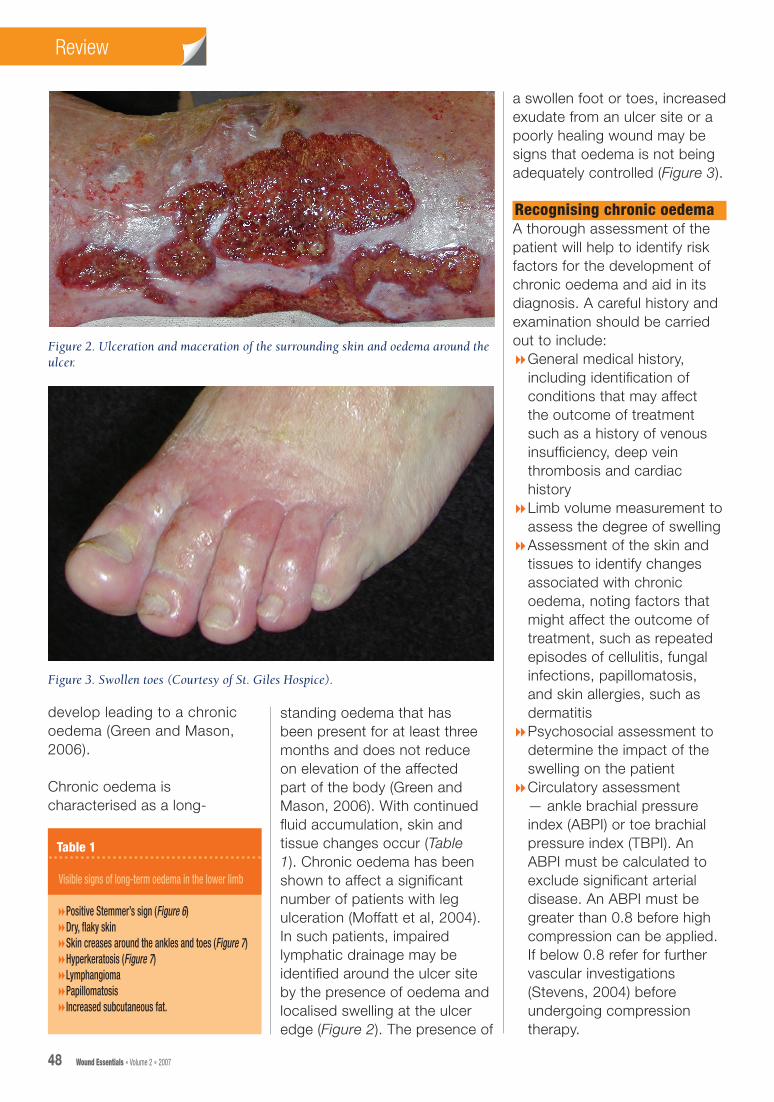
standing oedema that has been present for at least three months and does not reduce on elevation of the affected part of the body (Green and Mason, 2006). With continued fluid accumulation, skin and tissue changes occur (Table 1). Chronic oedema has been shown to affect a significant number of patients with leg ulceration (Moffatt et al, 2004). In such patients, impaired lymphatic drainage may be identified around the ulcer site by the presence of oedema and localised swelling at the ulcer edge (Figure 2). The presence of
a swollen foot or toes, increased exudate from an ulcer site or a poorly healing wound may be signs that oedema is not being adequately controlled (Figure 3).
Recognising chronic oedema A thorough assessment of the patient will help to identify risk factors for the development of chronic oedema and aid in its diagnosis. A careful history and examination should be carried out to include:8General medical history,
including identification of conditions that may affect the outcome of treatment such as a history of venous insufficiency, deep vein thrombosis and cardiac history
8Limb volume measurement to assess the degree of swelling
8Assessment of the skin and tissues to identify changes associated with chronic oedema, noting factors that might affect the outcome of treatment, such as repeated episodes of cellulitis, fungal infections, papillomatosis, and skin allergies, such as dermatitis
8Psychosocial assessment to determine the impact of the swelling on the patient
8Circulatory assessment — ankle brachial pressure index (ABPI) or toe brachial pressure index (TBPI). An ABPI must be calculated to exclude significant arterial disease. An ABPI must be greater than 0.8 before high compression can be applied. If below 0.8 refer for further vascular investigations (Stevens, 2004) before undergoing compression therapy.
Table 1
Visible signs of long-term oedema in the lower limb
8Positive Stemmer’s sign (Figure 6) 8Dry, flaky skin8Skin creases around the ankles and toes (Figure 7) 8Hyperkeratosis (Figure 7) 8Lymphangioma 8Papillomatosis 8Increased subcutaneous fat.
Figure 2. Ulceration and maceration of the surrounding skin and oedema around the ulcer.
Figure 3. Swollen toes (Courtesy of St. Giles Hospice).
46-58lymphoedema.indd 4 3/6/07 10:52:26 pm
ReviewReview
50 Wound Essentials • Volume 2 • 2007
Management of chronic odemaLymphoedema management comprises four components used in combination to provide the most effective reduction and maintenance of swelling. The British Lymphology Society (BLS) recommends a minimum standard of care for patients with lymphoedema (Figure 4).
Patients who require an intensive (decongestive) oedema treatment, i.e. those patients with large, swollen, distorted limbs with skin problems, would usually be treated with a course of multilayer lymphoedema bandaging (MLLB) and possibly manual lymphatic drainage (MLD), along with skin care, an exercise programme, psychosocial support and education. This intensive therapy programme is used to ‘decongest’ the limb and/or trunkal region by removing fl uid from the congested tissues, encouraging the movement of fl uid through the lymphatic pathways and reshaping the oedematous limb.
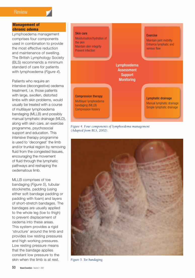
MLLB comprises of toe bandaging (Figure 5), tubular stockinette, padding (using either soft bandage padding or padding with foam) and layers of short-stretch bandages. The bandages are usually applied to the whole leg (toe to thigh) to prevent displacement of oedema into these areas. This system provides a rigid ‘structure’ around the limb and provides low resting pressures and high working pressures. Low resting pressure means that the bandage applies constant low pressure to the skin when the limb is at rest.
Figure 4. Four components of lymphoedema management(Adapted from BLS, 2002).
Skin care
Moisturisation/hydration of the skinMaintain skin integrityPrevent infection
LymphoedemaAssessment
SupportMonitoring
Exercise
Maintain joint mobilityEnhance lymphatic and venous fl ow
Compression therapy
Multilayer lymphoedema bandaging (MLLB)Compression hosiery
Lymphatic drainage
Manual lymphatic drainageSimple lymphatic drainage
Figure 5. Toe bandaging.
46-58lymphoedema.indd 6 3/6/07 10:52:32 pm

52 Wound Essentials • Volume 2 • 2007
ReviewReview
When the muscles contract and expand (e.g. during exercise) they press against the bandage and the pressure in the limb temporarily increases. This increased ‘working’ pressure stimulates lymphatic pumping and reabsorption of lymph (Moffatt, 2000).
Management of leg ulcersA graduated, sustained compression therapy is traditionally used in the management of leg ulcers, with a wound dressing selected as appropriate for the specific wound. A layered bandaging system is used and is said to be more effective than single layer systems (Fletcher et al, 1997). These systems are only applied below the knee and do not include the toes. The bandages are left in situ for up to one week. The choice of dressing and bandaging system will depend upon the patient’s condition, assessment and patient choice (Dowsett, 2005).
skin creases and even out the bandage shape to ensure the limb retains a cylindrical shape in order to maintain the correct compression gradient (Todd, 2000).
In many cases where traditional venous leg ulcer bandaging is used for patients with chronic oedema, oedema of the toes and forefoot develops. Oedema of the toes occurs when the bandages cause pressure on the dorsum of the foot, pushing fluid down into the toes. Where venous leg ulcer bandages have not been applied far enough down the foot, dorsal swelling may occur. If the lymphatics in this area become stagnant, secondary skin changes may take place (Tiwari, 2003), such as a positive Stemmer’s sign (Figure 6), thickened skin, or papillomatosis.
It has been suggested that traditional venous ulcer bandaging systems used for patients with chronic oedema may result in persistent oedema of the toes, knees and thighs, exacerbating ulceration and preventing healing (Williams, 2003). ‘Slippage’ of the bandage due to a reduction in swelling, particularly in the early days of treatment can lead to a tourniquet effect and can cause further distortion in the shape of the limb. Hence, MLLB may be more appropriate for this patient group.
Components of skin care
Skin careCare of the skin is a major concern when managing patients with ulceration
Management of leg ulcers and chronic oedema: which bandaging system?Patients with chronic oedema and ulceration require a different approach to treatment than those with venous ulceration alone. The specific issues associated with managing the patient with lymphoedematous ulceration include:8Limb shape distortion8Care of skin creases and folds 8Swelling of the toes
and forefoot.
These issues require a modified approach to the usual venous leg ulcer bandaging regimens (Moffatt et al, 2005).
Patients with limb distortion and skin folds or creases will require a more specialised approach to their care and, if available, working in conjunction with a lymphoedema specialist service is useful. The use of padding or foams is needed to correct the limb shape, pad out the
Figure 6. Positive Stemmer’s sign.
46-58lymphoedema.indd 8 3/6/07 10:52:33 pm

54 Wound Essentials • Volume 2 • 2007
ReviewReview
and oedema. Patients with chronic oedema are at an increased risk of infection if the skin is dry and cracked. This can lead to cellulitis and further deterioration in their oedema. The skin integrity of the unaffected skin must be maintained. Moisturisation of the skin is essential to maintain its hydration. An appropriate dressing should be used to protect the skin around the wound (peri-wound) from maceration (breakdown caused by moisture), which can occur if excess fluid (exudate) from the wound is left in contact with the skin. Consultation with tissue viability specialist will be of benefit in patients with complicated ulcers. Referral should be made if there is any doubt concerning management.
Compression and supportCompression and support are the foundations of good oedema management and can be used to reduce swelling and maintain the reduction (BLS, 2002). Often described as containment (Todd, 2000), external support in
combination with muscular activity stimulates both venous and lymphatic drainage. MLLB is recognised as an essential component of the intensive phase of lymphoedema management (Casley-Smith and Casley-Smith, 1997). In addition to the reduction of oedema, MLLB restores limb shape, reduces the risk of skin changes and can help to soften subcutaneous tissues and support stretched skin (Williams, 2003). MLLB enhances lymph formation by increasing movement of interstitial fluid into the initial lymphatics by increasing tissue pressure when the patient moves (Leduc et al, 1990; Thomas, 1996; Casley-Smith and Casley-Smith 1997).
MLLB is indicated for patients with:8Fragile/damaged or ulcerated
skin 8Fibrotic skin changes, e.g.
hyperkeratosis, papillomatosis8Lymphangioma8Lymphorrhoea (Figure 8)8Solid, non-pitting, fibrotic
subcutaneous tissues8Skin folds/creases.
MLLB is contraindicated for patients with:8Acute infection, e.g. cellulitis 8Arterial insufficiency8Deep vein thrombosis (DVT)8Severe cardiac failure.
Components of the MLLB system such as padding, toe bandaging and full leg bandaging may be incorporated into venous bandaging systems, as they may help to reduce the oedema thereby enhancing healing by improving delivery of nutrients to the cells and speeding up the removal of waste products (Williams and Keller, 2005). However, bandaging the full leg does require some skill and the four-layer system may not lend itself readily to this technique due to the high resting pressures exerted by the bandages at the knee joints and thigh.
Ideally, MLLB should be reapplied daily especially in the early stages of treatment as the bandages may become loose or slip due to reduction in limb volume. However, as the treatment continues the use of cohesive short-stretch bandages within the MLLB system can reduce slippage
Figure 8. Lymphorrhoea. (Courtesy of St. Giles Hospice).
Figure 7. Hyperkeratosis and skin creases (courtesy of St Giles Hospice).
46-58lymphoedema.indd 10 3/6/07 10:52:35 pm

56 Wound Essentials • Volume 2 • 2007
ReviewReview
and allow bandages to stay in place for longer (Williams, 2006), thereby reducing nursing time and frequency of dressing change.
The choice of ulcer dressing should also be considered to prevent skin irritation and reaction. Using a tubular bandage over the dressing and under the padding should help to reduce skin reactions and provide protection to the tissues. Toe bandaging in conjunction with venous bandaging systems can help to reduce toe swelling. However, it is also important that bandages are applied from toe to knee with the correct technique to avoid forefoot swelling.
For immobile patients, a combination of venous bandaging that produces a higher resting pressure may be more useful than MLLB due to the reduced muscle activity (Moffatt et al, 2005).
For every patient, the clinician needs to assess limb shape, tissue condition, and the extent of oedema in order to decide which bandage technique should be used. If in doubt, management should be discussed with a specialist.
Exercise and movementMobility and exercise play an important role in the management of patients with lymphoedema and ulceration, especially if the patient is undergoing MLLB. Movement can improve lymph flow by enhancing the muscle pump action which has an effect on lymphatic and venous flow,
thereby removing fluid from the tissues.
Mortimer et al (1990) and Goody et al (2001) suggest that passive movement of the foot can cause significant volume change in the lower limbs (Vaqas and Ryan, 2003). Foot and ankle exercises should be encouraged to promote calf pump action and thigh and hip exercises can help with fluid movement from the thigh.
Manual lymphatic drainageIn some cases it may be appropriate to include manual lymphatic drainage (MLD) in the treatment plan of patients with ulceration and chronic oedema. Manual lymphatic drainage is a type of massage which is designed to utilise the skin lymphatic drainage routes to redirect fluid from the oedematous regions to areas with healthy lymphatics. It requires specific gentle movements on the skin to stimulate lymph flow and open up collateral lymphatic vessels.
This may accelerate and stabilise the treatment results (Strossenreuter, 2003). MLD may also be performed on the ulcer site. This will require referral to a lymphoedema specialist practitioner for appropriate treatment.
A case study will now be presented that demonstrates the effective management of a patient with lymphovenous oedema and leg ulceration.
Case study Patient X, a 45-year-old male had significant skin and muscle
loss in both thighs following a crush injury. He consequently developed lymphoedema of both legs and underwent a course of decongestive lymphatic therapy to reduce the swelling to his right leg in 1999. Following the use of compression hosiery on both limbs, the swelling was contained. In 2005 he developed a deep vein thrombosis (DVT) in his left thigh and went on to develop symptoms of chronic venous insufficiency in addition to lymphoedema. He developed a small ulcer to the medial aspect of the ankle above the mallelous of his left leg that was treated successfully.
Figure 10. Compression hosiery was used to maintain limb volume reduction.
Figure 9. Ulcer healing after 2 weeks of treatment.
46-58lymphoedema.indd 12 3/6/07 10:52:37 pm
ReviewReview
Wound Essentials • Volume 2 • 2007 57
Unfortunately, early in 2006 he developed further ulceration to his left limb. He was seen by a healthcare professional who informed him that he could not receive compression bandaging because he had lymphoedema. At that point a hydrocolloid dressing was applied to the ulcer site. Patient X was told to remove the dressing seven days later and come back if he had further problems. After two days he was experiencing excruciating pain at the ulcer site and contacted the lymphoedema service. On assessment he had an ulcer to the gaiter area of the left leg that was odorous and producing exudate. The skin surrounding the ulcer was macerated and inflamed.
Patient X’s leg was oedematous on the foot and from mid-calf to thigh, but the ankle area was indurated and he had developed a typical ‘inverted champagne bottle’ appearance. This was a problem because when normal venous leg ulcer bandaging was applied, the shape of his leg caused the bandages to slip and further distort the shape of his lower leg.
ABPI was normal and there were no signs of arterial insufficiency. A swab was taken for micro-culture and sensitivity. A non-adhesive dressing was applied to the ulcer. The limb was bandaged using a modified multilayer system comprising toe bandages, tubifast, soft wool padding and cohesive short-stretch bandages. A full leg bandage was not applied as Patient X had restricted movement in his right leg
due to the rigidity of his knee following the accident. It was important not to impede his mobility further by restricting the movement in his left knee through bandage application. It was also necessary to pad out the distorted shape at the ankle to avoid excessive pressure. The wound swab came back positive for Staphylococcus aureus and Patient X was prescribed flucloxacillin. The district nurses replaced his bandages a few days later and he returned to the clinic a week after his first appointment. There were already signs that the ulcer was healing and he had not experienced any pain since having the bandages applied. This treatment plan was continued for a further two weeks with the ulcer beginning to heal very quickly (Figure 9). He was monitored for signs of swelling to the knee or thigh and, after three weeks, was fitted with custom-made compression hosiery to maintain limb volume reduction (Figure 10).
ConclusionRecognition of chronic oedema in patients with ulceration is essential if treatment is to be managed effectively. A combined approach to management is essential to facilitate successful outcomes for this client group. Assessment and correct diagnosis is crucial to the recognition of lymphoedema. Referral to the lymphoedema specialist and/or tissue viability services will benefit the patient in the long-term and is an essential component of multidisciplinary teamwork.
Simple changes to bandaging techniques to address the lymphatic component of swelling can bring about a quick response to treatment. This not only improves the patients’ quality of life but reduces the costs of dressings and nursing time (Stalbow, 2004). Leg ulcer practioners and lymphoedema services should adopt an integrated approach to meet the needs of patients with lymphoedema/chronic oedema and leg ulcers.
Armer J, Fu MR, Wainstock JH, Zagar E, Jacobs LK (2004) Lymphoedema following breast cancer treatment, including sentinel lymph node biopsy. Lymphology 37: 73–91
Badger CM, Peacock JL, Mortimer PS (2000) A randomised, controlled, parallel-group clinical trial comparing multilayer bandaging followed by hosiery versus hosiery alone in the treatment of patients with lymphoedema of the limb. Cancer 88(12): 2832–7
Board J, Harlow W (2002) Lymphoedema 1: components and function of the lymphatic system. Br J Nurs 11(5): 304–9
Key Points
8 Chronic oedema affects a significant proportion of the population.
8 Chronic oedema and ulceration require a modified, succinct approach to management.
8 Thorough assessment of a chronically swollen limb is essential if the appropriate treatment is to be carried out.
WE
46-58lymphoedema.indd 13 3/6/07 10:52:37 pm

ReviewReview
58 Wound Essentials • Volume 2 • 2007
Board J, Harlow W (2002) Lymphoedema 2: classification, signs, symptoms and diagnosis. Br J Nurs 11(6): 389–95
Board J, Harlow W (2002) Lymphoedema 3: the available treatments for lymphoedema. Br J Nurs 11(7): 438–50
British Lymphology Society (2002) Strategy for Lymphoedema Management. BLS, Sevenoaks, Kent
Callum M (1992) Prevalence of chronic leg ulceration and severe chronic venous disease in western countries. Phlebology 1 (suppl): 6–12
Casley-Smith J, Casley-Smith JR (1997) Modern Treatment for Lymphoedema. 5th edn. Terrace Printing, Adelaide, Australia
Dowsett C (2005) Assessment and management of patients with leg ulcers. Nurs Stand 19(32): 65–72
Fletcher A, Cullum N, Sheldon TA (1997) A systematic review of compression treatment for venous leg ulcers. Br Med J 315(7108): 576–80
Godoy JMP, Godoy MFG (2001) Development and evaluation of an apparatus for lymph drainage. Eur J Lymphology 9: 121
Green T, Mason W (2006) Chronic oedemas: identification and referral pathways. Br J Comm Nurs 11(4): S8–S16
Leduc A et al (1990) Le traitement Physique de l’oedeme du bras. Monographies de Bois-larris, 2nd edn. Masson, Paris
Levick JR, McHale N (2003) The physiology of lymph production and propulsion In: Browse N, Burnand KG, Mortimer PS, eds. Diseases of the Lymphatics. Arnold, London: 44–64
Moffatt CJ (2000) Compression Therapy. J Community Nurs 14: 26–36
Moffatt CJ, Franks PJ, Doherty DC et al (2003) Lymphoedema: an underestimated health problem. QJM 96(10): 731–8
Moffatt CJ, Franks PJ, Doherty DC, Martin R, Blewett R, Ross F (2004) Prevalence of leg ulceration in a London population. QJM 97(7): 431–7
Moffatt CJ, Morgan P, Doherty D (2005) The Lymphoedema Framework: a consensus on lymphoedema bandaging. In: European Wound Management Association (EWMA) Focus Document: Lymphoedema Bandaging in Practice. MEP Ltd, London
Moffatt CJ, Franks PJ, Doherty DC, Martin R (2004) Prevalence of leg ulceration in a London population. QJM 97: 431–7
Morgan PA, Moody M, Franks PJ, Moffatt CJ, Doherty DC (2005) Assessing community nurses’ level of knowledge of lymphoedema. Br J Nurs 14(1): 8–13
Mortimer PS (1998) The pathophysiology of lymphoedema. Cancer (suppl) 83(12): 2798–802
Mortimer P (2000) Acute Inflammatory Episodes In: Tywcross R, Jenns K, Todd J, eds (2000) Lymphoedema. Radcliffe Medical Press. Oxon: 130–9
Mortimer P, Browse N (2003) The relationship between the lymphatics and chronic venous disease. In: Browse N, Burnand KG, Mortimer PS (2003) Diseases of the Lymphatics. Arnold, London: 293–6
Mortimer PS, Levick JR (2004) Chronic peripheral oedema: the critical role of the lymphatic system. Clin Med 4(5): 448–53
Mortimer PS, Simmonds R, Rezvani M, Robbins M, Hopewell JW, Ryan T (1990) The measurement of sklin lymph flow by isotope clearance-reliability, reproducibility, injection dynamics and the effect of massage. J Invest Dermatol 95: 677–82
Stalbow J (2004) Preventing cellulitis in older people with persistent lower limb oedema. Br J Nurs 13(12): 725–32
Stanton A, Modi S, Mellor S, Levick R, Mortimer P (2006) Diagnosing breast cancer-related lymphoedema in the arm. J Lymphoedema 1(1): 12–15
Stevens J (2004) Diagnosis, assessment and management of mixed aetiology ulcers using reduced compression. J Wound Care 13(8): 339–43
Strossenreuter RHK (2003) Practical instructions for therapists-general requirements for compression bandages. In: Foldi M, Foldi E, Kubik S (eds) Textbook of Lymphology for Physicians and Lymphoedema Therapists. Urban and Fischer, San Francisco: 564–7
Thomas S (1996) High compression bandages. J Wound Care 5: 40–3
Tiwari A, Cheng, KS, Button M et al (2003) Differential diagnosis, investigation and current treatment of lower limb lymphoedema. Arch Surg 138(2): 152–61
Todd J (2000) Containment in the management of lymphoedema. In: Tywcross R, Jenns J, Todd J, eds. Lymphoedema. Radcliffe Medical Press, Oxford
Vaqas B, Ryan TJ (2003) Lymphoedema: Pathophysiology and management in resource-poor settings — relevance for lymphatic filariasis control programmes. Filaria J 2(1): 4
Williams AF (2003) The management of lymphoedema of the lower limbs. J Comm Nurs 17(8): 34–44
Williams AF, Keller M (2005) Practical guidance on lymphoedema bandaging of the upper and lower limbs. In: European Wound Management Association (EWMA). Focus Document: Lymphoedema Bandaging in Practice. MEP Ltd, London
Williams A (2006) A clinical audit of Actico cohesive short stretch bandages in lymphoedema. J Comm Nurs 20(2): 4–8
46-58lymphoedema.indd 14 3/6/07 10:52:38 pm