review of the effectiveness of infrared thermal imaging ... · review of the effectiveness of...
TRANSCRIPT

NZHTA TECH BRIEF SERIESJuly 2004Volume 3 Number 3
Review of the effectiveness of infraredthermal imaging (thermography) forpopulation screening and diagnostic
testing of breast cancer
Jane Kerr
New ZealandHealth Technology Assessment
Department of Public Health and General PracticeChristchurch School of Medicine
Christchurch, NZ.
Division of Health Sciences, University of Otago
NEW ZEALAND HEALTH TECHNOLOGY ASSESSMENT (NZHTA)
Department of Public Health and General PracticeChristchurch School of Medicine and Health SciencesChristchurch, New Zealand
Review of the effectiveness of infraredthermal imaging (thermography) forpopulation screening and diagnostictesting of breast cancer
Jane Kerr
NZHTA TECH BRIEF SERIESJuly 2004 Volume 3 Number 3

This report should be referenced as follows:
Kerr, J. Review of the effectiveness of infrared thermal imaging (thermography) for populationscreening and diagnostic testing of breast cancer. NZHTA Tech Brief Series 2004; 3(3)
Titles in this Series can be found on the NZHTA website:http://nzhta.chmeds.ac.nz/thermography_breastcancer.pdf
2004 New Zealand Health Technology Assessment (NZHTA)
ISBN 1-877235-64-4ISSN 1175-7884

REVIEW OF THE EFFECTIVENESS OF INFRARED THERMAL IMAGING (THERMOGRAPHY) FOR POPULATION
SCREENING AND DIAGNOSTIC TESTING OF BREAST CANCER
i
ACKNOWLEDGEMENTS
This Tech Brief was commissioned by the National Screening Unit of the New Zealand Ministry ofHealth.
The report was prepared by Dr Jane Kerr (Research Fellow) who selected and critically appraised theevidence. The research protocol for this report was developed by Ms Marita Broadstock (ResearchFellow). The literature search strategy was developed and undertaken by Mrs Susan Bidwell(Information Specialist Manager). Mrs Ally Reid (Administrative Secretary) provided documentformatting. Internal peer review was provided by Dr Robert Weir (Senior Research Fellow),Dr Ray Kirk (Director) and Ms Broadstock.
We are grateful to Dr Simon Baker (Public Health Physician, National Screening Unit, Ministry ofHealth), Dr Madeleine Wall (Clinical Leader, Breastscreen Aotearoa, National Screening Unit,Ministry of Health) and Dr Richard Tremewan (Medical Physics and Bioengineering Department,Christchurch Hospital) who provided valuable advice and input.
We are grateful to the authors who provided assistance in the retrieval of articles for this review:Professor E. F. Ring (Medical Imaging Research Group, School of Computing, University ofGlamorgan), Dr N. A. Diakides (Advanced Concepts Analysis Inc., Falls Church, Virginia) andProfessor R. G. Margolese (Jewish General Hospital, McGill University, Montreal). We are alsograteful to those from the fields of breast screening, medical physics/bioengineering, radiology andinfrared thermal imaging who provided advice regarding available literature.
DISCLAIMER
NZHTA takes great care to ensure the accuracy of the information supplied within the projecttimeframe, but neither NZHTA nor the University of Otago can accept responsibility for any errors oromissions that may occur. NZHTA and the University of Otago along with their employees accept noliability for any loss of whatever kind, or damage, arising from the reliance in whole or part, by anyperson, corporate or natural, on the contents of this paper. This document is not intended to be used aspersonal health advice; people seeking individual medical advice are referred to their physician. Theviews expressed in this report are those of NZHTA and do not necessarily represent those of theUniversity of Otago, or the New Zealand Ministry of Health.
COPYRIGHT
This work is copyright. Apart from any use as permitted under the Copyright Act 1994 no part may bereproduced by any process without written permission from New Zealand Health TechnologyAssessment. Requests and inquiries concerning reproduction and rights should be directed to theDirector, New Zealand Health Technology Assessment, Christchurch School of Medicine and HealthSciences, P O Box 4345, Christchurch, New Zealand.

REVIEW OF THE EFFECTIVENESS OF INFRARED THERMAL IMAGING (THERMOGRAPHY) FOR POPULATION
SCREENING AND DIAGNOSTIC TESTING OF BREAST CANCER
ii
CONTACT DETAILS
New Zealand Health Technology Assessment (NZHTA)Department of Public Health and General PracticeChristchurch School of Medicine and Health SciencesPO Box 4345ChristchurchNew ZealandTel: +64 3 364 3696 Fax: +64 3 364 3697
Email: [email protected]
Website: http://nzhta.chmeds.ac.nz
LEVEL OF EVIDENCE CONSIDERED IN TECH BRIEFS
Tech Briefs are rapidly produced assessments of the best available evidence for a topic of highlylimited scope. They are less rigorous than systematic reviews. Best evidence is indicated by researchdesigns which are least susceptible to bias according to evidence levels adapted from Bandolier(http://www.jr2.ox.ac.uk/bandolier/band70/b70-5.html, accessed 22 June 2004) that categorise studiesof diagnostic methods according to susceptibility for bias (see Appendix 1). Where methodologicallyacceptable and applicable, appraised evidence is limited to systematic reviews, meta-analyses,evidence-based clinical practice guidelines, health technology assessments and randomised controlledtrials (RCTs). Where not available, poorer quality evidence may be considered.
CONFLICT OF INTEREST
None.
REVIEW OF THE EFFECTIVENESS OF INFRARED THERMAL IMAGING (THERMOGRAPHY) FOR POPULATION
SCREENING AND DIAGNOSTIC TESTING OF BREAST CANCER
iii
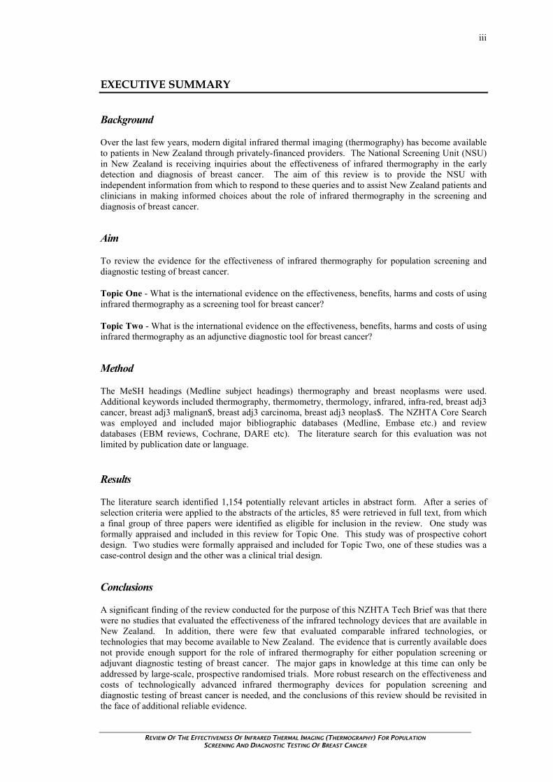
EXECUTIVE SUMMARY
Background
Over the last few years, modern digital infrared thermal imaging (thermography) has become availableto patients in New Zealand through privately-financed providers. The National Screening Unit (NSU)in New Zealand is receiving inquiries about the effectiveness of infrared thermography in the earlydetection and diagnosis of breast cancer. The aim of this review is to provide the NSU withindependent information from which to respond to these queries and to assist New Zealand patients andclinicians in making informed choices about the role of infrared thermography in the screening anddiagnosis of breast cancer.
Aim
To review the evidence for the effectiveness of infrared thermography for population screening anddiagnostic testing of breast cancer.
Topic One - What is the international evidence on the effectiveness, benefits, harms and costs of usinginfrared thermography as a screening tool for breast cancer?
Topic Two - What is the international evidence on the effectiveness, benefits, harms and costs of usinginfrared thermography as an adjunctive diagnostic tool for breast cancer?
Method
The MeSH headings (Medline subject headings) thermography and breast neoplasms were used.Additional keywords included thermography, thermometry, thermology, infrared, infra-red, breast adj3cancer, breast adj3 malignan$, breast adj3 carcinoma, breast adj3 neoplas$. The NZHTA Core Searchwas employed and included major bibliographic databases (Medline, Embase etc.) and reviewdatabases (EBM reviews, Cochrane, DARE etc). The literature search for this evaluation was notlimited by publication date or language.
Results
The literature search identified 1,154 potentially relevant articles in abstract form. After a series ofselection criteria were applied to the abstracts of the articles, 85 were retrieved in full text, from whicha final group of three papers were identified as eligible for inclusion in the review. One study wasformally appraised and included in this review for Topic One. This study was of prospective cohortdesign. Two studies were formally appraised and included for Topic Two, one of these studies was acase-control design and the other was a clinical trial design.
Conclusions
A significant finding of the review conducted for the purpose of this NZHTA Tech Brief was that therewere no studies that evaluated the effectiveness of the infrared technology devices that are available inNew Zealand. In addition, there were few that evaluated comparable infrared technologies, ortechnologies that may become available to New Zealand. The evidence that is currently available doesnot provide enough support for the role of infrared thermography for either population screening oradjuvant diagnostic testing of breast cancer. The major gaps in knowledge at this time can only beaddressed by large-scale, prospective randomised trials. More robust research on the effectiveness andcosts of technologically advanced infrared thermography devices for population screening anddiagnostic testing of breast cancer is needed, and the conclusions of this review should be revisited inthe face of additional reliable evidence.

REVIEW OF THE EFFECTIVENESS OF INFRARED THERMAL IMAGING (THERMOGRAPHY) FOR POPULATION
SCREENING AND DIAGNOSTIC TESTING OF BREAST CANCER
iv
GLOSSARY
Asymptomatic - asymptomatic people are those who do not have a symptom (e.g., breast lump) thatmay be due to a disease (e.g., breast cancer).
Benign tumour - a benign tumour is an abnormal growth that is neither malignant, nor a cancer. Abenign tumour is not capable of spreading, and usually does not recur after being removed.
Bias - deviation of results or inferences from the truth, or processes leading to such deviation. Anytrend in the collection, analysis, interpretation, publication, or review of data that can lead toconclusions that are systematically different from the truth.
Biopsy - in a breast biopsy, a sample of tissue is removed to be examined under a microscope, as an aidto diagnosis.
Breast cancer - a histologically-proven malignant lesion that is classified as ductal carcinoma in situ orinvasive breast cancer.
Breast cancer classification - breast cancers are classified in terms of tumour cell type, grade, size,lymph node involvement and stage.
Breast implant - a round or tear-shaped sack inserted into the chest in order to restore or enhance theshape of the breast. A breast implant may be filled with saline, silicone or a synthetic material.
Blinded study - a study in which observers and/or subjects are kept ignorant of the group to whichthey are assigned.
Cancer - a general term for a large number of diseases that all display uncontrolled growth and spreadof abnormal cells (also called malignant tumours). Cancer cells have the ability to continue to grow,invade and destroy surrounding tissue, and leave the original site and travel via the lymph or bloodsystems to other parts of the body where they may establish further cancerous tumours.
Carcinoma - a malignant tumour made up of epithelial cells that may infiltrate surrounding tissues andspread to other parts of the body via the blood or lymph nodes systems.
Case-control study - an epidemiological study involving the observation of cases (persons with thedisease, such as breast cancer) and a suitable control (comparison, reference) group of persons withoutthe disease. The relationship of an attribute (e.g., positive diagnostic test result) to the disease isexamined by comparing retrospectively the past history of the people in the two groups with regard tohow frequently the attribute is present.
Cohort study - an epidemiological study in which subsets of a defined population can be identifiedwho are, have been, or in the future may be exposed or not exposed in different degrees, to a factor orfactors (e.g., receiving a screening test for breast cancer) hypothesised to influence the probability ofoccurrence of a given disease or other outcome (e.g., positive biopsy for breast cancer). Studies usuallyinvolve the observation of a large population, for a prolonged period (years), or both.
Confidence interval (CI) - the computed interval with a given probability – e.g., 95%, that the truevalue of a variable such as a mean, proportion, or rate is contained within the interval. The 95% CI isthe range of values in which it is 95% certain that the true value lies for the whole population.
Confounder - a third variable that indirectly distorts the relationship between two other variables.
Core Biopsy - needle is used to remove a core of tissue for histological examination.
Coverage - the number, percent, or proportion of eligible women reached by a programme.

REVIEW OF THE EFFECTIVENESS OF INFRARED THERMAL IMAGING (THERMOGRAPHY) FOR POPULATION
SCREENING AND DIAGNOSTIC TESTING OF BREAST CANCER
v
Cross-sectional study - a study that examines the relationship between diseases or other health relatedcharacteristics (e.g., patient diagnosed with definite breast cancer) and other variables of interest (e.g.,positive diagnostic test result for breast cancer) as they exist in a defined population at one particulartime.
Descriptive study - a study concerned with, and designed only, to describe the existing distribution ofa variable, without regard to causal or other hypotheses.
Diagnosis - the process of identifying a disease by its characteristic signs, symptoms and findings oninvestigation.
Diagnostic test efficacy - the impact and usefulness of a diagnostic test expressed in terms of itstechnical properties.
Ductal carcinoma-in situ (DCIS) - a form of breast cancer, which spreads along the ducts of thebreast, but has not invaded the duct wall.
Effectiveness - a measure of the extent to which a specific intervention, procedure, regimen, or service,when deployed in the field in routine circumstances, does what it is intended to do for a specifiedpopulation.
Epidemiology - the study of the distribution and determinants of health-related states or events inspecified populations.
Evidence based - based on valid empirical information.
Evidence table - a summary display of selected characteristics (e.g., of methodological design, results)of studies of a particular intervention or health problem.
False negative result - a negative test result in a person who does have the condition being tested for.
False positive result - a positive test result in a person who does not have the condition being testedfor.
Fine needle aspiration biopsy (FNAB) - a fine needle is used to remove some cells or fluid from asuspicious area for histological examination.
Generalisability - applicability of study results to other populations.
Histology - the microscopic study of the minute structure and composition of tissues.
Lesion - an area of tissue damaged by disease or injury.
Mammogram - a specialised X-ray of the breast. It may be used as a screening test in women with nosigns or symptoms of breast cancer, or it may be used to evaluate breast symptoms (e.g., breast lump).
Mammography - the process of taking a mammogram.
Māori - the indigenous people of New Zealand.
Mean - calculated by adding all the individual values in the group and dividing by the number ofvalues in the group.
Median - any value that divides the probability distribution of a random variable in half. For a finitepopulation or sample, the median is the middle value of an odd number of values (arranged inascending order) or any value between the two middle values of an even number of values.

REVIEW OF THE EFFECTIVENESS OF INFRARED THERMAL IMAGING (THERMOGRAPHY) FOR POPULATION
SCREENING AND DIAGNOSTIC TESTING OF BREAST CANCER
vi
Meta-analysis - the process of using statistical methods to combine the results of different studies.The systematic and organised evaluation of a problem, using information from a number ofindependent studies of the problem.
Microcalcification - microcalcification means “very small calcification”. Calcifications (areas wherecalcium has been deposited) are frequently seen on mammograms of the breast, and are usually due tobenign breast processes. Certain appearances and patterns of calcification are associated withmalignancy.
Morbidity - illness.
Mortality - the number of deaths from a specified disease that are diagnosed or reported during adefined period of time in a given population.
Negative predictive value (NPV) - the probability that a person having a negative result on a test doesnot have the condition that the test is designed to detect.
Positive predictive value (PPV) - the probability that a person having a positive result on a test has thecondition that the test is designed to detect.
Population-based screening programme - a population-based screening programme is one in whichscreening is systematically offered by invitation to a defined, identifiable population: this requires ameans of identifying and inviting the target population – e.g., through a population register.
Randomised controlled trial (RCT) - an epidemiologic experiment in which subjects in a populationare randomly allocated into groups to receive or not receive an experimental preventive or therapeuticprocedure, manoeuvre or intervention. The groups are compared prospectively. RCTs are generallyregarded as the most scientifically rigorous method of hypothesis testing available in epidemiology.
Reference standard - an independently applied test that is compared to a screening or diagnostic testbeing evaluated in order to verify the latter’s accuracy. A reference standard therefore provides anaccurate or “truth” diagnosis for verification of positive and negative diagnoses. It is sometimesreferred to as providing “final truth determination”.
Screening - screening is the examination of asymptomatic people in order to classify them as likely orunlikely to have the disease that is the object of screening. The aim of screening is to detect diseasebefore it is clinically apparent, and for this to improve the outcome for people with the disease.
Selection bias - error due to systematic differences in characteristics between those who are selectedfor inclusion in a study and those who are not (or between those compared within a study and thosewho are not).
Sensitivity (Se) - sensitivity is the proportion of truly diseased persons in a tested population who areidentified as diseased by a test. Sensitivity is a measure of the probability of correctly diagnosing acase, or the probability that any given case will be identified by the test.
Specificity (Sp) - specificity is the proportion of truly non-diseased persons in a tested population whoare so identified by a test. Specificity is a measure of the probability of correctly identifying a non-diseased person with a test.
Surgical (open) biopsy - the surgical removal (performed under a local or general anaesthetic) of all,or part, of a suspicious area of tissue for histological examination.
Symptomatic - symptomatic people are those who do have a symptom (e.g., breast lump) that may bedue to a disease (e.g., breast cancer).
Systematic review - literature review reporting a systematic method to search for, identify andappraise a number of independent studies.

REVIEW OF THE EFFECTIVENESS OF INFRARED THERMAL IMAGING (THERMOGRAPHY) FOR POPULATION
SCREENING AND DIAGNOSTIC TESTING OF BREAST CANCER
vii
True negative - a test correctly identifies a person without the disease.
True positive - a test correctly identifies a person with the disease.
Tumour - an abnormal growth of tissue. A breast tumour may be: localised without potential(benign); malignant and growing inside the milk ducts (DCIS); malignant and invading nearby tissues(invasive), or; malignant and invading distant tissues (metastatic).
Ultrasound - the use of high frequency sound waves to study an organ or tissue. Ultrasound isparticularly useful for distinguishing fluid-filled structures from solid lesions.


REVIEW OF THE EFFECTIVENESS OF INFRARED THERMAL IMAGING (THERMOGRAPHY) FOR POPULATION
SCREENING AND DIAGNOSTIC TESTING OF BREAST CANCER
1
OBJECTIVE OF THIS REPORT AND RESEARCH QUESTIONS
The aim of this Tech Brief was to review the evidence for the effectiveness of infrared thermographyfor population screening and diagnostic testing of breast cancer. Because there were two topics underconsideration, the research questions and results of this review for each topic have been consideredseparately.
Topic One – Screening
What is the international evidence on the effectiveness, benefits, harms and costs of using infraredthermography as a screening tool for breast cancer?
Topic Two – Diagnosis
What is the international evidence on the effectiveness, benefits, harms and costs of using infraredthermography as an adjunctive diagnostic tool for breast cancer?

REVIEW OF THE EFFECTIVENESS OF INFRARED THERMAL IMAGING (THERMOGRAPHY) FOR POPULATION
SCREENING AND DIAGNOSTIC TESTING OF BREAST CANCER
2
BACKGROUND
This Tech Brief was requested by Simon Baker, Public Health Physician, National Screening Unit,Ministry of Health, New Zealand.
Rationale for this Report
In New Zealand, breast cancer is an important health concern. For non-Māori women, it is thecommonest cause of cancer registrations and death, and for Māori women it is the second commonest,after lung cancer (BreastScreen Aotearoa, 2004). Over the last few years, modern digital infraredthermal imaging (thermography) has become available to patients in New Zealand through privately-financed providers. The National Screening Unit (NSU) in New Zealand is receiving inquiries aboutthe effectiveness of infrared thermography in the early detection and diagnosis of breast cancer. Theaim of this review is to provide the NSU with independent information from which to respond to thesequeries and to assist New Zealand patients and clinicians in making informed choices about the role ofinfrared thermography in the screening and diagnosis of breast cancer.
Definitions of Thermography
Thermography is the recording of temperature, and has various applications in industrial (e.g., fortesting of materials) and military (e.g., for surveillance) contexts (Anbar, 1995). Clinical thermographyis the recording of temperature to form an image (thermogram) of the temperature distribution on thesurface of the body. Clinical thermography has been considered as a diagnostic instrument for avariety of medical conditions since the 1960s, although its application for many of these remainscontroversial (McLean, 1999). There are several thermography methods (also known as thermometryand thermology). These include “contact thermography” (where a needle inserted into a suspiciousarea measures temperature directly, or a sheet of heat-sensitive liquid crystal film is applied to thebreast), and “microwave thermography” (where temperature distribution is indicated by microwaveradiation emitted by the surface of the body).
The most common thermography method is “infrared thermography” (where infrared radiation emittedby the skin surface is detected). Information from an infrared detector is relayed to a processingsystem, which produces images of temperature distribution. Since the 1970s, it has been possible toprocess the infrared information using computers, that can then display and store images of the infraredthermal patterns (Ring and Ammer, 2000). Infrared thermal imaging can be either ‘static’ or‘dynamic’. Static imaging (also sometimes called ‘steady-state’ imaging) is when a patient’s infrareddata is detected under controlled conditions (Ohashi and Uchida, 2000). During dynamic infraredimaging, the patient is also subjected to one of several methods of cooling, such as cold air blowingover the breast area being imaged, or cold water submersion of the hands (Ohashi and Uchida, 2000).The manner and rate by which the temperature of the skin on the breasts’ surface recovers itsequilibrium is then measured by the infrared detector (Jones, 1998).
Infrared thermography using digital computer processing systems (sometimes also referred to as digitalinfrared thermal imaging) is currently being marketed in New Zealand for breast cancer detection, andis the focus of this report. Obvious potential benefits of infrared thermography are that it is a non-invasive technique and that it does not involve any patient (or operator) exposure to ionising radiation.However, it is limited in its role as a primary (stand-alone) breast cancer diagnostic modality by itsinability to inform clinicians where an abnormality lies to permit even a blind biopsy (Homer, 1985;van Dam et al., 1988; Head et al., 2000, Head and Elliott, 2002; Arena et al., 2003).
Current understandings of the underlying pathological mechanisms for increased temperature in breastcancer are that breast cancer cells produce nitric oxide (NO). This NO interferes with the normalneuronal (nervous system) control of breast tissue blood vessel flow by causing regional vasodilation inthe early stages of cancerous cell growth, and enhancing angiogenesis (new blood vessel formation) inlater stages (Anbar, 1998; Anbar et al., 2001). The subsequent increased blood flow in the area causesa temperature increase relative to the normal breast temperature, and even deep breast lesions seem to

REVIEW OF THE EFFECTIVENESS OF INFRARED THERMAL IMAGING (THERMOGRAPHY) FOR POPULATION
SCREENING AND DIAGNOSTIC TESTING OF BREAST CANCER
3
have the ability to induce changes in skin temperature (Snyder et al., 2000). Breast cancer metabolicprocesses may also contribute to the detectable increase in heat (Foster, 1998). These changes relate tophysiological breast processes. It is believed that in healthy individuals, temperature is generallysymmetrical across the midline of the body (McLean, 1999; Ng et al., 2002). Subjective interpretationof many diagnostic imaging modalities, including infrared thermographic images relies on theunderlying philosophy that normal contralateral images are relatively symmetrical, and that smallasymmetries may indicate a suspicious lesion (Qi and Head, 2001). Therefore, in breast cancer,infrared thermography detects disease by identifying areas of asymmetric temperature distribution onthe breasts’ surface (Qi and Head, 2001).
Medical History of Thermography
The use of thermography as a means of detecting breast cancer has a substantial history. Earlierresearch is still referred to in promotional material – for example, in references to “800 peer reviewedbreast thermography studies” (International Academy of Clinical Thermology [IACT], websiteaccessed 20 March 2004-IACT is a non-profit, non-regulatory agency with interests in research,education, and the establishment of standards and guidelines in clinical thermography).
Before the passage of the Medical Device Amendment in 1976 requiring FDA approval for devices,thermography was actively studied and used as a tool for the early detection of breast cancer in the US(Nass et al., 2001). Up to 3,000 thermography clinics operated in the US during this time (Foster,1998). Data generated by researchers from between the 1960s and the early 1970s suggested thatthermography may be a viable screening tool for breast cancer. A subsequent reanalysis of this datahas identified major problems with its relevance to screening, including that much of it was based inresearch generated from symptomatic patients, rather than asymptomatic populations (see Moskowitz,1995, for an overview). However, several important studies in this period that were undertaken toevaluate breast screening methods did include thermography, and shall be briefly summarised here.
Twenty-seven Breast Cancer Detection Demonstration Projects (BCDDP) were set up in 1972 in theUnited States, with the aim of screening 10,000 asymptomatic women per project over a five yearperiod, with an additional five year follow-up (Letton and Mason, 1986). The plan was to comparethermography, mammography, and clinical examination as screening tools, using state of the artequipment for the time. However, thermography was dropped early in these studies due to high falsepositive rates and low sensitivity (Moskowitz, 1985; Moskowitz, 1995; Foster, 1998). Somecontroversy followed after thermography was dropped from the BCDDPs. The controversy mainlyconcerned how well the investigators had been trained in thermography methods and interpretation, butthe equipment used was state of the art for that time (Moskowitz, 1995). Furthermore, Threatts et al.(1980) subsequently reported the results of an investigative review in which ten experiencedthermographers conducted a blinded interpretation of 576 thermograms from the BCDDP. Theyconcluded that the overall detection rate of thermography for all breast cancers (including in situ andminimal cancers) for the population studied was similar to that that would result from chance (Threattset al., 1980, in Margolese, 1998).
Feig et al. (1977) compared the use of thermography, an early form of mammography(xeromammography), and clinical examination as a screening tool in a separate clinical trial including16,000 women. Overall, thermography’s sensitivity and specificity were 39% and 82%, respectively,compared with xeromammography’s 78% sensitivity and 98% specificity.
Following these trials, medical interest in thermography has steadily decreased (Foster, 1998). In1984, the American College of Radiology issued a policy statement that thermography was still anexperimental procedure with no established clinical indications for the detection of breast cancer(Dankiw, 1990).
Since 1990, two Health Technology Assessment (HTA) reviews of thermography have been published.These reviews included evaluations of the evidence for its application in the detection and managementof breast cancer. The first of these reports, in the form of a narrative review (which included anoverview of existing institutional reviews of the technology), found no conclusive evidence supportingthe use of thermography as either a screening or diagnostic tool for breast cancer (Dankiw, 1990). Thesecond HTA report considered both contact and infrared thermography methods, and reviewed the

REVIEW OF THE EFFECTIVENESS OF INFRARED THERMAL IMAGING (THERMOGRAPHY) FOR POPULATION
SCREENING AND DIAGNOSTIC TESTING OF BREAST CANCER
4
literature from 1983 to 1999, as well as pertinent earlier articles identified by this process. This reportalso found insufficient evidence to support the use of thermography in any aspect of the management ofbreast cancer, and in addition concluded that it had “…been shown convincingly that thermography hasno place in mass primary or secondary screening for breast cancer…” (McLean, 1999, p7).
Also, in Canada, Margolese et al. (1998) developed a set of clinical guidelines for the management ofwomen presenting with a palpable breast lump, after conducting a systematic review of the literaturepublished to January 1996 (with non-systematic review to January 1997) to gather information forthese guidelines. The systematic review itself was not published, but in the published report of thefinal guidelines, the authors clearly stated that thermography was not a recommended diagnosticprocedure in the work-up for women presenting with a palpable breast lump. The authors of theguidelines further concluded that there was no role for thermographic techniques at that time, outsidethe testing of improved technology in structured clinical trials (Margolese et al., 1998).
Technological History of Infrared Thermography
A brief outline of the technological history of infrared thermography is presented to enableunderstanding of the subsequent rationale for the focus of this current report. Although infraredthermal imaging has been used in medicine since the early 1960s, infrared camera technologies thatwere available from the 1970s up until the mid-1980s suffered from several limitations. Theselimitations included poor spatial resolution (uncertainty up to 1cm²), poor thermal resolution (only ableto diagnose abnormalities > 1�C difference), slow systems (taking up to 4 seconds per image) thattherefore also substantially reduced both spatial and temperature resolutions, poor calibration systems,and optical aberrations on the camera lenses that resulted in unreliability at image peripheries (Anbar,1998). In addition, older infrared cameras required the addition of liquid nitrogen to cool the detector.This required a period of equilibrium before image acquisition, limited the angle of camera orientation,and necessitated regular camera maintenance. Newer systems contain efficient electronic coolingsystems, which have overcome these problems (Jones, 1998; Ring and Ammer, 2000). By the mid-1980s, some thermography practitioners stated that, at that time, new and more accurate thermographysystems had supplanted “older technology” (Gautherie et al., 1987).
The advances in infrared camera technology over the last decade have been accompanied by progressin computerised image processing systems (Diakides, 1998; Foster, 1998; Jones, 1998). It was notuntil the 1970s that data acquired by infrared cameras was processed by computers into digital imagesfor viewing (Ring and Ammer, 2000). Now, some sophisticated modelling programmes can enhancethe spatial resolution of images already acquired (Snyder et al., 2000). In addition, although infraredimages are often analysed subjectively, which is therefore dependent on interpreter experience, recentlysome researchers have been trialling new computer software statistical programmes that analyse thedata from infrared breast images quantitatively. Examples of the uses that such software programmesallow include the development of algorithms to calculate quantitative scores of risk for breast lesions(Arena et al., 2003), and the objective detection of small asymmetries on infrared images (Head andElliott, 1997; Qi and Head, 2001). Technological advances in recent years have resulted primarilyfrom military developments in infrared surveillance, and have led to renewed medical interest in thetechnology (Diakides, 1998; Nass et al., 2001). Given the advances in both infrared cameras anddigital processing of the acquired images, several authors have suggested that the systems currentlyavailable are not comparable to those that were previously used (Anbar, 1998; Ring and Ammer, 2000;Nass et al., 2001; Arena et al., 2003).
The digital infrared thermal imaging (DITI) device “Med 2000®” was developed around 1990(Hobson, 2002). MedithermNZ is promoting this technology from a base in Auckland. In addition, an“Infrared Thermal Imaging Medical Clinic” has been established in Tauranga, offering infrared digitalimaging with a system from Micro Health Systems, Inc. (MHS 5000 Thermal Imager®). The detectorsin these systems are lightweight, do not require liquid nitrogen cooling, and have thermal sensitivitiesranging from 0.01 to 0.1°C (http://www.meditherm.com/mms_products.htm, accessed 22 March 2004;http://www.mhs5000.com/specs.htm, accessed 23 March 2004). Computer software specific for eachsystem enables the images acquired by the infrared detector to be viewed, manipulated, and stored oncomputers. The providers of infrared thermal imaging in New Zealand therefore use infrared imagingsystems that are technologically more advanced than those available up until the mid-1980s.
REVIEW OF THE EFFECTIVENESS OF INFRARED THERMAL IMAGING (THERMOGRAPHY) FOR POPULATION
SCREENING AND DIAGNOSTIC TESTING OF BREAST CANCER
5
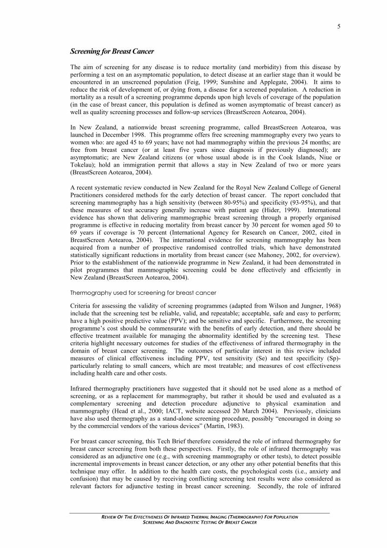
Screening for Breast Cancer
The aim of screening for any disease is to reduce mortality (and morbidity) from this disease byperforming a test on an asymptomatic population, to detect disease at an earlier stage than it would beencountered in an unscreened population (Feig, 1999; Sunshine and Applegate, 2004). It aims toreduce the risk of development of, or dying from, a disease for a screened population. A reduction inmortality as a result of a screening programme depends upon high levels of coverage of the population(in the case of breast cancer, this population is defined as women asymptomatic of breast cancer) aswell as quality screening processes and follow-up services (BreastScreen Aotearoa, 2004).
In New Zealand, a nationwide breast screening programme, called BreastScreen Aotearoa, waslaunched in December 1998. This programme offers free screening mammography every two years towomen who: are aged 45 to 69 years; have not had mammography within the previous 24 months; arefree from breast cancer (or at least five years since diagnosis if previously diagnosed); areasymptomatic; are New Zealand citizens (or whose usual abode is in the Cook Islands, Niue orTokelau); hold an immigration permit that allows a stay in New Zealand of two or more years(BreastScreen Aotearoa, 2004).
A recent systematic review conducted in New Zealand for the Royal New Zealand College of GeneralPractitioners considered methods for the early detection of breast cancer. The report concluded thatscreening mammography has a high sensitivity (between 80-95%) and specificity (93-95%), and thatthese measures of test accuracy generally increase with patient age (Hider, 1999). Internationalevidence has shown that delivering mammographic breast screening through a properly organisedprogramme is effective in reducing mortality from breast cancer by 30 percent for women aged 50 to69 years if coverage is 70 percent (International Agency for Research on Cancer, 2002, cited inBreastScreen Aotearoa, 2004). The international evidence for screening mammography has beenacquired from a number of prospective randomised controlled trials, which have demonstratedstatistically significant reductions in mortality from breast cancer (see Mahoney, 2002, for overview).Prior to the establishment of the nationwide programme in New Zealand, it had been demonstrated inpilot programmes that mammographic screening could be done effectively and efficiently inNew Zealand (BreastScreen Aotearoa, 2004).
Thermography used for screening for breast cancer
Criteria for assessing the validity of screening programmes (adapted from Wilson and Jungner, 1968)include that the screening test be reliable, valid, and repeatable; acceptable, safe and easy to perform;have a high positive predictive value (PPV); and be sensitive and specific. Furthermore, the screeningprogramme’s cost should be commensurate with the benefits of early detection, and there should beeffective treatment available for managing the abnormality identified by the screening test. Thesecriteria highlight necessary outcomes for studies of the effectiveness of infrared thermography in thedomain of breast cancer screening. The outcomes of particular interest in this review includedmeasures of clinical effectiveness including PPV, test sensitivity (Se) and test specificity (Sp)-particularly relating to small cancers, which are most treatable; and measures of cost effectivenessincluding health care and other costs.
Infrared thermography practitioners have suggested that it should not be used alone as a method ofscreening, or as a replacement for mammography, but rather it should be used and evaluated as acomplementary screening and detection procedure adjunctive to physical examination andmammography (Head et al., 2000; IACT, website accessed 20 March 2004). Previously, clinicianshave also used thermography as a stand-alone screening procedure, possibly “encouraged in doing soby the commercial vendors of the various devices” (Martin, 1983).
For breast cancer screening, this Tech Brief therefore considered the role of infrared thermography forbreast cancer screening from both these perspectives. Firstly, the role of infrared thermography wasconsidered as an adjunctive one (e.g., with screening mammography or other tests), to detect possibleincremental improvements in breast cancer detection, or any other any other potential benefits that thistechnique may offer. In addition to the health care costs, the psychological costs (i.e., anxiety andconfusion) that may be caused by receiving conflicting screening test results were also considered asrelevant factors for adjunctive testing in breast cancer screening. Secondly, the role of infrared

REVIEW OF THE EFFECTIVENESS OF INFRARED THERMAL IMAGING (THERMOGRAPHY) FOR POPULATION
SCREENING AND DIAGNOSTIC TESTING OF BREAST CANCER
6
thermography was considered as a comparative one (e.g., thermography versus mammography as aprimary screening tool).
The IACT website (accessed 20 March 2004) proposes that thermography can act as an “earlywarning” system by identifying signs of possible cancer or pre-cancerous cell growth that would not bedetectable by other screening methods for up to ten years. As detailed in Wilson and Jungner’s (1968)screening programme criteria above, for this to be an advantage, studies would need to demonstratehow thermography leads to the earlier detection and treatment of cancer, and therefore reducedmortality from the disease, compared with conventional screening approaches. Evaluating the use ofthermography in risk assessment (i) without cancer detection and mortality outcomes, or (ii) withoutbeing part of a multimodal screening programme, was therefore not within the scope of this review.
Diagnosis of Breast Cancer
The role of a diagnostic test for any disease is to further evaluate abnormalities that have been pre-detected by either clinical symptoms, or screening tests. To be clinically efficacious, a diagnostictechnique must permit a confident characterisation of the nature of a lesion that can be shown to alterpatient management (Orel and Troupin, 1993).
For breast cancer diagnosis in New Zealand, current recommendations are that the “triple test” is themethod of choice for diagnosing any palpable abnormality of the breast (Royal New Zealand Collegeof General Practitioners, 1999). The components of the “triple test” are: clinical breast examination;diagnostic mammography, and; fine-needle aspiration biopsy (FNAB). This combination is consideredpositive if any of the three components are positive, and negative if all three components are negative.Most studies have found that the combination of tests in the “triple test” is more reliable than eachalone for evaluating a breast mass (Hider, 1999; Donegan, 2002). Furthermore, when the results areconcordant, the sensitivity is around 99%, and when all three tests are negative, breast cancer is highlyunlikely (Hider, 1999; Donegan, 2002). FNAB appears to be the most accurate part of the triple test,but this partly results from its incremental value after clinical examination and diagnosticmammography have occurred (Hider, 1999). Ultrasound is unlikely to add any diagnostic informationbeyond the triple test, but is recommended in certain clinical situations – e.g., if clinical findings aresuggestive of a cystic lesion (Hider, 1999). For younger women (under 35 years) with symptomaticbreast lesions, ultrasound is more sensitive than mammography, and is recommended as the firstimaging test (Royal New Zealand College of General Practitioners, 1999). Other imaging tests that aresometimes used in the diagnosis of breast cancer in New Zealand include scintomammography, colourdoppler and Magnetic Resonance Mammography, but these are not currently recommended as routinediagnostic tests (Royal New Zealand College of General Practitioners, 1999).
If an asymptomatic woman has a possible breast abnormality detected by a screening mammogramfrom BreastScreen Aotearoa (the nationwide screening programme in New Zealand), the currentrecommendations are for that women to have further mammographic assessment, and then ultrasoundor other work-up as considered appropriate by radiologists experienced in breast imaging. If anabnormality cannot be confirmed as benign by these tests, the next recommendation is for a clinicalexamination to be performed by an experienced clinician, and then FNAB or breast tissue biopsy asappropriate (BreastScreen Aotearoa, 2004).
Thermography used for diagnosis of breast cancer
As noted previously, infrared thermography is limited in its role as a primary (stand-alone) breastcancer diagnostic modality by its inability to inform clinicians where an abnormality lies to permit evena blind biopsy (Homer, 1985; van Dam et al., 1988; Head et al., 2000, Head and Elliott, 2002; Arena etal., 2003). For breast cancer diagnosis, the role of infrared thermography was therefore considered bythis Tech Brief as an adjunctive one (e.g., with clinical breast examination, mammogram, and/orultrasound) in studies of patients with suspected breast cancer, to detect possible incrementalimprovements in breast cancer detection, or any other potential benefits that this technique may allow.The use of thermography in predicting prognosis for patients with breast cancer, or in monitoringtreatments of breast cancer was not considered.

REVIEW OF THE EFFECTIVENESS OF INFRARED THERMAL IMAGING (THERMOGRAPHY) FOR POPULATION
SCREENING AND DIAGNOSTIC TESTING OF BREAST CANCER
7
TECH BRIEF METHODS – SELECTION CRITERIA
Explanations of selection criteria are detailed in Appendix 2.
Topic 1: Screening
Study inclusion criteria
Publication type
Studies published between 1985 and 20 May 2004 (inclusive) in the English language, includingprimary (original) research (published as full original reports) and secondary research (systematicreviews and meta-analyses).
Setting
Studies evaluating the use of infrared thermography as an adjunctive or stand-alone tool for thepopulation screening of breast cancer.
Population
Asymptomatic women at (any) risk for breast cancer. Notably, thermography has been promoted inNew Zealand as particularly useful in the detection of abnormalities in women aged 30 to 50 years,women with small breasts, and women with breast implants (Wharton, 2003), so these factors were notconsidered to be exclusion criteria.
Sample size
Studies with samples of at least 20 participants.
Index test
� studies considering “technologically advanced” infrared imaging devices that are (or arecomparable with those) currently available, or which may become available in the future topatients in New Zealand. To identify devices as “technologically advanced”, publications from1985 were considered for appraisal.
Comparators
� studies evaluating multimodal screening approaches (e.g., screening mammography, clinical breastexamination) including infrared thermography compared with the same approaches withoutinfrared thermography
� studies evaluating infrared thermography compared with mammography as primary screeningtools.
Reference standard
Histological confirmation – e.g., core breast biopsy, open surgical breast biopsy.
Outcomes
Measures including any of the following presented in the results:
� significant differences in estimates of sensitivity (Se), specificity (Sp), positive predictive value(PPV) or negative predictive value (NPV), detection of disease at an earlier stage betweencomparators in the detection of cancer
� quality of life (including psychological costs)

REVIEW OF THE EFFECTIVENESS OF INFRARED THERMAL IMAGING (THERMOGRAPHY) FOR POPULATION
SCREENING AND DIAGNOSTIC TESTING OF BREAST CANCER
8
� health care costs
� safety outcomes
� reduction in breast cancer mortality.
Study exclusion criteria
Research papers were excluded if they:
� were not published in English
� concerned studies of infrared thermography devices that are no longer available (or that are nottechnologically comparable to those available in New Zealand)
� concerned studies of methods of thermography that were not infrared thermography, or did notspecify the method of thermography under study
� reported on the use of thermography in aspects of breast cancer management other than screening
� had only an abstract available or were conference proceedings
� were an expert opinion commentary, a narrative review or a book chapter
� were discussion articles – e.g., presented as a letter or editorial
� reported a sample size lower than 20 participants or a case presentation
� reported animal studies
� did not clearly describe their methods and results, or had discrepancies in data within the article.
Topic 2: Diagnosis
Study inclusion criteria
Publication type
Studies published between 1985 to 20 May 2004 (inclusive) in the English language, including primary(original) research (published as full original reports) and secondary research (systematic reviews andmeta-analyses).
Setting
Studies evaluating the use of infrared thermography as an adjunctive tool for the diagnosis of breastcancer.
Population
Patients symptomatic for breast cancer (e.g., presenting with a breast lump, thickening, asymmetricalglandular prominence, pain, or nipple discharge) or who have had an abnormal mammogram.
Notably, thermography has been promoted in New Zealand as particularly useful in the detection ofabnormalities in women aged 30 to 50 years, women with small breasts, and women with breastimplants (Wharton, 2003), so these factors were not considered to be exclusion criteria.
Sample size
Studies with samples of at least 20 participants.

REVIEW OF THE EFFECTIVENESS OF INFRARED THERMAL IMAGING (THERMOGRAPHY) FOR POPULATION
SCREENING AND DIAGNOSTIC TESTING OF BREAST CANCER
9
Index test
� studies considering “technologically advanced” infrared imaging devices that are (or arecomparable with those) currently available, or which may become available in the future topatients in New Zealand. To identify devices as “technologically advanced”, only publicationsfrom 1985 were considered for appraisal.
Comparators
� studies evaluating multimodal diagnostic approaches for patients with suspected breast cancer(e.g., clinical breast examination, mammogram, and/or ultrasound) including infraredthermography compared with the same approaches without infrared thermography.
Reference standard
Histological confirmation – e.g., core breast biopsy, open surgical breast biopsy).
Outcomes
Measures including any of the following presented in the results:
� significant differences in estimates of sensitivity (Se), specificity (Sp), positive predictive value(PPV) or negative predictive value (NPV), detection of disease at an earlier stage betweencomparators in the detection of cancer
� quality of life (including psychological costs)
� health care costs
� safety outcomes
� reduction in breast cancer mortality.
Study exclusion criteria
Research papers were excluded if they:
� were not published in English
� concerned studies of infrared thermography devices that are no longer available (or that are nottechnologically comparable to those available in New Zealand)
� concerned studies of methods of thermography that were not infrared thermography, or did notspecify the method of thermography under study
� reported on the use of thermography in aspects of breast cancer management other than diagnosis
� had only an abstract available or were conference proceedings
� were an expert opinion commentary, a narrative review or a book chapter
� were discussion articles – e.g., presented as a letter or editorial
� reported a sample size lower than 20 participants or a case presentation
� reported animal studies
� did not clearly describe their methods and results, or had discrepancies in data within the article.

REVIEW OF THE EFFECTIVENESS OF INFRARED THERMAL IMAGING (THERMOGRAPHY) FOR POPULATION
SCREENING AND DIAGNOSTIC TESTING OF BREAST CANCER
10
MAIN SEARCH TERMS
MeSH headings (Medline subject headings): thermography/ breast neoplasms/
Additional keywords: thermography, thermometry, thermology, infrared, infra-red, breast adj3 cancer,breast adj3 malignan$, breast adj3 carcinoma, breast adj3 neoplas$,
Full details of the search strategies for the main sources are presented in Appendix 3.
SEARCH SOURCES
The NZHTA CORE Search was employed. Characteristics of the Core search include: essentialsources only, major databases and secondary sources, and mostly published and indexed literature. Formore detail about the search sources, refer to the NZHTA Search Protocol athttp://nzhta.chmeds.ac.nz/nzhtainfo/protocol.htm Steps 1-9 (Core sections).
Bibliographic databases
� Medline� Embase� Current Contents� Cinahl� WebofScience (Science Citation Index)� Cochrane Library Controlled Trials Register
Review databases� Cochrane Database of Systematic Reviews� Database of Abstracts of Reviews of Effectiveness (DARE)� NHS Economic Evaluation Database (EED)� Health Technology Assessment (HTA) Database� ACP Journal Club
In addition to the sources above, the bibliographies of retrieved papers were scanned for references thathad not been identified in the search. Experts in the field of breast screening, medicalphysics/bioengineering, radiology and infrared thermal imaging provided advice regarding availableliterature. Relevant websites, including those of manufacturers, were searched where possible.Authors were contacted to attain copies of publications identified by the search strategy that could notbe retrieved through our usual sources.
An initial scoping search of Medline, Current Contents, and the review databases was done inNovember 2003. This search was updated on 4 to 5 March 2004, and a full search of the other sourcesundertaken on the same days.
Animal studies and correspondence were excluded in the search where this was possible within thelimitations of the individual database. Because earlier research is still referred to in promotionalmaterial, to provide an overview for the client of the quality and relevance of all published research, theliterature search for this evaluation was not limited by publication date or language. The overview ispresented on page 12.
An update of the search was done on 19 and 20 May 2004. Databases searched were Current Contents,Pubmed (last 90 days), WebofScience, Medline and Premedline (Ovid version), and Embase. TheCRD databases (DARE, NHS EED and HTA) and clinical trials websites were checked on20 May 2004 for any additional information.

REVIEW OF THE EFFECTIVENESS OF INFRARED THERMAL IMAGING (THERMOGRAPHY) FOR POPULATION
SCREENING AND DIAGNOSTIC TESTING OF BREAST CANCER
11
APPRAISAL METHODOLOGY
Summaries of appraisal results are shown in tabular form (known as Evidence Tables) which detailstudy aim and design, study setting, study comparators and reference standard, sample, methods,results, reported conclusions and NZHTA reviewer conclusions/comments based on the limitations andvalidity of the study.
The evidence presented in the selected studies were assessed and classified according to evidencelevels adapted from Bandolier (http://www.jr2.ox.ac.uk/bandolier/band70/b70-5.html, accessed22 June 2004) that categorise studies of diagnostic methods according to susceptibility for bias(Appendix 1).
ESTIMATION OF INDEX TEST RESULTS
The diagnostic test performance includes consideration of validity and reliability of the test (infraredthermography). Specifically, sensitivity, specificity, and positive and negative predictive values werecalculated when possible to assess the validity of infrared thermography testing. These measures werecalculated based on presentation of results as shown in Table 1 below.
Table 1. Assessment of validity of a diagnostic test
Reference testPositive Negative
Positive a bDiagnostic test
Negative c d
Total sample size n1 n2
Based on Table 1 above, measures of validity and 95 percent confidence intervals were calculatedusing the following formulae:
Sensitivity = a/(a+c)
= a/n1
Confidence interval for sensitivity: p � 1.96(pq/n1)1/2
Where p = a/(a+c)
q = c/(a+c)
Specificity = d/(b+d)
= d/n2
Confidence interval for specificity: p � 1.96(pq/n2)1/2
Where p = d/(b+d)q = b/(b+d)
Positive predictive value (PPV) = a/(a+b)
Confidence interval for specificity: p � 1.96(pq/n2)1/2
Where p = a/(a+b)q = b/(b+d)

REVIEW OF THE EFFECTIVENESS OF INFRARED THERMAL IMAGING (THERMOGRAPHY) FOR POPULATION
SCREENING AND DIAGNOSTIC TESTING OF BREAST CANCER
12
Negative predictive value (NPV) = d/(c+d)
Confidence interval for specificity: p � 1.96(pq/n2)1/2
Where p = d/(c+d)q = c/(b+d)
If either n*p or n(1-p) were less than five for sensitivity or specificity, confidence intervals based onthe normal approximation to the binomial distribution using the formulae above were consideredunreliable and exact methods based on the binomial distribution were used to calculate the confidenceinterval. Stata version 7.0 was used for these calculations (StataCorp, 2001).
RESULTS OF SEARCH STRATEGY
From the above search strategy, 1,154 potentially relevant articles/abstracts were identified of which 85were retrieved.
An overview of the quality and relevance of all published research is provided by the following list ofbroad reasons for non-retrieval at the abstract stage (it should be noted that papers were often excludedfor several reasons, and the following list is only indicative of the key reason for exclusion). Thesereasons were:
� non-English (485)
� non-thermographic technology (198)
� non-relevant thermography devices (this category included non-infrared thermography devices aswell as technologically outdated infrared thermography devices) (215)
� articles relating aspects of breast cancer management other than screening or diagnosis (42)
� technological articles about physical aspects of devices or computational processes (51)
� conference proceedings (8)
� expert opinion commentary, a narrative review or a book chapter (12)
� discussion articles (including letters and editorials) (48)
� case presentations (3)
� animal studies (7).
Of the retrieved articles, 82 were excluded. These were excluded for the following reasons:
� non-thermographic technology (2)
� concerned infrared thermography devices that are no longer available (or that are nottechnologically comparable to those available in New Zealand) (3)
� concerned methods of thermography that were not infrared thermography, or did not specify themethod of thermography under study (12)
� articles relating to aspects of breast cancer management other than screening or diagnosis (4)
� technological articles about physical aspects of devices or computational processes (12)
� conference proceedings (3)
� expert opinion commentary, a narrative review or a book chapter (33)
� discussion articles (including letters and editorials (11)
� did not clearly describe methods and results (1)
� not retrieved within the review timeframe (1).

REVIEW OF THE EFFECTIVENESS OF INFRARED THERMAL IMAGING (THERMOGRAPHY) FOR POPULATION
SCREENING AND DIAGNOSTIC TESTING OF BREAST CANCER
13
These papers, annotated with the reason for exclusion and a brief description of content, are listed inAppendix 4.
TOPIC 1: RESULTS RELEVANT FOR SCREENING
One retrieved article was eligible for review and was appraised for the screening topic (listed inAppendix 5). The included paper is presented in the evidence table below (see Table 2, page 14).There were no papers providing level one, two or three evidence. The study that was included andcritically appraised was level four.

REVIEW OF THE EFFECTIVENESS OF INFRARED THERMAL IMAGING (THERMOGRAPHY) FOR POPULATION
SCREENING AND DIAGNOSTIC TESTING OF BREAST CANCER
14
Table 2. Evidence table of appraised article relating to the use of infrared thermography as a screening tool
AuthorsCountryEvidence Grading
Study Design, Index Test,Comparators and ReferenceStandard
Study Aim, Methodsand Selection Criteria
Sample Characteristics Results Limitations and Conclusions
Williams et al. (1990)
Bath,United Kingdom
Grade: Level Four
Study settingBreast screening clinic, RoyalUnited Hospital, Bath.
Study designProspective cohort.
Index testThermography (TH) using either asystem by Barr and Stroud®(digitally recorded on magnetictape) or a system by RankPrecision Industries® (recordedon black and white film).
TH result assessed by examiningdoctor and classified by them aspositive or negative accordingto predetermined criteria.If a pre-menopausal womanhad an abnormal TH result, shewas invited back for a secondTH test in second week ofmenstrual cycle.
Other diagnostic testsPhysical examination (PE) wasperformed on all participants,but PE findings were notspecified. If either TH or PE wasclassified as positive at initial visit,then woman referred straight formammography and other testsas deemed clinically required.
Reference standardNot specified.
Study aimTo determine the sensitivityand specificity of TH as ascreening test for breastcancer, and to showwhether or not it could beused to identify women athigh-risk for developing thedisease over five years.
MethodsParticipants identified bytwo methods. (1) Womenbetween 40-65 yearsinvited to participate fromthe registers of six generalpractices (GPs). (2) Womenvolunteered to participateas a result of studypublicity.
Once enrolled, eachwoman gave medicalhistory, underwent TH, andthen underwent physicalexamination.
Documentary follow-upconducted of eachwoman five years laterthrough GP records toidentify those whodeveloped breast cancer.
Participants(1) From 8235 women invited
from GP records, 52%accepted, n=4284
(2) Volunteers from publicity,n=5954. (Of these, n=229were symptomatic).
Total n = 10,238 (female)
Age range 40-65 years. No otherdemographic details specified.
Inclusion criteriaNone specified.
Exclusion criteriaNone specified.
After first tests, missing data n=9.
Results left n=10,229.
59 women found to have breastcancer at time of tests. 19/59 weresymptomatic (16 positive TH, 3negative TH). Not specified howmany symptomatic womenwithout breast cancer had positiveand negative TH.
TH results: Positive, n=2681 (Breastcancer n=36, no breast cancern=2645). Negative n=7548 (Breastcancer n=23, no breast cancern=7525).
(NB: From positive TH n=2681,n=2444 were negative on PE andM, but data not provided of actualcancer status for this n=2444).
From this for TH: Se=61% (95%CI=49,73), Sp=74% (73, 75), PPV=1%(0, 2), NPV=99.7% (99.6, 99.8).
Remove symptomatic women withbreast cancer, approximatecalculations for asymptomaticwomen for TH: Se=50% (35, 66),Sp=74% (73, 75), PPV=1% (0,1),NPV=99.7%, (99.6, 99.9).
After 5 years, from n=10170, datafrom n=9819 (97%) available.Positive TH results found in 72% whodeveloped breast cancer and in73% of those who did not developbreast cancer.
LimitationsInsufficient information on type of TH deviceused. Reviewer unable to source any furtherinformation about these devices. Unclear ifall participants were tested by both THdevices or only one.
Study population included bothsymptomatic and asymptomatic women,and not all data for these subgroupspresented separately, so cannotextrapolate results to an asymptomaticscreening situation.
Large number of refusals to participate fromGP approach introduces potential forrecruitment bias. Potential for selection bias-selection criteria are not specified. Potentialinformation bias due to lack of blinding.
Possible verification bias as possibility that THpositive patients were followed more closelythan those negative-this would favour theindex test (TH).
Authors’ conclusionsTH is not sufficiently sensitive to be used as ascreening tool for breast cancer (nor as anindicator of risk).
Reviewer conclusionsSensitivity and specificity estimates do notsuggest the use of TH would be appropriateas a replacement test. It was not possible toevaluate the use of TH as an adjunctive testin this study. Methodological flaws in thedesign of this study are not clearlydiscounted in the report.

REVIEW OF THE EFFECTIVENESS OF INFRARED THERMAL IMAGING (THERMOGRAPHY) FOR POPULATION
SCREENING AND DIAGNOSTIC TESTING OF BREAST CANCER
15
Summary of Results Relevant to Screening
The study by Williams et al. (1990) was appraised for this topic and used infrared thermographydevices for which technological details were not specified. It was also unable to allow for anycomparison to be made between infrared thermography and any other screening modality, as either astand-alone or adjunctive modality. However, it is the only study conducted since 1985 found as aresult of the search strategy employed here that has examined the role of infrared thermography in ascreening situation, and therefore is presented as the best available evidence.
Williams et al.’s (1990) study was graded as level four evidence because there was no maskedreference standard applied. Several other methodological and reporting flaws detailed in Table 2(page 14), contributed to why this study did not provide adequate evidence relating to the use ofinfrared thermography in breast cancer screening. Of these, perhaps the most significant was that thepopulation studied included both symptomatic and asymptomatic women, without presenting abreakdown of the data that allowed for these sub-groups to be considered separately, which limited thegeneralisability of these results to a population screening situation.
The study by Williams et al. (1990) employed a prospective cohort design. Leitch (2002) recentlydiscussed the possible options for improvements in breast cancer screening. She concluded thatscrutiny in prospective randomised controlled trials was essential for the benefit of screeningmammography to be accepted, and that no other imaging technique has been sufficiently evaluated toreplace mammography as the best available tool (Leitch, 2002).
The outcomes of particular interest in this review included measures of clinical effectiveness includingPPV, test sensitivity (Se) and test specificity (Sp) – particularly relating to early (small) cancers, whichare most treatable; and measures of cost effectiveness including health care and other costs. There wasno evidence from the paper appraised to support the clinical effectiveness of infrared thermography asa screening tool for breast cancer. An abnormal result in a screening situation may lead to indefinitemonitoring through mammography and breast palpation (Homer, 1985). Avoidable investigation ofbenign lesions (i.e., investigation of false positives) will drive up the cost of screening, causeunnecessary morbidity and anxiety for those women involved, and may also be used as a criticism ofscreening programmes (Feig, 1999). This review found no papers examining the costs of usingcurrently available infrared thermography as a screening tool for breast cancer, and this is highlightedas a major gap in knowledge.
TOPIC 2: RESULTS RELEVANT FOR DIAGNOSIS
Two retrieved articles were appraised for the diagnosis topic (listed in Appendix 5). Included papersare presented in Table 3 (pages 16-17) in alphabetical order of authors’ surname. There were nopapers with level one, two, or three evidence. The studies that were included and critically appraisedwere both level four.

REVIEW OF THE EFFECTIVENESS OF INFRARED THERMAL IMAGING (THERMOGRAPHY) FOR POPULATION
SCREENING AND DIAGNOSTIC TESTING OF BREAST CANCER
16
Table 3. Evidence table of appraised articles relating to the use of infrared thermography as an adjunctive diagnostic tool
AuthorsCountryEvidence Grading
Study Design, Index Test,Comparators and ReferenceStandard
Study Aim, Methods andSelection Criteria
Sample characteristics Results Limitations and Conclusions
Keyserlingk et al.(1998)
Montreal, Canada
Grade: Level Four
Study settingVille Marie Breast and OncologyCenter.
Study designCase Control.
Index testInfrared imaging (IR) (BalesScientific®*). High-resolution,scanning, electronically cooledsystem. Four images. Computerreading graded by examiningphysician. Final resultscategorised as ‘normal’ or‘abnormal’.
ComparatorsMammography (M). At least fourstandard-view images.Interpreted by examiningphysician and radiologist.Graded as ‘suspicious’,‘equivocal’ or ‘non-specific’.
Physical examination (PE)Details not specified. Graded as‘suspicious’, ‘equivocal’ or ‘non-specific’.
Reference standardHistological diagnosis (surgicalbiopsy).
* This company has now beentaken over by ComputerizedThermal Imaging®.
Study aimTo assess the potentialcontribution of currentlyavailable high-resolutiondigital IR as an adjuvantimaging technique in theearly detection of breastcancer
MethodsRetrospective chart reviewfrom August 1995 onwards(end date not specified) toidentify consecutive cases(post-operative patientshaving initial diagnosis ofbreast cancer, with finalstaging as either DCIS,stage I, or stage II) andcontrols (post-operativepatients with benign breasthistology following opensurgical breast biopsy).
Inclusion criteriaPatient pre-operativeevaluation included clinicalexam, mammography andIR imaging.
Definitive surgicalmanagement as firsttherapeutic modality.
Exclusion criteriaNone specified.
Participantsn = 200 (gender not clearlyspecified).
Clinical reason for referral tobreast centre not specified.
Cases n=100 (from 128 chartsreviewed)Age range 31-84 years (mean=53).Final staging of breast cancerDCIS (n=4), stage I (n=42) stage II(n=54). Mean tumour size 2.5cm.
Controls n = 100 (from unknownnumber of charts reviewed)No further demographicsprovided for this group.No pathological diagnosticdetails supplied for this patientgroup.
No statistical analyses presentedcomparing demographics ofpatient groups at baseline.
CasesPE (suspicious)=61%M (susp)=66%PE or M (suspicious)=83%IR (abnormal)=83%M (suspicious or equivocal)=85%IR (abn) or M (susp)=93%IR (abn) or M (susp or equiv)=95%IR (abn) or M (susp) or PE (susp)=98%ControlsIR (abn)=19%M* (‘abnormal’)= 30%
Independent calculations (by JK)of diagnostic accuracy:
IR alone Se 83% (95%CI=76, 90)Sp=81% (73, 89) PPV=81% (74, 89)NPV=83% (75, 90).
M* alone Se=85% (78,92) Sp=70%(61,78) PPV=74% (66, 82) NPV=82%(74, 90).
CasesIncremental difference between M(66%) and IR +M (93%) ExactMcNemars �² value=27 (1d.f.),p<0.0001.
ControlsComparing M* (30%) with IR (19%),Yates corrected �² value=2.70(1d.f.), p= 0.10.
M*=Definition of ‘abnormal’mammography assumed assuspicious + equivocal.
LimitationsUnable to fully assess adjuvant diagnosticvalue of IR as insufficient informationprovided about results of IR, M and PE forcontrol group. Specifically, there wereinsufficient data to compare IR + M* versusM* in the control group. Statistical analysisfor IR alone versus M* for control groupcannot exclude the role of chance.
Potential selection bias. Method of selectionfor control group unclear. Potentialinterpretation bias, as interpretation of boththe index test and the comparators werenot blinded, and it is unclear whatinformation was available to thoseinterpreting the reference standard. Unclearwhether there was a significant gap in timebetween IR and surgical biopsy for anypatients.
Authors’ conclusionsAlthough study results suggest that IR canprovide safe, objective and practicaladditional information to both PE and M,the results should not be extrapolatedoutside controlled diagnostic environmentswithout further, prospective, evaluation.
Reviewer conclusionsAlthough the sensitivity results present highlysignificant data suggesting an additivebenefit of IR to M, there is inadequate datareported, specifically the proportion of falsepositive results, to fully confirm a beneficialrole of IR as an adjuvant diagnosticmodality for breast cancer.
REVIEW OF THE EFFECTIVENESS OF INFRARED THERMAL IMAGING (THERMOGRAPHY) FOR POPULATION
SCREENING AND DIAGNOSTIC TESTING OF BREAST CANCER
17
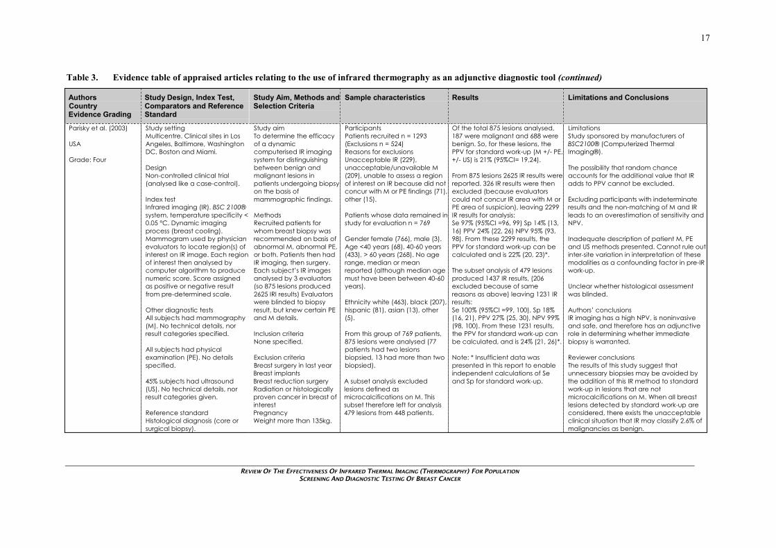
Table 3. Evidence table of appraised articles relating to the use of infrared thermography as an adjunctive diagnostic tool (continued)
AuthorsCountryEvidence Grading
Study Design, Index Test,Comparators and ReferenceStandard
Study Aim, Methods andSelection Criteria
Sample characteristics Results Limitations and Conclusions
Parisky et al. (2003)
USA
Grade: Four
Study settingMulticentre. Clinical sites in LosAngeles, Baltimore, WashingtonDC, Boston and Miami.
DesignNon-controlled clinical trial(analysed like a case-control).
Index testInfrared imaging (IR). BSC 2100®system, temperature specificity <0.05 °C. Dynamic imagingprocess (breast cooling).Mammogram used by physicianevaluators to locate region(s) ofinterest on IR image. Each regionof interest then analysed bycomputer algorithm to producenumeric score. Score assignedas positive or negative resultfrom pre-determined scale.
Other diagnostic testsAll subjects had mammography(M). No technical details, norresult categories specified.
All subjects had physicalexamination (PE). No detailsspecified.
45% subjects had ultrasound(US). No technical details, norresult categories given.
Reference standardHistological diagnosis (core orsurgical biopsy).
Study aimTo determine the efficacyof a dynamiccomputerised IR imagingsystem for distinguishingbetween benign andmalignant lesions inpatients undergoing biopsyon the basis ofmammographic findings.
MethodsRecruited patients forwhom breast biopsy wasrecommended on basis ofabnormal M, abnormal PE,or both. Patients then hadIR imaging, then surgery.Each subject’s IR imagesanalysed by 3 evaluators(so 875 lesions produced2625 IRI results) Evaluatorswere blinded to biopsyresult, but knew certain PEand M details.
Inclusion criteriaNone specified.
Exclusion criteriaBreast surgery in last yearBreast implantsBreast reduction surgeryRadiation or histologicallyproven cancer in breast ofinterestPregnancyWeight more than 135kg.
ParticipantsPatients recruited n = 1293(Exclusions n = 524)Reasons for exclusionsUnacceptable IR (229),unacceptable/unavailable M(209), unable to assess a regionof interest on IR because did notconcur with M or PE findings (71),other (15).
Patients whose data remained instudy for evaluation n = 769
Gender female (766), male (3).Age <40 years (68), 40-60 years(433), > 60 years (268). No agerange, median or meanreported (although median agemust have been between 40-60years).
Ethnicity white (463), black (207),hispanic (81), asian (13), other(5).
From this group of 769 patients,875 lesions were analysed (77patients had two lesionsbiopsied, 13 had more than twobiopsied).
A subset analysis excludedlesions defined asmicrocalcifications on M. Thissubset therefore left for analysis479 lesions from 448 patients.
Of the total 875 lesions analysed,187 were malignant and 688 werebenign. So, for these lesions, thePPV for standard work-up (M +/- PE,+/- US) is 21% (95%CI= 19,24).
From 875 lesions 2625 IR results werereported. 326 IR results were thenexcluded (because evaluatorscould not concur IR area with M orPE area of suspicion), leaving 2299IR results for analysis:Se 97% (95%CI =96, 99) Sp 14% (13,16) PPV 24% (22, 26) NPV 95% (93,98). From these 2299 results, thePPV for standard work-up can becalculated and is 22% (20, 23)*.
The subset analysis of 479 lesionsproduced 1437 IR results, (206excluded because of samereasons as above) leaving 1231 IRresults:Se 100% (95%CI =99, 100), Sp 18%(16, 21), PPV 27% (25, 30), NPV 99%(98, 100). From these 1231 results,the PPV for standard work-up canbe calculated, and is 24% (21, 26)*.
Note: * Insufficient data waspresented in this report to enableindependent calculations of Seand Sp for standard work-up.
LimitationsStudy sponsored by manufacturers ofBSC2100® (Computerized ThermalImaging®).
The possibility that random chanceaccounts for the additional value that IRadds to PPV cannot be excluded.
Excluding participants with indeterminateresults and the non-matching of M and IRleads to an overestimation of sensitivity andNPV.
Inadequate description of patient M, PEand US methods presented. Cannot rule outinter-site variation in interpretation of thesemodalities as a confounding factor in pre-IRwork-up.
Unclear whether histological assessmentwas blinded.
Authors’ conclusionsIR imaging has a high NPV, is noninvasiveand safe, and therefore has an adjunctiverole in determining whether immediatebiopsy is warranted.
Reviewer conclusionsThe results of this study suggest thatunnecessary biopsies may be avoided bythe addition of this IR method to standardwork-up in lesions that are notmicrocalcifications on M. When all breastlesions detected by standard work-up areconsidered, there exists the unacceptableclinical situation that IR may classify 2.6% ofmalignancies as benign.

REVIEW OF THE EFFECTIVENESS OF INFRARED THERMAL IMAGING (THERMOGRAPHY) FOR POPULATION
SCREENING AND DIAGNOSTIC TESTING OF BREAST CANCER
18
Summary of Results Relevant to Diagnosis
Of the two studies appraised, one (Keyserlingk et al., 1998) considered the potential adjuvant benefitthat could be gained from using infrared imaging in a multi-modality diagnostic setting, and used aretrospective case-control design. This study was classed as level four evidence because interpretationof both the index test and the comparators were not blinded, and it is unclear what information wasavailable to those interpreting the reference standard. The other study (Parisky et al., 2003) consideredthe efficacy of infrared imaging when used specifically for the purpose of further clarifying, fromroutine non-invasive diagnostic modalities, whether to proceed to a more invasive test. This study useda clinical trial design. This study was also classed as level four, because it was unclear whetherhistological assessment was blinded. The other major flaws identified are detailed in Table 3(pages 16-17) for this topic. The quality of evidence from many studies of diagnostic techniques hasbeen questioned by authors such as Knottnerus et al. (2002), who commented that methodologicalflaws are common in diagnostic studies.
The retrospective case-control study by Keyserlingk et al. (1998) provided efficacy data that washighly suggestive that infrared thermography provided an adjuvant benefit. However, as the authorsthemselves cautioned this needs to be further investigated in a controlled, prospective manner. Thenon-controlled clinical trial by Parisky et al. (2003) suggested that for a subset of patients (thosewithout microcalcifications on mammography), the addition of infrared imaging would indeed providea valuable tool if used under the same conditions in clinical practice as it was in their trial. However,this finding cannot be extrapolated to all patients who undergo breast investigations. When the wholestudy population was considered, a major factor of potential clinical concern was that infrared imagingmisdiagnosed 2.6% of breast malignancies as benign.
Thermograms need to be captured under controlled conditions, as they are sensitive to environmentalchanges in temperature, humidity, and air circulation. Such parameters must therefore be consideredwhen choosing a venue for patient examination (Jones, 1998). The patient is usually required to restfor 10 to 20 minutes with clothing removed from the relevant area to achieve thermal equilibrium(Jones, 1998). In addition, on the day the test is performed, patients are ideally required to avoid othercontrollable factors that could potentially influence skin temperature such as alcohol consumption,physical exertion and application of cosmetic preparations (Ring and Ammer, 2000). Some interest hasalso focused on physiological factors such as hormonal fluctuations, and temperature fluctuationsduring the normal female menstrual cycle, as potential affecting the stability of thermographicmeasurement (Ng et al., 2002). Neither of the studies appraised for the diagnostic topic have reportedthat menstrual factors were considered, nor report the menstrual status of their participants, despitehaving in their participant populations women who may have been at differing stages in theirreproductive lives.
A 1988 study by van Dam et al. illustrates the necessity for large sample sizes in studies that aim todetect incremental differences in diagnostic accuracy in a multi-modality situation (see Table 4,page 48 in Appendix 4 for a detailed overview of this study). Although their study used an infrareddevice that is no longer available, van Dam et al. (1988) aimed to evaluate the diagnostic characteristicsof independently conducted physical examination, mammography, ultrasound scanning, infraredthermography, and various combinations of these tests in the evaluation of solid palpable breastmasses. Their study included 201 patients, but was unable to show any statistically significantdifferences in effectiveness between any of the clinically relevant combinations of the diagnosticmodalities examined.
To assess validity and reliability of diagnostic tests, the ideal study design is a cross-sectional survey inwhich both the new test and the reference standard are performed (Greenhalgh, 1997). However, toassess the usefulness of a diagnostic test in a situation of clinical decision-making ideally requires aprospective comparison between using and not using the test, for which the randomised controlled trialis the most robust method (Knottnerus et al., 2002). Therefore, one major flaw identified by thisreview is that there was no evidence from randomised controlled trials to support or refute the clinicaleffectiveness, benefits or harms that may occur from the adjuvant diagnostic use of infraredthermography in clinical decision-making. An equally important finding of this review is that therewere no papers identified that examined economic or other costs of relevant infrared thermographydevices in an adjuvant diagnostic setting.

REVIEW OF THE EFFECTIVENESS OF INFRARED THERMAL IMAGING (THERMOGRAPHY) FOR POPULATION
SCREENING AND DIAGNOSTIC TESTING OF BREAST CANCER
19
CONCLUSIONS OF THIS REVIEW
A significant finding of the review conducted for the purpose of this Tech Brief was that there were nostudies that evaluated the effectiveness of the infrared technology devices that are available inNew Zealand. In addition, there were few that evaluated comparable infrared technologies, ortechnologies that may become available to New Zealand. This was despite using a systematicapproach, which included a comprehensive search strategy that identified 1,154 abstracts. Research inthis area is hindered by lack of consistency between manufacturers’ device descriptions andspecifications. The review found that much of the recent literature on infrared thermography is in theform of narrative review, discussion or opinion articles. Most of the published study reports oninfrared thermography refer to studies of infrared devices that are outdated or no longer available, ornon-infrared methods of thermography. No studies of this technology have been conducted inNew Zealand. The evidence that is currently available does not provide enough support for the role ofinfrared thermography for either the population screening or adjuvant diagnostic testing of breastcancer. The major gaps in knowledge at this time can only be addressed by large-scale, prospectiverandomised trials.
Our conclusions are consistent with recommendations by key professional clinician groups. The RoyalAustralian and New Zealand College of Radiologists Breast Imaging Reference Group do notrecommend the use of thermography for the early detection of breast cancer in their policy (2001).Similarly, the American Medical Association do not recommend it as a technique for diagnosticpurposes, although they note that research protocols using thermography are continuing and dataderived from these studies will require careful evaluation (American Medical Association, websiteaccessed 20 May 2004).
More robust research on the effectiveness and costs of technologically advanced infrared thermographydevices for population screening and diagnostic testing of breast cancer is needed, and the conclusionsof this review should be revisited in the face of additional reliable evidence.
REFERENCES
Anbar, M. (1995). Quantitative and dynamic telethermometry - A fresh look at clinical thermology.IEEE Engineering in Medicine & Biology Magazine, 14, 15-16.
Anbar, M. (1998). Clinical thermal imaging today. IEEE Engineering in Medicine and BiologyMagazine, 17, 25-33.
Anbar, M., Milescu, L., Naumov, A., Brown, C., Button, T., Carty, C., & Al Dulaimi, K. (2001).Detection of cancerous breasts by dynamic area telethermometry. IEEE Engineering in Medicine &Biology Magazine, 20, 80-91.
American Medical Association. (undated). Thermography update H-175.988: AMA Policy Finder.Available from: http://www.ama-assn.org/ Accessed on 20.5.04.
Arena, F., Barone, C., & DiCicco, T. (2003). Use of digital infrared imaging in enhanced breast cancerdetection and monitoring of the clinical response to treatment. IEEE EMBC, 1129-1132.
BreastScreen Aotearoa (2004). National policy and quality standards for BreastScreen Aotearoa.Wellington: Ministry of Health National Screening Unit.
Dankiw, W. (1990). Medical thermography. Health care technology series No. 4. Canberra: AustralianInstitute of Health.
Diakides, N.A. (1998). Infrared imaging: an emerging technology in medicine. IEEE Engineering inMedicine & Biology Magazine, 17, 17-18.

REVIEW OF THE EFFECTIVENESS OF INFRARED THERMAL IMAGING (THERMOGRAPHY) FOR POPULATION
SCREENING AND DIAGNOSTIC TESTING OF BREAST CANCER
20
Donegan, W. L. (2002). Diagnosis of breast cancer. In W. L. Donegan & J. S. Spratt (Eds.), Cancer ofthe Breast (pp. 311-345). Philadelphia: W B Saunders.
Feig, S. A., Shaber, G. S., Schwartz, G. F., Patchefsky, A., Libshitz, H. I., Edeiken, J., Nerlinger, R., etal. (1977). Thermography, mammography, and clinical examination in breast cancer screening.Review of 16,000 studies. Radiology, 122, 123-127.
Feig, S. A. (1999). Role and evaluation of mammography and other imaging methods for breast cancerdetection, diagnosis, and staging. Seminars in Nuclear Medicine, 29, 3-15.
Foster, K. R. (1998). Thermographic detection of breast cancer. IEEE Engineering in Medicine &Biology Magazine, 17, 10-14.
Greenhalgh, T. (1997). How to read a paper: getting your bearings (deciding what the paper is about).BMJ, 315, 243-246.
Gautherie, M., Haehnel, P., Walter, J. P., & Keith, L. G. (1987). Thermovascular changes associatedwith in situ and minimal breast cancers. Results of an ongoing prospective study after four years.Journal of Reproductive Medicine, 32, 833-842.
Head, J. F., Wang, F., Lipari, C. A., & Elliott, R. L. (2000). The important role of infrared imaging inbreast cancer - New technology improves applications in risk assessment, detection, diagnosis, andprognosis. IEEE Engineering in Medicine and Biology Magazine, 19, 52-57.
Head, J.F., & Elliott, R.L. (2002). Infrared imaging: making progress in fulfilling its medical promise.IEEE Engineering in Medicine & Biology Magazine, 21, 80-85.
Hider, P., & Nicholas, B. (1999). The early detection and diagnosis of breast cancer: a literature review- an update. NZHTA Report, 2, 1-150.
Hobson, J. (2002). New techniques in medicine: thermal imaging. Choice, September, 60-61.
Homer, M. J. (1985). Breast imaging: pitfalls, controversies, and some practical thoughts. RadiologicClinics of North America, 23, 459-472.
International Academy of Clinical Thermology. (undated). Mammography vs thermography. Availablefrom: http://www.iact-org.org/patients/breastthermography/mammography-vs-therm.html Accessed on20.3.04
Jones, B. F. (1998). A reappraisal of the use of infrared thermal image analysis in medicine. IEEETransactions on Medical Imaging, 17, 1019-1027.
Keyserlingk, J. R., Ahlgren, P. D., Yu, E., & Belliveau, N. (1998). Infrared imaging of the breast:Initial reappraisal using high-resolution digital technology in 100 successive cases of stage I and IIbreast cancer. Breast Journal, 4, 245-251.
Knottnerus, J. A., van Weel, C., & Muris, J. W. (2002). Evaluation of diagnostic procedures. BMJ, 324,477-480.
Leitch, A. M. (2002). Screening for breast cancer. In W. L. Donegan & J. S. Spratt (Eds.), Cancer ofthe Breast (pp. 269-286). Philadelphia: W B Saunders.
Mahoney, M. C. (2002). Breast imaging: mammography, sonography, and emerging technology. InW. L. Donegan & J. S. Spratt (Eds.), Cancer of the Breast (pp. 287-310). Philadelphia: W B Saunders.
Margolese, R. G., Cantin, J., Bouchard, F., Caines, J., Beaulieu, M. D., Little, C. D., Levine, M. N., etal. (1998). 1. The palpable breast lump: information and recommendations to assist decision-makingwhen a breast lump is detected. Canadian Medical Association Journal, 158, S3-S8.

REVIEW OF THE EFFECTIVENESS OF INFRARED THERMAL IMAGING (THERMOGRAPHY) FOR POPULATION
SCREENING AND DIAGNOSTIC TESTING OF BREAST CANCER
21
Martin, J. E. (1983). Breast imaging techniques. Mammography, ultrasonography, computedtomography, thermography, and transillumination. Radiologic Clinics of North America, 21, 149-153.
McLean, R. (1999). Thermography: technology brief. Montreal: Conseil d'Evaluation des Technologiesde la Santé.
Moskowitz, M. (1985). Thermography as a risk indicator of breast cancer. Results of a study and areview of the recent literature. Journal of Reproductive Medicine, 30, 451-459.
Moskowitz, M. (1995). Breast imaging. In W. L. Donegan & J. S. Spratt (Eds.), Cancer of the Breast(pp. 206-239). Phildelphia, PA.: W B Saunders.
Nass, S. J., Henderson, C., & Lashof, J. C. (2001). Mammography and beyond: developingtechnologies for the early detection of breast cancer. Washington, DC: National Academy Press.
Ng, E. Y., Fok, S. C., Peh, Y. C., Ng, F. C., & Sim, L. S. (2002). Computerized detection of breastcancer with artificial intelligence and thermograms. Journal of Medical Engineering & Technology, 26,152-157.
Ohashi, Y., & Uchida, I. (2000). Applying dynamic thermography in the diagnosis of breast cancer -Techniques for improving sensitivity of breast thermography. IEEE Engineering in Medicine andBiology Magazine, 19, 42-51.
Orel, S. G., & Troupin, R. H. (1993). Nonmammographic imaging of the breast: current issues andfuture prospects. Seminars in Roentgenology, 28, 231-241.
Parisky, Y. R., Sardi, A., Hamm, R., Hughes, K., Esserman, L., Rust, S., & Callahan, K. (2003).Efficacy of computerized infrared imaging analysis to evaluate mammographically suspicious lesions.AJR. American Journal of Roentgenology, 180, 263-269.
Qi, H., & Head, J. F. (2001). Asymmetry analysis using automatic segmentation and classification forbreast cancer detection in thermograms. Proceedings of the 23rd Annual International ConferenceIEEE Engineering in Medicine and Biology Society, 2866-2869.
Ring, E. F., & Ammer, K. (2000). The technique of infra red imaging in medicine. ThermologyInternational, 10, 7-14.
Royal Australian and New Zealand College of Radiologists Breast Imaging Reference Group. (2001).Policy on the use of thermography to detect breast cancer. Available from:http://www.ranzcr.edu.au/open/policies/diagnostic_imaging/pol7_3.htm Accessed on 25.6.04
Royal New Zealand College of General Practitioners (1999). Early detection of breast cancer:guidelines for primary care providers. Wellington: The College.
Snyder, W., Qi, H., Elliott, R. L., Head, J. F., & Wang, C. X. (2000). Increasing the effective resolutionof thermal infrared images: an algorithm based on mean-field annealing that also removes noise andpreserves image edges. IEEE Engineering in Medicine & Biology, 19, 63-70.
StataCorp (2001). Stata statistical software: release 7.0. College Station, TX.: Stata Corporation.
Sunshine, J. H., & Applegate, K. E. (2004). Technology assessment for radiologists. Radiology, 230,309-314.
van Dam, P. A., Van Goethem, M. L., Kersschot, E., Vervliet, J., Van den Veyver, I. B., De Schepper,A., & Buytaert, P. (1988). Palpable solid breast masses: retrospective single- and multimodalityevaluation of 201 lesions. Radiology, 166, 435-439.
Wharton, L. (2003). Thermography offers alternative in breast cancer detection. National BusinessReview, October 17, 56.

REVIEW OF THE EFFECTIVENESS OF INFRARED THERMAL IMAGING (THERMOGRAPHY) FOR POPULATION
SCREENING AND DIAGNOSTIC TESTING OF BREAST CANCER
22
Williams, K. L., Phillips, B. H., Jones, P. A., Beaman, S. A., & Fleming, P. J. (1990). Thermography inscreening for breast cancer. Journal of Epidemiology & Community Health, 44, 112-113.
Wilson, J. M., & Jungner, G. (1968). Principles and practice of screening for disease. Geneva: WorldHealth Organisation.

REVIEW OF THE EFFECTIVENESS OF INFRARED THERMAL IMAGING (THERMOGRAPHY) FOR POPULATION
SCREENING AND DIAGNOSTIC TESTING OF BREAST CANCER
23
APPENDIX 1: EVIDENCE LEVELS FOR DIAGNOSTIC TESTS
Susceptibility for bias in studies of diagnostic tests
[Adapted from Diagnostic Testing emerging from the gloom?Bandolier http://www.jr2.ox.ac.uk/bandolier/band70/b70-5.html]
Level One An independent, masked comparison with reference standard among an appropriatepopulation of consecutive patients.
Level Two An independent, masked comparison with reference standard among non-consecutivepatients or confined to a narrow population of study patients.
Level Three An independent, masked comparison with an appropriate population of patients, butreference standard not applied to all study patients.
Level Four Reference standard not applied independently or masked.
Level Five Expert opinion with no explicit critical appraisal, based on physiology, bench research, orfirst principles.

REVIEW OF THE EFFECTIVENESS OF INFRARED THERMAL IMAGING (THERMOGRAPHY) FOR POPULATION
SCREENING AND DIAGNOSTIC TESTING OF BREAST CANCER
24
APPENDIX 2: EXPLANATION OF SELECTION CRITERIA
Inclusion criteria
Publication Type
Studies published between 1985 and 20 May 2004 (inclusive) - These dates were chosen to identifystudies that concerned the index test of interest to this review.
English language - Tech Briefs are rapidly produced assessments of the best available evidence.Because of time restrictions and the resultant limitation on resources for translation, it is usual toexclude non-English articles. Nevertheless, it should be noted that during the review of abstracts forthis review, there were no significant non-English language studies identified that fulfilled all the otherselection criteria.
Setting
Topic One - The rationale for this inclusion criteria for screening is explained in the Backgroundsection of this report, under the sub-heading “Thermography used for screening for breast cancer”.
Topic Two - The rationale for this inclusion criteria is explained in the Background section of thisreport, under the sub-heading “Thermography Used for Diagnosis of Breast Cancer”.
Population
Topic One - The aim of screening for any disease is to reduce mortality (and morbidity) from thisdisease by performing a test on an asymptomatic population, which is why asymptomatic women werethe population of interest for this topic.
Topic Two - The role of a diagnostic test for any disease is to further evaluate abnormalities that havebeen pre-detected by either clinical symptoms, or screening tests, which is why patients symptomaticfor breast cancer or who have had an abnormal mammogram were the population of interest for thistopic.
Sample Size
Studies with small sample size are prone to sampling error. A lower limit of 20 is usual for criticalreviews.
Index Test
The focus of this Tech Brief was to assess evidence relevant to infrared imaging devices that are (or arecomparable with those) currently available, or which may become available in the future to patients inNew Zealand. As outlined in the Background section under the heading “Technological History ofInfrared Imaging”, infrared cameras that were available up until the mid-1980s suffered from severallimitations that have been overcome since that time, and there have been accompanying improvementsin computerised processing systems. This provided the rationale for selecting for appraisal only studiesthat have been published since 1985.
Comparators
Topic One - The rationale for this inclusion criteria for screening is explained in the Backgroundsection of this report, under the sub-heading “Thermography used for screening for breast cancer”.
Topic Two - The rationale for this inclusion criteria is explained in the Background section of thisreport, under the sub-heading “Thermography Used for Diagnosis of Breast Cancer”.

REVIEW OF THE EFFECTIVENESS OF INFRARED THERMAL IMAGING (THERMOGRAPHY) FOR POPULATION
SCREENING AND DIAGNOSTIC TESTING OF BREAST CANCER
25
Index Test
Histological diagnosis is generally considered to be the “gold-standard” measure for detection ofcancer.
Outcomes
The outcome measures chosen for this Tech Brief are standard for critical reviews of diagnosticmodalities.
Exclusion Criteria
English language - As explained above for inclusion criteria
Concerned studies of infrared devices that are no longer available (or that are not technologicallycomparable to those available to those available in New Zealand - As explained above for indextest.
Concerned studies of methods of thermogaphy that were not infrared thermography, or did notspecify the method of thermography under study - The focus of this review was on infraredthermography devices, because that is the type of thermography that is available in New Zealand,therefore studies of other types of thermography were not relevant to this review. A realisticassessment of a study could not be made in the context of this review if the method of thermographyused for the study was not specified.
Reported on the use of thermography in aspects of breast cancer management other thanscreening (Topic One) or diagnosis (Topic Two) - Screening and diagnosis were the aspects of breastcancer management under consideration in this review. Therefore, articles concerning thermographyuse in other aspects of breast cancer management, including technological articles about physicalaspects of devices or computational processes, were not relevant to the aims of this review.
Had only an abstract available, or were conference proceedings - These types of articles containlimited data on the methods or results of studies.
Were an expert opinion commentary, a narrative review or a book chapter - These types of articleare opinion articles that usually collect and report information in a non-systematic manner. They aretherefore prone to selection bias and/or reporting bias, and furthermore frequently do not present datathat can be used to assess effectiveness.
Were discussion articles (e.g., presented as a letter or editorial) - These types of article are opinionarticles that usually collect and report information in a non-systematic manner. They are thereforeprone to selection bias and/or reporting bias, and furthermore frequently do not present data that can beused to assess effectiveness.
Reported studies with fewer than 20 participants or were a case presentation - Studies with smallsample size are prone to sampling error. A lower limit of 20 is usual for critical reviews.
Reported animal studies - Humans were the focus of this review, therefore animal studies were notconsidered relevant.
Did not clearly describe their methods or results, or had discrepancies in data within the article -Such articles do not provide reliable data that can be used to assess effectiveness using rigorous criticalappraisal techniques.

REVIEW OF THE EFFECTIVENESS OF INFRARED THERMAL IMAGING (THERMOGRAPHY) FOR POPULATION
SCREENING AND DIAGNOSTIC TESTING OF BREAST CANCER
26
APPENDIX 3: SEARCH STRATEGY
Medline & Cochrane Controlled Trials Register1 thermography/2 thermal imaging.mp.3 (infrared imag$ or infra-red imag$).mp.4 or/1-35 breast neoplasms/6 breast cancer.ti,ab.7 5 or 68 4 and 79 letter.pt.10 8 not 911 animal/12 animal/ and human/13 11 not 1214 10 not 1315 (thermology or thermometry or thermography).tw.16 (breast adj3 carcinoma).mp.17 (breast adj3 malignan$).tw.18 (breast adj3 neoplas$).tw.19 (breast adj3 cancer$).tw.20 or/16-1921 15 and 722 (4 or 15) and 2023 14 or 22
Embase1 thermography/ or thermometry/2 (thermal imaging or thermography or thermometry or thermology).tw.3 (infra-red imag$ or infrared imag$).mp.4 or/1-35 Breast Cancer/6 (breast adj3 cancer).tw.7 (breast adj3 malignan$).tw.8 (breast adj3 carcino$).mp.9 (breast adj3 adenocarcino$).mp.10 or/5-911 cancer screening/12 4 and 1013 4 and 1114 13 not 1215 12 or 1316 letter.pt.17 15 not 16
Cinahl & Amed1 thermography/2 thermal imaging.mp.3 (infrared imag$ or infra-red imag$).mp.4 (thermology or thermometry or themography).tw.5 (breast adj3 carcinoma).mp.6 (breast adj3 malignan$).tw.7 (breast adj3 neoplas$).tw.8 (breast adj3 cancer$).tw

REVIEW OF THE EFFECTIVENESS OF INFRARED THERMAL IMAGING (THERMOGRAPHY) FOR POPULATION
SCREENING AND DIAGNOSTIC TESTING OF BREAST CANCER
27
9 (breast adj3 carcino$).mp.10 (breast adj3 adenocarcino$).mp.11 Breast Neoplasms/12 or/1-413 or/5-1114 12 and 13
Current Contents/Science Citation Index1 thermometry OR thermal imaging OR thermology OR thermography2 (infrared OR infra-red) SAME imag*3 #1 OR #24 breast SAME cancer5 Breast SAME carcinoma6 breast SAME malignanc*7 breast SAME adenocarcin*8 breast SAME neoplas*9 #4 OR #5 OR #6 OR #7 OR #810 #3 AND #9
The vocabulary used in the strategies above was adapted for use in remaining sources.

REVIEW OF THE EFFECTIVENESS OF INFRARED THERMAL IMAGING (THERMOGRAPHY) FOR POPULATION
SCREENING AND DIAGNOSTIC TESTING OF BREAST CANCER
28
APPENDIX 4: EXCLUDED RETRIEVED PAPERS
Note: The summaries of the excluded retrieved papers in this appendix are not the result of a criticalappraisal. Any conclusions presented in these summaries are those of the author(s) of the individualpaper only, and not the author of this report.
Anbar, M. (1995). Quantitative and dynamic telethermometry - A fresh look at clinical thermology.IEEE Engineering in Medicine & Biology Magazine, 14, 15-16.
This article was excluded because it was an expert opinion article. This short article briefly outlinesthe academic history of clinical thermal imaging, explaining that a scarcity of academic research inthis field has (understandably) meant that the merits of clinical thermology have not been firmlyestablished by systematic academic studies. The author opines that it is essential to understand themechanisms that underlie thermal changes in breast tissue, and that this understanding will enable theefficacy of diagnostic testing by this modality to be optimised. The author states that reliable, modern,and fast, computerised equipment must be used and studied to achieve this aim.
Anbar, M. (1998). Clinical thermal imaging today. IEEE Engineering in Medicine and BiologyMagazine, 17, 25-33.
This article was excluded because it was a narrative review. It outlines recent technological advancesin infrared equipment and also discusses current understandings of the physiological processes thatunderlie thermal changes in the breast and other tissues.
Anbar, M. (2002). Assessment of physiologic and pathologic radiative heat dissipation using dynamicinfrared imaging. Annals of the New York Academy of Sciences, 972, 111-118.
This report was excluded due a lack of clear description of methods and results. Most of this article isa narrative review that outlines in detail the physiological processes behind skin temperatureregulation. The author explains that because the skin’s temperature modulation is so complex, it canonly be accurately measured using highly sensitive dynamic infrared detectors. These allow formeasurement across multiple frequencies, and additionally for computer-generated procedures toobjectively assess localised attenuations of temperature modulation. The local attenuations signalpossible underlying pathology. The author then presents some graphical results from an objectivecomputer-generated assessment of different breast lesions. The methods used to attain the graphicalresults were not clearly described, including study design, sample selection and characteristics, therewas no comparator diagnostic modality used, and no numerical results presented in this report. Theauthor concludes that this type of computer-assisted interpretation has potential usefulness in breastcancer diagnosis.
Anbar, M., Brown, C.A., & Milescu, L. (1999). Objective identification of cancerous breasts bydynamic area telethermometry (DAT). Thermology International, 9, 137-143.
This paper was requested by electronic mail from the first author, after the failure to attain the journalthrough all our usual library sources, but it was not retrieved within the timeframe (several months).The paper is likely to have presented a study report concerning the development of an objectivediagnostic parameter using infrared imaging techniques and quantitative computational processes.

REVIEW OF THE EFFECTIVENESS OF INFRARED THERMAL IMAGING (THERMOGRAPHY) FOR POPULATION
SCREENING AND DIAGNOSTIC TESTING OF BREAST CANCER
29
Anbar, M., Brown, C., Milescu, L., Babalola, J., & Gentner, L. (2000). The potential of dynamic areatelethermometry in assessing breast cancer. IEEE Engineering in Medicine & Biology Magazine, 19,58-62.
An article describing the mechanisms of using ‘dynamic’ IR imaging techniques, where highly sensitiveand fast responding IR detectors are coupled with quantitative computer analysis to measure over timeareas of abnormal modulation of skin temperature. This article was excluded because it mainlydescribed technological aspects of this process, and although it presented some clinical examples ofuse, these were in case report form. The authors describe how the use of this technique is based on thehypothesis that abnormal secretion of nitrous oxide (NO) by breast cancer cells affects the usualvascular control of blood vessel flow, and therefore identifying the areas of abnormal blood flowlocates abnormal or malignant breast tissue.
Anbar, M., Milescu, L., Naumov, A., Brown, C., Button, T., Carty, C., & Al Dulaimi, K. (2001).Detection of cancerous breasts by dynamic area telethermometry. IEEE Engineering in Medicine &Biology Magazine, 20, 80-91.
Report from a study that was undertaken using a high-speed infrared detector to develop a singlediagnostic parameter that distinguishes benign from malignant breast lesions. This paper wasexcluded because it focuses on reporting in detail the various computational and statistical processesthat were undertaken to develop this single objective diagnostic parameter. Furthermore, there wasinsufficient data presented to estimate any of the pre-specified outcomes of interest for this review. Thestudy was based on the assumption that breast cancer cells secrete nitrous oxide, that the nitrous oxideattenuates the normal vascular control of the breast tissue, and that this produces minute changes inskin temperature over time. To develop this diagnostic parameter, a total of 100 women (withpresumed benign breast lesions, or in-situ breast cancer, or invasive breast cancer on excisionalbiopsy) were imaged. The authors conclude that by allowing a computer to assess the infrared image,this type of dynamic imaging can yield meaningful diagnostic information, and that their methodwarrants testing in clinical trial situations to determine its scope and limitations.
Anonymous (1985). The use of diagnostic tests for screening and evaluating breast lesions. Health andPublic Policy Committee, American College of Physicians. Annals of Internal Medicine, 103, 143-146.
This article was excluded because it was a narrative review article. It presents an author’s assessmentof breast self-examination, mammography, thermography and other techniques for the screening ofasymptomatic women and an assessment of mammography, thermography, ultrasound and fine-needleaspiration for the diagnostic evaluation of breast masses. For each modality, a description ispresented followed by the author’s assessment of efficacy, safety (where applicable) and cost. Forscreening, thermography is assessed as safe, but of insufficient accuracy, with cost difficult to assess.For the diagnosis of breast lumps, this author concluded that thermography was still generally nothelpful for the evaluation of breast masses. The author’s final conclusions are that mammography is anecessary part of breast screening, although ages for screening are still controversial, and that for theassessment of breast masses mammography and/or ultrasound plus fine-needle aspiration providesufficient information in most cases.
Anonymous (1986). Diagnosis and first-line treatment of breast cancer in Italian general hospitals.G.I.V.I.O. (Interdisciplinary Group for Cancer Care Evaluation) - Italy. Tumori, 72, 273-283.
This report was excluded because it was a descriptive report that outlines and discusses the quality ofdiagnostic procedures and treatment processes used in the management of women with breast cancerat 63 Italian hospitals between March 1983 and April 1984. It is mainly concerned with breast cancertreatment issues.

REVIEW OF THE EFFECTIVENESS OF INFRARED THERMAL IMAGING (THERMOGRAPHY) FOR POPULATION
SCREENING AND DIAGNOSTIC TESTING OF BREAST CANCER
30
Arena, F., Barone, C., & DiCicco, T. (2003). Use of digital infrared imaging in enhanced breast cancerdetection and monitoring of the clinical response to treatment. IEEE EMBC, 1129-1132.
Report from a study that had the aim of demonstrating that an automated computer generatedalgorithm can be developed to predict likely risk of breast cancer when advanced computer hardwareand software is used in conjunction with advanced high-resolution digital infrared technology. Thisstudy was excluded from appraisal because it was a report from conference proceedings. It lacked aclear description of methods and results. The researchers in the study pre-selected three patientpopulations on basis of: assumed normal; known biopsy results of wide variety of cancer; or previouscancer treatment. Although the report details the mechanism and nature of the IR examinationsundergone by the study population, there is little information given concerning the results of any otherinvestigations that the study population may have undergone.
Barraclough, B. M., Picker, R. H., & Barraclough, B. H. (1985). Toward breast screening. AustralasianRadiology, 29, 181-184.
Two-part journal article. The first part discusses potential breast screening methods, includingthermography. The second part presents an overview of the results from a small study in which 100female patients underwent ultrasound scanning as part of their breast examinations in a hospital inSydney, Australia, and was excluded from appraisal because it only provided information aboutultrasound scanning in these patients. The authors surmised that ultrasound scanning should beconsidered for inclusion in a breast-screening programme.
Blume, S. S. (1993). Social process and the assessment of a new imaging technique. InternationalJournal of Technology Assessment in Health Care, 9, 335-345.
This article was excluded because it was a narrative review. It presents an historical review of thesociological processes that have occurred since the initial discovery of infrared thermography, tracingthe various changes in medical, industrial, and thermography proponents’ opinions and attitudes to thetechnology since 1955. Concludes that the history of thermography shows how the process ofcontinuous assessment of medical technologies is dominated by certain values, priorities andassumptions. Not directly relevant to the topic, focussing on sociological issues not scientific evidence.
Cady, B., Steele, G. D., Jr., Morrow, M., Gardner, B., Smith, B. L., Lee, N. C., Lawson, H. W., et al.,(1998). Evaluation of common breast problems: guidance for primary care providers. Ca: a CancerJournal for Clinicians, 48, 49-63.
This report was excluded because it was a narrative review/expert opinion article that describes anorganised approach for the management of women presenting to primary care providers with breastsymptoms or a positive screening test. It mentions thermography only to state that it has no currentplace in breast cancer screening or diagnostic evaluation.
Ciatto, S., Palli, D., Rosselli del Turco, M., & Catarzi, S. (1987a). Diagnostic and prognostic role ofinfrared thermography. Radiologia Medical, 74, 312-315.
Report from an Italian study that used an older infrared device that is no longer available (AGAThermovision 680). This study was excluded from appraisal because it used older equipment that is nolonger obtainable, and for which device specifications were not easily available. The study had twoaims, firstly to evaluate the diagnostic accuracy of infrared thermography (TH) compared withphysical examination (PE) and mammography (M), and secondly to determine whether thermographicpatterns are related to prognostic outcome. Between 1976 and 1983, 6,832 women attending aspecialist oncology centre were examined by PE and M, and were selected to have TH on a “clinical

REVIEW OF THE EFFECTIVENESS OF INFRARED THERMAL IMAGING (THERMOGRAPHY) FOR POPULATION
SCREENING AND DIAGNOSTIC TESTING OF BREAST CANCER
31
basis without well defined criteria” (p 312). From this population, 469 women with breast cancerwere identified, with their diagnoses confirmed by either cytology or histology. The authors found thatthe sensitivity of TH as a diagnostic method in their study was 47 percent overall, for M was 89 percentand for PE was 86 percent, and furthermore that combining TH with PE or M did not appear toincrease the combined sensitivity. These authors concluded that their study results did not encouragethe use of infrared thermography in either diagnostic or prognostic processes.
Ciatto, S., Rosselli Del Turco, M., Cecchini, S., Grazzini, G., & Iossa, A. (1989). Telethermographyand breast cancer risk prediction. Tumori, 75, 110-112.
Report from a study that aimed to assess the association between different infrared thermographicpatterns and likelihood of developing breast cancer. This paper was excluded from appraisal becauseit was concerned with risk factor identification, did not provide evidence that earlier detection ofbreast cancer occurred as a result of utilising thermography in this manner, and the type of infraredthermography device was not reported. The study used a retrospective chart review method to identifypatients who had had certain thermographic patterns. Patient histories were then reviewed toascertain their subsequent breast health. As a result of this study, the authors concluded that they hadfound no association between abnormal thermographic patterns and risk of developing breast cancer.
Ciatto, S., Smith, A. H., Di Maggio, C., Pescarini, L., Lattanzio, E., Ancona, A., Punzo, C., et al.(1987b). Breast cancer diagnosis under the age of forty years. Tumori, 73, 457-461.
Report from an Italian study that aimed to assess the sensitivity of single tests, and combinations oftests, for the diagnosis of breast cancer in women under the age of 40 years. This study was excludedbecause it did not specify the method of thermography used. The study was conducted in 1984, and themethod employed was a retrospective review of case records of women aged 39 years and under withbreast cancer from six Italian institutions. The time period varied between centres, but went as farback as 15 years prior to 1984 in some centres. The authors report the rates of histologically-positivecases that were correctly diagnosed by each modality, and were only able to determine from theirresults that physical examination was the test of choice for women in this study (although it was onlypositive in 77% of all the cancers). The authors conclude that physical examination cannot be usedalone, that their study was unable to define the best test(s) to be combined with physical examination,but that thermography could probably be ruled out from the poor results that they had found in theirpreliminary diagnostic calculations.
Cotton, P. (1992). AMA's Council on Scientific Affairs takes a fresh look at thermography. JAMA-Journal of the American Medical Association, 267, 1885-1887.
This paper was excluded because it was a discussion article, which presents a brief review of theapplications of thermography in the USA, including issues of health insurance reimbursement.
Cuttino, J. T., Jr. (1987). Breast imaging techniques. Current status. North Carolina Medical Journal,48, 303-304.
This paper was excluded because it was a narrative review article. It is a short article that describesthe various diagnostic modalities available to image the breast. The author concludes that, at thatpoint in time, mammography is the best modality for breast screening, that ultrasound is useful as anadjunctive diagnostic modality but not for screening, that CT and MRI are still at developmentalstages, and that thermography has no role at all in either screening or diagnostic situations.

REVIEW OF THE EFFECTIVENESS OF INFRARED THERMAL IMAGING (THERMOGRAPHY) FOR POPULATION
SCREENING AND DIAGNOSTIC TESTING OF BREAST CANCER
32
de Thibault de Boesinghe, L. (1990). The value of thermography for the diagnosis, prognosis andsurveillance of non-palpable breast cancer. Journal Belge de Radiologie, 73, 375-378.
This paper was excluded because it was a narrative review article. It briefly outlines the history andtechnique of thermography, and the interpretation of infrared thermograms. The author concludeswith favourable comments concerning the role of infrared thermography as a technique to be combinedwith other available imaging modalities for the screening and diagnosis of breast cancer.
Diakides, N. A. (1997). New developments in low cost IR imaging systems. European Journal ofThermology, 7, 213-215.
This paper was excluded because it was a narrative review article. It outlines developments in infraredtechnologies, their applications and their future potentials. The author states that there is potential forclinical research to determine the benefits of advanced thermal imaging in comparison to otherimaging modalities.
Diakides, N.A. (1998). Infrared imaging: an emerging technology in medicine. IEEE Engineering inMedicine & Biology Magazine, 17, 17-18.
This paper was excluded because it was an editorial article. It introduces a journal issue that isdedicated to the subject of the application of new technologies in infrared imaging that have emergedsince the early 1990s.
Donnelly, L. F., Freundlich, I. M., Wallace, J. D., & Dodd, G. D. (2000). Thermography and thevenous diameter ratio in the detection of the non-palpable breast carcinoma. 1968. AJR. AmericanJournal of Roentgenology, 174, 1092.
This paper was excluded because it was an historical pictorial article that shows a reproduction of a1968 journal illustration.
Feig, S. A. (1999). Role and evaluation of mammography and other imaging methods for breast cancerdetection, diagnosis, and staging. Seminars in Nuclear Medicine, 29, 3-15.
This paper was excluded because it was a narrative review/expert opinion article. It outlines therequirements that the author thinks that any new supplementary imaging modality would have to meetto provide additional benefits over mammography alone in breast cancer screening, or overmammography plus the standard additional radiological techniques in breast cancer diagnosis.
Foster, K. R. (1998). Thermographic detection of breast cancer. IEEE Engineering in Medicine &Biology Magazine, 17, 10-14.
This paper was excluded because it was a narrative review article. It outlines and discusses the historyand major studies of thermography and mammography as techniques for breast cancer screening anddiagnosis. The author stresses that it is important that sufficient evidence is gained from extensiveclinical studies before a test is adopted as a tool for the widespread screening of asymptomaticpopulations.
REVIEW OF THE EFFECTIVENESS OF INFRARED THERMAL IMAGING (THERMOGRAPHY) FOR POPULATION
SCREENING AND DIAGNOSTIC TESTING OF BREAST CANCER
33
Gautherie, M., Haehnel, P., Walter, J. P., & Keith, L. G. (1987). Thermovascular changes associatedwith in situ and minimal breast cancers. Results of an ongoing prospective study after four years.Journal of Reproductive Medicine, 32, 833-842.
A report from a study that aimed to assess the thermal and vascular disorders associated with the earlystages of breast malignancy, with the aim of identifying those women who may be a higher risk forbreast cancer. This study was excluded because it used both contact thermography and infraredthermography methods, and the data presented in this report did not allow for a distinction to be madebetween these thermographic methods. Furthermore, the study focussed on thermography patterns asbeing a risk factor for breast cancer, and the authors state that their study was not designed to assessthe sensitivity and specificity of thermography versus other diagnostic methods. A cohort of 25,782women, which included both asymptomatic women (some of whom had a positive family history ofbreast cancer) and women who had presented with a variety of breast symptoms, was followed for fouryears. At first visit, all women were examined by thermography (76% by both contact and infraredmethods), then had mammography, and physical examination. The authors’ report that 1.2 percent ofthe asymptomatic women in their study were found to have breast cancer, which was higher than theywould have expected in an asymptomatic population. The latter part of this paper discusseshypotheses, which were current at that time, to explain why breast tumours generate heat.
Ghys, R. (1987). Cancer diagnosis in a "breast clinic": a 15 years retrospection. Journal of NuclearMedicine & Allied Sciences, 31, 315-320.
A report from a study that aimed to assess the independent diagnostic accuracies of the techniques ofclinical examination, mammography, thermography, anamnesis and diaphanoscopy, as well as theadjunctive value of the imaging modalities when added to clinical diagnosis for breast cancer. Thetechniques of anamnesis and diaphanoscopy were not explained in the report. This study was excludedbecause it used old thermographic equipment that is no longer easily available, and for whichtechnical specifications are not clearly reported. The authors conclude from their study that thecombined use of diaphanoscopy, thermography and mammography provided the best detection ofbreast cancers that were not clinically obvious.
Gold, R. H., Bassett, L. W., & Kimme-Smith, C. (1986). Breast imaging. State-of-the-art. InvestigativeRadiology, 21, 298-304.
This paper was excluded because it was a narrative review article of various breast imagingprocedures. It includes a short paragraph about thermography, in which the authors state thatthermography cannot reliably differentiate benign from malignant breast disease.
Grenn, M. W. (1996). Recent advances in portable infrared imaging systems Amsterdam. Proc 18thInternational Conference of the IEEE Engineering in Medicine and Biology Society. Paper 1091, 2083-2084.
This paper was excluded because it was the results of a conference proceedings. It is a shortdescriptive article that details technical specifications of portable infrared imaging systems thatdeveloped over the five years prior to that time.

REVIEW OF THE EFFECTIVENESS OF INFRARED THERMAL IMAGING (THERMOGRAPHY) FOR POPULATION
SCREENING AND DIAGNOSTIC TESTING OF BREAST CANCER
34
Harrison, B., & Mabuchi, K. (1998). Biomedical infrared imaging in Japan. IEEE Engineering inMedicine & Biology Magazine, 17, 66-70.
This paper was excluded because it was a discussion article about the status and use of infraredimaging in Japan. It notes particularly the cultural and language barriers that are present forJapanese researchers in this field.
Head, J. F., & Elliott, R. L. (1997). Thermography. Its relation to pathologic characteristics,vascularity, proliferation rate, and survival of patients with invasive ductal carcinoma of the breast.Cancer, 79, 186-188.
This paper was excluded because it was a journal correspondence article. It discusses the results froma study in which the authors had used infrared thermography to assess disease characteristics andprognosis for patients with breast cancer.
Head, J.F., & Elliott, R.L. (2002). Infrared imaging: making progress in fulfilling its medical promise.IEEE Engineering in Medicine & Biology Magazine, 21, 80-85.
This paper was excluded because it was a narrative review/expert opinion article. It discusses thebackground to, and current and potential applications of, infrared imaging in medicine. The authors’opinion is that thermography has not yet received the attention it merits as a potential part of multi-modal screening for breast cancer. The authors support thermography as a risk assessment andprognostic tool for breast cancer. The article states that current issues in medical applications of IRtechnologies revolve around poor quality IR images, which require visual (rather than quantitative)analysis, and therefore require training and agreed diagnostic standards. The paper outlinesdifferences between different generations of IR technologies, and upcoming developments. Theauthors’ opinion is that early instruments were not sensitive enough to detect the subtle changes intemperature required to accurately detect and monitor disease. The authors state that Dynamic Areatelethermometry (DAT) (developed by Anbar et al., 2000) may be the most promising new developmentin infrared imaging.
Head, J. F., & Lipari, C. A. (1999). Comparison of mammography and breast infrared imaging:sensitivity, specificity, false negatives, false positives, positive predictive value and negative predictivevalue. Proceeding of the 21st Annual International Conference IEEE Engineering in Medicine andBiology Society, 1116.
This paper was excluded because it was proceedings from a conference. The authors report havingundertaken a literature search to identify papers for a literature review, but do not clearly specify themethods and selection criteria that they employed to search and identify papers for their review. Theliterature review was followed by an averaging of the performance characteristics of mammographyfrom seven papers (reporting from various different diagnostic settings), and a comparison with theaveraged results of the performance characteristics of infrared imaging derived from one study ofinfrared imaging in a screening setting. The authors conclude from their review that infrared imaginghas performance characteristics similar to mammography for the detection of breast cancer.
REVIEW OF THE EFFECTIVENESS OF INFRARED THERMAL IMAGING (THERMOGRAPHY) FOR POPULATION
SCREENING AND DIAGNOSTIC TESTING OF BREAST CANCER
35
Head, J. F., Wang, F., & Elliott, R. L. (1993). Breast thermography is a noninvasive prognosticprocedure that predicts tumor growth rate in breast cancer patients. Annals of the New York Academy ofSciences, 698, 153-158.
A report from a two-part study that used data from a specialist breast clinic to investigate theprognostic accuracy of breast thermography. This study was excluded because it examined the role ofthermography in predicting prognosis in breast cancer patients. The first part of the study comparedpreviously acquired thermographic results from 126 deceased breast cancer patients, 100 living breastcancer patients and 100 non-cancer patients, finding that a significantly higher percentage of thedeceased group had ‘abnormal’ thermograms. No indication was given of whether the results of thethermographic interpretation were taken from the original examination, or whether the originalthermograms were re-reviewed for the purposes of this study. No information is given as to the resultsof any other clinical or radiological investigations that these patients may have undergone. Theauthors conclude that this study demonstrates the prognostic significance of thermography. Thesecond part of this study compared the thermographic results from an unspecified number of breastcancer patients to other known/suspected prognostic factors, finding that only factors relating totumour size and growth rate were significantly related to thermographic results. The authors concludethat this suggests that breast cancer patients with abnormal thermograms have faster growing, moreaggressive tumours.
Head, J. F., Wang, F., Lipari, C. A., & Elliott, R. L. (2000). The important role of infrared imaging inbreast cancer - New technology improves applications in risk assessment, detection, diagnosis, andprognosis. IEEE Engineering in Medicine and Biology Magazine, 19, 52-57.
A paper reporting from authors who undertook two separate studies to assess the use of infrared (IR)imaging in breast cancer management. The first study aimed to assess risk assessment, detection andprognosis, and used a retrospective case-control design to review IR results of patients who hadattended a specialist breast centre since 1973. This study was excluded because the type of IRdevice(s) used was unspecified, but in addition it focuses mainly on the use of IR in predictingprognosis in breast cancer patients. The selection criteria for this study were not specified, but threegroups of patients were studied: 126 who had died of breast cancer since 1973 (who had had IRimaging within one-year of breast cancer diagnosis); 100 randomly selected living ‘breast cancer’patients (who also had had IR imaging within one-year of breast cancer diagnosis); 100 patients with avariety of breast conditions who had not been diagnosed as having breast cancer (who then underwentIR imaging and were classified as high risk if certain IR patterns were seen). No other clinicalinformation, other imaging modality results, nor pathological confirmation for breast disease status isspecified in this report. When the IR results between the patient groups are presented, the deceasedbreast cancer group had 88 percent abnormal IR results, the living breast cancer group had 65 percentabnormal IR results, and the other breast disease group had 28 percent abnormal results (p<0.0001).In addition, when the size of 74 breast cancer tumours was considered in relation to IR results, therewas a statistically significant association between increased tumour size and likelihood of an abnormalIR result (it is noted that, of these tumours, 47 percent that were <2cm in size had a normal IR result).The authors surmised from this that an increased incidence of abnormal IR results is related to thelikelihood of progression of disease. Given that few details of study methods and patients’ clinicalstatus are specified, the validity of the authors’ conclusion is difficult to confirm.
The second study reported by these authors used a cross-sectional method to compare the quality of IRimages produced by two modern IR detectors. This study was excluded because it concerned mainlytechnical aspects of infrared detector systems. One device had a scanning IR detector from whichanalysed IR images were categorised as normal, slightly abnormal, or abnormal. The other device hada focal plane array detector from which IR images were categorised quantitatively into one of eightnumerical categories. The authors reported that the focal plane array detector produced better qualityimages than the scanning detector. Images were taken using both detectors from a group of 220patients, whose clinical status was not specified in the report other than that the group consisted ofwomen who were both ‘normal’ and ‘high-risk’. A physician had categorised the patients as ‘high-risk’ or ‘normal’ by subjectively assessing their past medical history. The report does not specify anyfurther clinical details for these patients, nor results from any other diagnostic modality that these

REVIEW OF THE EFFECTIVENESS OF INFRARED THERMAL IMAGING (THERMOGRAPHY) FOR POPULATION
SCREENING AND DIAGNOSTIC TESTING OF BREAST CANCER
36
patients may have undergone. The authors compared the IR results that were classified as possiblyabnormal to the risk categorisations form the medical histories, but found no correlation. Theysurmised from this that the IR categories that represented abnormal thermal patterns were therefore anindependent risk factor for breast cancer. It is difficult to accept this conclusion, given the lack ofclinical data provided and the fact that these patients were not followed prospectively to determine ifthey then developed breast cancer.
At the end of this paper, the authors conclude that the role of IR imaging in breast cancer riskassessment, detection, diagnosis and prognosis has not yet been determined, and also that IR imagingcan only be used to complement mammography and physical exam in the detection and diagnosis ofbreast cancer.
Hearn, K. (1991). Choosing the right tools. Radiologic Technology, 62, 240-242.
This paper was excluded because it was a narrative article. It is a brief paper, written by aradiography student, which describes the different techniques of clinical thermography. The authorsuggests that thermography may be a useful breast imaging modality in combination with otherscreening methods.
Heywang-Kobrunner, S. H. (1992). Nonmammographic breast imaging techniques. Current Opinion inRadiology, 4, 146-154.
This paper was excluded because it was a narrative review article. It discusses and compares thepotential of available non-mammographic imaging techniques to improve diagnostic capabilities in theearly detection of breast malignancy. The author considers that there is no role for thermography inbreast screening, diagnosis, prognostic prediction, or detection of recurrence. This paper has anannotated bibliography.
Homer, M. J. (1985). Breast imaging: pitfalls, controversies, and some practical thoughts. RadiologicClinics of North America, 23, 459-472.
This paper was excluded because it was a narrative review/expert opinion article. It covers the topicof breast imaging. The first section discusses the benefits and controversies of mammography, and thesecond section discusses other imaging modalities that may be of use for breast imaging. A subsectionon thermography states that the role of thermography in the evaluation of breast disease is unclear.The author concludes that it seems reasonable to await further scientific investigations to establish thecorrect place of thermography, before it can be used with confidence for either screening or diagnosticbreast imaging.
Isard, H. J. (1985). Thermography for breast-cancer diagnosis. JAMA-Journal of the American MedicalAssociation, 253, 41-42.
This paper was excluded because it was a journal correspondence article (letter to the editor). Theauthor expresses an opinion that the American College of Radiology has dismissed too lightly thepotential benefit of thermography as an adjunct to other breast cancer screening and diagnosticmodalities.
REVIEW OF THE EFFECTIVENESS OF INFRARED THERMAL IMAGING (THERMOGRAPHY) FOR POPULATION
SCREENING AND DIAGNOSTIC TESTING OF BREAST CANCER
37
Isard, H.J. (1992). Thermography in breast-cancer. JAMA-Journal of the American MedicalAssociation, 268, 3074-3074.
This paper was excluded because it was a journal correspondence article (letter to the editor). Theauthor expresses an opinion that the discussion article by Cotton (1992) published in a previousedition of JAMA presented an unbalanced view of the usefulness of thermography. The author pointsout that a 1972 study that followed 10,000 women for four years found that from 5,662 symptomaticpatients, 270 had proven breast cancer, and that for these symptomatic women thermography had atrue-positive rate of 72 percent. The author further states that breast thermography may have someuse as a prognostic indicator for cancer survival. This letter from Isard is followed by a reply fromCotton, who justifies his previous view of thermography on the basis that the findings from the 1972study had not been replicated in several subsequent attempts. Cotton then agrees that thermographymay well be useful as a prognostic indicator for breast cancer survival, but points out that there was asyet insufficient evidence for use in this role outside controlled clinical trials.
Jackson, V. P., Hendrick, R. E., Feig, S. A., & Kopans, D. B. (1993). Imaging of the radiographicallydense breast. Radiology, 188, 297-301.
This paper was excluded because it was a narrative review/expert opinion article. It discusses theissues and alternative options for imaging mammographically dense breasts. The authors do notconsider thermography as providing potential assistance in this clinical situation.
Jones, B. F. (1998). A reappraisal of the use of infrared thermal image analysis in medicine. IEEETransactions on Medical Imaging, 17, 1019-1027.
This paper was excluded because it was a narrative review/expert opinion article. It assesses the valueof IR thermography to medicine in the light of new developments in camera technology and softwaretechniques for dynamic analysis of temperature variation. It states that the potential of infraredthermography has been revolutionised by the development of lightweight digital cameras that areelectrically cooled or uncooled, with high spatial and temperature resolutions.
Joseph, L. P. (1999). Breast cancer detection. Science, 284, 743.
This paper was excluded because it was a correspondence article. It is a letter from the Devices andRadiological Health Department of the FDA responding to a recent public statement made by an actorin the USA who had recommended thermography because “mammograms are now known to becausing cancer”. The letter states that thermography has not been approved for screening by the FDA,that the American Medical Association declined to recommend it for medical applications, and thatstudies have proven that mammograms are the most effective way to detect early breast cancer.
Kaczmarek, M., & Nowakowski, A. (2003). Analysis of transient thermal processes for improvedvisualization of breast cancer using IR imaging. IEEE EMBC, 1113-1116.
This paper was excluded because it was a descriptive report of the technical development of amechanism for performing ‘active dynamic thermography’, in which the target object (the breast) isthermally excited and then multiple images are taken. The aim of this process is to improve resolution,and improve recognition of internal structures of the breast. The authors concluded that this is apromising new technique that needs to be studied further.

REVIEW OF THE EFFECTIVENESS OF INFRARED THERMAL IMAGING (THERMOGRAPHY) FOR POPULATION
SCREENING AND DIAGNOSTIC TESTING OF BREAST CANCER
38
Kastberger, G., & Stachl, R. (2003). Infrared imaging technology and biological applications. BehaviorResearch Methods Instruments & Computers, 35, 429-439.
This paper was excluded because it was a discussion article about the biological applications of IRtechnology. The paper describes some of the technological details of infrared thermography detectorsand then focuses on the applications of infrared thermography in the study of animal behaviours.
Keith, L. G., Oleszczuk, J. J., & Laguens, M. (2001). Circadian rhythm chaos: a new breast cancermarker. International Journal of Fertility & Womens Medicine, 46, 238-247.
This paper was excluded because it was a narrative review article. It focuses on evidence for aconnection between disturbances of normal circadian thermal variations and breast cancer. The paperprovides a brief overview of some of the historical aspects of thermography up until the mid-1980s, anduses results adapted from a 1982 study of liquid crystal thermography to support a proposed theorythat an increased disturbance of thermal circadian rhythm may indicate a greater degree ofmalignancy of breast cancer.
Keith, L. G., Oleszczuk, J. J., & Laguens, M. (2002). Are mammography and palpation sufficient forbreast cancer screening? A dissenting opinion. Journal of Womens Health & Gender Based Medicine,11, 17-25.
This paper was excluded because it was a discussion/opinion article that outlines various drawbacks ofmammography as a screening modality. The authors propose that the detection of abnormal circadianfluctuations in breast temperature may be a sign for increased risk of breast cancer.
Keller, C. E., & Watt, A. C. (1985). Mammography: still the imaging standard. Henry Ford HospitalMedical Journal, 33, 82-87.
This paper was excluded because it was a narrative review article. It examines and discusses the useof a variety of imaging methods for the detection of breast cancer. The authors state that no data areavailable to establish thermography as a useful screening method in detecting early occult breastcancer and conclude that thermography remains an experimental method.
Keyserlingk, J. R., Ahlgren, P. D., Yu, E., Belliveau, N., & Yassa, M. (2000). Functional infraredimaging of the breast. IEEE Engineering in Medicine & Biology Magazine, 19, 30-41.
This paper was excluded because it was a narrative review article. It overviews the history of infraredimaging of the breast, discusses the major studies since the 1960s, their designs, and postulatesreasons for the poor acceptance of thermographic imaging by the medical community. This narrativereview includes a summary of the retrospective study of infrared imaging as an adjunctive technique inbreast cancer diagnosis reported by Keyserlingk et al. (1998). The authors conclude that newerinfrared imaging technologies are vastly superior to those previously available, although they needstringent regulations and quality control standards.

REVIEW OF THE EFFECTIVENESS OF INFRARED THERMAL IMAGING (THERMOGRAPHY) FOR POPULATION
SCREENING AND DIAGNOSTIC TESTING OF BREAST CANCER
39
Laine, H., Harjula, A., & Tallroth, K. (1985). Sonography in palpable breast tumours. Comparison withpalpation, thermography and X-ray mammography. Journal Belge de Radiologie, 68, 5-8.
This paper reports from a study that aimed to evaluate the reliability of ultrasound scanning in thediagnosis of palpable breast lumps. The study was excluded because it does not identify the method ofthermography used. Twenty-eight women with breast lumps were examined by mammography,ultrasound and thermography. Despite the small sample size, the authors concluded that ultrasoundwas the imaging modality of choice, and that thermography was useful but tended towards‘overdiagnosis’.
Lally, J. (1993). Thermography: a prototype of failed technology. Delaware Medical Journal, 65, 789-790.
This paper was excluded because it was an editorial article. It discusses the difficulties involved inaccurately assessing the effectiveness of new health technologies. The author argues that societalresources should not be used on technologies that do not have solid scientific evidence for their worth,and uses thermography as an example of such a technology.
Letton, A. H., & Mason, E. M. (1986). Routine breast screening. Survival after 10.5 years follow-up.Annals of Surgery, 203, 470-473.
This paper was excluded because it was a narrative review. It reports from one of 27 centres(Georgia) in the U.S., which undertook a Breast Cancer Detection Demonstration Project in the 1970s,stating that thermography was abandoned at that centre after only a few months.
Letton, A. H., Mason, E. M., & Ramshaw, B. (1996). Twenty-year follow-up breast screening project.Journal of the Medical Association of Georgia, 85, 31-32.
Report from a study of long-term 20 year follow-up of a cohort of 9,043 women who underwent breastcancer screening over a five year duration at one of the BCDDP centres from 1973. This study wasexcluded because it focused on non-thermographic imaging modalities. Thermography was dropped atan early stage at this centre after the finding that only 46 percent of the women with breast cancer hadpositive thermograms, and therefore screening consisted of mammography plus physical examination.Compared the long-term outcome for 128 women who had cancers detected during the five-yearscreening period with a matched group of 1,069 who had negative examination during the sameperiod, and were followed-up as well. It found that the tumours detected by the routine screeningprocess were on average 0.8cm smaller than those subsequently detected by clinical symptoms orvoluntary mammograms.
Liszka, G., Herics, I., Bodo, M., & Ringwald, G. (1987). Diagnostic value of infrared thermography inbreast cancer, and in proliferative and non proliferative mastopathies. Archiv fur Geschwulstforschung,57, 487-491.
A report from a descriptive study that presents the thermographic results from a group of 201 patientswho each had a palpable breast mass. This study was excluded because it used an older infrareddetector, which is no longer manufactured. All patients had infrared thermography, as well asphysical examination, mammography, and needle aspiration cytology. However, only the results fromthermography are presented in this report, so no comparative or adjunctive comparisons ofperformance can be made with the other diagnostic modalities. Thermography results were classifiedas ‘suspicious’ or ‘malignant’ for 30 out of 37 breast carcinomas diagnosed in this group, but werealso ‘suspicious’ or malignant for 24 out of 50 ‘proliferative mastopathies’, and for 20 out of 114 ‘non-
REVIEW OF THE EFFECTIVENESS OF INFRARED THERMAL IMAGING (THERMOGRAPHY) FOR POPULATION
SCREENING AND DIAGNOSTIC TESTING OF BREAST CANCER
40
proliferative mastopathies’. The larger malignant tumours were more likely to have a ‘malignant’thermographic result. The authors concluded that infrared thermography by itself is not a reliablemethod to establish or exclude the diagnosis of breast cancer, but that they consider it as acomplementary examination. This is somewhat surprising, as the information reported by these authorsdoes not allow for an adequate assessment of the performances of thermography as an adjunctivediagnostic modality. However, these authors did compare their results with older studies that had usedthe same classification of thermography results as they had, pointed out that there was a largevariation in findings between different studies, but did not attempt to explain the differences.
Maisey, M. N. (1988). Imaging techniques in breast cancer. What is new? What is useful? - A review.European Journal of Cancer & Clinical Oncology, 24.
This paper was excluded because it was a narrative review/ expert opinion article. It examines various‘new’ imaging techniques and discusses their applications for the management of breast cancer underthe broad headings of: diagnosis and screening; imaging axillary nodes; staging procedures, and;choice and assessment of therapy. The author considers that, at that time, thermography is suitable forneither screening nor diagnosis of breast cancer.
Margolese, R. G., Cantin, J., Bouchard, F., Caines, J., Beaulieu, M. D., Little, C. D., Levine, M. N., etal. (1998). 1. The palpable breast lump: information and recommendations to assist decision-makingwhen a breast lump is detected. Canadian Medical Association Journal, 158, S3-S8.
This paper was excluded because it was a narrative report presenting a set of evidence-based clinicalguidelines to assist in the clinical decision-making process for women with palpable breast lumps,developed for the Canadian Steering Committee on Clinical Practice Guidelines for the Care andTreatment of Breast Cancer. A systematic review of published evidence to January 1996 (whichincluded a non-systematic review of literature until January 1997, as well as expert opinion) wasconducted to identify the evidence upon which the guidelines were based, and the guidelines weredeveloped as a result of a peer-reviewed, consensual research process. Thermography wasrecommended as having no current role in the management of women with breast lumps, except in thecontext of structured clinical trials. The first author of this report was contacted to request a reportfrom the original systematic review conducted for the guideline development, but the data was nolonger available.
May, T. S. (2002). Military surveillance system for breast cancer detection. Drug Discovery Today, 7,1111-1112.
This paper was excluded because it was a discussion article. It explains the various technologies thatwere previously used and that are now being developed in the field of infrared imaging, providingopinions from various experts in the field of thermal imaging. The author concludes that results fromlong-term, large clinical trials of modern infrared detectors are required before it can be determinedwhether infrared thermal imaging has sufficient sensitivity and specificity to be used for the diagnosisof breast cancer.
McKinna, J. A., Davey, J. B., Walsh, G. A., A'Hern, R. P., Curling, G., Frankland, H., & Viggers, J.(1992). The early diagnosis of breast cancer--a twenty-year experience at the Royal Marsden Hospital.European Journal of Cancer, 28A, 911-916.
A report of a observational study that assessed the rates of cancer detection in women attending aspecialist breast diagnostic unit in London from 1968 to 1987. This study was excluded because thethermography method and type of device are not specified. Two discrete patient populations wereidentified, as the clinic policy changed between 1981 and 1982. From 1968 to 1981, the population

REVIEW OF THE EFFECTIVENESS OF INFRARED THERMAL IMAGING (THERMOGRAPHY) FOR POPULATION
SCREENING AND DIAGNOSTIC TESTING OF BREAST CANCER
41
under study included both those with breast symptoms and also asymptomatic women attending forscreening (often because of familial history risk factors). From 1982 to 1987, only those women whohad been assessed at the clinic and then categorised as ‘high-risk’ for breast cancer were followed upby ongoing breast screening, entering a prospective ‘screening interval’ study. This clinic initiallyundertook thermography on all women. Following low sensitivities in both time periods, the use ofthermography was discontinued at the breast unit.
Moskowitz, M. (1985). Thermography as a risk indicator of breast cancer. Results of a study and areview of the recent literature. Journal of Reproductive Medicine, 30, 451-459.
This two-part paper firstly presents the results from a retrospective investigation into thermography asa risk factor for breast cancer that was undertaken by the author using data collected from 1973 to1983 in the Cincinnatti Breast Detection Demonstration Project (which had included thermography aspart of screening procedures between 1973 and 1976). This paper was excluded because it did notstate the method of thermography, nor type of device used and focused on risk prediction. The authorconcluded as a result of this study that there was no association between a ‘positive’ thermogram andincreased risk of developing breast cancer. The latter part of this paper presents a narrativereview/expert opinion discussion on the topic of thermography as a risk indicator for breast cancer.The author concludes that, at that point in time, thermography had not been proven useful in theevaluation of breast disease, and suggests that well-designed prospective trials needed to beundertaken to assess its true usefulness.
Mushlin, A. I. (1985). Diagnostic tests in breast cancer. Clinical strategies based on diagnosticprobabilities. Annals of Internal Medicine, 103, 79-85.
This paper was excluded because it was a narrative review/expert opinion article. It provides anoverview of the screening and diagnostic modalities then available for the detection of breast cancer.The author outlines the history of infrared thermography and other thermography methods, andreviews the major evidence for the technique of thermography to that date. As a result of his reviewthe author summarises that, although there is no radiation exposure involved, thermography is notsuited to screening due to low sensitivity and high false-positive results, and that mammography hashigher accuracy in the diagnostic situation and would not be assisted by thermography. For riskassessment, the author concludes that thermography’s role is limited because up to 20 percent ofwomen screened have abnormal thermographic results and only a small percentage of them go ontodevelop malignancy.
Negri, S., Bonetti, F., Capitanio, A., & Bonzanini, M. (1994). Preoperative diagnostic accuracy of fine-needle aspiration in the management of breast lesions: comparison of specificity and sensitivity withclinical examination, mammography, echography, and thermography in 249 patients. DiagnosticCytopathology, 11, 4-8.
A report from a study that aimed to verify the pre-operative diagnostic accuracy of physicalexamination (PE), mammography (M), ultrasound (US), thermography (TH) and fine needle aspiration(FNA). This study was excluded because it did not specify the method of thermography utilised. Theresearchers retrospectively reviewed the case notes of 2,178 women who had attended a breast unitwith “mammary problems” (p. 4) between April 1988 and February 1989, and identified 249 womenwho had undergone all five tests. There is no report of how or in what order the five tests were done,what equipment was used, nor how they were interpreted. From 249 women, 94 patients underwentexcision and histological examination on the basis of a result indicating malignancy from one or moreof the five tests. From this, 57 cases of histologically-proven breast cancer were identified. The 155women who did not have excision biopsy were re-examined after another 12 to 18 months to look forany false-negative results, but no further cases of breast cancer were identified after that time. Theauthors report only the data from the 94 patients who had the histological diagnosis. From this group,TH alone had a sensitivity (Se) = 32% and specificity (Sp) = 92%, compared to: PE alone Se=51%,

REVIEW OF THE EFFECTIVENESS OF INFRARED THERMAL IMAGING (THERMOGRAPHY) FOR POPULATION
SCREENING AND DIAGNOSTIC TESTING OF BREAST CANCER
42
Sp=89%; M alone Se=88%, Sp=57%; US alone Sp=56%; FNA alone Se=95%, Sp=92%. Incombinations: PE +TH Se=58%, Sp=81%; PE + M Se=89%, Sp=54%; PE + M + TH Se=89%,Sp=49%; PE + M + FNA Se=100%, Sp=49%; PE + M + FNA + TH Se=100%, Sp=40%, PE + US +M Se=89%, Sp=46%; PE + US + M + TH Se =89%, Sp=38%; PE + US + M + FNA Se=100%,Sp=40%, PE + US + M + FNA + TH Se= 100%, Sp=35%. The authors’ conclusion from this study isthat a single procedure is not sufficient to detect all breast cancers, that the best results are seen whenboth TH and US were excluded from the combination, and that the best diagnostic combination was PE+ M + FNA because each modality detected cases that the others did not (and that others have foundthat combination to be the most cost-effective). The authors further conclude that the low values ofsensitivity of TH make it unsuitable for the diagnosis and treatment of breast lesions.
Ng, E. Y., Fok, S. C., Peh, Y. C., Ng, F. C., & Sim, L. S. (2002). Computerized detection of breastcancer with artificial intelligence and thermograms. Journal of Medical Engineering & Technology, 26,152-157.
Report from a study in which the authors developed a statistical computer tool called an ‘artificialneural network’ (ANN) to be used in conjunction with infrared thermography. This paper wasexcluded because it reported on computational processes, and did not provide data that could be usedto estimate the outcomes of interest of this review. The purpose of the ANN was to objectively controlfor factors that may confound the temperature measurement of female breast tissue, such as thehormonal and temperature fluctuations during the normal female menstrual cycle. The study methodsare not well described, but the authors appear to have used data from between 200 to 207 femalepatients of unspecified age with normal breasts, benign breast disease and malignant breast disease todevelop their tool. The authors appear to have collected infrared data at different times during theparticipants’ menstrual cycles. The authors report that one ANN tool that was developed as a result oftheir research correctly predicted the health status of 124 of their participants. They conclude that therelatively small population of participants studied limited the diagnostic accuracy of this ANN, andthat a larger population group would need to be studied to develop a more accurate tool.
Ng, E. Y., & Sudharsan, N. M. (2004). Computer simulation in conjunction with medicalthermography as an adjunct tool for early detection of breast cancer. BMC Cancer, 4, 17(unpaginatedelectronic journal).
Report from a study that aimed to improve computerised infrared images of the female breast by usingnumerical modelling processes. This paper was excluded because it is concerned with the technicaland mathematical aspects of developing this computational model. The authors concluded that theirmodel may assist in the assessment of breast lumps.
Ng, E. Y., Ung, L. N., Ng, F. C., & Sim, L. S. (2001). Statistical analysis of healthy and malignantbreast thermography. Journal of Medical Engineering & Technology, 25, 253-263.
Report from a study that aimed to determine the feasibility of quantifying the analysis of breastthermograms. This paper was excluded because it was not clear what type of infrared thermographywas used. Ninety patients were chosen at random, but it is not clear from what population they werechosen (8 were excluded because of previous breast surgery or anatomical distortion). Three patientgroups:
Asymptomatic patients [n=30] (used mammography results as gold standard for this group).
Benign breast disease [n=48] (used mammography results as gold standard for this group, plushistology for 11 with suspicious mammograms- so all patients in this group did not receive the same).
Cancer patients [n=4] (used mammography as gold standard and histology for all, as mammographyresults were suspicious enough to warrant invasive test).

REVIEW OF THE EFFECTIVENESS OF INFRARED THERMAL IMAGING (THERMOGRAPHY) FOR POPULATION
SCREENING AND DIAGNOSTIC TESTING OF BREAST CANCER
43
The authors detail much of their bio-statistical development method of quantification of the thermalparameters. The authors conclude that they were able to quantify the temperature measurements ofboth breasts with cancer, and healthy breasts, although large amounts of thermal data were necessaryto do this. They finally state that further studies are needed to evaluate the role of thermography forbreast cancer imaging.
Ohashi, Y., & Uchida, I. (2000). Applying dynamic thermography in the diagnosis of breast cancer -Techniques for improving sensitivity of breast thermography. IEEE Engineering in Medicine andBiology Magazine, 19, 42-51.
Report from a study that compared the diagnostic accuracy of ‘steady state’ thermography with thediagnostic accuracy of ‘dynamic’ (sequential and subtraction) thermography methods. This study wasexcluded because the equipment used for the study was not specified, and it was not clear what methodof thermography was used. Examined approximately 728 patients with breast cancer for this study,and 100 with benign breast disease. It was not clear how these diagnoses were confirmed. It was notclear how the thermographic image analysis was done, whether by an operator or a computer. It wasnot clear whether the disease status of subjects was known to those interpreting the thermograms. Nocomparison with any other diagnostic modality was made. Major methodological flaws were presentin the design, conduct and reporting of this study.
Ohsumi, S., Takashima, S., Aogi, K., & Usuki, H. (2002). Prognostic value of thermographicalfindings in patients with primary breast cancer. Breast Cancer Research & Treatment, 74, 213-220.
Report from a study that evaluated the independent prognostic significance of a thermal difference(measured by infrared telethermometry) between the affected area of the diseased breast and thecorresponding area of the non-diseased breast in 340 women diagnosed with primary unilateral breastcancer. This study was excluded because it considered the role of infrared thermography in breastcancer prognosis. The patients in this study received various forms of treatment for their breastcancer, and were followed for a mean time length of 96 months. From the results of their study, theauthors conclude that, although the measurement of thermal differences between breast cancerpatients’ tumours using the infrared imaging device was straightforward, non-invasive and objective, itwas less important for disease prognosis than the conventional prognostic factors of lymph node status,tumor size and histological status, and oestrogen receptor status.
Orel, S. G., & Troupin, R. H. (1993). Nonmammographic imaging of the breast: current issues andfuture prospects. Seminars in Roentgenology, 28, 231-241.
This paper was excluded because it was a narrative review/expert opinion article. It discusses thecurrent value and potential applications of available radiological modalities in the screening anddiagnosis of breast cancer, comparing them to mammography. On the topic of thermography, theauthors present a brief overview of the history and literature, concluding that there was currently nodata to support the use of this technique in screening for, or diagnosis of, breast cancer.
Osman, M. M., & Afify, E. M. (1988). Thermal modeling of the malignant woman's breast. Journal ofBiomechanical Engineering, 110, 269-276.
This paper was excluded because it was a report from a technical study. It aimed to increaseknowledge on how malignant tumours’ size, location, and depth affect the temperature distribution inthe breast. Much of the report discusses detailed mathematical calculations. The authors concludethat hot spots on breast thermal contour plots may not be directly related to tumour below the surface.

REVIEW OF THE EFFECTIVENESS OF INFRARED THERMAL IMAGING (THERMOGRAPHY) FOR POPULATION
SCREENING AND DIAGNOSTIC TESTING OF BREAST CANCER
44
Paul, J. L., & Lupo, J. C. (2002). From tanks to tumors. IEEE Engineering in Medicine & BiologyMagazine, 21, 34-35.
This paper was excluded because it was a descriptive journal article that summarises the conferenceproceedings of a technological workshop that looked at means of utilising developments in datamanagement, image processing and IR sensor technology in the management of breast cancer.
Paulus, D. D. (1987). Imaging in breast cancer. Ca: a Cancer Journal for Clinicians, 37, 133-150.
This paper was excluded because it was a narrative review/expert opinion article. It discussesmammography and the technologies that compete with it for imaging in breast cancer management.Thermography is critiqued by the author as being an ineffective modality for breast cancer diagnosis,unhelpful for prognosis, questionable for risk-prediction, and considered as almost completelyworthless for screening asymptomatic women.
Pera, A., & Freimanis, A. K. (1987). The choice of radiologic procedures in the diagnosis of breastdisease. Obstetrics & Gynecology Clinics of North America, 14, 635-650.
This paper was excluded because it was a narrative review article. It discusses available radiologicprocedures for breast cancer detection in asymptomatic women, in women with indeterminate clinicalfindings, and in women with an obvious palpable breast mass. A short section on thermographydescribes it as a non-invasive modality that images the heat radiated by the breast, which has notproven to be useful for either screening or diagnosis.
Ramlau, C., & Sledzikowski, J. (1993). Combination of thermographic and ultrasound methods for thediagnosis of female breast cancer. European Journal of Gynaecological Oncology, 14, 152-154.
This paper was excluded because it is a technical study report. It describes an experimental projectwhere infrared thermography and ultrasound were combined together to develop a method of assessingbreast cancer risk.
Ring, E. F. (1998). Progress in the measurement of human body temperature. IEEE Engineering inMedicine & Biology Magazine, 17, 19-24.
This paper was excluded because it was a narrative article. It outlines the historical development oftemperature measurement as an indicator of disease from biblical times to modern infrared imagingtechniques.
Ring, E. F., & Ammer, K. (2000). The technique of infra red imaging in medicine. ThermologyInternational, 10, 7-14.
This paper was excluded because it was a narrative review/expert opinion article. It describes in non-expert language the specific equipment, location and patient requirements for the acquisition ofmedical thermological images. The authors’ opinion is that good technique and standardisation areessential, and achievable, for the clinical applications of infrared thermography.

REVIEW OF THE EFFECTIVENESS OF INFRARED THERMAL IMAGING (THERMOGRAPHY) FOR POPULATION
SCREENING AND DIAGNOSTIC TESTING OF BREAST CANCER
45
Runowicz, C. D., Goldberg, G. L., & Smith, H. O. (1993). Cancer screening for women older than 40years of age. Obstetrics & Gynecology Clinics of North America, 20, 391-408.
This paper was excluded because it was a narrative review. It outlines screening methods for cervical,ovarian and breast cancers. For breast screening, the authors describe thermography as having thedrawbacks of high false positives and a 25 percent false negative rate. The authors conclude that, forthose reasons, thermography has not achieved widespread clinical use for breast cancer screening.
Seymour, E. Q., & Stanley, J. H. (1985). The current status of breast imaging. American Surgeon, 51,591-595.
This paper was excluded because it was a narrative review of the status of breast imaging. It offersguidance for the use of the different imaging modalities. The authors briefly surmise thatthermography is not reliable for the detection of early non-palpable breast lesions, and that there isinsufficient evidence for the role of thermography in detecting women at high-risk for breast cancer.The authors conclude that low-dose mammography used with the (then) current guidelines can resultin a considerable reduction in mortality from breast cancer.
Simpson, H. W. (1996). Sir James Young Simpson Memorial Lecture 1995. Breast cancer prevention:a pathologist's approach. Journal of the Royal College of Surgeons of Edinburgh, 41, 359-370.
A report from a study of the female ‘breast menstrual cycle’ using a contact thermography method tocompare monthly breast temperatures in a group of female breast cancer patients with a control groupof non-cancer patients. This study was excluded because it used a non-infrared thermography method.The study found significant differences in the monthly breast temperature cycles between the twogroups. The author proposes that this may enable a calculation of lifetime breast cancer risk forparticular patients.
Stark, A. M. (1985). The value of risk factors in screening for breast cancer. European Journal ofSurgical Oncology, 11, 147-150.
Report from a study that aimed to identify whether a positive thermogram was a factor that mayclassify a woman as being at greater risk of developing breast cancer. This study was excluded fromappraisal because it looked at the role of thermography in identifying women at higher risk for breastcancer, but did not investigate whether or not identifying these risk factors led to earlier detection andtreatment of breast cancer than would have normally occurred. There was no comparison made with asimilar group of women who had not undergone thermographic examination. In addition, informationregarding the type of thermography, or model of thermographic device utilised during this study wasnot easily available (although the study started in 1969, so is likely to have used equipment that is nolonger available).
Stevens, R. G., & Beaman, S. A. (1988). The use of difference of Gaussian image filtering to assessobjectively the correlation between breast vascularity and breast cancer. Physics in Medicine &Biology, 33, 1417-1431.
Study report describing the development of a method to quantify the vascularity and associatedtemperature gradients of thermograms of the breast. This paper was excluded the report is concernedwith describing the detailed physics calculations and statistical analysis undertaken to quantify theparameters of interest. The resultant processing algorithm developed by this method was reported tocorrelate well with actual tissue vascularity, but the authors conclude that no part of it was of value asa breast screening technique.

REVIEW OF THE EFFECTIVENESS OF INFRARED THERMAL IMAGING (THERMOGRAPHY) FOR POPULATION
SCREENING AND DIAGNOSTIC TESTING OF BREAST CANCER
46
Ulmer, H. U., & Frischbier, H. J. (1987). The role of thermography in the identification of high-riskgroups in breast cancer. Breast Disease, 1, 19-27.
This paper reports from a study that aimed to investigate whether infrared thermography could identifyasymptomatic women who were at increased risk of developing breast cancer. This study was excludedbecause the type of infrared thermography device that was used in the study was not reported, and itlooked at the role of thermography in risk factor identification. From an initial cohort of 11,546asymptomatic women examined between 1973 and 1980 and followed up for a minimum of five years,thermographic abnormalities were found in 17.5 percent. The authors found a rate of breast cancerincidence of approximately 2% in women with at least one abnormal thermogram considered assuspicious or requiring further investigation. The authors conclude, as a result of their study, andtaking into consideration previous literature reports, that thermography was inappropriate forselecting women at high-risk of breast cancer for ongoing screening.
van Dam, P. A., Van Goethem, M. L., Kersschot, E., Vervliet, J., Van den Veyver, I. B., De Schepper,A., & Buytaert, P. (1988). Palpable solid breast masses: retrospective single- and multimodalityevaluation of 201 lesions. Radiology, 166, 435-439.
Report from a study that aimed to evaluate the diagnostic characteristics of independently conductedphysical examination, mammography, ultrasound, infrared thermography and various combinations ofthese tests in the evaluation of solid palpable breast lumps. This study was excluded because it utilisedan infrared device that is no longer available, and for which device specifications were not reported.
White, T., Butler, N., & Murphy, R. (1998). An uncooled IR sensor with digital focal plane array. IEEEEngineering in Medicine & Biology Magazine, 17, 60-65.
This paper was excluded because it was a detailed technical article. It reports on the development of amicrobolometer focal plane array infrared camera (bolometers are thermal detectors heated byincoming radiation, resulting in a temperature rise that is sensed as a change in the elementresistance). The paper uses individual case examples of clinical experiments to suggest potentialapplications for the particular camera system that the authors have been involved in developing.
Wright, T., & McGechan, A. (2003). Breast cancer: new technologies for risk assessment anddiagnosis. Molecular Diagnosis, 7, 49-55.
This paper was excluded because it was a narrative review article. It discusses the issues relating tothe identification of women at high risk of breast cancer, as well as issues relating to screening forbreast cancer. The authors describe various technologies (that are in various stages of developmentand testing) for potential use in both these clinical applications. The authors state that whilemammography is not perfect, it is currently the best tool for screening for breast cancer. The authorsreport that there are two types of new infrared thermography devices being studied to see if they aresuitable for the early detection of breast cancer, although the results of the studies will not be availablefor several years. One type of device, being developed by the US Office of Naval Research usingthermal data collected by two cameras, is a modification of a device that is used in militarysurveillance. The other type of device is being developed by OmniCorder Technologies, Inc., measureschanges in skin temperature over time, and is called dynamic area telethermometry.

REVIEW OF THE EFFECTIVENESS OF INFRARED THERMAL IMAGING (THERMOGRAPHY) FOR POPULATION
SCREENING AND DIAGNOSTIC TESTING OF BREAST CANCER
47
Zhou, X. H., & Gordon, R. (1989). Detection of early breast cancer: an overview and future prospects.Critical Reviews in Biomedical Engineering, 17, 203-255.
This paper was excluded because it was a narrative review/expert opinion article. It provides ahistorical overview on issues surrounding the early detection of breast cancer, focusing particularly onscreening. It discusses the imaging modalities in use at that time, and those being developed for theearly detection of breast cancer. After a discussion of the technique of thermography, the authorsconclude that, at that time, thermography has severe limitations in the early detection of breast cancer.

REVIEW OF THE EFFECTIVENESS OF INFRARED THERMAL IMAGING (THERMOGRAPHY) FOR POPULATION
SCREENING AND DIAGNOSTIC TESTING OF BREAST CANCER
48
Table 4. Evidence table of the appraised excluded article relating to the use of infrared thermography as an adjunctive diagnostic tool
AuthorsCountryEvidence Grading
Study Design, Index Test,Comparators and ReferenceStandard
Study Aim, Methodsand Selection Criteria
Sample Characteristics Results Limitations and Conclusions
van Dam et al. (1988)
Antwerp, Belgium.
Study settingDepartment of Gynaecology,Antwerp University Hospital.
Study designCross sectional survey (withretrospective blinded analysis).
Index testTelethermography (TH)*.Unit from Philips, Eindhoven.Details of equipment notspecified (nor available).
ComparatorsPhysical examination (PE). Alldone by same clinician to assessthe breast, and specific localstructures and characteristics ofthe mass.
Mammography (M)*.Conventional screen-filmcontact unit.
Ultrasound (US)*. 5MHz sectorscanner for first 183 patients,7.5MHz linear transducer scannerfor last 18 patients.
Reference standardHistological biopsy (tissueresection). Reviewed byexperienced breast pathologist.
Study aimTo evaluate the diagnosticcharacteristics ofindependently conductedPE, M, US, TH and variouscombinations of these testsin the evaluation of solidpalpable breast masses.
MethodsIdentified consecutivepatients who had a solidpalpable breast mass thathad been sampled bybiopsy, who hadundergone all fourdiagnostic modalities, andfor whom all necessarydata was readily availablefor retrospective review.
Results from all threeimaging modalities* wereseparately read andinterpreted by two seniorradiologists (blind to allpatient data, and to allother results). Discordantresults were re-reviewed forconsensus opinion.For PE, M and US‘malignant’ and ‘suspect’results were counted aspositive (require sameclinical management). TH‘suspect’ results werecounted as positive. For allfour modalities ‘benign’was counted as negative.
Participantsn = 201
All female.
Mean age 46.7 years, age range14-83 years.
Pre-menopausal n=94Peri-menopausal n=18Post-menopausal n=89.
Benign breast mass n=106(fibrocystic n=70, fibroadenoman=30, other n=6).
Malignant breast mass n=95(Invasive ductal carcinoman=79, intraductal carcinoman=5, other carcinoma n=11).
Breast mass size<2cm n=97(benign n=56, malignant n=41)>2cm n=104(benign n=50, malignant n=54).
Inclusion criteriaNone specified.
Exclusion criteriaNone specified.
TH alone: Se=49% (95% CI 39, 59)Sp=86% (79, 92), PPV=76% (65, 86)NPV=65% (58, 73)
PE alone: Se=88% (82, 95), Sp =71%(62, 79), PPV=73% (65, 81)NPV=87% (80, 94)
PE +TH: Se=92% (86, 97), Sp=65%(56, 74), PPV=70% (62, 78)NPV=90% (83, 96)
PE + M: Se =95% (90, 99), Sp =51%(41, 60), PPV=63% (55, 71)NPV=92% (84, 99)
PE + M +TH: Se=95% (90, 99), Sp=46% (37, 56), PPV=61% (53, 69)NPV=91% (83, 98)
PE +US: Se=94% (89, 99), Sp=66%(57, 75), PPV=71 (63, 79), NPV=92(86, 98)
PE +US +TH: Se=94% (89, 99), Sp=62% (53, 71), PPV=69% (61, 77)NPV=92% (85, 98)
PE+ M +US: Se=97% (91, 99), Sp=49% (39, 59), PPV=63% (55, 71)NPV=95% (85, 99)
PE+ M +US +TH: Se=97% (91, 99),Sp=43% (34, 53), PPV=61% (52, 68)NPV=94% (83, 99)
Three breast cancers wereincorrectly diagnosed as beingbenign masses by all fourdiagnostic modalities.
LimitationsDuring the period of this study 1,340 womenwere assessed at this centre by all fourdiagnostic modalities, but the reasons foronly including 201 in this study are notclearly explained.
The number of patients in this study wasinsufficient to permit discrimination ofdifferent multimodality protocols. Chancecannot be excluded as an explanation forany differences in effectiveness betweendiagnostic modalities.
Potential selection bias due to lowparticipation.
No explanation as to why a different type ofUS was used for 18 patients, nor whether thismay have affected the diagnosticaccuracy of this modality. Inadequatedetails of TH imaging are reported toexclude environmental and patient factorsas contributing to measurement error.
Authors’ conclusionsThermography did not provide additionalinformation in their 201 patients.
Reviewer conclusionsDespite the evident potential for selectionbias, this study does demonstrate that toshow any potential adjunctive benefit of TH(if one truly exists) in this diagnostic situation,a far larger number of patients would haveto be included.

REVIEW OF THE EFFECTIVENESS OF INFRARED THERMAL IMAGING (THERMOGRAPHY) FOR POPULATION
SCREENING AND DIAGNOSTIC TESTING OF BREAST CANCER
49
APPENDIX 5: APPRAISED RETRIEVED PAPERS
Keyserlingk, J. R., Ahlgren, P. D., Yu, E., & Belliveau, N. (1998). Infrared imaging of the breast: Initialreappraisal using high- resolution digital technology in 100 successive cases of stage I and II breast cancer.Breast Journal, 4, 245-251.
Parisky, Y. R., Sardi, A., Hamm, R., Hughes, K., Esserman, L., Rust, S., & Callahan, K. (2003). Efficacy ofcomputerized infrared imaging analysis to evaluate mammographically suspicious lesions. AJR. AmericanJournal of Roentgenology, 180, 263-269.
Williams, K. L., Phillips, B. H., Jones, P. A., Beaman, S. A., & Fleming, P. J. (1990). Thermography inscreening for breast cancer. Journal of Epidemiology & Community Health, 44, 112-113.