review of mbs items for specific ophthalmology services ... · review of mbs items for specific...
TRANSCRIPT
Protocol
Review of MBS Items for specific ophthalmology services under the
MBS Quality Framework
December 2010

This protocol was commissioned by the Medicare Benefits Division, Department of Health and Ageing, the Australian Government. Researchers:
Tracy Merlin Manager / Senior Lecturer
Jacqueline Street NHMRC Postdoctoral Researcher
Christine Holton Research Fellow Vineet Juneja Research Officer David Tamblyn Research Officer
Benjamin Ellery Research Officer Sophia Scrimgeour Research Assistant
Sophie Hennessy Research Assistant Adelaide Health Technology Assessment Discipline of Public Health School of Population Health and Clinical Practice
University of Adelaide

TABLE OF CONTENTS 1. INTRODUCTION TO QUALITY FRAMEWORK REVIEWS............................................................................1
1.1 Principles to guide MBS reviews...........................................................................................................2 1.2 Purpose of this document .....................................................................................................................2 1.3 Objectives of the review........................................................................................................................2
2. BACKGROUND ON OPHTHALMOLOGY SERVICES UNDER REVIEW......................................................3 2.1 Description of current services..............................................................................................................3 2.2 Context..................................................................................................................................................9 2.3 Justification for review.........................................................................................................................11
3. KEY STAKEHOLDERS ................................................................................................................................11 3.1 MBS Quality Framework Expert Advisory Committee .........................................................................11 3.2 Clinical Working Group .......................................................................................................................11 3.3 Clinical craft groups.............................................................................................................................12 3.4 Consumers and the general public......................................................................................................13 3.5 Consultants .........................................................................................................................................13 3.6 The Department of Health and Ageing................................................................................................13
4. REVIEW METHODOLOGY...........................................................................................................................14 4.1 MBS data ............................................................................................................................................17
Clinical/research questions ................................................................................................................17 4.2 Guideline concordance .......................................................................................................................17
Clinical/research question..................................................................................................................17 4.3 Literature review – mini-health technology assessments (mini-HTAs) ................................................19
Review questions and literature selection criteria ..............................................................................20 Literature search................................................................................................................................36 Critical appraisal of selected evidence...............................................................................................38 Explanatory notes ..............................................................................................................................39
4.4 Stakeholder consultation – community engagement...........................................................................41 4.5 Stakeholder negotiation ......................................................................................................................42 4.6 Economic evaluation...........................................................................................................................42 4.7 Review outcomes................................................................................................................................43
5. REVIEW TIMEFRAME..................................................................................................................................44 6. REFERENCES..............................................................................................................................................45 Attachment 1 CLINICAL PRACTICE GUIDELINES ...................................................................................46

Page 1
1. INTRODUCTION TO QUALITY FRAMEWORK REVIEWS
In the 2009‐10 Budget, the Australian Government agreed to put in place a new evidence‐based framework for managing the Medicare Benefits Schedule into the future through the measure Medicare Benefits Schedule – A quality framework for reviewing services (MBS Quality Framework).
A key component of the MBS Quality Framework is implementing a systematic approach to reviewing existing MBS items to ensure they reflect contemporary evidence, offer improved health outcomes for patients and represent value for money. The primary focus of the reviews framework is quality‐related issues with the key objective of identifying and evaluating current MBS services that present potential safety and quality issues or the opportunity to encourage more appropriate clinical use.
Adelaide Health Technology Assessment (AHTA), School of Population Health and Clinical Practice, at the University of Adelaide, as part of its contract with the Department of Health and Ageing will undertake a review of the evidence relating to MBS items for specific Ophthalmology Services (see Table 1).
Table 1 Ophthalmological Services listed on the Medicare Benefits Schedule and under review
SERVICE NAME MBS ITEM NOS Glaucoma 11200, 11203, 42746, 42749, 42752, 42770, 42771 Electroretinography 11204, 11205, 11210, 11211 Examination of optic fundi 11212 Retinal photography 11215, 11218 Perimetry 11221, 11222, 11224, 11225, 10940, 10941 Orbital echography 11237, 11240, 11241, 11242, 11243
Removal of foreign body 42551, 42554, 42557, 42560, 42563, 42566, 42569, 42644
Extirpation of tarsal cyst 42575 Lacrimal passages 42610, 42611, 42614, 42615
Cataract surgery 42698, 42701, 42702, 42703, 42704, 42707, 42710, 42713, 42716
Capsulectomy and lensectomy 42719, 42722, 42731 Vitrectomy 42725 Cryotherapy of retina 42728 Retinal services 42773, 42776, 42779, 42812, 42818 Eye injection (macular degeneration) 42740 Laser trabeculoplasty 42782, 42783 Retinal photocoagulation 42809 Removal of silicone oil 42815 Surgical assist 51315

Page 2
1.1 Principles to guide MBS reviews MBS Quality Framework reviews are underpinned by the following key principles:
• reviews have a primary focus on improving health outcomes and the financial sustainability of the MBS, through consideration of areas potentially representing:
o patient safety risk;
o limited health benefit; and/or
o inappropriate use (under or over use).
• reviews are evidence‐based, fit‐for‐purpose and consider all relevant data sources;
• reviews are conducted in consultation with key stakeholders including, but not limited to, the medical profession and consumers;
• review topics are made public, with identified opportunities for public submission and outcomes of reviews published;
• reviews are independent of Government financing decisions and may result in recommendations representing costs or savings to the MBS, as appropriate, based on the evidence;
• secondary investment strategies to facilitate evidence‐based changes in clinical practice are considered; and
• review activity represents efficie
1.2 Purpose of this document
nt use of Government resources.
This document is intended to outline the methodology in providing evidence based analysis to support the review of MBS items for specific Ophthalmology Services.
The objectives of the protocol are to:
• define the relevant clinical questions that the review will focus on;
• clarify the role of the identified Ophthalmology Services in current clinical practice;
• clarify the mechanisms for identifying evidence and provide an opportunity for discussion of clinical and methodological issues;
• clarify timelines associated with this project; and
• clarify roles and responsibilities of key stakeholders.
Once finalised, the protocol should not be altered as it provides the structure for the entire review process.
1.3 Objectives of the review To provide robust, evidence‐based analysis to inform recommendations aimed at strengthening the evidence‐base for specific Medicare‐funded ophthalmology items and their use.

Page 3
2. BACKGROUND ON OPHTHALMO
2.1 Description of current services
LOGY SERVICES UNDER REVIEW
The MBS services being reviewed are presented in Table 2, along with a description of each service, and the conditions/diseases for which the service is most relevant or commonly used. Initial listing, amendments to listings, setting of use of these services and the health professionals providing these services are given below Table 2.
Table 2 Description of MBS Ophthalmological items under review
Conditions/diseases relevant to the service
MBS Item Number
Item Descriptor for the Service Type of service
11200 PROVOCATIVE TEST OR TESTS FOR GLAUCOMA, including water drinking
11203 TONOGRAPHY in the investigation or management of glaucoma, 1 or both eyes using an electrical tonography machine producing a directly recorded tracing
Diagnostic
42746 GLAUCOMA, filtering operation for 42749 GLAUCOMA, filtering operation for, where previous filtering
operation has been performed 42752 GLAUCOMA, insertion of Molteno valve for, 1 or more stages 42770 CYCLODESTRUCTIVE procedures for the treatment of
intractable glaucoma, treatment to 1 eye, to a maximum of 2 treatments to that eye in a 2 year period
42771 CYCLODESTRUCTIVE PROCEDURES for the treatment of intractable glaucoma, treatment to one eye - where it can be demonstrated that a 3rd or subsequent treatment to that eye (including any treatments to which 42770 applies) is indicated in a 2 year period (Anaes.)
42782 LASER TRABECULOPLASTY - each treatment to 1 eye, to a maximum of 4 treatments to that eye in a 2 year period
Glaucoma
42783 LASER TRABECULOPLASTY - each treatment to 1 eye - where it can be demonstrated that a 5th or subsequent treatment to that eye (including any treatments to which item 42782 applies) is indicated in a 2 year period
Therapeutic
11204 ELECTRORETINOGRAPHY of one or both eyes by
computerised averaging techniques, including 3 or more studies performed according to current professional guidelines or standards
11205 ELECTROOCULOGRAPHY of one or both eyes performed according to current professional guidelines or standards
11210 PATTERN ELECTRORETINOGRAPHY of one or both eyes by computerised averaging techniques, including 3 or more studies performed according to current professional guidelines or standards
Various retinal diseases
11211 DARK ADAPTOMETRY of one or both eyes with a quantitative (log cd/m2) estimation of threshold in log lumens at 45 minutes of dark adaptations
Diagnostic
11212 OPTIC FUNDI, examination of, following intravenous dye
injection Eye
investigations/diseases 11215 RETINAL PHOTOGRAPHY, multiple exposures of 1 eye with
Diagnostic

Page 4
Conditions/diseases relevant to the service
MBS Item Number
Item Descriptor for the Service Type of service
intravenous dye injection 11218 RETINAL PHOTOGRAPHY, multiple exposures of both eyes
with intravenous dye injection
11221 FULL QUANTITATIVE COMPUTERISED PERIMETRY - (automated absolute static threshold) not being a service involving multifocal multichannel objective perimetry, performed by or on behalf of a specialist in the practice of his or her specialty, where indicated by the presence of relevant ocular disease or suspected pathology of the visual pathways or brain with assessment and report, bilateral - to a maximum of 2 examinations (including examinations to which item 11224 applies) in any 12 month period
Diagnostic
11222 FULL QUANTITATIVE COMPUTERISED PERIMETRY (automated absolute static threshold) not being a service involving multifocal multichannel objective perimetry, performed by or on behalf of a specialist in the practice of his or her specialty, with assessment and report, bilateral, where it can be demonstrated that a further examination is indicated in the same 12 month period to which Item 11221 applies due to presence of one of the following conditions:-
- established glaucoma (where surgery may be required within a six month period) where there has been definite progression of damage over a 12 month period;
- established neurological disease which may be progressive and where a visual field is necessary for the management of the patient; or
- monitoring for ocular disease or disease of the visual pathways which may be caused by systemic drug toxicity, where there may also be other disease such as glaucoma or neurological disease
- each additional examination
Diagnostic
11224 FULL QUANTITATIVE COMPUTERISED PERIMETRY - (automated absolute static threshold) not being a service involving multifocal multichannel objective perimetry, performed by or on behalf of a specialist in the practice of his or her specialty, where indicated by the presence of relevant ocular disease or suspected pathology of the visual pathways or brain with assessment and report, unilateral - to a maximum of 2 examinations (including examinations to which item 11221 applies) in any 12 month period
Diagnostic
Various eye, retinal, optic nerve and brain disorders
11225 FULL QUANTITATIVE COMPUTERISED PERIMETRY - (automated absolute static threshold) not being a service involving multifocal multichannel objective perimetry, performed by or on behalf of a specialist in the practice of his or her specialty, with assessment and report, unilateral, where it can be demonstrated that a further examination is indicated in the same 12 month period to which item 11224 applies due to presence of one of the following conditions:-
- established glaucoma (where surgery may be required within a 6 month period) where there has been definite progression of damage over a 12 month period;
- established neurological disease which may be progressive and where a visual field is necessary
Diagnostic

Page 5
Conditions/diseases relevant to the service
MBS Item Number
Item Descriptor for the Service Type of service
for the management of the patient; or - monitoring for ocular disease or disease of the
visual pathways which may be caused by systemic drug toxicity, where there may also be other disease such as glaucoma or neurological disease
- each additional examination
11237 OCULAR CONTENTS, simultaneous ultrasonic echography by both unidimensional and bidimensional techniques, for the diagnosis, monitoring or measurement of choroidal and ciliary body melanomas, retinoblastoma or suspicious naevi or simulating lesions, one eye, not being a service associated with a service to which items in Group I1 apply
11240 ORBITAL CONTENTS, unidimensional ultrasonic echography or partial coherence interferometry of, for the measurement of one eye prior to lens surgery on that eye, not being a service associated with a service to which items in Group I1 apply
11241 ORBITAL CONTENTS, unidimensional ultrasonic echography or partial coherence interferometry of, for bilateral eye measurement prior to lens surgery on both eyes, not being a service associated with a service to which items in Group I1 apply
11242 ORBITAL CONTENTS, unidimensional ultrasonic echography or partial coherence interferometry of, for the measurement of an eye previously measured and on which lens surgery has been performed, and where further lens surgery is contemplated in that eye, not being a service associated with a service to which items in Group I1 apply
Diagnosis, monitoring or measurement of orbital
masses or orbital measurement to inform
lens surgery and cataract surgery
11243 ORBITAL CONTENTS, unidimensional ultrasonic echography or partial coherence interferometry of, for the measurement of a second eye where surgery for the first eye has resulted in more than 1 dioptre of error or where more than 3 years have elapsed since the surgery for the first eye, not being a service associated with a service to which items in Group I1 apply
Diagnostic
42551 EYEBALL, PERFORATING WOUND OF, not involving
intraocular structures repair involving suture of cornea or sclera, or both, not being a service to which item 42632 applies
42554 EYEBALL, PERFORATING WOUND OF, with incarceration or prolapse of uveal tissue repair
42557 EYEBALL, PERFORATING WOUND OF, with incarceration of lens or vitreous repair
42560 INTRAOCULAR FOREIGN BODY, magnetic removal from anterior segment
42563 INTRAOCULAR FOREIGN BODY, nonmagnetic removal from anterior segment
42566 INTRAOCULAR FOREIGN BODY, magnetic removal from posterior segment
42569 INTRAOCULAR FOREIGN BODY, nonmagnetic removal from posterior segment
Eye trauma
42644 CORNEA OR SCLERA, removal of imbedded foreign body from
Therapeutic

Page 6
Conditions/diseases relevant to the service
MBS Item Number
Item Descriptor for the Service Type of service
Tarsal cysts/ chalazia 42575 TARSAL CYST, extirpation of Therapeutic
42610 NASOLACRIMAL TUBE (unilateral), removal or replacement
of, or LACRIMAL PASSAGES, probing for obstruction, unilateral, with or without lavage - under general anaesthesia
42611 NASOLACRIMAL TUBE (bilateral), removal or replacement of, or LACRIMAL PASSAGES, probing for obstruction, bilateral, with or without lavage - under general anaesthesia
42614 NASOLACRIMAL TUBE (unilateral), removal or replacement of, or LACRIMAL PASSAGES, probing to establish patency of the lacrimal passage and/or site of obstruction, unilateral, including lavage, not being a service associated with a service to which item 42610 applies (excluding aftercare)
Epiphora / dacryocystocele (Timo
cyst)
42615 NASOLACRIMAL TUBE (bilateral), removal or replacement of, or LACRIMAL PASSAGES, probing to establish patency of the lacrimal passage and/or site of obstruction, bilateral, including lavage, not being a service associated with a service to which item 42611 applies (excluding aftercare)
Therapeutic
42698 LENS EXTRACTION, excluding surgery performed for the
correction of refractive error except for anisometropia greater than 3 dioptres following the removal of cataract in the first eye
42701 ARTIFICIAL LENS, insertion of, excluding surgery performed for the correction of refractive error except for anisometropia greater than 3 dioptres following the removal of cataract in the first eye
42702 LENS EXTRACTION AND INSERTION OF ARTIFICIAL LENS, excluding surgery performed for the correction of refractive error except for anisometropia greater than 3 dioptres following the removal of cataract in the first eye
42703 ARTIFICIAL LENS, insertion of, into the posterior chamber and suture to the iris and sclera
42704 ARTIFICIAL LENS, REMOVAL or REPOSITIONING of by open operation, not being a service associated with a service to which item 42701 applies
42707 ARTIFICIAL LENS, REMOVAL of and REPLACEMENT with a different lens, excluding surgery performed for the correction of refractive error except for anisometropia greater than 3 dioptres following the removal of cataract in the first eye
42710 ARTIFICIAL LENS, removal of, and replacement with a lens inserted into the posterior chamber and sutured to the iris or sclera
42713 INTRAOCULAR LENSES, repositioning of, by the use of a McCannell suture or similar
Cataract
42716 CATARACT, JUVENILE, removal of, including subsequent needlings
Therapeutic
Removal of vitreous ± lens
42719 CAPSULECTOMY OR REMOVAL OF VITREOUS, or both, via the anterior chamber by any method, not being a service associated with a service to which item 42698, 42702 or
Therapeutic

Page 7
Conditions/diseases relevant to the service
MBS Item Number
Item Descriptor for the Service Type of service
42716 applies 42722 CAPSULECTOMY by posterior chamber sclerotomy OR
REMOVAL OF VITREOUS or VITREOUS BANDS, or both, from the anterior chamber by posterior chamber sclerotomy, by cutting and suction and infusion, not being a service associated with a service to which item 42698, 42702 or 42716 applies - 1 or both procedures
42731 CAPSULECTOMY or LENSECTOMY, or both, by posterior chamber sclerotomy in conjunction with the removal of vitreous or division of vitreous bands or removal of preretinal membrane from the posterior chamber by cutting and suction and infusion, not being a service associated with any other intraocular operation
Retinal detachment,
macular pucker, diabetic retinopathy, macular
holes, vitreous haemorrhage or opacity
42725 VITRECTOMY by posterior chamber sclerotomy including the removal of vitreous, division of bands or removal of preretinal membranes where performed, by cutting and suction and infusion
Therapeutic
42728 CRYOTHERAPY OF RETINA or other intraocular structures
with an internal probe, being a service associated with a service to which item 42725 applies
42818 RETINA, CRYOTHERAPY TO, as an independent procedure, with external probe Eye disease
42809 RETINA, photocoagulation of, not being a service associated with photodynamic therapy with verteporfin
Therapeutic
42773 DETACHED RETINA, diathermy or cryotherapy for, not being
a service associated with a service to which item 42776 applies
42776 DETACHED RETINA, buckling or resection operation for 42779 DETACHED RETINA, revision operation for
Retinal detachment
42812 DETACHED RETINA, removal of encircling silicone band from
Therapeutic
Retinal and subretinal vascular conditionsa,
bacterial, fungal and viral infections, intraocular
lymphoma, proliferative vitreoretinopathy
prophylaxis, submacular haemorrhage
42740 PARACENTESIS OF ANTERIOR OR POSTERIOR SEGMENT (including the vitreous) OR BOTH, for the injection of therapeutic substances, or the removal of aqueous or vitreous for diagnostic purposes, 1 or more of Diagnostic and
Therapeutic
Complex retinal detachments
42815 POSTERIOR CHAMBER, removal of silicone oil from
Therapeutic
Cataract 51315 Assistance at cataract and intraocular lens surgery covered
by item 42698,42701, 42702, 42704 or 42707, when performed in association with services covered by item 42551
Therapeutic

Page 8
Conditions/diseases relevant to the service
MBS Item Number
Item Descriptor for the Service Type of service
to 42569, 42653, 42656, 42746, 42749, 42752, 42776 or 42779
a eg choroidal neovascularisation, age‐related macular degeneration, diabetic macular oedema, diabetic retinopathy, retinal vein occlusion, and neovascular glaucoma. The MBS funded services listed above are primarily performed in the hospital setting (both day surgery and inpatient), and/or in the rooms of the consultant. For all of the services being investigated, the consultant or health professional performing the service is an ophthalmologist (specialist) only. MBS items for optometry are in a different section of the MBS – Category 1, Professional Attendances, Group A10, Optometric services (item numbers 10900 – 10943). With respect to the Assistance item (51315), the relevant medical practitioner is a Surgical Assistant. The MBS provides information on the year of introduction of each these ophthalmological items, and the year in which the current description was formulated. Of the 61 items being analysed,
• 29 commenced in 1991 and have not been amended – relating primarily to glaucoma, examination of optic fundi, retinal photography, removal of foreign body, extirpation of tarsal cyst, cataract surgery, cryotherapy of retina, retinal services, laser trabeculoplasty and removal of silicone oil;
• 12 items commenced in 1991 and have since been amended – 1 glaucoma item in 1996, 2 perimetry items in 2003, 1 lacrimal passages item in 1998 and 1 in 2001 , 2 cataract surgery items in 2001 and 1 in 2005, 1 (the sole) retinal photocoagulation item in 2002, 1 pars plana vitrectomy item and 3 capsulectomy and lensectomy items in 2005;
• 2 commenced in 1994 and have since been amended – 1 lacrimal passages item in 1998 and 1 in 2001;
• 2 commenced in 1996 – 1 cataract surgery item which was amended in 2001 and another 1 which has not been amended;
• 4 commenced in 1997 – 2 of these have not been amended (1 relating to laser trabeculoplasty, and 1 for surgical assist with cataracts); 2 perimetry items were amended in 2003;
• 1 commenced in 1999 and has since been amended – 1 orbital echography item in 2004.
• 5 commenced in 2001 and have not been amended – one relating to glaucoma, and the other 4 to electroretinography;
• 3 commenced in 2001 and have since been amended – 3 orbital echography items in 2004; and
• 1 item commenced in 2003 and has not been amended – relating to orbital echography.

Page 9
2.2 Context Incidence and prevalence of diseases relevant to the services under review
Following the 2007‐08 National Health Survey conducted by the Australian Bureau of Statistics, it was determined that 52% of the Australian population reported eyesight problems as a long‐term medical condition. An estimated 9.4% of Australians aged 55 years or older are visually impaired and 1.2% are blind. Approximately 30% of vision impaired Australians are believed to have untreated cataracts, with 27% having presbyopia (Australian Institute of Health and Welfare 2009).
The most prevalent causes of blindness relate to ageing – macular degeneration, cataracts, glaucoma, diabetic retinopathy, uncorrected refractive error, eye trauma and trachoma. Of the 1.2% prevalence of blindness in those aged 55 or older, 50% have age‐related macular degeneration as the primary cause, 16% glaucoma and 12% cataracts. Using AIHW data, Access Economics estimated that cataract affected more than 1.8 million Australians in 2009, with a prevalence ranging from 2.3% in those aged 40‐49 years to 76% in those 80+ years. The estimated incidence of overall five‐year cataract surgery was 5.7%, ranging from 0.3% in people aged 49‐54 years to 17.4% in those aged 75 years or more (Access Economics September 2009). Cataracts are the primary cause of 40% of cases of visual impairment, with macular degeneration the primary cause in 28% (Australian Institute of Health and Welfare 2005). The increasing diabetes problem in Australia has consequences for vision impairment as 15% of people with known diabetes and newly diagnosed diabetes have retinopathy (Australian Institute of Health and Welfare 2009). Similarly, around 3% of the population aged over 50 years has glaucoma (Mitchell, Smith et al. 1996). Although the ageing Australian population is driving the increasing prevalence of vision impairment, eye disorders (short‐ or long‐sightedness) are still among the top 5 long‐term health problems experienced by children (Australian Institute of Health and Welfare 2009).
Data available from the AIHW National Hospital Morbidity Database1, indicates that in 2008‐09, the number of hospital separations by principal diagnosis, for diseases of the eye and adnexa, was 70,660 in public hospitals and 172,995 in private hospitals; and for separations by procedure was 83,308 and 188,343 respectively. The total number of separations by AR‐DRG increased steadily from 175,883 in 1998‐99 to 280,824 in 2007‐08. These data are similar to principal diagnosis data determined by ICD‐10‐AM, with the number of separations for diseases of the eye and adnexa increasing from 160,340 in 1998‐99 to 230,805 in 2007‐08. Disorders of the lens accounted for approximately 70% of these eye diseases, with disorders of eyelid, lacrimal system and orbit the next highest at around 10%. The increasing prevalence of eye disease in Australia reflects the ageing nature of the population.
Surgical procedures accounted for approximately 95% of AR‐DRG hospital separations, with sameday lens procedures making up about 65% of these, and with retinal procedures being the next most common. Same‐day lens procedures (C16B) had the 7th highest number of public hospital separations for individual DRGs in 2008‐09 at 54,873; and 4th highest of private hospital separations at 127,970. Medical procedures increased slightly from 12,045 in 1998‐99 to 14,149 in 2007‐08, with the largest individual separation rates being for hyphema and medically managed trauma to the eye.
The cost of hospital separations for MDC 02 (diseases and disorders of the eye) for 2008‐09 was $556,536,000 ($250m in public hospitals, $305m private).
As such, eye disease is a significant health problem for the Australian population and has a significant impact on the national health system.
1 http://www.aihw.gov.au/hospitals/datacubes/index.cfm

Page 10
MBS item number usage and expenditure for ophthalmological items
Medicare Australia website statistics2 (item reports) indicate that the highest frequency of individual ophthalmology services across the period 1994‐2009 were:
• Item 11221 Perimetry 3,024,057
• Item 42702 Cataract surgery 1,262,741
• Item 11240 Orbital echography 997,874
• Item 42809 Retinal photocoagulation 648,476
• Item 42644 Removal foreign body (cornea) 644,441
The highest cost individual items for 2009 were:
• Item 42702 Cataract surgery $88,350,739
• Item 42740 Eye injection $24,915,656
• Item 42809 Retinal photocoagulation $15,973,258
• Item 11221 Perimetry $13,896,053
• Item 42782 Laser trabeculoplasty $8,813,409
For the 61 items being investigated, the total cost to Medicare for 2009 was $181,916,009. This includes safety net expenditure.
Alternate MBS funded services/comparator services
Within the optometry MBS items, there are two items which are currently comparable with the ones being investigated for ophthalmology. In the area of computerised perimetry, item 10940 is comparable to item 11221, and item 10941 is comparable to item 11224 (see below).
10940 COMPUTERISED PERIMETRY ‐ Full quantitative computerised perimetry (automated absolute static threshold) not being a service involving multifocal multichannel objective perimetry, performed by an optometrist, where indicated by the presence of relevant ocular disease or suspected pathology of the visual pathways or brain with assessment and report, bilateral ‐ to a maximum of 2 examinations (including examinations to which item 10941 applies) in any 12 month period, not being a service associated with a service to which item 10916, 10918, 10931, 10932 or 10933 applies.
10941 ‐ Full quantitative computerised perimetry (automated absolute static threshold) not being a service involving multifocal multichannel objective perimetry, performed by an optometrist, where indicated by the presence of relevant ocular disease or suspected pathology of the visual pathways or brain with assessment and report, unilateral ‐ to a maximum of 2 examinations (including examinations to which item 10940 applies) in any 12 month period, not being a service associated with a service to which item 10916, 10918, 10931, 10932 or 10933 applies.
2 http://www.health.gov.au/internet/mbsonline/publishing.nsf/Content/Medicare‐Benefits‐Schedule‐MBS‐1

Page 11
The comparable ophthalmology items will be included in a concordance exercise with relevant clinical practice guidelines (see Section 4.2). Given the linkages to the optometry items, the Department will use this opportunity to apply the findings of the guideline concordance to the optometry items in consultation with the relevant optometry craft group/s.
2.3 Justification for review Following amendments to the Schedule fee for several cataract items, it was agreed that a review of existing ophthalmology items listed on the MBS would be undertaken as part of the MBS Quality Framework. The review of ophthalmology items will inform recommendations aimed at strengthening the evidence‐base of Medicare‐funded ophthalmology services and their use. The relevant medical craft groups, the Royal Australian and New Zealand College of Ophthalmologists (RANZCO) and the Australian Society of Ophthalmologists have been involved in the development of the review approach, assisting in identifying existing items that may not appropriately reflect current clinical practice. In addition, RANZCO has nominated several experts to provide clinical input to the review.
3. KEY STAKEHOLDERS
3.1 MBS Quality Framework Expert Advisory Committee The Department is considering establishing an MBS Quality Framework Expert Advisory Committee (MQFEAC) to provide advice to the Department regarding new MBS listing and reviews of existing MBS items.
In relation to this review of ophthalmology services, it is envisaged that the MQFEAC will:
• provide comment on the draft review report, including recommendations, prior to the report going out for public comment;
• approve the final report should any significant changes be made following the public consultation period; and
While some of this work will be undertaken during face to face meetings, some work may also be completed out of session in order to ensure the review progresses in a timely manner.
3.2 Clinical Working Group A Clinical Working Group has been established for the duration of the review of MBS items for specific ophthalmology services to ensure the review reflects an understanding of current Australian clinical practice and draws valid conclusions from the available evidence. While this working group will be given the opportunity to comment on the review protocol and on the final report in their individual capacity, it is not able to make recommendations on future financing arrangements.
Members will be experts in the field being reviewed and identified by, although not representing, clinical craft groups.
The Clinical Working Group is chaired by the Department of Health and Ageing, and also includes a Medical Adviser from the Department.

Page 12
3.3 Clinical craft groups The main clinical craft groups that are likely to be affected by this review of MBS items are the:
• Royal Australian and New Zealand College of Ophthalmologists (RANZCO)
o The RANZCO is a professional body representing ophthalmologists and eye care specialists practicing in Australia. The mission statement of RANZCO indicates that the College’s role is to improve “the already high standard of eye care in Australia and New Zealand. In pursuit of this mission, the College provides a variety of services centered on its core roles as a higher educational institution and learned society”.3
• Australian Society of Ophthalmologists (ASO)
o The aim of the ASO is to represent the medico‐political interests of ophthalmologists within Australia. It has attracted 50% of membership across all Australian states and territories.4
• Optometrists Association Australia, National (OAA)
o The OAA represents 95% of all practising optometrists and is a not‐for‐profit organisation comprised of six state divisions, a national office and a National Board which acts as the governing body of the Association. The Association aims to “soundly and effectively lead the profession and ensure that optometry evolves as a respected and satisfying profession. The services and resources provided by the Association include: representation of optometrists and their interests to government and other bodies, development and sharing of information regarding vision standards, Medicare guidelines, practice management, financial, marketing and legal services. It also provides information and other services to the Australian public.”5
• Australian Association of Medical Surgical Assistants (AAMSA)
o The AAMSA is an organisation that “seeks to protect and promote the professional role of medical surgical assistants in the Australian Healthcare System. The members of AAMSA come from a variety of backgrounds, but all are qualified medical practitioners, who share a common interest in surgery. They are committed to their role in assisting Australian surgeons to provide the highest possible standards of safety and efficiency in their work”.6
• Royal Australasian College of Surgeons (RACS)
o The RACS is a non‐profit organisation with the responsibility of training surgeons and maintaining surgical standards in Australia and New Zealand. “The College's purpose is to be the unifying force for surgery in Australia and New Zealand, with FRACS standing for excellence in surgical care”.7 Interest in the MBS Quality Framework Review of Ophthalmology items will mainly come from the oculoplastic subspecialties represented within the College.
3 http://www.ranzco.edu/ 4 http://aso.asn.au/index.php?option=com_content&view=article&id=2&Itemid=20 5 http://www.optometrists.asn.au/AboutUs/tabid/74/language/en‐US/Default.aspx 6 http://www.aamsa.org.au/about.php 7 http://www.surgeons.org/Content/NavigationMenu/WhoWeAre/Overview/default.htm

Page 13
3.4 Consumers and the general public Consumers and the general public (which may include individual services providers) will be given multiple opportunities to comment on elements of the review, and are also involved in components of the review activity (see Section 4.4). The Consumer Health Forum will be approached directly to comment on the draft protocol and the draft review report. The protocol will be posted on the Department of Health and Ageing website8 for public comment for a period of three weeks. Written submissions will be invited and addressed individually by the consultants in a document that summarises the feedback received and how it was addressed. Where relevant, the protocol will be revised on the basis of this feedback.
Following review of the draft report by the MQFEAC, the report will be released for public consultation for a period of four weeks. Again written submissions will be invited through the medium of the website and will be analysed and addressed individually by the consultants, with incorporation of relevant information into the report, where appropriate.
3.5 Consultants Adelaide Health Technology Assessment (AHTA), School of Population Health and Clinical Practice, at the University of Adelaide is responsible for drafting the review protocol and identifying, analysing and synthesising the evidence related to the identified MBS items for specific Ophthalmology Services through the methodology identified in Section 4. AHTA will provide a review report at the completion of the project that will help inform the Government’s consideration of MBS subsidy of these services into the future.
As an academic applied research organisation, AHTA maintains an independent view of the ophthalmology items being reviewed as part of the MBS Quality Framework budgetary measure. AHTA has been conducting health technology assessments for a decade and has a wide experience of all types of health/medical interventions for diagnostic, monitoring and therapeutic purposes – having conducted health technology assessments on behalf of the Medical Services Advisory Committee (MSAC) and the Pharmaceutical Benefits Advisory Committee. AHTA staff apply best‐practice methodologies in their evaluation of all health services, in order to provide the most accurate information to policy makers. AHTA is a non‐profit organisation, without ties to industry, and is a member of the International Agencies for Health Technology Assessment (INAHTA).
3.6 The Department of Health and Ageing The Department of Health and Ageing (the Department) has contracted Adelaide Health Technology Assessment (AHTA), School of Population Health and Clinical Practice, at the University of Adelaide to undertake the review of MBS items for specific Ophthalmology Services and is responsible for the ongoing management of this contract.
The Department is also responsible for ensuring that the draft protocol and draft review report are made available online for public comment.
The Department will be responsible for negotiating with the relevant clinical craft groups with respect to potential minor wording changes of specific ophthalmology items as outlined in Section 4.5.
Following the finalisation of the review report, the Department will be responsible for providing advice to the Minister for Health and Ageing on future subsidy arrangement for the MBS items identified for
8 http://www.health.gov.au/internet/main/publishing.nsf/Content/MBRT‐Public_consultation‐reviews_of_existing_MBS_items

Page 14
this review of Ophthalmology Services. This advice will be informed by the review report but will also draw on other information such as budgetary considerations.
4. REVIEW METHODOLOGY
Table 3 provides an overview of the methodology that will be used for each of the ophthalmological items under review. The evaluation methodology that is proposed is a mixed method approach consisting of MBS data analysis, mini‐health technology assessments (HTAs) that are “fit‐for‐purpose” and a guideline concordance analysis. The proposed evaluation method will be tested on each of the 11 services suggested for evidence‐based analysis, as well as the 10 services suggested for guideline concordance analysis (with 4 services receiving both evidence‐based analysis and guideline concordance analysis). Six services (13 items) will be addressed through negotiation between the Department and the relevant stakeholders. In addition to the methodologies outlined in Table 3, a consumer engagement process will be undertaken on six services. The resulting outputs from each of these methods will be meta‐synthesised into an overall narrative that addresses each of the ophthalmological services under review. The draft protocol, including the defined review methodology for each service, was discussed with the Clinical Working Group and amended as appropriate.

Table 3 MBS item reviews and amendments
MBS ITEM SERVICE IDENTIFICATION SOURCE METHOD
RANZCO Guidelines Highest Other Mini HTA Stakeholder Guideline
submission utilisation (Professional Services Review)
review negotiation** concordance
11200, 11203, 42746, 42749, 42752, 42770, 42771
Glaucoma x x
x 11204, 11205, 11210, 11211 Electroretinography x
11212 Examination of optic fundi x x
11215, 11218 Retinal photography x x x 11221, 11222, 11224, 11225, 10940*, 10941*
Perimetry (*) x x x x
11237, 11240, 11241, 11242, 11243
Orbital echography x x
42551*, 42554*, 42557*, 42560, 42563, 42566, 42569, 42644 *
Removal of foreign body (*) x x x x (Item 42644)
42575 Expiration of tarsal cyst x x
42610, 42611, 42614, 42615 Lacrimal passages x x
42698, 42701, 42702, 42703*, 42704*, 42707*, 42710*, 42713*, 42716
Cataract surgery (*) x x x x x
42719, 42722, 42731* Capsulectomy and lensectomy (*)
x x x
42725 * Vitrectomy (*) x x 42728 Cryotherapy of retina x x x x 42773, 42776, 42779, 42812, 42818*
Retinal services (*) x x x x x
42740 Eye injection x x x x x
Page 15

Page 16
42782, 42783 Laser trabeculoplasty x x 42809 Retinal photocoagulation x x x x
42815 Removal of silicone oil x x
51315* Surgical assist (*) x x **
These items are flagged for minor amendments and will be progressed by the Department having regard to the broader review activity undertaken by the consultant * The Department will undertake stakeholder negotiation

Page 17
4.1 MBS data
Clinical/research questions 1. How frequent are claims for the MBS item numbers under review? 2. Are there any temporal or geographic trends associated with usage of these item numbers? 3. Are the Medicare claims data consistent with trends in the indicence/prevalence of the
conditions/diseases being addressed by the services? The project will commence with an analysis of Medicare claims data for 61 MBS ophthalmology item numbers being reviewed. Medicare Australia website statistics (item reports) will be canvassed as these provide details of the number of services provided (as counts) for each item since 1994, which will enable time‐trends to be established. The data will be analysed in terms of age group, gender, and geographical spread (by states). The time trends will be represented in graphical form, and the demographic data in tabular form. Data will also be requested from the Department of Health and Ageing regarding claims on items associated with each of the items under review, along with urban versus rural distribution of claims on each item number and a breakdown of claims by provider. AIHW National Hospital Morbidity database data will be investigated, as appropriate, to provide data on hospital separations related to eye conditions, by diagnosis‐related group AR‐DRG (Major Diagnostic Category 02), principal diagnosis in ICD‐10‐AM (group VII, H00‐H59), count of procedures ACHI (group III, chapters 160‐256) and cost. The data for each service will be presented graphically and in tables with associated interpretive text. The cost of the benefits paid by the government for each service will be obtained and reported for the most recent calendar year, 2009, as well as separately for the first six months of 2010. The analysis of data retrieved from all of these sources will provide insight into equity of access to each of the services under review, age groups likely to be affected if changes are made or items deleted, areas of under‐ or over‐utilisation, and demographic implications for future demand for the services. The demographics of patients claiming services will also be determined ie age, gender breakdown, in order to target consumer recruitment (see Section 4.4) to those that are appropriately representative of consumers currently receiving the service under review.
4.2 Guideline concordance
Clinical/research question 1. Is the descriptor for each MBS item number/service under review consistent with evidence‐
based (or in the absence of evidence, consensus‐based) recommendations provided in relevant clinical practice guidelines?
Concurrent with the MBS data analysis, an analysis of 10 ophthalmological services (36 items) identified as requiring a guideline concordance analysis will be assessed relative to “best practice” as
end ce in Australia. recomm ed in relevant Clinical Practice Guidelines and relevant to practi
Table 4 Ophthalmology items receiving guideline concordance analysis Service MBS Item Numbers Glaucoma 11200, 11203, 42746, 42749, 42752, 42770, 42771

Page 18
Electroretinography 11204, 11205, 11210, 11211 Retinal photography 11215, 11218 Perimetry 11221, 11222, 11224, 11225 Cataract surgery 42698, 42701, 42702, 42703, 42704, 42707, 42710,
42713, 42716 Cryotherapy of retina 42728 Retinal services 42773, 42776, 42779, 42812, 42818 Laser trabeculoplasty 42782, 42783 Eye injection 42740 Retinal photocoagulation 42809
Guidelines used in this concordance exercise are listed in Attachment 1, and will also be sourced from the NHMRC Guidelines Portal (http://www.clinicalguidelines.gov.au/) and the National Guidelines Clearinghouse (http://www.guideline.gov/). Guideline quality will be rated according to the Appraisal of Guidelines for Research and Evaluation (AGREE) appraisal instrument (http://www.agreecollaboration.org/instrument/), and the recommendations regarding the services under review in those evidence‐based Guidelines with a high AGREE score will be given more credence than lower rated Guidelines. Should the only available Guidelines be of poor quality, then this will be noted when rating the service’s concordance with the Guideline. The determination of Guideline concordance will be largely descriptive – firstly indicating the quality of the Guideline which is being used as the benchmark for the concordance assessment, describing the Guideline recommendations and relevant information presented in the Guideline text, and then indicating how well the MBS item descriptors reflect the pertinent recommendations and information in the Guideline, and finally suggesting revision or deletion of the MBS item descriptor. As part of the Guideline concordance exercise, MBS item 42740 will be assessed with regard to whether, as suggested by the RANZCO, this item number should be changed to 2 item numbers, on the basis that the existing item number is used for a variety of procedures of varying complexity and for different clinical situations. The most common use of this item is intravitreal injection of therapeutic substances, in particular ranibizumab (Lucentis) for wet age‐related macular degeneration. Its usage is expected to continue to increase. It is suggested that it would be more appropriate to have a separate item number for these injections as independent procedures (given they are primarily performed as such), and another item number for intravitreal injections performed during other ocular surgery, and also anterior chamber paracentesis +/‐ intracameral injection of therapeutic substances. This might allow more accurate tracking of the use of intravitreal therapies, which include Lucentis but also injection of other therapeutic agents.
Current definition of 42740: "PARACENTESIS OF ANTERIOR OR POSTERIOR SEGMENT (including the vitreous) OR BOTH, for the injection of therapeutic substances, or the removal of aqueous or vitreous for diagnostic purposes, 1 or more of (Anaes.) (Assist.) Suggested amendments: 1. Deletion of item number 42740 2. Creation of 2 new separate item numbers:

Page 19
A. Descriptor: "PARACENTESIS OF VITREOUS CAVITY, for the intravitreal injection of therapeutic substances, or the removal of vitreous for diagnostic purposes, 1 or more of, as a procedure associated with other intraocular surgery (Anaes)." B. Descriptor: "PARACENTESIS OF ANTERIOR CHAMBER and/or VITREOUS CAVITY, for the injection of therapeutic substances, or the removal of aqueous or vitreous for diagnostic purposes, 1 or more of, as an independent procedure (Anaes.) "
It is suggested that a surgical assistant is not required for this procedure, which is usually done on an outpatient basis, and a formal operating theatre is not mandatory as long as appropriate aseptic technique is used (per the RANZCO Guidelines on Intravitreal Therapy). RANZCO suggest that provision for an anaesthetic fee is appropriate for the very small number of circumstances in which the procedure requires an operating theatre environment with sedation or even general anaesthesia (eg in paediatric cases or adults requiring sedation etc). All other MBS items undergoing the Guideline concordance exercise will be reviewed more generally, in terms of “best practice” in undertaking the relevant service. Following the MBS data and guideline concordance analysis, specific services will be reviewed using two complementary analytic processes: (1) Literature review (see Section 4.3), and (2) Stakeholder consultation – community engagement (see Section 4.4).
4.3 Literature review – mini‐health technology assessments (mini‐HTAs) Eleven services (28 items) have been identified by the Department as requiring an evidence‐based analysis (see Table 5). Those MBS items of high utilisation being considered for this review but not prompted by amendments sought by RANZCO will be appraised generally in terms of their safety and effectiveness. For other MBS items identified as requiring revision by RANZCO, the review question has been adapted to address the issue identified by RANZCO. This analysis will be undertaken through literature review in the form of mini‐health technology assessment, with processes for literature searching and selection of relevant information using criteria specified a priori in order to ensure transparency and reduce bias in the selection of evidence to inform the respective review questions.
Table 5 Ophthalmology items receiving evidence‐based analysis Service MBS Item Numbers Optic fundi 11212 Retinal photography 11215, 11218 Orbital echography 11237, 11240, 11241, 11242, 11243 Removal of foreign body 42560, 42563, 42566, 42569 Extirpation of tarsal cyst 42575 Lacrimal passages 42610, 42611, 42614, 42615 Capsulectomy and lensectomy 42719, 42722, 42731 Cryotherapy of retina 42728 Retinal services 42773, 42776, 42779, 42812, 42818 Retinal photocoagulation 42809 Removal of silicone oil 42815

Page 20
The PICO (Population, Intervention, Comparator, Outcomes) criteria9 are used to develop well‐defined questions for each review. This involves focusing the question on the following four elements:
• the target population for the intervention;
• the intervention being considered;
• the comparator for the existing MBS service (where relevant); and
• the clinical outcomes that are most relevant to assess safety and effectiveness.
The PICO criteria have been determined on the basis of information provided in the literature, as well as clinical advice. These criteria will be applied when selecting literature for these mini‐HTAs. Additional criteria for selecting literature have also been outlined ie relevant study designs for assessing the safety and effectiveness of the service, time period within which the literature will be sourced, and language restrictions.
Review questions and literature selection criteria Three MBS items relate to examination of the retina and optic fundi, as such the relevant clinical question relates to all three.
1. Is there evidence supporting the poor diagnostic performance or safety of fluoroscein angioscopy (item number 11212) relative to fluoroscein angiography (items 11218, 11215) that would suggest removal of the item for fluoroscein angioscopy from the MBS is warranted?
Rationale for deletion: The relevant clinical craft group suggest that this item is outdated and infrequently used. The technology has evolved and fluorescein angioscopy is considered to be inaccurate compared with fluorescein angiography (items 11218 and 11215).
11212 OPTIC FUNDI, examination of, following intravenous dye injection 11215 RETINAL PHOTOGRAPHY, multiple exposures of 1 eye with intravenous
dye injection
Diabetic retinopathy, vein occlusions, retinal artery
occlusions, oedema of the optic disc, and tumours 11218 RETINAL PHOTOGRAPHY, multiple exposures of both eyes with
intravenous dye injection
Pre‐specified criteria for the selection of literature to address this question are provided in Table 6..
9 Richardson WS, Scott MD, Wilson MC et al. (1995) The well built clinical question: a key to evidence based decisions. ACP Journal Club, 123, ppA‐12.

Page 21
Table 6 Criteria for selecting studies to assess the safety and diagnostic accuracy of fluorescein angioscopy (item number 11212)
Characteristic Inclusion Criteria Study design Narrative and/or systematic reviews, diagnostic accuracy studies of any design, consensus or
evidence-based clinical practice guidelines. Only the most recent, good quality literature will be selected and reported – as determined by the NHMRC levels of evidence hierarchy (Table 17).
Population People undergoing eye investigations or evaluations for eye disease Intervention Fluorescein angioscopy with intravenous dye injection for examination of the optic fundus (as
described by MBS item number 11212) Comparator Fluorescein angiography (as described by MBS item numbers 11215 and 11218) Outcomes Latest occurrence (date) of published research on fluorescein angioscopy, preferred usage of
fluorescein angioscopy relative to fluorescein angiography, any safety and/or effectiveness concerns reported in the literature
Search period 2005 – 10/2010. Continue searching backwards (<2005) if literature is not available. Language English language only
The mini‐HTA on this topic is likely to find limited information as the clinical craft group suggest that it is a service that is infrequently used. As such, the ongoing use of this item number will be largely informed by the MBS data analysis component indicating where (rural vs urban) and how often this item number (11212) is used. Public consultation with retinal specialists will also inform the review as to the current utilisation of this service.
Five MBS items concerned with orbital echography will be reviewed, namely:
11237 OCULAR CONTENTS, simultaneous ultrasonic echography by both unidimensional and bidimensional techniques, for the diagnosis, monitoring or measurement of choroidal and ciliary body melanomas, retinoblastoma or suspicious naevi or simulating lesions, one eye, not being a service associated with a service to which items in Group I1 apply
11240 ORBITAL CONTENTS, unidimensional ultrasonic echography or partial coherence interferometry of, for the measurement of one eye prior to lens surgery on that eye, not being a service associated with a service to which items in Group I1 apply
11241 ORBITAL CONTENTS, unidimensional ultrasonic echography or partial coherence interferometry of, for bilateral eye measurement prior to lens surgery on both eyes, not being a service associated with a service to which items in Group I1 apply
11242 ORBITAL CONTENTS, unidimensional ultrasonic echography or partial coherence interferometry of, for the measurement of an eye previously measured and on which lens surgery has been performed, and where further lens surgery is contemplated in that eye, not being a service associated with a service to which items in Group I1 apply
Diagnosis, monitoring or measurement of orbital
masses or orbital measurement to inform lens surgery and cataract surgery
11243 ORBITAL CONTENTS, unidimensional ultrasonic echography or partial coherence interferometry of, for the measurement of a second eye where surgery for the first eye has resulted in more than 1 dioptre of error or where more than 3 years have elapsed since the surgery for the first eye, not being a service associated with a service to which items in Group I1 apply

Page 22
2. Are orbital echography and partial coherence interferometry safe, accurate and clinically effective measurement techniques?
Pre‐specified criteria for the selection of literature to address this question are provided in Table 7
Table 7 Criteria for selecting studies to assess the safety, measurement accuracy, and clinical effectiveness of orbital echography and partial coherence interferometry (item numbers 11237, 11240, 11241, 11242, 11243)
.
Characteristic Inclusion Criteria Study design Accuracy
Systematic reviews or narrative reviews of diagnostic accuracy/measurement concordance studies or diagnostic accuracy/measurement concordance studies alone are eligible. Systematic reviews or narrative reviews will initially be sought, and in their absence individual studies will be sought. Effectiveness Systematic reviews of randomised controlled trials, randomised controlled trials, systematic reviews or individual studies of a cohort and/or non-randomised design are eligible. A hierarchical step-wise method will be used to select studies according to study design. If there are no systematic reviews of randomised controlled trials available, then randomised controlled trials alone will be selected. Should trial data be unavailable then systematic reviews of non-randomised and/or cohort studies will be selected. In the event that these are not available non-randomised or cohort study designs alone will become eligible. In the event that there are no comparative studies, then recent narrative reviews of clinical practice guidelines will be sourced. Only the most recent, good quality literature will be selected and reported – as determined by the NHMRC levels of evidence hierarchy (Table 17).
Population People requiring (1) diagnosis, monitoring or measurement of orbital masses, or (2) orbital measurement to inform lens or cataract surgery
Intervention/tests (1) Uni-dimensional ultrasonic echography (2) Bi-dimensional ultrasonic echography
Comparator Laser interferometry (ie partial coherence interferometry) Outcome Safety
Adverse physical health outcomes as a consequence of the procedure. Accuracy Primary – measures of agreement or concordance (eg kappa), diagnostic accuracy measures (eg sensitivity, specificity, area under the receiver operator characteristic curve, and others) Effectiveness Primary – improvement or restoration of vision, refractive outcomes
Search period 2005 – 10/2010 Should there be limited data available during this period, the search will be extended back in five year increments until sufficient data are sourced.
Language English language only

Page 23
Two items regarding patient services to remove intraocular foreign bodies will be investigated for deletion from the MBS.
3. Is there evidence supporting the lack of clinical effectiveness or safety of the service described by item number 42560 ‐ “INTRAOCULAR FOREIGN BODY, magnetic removal from anterior segment” ‐ that would suggest its removal from the MBS is warranted?
Rationale for deletion: This item number is considered to reflect outdated technology (giant external magnet) which is no longer used. The item was used on only one occasion in Australia in 2008‐2009. It is suggested that intraocular foreign bodies are removed using microsurgical techniques regardless of their magnetic properties, and that the distinction is no longer relevant.
4. Is there evidence supporting the lack of clinical effectiveness or safety of the service described by item number 42566 ‐ “INTRAOCULAR FOREIGN BODY, magnetic removal from posterior segment” ‐ that would suggest its removal from the MBS is warranted?
Rational for deletion: As per rationale for item 42560 above, this item reflects outdated technology / surgical technique. This item was used on only one occasion in Australian in 2008‐2009.
Pre‐specified criteria for the selection of literature to address these questions are provided in

Page 24
Table 8.

Page 25
Table 8 Criteria for selecting studies to assess the safety and effectiveness of magnetic removal of intraocular foreign bodies (item numbers 42560, 42563, 42566, 42569)
Characteristic Inclusion Criteria Study design Effectiveness
Systematic reviews of randomised controlled trials, randomised controlled trials, systematic reviews or individual studies of a cohort and/or non-randomised design are eligible. A hierarchical step-wise method will be used to select studies according to study design. If there are no systematic reviews of randomised controlled trials available, then randomised controlled trials alone will be selected. Should trial data be unavailable then systematic reviews of non-randomised and/or cohort studies will be selected. In the event that these are not available non-randomised or cohort study designs alone will become eligible. In the event that there are no comparative studies, then recent narrative reviews of clinical practice guidelines will be sourced. Only the most recent, good quality literature will be selected and reported – as determined by the NHMRC levels of evidence hierarchy (Table 17).
Population People with trauma caused by foreign bodies which have penetrated the eyeball Interventions Magnetic removal of intraocular foreign bodies from either the anterior or posterior segment of the
eyeball (items 42560, 42566) Comparator Microsurgery to remove intraocular foreign bodies ie non-magnetic removal via forceps, sclerotomy,
scleral tunnel (items 42563, 42569) Outcome Safety
Adverse physical health outcomes as a consequence of procedure to remove the intraocular foreign body, including infection, vitreous loss, or other morbidity associated with the procedure. Effectiveness Primary – improvement or restoration of vision, reduction in pain or discomfort, healing rate Secondary – length of hospital stay
Search period 2005 – 10/2010 Should there be limited data available during this period, the search will be extended back in five year increments until sufficient data are sourced. If the service is only used infrequently, then a targeted search will be undertaken to determine the current ‘state-of-play’ of the procedure. The most recent narrative reviews and/or systematic reviews (if any) and Clinical Practice Guidelines will be identified and analysed to determine what international opinion is with respect to the service and whether there are any subgroups of patients where the procedures might still have a use.
Language English language only

Page 26
One high usage MBS item regarding extirpation of tarsal cyst will be reviewed, namely:
Tarsal cysts/ chalazia 42575 TARSAL CYST, extirpation of
5. Is tarsal cyst extirpation (item number 42575) a safe and effective procedure?
Pre‐specified criteria for the selection of literature to address this question are provided in Table 9.
Table 9 Criteria for selecting studies to assess the safety and effectiveness of tarsal cyst extirpation (item number 42575)
Characteristic Inclusion Criteria Study design Effectiveness
Systematic reviews of randomised controlled trials, randomised controlled trials, or systematic reviews or individual observational (cohort or large single arm studies) or non-randomised designs are eligible. A hierarchical step-wise method will be used to select studies according to study design. If there are no systematic reviews of randomised controlled trials available, then randomised controlled trials alone will be selected. Should trial data be unavailable then systematic reviews of non-randomised and/or observational studies will be selected. In the event that there are no good quality clinical studies available, then recent narrative reviews of clinical practice guidelines will be sourced. Only the most recent, good quality literature will be selected and reported – as determined by the NHMRC levels of evidence hierarchy (Table 17).
Population People with tarsal (meibomian) cysts/chalazia that are unresponsive to supportive care (warm compresses and antibiotics) and/or corticosteroid injection
Intervention Surgical extirpation/removal. Note: Intervention is last line therapy Comparator If comparative studies are available – then likely comparator would be extended versions of previous
line of therapy ie supportive care (warm compresses and antibiotics) and/or corticosteroid injection Outcome Safety
Adverse physical health outcomes as a consequence of the procedure. Effectiveness Primary – improvement or restoration of vision, reduction in pain or discomfort, resolution of the cyst
Search period 2005 – 10/2010 Should there be limited data available during this period, the search will be extended back in five year increments until sufficient data are sourced. If the service is only used infrequently, then a targeted search will be undertaken to determine the current ‘state-of-play’ of the procedure. The most recent narrative reviews and/or systematic reviews (if any) and Clinical Practice Guidelines will be identified and analysed to determine what international opinion is with respect to the service.
Language English language only

Page 27
Four MBS items concerned with removal of obstruction in lacrimal passages and/or removal/replacement of nasolacrimal tubes will be reviewed, namely:
42610 NASOLACRIMAL TUBE (unilateral), removal or replacement of, or LACRIMAL PASSAGES, probing for obstruction, unilateral, with or without lavage - under general anaesthesia
42611 NASOLACRIMAL TUBE (bilateral), removal or replacement of, or LACRIMAL PASSAGES, probing for obstruction, bilateral, with or without lavage - under general anaesthesia
42614 NASOLACRIMAL TUBE (unilateral), removal or replacement of, or LACRIMAL PASSAGES, probing to establish patency of the lacrimal passage and/or site of obstruction, unilateral, including lavage, not being a service associated with a service to which item 42610 applies (excluding aftercare)
Epiphora / dacryocystocele (Timo cyst)
42615 NASOLACRIMAL TUBE (bilateral), removal or replacement of, or LACRIMAL PASSAGES, probing to establish patency of the lacrimal passage and/or site of obstruction, bilateral, including lavage, not being a service associated with a service to which item 42611 applies (excluding aftercare)
6. Are probing techniques to assess lacrimal passage patency or removal of obstructions and/or removal or replacement of the nasolacrimal tube(s) (item numbers 42610, 42611, 42614 or 42615) safe and effective procedures?
Pre‐specified criteria for the selection of literature to address this question are provided in

Page 28
Table 10.

Page 29
Table 10 Criteria for selecting studies to assess the safety and effectiveness of lacrimal passage procedures (item numbers 42610, 42611, 42614, 42615)
Characteristic Inclusion Criteria Study design Effectiveness
Systematic reviews of randomised controlled trials, randomised controlled trials, systematic reviews or individual studies of a cohort and/or non-randomised design are eligible. A hierarchical step-wise method will be used to select studies according to study design. If there are no systematic reviews of randomised controlled trials available, then randomised controlled trials alone will be selected. Should trial data be unavailable then systematic reviews of non-randomised and/or cohort studies will be selected. In the event that these are not available non-randomised or cohort study designs alone will become eligible. Only the most recent, good quality literature will be selected and reported – as determined by the NHMRC levels of evidence hierarchy (Table 17).
Population 1. Adults with epiphora 2. Children with dacryocystocele (Timo cyst)
Intervention (1) Probing techniques to establish patency of lacrimal passages or remove obstruction (2) Remove or replace nasoclacrimal tube(s)
Comparator N/A Outcome Safety
Adverse physical health outcomes as a consequence of the procedures. Effectiveness
Primary – reduction in tear production, quality of life Secondary - reoperation
Search period 2005 – 10/2010 Should there be limited data available during this period, the search will be extended back in five year increments until sufficient data are sourced. If the service is only used infrequently, then a targeted search will be undertaken to determine the current ‘state-of-play’ of the procedure. The most recent narrative reviews and/or systematic reviews (if any) and Clinical Practice Guidelines will be identified and analysed to determine what international opinion is with respect to the service and whether there are certain subgroups of patients where the procedure has a use.
Language English language only
Two items addressing the repair of retinal tears will be reviewed:
42728 CRYOTHERAPY OF RETINA or other intraocular structures with an internal probe, being a service associated with a service to which item 42725 applies Eye disease
42818 RETINA, CRYOTHERAPY TO, as an independent procedure, with external probe
7. Are item numbers 42728 ‐ “CRYOTHERAPY OF RETINA or other intraocular structures with an internal probe” and 42818 ‐ “RETINA, CRYOTHERAPY TO, as an independent procedure, with external probe” – safe and effective procedures?

Page 30
8. Is it reasonable that item numbers 42728 and 42818 could be utilised under the same revised MBS item number?
Rationale for deletion of 42728: This item is infrequently used (total 124 services Australia‐wide in 08‐09). Endocryotherapy is rarely used in contemporary vitreoretinal surgery. It is likely that this item number is being used for other services, and is best deleted. The small number of actual uses of endocryotherapy could be billed using 42818 (see review question below).
Suggested amendment to 42818 and rationale:
RETINA, CRYOTHERAPY TO, as an independent procedure, with external probe not being a service to which item 42773 applies
The current restriction “as an independent procedure” may have had the unintended consequence of driving increased use of item 42773 (see below), which has a significantly higher fee and is for retinal detachment, rather than retinal tear.
42773 DETACHED RETINA, diathermy or cryotherapy for, not being a service associated with a service to
which item 42776 applies
Pre‐specified criteria for the selection of literature to address these related questions are provided in Table 11.
Table 11 Criteria for selecting studies to assess the safety and effectiveness of retinal cryotherapy (item numbers 42728, 42818)
Characteristic Inclusion Criteria Study design Narrative and/or systematic reviews, intervention studies of any design, consensus or evidence-based
clinical practice guidelines. Only the most recent, good quality literature will be selected and reported – as determined by the NHMRC levels of evidence hierarchy (Table 17).
Population Eye disease Interventions Retinal cryotherapy using an external probe, Retinal cryotherapy using an internal probe
(endocryotherapy) Comparator NA Outcome Latest occurrence (date) of published research on both procedures, circumstances where these
procedures might be used, any safety and/or effectiveness concerns reported in the literature regarding both procedures
Search period 2005 – 10/2010 Language English language only
NA = not applicable

Page 31
Eye disease
42809 RETINA, photocoagulation of, not being a service associated with photodynamic therapy with verteporfin
9. Is laser photocoagulation (item number 42809) a safe and effective procedure?
Pre‐specified criteria for the selection of literature to address this question are provided in Table 12.
Table 12 Criteria for selecting studies to assess the safety and effectiveness of laser photocoagulation (item number 42809)
Characteristic Inclusion Criteria Study design Narrative and/or systematic reviews, intervention studies of any design, consensus or evidence-based
clinical practice guidelines. Only the most recent, good quality literature will be selected and reported – as determined by the NHMRC levels of evidence hierarchy (Table 17).
Population Patients requiring laser photocoagulation of the retina (eg for panretinal photocoagulation for diabetes or other vaso-occlusive disease, treatment of retinal tears, local treatment for macroaneurysm or macular oedema)
Intervention Laser photocoagulation of the retina Comparator NA Outcome Latest occurrence (date) of published research on laser photocoagulation, circumstances where this
procedure might be used, any safety and/or effectiveness concerns reported in the literature regarding this intervention
Search period 2005 – 10/2010 Language English language only
NA = not applicable
Three related items regarding capsulectomy, when performed separately or in combination with vitrectomy, will be reviewed, namely:
42719 CAPSULECTOMY OR REMOVAL OF VITREOUS, or both, via the anterior chamber by any method, not being a service associated with a service to which item 42698, 42702 or 42716 applies
42722 CAPSULECTOMY by posterior chamber sclerotomy OR REMOVAL OF VITREOUS or VITREOUS BANDS, or both, from the anterior chamber by posterior chamber sclerotomy, by cutting and suction and infusion, not being a service associated with a service to which item 42698, 42702 or 42716 applies - 1 or both procedures
42731 CAPSULECTOMY or LENSECTOMY, or both, by posterior chamber sclerotomy in conjunction with the removal of vitreous or division of vitreous bands or removal of preretinal membrane from the posterior chamber by cutting and suction and infusion, not being a service associated with any other intraocular operation
There appears to be overlap between these items and a suggestion that the wording of the items could be modified to reflect modern terminology. These items also need to be kept distinct from the cataract (lens removal) and vitrectomy items outlined at 42698, 42702, 42716, 42725. This could result in the removal of one or more of these capsulectomy items and/or a rewording of the items.

Page 32
10. What types of procedure or combination of procedures are currently used to remove part of the lens capsule?
Pre‐specified criteria for the selection of literature to address this question are provided in Table 13.
Table 13 Criteria for selecting studies to assess the use of capsulectomy or related procedures to remove part of the lens capsule (item numbers 42722, 42719, 42731)
Characteristic Inclusion Criteria Study design Narrative and/or systematic reviews, intervention studies of any design, consensus or evidence-based
clinical practice guidelines. Only the most recent, good quality literature will be selected and reported – as determined by the NHMRC levels of evidence hierarchy (Table 17).
Population Patients requiring removal of vitreous or lens (usually subsequent to cataract surgery) Intervention Limbal (anterior) vitrectomy, pars plana lensectomy/vitrectomy Comparator NA Outcome Latest occurrence (date) of published research on limbal or pars plana lensectomy/vitrectomy,
circumstances where these vitrectomy/lensectomy procedures might be used, any safety and/or effectiveness concerns reported in the literature regarding these procedures
Search period 2005 – 10/2010. Continue searching backwards (<2005) if literature is not available. Language English language only
NA = not applicable

Page 33
Four MBS items related to retinal detachment will be reviewed.
42773 DETACHED RETINA, diathermy or cryotherapy for, not being a service associated with a service to which item 42776 applies
42776 DETACHED RETINA, buckling or resection operation for 42779 DETACHED RETINA, revision operation for
Retinal detachment
42812 DETACHED RETINA, removal of encircling silicone band from
There is a suggestion that the original intention of item 42779 was to relate to revision of scleral buckling surgery, a retinal reattachment operation, rather than revision of other retinal reattachment operations (eg vitrectomy). It is suggested that revision of scleral buckling is now infrequently performed, but may be significantly more complex than primary retina reattachment procedures such as primary scleral buckling (item 42776) or vitrectomy surgery (42725). This is reflected in higher reimbursement for this item compared with 42776 or 42725. The majority of revisions for retinal detachment procedures are not considered to be revisions of scleral buckles.
Re‐wording of the descriptor for item 42812 is suggested to: "Removal of scleral buckling material, from an eye having undergone previous scleral buckling surgery (Anaes) (Assist)" Rationale: under these circumstances the retina is usually reattached, so the descriptor "DETACHED RETINA" is incorrect. The material is not necessarily a silicone band, therefore "scleral buckling material" is more accurate. An Assistant's fee may be justified as the procedure, especially in the case of certain types of scleral buckling material, is considered to involve significant dissection, and risk of haemorrhage. It is suggested that the procedure may take over 60 minutes in complex cases, and may involve significant risk of ocular perforation and other potentially sight‐threatening complications.
11. What is the revision rate for primary scleral buckling retina reattachment surgery (item 42776), relative to other retinal re‐attachment procedures ie diathermy, cryotherapy (item 42773) or vitrectomy (42725)?
12. For scleral buckling (item 42812), for what reason does reoperation occur, other than for
failure of retina reattachment?
13. Are some of these revision operations more complex than others?
Pre‐specified criteria for the selection of literature to address these questions are provided in Table 14.

Page 34
Table 14 Criteria for selecting studies to assess revision following retinal re‐attachment surgery
Characteristic Inclusion Criteria Study design Effectiveness
Systematic reviews of randomised controlled trials, randomised controlled trials, systematic reviews or individual studies of a cohort and/or non-randomised design are eligible. A hierarchical step-wise method will be used to select studies according to study design. If there are no systematic reviews of randomised controlled trials available, then randomised controlled trials alone will be selected. Should trial data be unavailable then systematic reviews of non-randomised and/or cohort studies will be selected. In the event that these are not available, non-randomised or cohort study designs alone will become eligible. Only the most recent, good quality literature will be selected and reported – as determined by the NHMRC levels of evidence hierarchy (Table 17).
Population (1) People with retinal detachment (2) People having undergone a previous retinal attachment procedure
Interventionsa (1) Primary retinal attachment procedures (2) Revision of retinal attachment procedures
Comparatorsa (1) Primary retinal attachment procedures will be compared with each other (2) Revision procedures will be compared with each other
Outcomea Safety (2) Complications associated with the revision procedure
Effectiveness
(1) Revision rate (failure of primary retinal reattachment), reoperation rate for reasons other than revision (eg removal of scleral buckling material, silicone band)
(2) Procedure duration, procedure complexity/need for assistance Search period 2005 – 10/2010
Should there be limited data available during this period, the search will be extended back in five year increments until sufficient data are sourced. If the service is only used infrequently, then a targeted search will be undertaken to determine the current ‘state-of-play’ of the procedure. The most recent narrative reviews and/or systematic reviews (if any) and Clinical Practice Guidelines will be identified and analysed to determine what international opinion is with respect to the service.
Language English language only a Numbering relates to the population of interest ie population (1) or population (2) One MBS item concerned with removal of silicone oil following treatment for complex retinal detachments will be reviewed, namely:
Complex retinal
detachments 42815 POSTERIOR CHAMBER, removal of silicone oil from
There is a suggestion that this item’s descriptor should be amended to : "VITREOUS CAVITY, removal of silicone oil or other liquid vitreous substitute from (Anaes.) (Assist.)"
Rationale: "posterior chamber" is anatomically inaccurate; also there are other liquid vitreous substitutes such as perfluorocarbons or perfluoro‐octanes which may be used instead of silicone oil as a vitreous substitute on a temporary basis, and require removal in a similar fashion.

Page 35
14. What liquid vitreous substitutes are used in the treatment of complex retinal detachments?
Pre‐specified criteria for the selection of literature to address this question are provided in Table 15.
Table 15 Criteria for selecting studies to assess the the types of liquid vitreous substitutes used in the treatment of macular holes (item number 42815)
Characteristic Inclusion Criteria Study design Narrative or systematic reviews or any study design, consensus or evidence-based clinical practice
guidelines Only the most recent, good quality literature will be selected and reported – as determined by the NHMRC levels of evidence hierarchy (Table 17).
Population People with complex retinal detachments Intervention Use of liquid vitreous substitutes Comparator NA Outcome NA Search period 2005 – 10/2010 Language English language only NA = not applicable
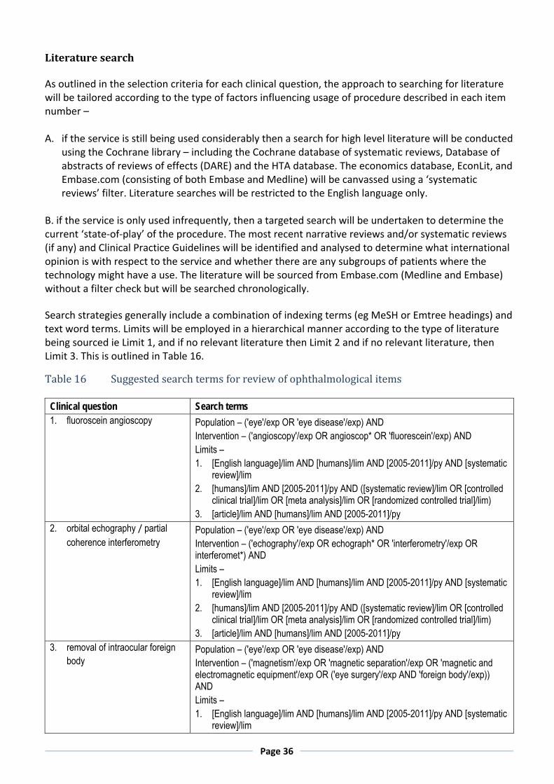
Page 36
Literature search As outlined in the selection criteria for each clinical question, the approach to searching for literature will be tailored according to the type of factors influencing usage of procedure described in each item number – A. if the service is still being used considerably then a search for high level literature will be conducted
using the Cochrane library – including the Cochrane database of systematic reviews, Database of abstracts of reviews of effects (DARE) and the HTA database. The economics database, EconLit, and Embase.com (consisting of both Embase and Medline) will be canvassed using a ‘systematic reviews’ filter. Literature searches will be restricted to the English language only.
B. if the service is only used infrequently, then a targeted search will be undertaken to determine the current ‘state‐of‐play’ of the procedure. The most recent narrative reviews and/or systematic reviews (if any) and Clinical Practice Guidelines will be identified and analysed to determine what international opinion is with respect to the service and whether there are any subgroups of patients where the technology might have a use. The literature will be sourced from Embase.com (Medline and Embase) without a filter check but will be searched chronologically. Search strategies generally include a combination of indexing terms (eg MeSH or Emtree headings) and text word terms. Limits will be employed in a hierarchical manner according to the type of literature being sourced ie Limit 1, and if no relevant literature then Limit 2 and if no relevant literature, then
is is Limit 3. Th outlined in Table 16.
Table 16 Suggested search terms for review of ophthalmological items
Clinical question Search terms 1. fluoroscein angioscopy
Population – ('eye'/exp OR 'eye disease'/exp) AND Intervention – ('angioscopy'/exp OR angioscop* OR 'fluorescein'/exp) AND Limits – 1. [English language]/lim AND [humans]/lim AND [2005-2011]/py AND [systematic
review]/lim 2. [humans]/lim AND [2005-2011]/py AND ([systematic review]/lim OR [controlled
clinical trial]/lim OR [meta analysis]/lim OR [randomized controlled trial]/lim) 3. [article]/lim AND [humans]/lim AND [2005-2011]/py
2. orbital echography / partial coherence interferometry
Population – ('eye'/exp OR 'eye disease'/exp) AND Intervention – ('echography'/exp OR echograph* OR 'interferometry'/exp OR interferomet*) AND Limits – 1. [English language]/lim AND [humans]/lim AND [2005-2011]/py AND [systematic
review]/lim 2. [humans]/lim AND [2005-2011]/py AND ([systematic review]/lim OR [controlled
clinical trial]/lim OR [meta analysis]/lim OR [randomized controlled trial]/lim) 3. [article]/lim AND [humans]/lim AND [2005-2011]/py
3. removal of intraocular foreign body
Population – ('eye'/exp OR 'eye disease'/exp) AND Intervention – ('magnetism'/exp OR 'magnetic separation'/exp OR 'magnetic and electromagnetic equipment'/exp OR ('eye surgery'/exp AND 'foreign body'/exp)) AND Limits – 1. [English language]/lim AND [humans]/lim AND [2005-2011]/py AND [systematic
review]/lim

Page 37
2. [humans]/lim AND [2005-2011]/py AND ([systematic review]/lim OR [controlled clinical trial]/lim OR [meta analysis]/lim OR [randomized controlled trial]/lim)
3. [article]/lim AND [humans]/lim AND [2005-2011]/py 4. tarsal cyst extirpation
Population – 'chalazion'/exp OR 'chalazi*' OR 'tarsal' NEAR/2 'cyst' OR 'meibomian' NEAR/2 'cyst' AND Intervention – Not required Limits – 1. [English language]/lim AND [humans]/lim AND [2005-2011]/py AND [systematic
review]/lim 2. [humans]/lim AND [2005-2011]/py AND ([systematic review]/lim OR [controlled
clinical trial]/lim OR [meta analysis]/lim OR [randomized controlled trial]/lim) 3. [article]/lim AND [humans]/lim AND [2005-2011]/py
5. lacrimal passage obstruction and remova/replacement of nasolacrimal tube
Population – 'lacrimal apparatus'/exp OR 'nasolacrimal tube' OR 'lacrimal passages' OR 'lacrimal gland disease'/exp OR dacryocyst* OR 'epiphora' AND Intervention – 'eye surgery'/exp OR 'eye surgery' Limits – 1. [English language]/lim AND [humans]/lim AND [2005-2011]/py AND [systematic
review]/lim 2. [humans]/lim AND [2005-2011]/py AND ([systematic review]/lim OR [controlled
clinical trial]/lim OR [meta analysis]/lim OR [randomized controlled trial]/lim) 3. [article]/lim AND [humans]/lim AND [2005-2011]/py
6. retinal cryotherapy Population – ('eye'/exp OR 'eye disease'/exp) AND Intervention – ('cryotherapy'/exp OR retin* near/2 cryo*) AND Limits – 1. [English language]/lim AND [humans]/lim AND [2005-2011]/py AND [systematic
review]/lim 2. [humans]/lim AND [2005-2011]/py AND ([systematic review]/lim OR [controlled
clinical trial]/lim OR [meta analysis]/lim OR [randomized controlled trial]/lim) 3. [article]/lim AND [humans]/lim AND [2005-2011]/py
7. laser photocoagulation Population – ('eye'/exp OR 'eye disease'/exp) AND Intervention – ('laser coagulation'/exp OR photocoagulat*) Limits – 1. [English language]/lim AND [humans]/lim AND [2005-2011]/py AND [systematic
review]/lim 2. [humans]/lim AND [2005-2011]/py AND ([systematic review]/lim OR [controlled
clinical trial]/lim OR [meta analysis]/lim OR [randomized controlled trial]/lim) 3. [article]/lim AND [humans]/lim AND [2005-2011]/py
8. capsulectomy/lensectomy/ removal of vitreous
Population – ('eye'/exp OR 'eye disease'/exp) AND Intervention – ('capsulotomy'/exp OR 'lensectomy'/exp OR 'vitrectomy'/exp) Limits – 1. [English language]/lim AND [humans]/lim AND [2005-2011]/py AND [systematic
review]/lim 2. [humans]/lim AND [2005-2011]/py AND ([systematic review]/lim OR [controlled
clinical trial]/lim OR [meta analysis]/lim OR [randomized controlled trial]/lim) 3. [article]/lim AND [humans]/lim AND [2005-2011]/py
9. revision of retinal attachment procedure
Population –'retina detachment'/exp OR detach* OR 'vitreous body detachment' AND Intervention – ('retina surgery'/exp OR ‘vitrectomy’/exp) AND Limits – 1. [English language]/lim AND [humans]/lim AND [2005-2011]/py AND [systematic
review]/lim 2. [humans]/lim AND [2005-2011]/py AND ([systematic review]/lim OR [controlled

Page 38
clinical trial]/lim OR [meta analysis]/lim OR [randomized controlled trial]/lim) 3. [article]/lim AND [humans]/lim AND [2005-2011]/py
10. liquid vitreous substitutes Population – 'vitreous'/exp OR 'retina detachment'/exp OR detach* OR 'vitreous body detachment'/exp OR 'vitrectomy'/exp AND Intervention – (intraocular NEAR/2 tamponade OR 'vitreous' NEAR/10 substitut* OR 'silicone gel'/exp OR 'organofluorine derivative'/exp OR 'glycosaminoglycan'/exp) AND Limits – 1. [English language]/lim AND [humans]/lim AND [2005-2011]/py AND [systematic
review]/lim 2. [humans]/lim AND [2005-2011]/py AND ([systematic review]/lim OR [controlled
clinical trial]/lim OR [meta analysis]/lim OR [randomized controlled trial]/lim) 3. [article]/lim AND [humans]/lim AND [2005-2011]/py
Critical appraisal of selected evidence The literature will be categorised according to NHMRC levels of evidence (see Table 17), critically appraised using checklists relevant for each type of literature, and then synthesised according to the evidence matrix for NHMRC Grades of recommendation (see Table 18). Relevant checklists include PRISMA for systematic reviews and health technology assessments (Liberati, Altman et al. 2009); SIGN checklists for appraising randomised and non‐randomised controlled trials and observational studies (SIGN 2008); and the QUADAS checklist for appraising diagnostic accuracy studies (Whiting 2003).

Page 39
Table 17 Designations of levels of evidence (Merlin T, Weston A et al. 2009; NHMRC 2009) Level Intervention 1 Diagnostic accuracy 2 I 4 A systematic review of level II
studies A systematic review of level II studies
II A randomised controlled trial A study of test accuracy with: an independent, blinded comparison with a valid reference standard,5 among consecutive persons with a defined clinical presentation6
III-1 A pseudorandomised controlled trial (i.e. alternate allocation or some other method)
A study of test accuracy with: an independent, blinded comparison with a valid reference standard,5 among non-consecutive persons with a defined clinical presentation6
III-2 A comparative study with concurrent controls: ▪ Non-randomised, experimental trial9 ▪ Cohort study ▪ Case-control study ▪ Interrupted time series with a control group
A comparison with reference standard that does not meet the criteria required for Level II and III-1 evidence
III-3 A comparative study without concurrent controls: ▪ Historical control study ▪ Two or more single arm study10
▪ Interrupted time series without a parallel control group
Diagnostic case-control study6
IV Case series with either post-test or pre-test/post-test outcomes
Study of diagnostic yield (no reference standard)11
Explanatory notes10
1 Definitions of these study designs are provided on pages 7-8 How to use the evidence: assessment and application of scientific evidence (NHMRC 2000b) and in the accompanying Glossary.
2 These levels of evidence apply only to studies of assessing the accuracy of diagnostic or screening tests. To assess the overall effectiveness of a diagnostic test there also needs to be a consideration of the impact of the test on patient management and health outcomes (Medical Services Advisory Committee 2005, Sackett and Haynes 2002). The evidence hierarchy given in the ‘Intervention’ column should be used when assessing the impact of a diagnostic test on health outcomes relative to an existing method of diagnosis/comparator test(s). The evidence hierarchy given in the ‘Screening’ column should be used when assessing the impact of a screening test on health outcomes relative to no screening or opportunistic screening.
4 A systematic review will only be assigned a level of evidence as high as the studies it contains, excepting where those studies are of level II evidence. Systematic reviews of level II evidence provide more data than the individual studies and any meta-analyses will increase the precision of the overall results, reducing the likelihood that the results are affected by chance. Systematic reviews of lower level evidence present results of likely poor internal validity and thus are rated on the likelihood that the results have been affected by bias, rather than whether the systematic review itself is of good quality. Systematic review quality should be assessed separately. A systematic review should consist of at least two studies. In systematic reviews that include different study designs, the overall level of evidence should relate to each individual outcome/result, as different studies (and study designs) might contribute to each different outcome.
5 The validity of the reference standard should be determined in the context of the disease under review. Criteria for determining the validity of the reference standard should be pre-specified. This can include the choice of the reference standard(s) and its timing in relation to the index test. The validity of the reference standard can be determined through quality appraisal of the study (Whiting et al 2003).
6 Well-designed population based case-control studies (eg. population based screening studies where test accuracy is assessed on all cases, with a random sample of controls) do capture a population with a representative spectrum of disease and thus fulfil the
10 Note ‐ only evidence hierarchies relevant to this review have been reproduced in Table 17, so tablenotes 7 and 8 have not been reproduced

Page 40
requirements for a valid assembly of patients. However, in some cases the population assembled is not representative of the use of the test in practice. In diagnostic case-control studies a selected sample of patients already known to have the disease are compared with a separate group of normal/healthy people known to be free of the disease. In this situation patients with borderline or mild expressions of the disease, and conditions mimicking the disease are excluded, which can lead to exaggeration of both sensitivity and specificity. This is called spectrum bias or spectrum effect because the spectrum of study participants will not be representative of patients seen in practice (Mulherin and Miller 2002).
9 This also includes controlled before-and-after (pre-test/post-test) studies, as well as adjusted indirect comparisons (ie. utilise A vs B and B vs C, to determine A vs C with statistical adjustment for B).
10 Comparing single arm studies ie. case series from two studies. This would also include unadjusted indirect comparisons (ie. utilise A vs B and B vs C, to determine A vs C but where there is no statistical adjustment for B).
11 Studies of diagnostic yield provide the yield of diagnosed patients, as determined by an index test, without confirmation of the accuracy of this diagnosis by a reference standard. These may be the only alternative when there is no reliable reference standard.
Note A: Assessment of comparative harms/safety should occur according to the hierarchy presented for each of the research questions, with the proviso that this assessment occurs within the context of the topic being assessed. Some harms (and other outcomes) are rare and cannot feasibly be captured within randomised controlled trials, in which case lower levels of evidence may be the only type of evidence that is practically achievable; physical harms and psychological harms may need to be addressed by different study designs; harms from diagnostic testing include the likelihood of false positive and false negative results; harms from screening include the likelihood of false alarm and false reassurance results.
Note B: When a level of evidence is attributed in the text of a document, it should also be framed according to its corresponding research question eg. level II intervention evidence; level IV diagnostic evidence; level III-2 prognostic evidence.
Note C: Each individual study that is attributed a “level of evidence” should be rigorously appraised using validated or commonly used checklists or appraisal tools to ensure that factors other than study design have not affected the validity of the results.
Source: Hierarchies adapted and modified from: NHMRC 1999; Bandolier 1999; Lijmer et al. 1999; Phillips et al. 2001.
The overall body of research evidence will be assessed and synthesised to address each clinical question according to Table 18. An evidence rating from A (excellent) to D (poor) will be assigned to the evidence, considering each of the components outlined in the body of evidence matrix. In the absence of such literature, expert opinion and narratives will be synthesised according to the credibility of the
chsource of su material.
Table 18 Body of evidence assessment matrix (adapted from (NHMRC 2009)) A B C D Component
Excellent Good Satisfactory Poor Evidence base1 one or more level I
studies with a low risk of bias or several level II studies with a low risk of bias
one or two level II studies with a low risk of bias or a SR/several level III studies with a low risk of bias
one or two level III studies with a low risk of bias, or level I or II studies with a moderate risk of bias
level IV studies, or level I to III studies/SRs with a high risk of bias
Consistency2 all studies consistent most studies consistent and inconsistency may be explained
some inconsistency reflecting genuine uncertainty around clinical question
evidence is inconsistent
Clinical impact very large substantial moderate slight or restricted
Generalisability population/s studied in body of evidence are the same as the target population
population/s studied in the body of evidence are similar to the target population
population/s studied in body of evidence differ to target population but it is clinically sensible to apply this evidence to target population3
population/s studied in body of evidence differ to target population and hard to judge whether it is sensible to generalise to target population

Page 41
Applicability directly applicable to Australian healthcare context
applicable to Australian healthcare context with few caveats
probably applicable to Australian healthcare context with some caveats
not applicable to Australian healthcare context
SR = systematic review; several = more than two studies 1 Level of evidence determined from the NHMRC evidence hierarchy – Table 17. 2 If there is only one study, rank this component as ‘not applicable’. 3 For example, results in adults that are clinically sensible to apply to children OR psychosocial outcomes for one cancer that may be applicable to patients with another cancer
4.4 Stakeholder consultation – community engagement Community engagement will be conducted to determine consumers’ “revealed preferences” and experiences concerning the services under review. Six services will be selected from those receiving Guideline concordance analysis and/or a mini‐HTA – four of these will be the most common services while the remaining two will be reserved for potentially problematic services (ie painful procedures, long hospital stay etc). Consumer engagement concerning the four most common services will occur mainly through the canvassing of internet blogs and qualitative literature, although telephone and internet interviewing of consumers will be undertaken, if possible. The aim will be to determine consumer experiences with the ophthalmological service under review, their preferences for modifying the service and/or to identify any consumer concerns or hardship should the service be removed. Should interviewing be conducted, consumers will be recruited via the Consumer Health Forum, local ophthalmologists or patient advocacy groups sending out letters, or via contact with authors of internet blogs. Eligibility for recruitment will be matched, as far as practicable, to the demographic characteristics that MBS data analysis indicates the primary consumers. Ethics approval has been received through the University approval process. The two additional services will receive a limited ‘revealed preference’ assessment via canvassing internet consumer blogs and the qualitative literature on the topic only. The qualitative literature search will be undertaken using the Embase.com and Scopus databases. Blogs (also known as weblogs) are on‐line journals documenting views and experiences of a single author, or in some cases a small group of authors. English language blogs from developed countries concerning the services under review will be identified through a Google advanced domain search and, where relevant, bloggers will be contacted and asked for an interview. Commercial blogs will be excluded. All literature will be imported into NVivo for thematic coding and analysis. Research questions will be drafted to address each of the six services once they are identified during the MBS data analysis phase.

Page 42
4.5 Stakeholder negotiation Several item descriptors for the ophthalmological items under review will be discussed and potentially amended through negotiation between the Department and relevant stakeholders. Most of these
nts w ble 19amendme ill relate to corrections of terminology. Items to be addressed are in Ta
Table 19 Ophthalmology items potentially being revised through negotiation
.
Service MBS Item Numbers Removal of foreign body 42551, 42554, 42557, 42644 Cataract surgery 42703, 42704, 42707, 42710, 42713 Capsulectomy and lensectomy 42731 Retinal services 42818 Vitrectomy 42725 Surgical assist 51315
As outlined previously, the Department will also be reviewing optometry perimetry items 10940 and 10941 separately, and will undertake stakeholder negotiation with the relevant optometry craft groups.
With respect to the surgical assist item (51315) given below, there is a suggestion that there is an anomaly in the wording as it does not allow for surgical assistants to participate in surgery where both phacoemulsification to remove the lens (42698, 42702) and vitrectomy (42725) are required.
Cataract 51315 Assistance at cataract and intraocular lens surgery covered by item 42698,42701,
42702, 42704 or 42707, when performed in association with services covered by item 42551 to 42569, 42653, 42656, 42746, 42749, 42752, 42776 or 42779
A suggested amendment to item 51315 is suggested as a consequence:
Suggested amendment and rationale: "Assistance at cataract and intraocular lens surgery covered by item 42698, 42701, 42702, 42704 or 42707, when performed in association with services covered by item 42551 to 42569, 42653, 42656, 42725, 42746, 42749, 42752, 42776 or 42779"
Pars plana vitrectomy (item 42725) is an intraocular procedure during which lens removal / IOL implantation may be required in selected cases, as with the other item codes already listed as associated services. This combination of surgery may be required to allow improved visualisation for the vitrectomy procedure (hence improved safety) and avoids the need for a second surgery (ie subsequent cataract surgery).
4.6 Economic evaluation Only a preliminary economic evaluation of the services will be conducted, relying on literature identified through the searches outlined in Section 4.3 and MBS item costs only.
In the literature searches, acceptable evidence would include trial‐based costing studies, cost analyses and economic modelling studies. Acceptable outcomes would include: cost, incremental cost‐effectiveness ratio eg cost per event avoided, cost per life year gained, cost per quality adjusted life year or disability adjusted life year. The applicability of any identified economic analyses to the Australian health system would be assessed.
A formal modelled economic evaluation of the different ophthalmology items will not be conducted during this review.

Page 43
4.7 Review outcomes The conclusions regarding the ophthalmological services that are assessed will be provided in a draft report. This report will be presented in chapters according to each service being reviewed. The results of the MBS item data analysis, guideline concordance activity, mini‐HTA and consumer engagement will be synthesised for each service under review. A summary statement and conclusion will be developed for each “service” chapter. Following public consultation and feedback, the report will be finalised. The evaluation method that is tested for this review will also be assessed and critiqued as part of the project, with suggestions for its modification/revision provided, along with the final report. Reviews are expected to result in primary and supplementary review outcomes as shown below: Primary review outcomes Where an evaluation suggests that an item under review is supported by the evidence, the likely recommendation will be that the MBS listing will be retained in its current form. However, should an evaluation suggest that listed MBS items or services are inconsistent with contemporary evidence in relation to its clinical use or effectiveness, direct amendments to the MBS may be recommended. These may include one or more of the following changes: • addition or removal of MBS items; • changes to the Schedule fee; • refinement of MBS item descriptors to better target patient groups, clinical indicators and/or promote the use of optimal clinical pathways; and/or • potential for interim‐listing pending the collection of item‐specific data. Potential amendments to the MBS arising from reviews will be undertaken through consultation with the relevant stakeholder groups. Supplementary review outcomes ‐ initiatives to facilitate evidence‐based changes in clinical practice In addition to primary review outcomes relating to MBS reimbursement, reviews may indicate the need for secondary investment strategies aimed at bridging the divide between current evidence, including clinical guidelines and current clinical practice. To achieve this, a number of strategies may be implemented following the evaluation of individual items or services. These may include, but are not limited to, the following: • development or revision of clinical practice guidelines for evaluated services where there is an identified need; • strengthening or targeting of auditing/compliance activities; • education and training initiatives for practitioners and/or consumers; • exploring incentive‐based initiatives to promote improved clinical practices or linking education and training programs to access incentives; and • the development of research opportunities where gaps in effective service provision are evident. The identification of mechanisms to support evidence‐based best practice will complement and reinforce any primary outcome MBS amendments to help improve health outcomes for patients, whilst ensuring the most efficient use of limited resources.

Page 44
5. REVIEW TIMEFRAME
The following key milestones are associated with this review of ophthalmological services:
Milestone Time Draft review protocol submitted to the Department for consideration
October, 2010
Draft review protocol available for public comment October, 2010 Completion of each evidence based review activity
- MBS data analysis - Guideline concordance - Community engagement - Evidence-based analysis / mini-HTAs
- November, 2010 - November, 2010 - November, 2010 - December, 2010
Submission of draft review report to MQFEAC for consideration
January – February, 2011
Draft review report available for public comment (minimum of four weeks)
January – February, 2011
Submission of final report March – April, 2011

Page 45
6. REFERENCES
Access Economics (September 2009). Cataract treatment in Australia, Access Economics Pty Ltd for Alcon Laboratories Australia Pty Ltd. Australian Institute of Health and Welfare (2005). Vision problems among older Australians. Canberra, AIHW. Australian Institute of Health and Welfare (2009). A guide to Australian eye health data. Canberra, AIHW. Liberati, A., D. Altman, et al. (2009). "The PRISMA Statement for Reporting Systematic Reviews and Meta‐Analyses of Studies That Evaluate Health Care Interventions: Explanation and Elaboration." PLoS Med 6(7): e1000100. Merlin T, Weston A, et al. (2009). "Extending an evidence hierarchy to include topics other than treatment: revising the Australian ‘levels of evidence’." BMC Medical Research Methodology 9(34 ). Mitchell, P., W. Smith, et al. (1996). "Prevalence of open‐angle glaucoma in Australia." Ophthalmology 103(10): 1661‐1669. NHMRC (2009). NHMRC levels of evidence and grades for recommendations for developers of guidelines. Canberra, ACT, National Health and Medical Research Council. 2010. SIGN (2008). A guideline developer's handbook. Edinburgh, Scottish Intercollegiate Guidelines Network. Whiting, P., Rutjes, A.W. et al (2003). "The development of QUADAS: a tool for the quality assessment of studies of diagnostic accuracy included in systematic reviews." BMC Medical Research Methodology 3(1): 25.

Page 46
Attachment 1
CLINICAL PRACTICE GUIDELINES
Glaucoma
• Guidelines for Screening, Prognosis, Diagnosis, Management and Prevention of Glaucoma (Draft). Please note, it is expected these guidelines will be endorsed in August 2010. Access from http://www.nhmrc.gov.au/guidelines/consult/consultations/glaucoma_screening_guidelines.htm
• NICE Guidance: Glaucoma –diagnosis and management of chronic open angle glaucoma and ocular hypertension (April 2009). Access from: http://guidance.nice.org.uk/CG85/Guidance/pdf/English
• RANZCO: Guidelines for Collaborative Care of Glaucoma Patients (undated). Access from
http://www.ranzco.edu/aboutus/ranzco‐policies‐and‐procedures/policy/GUIDELINES_FOR_COLLABORATIVE_CARE_OF_GLAUCOMA_PATIENTS.pdf
• American Academy of Ophthalmology Practice Patterns Open Angle Glaucoma • European Glaucoma Society guidelines. www.eugs.org/ • World Glaucoma Association publications. www.worldglaucoma.org/ • SEAGIG (Southeast Asian Glaucoma Interest Group) guidelines. www.seagig.org/ Retinal services
• American Academy of Ophthalmology: Posterior Vitreous Detachment, Retinal Breaks, and Lattice Degeneration – Preferred Practice Pattern (September 2008). Access from http://one.aao.org/CE/PracticeGuidelines/PPP.aspx?p=1
Macular degeneration
• American Academy of Ophthalmology: Age‐Related Macular Degeneration – Preferred Practice Pattern (September 2008). Access from http://one.aao.org/CE/PracticeGuidelines/PPP.aspx?p=1
• RANZCO: Medications for Age‐Related Macular Degeneration (AMD) (undated). Access from
http://www.ranzco.edu/aboutus/ranzco‐policies‐and‐procedures/policy/Additional%20information%20on%20PBS%20access%20to%20AMD%20therapies%20‐%20August%202007.pdf
Cataracts
• American Academy of Ophthalmology: Cataract in the Adult Eye – Preferred Practice Pattern (September 2006). Access from http://one.aao.org/CE/PracticeGuidelines/PPP.aspx?p=1
• Canadian Ophthalmological Society evidence‐based clinical practice guidelines for cataract surgery
in the adult eye (October 2008). Access from http://eyesite.ca/english/program‐and‐services/policy‐statements‐guidelines/index.htm

Page 47
• RANZCO: Cataract and Intraocular Lens Surgery (March 2006). Access from http://www.ranzco.edu/aboutus/ranzco‐policies‐and‐procedures/policy/Cataract_Surgery.pdf
Electroretinography • Standard for clinical electroretinography (2008 update). Access from
http://www.iscev.org/standards/index.html
• Standard for clinical electrooculography (2006). Access from http://www.iscev.org/standards/index.html
• Standard for clinical pattern electroretinography (2007 update). Access from http://www.iscev.org/standards/index.html
• Guidelines for clinical multifocal electroretinography (2007). Access from http://www.iscev.org/standards/index.html