european ophthalmology congress · • neuro-ophthalmology and research • innovative techniques...
TRANSCRIPT
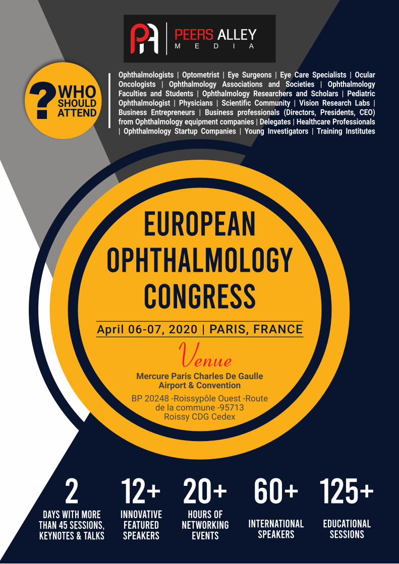
?WHOSHOULDATTEND
12+2 20+ 60+ 125+INNOVATIVEFEATUREDSPEAKERS
HOURS OF NETWORKING
EVENTSINTERNATIONAL
SPEAKERSEDUCATIONAL
SESSIONS
DAyS WITH MORE THAN 45 SESSIONS, KEyNOTES & TALKS
Ophthalmologists | Optometrist | Eye Surgeons | Eye Care Specialists | Ocular Oncologists | Ophthalmology Associations and Societies | Ophthalmology Faculties and Students | Ophthalmology Researchers and Scholars | Pediatric Ophthalmologist | Physicians | Scientific Community | Vision Research Labs | Business Entrepreneurs | Business professionals (Directors, Presidents, CEO) from Ophthalmology equipment companies | Delegates | Healthcare Professionals | Ophthalmology Startup Companies | Young Investigators | Training Institutes
EUROPEAN OPHTHALMOLOGy
CONGRESSApril 06-07, 2020 | PARIS, FRANCE
Mercure Paris Charles De GaulleAirport & Convention
BP 20248 -Roissypôle Ouest -Route de la commune -95713
Roissy CDG Cedex
Venue
http://ophthalmology.peersalleyconferences.com/
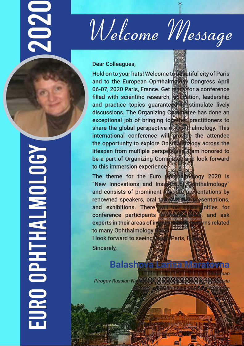
Dear Colleagues,
Hold on to your hats! Welcome to beautiful city of Paris and to the European Ophthalmology Congress April 06-07, 2020 Paris, France. Get ready for a conference filled with scientific research, education, leadership and practice topics guaranteed to stimulate lively discussions. The Organizing Committee has done an exceptional job of bringing together practitioners to share the global perspective of Ophthalmology. This international conference will provide the attendee the opportunity to explore Ophthalmology across the lifespan from multiple perspectives. I am honored to be a part of Organizing Committee and look forward to this immersion experience.
The theme for the Euro Ophthalmology 2020 is “New Innovations and Insights of Ophthalmology” and consists of prominent keynote presentations by renowned speakers, oral talks, poster presentations, and exhibitions. There will be opportunities for conference participants to learn, share, and ask experts in their areas of interests and concerns related to many Ophthalmology fields.I look forward to seeing you in Paris, France.
Sincerely,
EURO
OPH
THAL
MOL
OGY
2020
Balashova Larisa MaratovnaChairman
Pirogov Russian National Research Medical University, RussiaMinistry of Health of the Russian Federation
Welcome Message
PRESENTATION TIME TO
WITH YOURCONNECT
PEERS
Register & Participate
in
EURO OPHTHALMOLOGy
2020
FORUM
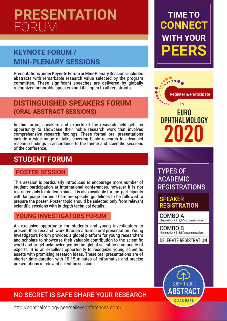
KEYNOTE FORUM / MINI-PLENARY SESSIONS
DISTINGUISHED SPEAKERS FORUM(ORAL ABSTRACT SESSIONS)
STUDENT FORUM
Presentations under Keynote Forum or Mini-Plenary Sessions includes abstracts with remarkable research value selected by the program committee. These significant speeches are delivered by globally recognized honorable speakers and it is open to all registrants.
In this forum, speakers and experts of the research field gets an opportunity to showcase their noble research work that involves comprehensive research findings. These formal oral presentations include a wide range of talks covering basic research to advanced research findings in accordance to the theme and scientific sessions of the conference.
TYPES OF ACADEMICREGISTRATIONS
This session is particularly introduced to encourage more number of student participation at international conferences, however it is not restricted only to students since it is also available for the participants with language barrier. There are specific guidelines to be followed to prepare the poster. Poster topic should be selected only from relevant scientific sessions with in-depth technical details.
An exclusive opportunity for students and young investigators to present their research work through a formal oral presentation. Young Investigators Forum provides a global platform for young researchers and scholars to showcase their valuable contribution to the scientific world and to get acknowledged by the global scientific community of experts. It is an excellent opportunity to recognize young scientific assets with promising research ideas. These oral presentations are of shorter time duration with 10-15 minutes of informative and precise presentations in relevant scientific sessions.
NO SECRET IS SAFE SHARE YOUR RESEARCH
SPEAKERREGISTRATIONCOMBO A(Registration + 2 night’s accommodation)
COMBO B(Registration + 3 night’s accommodation)
POSTER SESSION
YOUNG INVESTIGATORS FORUM
DELEGATE REGISTRATION
http://ophthalmology.peersalleyconferences.com/
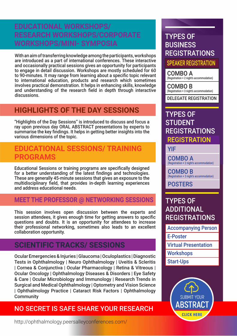
EDUCATIONAL WORKSHOPS/ RESEARCH WORKSHOPS/CORPORATE WORKSHOPS/MINI- SYMPOSIA
HIGHLIGHTS OF THE DAY SESSIONS
EDUCATIONAL SESSIONS/ TRAINING PROGRAMS
MEET THE PROFESSOR @ NETWORKING SESSIONS
SCIENTIFIC TRACKS/ SESSIONS
With an aim of transferring knowledge among the participants, workshops are introduced as a part of international conferences. These interactive and occasionally practical sessions gives an opportunity for participants to engage in detail discussion. Workshops are mostly scheduled for 60 to 90-minutes. It may range from learning about a specific topic relevant to international education, products and research which sometimes involves practical demonstration. It helps in enhancing skills, knowledge and understanding of the research field in depth through interactive discussions.
“Highlights of the Day Sessions” is introduced to discuss and focus a ray upon previous day ORAL ABSTRACT presentations by experts to summarise the key findings. It helps in getting better insights into the various dimensions of the topic.
Educational Sessions or training programs are specifically designed for a better understanding of the latest findings and technologies. These are generally 45-minute sessions that gives an exposure to the multidisciplinary field, that provides in-depth learning experiences and address educational needs.
This session involves open discussion between the experts and session attendees, it gives enough time for getting answers to specific questions and doubts. It is an opportunity for attendees to increase their professional networking, sometimes also leads to an excellent collaboration opportunity.
Ocular Emergencies & Injuries | Glaucoma | Oculoplastics | Diagnostic Tests in Ophthalmology | Neuro Ophthalmology | Uveitis & Scleritis | Cornea & Conjunctiva | Ocular Pharmacology | Retina & Vitreous | Ocular Oncology | Ophthalmology Diseases & Disorders | Eye Safety & Care | Ocular Microbiology and Immunology | Research Trends in Surgical and Medical Ophthalmology | Optometry and Vision Science | Ophthalmology Practice | Cataract Risk Factors | Ophthalmology Community
TYPES OF BUSINESSREGISTRATIONS
TYPES OF STUDENTREGISTRATIONS
TYPES OFADDITIONALREGISTRATIONS
SPEAKER REGISTRATION
REGISTRATION
COMBO A(Registration + 2 night’s accommodation)
YIF
COMBO B(Registration + 3 night’s accommodation)
DELEGATE REGISTRATION
Accompanying PersonE-PosterVirtual PresentationWorkshopsStart-Ups
POSTERS
COMBO A(Registration + 2 night’s accommodation)
COMBO B(Registration + 3 night’s accommodation)
NO SECRET IS SAFE SHARE YOUR RESEARCH
http://ophthalmology.peersalleyconferences.com/
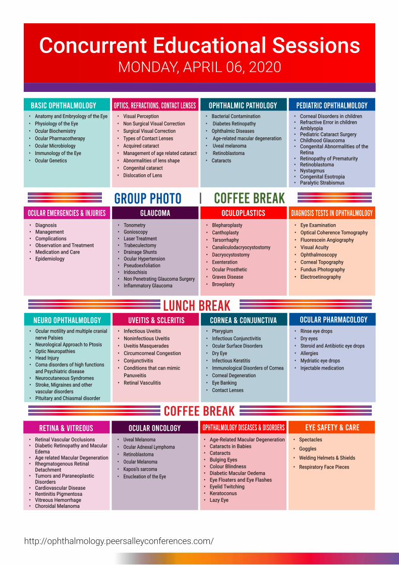
GROUP PHOTO
LUNCH BREAK
COFFEE BREAK
COFFEE BREAK
BASIC OPHTHALMOLOGy
OCULAR EMERGENCIES & INjURIES
OPTICS, REFRACTIONS, CONTACT LENSES
GLAUCOMA
OPHTHALMIC PATHOLOGy
OCULOPLASTICS
PEDIATRIC OPHTHALMOLOGy
DIAGNOSIS TESTS IN OPHTHALMOLOGy
• Anatomy and Embryology of the Eye• Physiology of the Eye• Ocular Biochemistry• Ocular Pharmacotherapy• Ocular Microbiology• Immunology of the Eye• Ocular Genetics
• Diagnosis• Management• Complications• Observation and Treatment• Medication and Care• Epidemiology
• Visual Perception• Non Surgical Visual Correction• Surgical Visual Correction• Types of Contact Lenses• Acquired cataract• Management of age related cataract• Abnormalities of lens shape• Congenital cataract• Dislocation of Lens
• Tonometry• Gonioscopy• Laser Treatment• Trabeculectomy• Drainage Shunts• Ocular Hypertension• Pseudoexfoliation• Iridoschisis• Non Penetrating Glaucoma Surgery• Inflammatory Glaucoma
• Bacterial Contamination• Diabetes Retinopathy• Ophthalmic Diseases• Age-related macular degeneration• Uveal melanoma• Retinoblastoma• Cataracts
• Blepharoplasty• Canthoplasty• Tarsorrhaphy• Canaliculodacryocystostomy• Dacryocystostomy• Exenteration• Ocular Prosthetic• Graves Disease• Browplasty
• Corneal Disorders in children• Refractive Error in children• Amblyopia• Pediatric Cataract Surgery• Childhood Glaucoma • Congenital Abnormalities of the
Retina• Retinopathy of Prematurity• Retinoblastoma• Nystagmus• Congenital Esotropia• Paralytic Strabismus
• Eye Examination• Optical Coherence Tomography• Fluorescein Angiography• Visual Acuity• Ophthalmoscopy• Corneal Topography• Fundus Photography• Electroetinography
NEURO OPHTHALMOLOGy UVEITIS & SCLERITIS CORNEA & CONjUNCTIVA OCULAR PHARMACOLOGy• Ocular motility and multiple cranial
nerve Palsies • Neurological Approach to Ptosis• Optic Neuropathies• Head Injury• Coma disorders of high functions
and Psychiatric disease• Neurocutaneous Syndromes• Stroke, Migraines and other
vascular disorders• Pituitary and Chiasmal disorder
• Infectious Uveitis• Noninfectious Uveitis• Uveitis Masquerades • Circumcorneal Congestion• Conjunctivitis• Conditions that can mimic
Panuveitis• Retinal Vasculitis
• Pterygium• Infectious Conjunctivitis• Ocular Surface Disorders• Dry Eye• Infectious Keratitis• Immunological Disorders of Cornea• Corneal Degeneration• Eye Banking• Contact Lenses
• Rinse eye drops• Dry eyes• Steroid and Antibiotic eye drops• Allergies• Mydriatic eye drops• Injectable medication
RETINA & VITREOUS OCULAR ONCOLOGy OPHTHALMOLOGy DISEASES & DISORDERS EyE SAFETy & CARE
• Retinal Vascular Occlusions• Diabetic Retinopathy and Macular
Edema• Age related Macular Degeneration• Rhegmatogenous Retinal
Detachment• Tumors and Paraneoplastic
Disorders• Cardiovascular Disease• Rentinitis Pigmentosa• Vitreous Hemorrhage• Choroidal Melanoma
• Uveal Melanoma • Ocular Adnexal Lymphoma • Retinoblastoma • Ocular Melanoma• Kaposi’s sarcoma• Enucleation of the Eye
• Age-Related Macular Degeneration• Cataracts in Babies• Cataracts• Bulging Eyes• Colour Blindness• Diabetic Macular Oedema• Eye Floaters and Eye Flashes• Eyelid Twitching• Keratoconus• Lazy Eye
• Spectacles
• Goggles
• Welding Helmets & Shields
• Respiratory Face Pieces
http://ophthalmology.peersalleyconferences.com/
MONDAY, APRIL 06, 2020 Concurrent Educational Sessions
http://ophthalmology.peersalleyconferences.com/
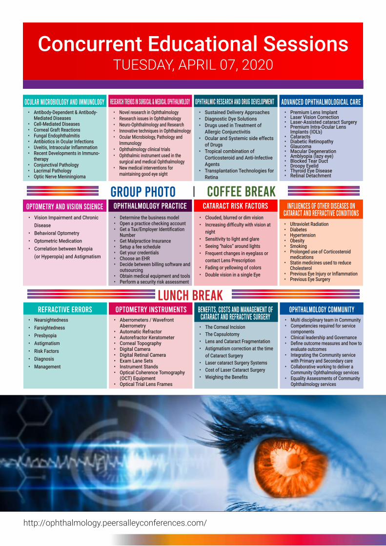
Concurrent Educational SessionsTUESDAY, APRIL 07, 2020
GROUP PHOTO
LUNCH BREAK
COFFEE BREAK
OCULAR MICROBIOLOGy AND IMMUNOLOGy
OPTOMETRy AND VISION SCIENCE
RESEARCH TRENDS IN SURGICAL & MEDICAL OPHTHALMOLOGy
OPHTHALMOLOGy PRACTICE
OPHTHALMIC RESEARCH AND DRUG DEVELOPMENT
CATARACT RISK FACTORS
ADVANCED OPHTHALMOLOGICAL CARE
INFLUENCES OF OTHER DISEASES ON CATARACT AND REFRACTIVE CONDITIONS
• Antibody-Dependent & Antibody- Mediated Diseases
• Cell-Mediated Diseases• Corneal Graft Reactions• Fungal Endophthalmitis• Antibiotics in Ocular Infections• Uveitis, Intraocular Inflammation• Recent Developments in Immuno-
therapy• Conjunctival Pathology• Lacrimal Pathology• Optic Nerve Meniningioma
• Vision Impairment and Chronic Disease
• Behavioral Optometry• Optometric Medication• Correlation between Myopia
(or Hyperopia) and Astigmatism
• Novel research in Ophthalmology• Research issues in Ophthalmology• Neuro-Ophthalmology and Research• Innovative techniques in Ophthalmology• Ocular Microbiology, Pathology and
Immunology• Ophthalmology clinical trials• Ophthalmic instrument used in the
surgical and medical Ophthalmology• New medical interventions for
maintaining good eye sight
• Determine the business model• Open a practice checking account• Get a Tax/Employer Identification
Number• Get Malpractice Insurance• Setup a fee schedule• Get your credentials• Choose an EHR• Decide between billing software and
outsourcing• Obtain medical equipment and tools• Perform a security risk assessment
• Sustained Delivery Approaches• Diagnostic Dye Solutions• Drugs used in Treatment of
Allergic Conjunctivitis• Ocular and Systemic side effects
of Drugs• Tropical combination of
Corticosteroid and Anti-Infective Agents
• Transplantation Technologies for Retina
• Clouded, blurred or dim vision• Increasing difficulty with vision at
night• Sensitivity to light and glare• Seeing “halos” around lights• Frequent changes in eyeglass or
contact Lens Prescription• Fading or yellowing of colors• Double vision in a single Eye
• Premium Lens Implant• Laser Vision Correction• Laser-Assisted cataract Surgery• Premium Intra-Ocular Lens
Implants (IOL’s)• Cataracts• Diabetic Retinopathy• Glaucoma• Macular Degeneration• Amblyopia (lazy eye)• Blocked Tear Duct• Droopy Eyelid• Thyroid Eye Disease• Retinal Detachment
• Ultraviolet Radiation• Diabetes• Hypertension• Obesity• Smoking• Prolonged use of Corticosteroid
medications• Statin medicines used to reduce
Cholesterol• Previous Eye Injury or Inflammation• Previous Eye Surgery
REFRACTIVE ERRORS OPTOMETRy INSTRUMENTS BENEFITS, COSTS AND MANAGEMENT OF CATARACT AND REFRACTIVE SURGERy
OPHTHALMOLOGy COMMUNITy• Nearsightedness• Farsightedness• Presbyopia• Astigmatism• Risk Factors• Diagnosis• Management
• Aberrometers / Wavefront Aberrometry
• Automatic Refractor • Autorefractor Keratometer• Corneal Topography • Digital Camera • Digital Retinal Camera• Exam Lane Sets • Instrument Stands • Optical Coherence Tomography
(OCT) Equipment • Optical Trial Lens Frames
• The Corneal Incision• The Capsulotomy• Lens and Cataract Fragmentation• Astigmatism correction at the time
of Cataract Surgery• Laser cataract Surgery Systems• Cost of Laser Cataract Surgery• Weighing the Benefits
• Multi disciplinary team in Community• Competencies required for service
components• Clinical leadership and Governance• Define outcome measures and how to
evaluate outcomes• Integrating the Community service
with Primary and Secondary care• Collaborative working to deliver a
Community Ophthalmology services• Equality Assessments of Community
Ophthalmology services
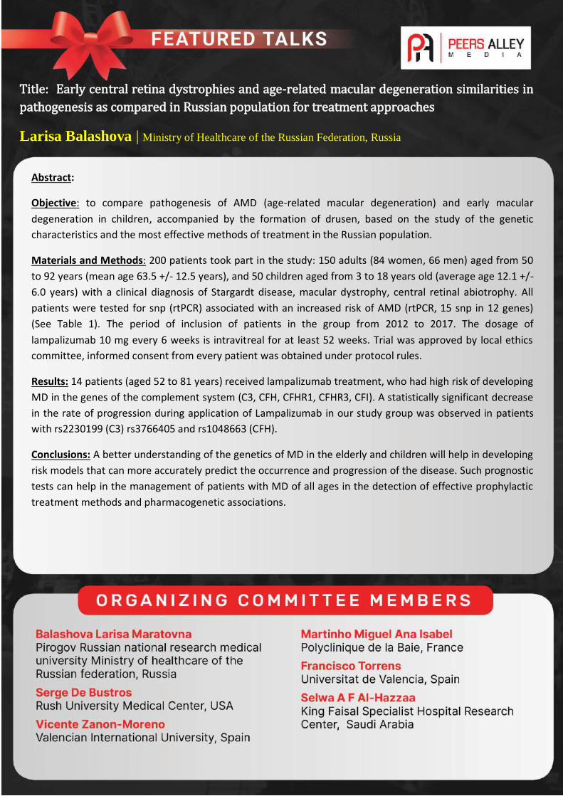
Title: Early central retina dystrophies and age-related macular degeneration similarities in
pathogenesis as compared in Russian population for treatment approaches
Larisa Balashova | Ministry of Healthcare of the Russian Federation, Russia
Abstract:
Objective: to compare pathogenesis of AMD (age-related macular degeneration) and early macular
degeneration in children, accompanied by the formation of drusen, based on the study of the genetic
characteristics and the most effective methods of treatment in the Russian population.
Materials and Methods: 200 patients took part in the study: 150 adults (84 women, 66 men) aged from 50
to 92 years (mean age 63.5 +/- 12.5 years), and 50 children aged from 3 to 18 years old (average age 12.1 +/-
6.0 years) with a clinical diagnosis of Stargardt disease, macular dystrophy, central retinal abiotrophy. All
patients were tested for snp (rtPCR) associated with an increased risk of AMD (rtPCR, 15 snp in 12 genes)
(See Table 1). The period of inclusion of patients in the group from 2012 to 2017. The dosage of
lampalizumab 10 mg every 6 weeks is intravitreal for at least 52 weeks. Trial was approved by local ethics
committee, informed consent from every patient was obtained under protocol rules.
Results: 14 patients (aged 52 to 81 years) received lampalizumab treatment, who had high risk of developing
MD in the genes of the complement system (C3, CFH, CFHR1, CFHR3, CFI). A statistically significant decrease
in the rate of progression during application of Lampalizumab in our study group was observed in patients
with rs2230199 (C3) rs3766405 and rs1048663 (CFH).
Conclusions: A better understanding of the genetics of MD in the elderly and children will help in developing
risk models that can more accurately predict the occurrence and progression of the disease. Such prognostic
tests can help in the management of patients with MD of all ages in the detection of effective prophylactic
treatment methods and pharmacogenetic associations.
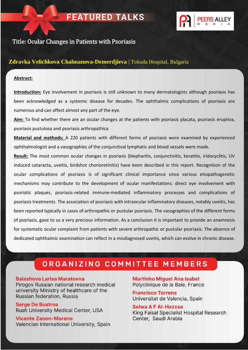
Title: Ocular Changes in Patients with Psoriasis
Zdravka Velichkova Chalmanova-Demerdjieva | Tokuda Hospital, Bulgaria
Abstract:
Introduction: Eye involvement in psoriasis is still unknown to many dermatologists although psoriasis has
been acknowledged as a systemic disease for decades. The ophthalmic complications of psoriasis are
numerous and can affect almost any part of the eye.
Aim: To find whether there are an ocular changes at the patients with psoriasis placata, psoriasis eruptiva,
psoriasis pustulosa and psoriasis arthropathica
Material and methods: A 220 patients with different forms of psoriasis were examined by experienced
ophthalmologist and a vasographies of the conjunctival lymphatic and blood vessels were made.
Result: The most common ocular changes in psoriasis (blepharitis, conjunctivitis, keratitis, iridocyclitis, UV
induced cataracta, uveitis, birdshot chorioretinitis) have been described in this report. Recognition of the
ocular complications of psoriasis is of significant clinical importance since various etiopathogenetic
mechanisms may contribute to the development of ocular manifestations: direct eye involvement with
psoriatic plaques, psoriasis-related immune-mediated inflammatory processes and complications of
psoriasis treatments. The association of psoriasis with intraocular inflammatory diseases, notably uveitis, has
been reported typically in cases of arthropathic or pustular psoriasis. The vasographies of the different forms
of psoriasis, gave to us a very precious information. As a conclusion it is important to provide an anamnesis
for systematic ocular complaint from patients with severe arthropathic or pustular psoriasis. The absence of
dedicated ophthalmic examination can reflect in a misdiagnosed uveitis, which can evolve in chronic disease.
All patients with psoriasis should undergo a routine ophthalmic evaluation and care. Dermatologists should
regularly monitor their psoriatic patients for eye symptoms.
Title: Efficacy of Affordable Combination Microinvasive Glaucoma Surgery: Combined 23-Gauge Cytotome
Goniotomy and Ciliary Sulcus Suprachoroidal Microtube Surgery in Patients with Moderate to Advanced
Glaucoma: A 6-month retrospective study
Daniel Laroche | New York Eye and Ear Infirmary of Mount Sinai, USA
Abstract:
PURPOSE: The aim of this study was to determine the efficacy of a novel technique of combined 23-gauge
goniotomy ($4US) and ciliary sulcus suprachoroidal microtube ($125US) in patients with primary open angle
glaucoma.
METHODS: Single-center, retrospective study of predominantly Black and Afro-Latino patients with primary
open angle glaucoma whom underwent a novel procedure of 23-gauge goniotomy and ciliary suprachoroidal
microtube insertion. Patients who underwent the above procedure with 6 months follow up were included
in the study. Investigated parameters were intraocular pressure (IOP), number of medications, and visual
acuity.
RESULTS: Ten (10) patients with primary angle glaucoma underwent 23-gauge cystotome goniotomy and
ciliary sulcus suprachoroidal microtube. The mean pre-op IOP was 19.4mmHG, mean number of medications
were 4.75 and visual acuity log MAR 0.7 (20/100 VA). At 1 month, 3month and 6 months follow up; the
mean IOPs reduced to 16.2 mmHG, 14.8mmHG, and 13.4mmHG, the mean number of medications also
decreased to 1.7, 1.8 and 2.0 respectively. Patients saw an improvement in vision to Log MAR 0.5 (20/60 VA)
at 6 months. The average mean deviation on visual field test was -17.62.
DISCUSSION: 23-gauge cystome goniotomy was performed with a straight 25mm 23-gauge cystotome by
Eagle labs.1 Ciliary sulcus suprachoroidal microtube technique was performed with the tube extender by
New World Medical.2
CONCLUSION: Combination microinvasive glaucoma surgery with a 23-gauge cystotome and ciliary sulcus
suprachoroidal microtube can safely lower intraocular pressure at 6 months in patients with moderate to
advance primary open angle glaucoma

Title: VEGF is a master regulator for Neuronal integrity in Diabetic Retinopathy and
Hypoxic Retinal Diseases
Yun-Zheng Le | Harold Hamm Chair in Diabetes Research, USA
Abstract:
Objective: to compare pathogenesis of AMD (age-related macular degeneration) and early macular
degeneration in children, accompanied by the formation of drusen, based on the study of the genetic
characteristics and the most effective methods of treatment in the Russian population.
Materials and Methods: 200 patients took part in the study: 150 adults (84 women, 66 men) aged from 50
to 92 years (mean age 63.5 +/- 12.5 years), and 50 children aged from 3 to 18 years old (average age 12.1 +/-
6.0 years) with a clinical diagnosis of Stargardt disease, macular dystrophy, central retinal abiotrophy. All
patients were tested for snp (rtPCR) associated with an increased risk of AMD (rtPCR, 15 snp in 12 genes)
(See Table 1). The period of inclusion of patients in the group from 2012 to 2017. The dosage of
lampalizumab 10 mg every 6 weeks is intravitreal for at least 52 weeks. Trial was approved by local ethics
committee, informed consent from every patient was obtained under protocol rules.
Results: 14 patients (aged 52 to 81 years) received lampalizumab treatment, who had high risk of developing
MD in the genes of the complement system (C3, CFH, CFHR1, CFHR3, CFI). A statistically significant decrease
in the rate of progression during application of Lampalizumab in our study group was observed in patients
with rs2230199 (C3) rs3766405 and rs1048663 (CFH).
Conclusions: A better understanding of the genetics of MD in the elderly and children will help in developing
risk models that can more accurately predict the occurrence and progression of the disease. Such prognostic
tests can help in the management of patients with MD of all ages in the detection of effective prophylactic
treatment methods and pharmacogenetic associations.
Title: VEGF is a Trophic factor for cone Photoreceptor survival under Hypoxia, an
AMD-like Retinal Environment
Meili Zhu | University of Oklahoma Health Sciences Center, USA
Abstract:
Objectives and scope: Vascular endothelial growth factor-A (VEGF-A or VEGF) is an essential growth factor
required for vascular development and physiological angiogenesis in the eye. However, VEGF is also a major
pathogenic factor in diabetic retinopathy (DR) and age-related macular degeneration (AMD). To determine
the impact of long-term anti-VEGF treatments for these diseases on photoreceptors, we conditionally
disrupted VEGF receptor-2 (VEGFR2), major VEGF receptor in cones, and characterized the conditional
VEGFR2 knockout (KO) mice under normal and hypoxic conditions. We further investigated the mechanism
of VEGFR2-mediated cone viability using 661W cells.
Methods: Hypoxia was generated with cobalt chloride. Gene expression was measured with
immunoblotting. Cone density was measured with immunohistochemistry.
Results: Cone-specific VEGFR2 KO mice did not show any alteration in cone density normally. However,
hypoxia exacerbated the cone degeneration in conditional VEGFR2 KO mice. Western blot analysis of
cultured 661W cells grown under hypoxic condition showed that phosphorylated AKT was downregulated in
the presence of VEGFR2-specific inhibitor SU5416.
Conclusions: VEGF signalling through VEGFR2 promoted cone photoreceptor viability under hypoxic
conditions, most likely through the VEGFR2-AKT survival pathway. Our results support the notion that
VEGFR2-mediated neuroprotection may be required for cone photoreceptor survival in hypoxia during the
development of AMD and DR and anti-VEGF treatment of these diseases.
Title: Prism Treatment of Infantile Squint
Elfriede Stangler-Zuschrott | Medical University Vienna, Austria
Abstract:
Background: Infantile concomitant squint is based on sensorial and motor defects. Successful treatment is required to
modulate both components and not merely the eye position. Material and method: Esotropic or intermittent
divergent squinting children aged 2 - 14 years, without amblyopia, with a squint deviation not more than 30°, wore
fully corrective glasses and prismatic compensation of the squint angle. Press-on prisms, split to both glasses, were
tolerated well and never caused amblyopia, which may occur only when a microtropia has developed behind the
prisms. Ground-in prism glasses for schoolchildren prevent non-compliance. Results: The study consists of two parts:
1) Long-term prism treatment over one year prior to squint surgery. 100 prism-treated esotropic patients were
compared to 78 children without prisms; the same surgeon used the same method. Patients treated with a prism
achieved the following statistically proven benefits: a) the number of alignments after surgery increased from 68% to
93% (p=0.0001), b) the postoperative recurrent increase of squint deviation was less (50%), c)fewer second operations
were needed in one and the same patient (8% instead of 25,6%, p=0.001),and d) improvements of the binocular state
led to the development of peripheral fusion even in cases of congenital strabismus. 2.) Long-term wearing of prisms as
conservative treatment. In a first study 44 patients were treated over a period of 2 -6, 5 years irrespective of the type
and angle of the squint. The results were 78% alignment without surgery. The limitations of this method are: The
squint angle should not exceed 15° in convergent and 10° in divergent cases. Starting the treatment at an early age
will reduce the time of recovery. Conclusions: Long-term prism treatment of concomitant strabismus improves the
sensorial and motor functions of binocularity; the sole disadvantage is the length of time it takes.
Title: Dynamical and real-time measurement of Monochromatic Ocular wave front Aberrations:
Applications
Sandra Franco | University of Minho, Portugal
Abstract:
The optical properties of the eye are not static and change continuously over time with factors such as pupil
size, the tear film stability and accommodation. Dynamical and real-time measurement of the
monochromatic ocular wavefront aberrations, during changes in the accommodation stimulus, provides
insights into the dynamics of the mechanisms that control accommodation.
Wavefront aberrations were measured in a dynamic and real-time mode while the accommodation state is
changed through lens interposition in front of the contralateral eye.
The ocular wavefront aberrations, during accommodative changes, were acquired. In addition, from the
acquired data, we evaluated the accommodative response’s characteristics, such as its inaccuracies during
sustained near vision, both in terms of response errors (lag or lead of accommodation), the latency of the
response and its microfluctuations
This methodology was applied in several subjects, focusing on the applications to symptomatic patients who
present difficulties with near vision tasks.
Results showed that the real-time measurement of the ocular aberrations has several applications, for
example for studying the variations of the optical proprieties of the eye to have a better and comprehensive
knowledge of the ocular accommodation and in helping in the clinical diagnosis of patients with
accommodative dysfunctions and posterior follow-up.

Title: Identification of MicroRNAs in tears as Biomarkers for primary Open-angle
Glaucoma
Vicente Zanon-Moreno | Valencian International University, Spain
Abstract:
Purpose: To study the expression profile of microRNAs in tears of patients diagnosed of primary open-angle
glaucoma (POAG) and patients with ocular hypertension (OHT) with the aim of finding molecular biomarkers
for better managing OHT and to achieve the preclinical POAG diagnosis.
Methods: A case-control study was carried out in 48 patients, 24 POAG vs 24 OHT, matched by age and
gender. Total RNA was isolated using the miRNeasy Serum/Plasma Kit (Qiagen) and quantified in a 2100
Bioanalyzer (Agilent). The microRNAs in tear samples were identified and analysed by next generation
sequencing using the Illumina NextSeq500 Technology. Finally, genes related to miRNAs showing
significantly different expression level were used to perform a gene ontology functional analysis.
Bioinformatic analyses were performed by specific programs.
Results: We identified 120 microRNAs in tears of the participants. Among them, only 8 showed significant
differences in expression between groups (p < 0.05) (2 microRNAs down-regulated and 8 up-regulated in
POAG tears). Receiver operating characteristic curve (ROC) was obtained for all of them, and 4 microRNAs
showed an area under the curve higher than 0.75.
Conclusions: Tear samples have provided essential information to differentially identify 8 microRNAs, 4 of
which presumptively might be used as biomarkers for early identification of the POAG risk in OHT
individuals.
Title: A new RNA-Seq derived perspective on Retinitis pigmentosa: New identified
pathways related to Oxidative stress
Luigi Donato | University of Messina, Italy
Abstract:
Among main inherited retinal dystrophies (IRDs), retinitis pigmentosa (RP) represents one of the most heterogeneous
groups characterized by progressive retinal degeneration. One of the principal causes of RP is oxidative stress in the
retinal pigment epithelium (RPE), which involves in the toxification mechanisms leading to N-retinylidene-N-
retinylethanolamine (A2E) accumulation and degradation. A2E is a toxic bis-retinoid derivative, produced from
condensation and oxidation of vitamin A (trans-retinal). Throughout life, A2E and other complex lipids accumulate and
form lipofuscin in the RPE, ultimately resulting in rod and cone degeneration. In order to find new ways by which
oxidative stress could act on RP etiopathogenesis and progression, we realized a comparative RNA-Seq experiment on
human RPE cells, treated with A2E 20µM and untreated, in three time points (time zero, 3h, 6h). An Illumina
NovaSeqTM sequencing system was used for the experiment, which was repeated thrice. Data analysis was carried out
with CLC Genomics Workbench and DNASTAR Lasergene Suite software, including low quality reads trimming and
gene expression quantification based on RPKM and EM algorithm. Upregulated (fold change >1.00) and
downregulated (fold change <−1.00) genes were, then, functionally annotated and, in order to discover pathways with
over-represented genes, enriched by Gene Set Enrichment Analysis (GSEA). Finally, the resulted pathways were
clustered into several “macro-pathways”, based on the highest degree of different gene expression and considering
the 5% most changed genes, at each time point, as “survived”. From this classification, several sub-pathways not yet
linked to RP were analyzed, such as: blood coagulation, protein sumoylation, Wnt receptor signaling pathway, planar
cell polarity pathway, sensory perception of sound and insulin receptor signaling pathway. These data suggest that a
huge number of new candidate genes, involved in identified pathways and still never associated to RP, could play a
role in the etiopathogenesis of this pathology.
Title: Implantable collamer lens surgery in patients with primary iris and/or ciliary
body cysts
Zhen Li | The People’s Hospital of Leshan, China
Abstract:
Background: The prevalence of primary iris and/or ciliary body cysts is common in myopia, though asymptomatic in
nearly all cases. It’s a very valuable thing to study the clinical safety and reliability of implantable collamer lens (ICL)
surgery in patients with primary iris and/or ciliary body cysts.
Methods: A total of 108 patients (201 eyes) were included in this retrospective study. All eyes had been implanted
with V4c implantable collamer lens (ICLV4c). According to the eyes with or without primary iris and/or ciliary body
cysts, all eyes were divided into two groups. We observed preoperative and postoperative uncorrected distance visual
acuity (UDVA), corrected distance visual acuity)(CDVA), intra-ocular pressure(IOP), anterior chamber volume(ACV),
anterior chamber depth(ACD), trabecular-iris angle (TIA), angle opening distance at 500 μm (AOD500) vertical central
distance between the corneal endothelium and the front surface of ICL(CE-ICL), and the central vault. The follow-up
periods covered 12 months.
Results: Among all the 201 eyes, primary iris and/or ciliary body cysts were detected in 54 eyes (26.87%),but the
prevalence was account to 36.11%(18males,21females).There were 30 eyes (55.56%) with unilateral single cyst, 12
eyes (22.22%) with unilateral double cysts, 12 eyes (22.22%) eyes with unilateral multiple and/or multi-quadrants
cysts, the mean size of cysts was (0.714 ± 0.149)mm(range from 0.510 to 1.075 mm).30.4% of the cysts were located
at iridociliary sulcus, 65.5% in pars plicata, and 4.1% in midzonal iris, which showed a characteristic distribution
pattern, with cysts found predominantly in the inferior and temporal quadrants. The postoperative size and the
number of cysts showed nearly no changes. The postoperative ACV, AOD500 and TIA showed a statistical reduction in
both two groups (P < 0.05), but with no statistical significant between the two groups (P > 0.05), the parameters of
postoperative IOP,CE-ICL and central vault also showed the same results as which. We did not observe serious
complication and IOP elevating in the whole follow-up periods.
Conclusion: Primary iris and/or ciliary body cysts are not absolutely contraindication for ICL surgery. For some single
cyst smaller than 1.075 mm or single quadrant cysts located at ciliary body are rare to lead some serious
complications. But, for some multiple cysts, especially multi-quadrants cysts located at iridociliary sulcus, we still
should remain cautions.

Title: Classification of Diabetic Retinopathy Images Using Neural Network Approach
Anoop Balakrishnan Kadan | ISBAT University
Abstract:
Vision loss occurs due to clouding in the eye structures, damages in the nerve layers of eye
that carries the light, and retinal detachment that will lead to blurred vision. Diabetic
retinopathy is eye disease that will lead to the loss of vision. Exudates are one of the
symptoms of the diabetic retinopathy where hard exudates are not common. It is very
difficult to treat the eye that consists of hard exudates. Therefore, in this work by
morphological image processing hard exudates are detected. Treatment of the eye at an
early stage can help to prevent vision loss. By interfacing with fuzzy algorithm it will show
how much the eye is affected by exudates. Proposed technique is by applying a neural
network approach (Bayesian) to classify the eye whether it is a normal eye or an abnormal.
Thereby, with the help of advanced treatment ophthalmologists will be able to treat the
patient effectively.

Title: Effectiveness of using Dexmedetomidine as an adjunct to local anaesthesia in
peribulbar block in ocular surgeries
Arvind Kumar Morya | AIIMS
Abstract:
Purpose: To evaluate the effect of dexmedetomidine, a centrally acting α2-adrenergic receptor agonist as an adjunct
to local anaesthetics in peribulbar block in ocular surgeries.
Methods: 120 patients of either gender undergoing cataract, squint, glaucoma surgeries under local anaesthesia
(peribulbar blockade) willingly giving consent to participate in this study were divided into two groups of 60 each -
Group A receiving - 3ml lignocaine+ 2ml bupivacaine+ 150 IU hyaluronidase; Group B receiving -3ml lignocaine+ 2ml
bupivacaine+ 150 IU hyaluronidase + 25mcg dexmedetomidine. We evaluated the effect of dexmedetomidine on
onset of globe akinesia and duration of block, intraocular pressure, heart rate, mean arterial pressure. Sedation levels
were assessed using Ramsay Sedation scale every 10 minutes during surgery. Pain score was assessed using visual
analogue scale and surgeon satisfaction was assessed using a 5 point likert like verbal rating scale.
Results: Time of onset of action was shortened and duration of block was increased significantly in group B as
compared to group A (p<0.05).The intraocular pressure decreased throughout the first 10 min after the administration
of the local anaesthesia in Group B (p<0.05). As regards the conscious level in the two groups, there was a significant
difference (P < 0.0001). Group A is higher regarding score 1 and 2, and Group B higher in score 3. Pain score was
significantly decreased in the dexmedetomidine group. However, group B showed more changes in heart rate and
blood pressure.
Conclusion: Addition of dexmedetomidine to lignocaine and bupivacaine in a peribulbar block significantly shortened
onset time, decreased the IOP, and prolonged the duration of anaesthesia that lead to increased patient cooperation
and surgeon satisfaction.
Title: Trehalose-a novel disaacharide against dry eye.
Morya AK1and Solanki K2
Arvind Kumar Morya | AIIMS
Abstract:
Aim: To compare effects of Trehalose 3% (Group A) with Sodium hyaluronate 1% (Group B) eyedrops in the
treatment of dry eye disease.
Material and methods: 394 patients evaluated for the dry eye using Schirmir's test, Tear film height, Tear
film break-up time on slit lamp , Rose bengal staining and found to be affected are divided in two groups of
199 in Group A and 195 in Group B each. Each patient’s history and symptoms were noted according to
Ocular Surface Discomfort Score. Group A received eyedrop trehalose 3%. Group B received eye drops 1%
Sodium hyaluronate both of which were administered QID. Comparison of relief in symptoms and signs
including Schirmir's test, Tear film height, Tear film break-up time on slit lamp, Rose bengal staining was
done at 4 weeks, 8 weeks and 12 weeks. Data was entered using SPSS version 22. Mean age of presentation
was 37.6+/- 14.39 years with range of 15-74 years. There was a significant difference in Schirmer’s test
(P=0.0001) and TBUT (P= <0.0001) and improvement in OSDI (P= 0.001) at second follow up maintained at
3rd follow-up.
Discussion: Trehalose- a potent osmo- and bioprotectant has been recently introduced that works on the
priniciple of autophagy. It stabilizes bilipid membranes and labile proteins and is protective against the
dessication. Trehalose has been found to be effective in the management of dry eye with better outcomes in
terms of Schirmer’s, TBUT and OSDI.

Title: Predicting Visual outcome after Open globe injury using classification and regression tree
model: the Moradabad Ocular trauma study.
Richa Gupta | C L Gupta Eye Institute
Abstract:
Objective: To identify factors associated with visual outcome in patients with open globe injuries (OGIs) using CART
model.
Scope: To utilize the CART model in predicting final visual acuity based on presenting clinical features following open
globe injury.
Methodology: Retrospective review of OGIs presenting to a tertiary eye care institute in North India from October
2009 to December 2016 was conducted. A total of 157 patients with open globe injury have been included in the
study. Multivariate analysis to ascertain the effects of different identified variables on the likelihood of poor visual
outcome was done using binomial logistic regression. "Visual survival" (counting fingers or better) versus "minimal/no
vision" (hand motion, light perception, and no light perception) was predicted using the classification and regression
tree (CART) model.
Results: Univariate analysis determined 9 predictors associated with poor visual outcome. Out of these, presence of
relative afferent pupillary defect (RAPD), poor presenting visual acuity, presence of adnexal injuries, and location of
injuries were the most significant predictors of vision loss. Absence of RAPD led to 79% chance of vision survival. Sixty-
eight percent of patients with RAPD and initial visual acuity (VA) of less than 6/60 resulted in poor vision.
Conclusion: The CART model is useful in predicting final VA based on some prognostic factors present initially, like
presence of RAPD, poor presenting visual acuity, presence of adnexal injuries, and location of injuries.
Title: Risk factors associated with Cataract surgery: An Analytical Overview
Subhash Mishra | Directorate of Health Services
Abstract:
There are many factors which affect the outcome of cataract surgery sometimes resulting into serious
complications. Outcome of Same surgeon with same protocol and IOL and medicines may be different due
to patient factor, similarly medicine factor, IOL factor assistant factor, sterilization factor lead to different
complications varying from visual unhappiness to loss of eye. These factors are grouped and discussed to
make an eye surgeon understand and capable of dealing with them to provide a safe and effective sight
restoration in cataract surgery. Some instances of mass post operative end-ophthalmitis are mentioned with
analysis of possible causes.
Abstract:
Purpose: To evaluate the efficacy of subcutaneous triamcinolone acetonide (SCTA) injection for treatment of
upper eyelid retraction and swelling associated with thyroid eye disease (TED).
Methods: In this retrospective, nonrandomized case series, 102 TED patients with inflammatory and
enlargement of levator palpebrae muscle observed on MRI evaluation as the cause of the upper eyelid
retraction and swelling, were managed clinically between June 2012 and December 2015. TA was injected
20mg/0.5ml subcutaneously for each eyelid. Patients showing no signs of improvement after first trial
received a reinjection, and were followed up with post-injection until lid retraction and swelling had reduced.
Treatment was evaluated after 12 months of SCTA injection, and the outcome was determined by eyelid signs
such as eyelid retraction, swelling, and eyelid lag. Each was graded on a 0-3 scale.
Results: Ninety-five of 102 patients (93.1%) experienced significant improvement in the amount of eyelid
signs after SCTA. The average score before and after treatment was from 1.62 to 0.12 in eyelid retraction,
1.32 to 0.26 in swelling, and 1.72 to 0.30 in eyelid lag respectively. Of 108 eyelids injected, 65 (60.2%) were
improved by a single SCTA, and 43 needed reinjection. In the single SCTA group, clinical activity score is lower
than the other group (p< 0.01), however TRAbs level and MRI findings were insignificant. None of the treated
eyelids exhibited intraocular pressure elevation. Eight female patients experienced menstrual disorder. Seven
patients had developed diplopia and were treated systemic steroid therapy.
Conclusions: Subcutaneous triamcinolone acetonide injection provides an effective treatment to improve
upper eyelid retraction and swelling associated with thyroid eye disease in the inflammatory levator
enlargement stage. It is easy to administer and few untoward effects can be resolved spontaneously.
Title: Subcutaneous Injections of triamcinolone acetonide for upper eyelid retraction and
swelling associated with thyroid eye disease
Ai Kozaki | Olympia Eye Hospital

Abstract:
Purpose: Vessel density (VD), assessed by OCT Angiography (OCTA), is thought to be relevant in the
prognosis of glaucoma. We intended to verify whether a rapid decrease of intra-ocular pressure (IOP) during
the post-operative period of glaucoma surgery was accompanied by an increase in VD.
Methods: We present a prospective study from April to September 2018, of patients with recent (<6weeks)
glaucoma surgery, with IOP ≥ 21mmHg, who accepted to participate. Other pathologies were excluded. We
performed an OCTA (Triton, Topcon®) with scans of 4.5mm centered in the optic disc before consultation,
and 15 minutes after a manoeuvre to decrease the IOP (releasable suture removal, laser suture lysis or
goniopuncture); the IOP measure was repeated.
Results: Four patients (3 males, 1 female; 15-71 years old) were included. In all cases, a rapid decrease in
IOP (mean IOP lowering of 7,75 mmHg) after these manoeuvres was associated with a marked improvement
in whole-image optic disc VD.
Conclusion: To our knowledge, we are the first to identify an immediate substantial increase in the vessel
density, in the early glaucoma post-operative period. Further larger studies are needed.
Title: Improvement of vessel density after glaucoma surgery
Ana Isabel Martinho Miguel | Polyclinique de la Baie
Title: Histone deacetylase inhibitor attenuates experimental fungal keratitis in mice
Xiaohua Li | Zhengzhou University
Abstract:
Fungal keratitis is one of the leading causes of blindness of infected corneal diseases, but the pathogenesis
of fungal keratitis is not fully understood and therefore the treatment of the disease by medication is still
under investigation. In the current study, we sought to study the effect of HDAC inhibitor suberoylanilide
hydroxamic acid (SAHA) on experimental fungal keratitis in mice. SAHA (25 mg/kg) (n = 30) or vehicle (DMSO)
(n = 30) was delivered through intraperitoneal injection (IP) 24 hours after the fungal inoculation, and the
same amount of SAHA injection or DMSO was followed at day 2. The expression of histone H3 (H3),
acetylated histone H3 (AC-H3), histone deacetylase 1 (HDAC)1, tumor necrosis factor-α (TNFα), and Toll-like
receptor 4 (TLR4) in surgically excised specimens from the patients and mice with fungal keratitis were
detected by immunohistochemistry. The expression of mRNAs for Interleukin-1β (IL-1β), TNFα, and TLR4
were evaluated in the corneas of the mice with fungal infection and the control corneas by real-time PCR.
The quantification of IL-1β and TNFα in the corneas of the mice with fungal infection was determined by
ELISA. The inhibitory effect of SAHA on mice fungal keratitis was revealed by GMS and H&E staining. We
found that the downregulation of histone acetylation and upregulation of HDAC1 expression were
associated with the increased inflammation response in fungal keratitis not only in humans but also
in experimental animals. SAHA was able to inhibit experimental fungal keratitis in mouse by suppressing
TLR4 and inflammatory cytokines such as TNFα and IL-1β; the inhibition of HDAC may be a potential
therapeutic approach for the treatment of fungal keratitis.
Title: MeCP2 plays a critical role in the pathogenesis of proliferative vitreoretinopathy
Shikun He | Zhengzhou University
Abstract:
Purpose: Proliferative vitreoretinopathy (PVR) is one of blindness eye disease. The pathogenesis of PVR is
still under investigation. In the current study, we sought to investigate the role of the methyl-CpG-binding
protein 2(MeCP2) especially phosphorylated P-MeCP2-421 (P-MeCP2-421) in the pathogenesis of PVR.
Methods: The expressions of P-MeCP2-421, 80, PPAR-γ and the double labeling of P-MeCP2-421 with α-
SMA ,cytokeratin, TGFβ,GFAP and PPAR-γ in human PVR membranes were analyzed by immunochemistry;
The effect of knocking down MeCP2 using siRNA on the expressions of α-SMA, phospho-Samd/2/3, collagen
1 and fibronectin and PPAR-γ was studied by immunoblot in cultured ARPE-19. The expression of α-SMA
stimulated by recombinant MeCP2 and P-MeCP2-421 activation by TNF in the cells was evaluated using
western bot. The effect of TGF-β and 5-AZA treatment on PPAR-γ expression was analyzed by immunoblot
as well. Chromatin immunoprecipitation was used to determine the biding of MeCP2 to TGFβ.
Results: Our results showed that P-MeCP2-421 was highly expressed in PVR membranes and was double
labeled with cells positive for α-SMA, cytokeratin, GFAP and TGFΒ. knock-down MeCP2 inhibited the
activation of Smad2/3 and the expression of collagen 1 and fibronectin induced by TGFβ. TGFβ inhibited the
expression of PPAR-γ, silence of MeCP2 by siRNA or using MeCP2 inhibitor (5-AZA) increased the expression
of PPAR-γ. α-SMA was upregulated by the treatment of recombinant MeCP2. TNF stimulated the activation
of P-MeCP2-421. Importantly, we found that MeCP2 boned to TGFβ as demonstrated by Chip assay.
Conclusion. The results from current study suggest that MeCP2 especially phosphorylated P-MeCP2-421
may play a significant role in the pathogenesis of PVR and targeting MeCP2 may be a potential of
therapeutically approach for the treatment of PVR.
Title: New Insights into Eye Development
Tammy Z Movsas | Zietchick Research Institute
Abstract:
Preterm infants are born with immature retinas which need to continue to vascularize after birth. To accomplish this, proper intraocular levels of vascular endothelial growth factor (VEGF) must be maintained. If VEGF levels in the preterm infant fall too low, retinal vessel formation halts, which leads to early stages of retinopathy of prematurity (ROP). If VEGF levels rise too high, retinal neo-vascularization occurs; this is the hallmark of late stages of ROP. Oxygen strongly influences intraocular VEGF production: high oxygen negates VEGF expression and low oxygen over-stimulates VEGF expression. This certainly explains why the exposure of premature infants to varying levels of oxygen is strongly associated with ROP development. But…what drives ocular VEGF production in utero when oxygen levels are physiologically-appropriate? The answer to this question is the key to ROP prevention.
Dr. Movsas has been challenging the current thought that physiologic hypoxia regulates all VEGF production during normal eye development. She and colleagues have been studying the role of luteinizing hormone (LH) and human chorionic gonadotropin (hCG) in the developing eye. She will discuss her novel findings summarized below:
The strong correlation that exists between LH and VEGF in vitreous from both bovine and porcine eyes
The ERG retinal amplitudes of a-waves and b-waves at high stimulus intensities are reduced in genetically modified mice without LH receptors, known as lhrkos.
Ocular VEGF and retinal vascular volumes are significantly reduced in lhrko eyes from neonatal mice. This provides the first evidence that LH signaling likely plays a role in VEGF regulation and vascularization in the developing mouse eye.
Human preterm infants produce hCG after birth and low levels are this hormone are associated with non-proliferative ROP. This study is the first to demonstrate a potential relationship between hCG and human angiogenesis.
Title: A prospective study with high frequency femtosecond Laser assisted cataract surgery: Clinical and Physical Advantages compare to Manual Cataract Procedure
Bojan Pajic | Swiss Eye Research Foundation
Abstract:
Background: The aim of our study was to investigate prospectively the safety and efficacy of the LDV Z8
femtosecond laser in cataract surgery compared to the conventional procedure.
Methods: The study included 130 eyes from 130 patients: 68 treated with femtosecond laser-assisted
cataract surgery (FLACS) using the FEMTO LDV Z8 and 62 treated with conventional phacoemulsification.
Capsulotomy and lens fragmentation in the laser group were performed with the FEMTO LDV Z8
femtosecond laser system, which employs a new, low-energy, high repetition rate laser process for cataract
surgery. In the conventional group, the capsulotomy was performed by a cystotome, and lens
fragmentation was achieved by the phaco-shop.
Results: Ease of phacoemulsification (on a 4-point scale), the completeness of capsulotomy (on a 10-point
scale), effective phacoemulsification time (seconds), uncorrected distance visual acuity (UCVA), best
spectacle-corrected distance visual acuity (BSCVA), spherical equivalent (SE), and safety of the procedure
were evaluated. The total follow-up time was three months. The intended capsulotomy diameter was in
group 1, high frequency femtosecond laser, significant more precise (p<0.001) than in group 2, conventional
procedure. The mean phacoemulsification time in Group 1 was 1.9 ± 2.25 and in Group 2, 2.3 ± 2.41 s which
is significant (p = 0.042). The effective phacoemulsification time (EPT) in Group 1 was found to be 1.48 ±
1.80 s and in Group 2 1.81 ± 1.93 s, which is significant (p = 0.044). Overall surgery time in Group 1 was 7.5 ±
1.22 min (range: 5–12 min) and 6.6 ± 1.76 min (range: 4.6–12 min) in Group 2, that is slightly significant (p =
0.048). The total follow-up time was three months.
Conclusions: FLACS with femtosecond laser system was characterized by complete and reproducible precise
capsulotomy and highly effective lens fragmentation.
Title: The efficacy and safety of aflibercept and conbercept in diabetic macular edema
Siwei Cai | Tianjin Medical University Eye Hospital
Abstract:
Diabetic macular edema (DME) has shown an increasing prevalence during the past years and is the
leading cause of diabetic retinopathy blindness. Traditional treatment modalities include laser and
corticosteroid therapy, which, however, either act through unclear mechanisms or cause cataracts and
elevated intraocular pressure. In recent years, as the pathogenic role of VEGF in DME has been well-
recognized, the intravitreal injection of anti-VEGF drugs has become the first-line treatment of DME due to
their great efficacy in improving visual acuity and mitigating macular edema. Advantages have been shown
for aflibercept and conbercept, the two recombinant decoy receptors that can bind VEGF with high
specificity and affinity, in DME treatment in clinical trials conducted both worldwide and in People’s
Republic of China. This review introduces the structural characteristics and molecular mechanisms of
action of these two anti-VEGF drugs, and summarizes the clinical trials evaluating their efficacy and safety,
with the hope to provide clues for designing optimal and personalized therapeutic regimens for DME
patients.

NO SECRET IS SAFE SHARE YOUR RESEARCH
Sponsors | Media Partners
http://ophthalmology.peersalleyconferences.com/

A right choice of conference destination is an important aspect of any international conference and keeping that in consideration, Euro Ophthalmology 2020 is scheduled in the Beautiful city ‘’Paris’’.
NETWORKING...CONFERENCING...FOSTERING
ATTENDING A CONFERENCE ISN’T ALL ABOUT LEARNING AND NETWORKING
A NEW PLACE , PEOPLE AND CULTUREDISCOVERING
Avenue des Champs Élysées
Arc de Triomphe
Musical Concerts at Sainte Chapelle
Palais Garnier, Opéra National de Paris
Bustling Boulevards and Legendary Cafés
Eiffel Tower
Musée d’Orsay
Place de la Concorde
Cathédrale Notre Dame de
Luxembourg Gardens
Musée du Louvre
Seine River Cruises

Connect with us
Mercure Paris Charles De GaulleAirport & ConventionBP 20248 -Roissypôle Ouest -Route de la commune -95713 Roissy CDG CedexVenue
Juliana Katelyn Program Director | Euro Ophthalmology 2020
Peers Alley Media1126 59 Ave East, V5X 1Y9
Vancouver BC, CanadaContact us: [email protected]
Ph : +1-778-766-2134
http://ophthalmology.peersalleyconferences.com/
Contact Us