review crimean–congo hemorrhagic fever · rpp-04-433 9. sponsoring/monitoring agency name(s) and...
TRANSCRIPT
Antiviral Research 64 (2004) 145–160
Review
Crimean–Congo hemorrhagic fever
Chris A. Whitehouse∗,1
Diagnostic Systems Division, United States Army Institute of Infectious Diseases (USAMRIID), Fort Detrick, Frederick, MD 21702-5011, USA
Received 28 June 2004; accepted 3 August 2004
Abstract
Crimean–Congo hemorrhagic fever (CCHF) is a tick-borne disease caused by the arbovirus Crimean–Congo hemorrhagic fever virus(CCHFV), which is a member of theNairovirusgenus (familyBunyaviridae). CCHF was first recognized during a large outbreak amongagricultural workers in the mid-1940s in the Crimean peninsula. The disease now occurs sporadically throughout much of Africa, Asia, andEurope and results in an approximately 30% fatality rate. After a short incubation period, CCHF is characterized by a sudden onset of high fever,chills, severe headache, dizziness, back, and abdominal pains. Additional symptoms can include nausea, vomiting, diarrhea, neuropsychiatric,and cardiovascular changes. In severe cases, hemorrhagic manifestations, ranging from petechiae to large areas of ecchymosis, develop.
are knownreal-timeease anddegreesno antiviralcreasedclinicalresented,
1466146
147
47
49
osition,
Numerous genera of ixodid ticks serve both as vector and reservoir for CCHFV; however, ticks in the genusHyalommaare particularlyimportant to the ecology of this virus. In fact, occurrence of CCHF closely approximates the known world distribution ofHyalommaspp.ticks. Therefore, exposure to these ticks represents a major risk factor for contracting disease; however, other important risk factorsand are discussed in this review. In recent years, major advances in the molecular detection of CCHFV, particularly the use ofreverse transcription-polymerase chain reaction (RT-PCR), in clinical and tick samples have allowed for both rapid diagnosis of dismolecular epidemiology studies. Treatment options for CCHF are limited. Immunotherapy and ribavirin have been tried with varyingof success during sporadic outbreaks of disease, but no case-controlled trials have been conducted. Consequently, there is currentlytreatment for CCHF approved by the U.S. Food and Drug Administration (FDA). However, renewed interested in CCHFV, as well as inknowledge of its basic biology, may lead to improved therapies in the future. This article reviews the history, epidemiology, ecology,features, pathogenesis, diagnosis, and treatment of CCHF. In addition, recent advances in the molecular biology of CCHFV are pand issues related to its possible use as a bioterrorism agent are discussed.© 2004 Elsevier B.V. All rights reserved.
Keywords:Crimean–Congo hemorrhagic fever; Tick-borne virus; Epidemiology, pathogenesis and treatment of CCHF
Contents
1. Historical perspective. . . . . . . . . . . . . . . . . . . . . . . . . . . . . . . . . . . . . . . . . . . . . . . . . . . . . . . . . . . . . . . . . . . . . . . . . . . . . . . . . . . . . . . . . . . . . . . . .1.1. Early history of Crimean–Congo hemorrhagic fever (CCHF). . . . . . . . . . . . . . . . . . . . . . . . . . . . . . . . . . . . . . . . . . . . . . . . . . . . . . . 141.2. Discovery of the virus. . . . . . . . . . . . . . . . . . . . . . . . . . . . . . . . . . . . . . . . . . . . . . . . . . . . . . . . . . . . . . . . . . . . . . . . . . . . . . . . . . . . . . . . . .
2. Classification of the virus. . . . . . . . . . . . . . . . . . . . . . . . . . . . . . . . . . . . . . . . . . . . . . . . . . . . . . . . . . . . . . . . . . . . . . . . . . . . . . . . . . . . . . . . . . . . .
3. Structure and molecular biology of the virus. . . . . . . . . . . . . . . . . . . . . . . . . . . . . . . . . . . . . . . . . . . . . . . . . . . . . . . . . . . . . . . . . . . . . . . . . . . . 1
4. Strain variation and phylogenetic relationships. . . . . . . . . . . . . . . . . . . . . . . . . . . . . . . . . . . . . . . . . . . . . . . . . . . . . . . . . . . . . . . . . . . . . . . . . . 1
∗ Tel.: +1 301 619 2098; fax: +1 301 619 2492.E-mail address:[email protected].
1 The views, opinions, and findings contained herein are those of the author and should not be construed as an official Department of the Army ppolicy, or decision unless so designated by other documentation.
0166-3542/$ – see front matter © 2004 Elsevier B.V. All rights reserved.doi:10.1016/j.antiviral.2004.08.001

Report Documentation Page Form ApprovedOMB No. 0704-0188
Public reporting burden for the collection of information is estimated to average 1 hour per response, including the time for reviewing instructions, searching existing data sources, gathering andmaintaining the data needed, and completing and reviewing the collection of information. Send comments regarding this burden estimate or any other aspect of this collection of information,including suggestions for reducing this burden, to Washington Headquarters Services, Directorate for Information Operations and Reports, 1215 Jefferson Davis Highway, Suite 1204, ArlingtonVA 22202-4302. Respondents should be aware that notwithstanding any other provision of law, no person shall be subject to a penalty for failing to comply with a collection of information if itdoes not display a currently valid OMB control number.
1. REPORT DATE 3 AUG 2004
2. REPORT TYPE N/A
3. DATES COVERED -
4. TITLE AND SUBTITLE Crimean-Congo hemorrhagic fever, Antiviral Research 64:145 - 160
5a. CONTRACT NUMBER
5b. GRANT NUMBER
5c. PROGRAM ELEMENT NUMBER
6. AUTHOR(S) Whitehouse, CA
5d. PROJECT NUMBER
5e. TASK NUMBER
5f. WORK UNIT NUMBER
7. PERFORMING ORGANIZATION NAME(S) AND ADDRESS(ES) United States Army Medical Research Institute of Infectious Diseases,Fort Detrick, MD
8. PERFORMING ORGANIZATIONREPORT NUMBER RPP-04-433
9. SPONSORING/MONITORING AGENCY NAME(S) AND ADDRESS(ES) 10. SPONSOR/MONITOR’S ACRONYM(S)
11. SPONSOR/MONITOR’S REPORT NUMBER(S)
12. DISTRIBUTION/AVAILABILITY STATEMENT Approved for public release, distribution unlimited
13. SUPPLEMENTARY NOTES The original document contains color images.
14. ABSTRACT Crimean-Congo hemorrhagic fever (CCHF) is a tick-borne disease caused by the arbovirusCrimean-Congo hemorrhagic fever virus (CCHFV), which is a member of the Nairovirus genus (familyBunyaviridae). CCHF was first recognized during a large outbreak among agricultural workers in themid-1940s in the Crimean peninsula. The disease now occurs sporadically throughout much of Africa,Asia, and Europe and results in an approximately 30% fatality rate. After a short incubation period,CCHF is characterized by a sudden onset of high fever, chills, severe headache, dizziness, back, andabdominal pains. Additional symptoms can include nausea, vomiting, diarrhea, neuropsychiatric, andcardiovascular changes. In severe cases, hemorrhagic manifestations, ranging from petechiae to large areasof ecchymosis, develop. Numerous genera of ixodid ticks serve both as vector and reservoir for CCHFV;however, ticks in the genus Hyalomma are particularly important to the ecology of this virus. In fact,occurrence of CCHF closely approximates the known world distribution of Hyalomma spp. ticks.Therefore, exposure to these ticks represents a major risk factor for contracting disease; however, otherimportant risk factors are known and are discussed in this review. In recent years, major advances in themolecular detection of CCHFV, particularly the use of real-time reverse transcription-polymerase chainreaction (RT-PCR), in clinical and tick samples have allowed for both rapid diagnosis of disease andmolecular epidemiology studies. Treatment options for CCHF are limited. Immunotherapy and ribavirinhave been tried with varying degrees of success during sporadic outbreaks of disease, but nocase-controlled trials have been conducted. Consequently, there is currently no antiviral treatment forCCHF approved by the U.S. Food and Drug Administration (FDA). However, renewed interested inCCHFV, as well as increased knowledge of its basic biology, may lead to improved therapies in the future.This article reviews the history, epidemiology, ecology, clinical features, pathogenesis, diagnosis, andtreatment of CCHF. In addition, recent advances in the molecular biology of CCHFV are presented, andissues related to its possible use as a bioterrorism agent are discussed.
15. SUBJECT TERMS Crimean-Congo hemorrhagic fever, antiviral, pathogenesis, review
16. SECURITY CLASSIFICATION OF: 17. LIMITATION OF ABSTRACT
SAR
18. NUMBEROF PAGES
16
19a. NAME OFRESPONSIBLE PERSON
a. REPORT unclassified
b. ABSTRACT unclassified
c. THIS PAGE unclassified
Standard Form 298 (Rev. 8-98) Prescribed by ANSI Std Z39-18

146 C.A. Whitehouse / Antiviral Research 64 (2004) 145–160
5. Ecology and epidemiology of CCHFV. . . . . . . . . . . . . . . . . . . . . . . . . . . . . . . . . . . . . . . . . . . . . . . . . . . . . . . . . . . . . . . . . . . . . . . . . . . . . . . . . 1495.1. Vertebrate reservoir hosts. . . . . . . . . . . . . . . . . . . . . . . . . . . . . . . . . . . . . . . . . . . . . . . . . . . . . . . . . . . . . . . . . . . . . . . . . . . . . . . . . . . . . . . 1495.2. Tick vectors. . . . . . . . . . . . . . . . . . . . . . . . . . . . . . . . . . . . . . . . . . . . . . . . . . . . . . . . . . . . . . . . . . . . . . . . . . . . . . . . . . . . . . . . . . . . . . . . . . . 1515.3. Geographical distribution. . . . . . . . . . . . . . . . . . . . . . . . . . . . . . . . . . . . . . . . . . . . . . . . . . . . . . . . . . . . . . . . . . . . . . . . . . . . . . . . . . . . . . . 152
6. Clinical features. . . . . . . . . . . . . . . . . . . . . . . . . . . . . . . . . . . . . . . . . . . . . . . . . . . . . . . . . . . . . . . . . . . . . . . . . . . . . . . . . . . . . . . . . . . . . . . . . . . . . . 152
7. Pathogenesis/clinical pathology. . . . . . . . . . . . . . . . . . . . . . . . . . . . . . . . . . . . . . . . . . . . . . . . . . . . . . . . . . . . . . . . . . . . . . . . . . . . . . . . . . . . . . . . 153
8. Diagnosis. . . . . . . . . . . . . . . . . . . . . . . . . . . . . . . . . . . . . . . . . . . . . . . . . . . . . . . . . . . . . . . . . . . . . . . . . . . . . . . . . . . . . . . . . . . . . . . . . . . . . . . . . . . 1538.1. Laboratory diagnosis. . . . . . . . . . . . . . . . . . . . . . . . . . . . . . . . . . . . . . . . . . . . . . . . . . . . . . . . . . . . . . . . . . . . . . . . . . . . . . . . . . . . . . . . . . . 154
8.1.1. Virus isolation. . . . . . . . . . . . . . . . . . . . . . . . . . . . . . . . . . . . . . . . . . . . . . . . . . . . . . . . . . . . . . . . . . . . . . . . . . . . . . . . . . . . . . . . . 1548.1.2. Immunological assays. . . . . . . . . . . . . . . . . . . . . . . . . . . . . . . . . . . . . . . . . . . . . . . . . . . . . . . . . . . . . . . . . . . . . . . . . . . . . . . . . . 1548.1.3. Molecular diagnostic assays. . . . . . . . . . . . . . . . . . . . . . . . . . . . . . . . . . . . . . . . . . . . . . . . . . . . . . . . . . . . . . . . . . . . . . . . . . . . . 154
9. Treatment. . . . . . . . . . . . . . . . . . . . . . . . . . . . . . . . . . . . . . . . . . . . . . . . . . . . . . . . . . . . . . . . . . . . . . . . . . . . . . . . . . . . . . . . . . . . . . . . . . . . . . . . . . . 154
10. Prevention and control. . . . . . . . . . . . . . . . . . . . . . . . . . . . . . . . . . . . . . . . . . . . . . . . . . . . . . . . . . . . . . . . . . . . . . . . . . . . . . . . . . . . . . . . . . . . . . . 15510.1. Risk factors. . . . . . . . . . . . . . . . . . . . . . . . . . . . . . . . . . . . . . . . . . . . . . . . . . . . . . . . . . . . . . . . . . . . . . . . . . . . . . . . . . . . . . . . . . . . . . . . . . 15510.2. Control measures. . . . . . . . . . . . . . . . . . . . . . . . . . . . . . . . . . . . . . . . . . . . . . . . . . . . . . . . . . . . . . . . . . . . . . . . . . . . . . . . . . . . . . . . . . . . . 156
11. Potential bioterrorism concerns. . . . . . . . . . . . . . . . . . . . . . . . . . . . . . . . . . . . . . . . . . . . . . . . . . . . . . . . . . . . . . . . . . . . . . . . . . . . . . . . . . . . . . . 156
Acknowledgements. . . . . . . . . . . . . . . . . . . . . . . . . . . . . . . . . . . . . . . . . . . . . . . . . . . . . . . . . . . . . . . . . . . . . . . . . . . . . . . . . . . . . . . . . . . . . . . . . . . . . . . 156
References. . . . . . . . . . . . . . . . . . . . . . . . . . . . . . . . . . . . . . . . . . . . . . . . . . . . . . . . . . . . . . . . . . . . . . . . . . . . . . . . . . . . . . . . . . . . . . . . . . . . . . . . . . . . . . . 156
1. Historical perspective
infectious hemorrhagic disease, and Uzbekistan hemorrhagicfever have been known for centuries to produce a disease sim-i
1
tiono clini-c per-
sonnel were infected during an epidemic in war-torn Crimea
sy-la-tsdated
Fion
heneli-rn
-an,
inctualed
diesden-m
various geographic regions. In fact, several agents of tick-borne hemorrhagic fevers from Kazakhstan and Uzbekistana in-d rks mtv goaT re,
lar to CCHF (Chumakov et al., 1976).
.2. Discovery of the virus
Crimean hemorrhagic fever (CHF) came to the attenf modern medical science and was first described as aal entity in 1944–1945 when about 200 Soviet military
nd from various areas across Africa were found to beistinguishable from each other. This ultimately led to wohowing CHF virus was antigenically indistinguishable frohe Congo virus (Casals, 1969; Chumakov et al., 1969), airus originally isolated from human patients from the Connd Uganda (Simpson et al., 1967; Woodall et al., 1967).he realization that the Congo virus and CHF virus we
1.1. Early history of Crimean–Congo hemorrhagic fever(CCHF)
A disease now considered to be CCHF was described by aphysician in the 12th century from the region that is presentlyTadzhikistan. The description was of a hemorrhagic diseasewith the presence of blood in the urine, rectum, gums, vomi-tus, sputum, and abdominal cavity and was said to be causedby a louse or tick, which normally parasitizes a blackbird(Hoogstraal, 1979). The arthropod described may well havebeen a species ofHyalommatick larvae which are frequentlyfound on blackbirds. CCHF has also been recognized for cen-turies under at least three names by the indigenous people ofsouthern Uzbekistan:khungribta(blood taking),khunymuny(nose bleeding), orkarakhalak(black death) (Chumakov,1974; Hoogstraal, 1979). [The term “black death,” now com-monly used to refer to plague (Yersina pestis), did not appearin the Oriental literature on plague, and was not commonlyused in European languages until the 16th and 17th cen-turies (Dols, 1977).] In Central Asia, various hemorrhagicdiseases including acute infectious capillarotoxicosis, acute
(Chumakov, 1945, 1947). Shortly thereafter, a viral etiologywas suggested by reproducing a febrile syndrome in pchiatric patients undergoing pyrogenic therapy after inocution with a filterable agent from the blood of CHF patien(Chumakov, 1974). Further evidence of a viral etiology anof a suspected tick-borne route of infection was demonstrby inducing a mild, but characteristic, clinical course of CHin healthy human volunteers 2 days after their inoculatwith filtered suspensions of nymphalHyalomma margina-tumticks in the presence of antibiotics (Chumakov, 1974).
In 1967, a breakthrough in CHF research came wChumakov and his colleagues at the Institute of Poliomytis and Viral Encephalitides in Moscow first used newbowhite mice for CHF virus isolation (Butenko et al., 1968;Chumakov et al., 1968). The resulting Drosdov strain, isolated by this method from a patient (Drosdov) in Astrakhbecame the prototype strain for much experimental workRussia and elsewhere. This now gave researchers an avirus to use in a variety of experimental studies and allowfor the production of the necessary reagents (i.e., antiboand antigens) needed for serological surveys and for the itification and classification of viral isolates obtained fro
C.A. Whitehouse / Antiviral Research 64 (2004) 145–160 147
in fact, the same virus lead to the new name, CHF–Congovirus. However, many authors found the name awkward,and have adopted Crimean–Congo hemorrhagic fever virus(Hoogstraal, 1979).
2. Classification of the virus
CCHFV is a member of theNairovirusgenus of the fam-ily Bunyaviridae. Other genera within the family includeOrthobunyavirus, Hantavirus, Phlebovirus, andTospovirus.According to the most recent report from the InternationalCommittee on the Taxonomy of Viruses, there are seven rec-ognized species in the genusNairoviruscontaining 34 viralstrains (Elliott et al., 2000), all of which are believed to betransmitted by either ixodid or argasid ticks (i.e., hard orsoft ticks, respectively). The most important serogroups arethe CCHF group, which includes CCHFV, and Hazara virus,which has not been demonstrated to be pathogenic to humans,and the Nairobi sheep disease group, which includes Nairobisheep disease (NSD) and Dugbe viruses. Only three mem-bers are known to be pathogens of humans, namely, CCHFV,Dugbe and Nairobi sheep disease viruses, although the lat-ter is primarily a pathogen of sheep and goats. Dugbe viruscauses a mild febrile illness and thrombocytopenia in humans
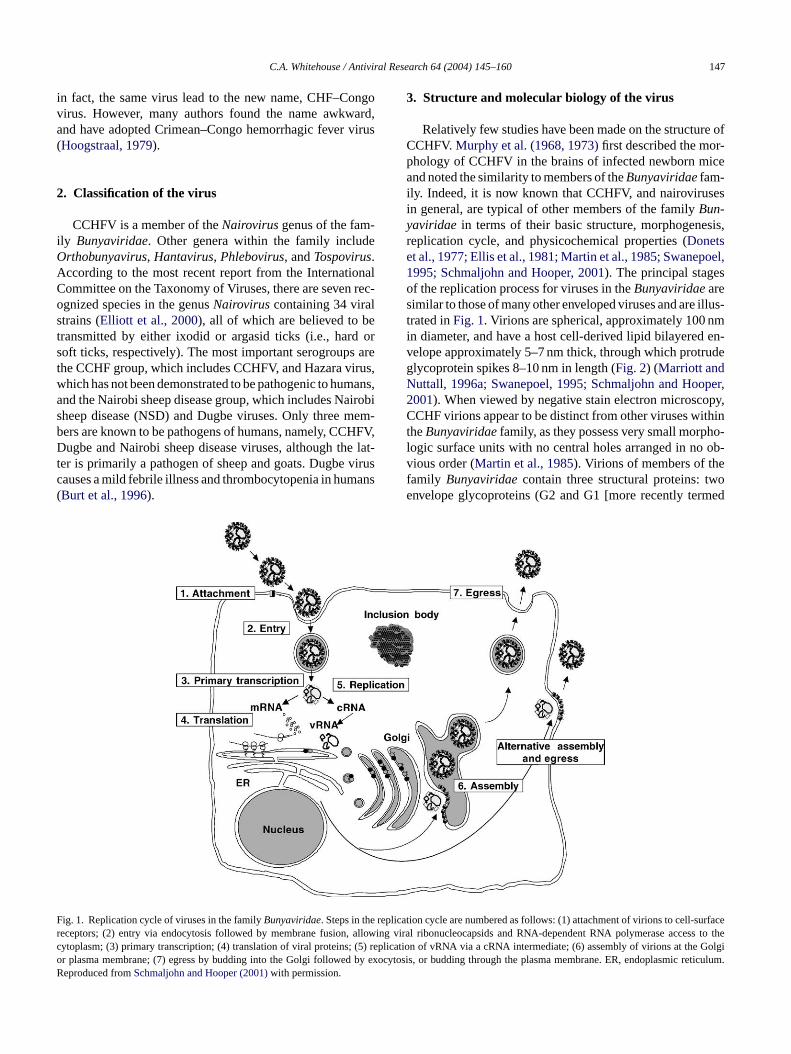
Fig. 1. Replication cycle of viruses in the familyBunyaviridae. Steps in the replicreceptors; (2) entry via endocytosis followed by membrane fusion, allowincytoplasm; (3) primary transcription; (4) translation of viral proteins; (5) replior plasma membrane; (7) egress by budding into the Golgi followed by exoReproduced fromSchmaljohn and Hooper (2001)with permission.
3. Structure and molecular biology of the virus
Relatively few studies have been made on the structure ofCCHFV.Murphy et al. (1968, 1973)first described the mor-phology of CCHFV in the brains of infected newborn miceand noted the similarity to members of theBunyaviridaefam-ily. Indeed, it is now known that CCHFV, and nairovirusesin general, are typical of other members of the familyBun-yaviridae in terms of their basic structure, morphogenesis,replication cycle, and physicochemical properties (Donetset al., 1977; Ellis et al., 1981; Martin et al., 1985; Swanepoel,1995; Schmaljohn and Hooper, 2001). The principal stagesof the replication process for viruses in theBunyaviridaearesimilar to those of many other enveloped viruses and are illus-trated inFig. 1. Virions are spherical, approximately 100 nmin diameter, and have a host cell-derived lipid bilayered en-velope approximately 5–7 nm thick, through which protrudeglycoprotein spikes 8–10 nm in length (Fig. 2) (Marriott andNuttall, 1996a; Swanepoel, 1995; Schmaljohn and Hooper,2001). When viewed by negative stain electron microscopy,CCHF virions appear to be distinct from other viruses withintheBunyaviridaefamily, as they possess very small morpho-logic surface units with no central holes arranged in no ob-vious order (Martin et al., 1985). Virions of members of thefamily Bunyaviridaecontain three structural proteins: twoe med
nvelope glycoproteins (G2 and G1 [more recently ter (Burt et al., 1996).ation cycle are numbered as follows: (1) attachment of virions to cell-surfaceg viral ribonucleocapsids and RNA-dependent RNA polymerase access to thecation of vRNA via a cRNA intermediate; (6) assembly of virions atthe Golgicytosis, or budding through the plasma membrane. ER, endoplasmic reticulum.

148 C.A. Whitehouse / Antiviral Research 64 (2004) 145–160
Fig. 2. Schematic cross-section of aBunyaviridaevirion. The three RNAgenome segments (S, M, and L) are complexed with nucleocapsid protein toform ribonucleocapsid structures. The nucleocapsids and RNA-dependentRNA polymerase are packaged within a lipid envelope that contains theviral glycoproteins, G1 and G2 (also referred to as Gn and Gc, respectively).Reproduced fromSchmaljohn and Hooper (2001)with permission.
Gn and Gc, respectively, named in accordance with their rel-ative proximity to the amino or carboxy terminus of the Msegment encoded polyprotein]) and a nucleocapsid protein(N), plus a large polypeptide (L) (approximately 200 kDa),which is the viron-associated RNA-dependent RNA poly-merase (Schmaljohn and Hooper, 2001; Marriott andNuttall, 1996b). Recently, two independent research groupspublished the complete nucleotide sequence of the CCHFVL segment (Honig et al., 2004; Kinsella et al., 2004). The se-quence is approximately 60% identical both at the nucleotideand amino acid levels to the L segment of Dugbe virus,the only otherNairovirus genome to be fully sequenced,with the most highly conserved area being that encoding theregion corresponding to the core catalytic domains of theRNA-dependent RNA polymerase. Further analysis of the Lsegment sequences from both CCHFV and Dugbe virus re-vealed the presence of a zinc-finger domain and a leucinezipper motif, suggesting that nairovirus L segments displaycharacteristics of viral helicases (i.e., having both helicaseactivity and polymerase activity stemming from one polypro-tein), most often seen in positive-strand RNA virus replicases.Furthermore, high-sequence homology with a newly formedsuperfamily of predicted cysteine proteases, termed ovar-ian tumor (OTU)-like proteases, was discovered, which wasalso suggested from the L segment sequence of Dugbe virus( edtL toy ae ro-t tes,s ovirusLo om-
plex nature of the protein products encoded by the CCHFVL segment.
The genome is characteristic of other members of the fam-ily and is composed of three negative-strand RNA segments,S, M, and L, encoding the N nucleocapsid, Gn and Gc glyco-proteins, and the L polymerase, respectively. The RNA seg-ments are complexed with N to form individual S, M, and Lnucleocapsids, which appear to be circular or loosely helical(Bishop, 1996). The M segment of nairoviruses is 30–50%larger than the M segments of members of other genera intheBunyaviridaefamily and has the potential coding capac-ity of up to 240 kDa of protein (Elliott, 1990). At least oneof each of the S, M, and L ribonucleocapsids must be con-tained in a virion for infectivity; however, equal numbers ofnucleocapsids may not always be packaged in mature virions(Schmaljohn and Hooper, 2001). Recent data show that the Nprotein is targeted to the perinuclear region of infected cellsin the absence of native RNA segments and that this targetingis actin filament dependent (Andersson et al., 2004a,b). Thefirst 8–13 nucleotide bases at the 3′ termini of all three RNAsegments have a sequence (3′-AGAGUUUCU. . .) that is con-served within viruses of the genus (Clerex-van Haaster et al.,1982), with a complementary consensus sequence at the 5′termini. Base-pairing of the terminal nucleotides is predictedto form stable panhandle structures and noncovalently closedc lec-t rus,U
forr usesw rnal-i lasm( micr icha leasev tw f theg dt andG co-p edt ein,w kDap G2)a uslyr FVu y re-l rsorche
d forC Ap de-v virus( se
Makarova et al., 2000). From these data, it is hypothesizhat the OTU-like protease may function in theNairovirus
protein by autoproteolytically cleaving the polyproteinield a polymerase and a helicase (Honig et al., 2004; Kinsellt al., 2004). Other suggested functions of the OTU-like p
ease include involvement with deubiquitination activitiuch that has recently been demonstrated for the aden3 23 K proteinase (Balakirev et al., 2002). Clearly, this isnly the beginning of future studies to elucidate the c
ircular RNAs, which have been directly observed by eron microscopy of RNA extracted from another bunyaviukuniemi, virions (Hewlett et al., 1977).The viral glycoproteins are believed to be responsible
ecognition of receptor sites on susceptible cells. Virhich attach to receptors on susceptible cells are inte
zed by endocytosis, and replication occurs in the cytopseeFig. 1). Virions mature by budding through endoplaseticulum into cytoplasmic vesicles in the Golgi region, whre presumed to fuse with the plasma membrane to reirus (Donets et al., 1977; Ellis et al., 1981). Much recenork has been done on the molecular characterization olycoproteins of CCHFV.Sanchez et al. (2002)demonstrate
hat during CCHFV infection, the mature Gn (37-kDa)c (75-kDa) proteins form the predominant structural glyrotein components of the virus. Additionally they show
hat the M RNA segment of CCHFV encodes a polyprothich undergoes proteolytic processing to yield a 140-recursor protein of Gn (PreGn, previously referred to Prend an 85-kDa precursor protein of Gc (PreGc, previoeferred to as PreG1). It was recently shown that CCHses, at least in part, the subtilase SKI-1 and possibl
ated cellular proteases for the major glycoprotein preculeavage events (Sanchez et al., 2002; Vincent et al., 2003), asas been demonstrated for theArenavirus, Lassa virus (Lenzt al., 2001).
Recently, a reverse genetics system was developeCHFV (Flick et al., 2003), which was based on the RNolymerase I transcription system recently used in theelopment of a reverse genetics system for UukuniemiFlick and Pettersson, 2001). The development of a rever

C.A. Whitehouse / Antiviral Research 64 (2004) 145–160 149
genetics system for CCHFV was a major step forward inefforts to understand the biology of the virus. The develop-ment of an infectious clone for CCHFV will allow for moreextensive studies of its biology and pathogenesis, and may ul-timately lead to better therapeutic and prophylactic measuresagainst CCHFV infections.
4. Strain variation and phylogenetic relationships
Many early studies, based on serological testing, sug-gested that there are very few significant differences amongstrains of CCHFV. For example, studies employing modifiedagar gel diffusion precipitation, neutralization, cell culture in-terference, and complement fixation tests demonstrated thatthere were no apparent antigenic differences among strainsfrom several different geographic locations in the former So-viet Union and Africa (Casals, 1969; Casals et al., 1970;Chumakov et al., 1969; Tignor et al., 1980). However,more recent data based on nucleic acid sequence analysishave revealed extensive genetic diversity. The first publishedCCHFV sequence data was of the S RNA segment (whichencodes the viral nucleoprotein) of the Chinese sheep iso-late C68031 (Marriott and Nuttall, 1992). Since then, severaladditional S segment sequences from CCHFV isolates fromde 02a;D esF PCRp the1 AE)s witht e nu-c .,1e se-q 200,w i-n ke-w selyr eredte if-f and2 ians p byi cef s-s hinaa woC dif-f meand ige-rs rom
Uzbekistan (TI10145) differed in their S segment sequenceby 14.9% and 13.2%, respectively, from the Nigerian IbAr10200. Based on the S RNA sequence, the southern Russianstrain was most closely related to Drosdov (4.7% difference)and the sequence of the Uzbekistan strain was most closelyrelated to the Chinese strains (3.8% and 3.7% difference fromstrain 8402 and HY13, respectively) (Yashina et al., 2003).
More recent work has begun to shed light on the geneticdiversity of the M RNA segment. The first published charac-terization of the CCHFV M RNA segment was of the Chi-nese strains, BA66019 and BA8402, isolated in 1965 and1984 from a CCHF patient andHyalommaspp. ticks, re-spectively (Papa et al., 2002c), although a complete M RNAsequence of the reference strain IbAr 10200 was deposited inGenBank (accession number U39455) previously. The cod-ing nucleotide sequences of the two Chinese strains differedfor the Nigerian strain IbAr 10200 by a mean of 22%, supply-ing further evidence of the extent of genetic diversity amongthese viruses. Recently, sequence analysis of the M RNA seg-ments from CCHFV isolates from Russia and from CentralAsia (Tajikistan) indicated that they each form separate phy-logenetically distinct groups (Yashina et al., 2003; Sereginet al., 2004).
5
5
en-e otict romn attlea( s( .,1 bS ies toC cteda eys,s , andA eno tile,ai Vv ,1 iche de-te
ctedw d/ora ctoryt ntsb tali FV,
ifferent regions of the world have been published (Schwarzt al., 1996; Rodriguez et al., 1997; Papa et al., 20rosten et al., 2002b; Yashina et al., 2003). Analysis of thesequences show considerable genetic differences (Fig. 3).or example, several CCHFV S segment sequences ofroducts obtained directly from infected patients from994–1995 outbreak in the United Arab Emirates (Uhowed a divergence of 10.0–11.8% when comparedhe Chinese sheep isolate C68031; however, most of thleotide changes were in the third position (Schwarz et al996). Two isolates obtained from Kosovo in 2001 (Papat al., 2002b) showed a 17% difference in nucleotideuence in the S segment from the Nigerian strain IbAr 10hile differing only by 4% from the Drosdov strain, origally isolated from the blood of a patient in Russia. Liise, strains obtained in neighboring Albania were clo
elated to those from Kosovo and phylogenetically clustogether along with the Drosdov strain from Russia (Papat al., 2002a). It is interesting that the Greek strain AP92 d
ered greatly from other European strains (e.g., 24.3%5.3% nucleotide difference from the Kosovo and Albantrains, respectively), and therefore, clusters in a groutself (Fig. 3). AP92 strain was originally isolated in Greerom a Rhipicephalus bursatick and has not yet been aociated with disease in humans. Also, strains from Cre known to be greatly divergent from African strains. Thinese strains (BA66019 and BA8402) exhibited a 15%
erence in nucleotide sequence in the S segment and aifference of 22% in the M segment from those of the Nian strain IbAr 10200 (Papa et al., 2002c). Additionally, atrain from southern Russia (STV/HU29223) and one f
. Ecology and epidemiology of CCHFV
.1. Vertebrate reservoir hosts
Like other tick-borne zoonotic agents, CCHFV grally circulates in nature unnoticed in an enzo
ick–vertebrate–tick cycle. CCHFV has been isolated fumerous domestic and wild vertebrates, including cnd goats (Woodall et al., 1965; Causey et al., 1970), sheepYu-Chen et al., 1985), hares (Chumakov, 1974), hedgehogCausey et al., 1970), aMastomysspp. mouse (Saluzzo et al985), and even domestic dogs (Shepherd et al., 1987a,).era from several species of wild mammals have antibodCHFV and seroepidemiological studies have also detentibodies to CCHFV in domestic cattle, horses, donkheep, goats, and pigs from various parts of Europe, Asiafrica (Watts et al., 1989a,b). Interestingly, there has benly one report of antibody to CCHFV detected from a reptortoise from Tadzhikistan (Pak et al., 1971) even though
mmatureHyalomma anatolicumticks, a common CCHFector, are known to sometimes feed on lizards (Hoogstraal979). For a comprehensive listing of vertebrates from whither CCHFV has been isolated or antibody to CCHFV
ected, the reader is referred toHoogstraal (1979)andWattst al. (1989a,b).
Although many domestic and wild vertebrates are infeith CCHFV, as evidenced by development of viremia anntibody response, birds, in general, appear to be refra
o infection with CCHFV. For instance, early experimey Berezin et al. (1971a,b)showed that after experimen
noculation of birds (rooks and rock doves) with CCH

150 C.A. Whitehouse / Antiviral Research 64 (2004) 145–160
Fig. 3. Phylogenetic relationships inferred from comparing partial sequences of the S segment RNA of CCHFV. Sequences were aligned by the multisequencealignment program GeneDoc (version 2.6.002) and analyzed by a neighbor-joining method with Kimura two-parameter distances by using MEGA software(version 2.1). The lengths of the horizontal branches are proportional to the number of nucleotide differences between taxa. Vertical branches are for visualclarity only. Bootstrap values above 50%, obtained from 500 replicates of the analysis, are shown at the appropriate branch points. CCHFV strains aredescribed as strain designation/country of origin. The GenBank accession numbers of the CCHFV S RNA segment sequences used are as follows: IbAr 10200(U88410); Drosdov, HY13, and JD206 (U88412, U88413 and U88414, respectively); 9553/2001 (AF428144), 9717/01 (AF428145), Kosovo (AF404507),U3010 (U88416), 66019 (AJ010648), TI10145 (AF481799), 8402 (AJ010649), C68031 (M86625), HU9447547 (U75670), ArMg951 (U15024), HU9509853(U75672), TI9538886 (U75673), TI9538889 (U75669), HU9509854 (U75671), ArB604 (U15092), HD38562 (U15093), RSA (U75675), SPU415/85 (U88415),ArD97268 (U15091), ArD39554 (U15089), AnD15786 (U15020), ArD8194 (U1502), DAK8194 (U88411), ArTeh193-3 (U15022), HD49199 (U15023), AP92(U04958). Two outgroup taxa included Dugbe virus (strain KT 281/75, AF434165) and Hazara virus (strain JC280, M86624). Scale bar, 5% divergence.
they remained healthy, and evidence of viremia or an an-tibody response could not be demonstrated. Furthermore,work by the same group, showed that even though CCHFVcould be isolated from nymphal ticks collected from over600 birds, the birds remained serologically negative for an-tibody to CCHFV. Attempts to isolate virus from the bloodand organs of 360 of those birds were uniformly negative.Several additional examples are known from the 1970s inwhich CCHFV has been isolated from ticks infecting numer-ous species of birds, which remain serologically negative for
the virus (Hoogstraal, 1979). Taken together, these studiessuggest that birds appear to be refractory to CCHF viremiaeven though they can support large numbers of CCHFV-infected ticks. However, some exceptions do exist; detectedantibodies to CCHFV in 1 of 428 sera tested from chickensand ducks in Kazakhstan and also detected CCHFV antibod-ies in the serum of a magpie (Pica pica). However, in morerecent pathogenicity studies, domestic chickens proved to berefractory to CCHFV infection (Shepherd et al., 1987a,b).Also, in the same study, low CCHF viremia was detected

C.A. Whitehouse / Antiviral Research 64 (2004) 145–160 151
in a blue-helmeted guinea fowl (Numidia meleagris) afterexperimental infection with CCHFV. In another more re-cent study, antibodies were detected after CCHFV inocula-tion in a red-beaked hornbill and a glossy starling (but notin two laughing doves or six domestic chickens); however,none of the birds showed detectable viremia (Zeller et al.,1994).
Another interesting exception has been the disease’s ap-parent association with the commercial ostrich meat indus-try in South Africa. In 1984, a case of CCHF occurred in aworker who became ill after slaughtering ostriches (Struthiocamelus) on a farm in South Africa (Van Eeden et al., 1985).Antibody to CCHFV was detected in 24% of ostriches fromsurrounding farms, including six of nine ostriches from thefarm where the patient worked. Interestingly, none of 460birds of 37 other species tested during that study had de-tectable antibodies to CCHFV (Shepherd et al., 1987a,b).Also, in 1996, there was an outbreak of 17 cases of CCHFamong workers at an ostrich abattoir (Swanepoel et al., 1998).In both instances, it was suspected that infection was acquiredeither by contact with ostrich blood or by inadvertently crush-ing infected ticks while skinning the ostriches. Ostriches havealso been experimentally infected with CCHFV (Swanepoelet al., 1998). The ostriches, which were experimentallyinfected with CCHFV subcutaneously, developed viremia1 vis-c con-c s ata e oft hisl eriodc ili-t
rolei tingv mayr rdst donei
bloodf s im-p xactr mis-s
5
s oft(l thef thef icks rs. Inm thesea irus,
but simply virus may be present in a recent blood meal froma viremic host. For example, the one instance where viruswas isolated from a biting midge, the midge was collectedby a light trap near a cattle shed in Nigeria and may havecontained undigested blood (Causey et al., 1970). Similarly,it seems unlikely that argasids are capable of transmittingCCHFV since the virus failed to replicate in three speciesof soft ticks (i.e.,Argas walkerae, Ornithodoros savignyi,andOrnithodoros porcinus) after intracoelomic inoculation(Shepherd et al., 1989) and the same was shown for the softtick Ornithodoros sonrai(Durden et al., 1993). Far more im-portant to the ecology and epidemiology of CCHF are ticksin the genusHyalomma. As early as 1944,Hyalommaspp.ticks were implicated in the ecology of CCHF based upona relationship between clinical cases and tick bite. In fact,the following year, a healthy volunteer subcutaneously in-oculated with a suspension of 370 nymphalH. marginatumticks developed a disease characteristic of mild CCHF. Thisnot only helped to prove the viral etiology of this disease,but also implicatedHyalommaspp. ticks as possible vectors;however, it was not until the late 1960s that the virus wasisolated from adultHyalomma, as well as several other tickspecies (Hoogstraal, 1979; Chumakov, 1971). In general, theknown occurrence of CCHFV in Europe, Asia, and Africacoincides with the world distribution of ticks of the genusH ,bC -ct rusw d re-p loodf iant ly allt lowf -s HF,t (i.e.,Rw bu-t nlya ture.E inlyo tedH fC ,1 heym her,p n ad-d s tos froml xper-ie rdone s-ma
–4 days after infection and virus was detectable ineral organs up to 5 days post-inoculation. It wasluded from these studies that infection in ostrichebattoirs could be prevented by keeping the birds fre
icks for a certain period of time before slaughter. Ted to the standard 30-day pre-slaughter quarantine purrently enforced in South African ostrich export facies.
Clearly, ground-feeding birds may play an importantn the ecology and epizootiology of CCHF by transporirus-infected ticks (even though the birds themselvesemain non-viremic). However, the role, if any, for the bihemselves are not clear and additional work needs to ben this area to resolve these issues.
In summary, vertebrates are essential as a source ofor vector ticks and the number of species of vertebratelicated in the natural history of CCHF is extensive, the eole, if any, of vertebrates in the maintenance and transion of the virus remains to be determined.
.2. Tick vectors
CCHFV has been isolated from at least 31 specieicks and one species of biting midge (Culicoides spp.)Hoogstraal, 1979; Linthicum and Bailey, 1994). Viral iso-ations from ticks have been made from two species inamily Argasidae (soft ticks) and from seven genera ofamily Ixodidae (hard ticks). Viral isolation alone from a tpecies, however, does not incriminate them as vectoany cases, in fact, there is no definitive evidence thatrthropods are capable of serving as vectors for the v
yalomma(Hoogstraal, 1956, 1979; Watts et al., 1989a).CHF viral replication and tissue tropism inHyalomma trunatumticks were examined byDickson and Turell (1992). Inicks that were experimentally infected with CCHFV, vias recovered in highest titers from salivary glands anroductive tissues and was positively associated with b
eeding. Additionally, virus was recovered from Malpighubules, midgut, muscle, and nervous tissues from nearhe ticks tested; however, viral titers were consistentlyrom these tissues. AlthoughHyalommaspp. ticks are conidered the most important in the epidemiology of CChe virus has been isolated from ticks in other generahipicephalus,Boophilus,Dermacentor, andIxodesspp.) asell, which may contribute to its wide geographical distri
ion. The biological role of ticks is also important, not os virus vectors, but also as reservoirs of the virus in navidence of this phenomenon for CCHFV is based man limited viral isolations from the eggs of field-collecyalommaspp. andDermacentorspp. ticks and isolation oCHFV from unfed ticks in the spring (Chumakov, 1965972; Pak et al., 1974). Because these ticks were unfed, tust have acquired the virus from their infected motassed through the eggs (transovarial transmission). Iition, virus can be passed directly from immature tickubsequent life stages (transstadial transmission) (i.e.,arvae to nymph to adult), and this has been shown ementally with CCHFV for several species of ticks (Logant al., 1989; Okorie, 1991; Shepherd et al., 1991; Got al., 1993; Dohm et al., 1996). Interestingly, venereal tranission of CCHFV from male to femaleH. truncatumhaslso been observed (Gonzalez et al., 1992).

152 C.A. Whitehouse / Antiviral Research 64 (2004) 145–160
Fig. 4. The worldwide geographic distribution of CCHF viral isolates andhuman disease.
5.3. Geographical distribution
The known distribution of CCHFV covers the greatest ge-ographic range of any tick-borne virus and there are reportsof viral isolation and/or disease from more than 30 coun-tries in Africa, Asia, southeast Europe, and the Middle East(Fig. 4) (Hoogstraal, 1979; Swanepoel, 1995). Evidence forits presence in France, Portugal, Egypt, and India is basedonly on limited serologic observations. Interestingly, afterseveral decades of only serologic evidence of the existenceCCHFV in Turkey, an outbreak of disease in the eastern BlackSea region of the country was recently reported (Karti et al.,2004). Additionally, viral isolates were made from two of thepatients, and phylogenetic analysis of the isolates suggeststhat two different genetic lineages of CCHFV are circulatingin Turkey. These closely resemble virus lineages found inKosovo and southwestern Russia and are clearly distinct fromthose associated with a recent CCHF outbreak in neighboringIran in 2002 (Mardani et al., 2003), consistent with CCHFsbeing enzootic in Turkey, rather than having been introducedfrom Iran by infected tick or livestock movement.
6. Clinical features
ichd trastt osts,h or-r butedt )T au-t .,c e( u-r varyg can
be as short as 1–3 days, but can much longer, depending onseveral factors including route of exposure. For example, inSouth Africa, among 21 patients for which reliable data wereobtained, the time to onset of disease after exposure by tickbite was 3.2 days, to blood or tissue of livestock was 5.0days, and to blood of human cases was 5.6 days (Swanepoelet al., 1987). It has been hypothesized that different hostscan induce phenotypic changes in CCHFV strains that mod-ulate viral virulence (Gonzalez et al., 1995). It is unclearwhether the variation observed in incubation times, and ulti-mately disease outcome, may be due to this phenomenon orother factors, such as viral dose. After the incubation period,the prehemorrhagic period is characterized by a sudden on-set of fever, chills, severe headache, dizziness, photophobia,and back and abdominal pains. Additional symptoms such asnausea, vomiting, diarrhea and an accompanying loss of ap-petite are common. Fever is often very high (39–41◦C) andcan be constantly elevated for 5–12 days or may be bipha-sic. It is interesting that neuropsychiatric changes have beenreported in some CCHF patients. These have included sharpchanges in mood, with feelings of confusion and aggressionand even some bouts of violent behavior (Swanepoel et al.,1987, 1989). Cardiovascular changes can also be seen andinclude bradycardia and low blood pressure. This is not al-ways the case, however; of the 11 cases of CCHF duringa gnso lti-m
mor-r pe-t r ont pperbl n byd ffeeg alh ina,g em-o
F 7–10d oel,N
Humans appear to be the only host of CCHFV in whisease is manifested (except for newborn mice). In con
o the inapparent infection in most other vertebrate human infection with CCHFV often results in severe hemhagic disease. The historical accounts of disease attrio CCHF have been reviewed in detail byHoogstraal (1979.he typical course of CCHF has been noted by some
hors as progressing through four distinct phases, i.ein-ubation, prehemorrhagic, hemorrhagic, andconvalescencHoogstraal, 1979); however, it is noteworthy that the dation and associated symptoms of these phases canreatly. In general, the incubation period after a tick bite
n outbreak in the UAE from 1994 to 1995, none had sif cardiovascular abnormalities, although eight (72.7%) uately died (Schwarz et al., 1997).In severe cases, 3–6 days after onset of disease, he
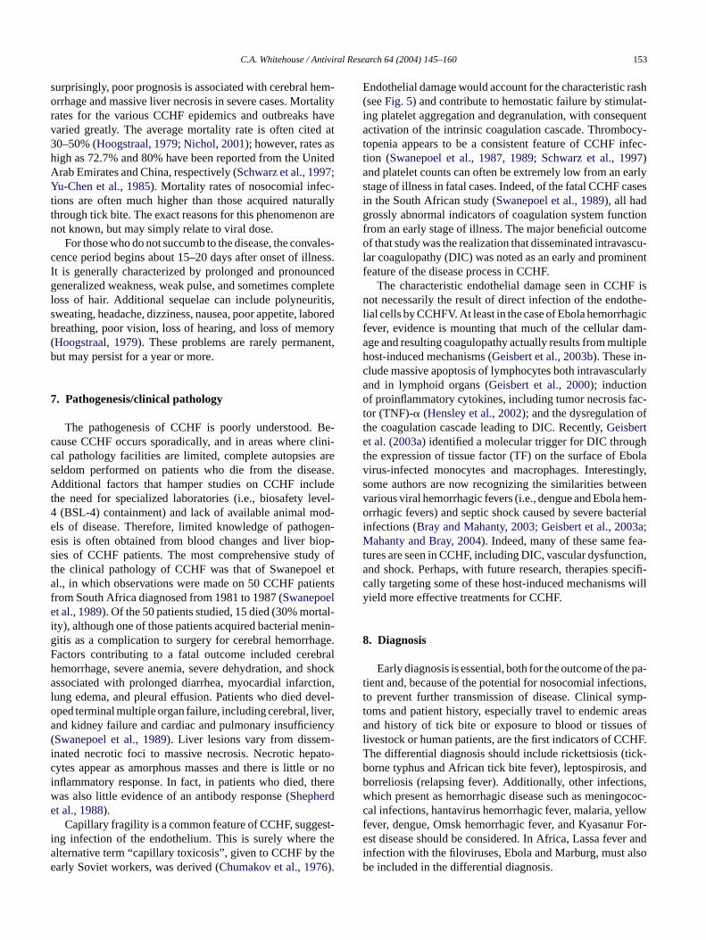
hagic manifestations develop. These can range fromechiae to large areas of ecchymosis and often appeahe mucous membranes and skin, especially on the uody and/or extremities (Fig. 5). Bleeding in the form of me-
ena, hematemesis, and epistaxis is also commonly seeay 4 or 5 and can often be characterized by dark “corounds” vomitus and tar-like stools resulting from intestinemorrhages. Bleeding from other sites including the vagingival bleeding and, in the most severe cases, cerebral hrrhage have been reported (Swanepoel et al., 1987). Not
ig. 5. Massive cutaneous ecchymosis on the arm of a CCHF patient,ays after clinical onset. Photograph courtesy of Dr. Robert Swanepational Institute of Virology, South Africa.
C.A. Whitehouse / Antiviral Research 64 (2004) 145–160 153
surprisingly, poor prognosis is associated with cerebral hem-orrhage and massive liver necrosis in severe cases. Mortalityrates for the various CCHF epidemics and outbreaks havevaried greatly. The average mortality rate is often cited at30–50% (Hoogstraal, 1979; Nichol, 2001); however, rates ashigh as 72.7% and 80% have been reported from the UnitedArab Emirates and China, respectively (Schwarz et al., 1997;Yu-Chen et al., 1985). Mortality rates of nosocomial infec-tions are often much higher than those acquired naturallythrough tick bite. The exact reasons for this phenomenon arenot known, but may simply relate to viral dose.
For those who do not succumb to the disease, the convales-cence period begins about 15–20 days after onset of illness.It is generally characterized by prolonged and pronouncedgeneralized weakness, weak pulse, and sometimes completeloss of hair. Additional sequelae can include polyneuritis,sweating, headache, dizziness, nausea, poor appetite, laboredbreathing, poor vision, loss of hearing, and loss of memory(Hoogstraal, 1979). These problems are rarely permanent,but may persist for a year or more.
7. Pathogenesis/clinical pathology
The pathogenesis of CCHF is poorly understood. Be-c clini-c ares ase.A udet vel-4 od-e gen-e iop-s y oft l eta ientsf le tal-i nin-g age.F bralh shoca tion,l evel-o ver,a ncy( -i ato-c or noi herewe
st-i thea thee
Endothelial damage would account for the characteristic rash(seeFig. 5) and contribute to hemostatic failure by stimulat-ing platelet aggregation and degranulation, with consequentactivation of the intrinsic coagulation cascade. Thrombocy-topenia appears to be a consistent feature of CCHF infec-tion (Swanepoel et al., 1987, 1989; Schwarz et al., 1997)and platelet counts can often be extremely low from an earlystage of illness in fatal cases. Indeed, of the fatal CCHF casesin the South African study (Swanepoel et al., 1989), all hadgrossly abnormal indicators of coagulation system functionfrom an early stage of illness. The major beneficial outcomeof that study was the realization that disseminated intravascu-lar coagulopathy (DIC) was noted as an early and prominentfeature of the disease process in CCHF.
The characteristic endothelial damage seen in CCHF isnot necessarily the result of direct infection of the endothe-lial cells by CCHFV. At least in the case of Ebola hemorrhagicfever, evidence is mounting that much of the cellular dam-age and resulting coagulopathy actually results from multiplehost-induced mechanisms (Geisbert et al., 2003b). These in-clude massive apoptosis of lymphocytes both intravascularlyand in lymphoid organs (Geisbert et al., 2000); inductionof proinflammatory cytokines, including tumor necrosis fac-tor (TNF)-� (Hensley et al., 2002); and the dysregulation ofthe coagulation cascade leading to DIC. Recently,Geisberte ht bolav ingly,s eenv hem-o teriali 3a;M ea-t tion,a ecifi-c s willy
8
e pa-t ions,t mp-t reasa s ofl HF.T ick-b andb ns,w ococ-c llowf For-e r andi lsob
ause CCHF occurs sporadically, and in areas whereal pathology facilities are limited, complete autopsieseldom performed on patients who die from the disedditional factors that hamper studies on CCHF incl
he need for specialized laboratories (i.e., biosafety le(BSL-4) containment) and lack of available animal m
ls of disease. Therefore, limited knowledge of pathosis is often obtained from blood changes and liver bies of CCHF patients. The most comprehensive studhe clinical pathology of CCHF was that of Swanepoel., in which observations were made on 50 CCHF pat
rom South Africa diagnosed from 1981 to 1987 (Swanepoet al., 1989). Of the 50 patients studied, 15 died (30% mor
ty), although one of those patients acquired bacterial meitis as a complication to surgery for cerebral hemorrhactors contributing to a fatal outcome included cereemorrhage, severe anemia, severe dehydration, andssociated with prolonged diarrhea, myocardial infarc
ung edema, and pleural effusion. Patients who died dped terminal multiple organ failure, including cerebral, lind kidney failure and cardiac and pulmonary insufficieSwanepoel et al., 1989). Liver lesions vary from dissemnated necrotic foci to massive necrosis. Necrotic hepytes appear as amorphous masses and there is littlenflammatory response. In fact, in patients who died, tas also little evidence of an antibody response (Shepherdt al., 1988).
Capillary fragility is a common feature of CCHF, suggeng infection of the endothelium. This is surely wherelternative term “capillary toxicosis”, given to CCHF byarly Soviet workers, was derived (Chumakov et al., 1976).
k
t al. (2003a)identified a molecular trigger for DIC throughe expression of tissue factor (TF) on the surface of Eirus-infected monocytes and macrophages. Interestome authors are now recognizing the similarities betwarious viral hemorrhagic fevers (i.e., dengue and Ebolarrhagic fevers) and septic shock caused by severe bac
nfections (Bray and Mahanty, 2003; Geisbert et al., 200ahanty and Bray, 2004). Indeed, many of these same f
ures are seen in CCHF, including DIC, vascular dysfuncnd shock. Perhaps, with future research, therapies spally targeting some of these host-induced mechanismield more effective treatments for CCHF.
. Diagnosis
Early diagnosis is essential, both for the outcome of thient and, because of the potential for nosocomial infecto prevent further transmission of disease. Clinical syoms and patient history, especially travel to endemic and history of tick bite or exposure to blood or tissue
ivestock or human patients, are the first indicators of CChe differential diagnosis should include rickettsiosis (torne typhus and African tick bite fever), leptospirosis,orreliosis (relapsing fever). Additionally, other infectiohich present as hemorrhagic disease such as meningal infections, hantavirus hemorrhagic fever, malaria, yeever, dengue, Omsk hemorrhagic fever, and Kyasanurst disease should be considered. In Africa, Lassa feve
nfection with the filoviruses, Ebola and Marburg, must ae included in the differential diagnosis.

154 C.A. Whitehouse / Antiviral Research 64 (2004) 145–160
8.1. Laboratory diagnosis
8.1.1. Virus isolationAny attempts at isolating and culturing the virus should
only be performed in a maximum biocontainment laboratory(i.e., BSL-4). The traditional method for CCHFV isolationhas been by intracranial (i.c.) or intraperitoneal (i.p.) inocu-lation of a sample (e.g., blood from an acute-phase patient orground tick pools) into newborn mice. Isolation in cell cultureis far simpler and provides a more rapid result, but is generallyconsidered less sensitive (Shepherd et al., 1986) and can gen-erally only detect high concentrations of virus. Nevertheless,virus can be isolated from blood and organ suspensions in awide variety of susceptible cell lines including LLC-MK2,Vero, BHK-21, and SW-13 cells with maximal virus yields(107–108 plaque-forming units/ml) after 4–7 days of incu-bation (Nichol, 2001). Depending on the cell line and strain,the virus may produce little or no cytopathic effect (CPE) anddevelop into a noncytopathic persistent infection of the cells;however, virus can be identified by performing immunoflu-orescence assay (IFA) with specific monoclonal antibodies.Additionally, CPE and the visualization of plaques may occuronly after serial passage of virus (Shepherd et al., 1986).
8.1.2. Immunological assaysfec-
t dif-f ackol cultt werel FA( ofe an-t sa nessae elsb lsob on-s n isc ld org s, orIb nosisi umo l as-s avebi ts
8verse
t videa rve as
the front-line tool in the diagnosis of CCHF, as well as otherviral hemorrhagic fevers (Drosten et al., 2003). The bene-fits of using such assays are many. Because RT-PCR detectsthe genetic material of the virus, and can be designed to behighly specific, it is possible to make a presumptive diag-nosis of CCHF without the need to culture the virus, whichwould require the use of specialized biocontainment labora-tory facilities. Indeed, due to the high sensitivity of RT-PCR,positive results can often be obtained from samples which areculture negative (Schwarz et al., 1996). In addition, the assaycan be applied retrospectively to stored serum samples. Inone such study, viral RNA could be detected in samples up today 16 of illness; whereas, infective virus was progressivelycleared from the serum after the first week of illness (Burtet al., 1998). Another benefit to molecular diagnostic assaysis their rapidity compared to virus culture, often allowing apresumptive diagnosis to be reported within 8 h of receivingthe first specimen (Burt et al., 1998).
Likewise, RT-PCR assays for CCHFV have greatly en-hanced epidemiological studies, for example, being able todetect viral nucleic acid directly from field-collected ticks. Anadded benefit of these techniques is that they allow for molec-ular epidemiology to be performed. Amplified viral comple-mentary DNA (cDNA) can be sequenced and subjected tophylogenetic analysis. Using this approach, the source of aC ined( alv
as-s ssays.T nven-t ate,h vid-i atorsh etect-i ,2 l.,2 ,2 01;Ha V us-i d theD CRpp lvedb Ra B)p obel
9
e-d ndi the
Serologic tests used to study and diagnose CCHFV inion before 1980, such as complement fixation, immunousion, and hemagglutination inhibition, suffered from a lf sensitivity and reproducibility (Hoogstraal, 1979). Simi-
arly, the neutralizing antibody response is weak and diffio demonstrate in CCHF infections. These problemsargely overcome with the introduction of the indirect IZgurskaya and Chumakov, 1977) and the developmentnzyme-linked immunoassays for detecting IgG and IgM
ibodies (Donets et al., 1982). Both IgG and IgM antibodiere detectable by IFA by about 7 days after onset of illnd are present in the sera of survivors by day 9 (Shepherdt al., 1989). The IgM antibody declines to undetectable levy the fourth month after infection, and IgG titers may aegin to decline gradually at this time, but remain demtrable for at least 5 years. Recent or current infectioonfirmed by demonstrating seroconversion, or a fourforeater increase in antibody titer in paired serum sample
gM antibody in a single sample (Swanepoel, 1995). An anti-ody response is rarely detectable in fatal cases and diag
s usually confirmed by isolation of the virus from the serr liver biopsy specimens. Recently, new immunologicaays incorporating recombinant CCHFV nucleoprotein heen developed and used in an IFA (Saijo et al., 2002b) or
n an ELISA (Saijo et al., 2002a; Tang et al., 2003) to detecerum antibodies from infected patients.
.1.3. Molecular diagnostic assaysMolecular-based diagnostic assays, such as the re
ranscription-polymerase chain reaction (RT-PCR), prouseful complement to serodiagnosis and now often se
CHF outbreak in the United Arab Emirates was determRodriguez et al., 1997) and phylogenetically distinct virariants were identified (Schwarz et al., 1996).
A further improvement on the conventional RT-PCRay has been the development of automated real-time ahe real-time PCR assay has many advantages over co
ional RT-PCR methods, including lower contamination righer sensitivity and specificity, and they are rapid, pro
ng results in minutes instead of hours. Several investigave reported the use of real-time PCR assays for d
ng some viral causes of hemorrhagic fevers (Drosten et al.002a), including Ebola (Gibb et al., 2001; Towner et a004; Weidmann et al., 2004), Rift Valley fever (Garcia et al.001), and dengue (Laue et al., 1999; Callahan et al., 20oung et al., 2000) viruses.Drosten et al. (2002a)developedone-step real-time RT-PCR assay for detecting CCHF
ng primers to the nucleoprotein gene; however, they useNA-intercalating dye, SybrGreen I, for detecting the Product because no conserved binding site for a 5′-nucleaserobe could be found. This problem has been partially soyGarrison et al. (2003), who developed a real-time RT-PCssay using TaqMan-minor groove binding protein (MGrobe, allowing for greater specificity with a shorter pr
ength.
. Treatment
Treatment options for CCHF are limited. Early remies included giving rutin (a bioflavonoid compound fou
n buckwheat), ascorbic acid, and calcium chloride for
C.A. Whitehouse / Antiviral Research 64 (2004) 145–160 155
treatment of the hemorrhagic syndrome. It was also suggestedthat with extensive blood loss, transfusions and blood substi-tutes such as polyglutin, plasma, and hemodes were neces-sary and intravenous injections of gelatin and aminocaproicacid were also indicated. Much emphasis was also placedon preventing reinfection, including the necessity of remov-ing blood crusts from the oral cavity, brushing the teethregularly, and painting with Vaseline any sores on the lipsor tongue. There was an early recognition of the possi-ble benefits of treatments using serum prepared from theblood of recovered CCHF patients or gammaglobulin ob-tained from immunization of horses (Hoogstraal, 1979). Inmore recent times, immunotherapy was attempted via passivetransfer of CCHF survivor convalescent plasma (Vasilenkoet al., 1990). Although seven patients with severe CCHFwho received immune plasma recovered, this was an un-controlled experiment, and firm evidence of its value islacking. There is currently no specific antiviral therapy forCCHF approved for use in humans by the FDA. However,the antiviral drug, ribavirin, has shown the most promiseover the years. It has been shown to inhibit in vitro vi-ral replication in Vero cells (Watts et al., 1989a,b) and re-duce the mean-time-to-death in a suckling mouse model ofCCHF (Tignor and Hanham, 1993). Additionally, severalcase reports have been published that suggest oral or intra-v ns( t al.,2 so-c r 10d .,1 ffi-c edC d,c con-fi er rror-i ffortsb ningp FV( theb ned,w le in-hA tiono on,i rei n su-p ivitya sesa ,A ini xAc onso tiono in-f
clearly a critical need to identify new effective treatments forthis disease, and if the past few years are any example, as morebasic research is conducted on this virus, novel approachesto its control will surely evolve.
10. Prevention and control
10.1. Risk factors
There are several groups of individuals who are consideredto be at-risk of contracting CCHFV. Specifically, people fromendemic areas who are susceptible to tick bite, particularlyfrom Hyalommaspp. ticks. These would include individu-als who work outdoors, particularly those who work withlarge domestic animals. Although CCHFV has been isolatedfrom numerous species of ticks (see Section5), those of theHyalommagenus are considered the primary vector in CCHFenzootic and endemic areas. The distribution of CCHFV co-incides precisely with the distribution ofHyalommaticks(Hoogstraal, 1956); therefore, there appears to be little or norisk in areas outside the known distribution of these ticks.Exposures such as crushing infected ticks and butchering in-fected animals have also been a frequent source of CCHFVinfection. Other groups who are at-risk include those caringf ioni ex-t d ofd 95;P in-c al inP al,1 icalC erd’sf ital-i eatedb thes ands,s , butu f hish hileo geonw ctedC singa daysl ncedb therp o be-c hos-p withC ess.A os-p rk-e edlep otherc
enous ribavirin is effective for treating CCHFV infectioFisher-Hoch et al., 1995; Papa et al., 2002a; Mardani e003; Tang et al., 2003). For example, in Pakistan, three noomial cases of CCHF were treated with oral ribavirin foays, and they made a complete recovery (Fisher-Hoch et al995). More recently, in a large cohort study in Iran, the eacy of oral ribavirin was 80% among patients with confirmCHF (Mardani et al., 2003). But, to date, no randomizeontrolled studies have been performed to rigorouslyrm the efficacy of ribavirin for treating CCHF. With thecent interest in CCHFV as a potential agent of biotesm/biowarfare, there have been increased research ey several groups. In particular, rapid methods of screeotential antiviral compounds are being applied to CCHParagas et al., 2004), as well as increased knowledge ofasic biology of the virus and its disease is being gaihich may lead to improved therapies, such as possibibitors of the viral protein processing (Pullikotil et al., 2004).nother area of interest with promise is the identificaf interferon-induced proteins that inhibit viral replicati
n particular, the Mx family of proteins. Mx proteins anterferon-induced GTPases that belong to the dynamierfamily of large GTPases, which possess antiviral actgainst a wide range of RNA viruses, including bunyavirund orthomyxoviruses (Haller and Kochs, 2002). Recentlyndersson et al. (2004a,b)showed that human MxA prote
nhibits replication of CCHFV. They demonstrated that Mo-localizes with the NP of CCHFV in the perinuclear regif infected cells and that this interaction prevents replicaf viral RNA and thereby inhibits the production of new
ectious viral particles (Andersson et al., 2004a,b). There is
or CCHF patients. In fact, the risk of nosocomial infectn health-care workers is well documented and can beremely high, especially during the hemorrhagic perioisease (Van Eeden et al., 1985; Fisher-Hoch et al., 19apa et al., 2002a). This is exemplified by a nosocomialident that occurred in the Central Government Hospitakistan in January 1976 (Burney et al., 1980; Hoogstra979). A shepherd was brought to the hospital with typCHF symptoms and died the same night. The sheph
ather, who cared for his sick son at home, was hospzed and died 2 days later despite intensive care and replood transfusions. A female physician, who admittedhepherd, when he vomited blood onto her face and hhowed signs of CCHFV infection and was hospitalizedltimately recovered. The boy had surgery on the day oospital admission. The surgeon, who cut his finger wperating, died of CCHF 2 weeks later. An assistant surho pricked his finger during the operation also contraCHF and was hospitalized; he later recovered. A nurttendant who assisted in the operation died of CCHF 3
ater. The anesthesiologist also became ill and experieleeding from the gums, but recovered. Five of seven oersons in the operating theatre during the procedure alsame ill and were hospitalized; all recovered. Of the 12ital personnel attending the shepherd, 10 became illCHF; two died and eight recovered after severe illnnother nosocomial outbreak occurred at Tygerberg Hital in South Africa. Thirty-three percent of medical wors who had contact with patients through accidental nericks developed CCHF and 8.7% contracted disease byontacts with the patients’ blood (van de Wal et al., 1985).

156 C.A. Whitehouse / Antiviral Research 64 (2004) 145–160
Laboratory workers handling viral material are also at highrisk of contracting the disease as evidenced by several cases oflaboratory-acquired CCHF in Africa (Simpson et al., 1967),and several cases in Russia in which aerosol and/or droplet-respiratory route of infection were suspected (Hoogstraal,1979). For these reasons, in the U.S., the Centers for Dis-ease Control and Prevention (CDC) has classified CCHFVas a biosafety level-4 pathogen (Richmond and McKinney,1999).
10.2. Control measures
The best means of preventing disease is to avoid or min-imize exposure to the virus. This can be accomplished ina number of ways. Persons in high-risk occupations (i.e.,slaughterhouse workers, veterinarians, sheep herders, etc.)should take every precaution to avoid exposure to virus-infected ticks or virus-contaminated animal blood or othertissues. For example, wearing gloves and limiting exposureof naked skin to fresh blood and other tissues of animals areeffective practical control measures. Likewise, medical per-sonnel who care for suspected CCHF patients should prac-tice standard barrier-nursing techniques. Tick control maynot always be practical in many regions of the world whereHyalommaticks are most prevalent. However, acaricide treat-ment of livestock in CCHFV endemic areas is effective inr m-m ide[ atedw ites.A odya izet
hasb e andt for-m ands e Nt nt re-sr on-t ctiono y.
1
thef d/orb AIDC ittedf mayb ilityt d asa ualtyw ca-
tion as a Category A or B pathogen. The highly lethal natureof the virus has restricted research to BSL-4 laboratories andhas consequently had limited research investigations.
Acknowledgements
I thank Mike Bray (NIH) for many useful discussionsregarding this work. I also thank Katheryn Kenyon (US-AMRIID), Aysegul Nalca (Southern Research Institute), andMike Bray (NIH) for critically reading the manuscript andproviding useful editorial suggestions. Many thanks go toChristopher Mores (University of Florida) for helpful dis-cussions regarding phylogenetic analyses and to ConnieSchmaljohn (USAMRIID) for supplyingFigs. 1 and 2. Addi-tionally, the author is indebted to members of the NAMRU-3(Cario, Egypt) medical library for providing English trans-lations for many of the original Russian works used in thisreview.
References
Andersson, I., Bladh, L., Mousavi-Jazi, M., Magnusson, K.-E., Lundkvist,A., Haller, O., Mirazimi, A., 2004a. Human MxA protein inhibits the
78,
A A.,ingin to3.
B biq-331.
B .N.,mor-tits.
B 1b.virused.2).
B . In:p.
B .M.,ole,burg,M.,
, P.,ons:287,
B hock.
B 976.ean
Hyg.
B of aon ofagic
educing the population of infected ticks. Applying coercially available insect repellents (i.e., diethyl toluam
DEET]) to exposed skin and the use of clothing impregnith permethrin can give some protection against tick bs with other tick-borne diseases, inspection of one’s bnd clothes for ticks, and their prompt removal can minim
he risk of infection.A suckling mouse brain, formalin-inactivated vaccine
een used in Bulgaria and other parts of Eastern Europhe former Soviet Union. In the Rostov region of theer Soviet Union, 1500 persons received the vaccine
howed a high frequency of detectable antibody by thest (Tkachenko et al., 1971). Likewise, vaccine was giveo several hundred human volunteers in Bulgaria, withulting high antibody induction (Vasilenko, 1973). With theelatively small target population of persons at-risk for cracting CCHFV, the large-scale development and produf a CCHF vaccine by modern standards seems unlikel
1. Potential bioterrorism concerns
The highly pathogenic nature of the CCHFV has led toear that it might be used as an agent of bioterrorism aniowarfare and has resulted in its inclusion as a CDC/NIategory C Priority Pathogen. CCHFV can be transm
rom person to person, has a high case-fatality rate, ande transmissible by small-particle aerosol; but, its inab
o replicate to high concentrations in cell culture is citemajor impediment to its development as a mass caseapon (Borio et al., 2002), and thus precludes its classifi
replication of Crimean–Congo hemorrhagic fever virus. J. Virol.4323–4329.
ndersson, I., Simon, M., Lundkvist, A., Nilsson, M., Holmstrom,Elgh, F., Mirazimi, A., 2004b. Role of actin filaments in targetof Crimean–Congo hemorrhagic fever virus nucleocapsid proteperinuclear regions of mammalian cells. J. Med. Virol. 72, 83–9
alakirev, M.Y., Jaquinod, M., Haas, A.L., Chroboczek, J., 2002. Deuuitinating function of adenovirus proteinase. J. Virol. 76, 6323–6
erezin, V.V., Chumakov, M.P., Reshetnikov, I.A., Zgurskaya, G1971a. Study of the role of birds in the ecology of Crimean herhagic fever virus. Mater. 6 Simp. Izuch. Virus Ekol. Svyazan. P(Omsk, 1971), 94–95 (in Russian; in English, NAMRU3-T721).
erezin, V.V., Chumakov, M.P., Stolbov, D.N., Butenko, A.M., 197On the problem of natural hosts of Crimean hemorrhagic feverin Astrakhan region. Tr. Inst. Polio Virusn. Entsefalitov Akad. MNauk SSSR 19, 210–216 (in Russian; in English, NAMRU3-T91
ishop, D.H., 1996. Biology and molecular biology of bunyavirusesElliott, R.M. (Ed.), TheBunyaviridae. Plenum Press, New York, p19–61.
orio, L., Inglesby, T., Peters, C.J., Schmaljohn, A.L., Hughes, JJahrling, P.B., Ksiazek, T., Johnson, K.M., Meyerhoff, A., O’ToT., Ascher, M.S., Bartlett, J., Breman, J.G., Eitzen Jr., E.M., HamM., Hauer, J., Henderson, D.A., Johnson, R.T., Kwik, G., Layton,Lillibridge, S., Nabel, G.J., Osterholm, M.T., Perl, T.M., RussellTonat, K., 2002. Hemorrhagic fever viruses as biological weapmedical and public health management. J. Am. Med. Assoc.2391–2405.
ray, M., Mahanty, S., 2003. Ebola hemorrhagic fever and septic sJ. Infect. Dis. 188, 1613–1617.
urney, M.I., Ghafoor, A., Saleen, M., Webb, P.A., Casals, J., 1Nosocomial outbreak of viral hemorrhagic fever caused by Crimhemorrhagic fever–Congo virus in Pakistan. Am. J. Trop. Med.29, 941–947.
urt, F.J., Leman, P.A., Smith, J.F., Swanepoel, R., 1998. The usereverse transcription-polymerase chain reaction for the detectiviral nucleic acid in the diagnosis of Crimean–Congo haemorrhfever. J. Virol. Methods 70, 129–137.

C.A. Whitehouse / Antiviral Research 64 (2004) 145–160 157
Burt, F.J., Spencer, D.C., Leman, P.A., Patterson, B., Swanepoel, R., 1996.Investigation of tick-borne viruses as pathogens of humans in SouthAfrica and evidence of Dugbe virus infection in a patient with pro-longed thrombocytopenia. Epidemiol. Infect. 116, 353–361.
Butenko, A.M., Chumakov, M.P., Bashkirtsev, V.N., Zavodova, T.I.,Tkachenko, E.A., Rubin, S.G., Stolbov, D.N., 1968. Isolation and in-vestigation of Astrakhan strain (“Drozdov”) of Crimean hemorrhagicfever virus and data on serodiagnosis of this infection. Mater. 15Nauchn. Sess. Inst. Polio Virus Entsefalitov (Moscow) 3, 88–90 (inRussian; in English, NAMRU3-T866).
Callahan, J.D., Wu, S.J., Dion-Schultz, A., Mangold, B.E., Peruski, L.F.,Watts, D.M., Porter, K.R., Murpgy, G.R., Suharyono, W., King, C.C.,Hayes, C.G., Temenak, J.J., 2001. Development and evaluation ofserotype- and group-specific fluorogenic reverse transcriptase PCR(TaqMan) assays for dengue virus. J. Clin. Microbiol. 39, 4119–4124.
Casals, J., 1969. Antigenic similarity between the virus causing Crimeanhemorrhagic fever and Congo virus. Proc. Soc. Exp. Biol. Med. 131,233–236.
Casals, J., Henderson, B.F., Hoogstraal, H., Johnson, K.M., Shelokov, A.,1969. A review of Soviet viral hemorrhagic fevers. J. Infect. Dis. 122,437–453.
Causey, O.R., Kemp, G.E., Madbouly, M.H., David-West, T.S., 1970.Congo virus from domestic livestock, African hedgehogs, and arthro-pods in Nigeria. Am. J. Trop. Med. Hyg. 19, 846–850.
Chumakov, M.P., 1945. A new tick-borne virus disease—Crimean hem-orrhagic fever. In: Sokolov, A.A., Chumakov, M.P., Kolachev, A.A.(Eds.), Crimean Hemorrhagic Fever (Acute Infectious Capillary Tox-icosis). Izd. Otd. Primorskoi Armii, Simferopol, Moscow, pp. 13–45(in Russian).
Chumakov, M.P., 1965. A short study of the investigation of the virus ofkad.U3-
C fever.
C andsn.lish,
C d thenat-
ries.irsk,).
C hem-auk
C .I.,.G.,.V.,mor-
ish,
C L.I.,Pak,evers on
C c re-feveress.
; in
C ller,n-ce93.
Dickson, D.L., Turell, M.J., 1992. Replication and tissue tropisms ofCrimean–Congo hemorrhagic fever virus in experimentally infectedadult Hyalomma truncatum(Acari: Ixodidae). J. Med. Entomol. 29,767–773.
Dohm, D.J., Logan, T.M., Linthicum, K.J., Rossi, C.A., Turell, M.J.,1996. Transmission of Crimean–Congo hemorrhagic fever virus byHyalomma impeltatum(Acari: Ixodidae) after experimental infection.J. Med. Entomol. 33, 848–851.
Dols, M.W., 1977. The Black Death in the Middle East. Princeton Uni-versity Press, Princeton, NJ, 390 pp.
Donets, M.A., Chumakov, M.P., Korolev, M.B., Rubin, S.G., 1977.Physicochemical characteristics, morphology and morphogenesis ofvirions of the causative agent of Crimean hemorrhagic fever. Intervi-ology 8, 294–308.
Donets, M.A., Rezapkin, G.V., Ivanov, A.P., Tkachenko, E.A., 1982. Im-munosorbent assays for diagnosis of Crimean–Congo haemorrhagicfever (CCHF). Am. J. Trop. Med. Hyg. 31, 156–162.
Drosten, C., Gottig, S., Schilling, S., Asper, M., Panning, M., Schmitz,H., Gunther, S., 2002a. Rapid detection and quantification of RNA ofEbola and Marburg viruses, Lassa virus, Crimean–Congo hemorrhagicfever virus, Rift Valley fever virus, dengue virus, and yellow fevervirus by real-time reverse transcription-PCR. J. Clin. Microbiol. 40,2323–2330.
Drosten, C., Kummerer, B.M., Schmitz, H., Gunther, S., 2003. Moleculardiagnostics of viral hemorrhagic fevers. Antiviral Res. 57, 61–87.
Drosten, C., Minnak, D., Emmerich, P., Schmitz, H., Reinicke, T., 2002b.Crimean–Congo hemorrhagic fever in Kosovo. J. Clin. Microbiol. 40,1122–1123.
Durden, L.A., Logan, T.M., Wilson, M.L., Linthicum, K.J., 1993. Ex-perimental vector incompetence of a soft tick,Ornithodoros sonrai
s. J.
E .
E .T.,000..,
loff,s.),Tax-1.
E , S.,feverl.
F Mc-with
F s for6.
F Uuku-ion
G uloy,lleynds.
G less,MGBhagice on
G .K.,ring6.
G ens-lities
Crimean hemorrhagic fever. Tr. Inst. Polio Virusn. Entsefalitov AMed. Nauk SSSR 7, 193–196 (in Russian; in English, NAMRT189).
humakov, M.P., 1947. A new virus disease—Crimean hemorrhagicNov. Med. 4, 9–11 (in Russian; in English, NAMRU3-T900).
humakov, M.P., 1971. Some results of investigation of the etiologyimmunology of Crimean hemorrhagic fever. Tr. Inst. Polio ViruEntsefalitov Akad. Med. Nauk SSSR 19, 7 (in Russian; in EngNAMRU3-T953).
humakov, M.P., 1972. Investigations of arboviruses in the USSR anquestion of possible association through migratory birds betweenural arbovirus infection foci in the USSR and warm-climate countIn: Mater. 5 Izuch. Roli Pereletn. Ptits. Rasp. Arbovirus, NovosibJuly 1969, pp. 133–138 (in Russian; in English, NAMRU3-T876
humakov, M.P., 1974. On 30 years of investigation of Crimeanorrhagic fever. Tr. Inst. Polio Virusn. Entsefalitov Akad. Med. NSSSR 22, 5–18 (in Russian; in English, NAMRU3-T950).
humakov, M.P., Butenko, A.M., Shalunova, N.V., Mart’yanova, LSmirnova, W.E., Bashkirtsev, V.N., Zavodova, T.I., Rubin, STkachenko, E.A., Karmy-Sheva, V.Ya, Reingold, V.N., Popov, GSavinov, A.P., 1968. New data on the virus causing Crimean herhagic fever (CHF). Vopr. Virusol. 13, 377 (in Russian; in EnglNAMRU3-T596).
humakov, M.P., Smirnova, S.E., Shalunova, N.Y., Mart’yanova,Fleer, G.P., Zgurskaya, G.N., Maksumov, S.S., Kasymov, K.Y.,T.P., 1976. Proofs of etiological identity to Crimean hemorrhagic fin Central Asian hemorrhagic fever. In: IX International CongresTropical Medicine and Malaria, Athens, vol. 1, pp. 33–34.
humakov, M.P., Smirnova, S.E., Tkachenko, E.A., 1969. Antigenilationships between the Soviet strains of Crimean hemorrhagicvirus and the Afro-Asian Congo virus strains. Mater. 16 Nauchn. SInst. Polio Virus Entsefalitov (Moscow) 2, 152–154 (in RussianEnglish, NAMRU3-T614).
lerex-van Haaster, C.M., Clerex, J.P., Ushijima, H., Akashi, H., FuF., Bishop, D.H., 1982. The 3′ terminal RNA sequences of buyaviruses and nairoviruses (Bunyaviridae): evidence of end sequengeneric differences within the virus family. J. Gen. Virol. 61, 289–2
(Acari: Argasidae), for Crimean–Congo hemorrhagic fever viruMed. Entomol. 30, 493–496.
lliott, R.M., 1990. Molecular biology of theBunyaviridae. J. Gen. Virol71, 501–522.
lliott, R.M., Bouloy, M., Calisher, C.H., Goldbach, R., Moyer, JNichol, S.T., Pettersson, R., Plyusnin, A., Schmaljohn, C.S., 2Family Bunyaviridae. In: van Regenmortel, M.H.V., Fauquet, C.MBishop, D.H.L., Carstens, E.B., Estes, M.K., Lemon, S., ManiJ., Mayo, M.A., McGeogch, D., Pringle, C.R., Wickner, R.B. (EdVirus Taxonomy. Seventh Report International Committee for theonomy of Viruses. Academic Press, San Diego, CA, pp. 599–62
llis, D.S., Southee, T., Lloyd, G., Platt, G.S., Jones, N., StamfordBowen, E.T., Simpson, D.I., 1981. Congo/Crimean haemorrhagicvirus from Iraq, 1979. I. Morphology in BHK 21 cells. Arch. Viro70, 189–198.
isher-Hoch, S.P., Khan, J.A., Rehman, S., Mirza, S., Khurshid, M.,Cormick, J.B., 1995. Crimean–Congo haemorrhagic fever treatedoral ribavirin. Lancet 346, 472–475.
lick, R., Flick, K., Feldmann, H., Elgh, F., 2003. Reverse geneticCrimean–Congo hemorrhagic fever virus. J. Virol. 77, 5997–600
lick, R., Pettersson, R.F., 2001. Reverse genetics system forniemi virus (Bunyaviridae): RNA polymerase I-catalysed expressof chimeric viral RNAs. J. Virol. 75, 1643–1655.
arcia, S., Crance, J.M., Billecocq, A., Peinnequin, A., Jouan, A., BoM., Garin, D., 2001. Quantitative real-time detection of Rift Vafever virus and its application to evaluation of antiviral compouJ. Clin. Microbiol. 39, 4456–4461.
arrison, A.R., Kulesh, D., Norwood, D., Whitehouse, C.A., LoveB., Endy, T.P., Paragas, J., 2003. Development of a TaqMan-assay for detection and quantitation of Crimean–Congo hemorrfever virus. In: Proceedings of the 12th International ConferencNegative Strand Viruses, Pisa, Italy.
eisbert, T.W., Hensley, L.E., Gibb, T.R., Steele, K.E., Jaax, NJahrling, P.B., 2000. Apoptosis induced in vitro and in vivo duinfection by Ebola and Marburg viruses. Lab. Invest. 80, 171–18
eisbert, T.W., Young, H.A., Jahrling, P.B., Davis, K.J., Kagan, E., Hley, L.E., 2003a. Mechanisms underlying coagulation abnorma

158 C.A. Whitehouse / Antiviral Research 64 (2004) 145–160
in Ebola hemorrhagic fever: overexpression of tissue factor in pri-mate monocyte/macrophages is a key event. J. Infect. Dis. 188, 1618–1629.
Geisbert, T.W., Young, H.A., Jahrling, P.B., Davis, K.J., Larson, T., Ka-gan, E., Hensley, L.E., 2003b. Pathogenesis of Ebola hemorrhagicfever in primate models: evidence that hemorrhage is not a directeffect of virus-induced cytolysis of endothelial cells. Am. J. Pathol.163, 2371–2382.
Gibb, T.R., Norwood, D.A., Woollen, N., Henchal, E.A., 2001. Develop-ment and evaluation of a fluorogenic 5′ nuclease assay to detect anddifferentiate between Ebola virus subtypes Zaire and Sudan. J. Clin.Microbiol. 39, 4125–4130.
Gonzalez, J.P., Camicas, J.L., Cornet, J.P., Faye, O., Wilson, M.L., 1992.Sexual and transovarian transmission of Crimean–Congo haemor-rhagic fever virus inHyalomma truncatumticks. Res. Virol. 143,23–28.
Gonzalez, J.P., Wilson, M.L., Cornet, J.P., Camicas, J.L., 1995. Host-passage-induced phenotypic changes in Crimean–Congo haemorrhagicfever virus. Res. Virol. 146, 131–140.
Gordon, S.W., Linthicum, K.J., Moulton, J.R., 1993. Transmission ofCrimean–Congo hemorrhagic fever virus in two species ofHyalommaticks, from infected adults to cofeeding immature forms. Am. J. Trop.Med. Hyg. 48, 576–580.
Haller, O., Kochs, G., 2002. Interferon-induced Mx proteins: dynamin-likeGTPases with antiviral activity. Traffic 3, 710–717.
Hensley, L.E., Young, H.A., Jahrling, P.B., Geisbert, T.W., 2002. Proin-flammatory response during Ebola virus infection of primate models:possible involvement of the tumor necrosis factor receptor superfam-ily. Immunol. Lett. 80, 169–179.
Hewlett, M.J., Pettersson, R.F., Baltimore, D., 1977. Circular forms of21,
H hem-tein.
H l. I..
H ongo. 15,
H ction
K M.,ent,04.. 10,
K R.,rhagic
L virussing37,
L 001.pro-. 98,
L em-gicalNew
L R.,hagic.
Mahanty, S., Bray, M., 2004. Pathogenesis of filoviral hemorrhagic fevers.Lancet Infect. Dis. 4, 487–498.
Makarova, K.S., Aravind, L., Koonin, E.V., 2000. A novel superfamily ofpredicted cysteine proteases from eukaryotes, viruses andChlamydiapneumoniae. TIBS 25, 50–52.
Mardani, M., Jahromi, M.K., Naieni, K.H., Zeinali, M., 2003. The efficacyof oral ribavirin in the treatment of Crimean–Congo hemorrhagic feverin Iran. Clin. Infect. Dis. 36, 1613–1618.
Marriott, A.C., Nuttall, P.A., 1992. Comparison of the S RNA segmentsand nucleoprotein sequences of Crimean–Congo hemorrhagic fever,Hazara, and Dugbe viruses. Virology 189, 795–799.
Marriott, A.C., Nuttall, P.A., 1996a. Molecular biology of nairoviruses.In: Elliott, R.M. (Ed.), TheBunyaviridae. Plenum Press, New York,pp. 91–104.
Marriott, A.C., Nuttall, P.A., 1996b. Large RNA segment of Dugbenairovirus encodes the putative RNA polymerase. J. Gen. Virol. 77,1775–1780.
Martin, M.L., Lindsey-Regnery, H., Sasso, D.R., McCormick, J.B.,Palmer, E., 1985. Distinction betweenBunyaviridaegenera by sur-face structure and comparison with Hantaan virus using negative stainelectron microscopy. Arch. Virol. 86, 17–28.
Murphy, F.A., Harrison, A.K., Tzianabos, T., 1968. Electron microscopicobservations of mouse brain infected with Bunyamwera group ar-boviruses. J. Virol. 2, 1315–1325.
Murphy, F.A., Harrison, A.K., Whitfield, S.G., 1973.Bunyaviridae: mor-phologic and morphogenetic similarities of Bunyamwera serologicalsupergroup viruses and several other arthropod-borne viruses. Intervi-rology 1, 297–316.
Nichol, S.T., 2001. Bunyaviruses. In: Knipe, D.M., Howley, P.M. (Eds.),Fields Virology, vol. 1, fourth ed. Lippincott, Williams & Wilkins,
O f the
et.
P a,tan.–94
P yev,.I.,ts ofSSRSR
P avli-A.,nia.
P nto-ongo. 8,
P 02c.o53.
P assayagic
P entS1P.
R andffice.
R .S.,C.J.,reak
Uukuniemi virion RNA: an electron microscopic study. J. Virol.1085–1093.
onig, J.E., Osborne, J.C., Nichol, S.T., 2004. Crimean–Congoorrhagic fever virus genome L RNA segment and encoded proVirology 321, 29–35.
oogstraal, H., 1956. African Ixodoidea: Ticks of the Sudan, voNAMRU3 Res. Rep. Nm. 005 050.29.07, Cairo, Egypt, 1101 pp
oogstraal, H., 1979. The epidemiology of tick-borne Crimean–Chemorrhagic fever in Asia, Europe, and Africa. J. Med. Entomol307–417.
oung, H.H., Hritz, D., Kanesa-Thasan, N., 2000. Quantitative deteof dengue 2 virus using fluorogenic RT-PCR based on 3′-noncodingsequence. J. Virol. Methods 86, 1–11.
arti, S., Odabasi, S., Korten, Z., Yilmaz, V., Sonmez, M., Caylan,Akdogan, R., Eren, E., Koksal, N., Ovali, I., Erickson, E.B., VincM.J., Nichol, S.T., Comer, J.A., Rollin, P.E., Ksiazek, T.G., 20Crimean–Congo hemorrhagic fever in Turkey. Emerg. Infect. Dis1379–1384.
insella, E., Martin, S.G., Grolla, A., Czub, M., Feldmann, H., Flick,2004. Sequence determination of the Crimean–Congo hemorfever virus L segment. Virol. 321, 23–28.
aue, T., Emmerich, P., Schmitz, H., 1999. Detection of dengueRNA in patients after primary or secondary dengue infection by uthe TaqMan automated amplification system. J. Clin. Microbiol.2543–2547.
enz, O., ter Meulen, J., Klenk, H.-D., Seidah, N.G., Garten, W., 2The Lassa virus glycoprotein precursor GP-C is proteolyticallycessed by subtilase SKI-1/S1P. Proc. Natl. Acad. Sci. U.S.A12701–12705.
inthicum, K.J., Bailey, C.L., 1994. Ecology of Crimean–Congo horrhagic fever. In: Sonenshine, D.E., Mather, T.N. (Eds.), EcoloDynamics of Tick-Borne Zoonoses. Oxford University Press,York, pp. 392–437.
ogan, T.M., Linthicum, K.J., Bailey, C.L., Watts, D.M., Moulton, J.1989. Experimental transmission of Crimean–Congo hemorrfever virus byHyalomma truncatumKoch. Am. J. Trop. Med. Hyg40, 207–212.
Philadelphia, pp. 1603–1633.korie, T.G., 1991. Comparative studies on the vector capacity o
different stages ofAmbloyomma variegatumFabricius andHyalommarufipes Koch for Congo virus, after intracoelomic inoculation. VParasitol. 38, 215–223.
ak, T.P., Daniyarov, O.A., Kostyukov, M.A., Bulcyhev, V.P., KuimA.U., 1974. Ecology of Crimean hemorrhagic fever in TadzhikisMater. Resp. Simp. Kamenyuki ‘Belovezh Puscha’ (Minsk), 93(in Russian; in English, NAMRU3-T968).
ak, T.P., Smirnova, S.E., Zgurskaya, G.N., Yasinsky, A.V., BerdK.B., Apostoli, L.A., Karovkin, V.P., Feldman, E.M., Derlyatko, KGolovko, E.N., Makhmudov, R.K., Chumakov, M.P., 1971. Resula serological survey of Crimean hemorrhagic fever in Tadzhikin 1969. Tr. Inst. Polio Virusn. Entsefalitov Akad. Med. Nauk SS19, 72–79 (in Russian; in English, NAMRU3-T936).
apa, A., Bino, S., Llagami, A., Brahimaj, B., Papadimitriou, E., Pdou, V., Velo, E., Cahani, G., Hajdini, M., Pilaca, A., Harxhi,Antoniadis, A., 2001. Crimean–Congo hemorrhagic fever in AlbaEur. J. Clin. Microbiol. Infect. Dis. 21, 603–606.
apa, A., Bozovi, B., Pavlidou, V., Papadimitriou, E., Pelemis, M., Aniadis, A., 2002b. Genetic detection and isolation of Crimean–Chemorrhagic fever virus, Kosovo, Yugoslavia. Emerg. Infect. Dis852–854.
apa, A., Ma, B., Kouidou, S., Tang, Q., Hang, C., Antoniadis, A., 20Genetic characterization of the M RNA segment ofCrimean–Conghemorrhagic fever virusstrains, China. Emerg. Infect. Dis. 8, 50–
aragas, J., Whitehouse, C.A., Endy, T.P., Bray, M., 2004. A simplefor determining antiviral activity against Crimean–Congo hemorrhfever virus. Antiviral Res. 62, 21–25.
ullikotil, P., Vincent, M., Nichol, S.T., Seidah, N.G., 2004. Developmof protein-based inhibitors of the proprotein of convertase SKI-1/J. Biol. Chem. 279, 17338–17347.
ichmond, J.Y., McKinney, R.W., 1999. Biosafety in MicrobiologicalBiomedical Laboratories, fourth ed., U.S. Government Printing O
odriguez, L.L., Maupin, G.O., Ksiazek, T.G., Rollin, P.E., Khan, ASchwarz, T.F., Lofts, R.S., Smith, J.F., Noor, A.M., Peters,Nichol, S.T., 1997. Molecular investigation of a multisource outb

C.A. Whitehouse / Antiviral Research 64 (2004) 145–160 159
of Crimean–Congo hemorrhagic fever in the United Arab Emirates.Am. J. Trop. Med. Hyg. 57, 512–518.
Saijo, M., Qing, T., Niikura, M., Maeda, A., Ikegami, T., Prehaud, C.,Kurane, I., Morikawa, S., 2002a. Recombinant nucleoprotein-basedenzyme-linked immunosorbent assay for detection of immunoglobinG antibodies to Crimean–Congo hemorrhagic fever virus. J. Clin.Microbiol. 40, 1587–1591.
Saijo, M., Qing, T., Niikura, M., Maeda, A., Ikegami, T., Sakai, K.,Prehaud, C., Kurane, I., Morikawa, S., 2002b. Immunofluorescencetechnique using HeLa cells expressing recombinant nucleoprotein fordetection of immunoglobulin G antibodies to Crimean–Congo hem-orrhagic fever virus. J. Clin. Microbiol. 40, 372–375.
Saluzzo, J.F., Digoutte, J.P., Camicas, J.L., Chauvancy, G., 1985.Crimean–Congo haemorrhagic fever and Rift Valley fever in south-eastern Mauritania. Lancet 1, 116.
Sanchez, A.J., Vincent, M.J., Nichol, S.T., 2002. Characterization of theglycoproteins of Crimean–Congo hemorrhagic fever virus. J. Virol.76, 7263–7275.
Schmaljohn, C.S, Hooper, J.W., 2001.Bunyaviridae: the viruses and theirreplication. In: Knipe, D.M., Howley, P.M. (Eds.), Fields Virology,vol. 1, fourth ed. Lippincott, Williams & Wilkins, Philadelphia, pp.1581–1602.
Schwarz, T.F., Nsanze, H., Ameen, A.M., 1997. Clinical features ofCrimean–Congo haemorrhagic fever in the United Arab Emirates. In-fection 25, 364–367.
Schwarz, T.F., Nsanze, H., Longson, M., Nitschko, H., Gilch, S., Shurie,H., Ameen, A., Zahir, A.R., Acharya, U.G., Jager, G., 1996. Poly-merase chain reaction for diagnosis and identification of distinct vari-ants of Crimean–Congo hemorrhagic fever virus in the United ArabEmirates. Am. J. Trop. Med. Hyg. 55, 190–196.
S nse inl. 4),
S xperi-ongorop.
S . Com-em-
S . Fieldfever.
S ee, O.,fever
S earle,s in36,
S O.I.,.I.,char-agics 28,
S in-nde-tes.
S Ed.),5–
S J.H.,mor-
Swanepoel, R., Leman, P.A., Burt, F.J., Jardine, J., Verwoerd, D.J., Ca-pua, I., Bruckner, G.K., Burger, W.P., 1998. Experimental infection ofostriches with Crimean–Congo haemorrhagic fever virus. Epidemiol.Infect. 121, 427–432.
Swanepoel, R., Shepherd, A.J., Leman, P.A., Shepherd, S.P., McGillivray,G.M., Erasmus, M.J., Searle, L.A., Gill, D.E., 1987. Epi-demiologic and clinical features of Crimean–Congo hemorrhagicfever in southern Africa. Am. J. Trop. Med. Hyg. 36, 120–132.
Tang, Q., Saijo, M., Zhang, Y., Asiguma, M., Tianshu, D., Han, L., Shi-mayi, B., Maeda, A., Kurane, I., Morikawa, S., 2003. A patient withCrimean–Congo hemorrhagic fever serologically diagnosed by recom-binant nucleoprotein-based antibody detection systems. Clin. Diagn.Lab. Immunol. 10, 489–491.
Tignor, G.H., Hanham, C.A., 1993. Ribavirin efficacy in an in vivo modelof Crimean–Congo hemorrhagic fever virus (CCHF) infection. Antivi-ral Res. 22, 309–325.
Tignor, G.H., Smith, A.L., Casals, J., Ezeokoli, C.D., Okoli, J., 1980.Close relationships of Crimean hemorrhagic fever–Congo (CHF-C)virus strains by neutralizing antibody assays. Am. J. Trop. Med. Hyg.29, 676–685.
Tkachenko, E.A., Butenko, A.M., Badalov, M.E., Zavodov, T.I., Chu-makov, M.P., 1971. Investigation of the immunogenic activity ofkilled brain vaccine against Crimean hemorrhagic fever. Tr. Inst. Po-lio Virusn. Entsegalitov Akad. Med. Nauk SSSR 19, 119–129 (inRussian; in English, NAMRU3-T931).
Towner, J.S., Rollin, P.E., Bausch, D.G., Sanchez, A., Crary, S.M., Vin-cent, M., Lee, W.F., Spiropoulou, C.F., Ksiazek, T.G., Lukwiya, M.,Kaducu, F., Downing, R., Nichol, S.T., 2004. Rapid diagnosis of Ebolahemorrhagic fever by reverse transcription-PCR in an outbreak set-
ome.
v noso-rbergMed.
V k, A.,ongos. S.
V epi-em-
nt.32–
V rov,ngo
V , M.,feverirol.
W 9a.Ar-
aton,
W n ofby
W pro-
W s: aud-
W M.,Virus
hepherd, A.J., Swanepoel, R., Leman, P.A., 1988. Antibody respoCrimean–Congo hemorrhagic fever. Rev. Infect. Dis. 11 (SuppS801–S806.
hepherd, A.J., Swanepoel, R., Cornel, A.J., Mathee, O., 1989. Emental studies on the replication and transmission of Crimean–Chemorrhagic fever virus in some African tick species. Am. J. TMed. Hyg. 40, 326–331.
hepherd, A.J., Swanepoel, R., Leman, P.A., Sherpherd, S.P., 1986parison of methods for isolation and titration of Crimean–Congo horrhagic fever virus. J. Clin. Microbiol. 24, 654–656.
hepherd, A.J., Swanepoel, R., Leman, P.A., Shepherd, S.P., 1987aand laboratory investigation of Crimean–Congo haemorrhagicvirus (Nairovirus, familyBunyaviridae) infection in birds. Trans. RSoc. Trop. Med. Hyg. 81, 1004–1007.
hepherd, A.J., Swanepoel, R., Shepherd, S.P., Leman, P.A., Math1991. Viraemic transmission of Crimean–Congo haemorrhagicvirus to ticks. Epidemiol. Infect. 106, 373–382.
hepherd, A.J., Swanepoel, R., Shepherd, S.P., McGillivray, G.M., SL.A., 1987b. Antibody to Crimean–Congo hemorrhagic fever viruwild mammals from southern Africa. Am. J. Trop. Med. Hyg.133–142.
eregin, S.V., Samokhvalov, E.I., Petrova, I.D., Vyshemirskii,Samokhvalova, E.G., Lvov, D.K., Gutorov, V.V., Tyunnikov, GShchelkunov, S.N., Netesov, S.V., Petrov, V.S., 2004. Geneticacterization of the M RNA segment of Crimean–Congo hemorrhfever virus strains isolated in Russia and Tajikistan. Virus Gene187–193.
impson, D.I.H., Knight, H.E.M., Courtois, G., Williams, M.C., Webren, M.P., Kibukamusoke, J.W., 1967. Congo virus: a hitherto uscribed virus occurring in Africa. I. Human isolations–clinical noEast Afr. Med. J. 44, 87–92.
wanepoel, R., 1995. Nairovirus infections. In: Porterfield, J.S. (Exotic viral infections. Chapman & Hall, London, pp. 28293.
wanepoel, R., Gill, D.E., Shepherd, A.J., Leman, P.A., Mynhardt,Harvey, S., 1989. The clinical pathology of Crimean–Congo herhagic fever. Rev. Infect. Dis. 11 (Suppl. 4), S794–S800.
ting and assessment of patient viral load as a predictor of outcJ. Virol. 78, 4330–4341.
an de Wal, B.W., Joubert, J.R., van Eeden, P.J., King, J.B., 1985. Acomial outbreak of Crimean–Congo haemorrhagic fever at TygeHospital. Part IV. Preventive and prophylactic measures. S. Afr.J. 68, 729–732.
an Eeden, P.J., Joubert, J.R., van de Wal, B.W., King, J.B., de KocGroenewald, J.H., 1985. A nosocomial outbreak of Crimean–Chaemorrhagic fever at Tygerberg Hospital. Part I. Clinical featureAfr. Med. J. 68, 711–717.
asilenko, S.M., 1973. Results of the investigation on etiology,demiologic features and the specific prophylactic of Crimean horrhagic fever (CHF) in Bulgaria. In: Abstr. Inv. Pap. 9. ICongr. Trop. Med. Malar., Athens, October 1973, vol. 1, pp.33.
asilenko, S.M., Vassilev, T.L., Bozadjiev, L.G., Bineva, I.L., KazaG.Z., 1990. Specific intravenous immunoglobulin for Crimean–Cohaemorrhagic fever. Lancet 335, 791–792.
incent, M.J., Sanchez, A.J., Erickson, B.R., Basak, A., ChretienSeidah, N.G., Nichol, S.T., 2003. Crimean–Congo hemorrhagicvirus glycoprotein proteolytic processing by subtilase SKI-1. J. V77, 8640–8649.
atts, D.M., Ksiazek, T.G., Linthicum, K.J., Hoogstraal, H., 198Crimean–Congo hemorrhagic fever. In: Monath, T.P. (Ed.), Theboviruses: Epidemiology and Ecology, vol. II. CRC Press, Boca Rpp. 177–222.
atts, D.M., Ussery, M.A., Nash, D., Peters, C.J., 1989b. InhibitioCrimean–Congo hemorrhagic fever viral infectivity yields in vitroribavirin. Am. J. Trop. Med. Hyg. 41, 581–585.
eidmann, M., Muhlberger, E., Hufert, F.T., 2004. Rapid detectiontocol for filoviruses. J. Clin. Virol. 30, 94–99.
oodall, J.P., Williams, M.C., Simpson, D.I.H., 1967. Congo viruhitherto undescribed virus occurring in Africa. II. Identification sties. East Afr. Med. J. 44, 93–98.
oodall, J.P., Williams, M.C., Simpson, D.I.H., Ardoin, P., Lule,West, R., 1965. The Congo group of agents. Rep. East Afr.Res. Inst. 14, 34–36.

160 C.A. Whitehouse / Antiviral Research 64 (2004) 145–160
Yashina, L., Petrova, I., Seregin, S., Vyshemirskii, O., Lvov, D., Aristova,V., Kuhn, J., Morzunov, S., Gutorov, V., Kuzina, I., Tyunnikov, G.,Netesov, S., Petrov, V., 2003. Genetic variability of Crimean–Congohaemorrhagic fever virus in Russia and Central Asia. J. Gen. Virol.84, 1199–1206.
Yu-Chen, Y., Ling-Xiong, K., Ling, L., Yu-Qin, Z., Feng, L., Bao-Jian,C., Shou-Yi, G., 1985. Characteristics of Crimean–Congo hemorrhagicfever virus (Xinjiang strain) in China. Am. J. Trop. Med. Hyg. 34,1179–1182.
Zeller, H.G., Cornet, J.P., Camicas, J.L., 1994. Experimental transmissionof Crimean–Congo hemorrhagic fever virus by west African wildground-feeding birds toHyalomma marginatum rufipesticks. J. Trop.Med. Hyg. 50, 676–681.
Zgurskaya, G.N., Chumakov, M.P., 1977. Titration of antibodies toCrimean hemorrhagic fever virus in a drop from infected tis-sue culture suspension by the indirect immunofluorescence method.Vopr. Virusol. 22, 606–608 (in Russian; in English, NAMRU3-T1289).