review cisplatin and nanotechnology
DESCRIPTION
Review Cisplatin and NanotechnologyTRANSCRIPT
Send Orders for Reprints to [email protected] Current Cancer Drug Targets, 2014, 14, 000-000 1
1568-0096/14 $58.00+.00 © 2014 Bentham Science Publishers
Cisplatin Properties in a Nanobiotechnological Approach to Cancer: A Mini-Review
P.D. Marcato1,*, W.J. Fávaro2 and N. Durán3,4
1NanoBioLab, Faculty of Pharmaceutical Sciences of Riberão Preto, University of São Paulo, Ribeirão Preto, SP; 2Department of Structural and Functional Biology, Institute of Biology, Universidade Estadual de Campinas, Campinas, SP, Brazil; 3Chemistry Institute, Universidade Estadual de Campinas, Campinas, CEP 13083-970, SP, Brazil; 4Laboratory on Nanostructures Synthesis and Biosystems Interactions (NanoBioss)(UNICAMP/SP), Brazil
Abstract: For many years, cisplatin has been used to treat many types of cancer, including urogenital, skin and lung cancers. Unfortunately, treatment with this drug causes serious side effects, such as severe toxicity; including nephrotoxicity, neurotoxicity, gastrointestinal toxicity, peripheral neuropathy, ototoxicity, asthenia and hematological toxicity.Therefore, the clinical use of cisplatin has been hampered.The incidence of nephrotoxicity frequently prevents the use of high enough doses to maximize the antineoplastic effects, and strict attention must be given to the hydration of cisplatin-treated patients to minimize kidney damage.Nanobiotechnology, or nanomedicine, was developed to mitigate, or even eliminate,the toxic effects of pharmaceutical compounds; for example, drug-targeting systems were developed to enable site specificity and to control the delivery drug. Therefore, biomedical nanotechnology researchers attempted to develop nanostructures not only to deliver chemotherapeutics to the desired treatment site but also to control when and how quickly the compounds are released. To achieve these ends, a drug can either be encapsulated in a matrix or attached to a particle surface. Studies concerning the encapsulation of cisplatin in liposomes, polymeric nanoparticles, solid lipid nanoparticles and carbon nanotubes, as well as the immobilization of cisplatin on metallic nanoparticles, have already been published. The association of cancer treatment, particularly chemotherapeutics, with nanotechnology is currently one of the most exciting areas of research. In this mini-review, cisplatin will be discussed in terms of its efficacy against many cancers, including bladder cancer. Additionally, established nanostructure-based drug delivery systems for cisplatin and their efficacy against different types of cancer will be reviewed. Because cisplatin is a standard treatment with good performance statistics and with an effective renal function-glomerular filtration rate, we expect that this review will be helpful for future research.
Keywords: Cancer therapy, cisplatin, drug delivery system, nanobiotechnology, nanoparticles, urogenital cancer.
INTRODUCTION
Recent reviews have highlighted the importance of drug delivery systems as a critical sub-field of nanomedicine. Drug targeting systems should be able to accurately direct the active compound toward specific cells or tissue sites and to deliver the payload in a controlled manner [1, 2]; attaining these qualities is the main challenge facing nano biotechnological research. In these targeted delivery systems, the drug can either be encapsulated by the particle or attached to a particle’s surface. Different nanostructures, such as liposomes [3], polymeric nanoparticles [4-6], solid lipid nanoparticles [7-9], carbon nanotubes [10] and metallic nanoparticles [11-14], have been studied as drug carriers. Some of these nanostructures are also studied in other applications, such as biosensors [15], which are also important in nanomedicine.
Cisplatin (cis-diaminodichloroplatinum (II)) is a first-line chemotherapeutic agent and a powerful component of standard treatment regimens for several human malignancies,
*Address correspondence to this author at the NanobioLab, Faculty of Pharmaceutical Sciences of Riberão Preto, University of São Paulo, CP: 14040-903, Ribeirão Preto, SP; Tel/Fax: +55 16 36020490; E-mail: [email protected]
including bladder cancer. Bladder cancer is the fifth most common cancer in the USA; more than 73,000 estimated cases were diagnosed in 2012, and at least 14,000 bladder cancer patients die every year, according to recent epidemiological data [16, 17]. Systemic chemotherapy, which is the most common treatment for bladder cancer, enables long-term survival in some patients with advanced or metastatic cancer. The combination therapy, which includes methotrexate, vinblastine, doxorubicin, and cisplatin, is the current and most widely used treatment of this disease, with overall response rates of up to 72% and with a survival prognosis of approximately 1 year [18].
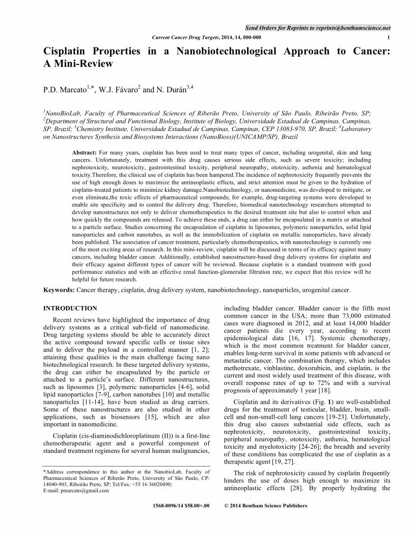
Cisplatin and its derivatives (Fig. 1) are well-established drugs for the treatment of testicular, bladder, brain, small-cell and non-small-cell lung cancers [19-23]. Unfortunately, this drug also causes substantial side effects, such as nephrotoxicity, neurotoxicity, gastrointestinal toxicity, peripheral neuropathy, ototoxicity, asthenia, hematological toxicity and myelotoxicity [24-26]; the breadth and severity of these conditions has complicated the use of cisplatin as a therapeutic agent [19, 27].
The risk of nephrotoxicity caused by cisplatin frequently hinders the use of doses high enough to maximize its antineoplastic effects [28]. By properly hydrating the
2 Current Cancer Drug Targets, 2014, Vol. 14, No. 5 Marcato et al.
patients, this effect can be minimized [19]. The time- and dose-dependent nephrotoxicity of cisplatin restricts the use of high doses, which prevents its maximum therapeutic efficacy; 25-35% of patients show evidence of nephrotoxicity following a single dose of cisplatin. Once cisplatin enters the cell, this drug is bio-activated by a hydrolytic process [29, 30]. Water molecules replace the chloride ligands, which transform the original platinum (Pt) species into mono- and di-aqua diammine platinum (II) species. These hydrated species are more reactive than the original platinum species, which are neutral, and proceed to create intra-strand DNA–cisplatin cross-linking, which causes cytotoxic lesions in tumors and in other rapidly dividing cells. Although most non-proliferating cells are relatively unaffected, proximal tubule cells are damaged by cisplatin [29, 30].
The mechanisms of tubular cell injury have been studied to aid in the development nephroprotective approaches [28, 31, 32]. Several pathways of apoptosis have been implicated in cisplatin-induced nephrotoxicity [32]. Cisplatin increases levels of proapoptotic proteins and, concurrently, decreases
anti-apoptotic proteins; this compound also activates initiator and executioner caspases [28, 32, 33]. Reactive oxygen species (ROS) also have a critical role in the pathogenesis of cisplatin-induced nephrotoxicity. ROS directly damage cell components, such as lipids, proteins, and DNA, and activate the mitochondrial pathway of apoptosis [31, 34]. Therefore, finding an effective way to prevent cisplatin-induced nephrotoxicity has become a priority.
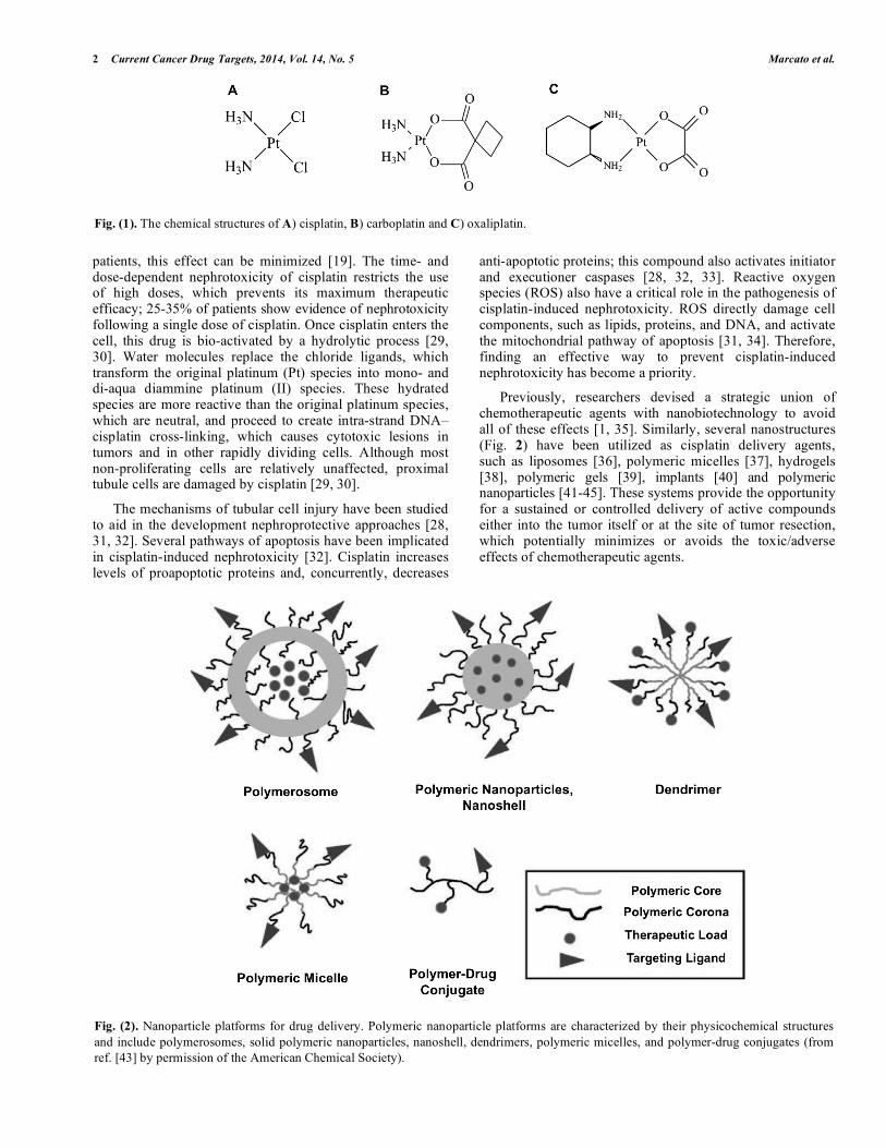
Previously, researchers devised a strategic union of chemotherapeutic agents with nanobiotechnology to avoid all of these effects [1, 35]. Similarly, several nanostructures (Fig. 2) have been utilized as cisplatin delivery agents, such as liposomes [36], polymeric micelles [37], hydrogels [38], polymeric gels [39], implants [40] and polymeric nanoparticles [41-45]. These systems provide the opportunity for a sustained or controlled delivery of active compounds either into the tumor itself or at the site of tumor resection, which potentially minimizes or avoids the toxic/adverse effects of chemotherapeutic agents.
Fig. (1). The chemical structures of A) cisplatin, B) carboplatin and C) oxaliplatin.
Fig. (2). Nanoparticle platforms for drug delivery. Polymeric nanoparticle platforms are characterized by their physicochemical structures and include polymerosomes, solid polymeric nanoparticles, nanoshell, dendrimers, polymeric micelles, and polymer-drug conjugates (from ref. [43] by permission of the American Chemical Society).
Cisplatin Properties in a Nanobiotechnological Approach to Cancer Current Cancer Drug Targets, 2014, Vol. 14, No. 5 3
In this mini-review, the importance of cisplatin as a treatment for bladder cancer and as an active chemotherapeutic agent utilized in tandem with nanobiotechnology will be discussed. Several of the nanostructures applied as carrier systems (liposomes, polymeric nanoparticles, carbon nanotubes, solid lipid nanoparticles, metallic and metallic oxides) for cisplatin will be covered in this review.
Liposomes
Liposomes, which have been used as drug carrier systems for cancer and for several other diseases, are vesicles that are composed of a lipid bilayer [1]. The purpose of this section is to highlight the benefits of and problems arising from the application of liposomes for bladder cancer treatment [46]. Table 1 shows some of the most important reports in this area. The use of liposomes to carry drugs can exhibit results in contrast to what is expected [47]. For example, cholesterol-pegylated liposomes with cisplatin formed unexpected composites (SPI-077), and due to the appearance of this new species, the liposomal structures retained the encapsulated cisplatin so strongly that the amount of drug released was insufficient to exert substantial cytotoxic activity against cancer cells [48]. This result caused the failure of SPI-077 in phase I/II clinical trials [49].
The use of liposomes in chemotherapy has been used to directly target tumors, to slow the release of the encapsulated
drug into the tumor site and to reduce side effects caused by the drug. A liposomal formulation, which has been named Lipoplatin™ and which contains an encapsulated cisplatin derivative, is already on the market [50]. The liposomes used for the formulation of Lipoplatin™, which have an average size of 110 nm, are composed of soy phosphatidyl choline (SPC-3), cholesterol, dipalmitoyl phosphatidyl glycerol and methoxy-polyethylene glycol-distearoyl phosphatidylethanolamine (mPEG2000-DSPE), as well as the cisplatin derivative encapsulated within. In phase I, II and III clinical trials, this formulation displayed a reduced incidence of renal toxicity, peripheral neuropathy, ototoxicity, myelotoxicity, nausea/vomiting and asthenia, which are associated with cisplatin usage, as well as an enhanced or similar efficacy to unencapsulated cisplatin [51]. This formulation’s anti-angiogenic activity was also verified during the phase III trials. The approval for phase II/III studies against pancreatic cancer currently remains in progress due to the orphan drug status granted to Lipoplatin by the European Medicine Agency [52].
Five bladder cancer patients were treated exclusively with LipoplatinTM,which was administered once every 2 weeks intravenously, in one clinical trial. Once little to no side effects was observed to occur, gemcitabine was also incorporated into the patients’ bi-weekly treatments. Sixteen patients with bladder cancer exhibited renal failure; 14 of these patients received treatment with the combination
Table 1. Liposomes and cisplatin on cancer.
Product Name Cancer Type Effect Reference
Cholesterol-pegylated liposomes
SPI-077 Bladder cancer Insufficient drug release. Failure at Phase I/II
[49]
Phosphatyl choline (SPC-3)/cholesterol/
dipalmitoyl phosphatidyl glycerol liposome
Lipoplatin Bladder cancer 16 patients exhibited renal failure, 14/16 treated with
Lipoplatin/gemcitabine 5/16 remission
[50-53]
Aroplatin Colorectal cancer Good responce [54-56]
Cis-dichlororidobis(2-stearol-hydrazide)platinum (II)
liposome (DCSP)
Ptsome Potential combination therapy.
Exhibited enhanced permeation and retention (EPR) in tumors
[57]
pH-sensitive liposome SpHL-cisplatin Solid tumor Little myelo- nephron- renal toxicity [58]
Cholesterol-tethered/succininc-acid bound cisplatinum (II)
nanoparticles (SACNs)
SACs Bladder cancer [59]
Trsnferrin-conjugated PEG liposomes
Tf-PEG Gastric cancer/bladder cancer Enhanced uptake by tumor cells [60]
Bile acid-cisplatin complex liposomes
Bamet-R2 Ovariam cells Bamet-R2 exhibited uptake of cisplatin
[61]
Conventional liposomes Cis-bisneo decanoato-trans-R,R-1,2-diamnocyclohexane
polatinum (II) (NDDP)
Metastatic tumor Enhancement cisplatin therapeutic [62-64]
Cationic liposome Polyetyleneimine (PEI) bounds cholesterol (PEI-ChoL)
Adenocarciinomic human alveolar basal epithelial cells
Enhancement internalization of cisplatin
[64, 65, 68]
4 Current Cancer Drug Targets, 2014, Vol. 14, No. 5 Marcato et al.
of LipoplatinTM and gemcitabine as their first line of chemotherapy, and 2 patients received the same regimen as their second line of chemotherapy. Five patients exhibited complete remission, whereas a partial response was observed in8 patients. Three patients maintained a stable level of disease. The median duration of response among the 16 patients with bladder cancer was 12 months (range 4-18 months). One patient with bladder cancer, who had achieved a complete response, had a tumor recurrence after 12 months. This patient survived for 48 months before dying of a heart attack [53].
Derivatives of cisplatin have also been encapsulated in other liposomal formulations. For example, AroplatinTMis a liposomal formulation using oxaliplatin. In the phase II clinical trials for this formulation, patients with advanced colorectal cancers that were resistant to 5-fluorouracil/ leucovorin, capecitabine and to irinotecan demonstrated a good response after treatment with AroplatinTM [54, 55]. The FDA also approved a liposomal product encapsulating another derivative of cisplatin, namely, carboplatin; this treatment was given the trade name ParaplatinTM [56].
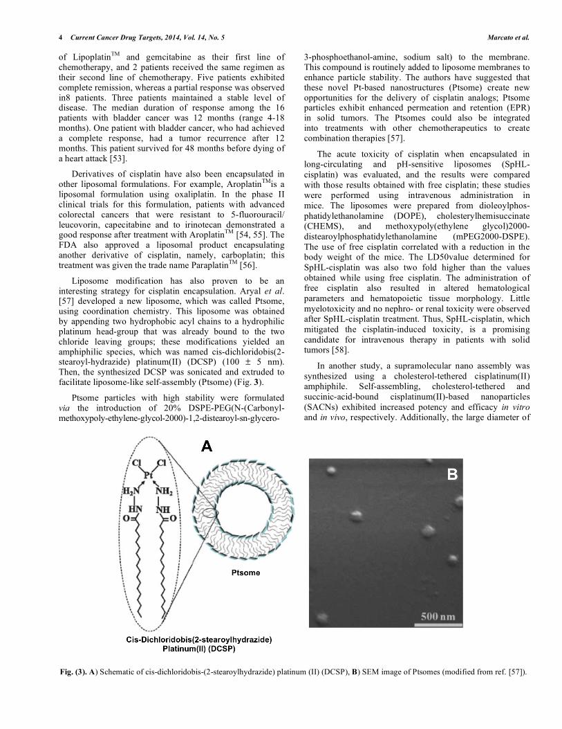
Liposome modification has also proven to be an interesting strategy for cisplatin encapsulation. Aryal et al. [57] developed a new liposome, which was called Ptsome, using coordination chemistry. This liposome was obtained by appending two hydrophobic acyl chains to a hydrophilic platinum head-group that was already bound to the two chloride leaving groups; these modifications yielded an amphiphilic species, which was named cis-dichloridobis(2-stearoyl-hydrazide) platinum(II) (DCSP) (100 ± 5 nm). Then, the synthesized DCSP was sonicated and extruded to facilitate liposome-like self-assembly (Ptsome) (Fig. 3).
Ptsome particles with high stability were formulated via the introduction of 20% DSPE-PEG(N-(Carbonyl-methoxypoly-ethylene-glycol-2000)-1,2-distearoyl-sn-glycero-
3-phosphoethanol-amine, sodium salt) to the membrane. This compound is routinely added to liposome membranes to enhance particle stability. The authors have suggested that these novel Pt-based nanostructures (Ptsome) create new opportunities for the delivery of cisplatin analogs; Ptsome particles exhibit enhanced permeation and retention (EPR) in solid tumors. The Ptsomes could also be integrated into treatments with other chemotherapeutics to create combination therapies [57].
The acute toxicity of cisplatin when encapsulated in long-circulating and pH-sensitive liposomes (SpHL-cisplatin) was evaluated, and the results were compared with those results obtained with free cisplatin; these studies were performed using intravenous administration in mice. The liposomes were prepared from dioleoylphos- phatidylethanolamine (DOPE), cholesterylhemisuccinate (CHEMS), and methoxypoly(ethylene glycol)2000-distearoylphosphatidylethanolamine (mPEG2000-DSPE). The use of free cisplatin correlated with a reduction in the body weight of the mice. The LD50value determined for SpHL-cisplatin was also two fold higher than the values obtained while using free cisplatin. The administration of free cisplatin also resulted in altered hematological parameters and hematopoietic tissue morphology. Little myelotoxicity and no nephro- or renal toxicity were observed after SpHL-cisplatin treatment. Thus, SpHL-cisplatin, which mitigated the cisplatin-induced toxicity, is a promising candidate for intravenous therapy in patients with solid tumors [58].
In another study, a supramolecular nano assembly was synthesized using a cholesterol-tethered cisplatinum(II) amphiphile. Self-assembling, cholesterol-tethered and succinic-acid-bound cisplatinum(II)-based nanoparticles (SACNs) exhibited increased potency and efficacy in vitro and in vivo, respectively. Additionally, the large diameter of
Fig. (3). A) Schematic of cis-dichloridobis-(2-stearoylhydrazide) platinum (II) (DCSP), B) SEM image of Ptsomes (modified from ref. [57]).
Cisplatin Properties in a Nanobiotechnological Approach to Cancer Current Cancer Drug Targets, 2014, Vol. 14, No. 5 5
SACN particles exceeds the size that can be cleared by a patient’s kidneys, therefore limiting nephrotoxicity. The authors demonstrated that by using a rational drug design, the size of their supramolecular assemblies could be increased from Angstrom to nano-scale, which confers unique biological properties. Therefore, this report supports the hypothesis that integrating a rational drug design and supramolecular nanochemistry can be a powerful strategy for drug development in the treatment of several cancers, including bladder cancer [59].
The antitumor activity of cisplatin, which was encapsulated by transferrin-conjugated polyethylene glycol liposomes (Tf-PEG liposomes), on human gastric cancer cells, which were peritoneally disseminated in nude mice,was evaluated. Small, unilamellar Tf-PEG encapsulated cisplatin was prepared by reverse-phase evaporation, which was followed by extrusion, and, finally, internalized by tumor cells via receptor-mediated endocytosis. The uptake of the Tf-PEG liposomes into healthy liver and spleen cells was significantly less prevalent than the uptake of conventional liposomes. Furthermore, this system exhibited increased in vivo antitumor activity relative to free cisplatin. This platinum complex also demonstrated increased water
solubility, which was due to its encapsulation into the liposomes, and demonstrated enhanced uptake by liver tumor cells [60]. Although this study’s findings are exclusive to human gastric cancer, this system still has a great potential for the treatment of bladder cancer.
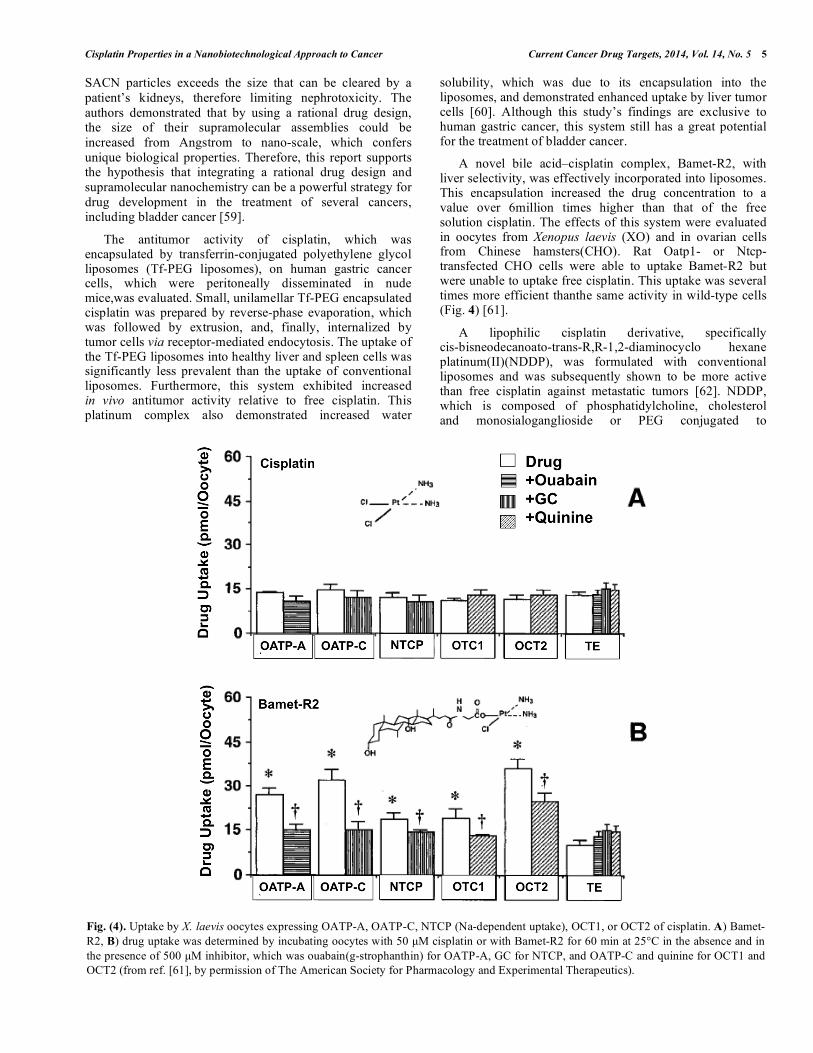
A novel bile acid–cisplatin complex, Bamet-R2, with liver selectivity, was effectively incorporated into liposomes. This encapsulation increased the drug concentration to a value over 6million times higher than that of the free solution cisplatin. The effects of this system were evaluated in oocytes from Xenopus laevis (XO) and in ovarian cells from Chinese hamsters(CHO). Rat Oatp1- or Ntcp-transfected CHO cells were able to uptake Bamet-R2 but were unable to uptake free cisplatin. This uptake was several times more efficient thanthe same activity in wild-type cells (Fig. 4) [61].
A lipophilic cisplatin derivative, specifically cis-bisneodecanoato-trans-R,R-1,2-diaminocyclo hexane platinum(II)(NDDP), was formulated with conventional liposomes and was subsequently shown to be more active than free cisplatin against metastatic tumors [62]. NDDP, which is composed of phosphatidylcholine, cholesterol and monosialoganglioside or PEG conjugated to
Fig. (4). Uptake by X. laevis oocytes expressing OATP-A, OATP-C, NTCP (Na-dependent uptake), OCT1, or OCT2 of cisplatin. A) Bamet-R2, B) drug uptake was determined by incubating oocytes with 50 µM cisplatin or with Bamet-R2 for 60 min at 25°C in the absence and in the presence of 500 µM inhibitor, which was ouabain(g-strophanthin) for OATP-A, GC for NTCP, and OATP-C and quinine for OCT1 and OCT2 (from ref. [61], by permission of The American Society for Pharmacology and Experimental Therapeutics).

6 Current Cancer Drug Targets, 2014, Vol. 14, No. 5 Marcato et al.
phosphatidylethanolamine, was developed to create a class of liposomesable to sustain prolonged circulation [63].
Liposome formulations were prepared by extrusion using 1,2-distearoyl-sn-glycero-3-phosphatidylcholine (DSPC), 1,2-dipalmitoyl-sn-glycero-3-phosphatidylcholine (DPPC), 1,2-distearoyl-sn-glycero-3-phosphatidylglycerol (DSPG) and cholesterol (CHOL). An optimization of the liposomal carrier for the enhancement of cisplatin therapeutic activity was performed, which resulted in the development of liposomes composed of DSPC: DPPC: DSPG: Chol (35 : 35 : 20 : 10).The encapsulated cisplatin, which had ahalf-life of8.3 hours, demonstratedhigh efficacy in a study with a P388 murine lymphocytic leukemia model [64].
The antitumor activities of free cisplatin, cisplatin encapsulated in a neutral liposomes and cisplatin encapsulated by lysosomes composed of the transfection agent polycation polyethylenimine (PEI) bound to cholesterol (PEI-Chol), were evaluated in A549 cells (adenocarcinomic human alveolar basal epithelial cells). The anticancer activity exhibited by the PEI-Chol-encapsulated cisplatin was much higher than that exhibited by cisplatin in neutral liposomes or by free cisplatin; the use of PEI-Cholwas proposed to aid in the internalization of cisplatin by the cells. A possible mechanism for the internalization of cationic liposomes into the cells was also included [64].
A possible mechanism for the internalization of cationic liposomes into the cells was proposed, which included the caveolae-mediated endocytosis pathway and clathrin- and actin-dependent uptake pathways [65]. Therefore, the cationic modification of liposomes with PEI is a promising method for antitumor drug delivery and will likely be applicable to bladder cancer.
In the treatment of bladder cancer, it is more desirable to preserve the bladder than to perform a partial or full cystectomy. To this effect, intravesical polo-like kinase-1 (PLK-1) small interfering RNA (siRNA) was studied as a treatment against bladder cancer. Bladder cancer patients who express high levels of PLK-1 are known to often have a poor prognosis. By using siRNA/cationic liposomes (DOTMA) [66], the expression of endogenous PLK-1 can be suppressed in a time- and dose-dependent manner within bladder cancer cells. In studies performed in vivo, which utilized an orthotopic mouse model and the LUC-labeled bladder cancer cell line UM-UC-3LUC, the PLK-1 siRNA was transfected into the cells, which subsequently reduced PLK-1 expression and, ultimately, inhibited the growth of bladder cancer [67].
Cationic liposomes as carrier systems for the LKB1 gene (LPs-pVAX-LKB1) (tumor suppressor gene in lung cancer), which is associated with low cisplatin concentration, were used to sensitize the response of lung cancer cells to cisplatin, to induce apoptosis and to inhibit cancer cell proliferation, invasion and metastasis. The effects of the intratumoral administration of Lps-pVAX-LKB1 in conjunction with the intraperitoneal injection of low-dose cisplatin were studied in mice transfected with metastatic A549 lung cancer cells. An additional study in which the administration of Lps-pVAX-LKB1 was injected intravenously in conjunction with the intraperitoneal injection of low-dose
cisplatin was also performed. The treatments exhibited an inhibition of tumor growth and a decrease in the number of metastases by lung tumor nodules. The life expectancies of the mice in the combination treatment groups were also significantly prolonged when compared with the life span of the control group. The authors proposed possible mechanisms for these interactions and suggested that the combination of LKB1 gene therapy and low-dose cisplatin-based chemotherapy may be a potent therapeutic strategy for lung cancer, with possible extrapolation to other cancers, such as bladder cancer [68].
Liposomes loaded with both cisplatin and quantum dots (CdSe or CdSe/ZnS) exhibited higher cytotoxic activity relative to an equal dose of free cisplatin. A large fluorescence signal and significant cisplatin accumulation were detected in the brain and in the skin, which were verified by ex vivo imaging and by drug distribution. Utilizing liposomes as carriers could reduce the reticulo endothelial system uptake of QDs and of cisplatin. The QDLs evaluated in this study exemplify a new method of cancer treatment with both diagnostic and therapeutic aspects [69].
Baruah and Surin [70] prepared liposomes by hydrating a 1,2-dioleoyl-sn-glycero-3-phosphocholine lipid with solutions of three ‘‘probe’’ molecules, which were cisplatin, guanosine 5´-monophosphate (5´-GMP), and 9-ethylguanine(9-EtG), in a phosphate-buffered saline solution and in an N-(2-hydroxyethyl)piperazine-N´-ethanesulfonic acid buffer. When cisplatin-containing liposomes mix with 5´-GMP- or 9-EtG-embedded liposomes, the N7 nitrogen atom of 5´-GMP or of 9-EtG binds to the cisplatin, which replaces the chloride leaving groups to form a bis-adduct. In these studies, cisplatin and a metal-based drug are incorporated into nanocapsules, which are composed of negatively charged liposome templates with positively charged polyelectrolytes. These deposited polyelectrolytes minimize the release of the cisplatin and, therefore, reduce the drug’s toxicity. These studies remain ongoing.
Treatment with liposomal doxorubicin (LD), which consisted of a pegylated phospholipid vesicle loaded with doxorubicin, against unresectable malignant pleural mesothelioma (MPM) was studied in the presence of 99mTc and was followed by functional physical examinations, which included the observation of dyspnea, cough, and chest/arm pain. Patients treated with LD demonstrated a significant improvement over traditional therapy. Three times more LD accumulated in tumors and in soft tissue than in liver tissue, and after one hour from the point of administration, the LD uptake in tumor tissue was higher than that in soft tissue. The utilization of the LD combined with cisplatin was an active therapeutic regimen for MPM and demonstrated a better toxicity profile, which resulted in an improvement in the quality of life for the patients [71].
Polymeric nanoparticles
Polymeric nanoparticles are used as carriersfor drugs, which are composited into the polymeric matrix, and have experienced an increased application in medicine. Table 2 shows some of the most important results published in the literature. One strategy described in the literature involved the encapsulation of a Pt4+ complex within polymeric

Cisplatin Properties in a Nanobiotechnological Approach to Cancer Current Cancer Drug Targets, 2014, Vol. 14, No. 5 7
nanoparticles, which were functionalized with a variety peptides, antibodies and aptamers to prevent degradation and to enhance the cellular accumulation of the active compound [72, 73]. The release of the Pt4+ complexes and their subsequent reduction in situ facilitated the controlled release of the active Pt2+ species.
Dhar et al. [72] encapsulated Pt4+ complex in polymeric nanoparticles, which were prepared from poly(D,L-lactic-co-glycolic acid) (PLGA) and from polyethylene-glycol (PEG). The surface of these particles was functionalized with aptamers for prostate-specific membrane antigen (PSMA), which facilitated targeted delivery to the prostate.
Previously, Avgoustakis et al. [74] also produced PLGA-mPEG nanoparticles containing cisplatin. These particles were prepared using a double emulsion method and were characterized by their morphology, size, zeta potential and drug loading. Although the intravenous administration of these cisplatin nanoparticles in mice did result in prolonged cisplatin circulation in the blood, this technique did not have a high enough loading efficiency to be considered further for therapeutic applications [74].
Another strategy employed to develop atumor-targeted drug delivery system was the production of micelles
composed of a polymer–metal complex, which consisted of cisplatin and PEG-poly(glutamic acid) block copolymers. These cisplatin micelles were 28 nm in size and exhibited a sustained drug release profile, remarkably prolonged blood circulation and effective accumulation in solid tumor sites [75]. All the data presented in this manuscript suggested that formulations incorporating cisplatin into the components of the micelles are a promising solution for targeting solid tumors in chemotherapy. Micelles with a hydrophobic inner core and with a hydrophilic outer shell allow the chemical integration of cisplatin into the micelles. The delivery of cisplatin-micelles to tumors is directed by the higher extracellular acidity of solid tumors relative to normal tissues [76].
Cisplatin has also been encapsulated in microspheres composed of degradable polysaccharides, such as starch microspheres (DSM). These particles were used in clinical trials to achieve the intensification of intra-arterial chemo- therapy for cancers occurring in the head and in the neck using high doses of cisplatin. Chemoembolization succeeded in 37 interventions, and the overall response after one cycle was 64.7% when using only the cisplatin crystal suspension (n=17) and86.6% when using the additional biodegradable starch microspheresin conjunction with cisplatin (n=15). All
Table 2. Polymeric nanoparticles and cisplatin on cancer.
Product Name Cancer Type Effect Reference
Poly(lactic-coglycolic acids (PLGA)/PEG)
Pt4+ complex-PLGA-PEG Associated to propstate specific
membrane antigen (PSMA)
Prostate cancer Prolonged cisplatin blood circulation
[72-74]
PEG-poly(glutamic acid) block polymer Mycelles of polymer-metal complex
Solid tumor Cisplatin prolonged blood circulation and effective accumulationin tumor
[76]
Polysaccharides microspheres Starch microspheres (DMS) Head and neck cancer
Cisplatin intensification of intra-aeterial chemotherapy
[77]
Poly(N-isopropylacrylamide
graphted hyaluronic acid
HPN copolymer
(grafted with gelatin formed HPNG copolymer)
Bladder cancer Sustained cisplatin release [78]
Hydrophobically glycol chitosan-cholanic acid conjugated (HGC)
Cisplatin –HGC nanoparticles Tumor tissues Sustained cisplatin release and reduced cytotoxicity
[79]
Poly(sebacic acid-co-ricinoleic acid esteranhydride)
Cisplatin-PSRA Bladder tumor Intratumoral and intraperotoneal application
[19]
Poly(ethylene glycol-b-poly(acrylic acid)/α-cyclodextrin
PEG-b-PAA/α-CD Hydrogel cisplatin
Bladder cancer Sustained delivery of cispatin [82]
PCL—b-P(PMA-click-MSA-co-PEGMA) polymer with bidentate dicarboxylate
groups/iron oxide(SPIONS)
SPIONs-Cispaltin-conjugated polymeric nanoparticles
(Pt-Fe-PNs)
Bladder cancer Mucoadhesives tendencies [85]
Poly(ethyleneglycol)-b-poly (metaacrylic acid)e
Cispçlatin-PEG-PMA Bladder cancer Prolonged blood circulation, increased tumor accumulation
[86]
Epidermal growth factor (EGF)-polyethylene glycol-polylactic-glycolic
acid-polylysine (mPEG-PLGA-PLL)
Cisplatin (CDDP)/mPEG-PLGA-PLL) nanoparticles (CDDP-NPs-EGF)
Bladder cancer Decreased nephrotoxicity of cisplatin
[87]

8 Current Cancer Drug Targets, 2014, Vol. 14, No. 5 Marcato et al.
subjects were assessed 3 weeks after treatment. Systemic toxicity was extremely low, and local side-effects, which included pain, swelling, and small necroses, were more pronounced after the additional delivery of DSM [77].
The modification of drug-loaded polymers has also provided benefits for therapeutic delivery systems. For example, poly(N-isopropylacrylamide) (PNIPAM) was grafted onto hyaluronic acid (HA) to obtain an HPN copolymer, which was subsequently grafted with gelatin to form an HPNG copolymer. The addition of the gelatin strengthened the fibrous structure in the matrix. The use of HA and gelatin also produced a different microstructure when compared with the parent PNIPAM hydrogel. The new material showed a sustained cisplatin release profile and interesting in vivo results. Aqueous cisplatin solution (control) was almost completely released (100%) after 8 h, whereas only 85% and 52% of the cisplatin incorporated into PNIPAM and into HPNG, respectively, was released over the sameperiod (8 h). These formulations were intravesically administered to rats. The cisplatin levels in the bladder tissues after the administration of PNIPAM-cisplatin and of HPNG-cisplatin showed seven fold and twofold enhancements, respectively, of the cisplatin concentration in the bladder wall. A histological examination showed that no adverse change in the urothelium occurred with the application of HPNG, whereas PNIPAM caused partial desquamation of umbrella cells. The authors suggested that thermosensitive hydrogels might be promising carriers for targeted drug delivery to the bladder [78].
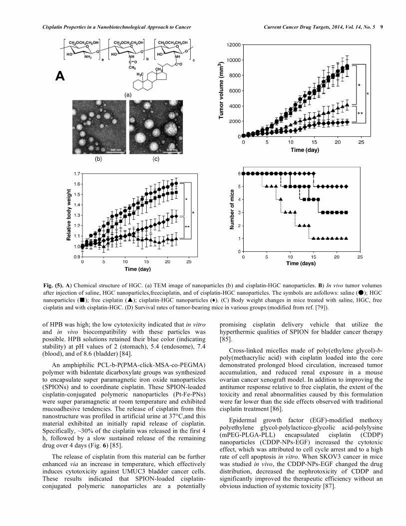
A hydrophobically modified glycol chitosan–cholanic acid conjugate (HGC) was prepared by chemically grafting 5β-cholanic acids to glycol chitosan in the presence of N-hydroxysuccinimide and of 1-ethyl-3-(3-dimethylaminopropyl)-carbodiimide hydrochloride, with cisplatin solution added at the end of the reaction (Fig. 5a). The in vivo antitumor efficacy of these cisplatin-HGC nanoparticles (300-500 nm) exhibited a sustained cisplatin release profile and reduced the cisplatin-induced cytotoxicity in vitro. Localized cisplatin-HGC nanoparticles in tumor tissue showed higher antitumor efficacy and lower toxicity compared with free cisplatin, which was demonstrated by changes in the tumor volumes (Fig. 5b), body weights (Fig. 5c), and survival rates (Fig. 5d), as well as by immunohistological TUNEL assay data. Therefore, HGC nanoparticles also have excellent potential as carriers for insoluble anticancer drugs [79].
A hyperbranched polyglycerol with modifications ofHPG-C8/10-MePEG with amine groups on the material’s surface results in highly positively charged nanoparticles (HPG-C8/10-MePEG-NH2) that have exhibited improved mucoadhesive properties. Docetaxel (DTX)-loaded into HPG-C8/10-MePEG-NH2nanoparticlesexhibited equivalent in vitro cytotoxicity to the commercially available Taxotere (Docetaxel from Sanofi-aventis). Additionally, in vivo, DTX-loaded HPG-C8/10-MePEG-NH2nanoparticles were an effective formulation to inhibit tumor growth in anorthotopic model of bladder cancer (intravesical administration). The application of these nanostructures to clinical trials for patients with resistant bladder cancer remains underway; the safety and efficacy of this mucoadhesive nanoparticulate with DTX formulation must first be evaluated [80].
The same group who previously described the HPGs functionalized with amine groups also demonstrated that modified HPGs with a well-defined carboxylic acid, MePEG and C8/10 alkyl core were also effective against cancer cells. The average size of these functionalized nanoparticles was less than 10 nm. The carboxylate functionalized HPGs (HPG-C8/10-MePEG6.5-COOH348and HPG-C8/10-MePEG6.5-COOH113) demonstrated good biocompatibility, and drug-loaded HPGs effectively inhibited proliferation of KU-7-luc human bladder cancer cells [81].
Cisplatin was encapsulated in poly (sebacic acid-co-ricinoleic acid ester anhydride) with effective loading ratios of 2:8 and 3:7. This drug delivery system was developed to probe the efficacy of intratumorally delivered cisplatin for heterotopic mouse bladder tumors (MBT). Free and encapsulatedcisplatin were administered by intratumoral (IT) andintraperitoneal (IP) routes; however, the IP-delivered drug inhibited tumor growth more efficiently when compared with the IT application of free cisplatin solution and was more efficient than the administration of only polymer for prolonging the life of the mice. In the cisplatin/polymer group, mice were injected intratumorally with cisplatin formulation 10 days after tumor inoculation. In 8 of 10mice, the tumors completely disappeared during the first 10 days after treatment, and mice remained tumor-free until the end of the study (40 days post-tumor cell inoculation) [19].
Supramolecular hydrogels with nanoparticles featuring drugs encapsulated in block copolymers were prepared by mixing poly(ethylene glycol)-b-poly(acrylic acid) (PEG-b-PAA polymer), cisplatin, and α-cyclodextrin (α-CD) in water. After the addition of either PEG or Pluronic F127, cisplatin-loaded nanoparticles within hydrogels were produced. In vitro cytotoxicity studies showed that the cisplatin-loaded nanoparticle-containing hydrogels inhibited the growth of human bladder carcinoma EJ cells with a similar potency to that of free cisplatin. Additionally, hydrogels without cisplatin showed no cytotoxicity. These results suggested that the cisplatin-coordinated PEG-b-PAA/α-CD supramolecular hydrogels exhibited a potential for use as an injectable system that exhibits a sustained delivery of cisplatin for cancer therapy [82].
Another interesting drug delivery system uses nanoparticles, which were synthesized via the reaction of K3[Fe(CN)6] x3H2O (PB) and HCl, featuring hollow interiors and microporous frameworks (HPB). Then, the solid PB crystals were used as a starting material. Hollow interiors were generated through controlled chemical etching with polyvinylpyrrolidone (PVP). The HPB obtained is this study exhibited a uniform particle size of 110 nm, with a hollow space 90 nm in diameter and with nanopores less than 2 nm in diameter [83]. For the loading of the cisplatin, HPB was added to an aqueous cisplatin solution and stirred for 24 h in the dark. Subsequently, the mixture was heated to 100°C for 10 min.
The cytotoxicity of the synthesized HPB was examined by 3-[4,5-dimethylthiazol-2-yl]-2,5-diphenyl tetrazolium bromide (MTT) assays using T24 cells, which showed that cell viability was approximately 70%, even when the dosage
Cisplatin Properties in a Nanobiotechnological Approach to Cancer Current Cancer Drug Targets, 2014, Vol. 14, No. 5 9
of HPB was high; the low cytotoxicity indicated that in vitro and in vivo biocompatibility with these particles was possible. HPB solutions retained their blue color (indicating stability) at pH values of 2 (stomach), 5.4 (endosome), 7.4 (blood), and of 8.6 (bladder) [84].
An amphiphilic PCL-b-P(PMA-click-MSA-co-PEGMA) polymer with bidentate dicarboxylate groups was synthesized to encapsulate super paramagnetic iron oxide nanoparticles (SPIONs) and to coordinate cisplatin. These SPION-loaded cisplatin-conjugated polymeric nanoparticles (Pt-Fe-PNs) were super paramagnetic at room temperature and exhibited mucoadhesive tendencies. The release of cisplatin from this nanostructure was profiled in artificial urine at 37°C,and this material exhibited an initially rapid release of cisplatin. Specifically, ~30% of the cisplatin was released in the first 4 h, followed by a slow sustained release of the remaining drug over 4 days (Fig. 6) [85].
The release of cisplatin from this material can be further enhanced via an increase in temperature, which effectively induces cytotoxicity against UMUC3 bladder cancer cells. These results indicated that SPION-loaded cisplatin-conjugated polymeric nanoparticles are a potentially
promising cisplatin delivery vehicle that utilize the hyperthermic qualities of SPION for bladder cancer therapy [85].
Cross-linked micelles made of poly(ethylene glycol)-b-poly(methacrylic acid) with cisplatin loaded into the core demonstrated prolonged blood circulation, increased tumor accumulation, and reduced renal exposure in a mouse ovarian cancer xenograft model. In addition to improving the antitumor response relative to free cisplatin, the extent of the toxicity and renal abnormalities caused by this formulation were far lower than the side effects observed with traditional cisplatin treatment [86].
Epidermal growth factor (EGF)-modified methoxy polyethylene glycol-polylacticco-glycolic acid-polylysine (mPEG-PLGA-PLL) encapsulated cisplatin (CDDP) nanoparticles (CDDP-NPs-EGF) increased the cytotoxic effect, which was attributed to cell cycle arrest and to a high rate of cell apoptosis in vitro. When SKOV3 cancer in mice was studied in vivo, the CDDP-NPs-EGF changed the drug distribution, decreased the nephrotoxicity of CDDP and significantly improved the therapeutic efficiency without an obvious induction of systemic toxicity [87].
Fig. (5). A) Chemical structure of HGC. (a) TEM image of nanoparticles (b) and cisplatin-HGC nanoparticles. B) In vivo tumor volumes after injection of saline, HGC nanoparticles,freecisplatin, and of cisplatin-HGC nanoparticles. The symbols are asfollows: saline (�); HGC nanoparticles (�); free cisplatin (�); cisplatin-HGC nanoparticles (♦). (C) Body weight changes in mice treated with saline, HGC, free cisplatin and with cisplatin-HGC. (D) Survival rates of tumor-bearing mice in various groups (modified from ref. [79]).
10 Current Cancer Drug Targets, 2014, Vol. 14, No. 5 Marcato et al.
Carbon nanotubes
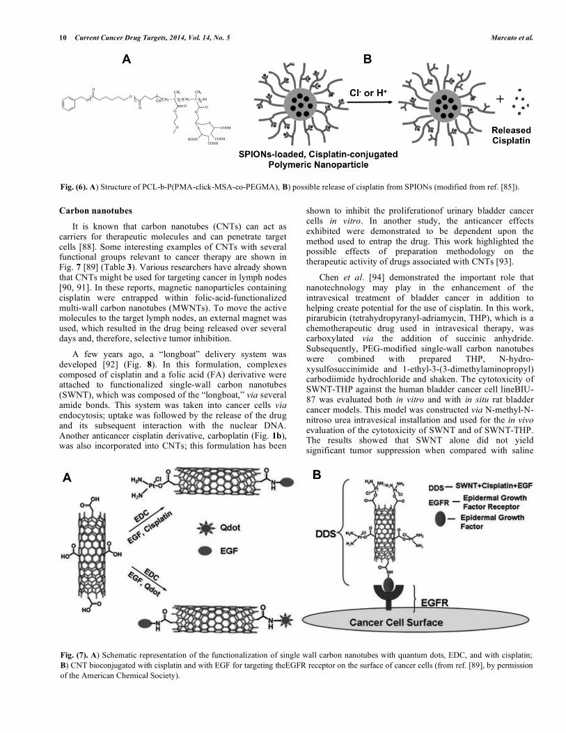
It is known that carbon nanotubes (CNTs) can act as carriers for therapeutic molecules and can penetrate target cells [88]. Some interesting examples of CNTs with several functional groups relevant to cancer therapy are shown in Fig. 7 [89] (Table 3). Various researchers have already shown that CNTs might be used for targeting cancer in lymph nodes [90, 91]. In these reports, magnetic nanoparticles containing cisplatin were entrapped within folic-acid-functionalized multi-wall carbon nanotubes (MWNTs). To move the active molecules to the target lymph nodes, an external magnet was used, which resulted in the drug being released over several days and, therefore, selective tumor inhibition.
A few years ago, a “longboat” delivery system was developed [92] (Fig. 8). In this formulation, complexes composed of cisplatin and a folic acid (FA) derivative were attached to functionalized single-wall carbon nanotubes (SWNT), which was composed of the “longboat,” via several amide bonds. This system was taken into cancer cells via endocytosis; uptake was followed by the release of the drug and its subsequent interaction with the nuclear DNA. Another anticancer cisplatin derivative, carboplatin (Fig. 1b), was also incorporated into CNTs; this formulation has been
shown to inhibit the proliferationof urinary bladder cancer cells in vitro. In another study, the anticancer effects exhibited were demonstrated to be dependent upon the method used to entrap the drug. This work highlighted the possible effects of preparation methodology on the therapeutic activity of drugs associated with CNTs [93].
Chen et al. [94] demonstrated the important role that nanotechnology may play in the enhancement of the intravesical treatment of bladder cancer in addition to helping create potential for the use of cisplatin. In this work, pirarubicin (tetrahydropyranyl-adriamycin, THP), which is a chemotherapeutic drug used in intravesical therapy, was carboxylated via the addition of succinic anhydride. Subsequently, PEG-modified single-wall carbon nanotubes were combined with prepared THP, N-hydro- xysulfosuccinimide and 1-ethyl-3-(3-dimethylaminopropyl) carbodiimide hydrochloride and shaken. The cytotoxicity of SWNT-THP against the human bladder cancer cell lineBIU-87 was evaluated both in vitro and with in situ rat bladder cancer models. This model was constructed via N-methyl-N-nitroso urea intravesical installation and used for the in vivo evaluation of the cytotoxicity of SWNT and of SWNT-THP. The results showed that SWNT alone did not yield significant tumor suppression when compared with saline
Fig. (6). A) Structure of PCL-b-P(PMA-click-MSA-co-PEGMA), B) possible release of cisplatin from SPIONs (modified from ref. [85]).
Fig. (7). A) Schematic representation of the functionalization of single wall carbon nanotubes with quantum dots, EDC, and with cisplatin; B) CNT bioconjugated with cisplatin and with EGF for targeting theEGFR receptor on the surface of cancer cells (from ref. [89], by permission of the American Chemical Society).
Cisplatin Properties in a Nanobiotechnological Approach to Cancer Current Cancer Drug Targets, 2014, Vol. 14, No. 5 11
controls in vitro. However, SWNT-THP exhibited higher tumor suppression than THP-saline both in vitro and in vivo. The present findings indicate that the SWNT delivery of THP matches the therapeutic efficacy of free THP in the treatment of bladder cancer with minimal side effects [94].
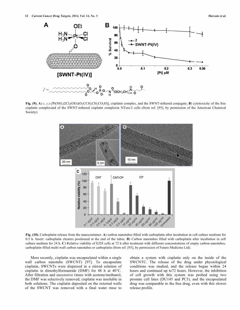
Feazell et al. [95] reported that the preparation of a Pt(IV)-based SWCNT pro-drug led to intracellular concentrations of Pt4+six times higher than when the cells were treated with the free drug (Fig. 9).
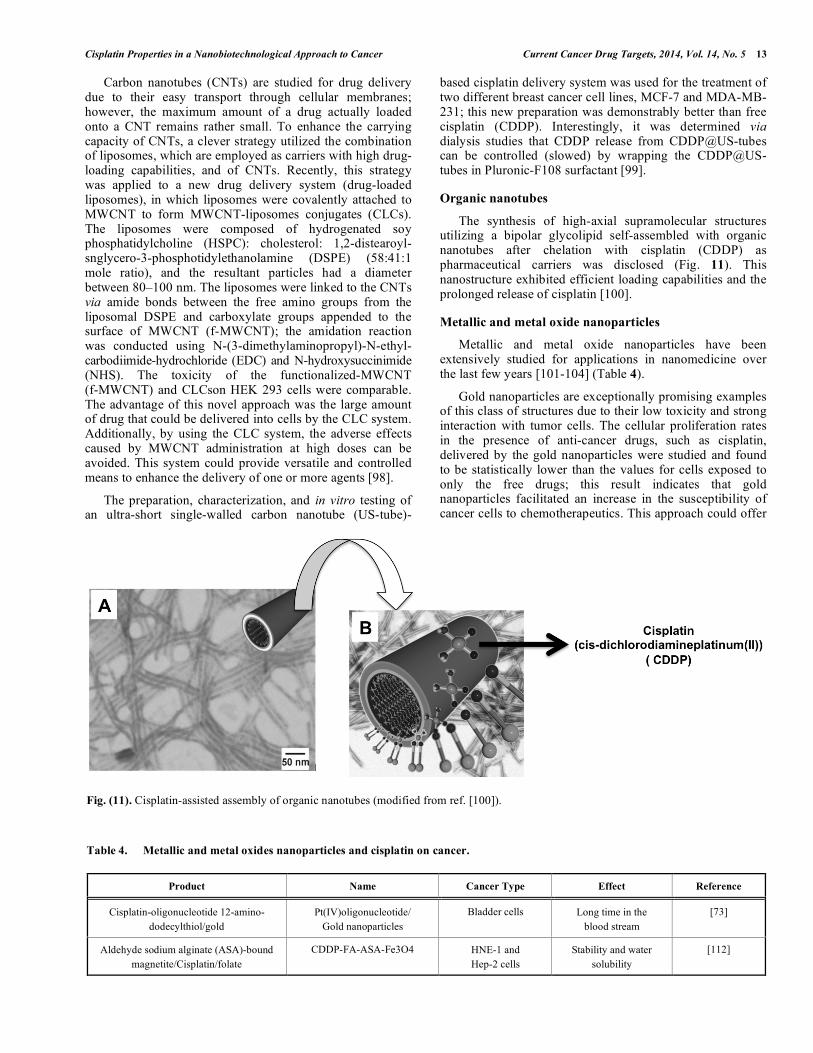
Filling the internal empty cavity of the nanotubes with carboplatin is also an interesting strategy [96]. The release of carboplatin was evaluated via the incubation of MWCNT-carboplatin in cell culture medium; at different times, the sample was analyzed by TEM. Generally, the filled yield of the cisplatin derivative was relatively high in the carboplatin clusters situated at the ends of the tubes (Fig. 10A). A reduction in the number of carboplatin clusters within the MWCNT-carboplatin was detected after an incubation period of 24 h, at which time only a few clusters could be detected inside the tubes (Fig. 10B).
To test the functionality of MWCNT-carboplatin as a therapeutic carrier system, the cytotoxicity was analyzed in systems with either empty or carboplatin-filled MWCNT. EJ28 cells, which are derived from human bladder cancer, were incubated with different concentrations of either sample (MWCNT empty and carboplatin filled). The results of the cell-viability assay using the cell-proliferation reagent WST-1 (4-[3-(4-iodophenyl)-2-(4-nitrophenyl)-2H-5-tetrazolio]-1,3-benzene disulfonate) revealed that empty MWCNTs (0.1-0.5 µg/µL) caused no reduction in cell viability 72 h after treatment (Fig. 10c). However, free carboplatin showed the expected toxicity in a concentration-dependent manner (Fig. 10c). Similar to free carboplatin, MWCNT-carboplatin caused a concentration-dependent cytotoxic effect, and a strong inhibition of viability was produced by the MWCNT-carboplatin at concentrations up to 0.5 µg/µL [96]. Although no results were obtained pertaining to carboplatin release, which precluded its meaningful comparison with the effectiveness of the free form of the drug, these results still confirmed that the nanotubes could be used to deliver cytotoxic drugs.
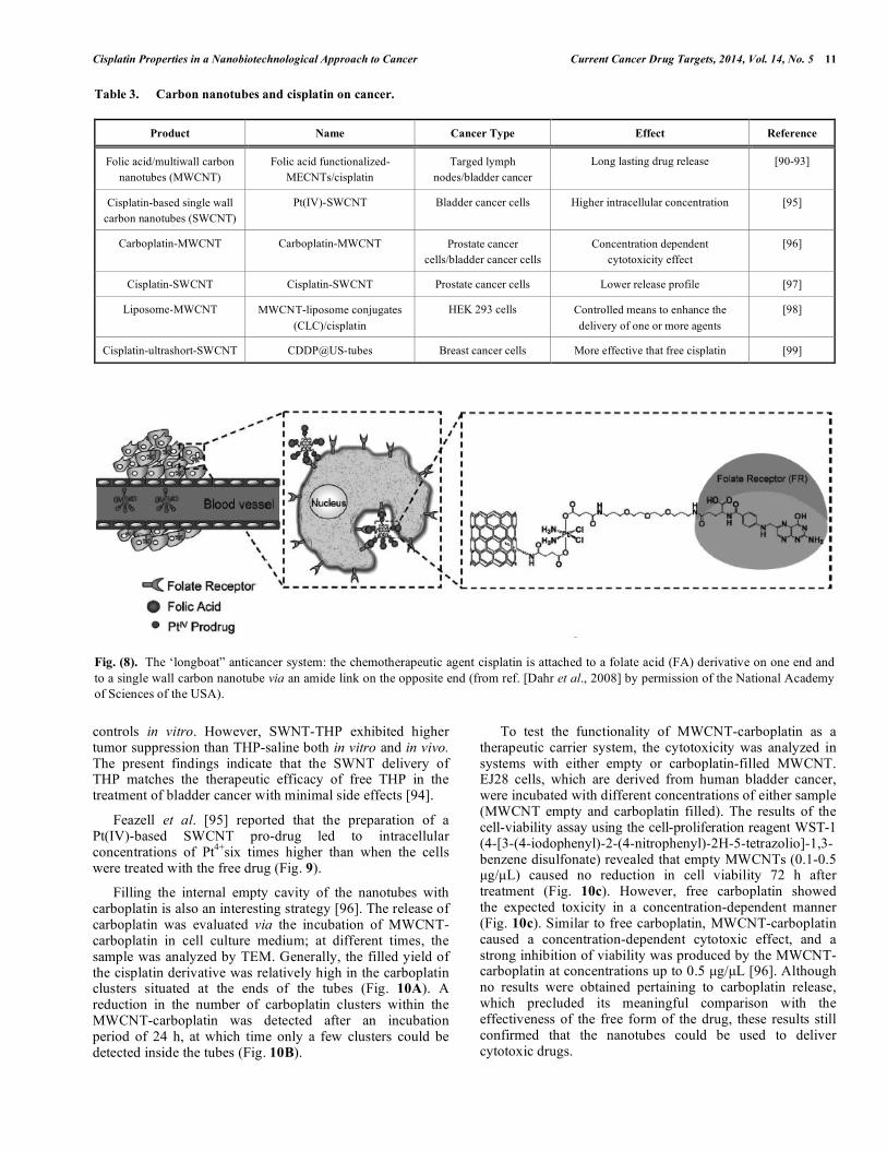
Table 3. Carbon nanotubes and cisplatin on cancer.
Product Name Cancer Type Effect Reference
Folic acid/multiwall carbon nanotubes (MWCNT)
Folic acid functionalized-MECNTs/cisplatin
Targed lymph nodes/bladder cancer
Long lasting drug release [90-93]
Cisplatin-based single wall carbon nanotubes (SWCNT)
Pt(IV)-SWCNT Bladder cancer cells Higher intracellular concentration [95]
Carboplatin-MWCNT Carboplatin-MWCNT Prostate cancer cells/bladder cancer cells
Concentration dependent cytotoxicity effect
[96]
Cisplatin-SWCNT Cisplatin-SWCNT Prostate cancer cells Lower release profile [97]
Liposome-MWCNT MWCNT-liposome conjugates (CLC)/cisplatin
HEK 293 cells Controlled means to enhance the delivery of one or more agents
[98]
Cisplatin-ultrashort-SWCNT CDDP@US-tubes Breast cancer cells More effective that free cisplatin [99]
Fig. (8). The ‘longboat” anticancer system: the chemotherapeutic agent cisplatin is attached to a folate acid (FA) derivative on one end and to a single wall carbon nanotube via an amide link on the opposite end (from ref. [Dahr et al., 2008] by permission of the National Academy of Sciences of the USA).
12 Current Cancer Drug Targets, 2014, Vol. 14, No. 5 Marcato et al.
More recently, cisplatin was encapsulated within a single wall carbon nanotube (SWCNT) [97]. To encapsulate cisplatin, SWCNTs were dispersed in a stirred solution of cisplatin in dimethylformamide (DMF) for 48 h at 40°C. After filtration and successive rinses with acetone/methanol, the DMF was selectively removed; cisplatin was insoluble in both solutions. The cisplatin deposited on the external walls of the SWCNT was removed with a final water rinse to
obtain a system with cisplatin only on the inside of the SWCNTC. The release of the drug under physiological conditions was studied, and the release began within 24 hours and continued up to72 hours. However, the inhibition of cell growth with this system was probed using two prostate cell lines (DU145 and PC3), and the encapsulated drug was comparable to the free drug, even with this slower release profile.
Fig. (9). A) c, c,t-[Pt(NH3)2Cl2(OEt)(O2CCH2CH2CO2H)], cisplatin complex, and the SWNT-tethered conjugate; B) cytotoxicity of the free cisplatin complexand of the SWNT-tethered cisplatin complexin NTera-2 cells (from ref. [95], by permission of the American Chemical Society).
Fig. (10). Carboplatin release from the nanocontainer. A) carbon nanotubes filled with carboplatin after incubation in cell culture medium for 0.5 h. Insert: carboplatin clusters positioned at the end of the tubes. B) Carbon nanotubes filled with carboplatin after incubation in cell culture medium for 24 h. C) Relative viability of EJ28 cells at 72 h after treatment with different concentrations of empty carbon nanotubes, carboplatin-filled multi-wall carbon nanotubes or carboplatin (from ref. [93], by permission of Future Medicine Ltd).
Cisplatin Properties in a Nanobiotechnological Approach to Cancer Current Cancer Drug Targets, 2014, Vol. 14, No. 5 13
Carbon nanotubes (CNTs) are studied for drug delivery due to their easy transport through cellular membranes; however, the maximum amount of a drug actually loaded onto a CNT remains rather small. To enhance the carrying capacity of CNTs, a clever strategy utilized the combination of liposomes, which are employed as carriers with high drug-loading capabilities, and of CNTs. Recently, this strategy was applied to a new drug delivery system (drug-loaded liposomes), in which liposomes were covalently attached to MWCNT to form MWCNT-liposomes conjugates (CLCs). The liposomes were composed of hydrogenated soy phosphatidylcholine (HSPC): cholesterol: 1,2-distearoyl-snglycero-3-phosphotidylethanolamine (DSPE) (58:41:1 mole ratio), and the resultant particles had a diameter between 80–100 nm. The liposomes were linked to the CNTs via amide bonds between the free amino groups from the liposomal DSPE and carboxylate groups appended to the surface of MWCNT (f-MWCNT); the amidation reaction was conducted using N-(3-dimethylaminopropyl)-N-ethyl-carbodiimide-hydrochloride (EDC) and N-hydroxysuccinimide (NHS). The toxicity of the functionalized-MWCNT (f-MWCNT) and CLCson HEK 293 cells were comparable. The advantage of this novel approach was the large amount of drug that could be delivered into cells by the CLC system. Additionally, by using the CLC system, the adverse effects caused by MWCNT administration at high doses can be avoided. This system could provide versatile and controlled means to enhance the delivery of one or more agents [98].
The preparation, characterization, and in vitro testing of an ultra-short single-walled carbon nanotube (US-tube)-
based cisplatin delivery system was used for the treatment of two different breast cancer cell lines, MCF-7 and MDA-MB-231; this new preparation was demonstrably better than free cisplatin (CDDP). Interestingly, it was determined via dialysis studies that CDDP release from CDDP@US-tubes can be controlled (slowed) by wrapping the CDDP@US-tubes in Pluronic-F108 surfactant [99].
Organic nanotubes
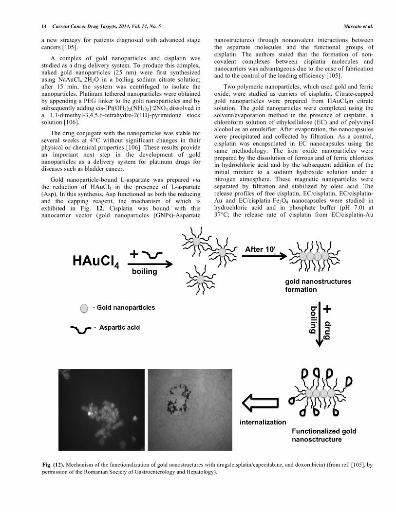
The synthesis of high-axial supramolecular structures utilizing a bipolar glycolipid self-assembled with organic nanotubes after chelation with cisplatin (CDDP) as pharmaceutical carriers was disclosed (Fig. 11). This nanostructure exhibited efficient loading capabilities and the prolonged release of cisplatin [100].
Metallic and metal oxide nanoparticles
Metallic and metal oxide nanoparticles have been extensively studied for applications in nanomedicine over the last few years [101-104] (Table 4).
Gold nanoparticles are exceptionally promising examples of this class of structures due to their low toxicity and strong interaction with tumor cells. The cellular proliferation rates in the presence of anti-cancer drugs, such as cisplatin, delivered by the gold nanoparticles were studied and found to be statistically lower than the values for cells exposed to only the free drugs; this result indicates that gold nanoparticles facilitated an increase in the susceptibility of cancer cells to chemotherapeutics. This approach could offer
Fig. (11). Cisplatin-assisted assembly of organic nanotubes (modified from ref. [100]).
Table 4. Metallic and metal oxides nanoparticles and cisplatin on cancer.
Product Name Cancer Type Effect Reference
Cisplatin-oligonucleotide 12-amino-dodecylthiol/gold
Pt(IV)oligonucleotide/ Gold nanoparticles
Bladder cells Long time in the blood stream
[73]
Aldehyde sodium alginate (ASA)-bound magnetite/Cisplatin/folate
CDDP-FA-ASA-Fe3O4 HNE-1 and Hep-2 cells
Stability and water solubility
[112]
14 Current Cancer Drug Targets, 2014, Vol. 14, No. 5 Marcato et al.
a new strategy for patients diagnosed with advanced stage cancers [105].
A complex of gold nanoparticles and cisplatin was studied as a drug delivery system. To produce this complex, naked gold nanoparticles (25 nm) were first synthesized using NaAuCl4·2H2O in a boiling sodium citrate solution; after 15 min, the system was centrifuged to isolate the nanoparticles. Platinum tethered nanoparticles were obtained by appending a PEG linker to the gold nanoparticles and by subsequently adding cis-[Pt(OH2)2(NH3)2]·2NO3 dissolved in a 1,3-dimethyl-3,4,5,6-tetrahydro-2(1H)-pyrimidone stock solution [106].
The drug conjugate with the nanoparticles was stable for several weeks at 4°C without significant changes in their physical or chemical properties [106]. These results provide an important next step in the development of gold nanoparticles as a delivery system for platinum drugs for diseases such as bladder cancer.
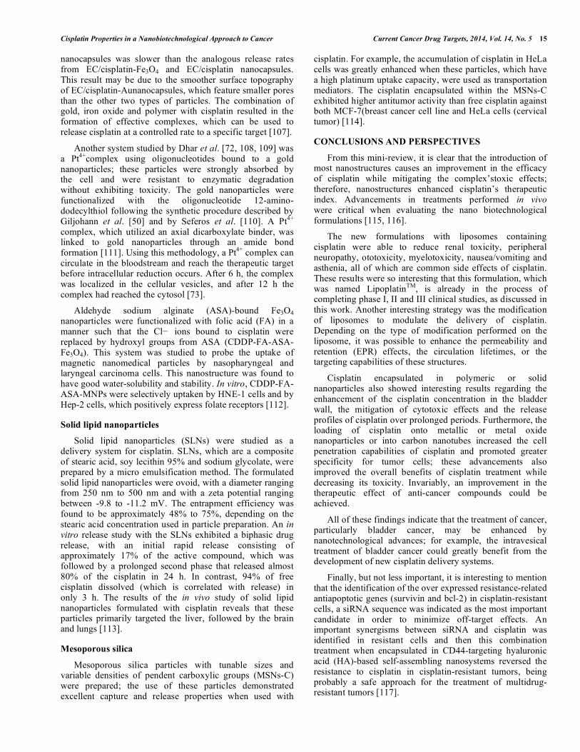
Gold nanoparticle-bound L-aspartate was prepared via the reduction of HAuCl4 in the presence of L-aspartate (Asp). In this synthesis, Asp functioned as both the reducing and the capping reagent, the mechanism of which is exhibited in Fig. 12. Cisplatin was bound with this nanocarrier vector (gold nanoparticles (GNPs)-Aspartate
nanostructures) through noncovalent interactions between the aspartate molecules and the functional groups of cisplatin. The authors stated that the formation of non-covalent complexes between cisplatin molecules and nanocarriers was advantageous due to the ease of fabrication and to the control of the loading efficiency [105].
Two polymeric nanoparticles, which used gold and ferric oxide, were studied as carriers of cisplatin. Citrate-capped gold nanoparticles were prepared from HAuCl4in citrate solution. The gold nanoparticles were completed using the solvent/evaporation method in the presence of cisplatin, a chloroform solution of ethylcellulose (EC) and of polyvinyl alcohol as an emulsifier. After evaporation, the nanocapsules were precipitated and collected by filtration. As a control, cisplatin was encapsulated in EC nanocapsules using the same methodology. The iron oxide nanoparticles were prepared by the dissolution of ferrous and of ferric chlorides in hydrochloric acid and by the subsequent addition of the initial mixture to a sodium hydroxide solution under a nitrogen atmosphere. These magnetic nanoparticles were separated by filtration and stabilized by oleic acid. The release profiles of free cisplatin, EC/cisplatin, EC/cisplatin-Au and EC/cisplatin-Fe3O4 nanocapsules were studied in hydrochloric acid and in phosphate buffer (pH 7.0) at 37°C; the release rate of cisplatin from EC/cisplatin-Au
Fig. (12). Mechanism of the functionalization of gold nanostructures with drugs(cisplatin/capecitabine, and doxorubicin) (from ref. [105], by permission of the Romanian Society of Gastroenterology and Hepatology).
Cisplatin Properties in a Nanobiotechnological Approach to Cancer Current Cancer Drug Targets, 2014, Vol. 14, No. 5 15
nanocapsules was slower than the analogous release rates from EC/cisplatin-Fe3O4 and EC/cisplatin nanocapsules. This result may be due to the smoother surface topography of EC/cisplatin-Aunanocapsules, which feature smaller pores than the other two types of particles. The combination of gold, iron oxide and polymer with cisplatin resulted in the formation of effective complexes, which can be used to release cisplatin at a controlled rate to a specific target [107].
Another system studied by Dhar et al. [72, 108, 109] was a Pt4+complex using oligonucleotides bound to a gold nanoparticles; these particles were strongly absorbed by the cell and were resistant to enzymatic degradation without exhibiting toxicity. The gold nanoparticles were functionalized with the oligonucleotide 12-amino-dodecylthiol following the synthetic procedure described by Giljohann et al. [50] and by Seferos et al. [110]. A Pt4+ complex, which utilized an axial dicarboxylate binder, was linked to gold nanoparticles through an amide bond formation [111]. Using this methodology, a Pt4+ complex can circulate in the bloodstream and reach the therapeutic target before intracellular reduction occurs. After 6 h, the complex was localized in the cellular vesicles, and after 12 h the complex had reached the cytosol [73].
Aldehyde sodium alginate (ASA)-bound Fe3O4 nanoparticles were functionalized with folic acid (FA) in a manner such that the Cl− ions bound to cisplatin were replaced by hydroxyl groups from ASA (CDDP-FA-ASA-Fe3O4). This system was studied to probe the uptake of magnetic nanomedical particles by nasopharyngeal and laryngeal carcinoma cells. This nanostructure was found to have good water-solubility and stability. In vitro, CDDP-FA-ASA-MNPs were selectively uptaken by HNE-1 cells and by Hep-2 cells, which positively express folate receptors [112].
Solid lipid nanoparticles
Solid lipid nanoparticles (SLNs) were studied as a delivery system for cisplatin. SLNs, which are a composite of stearic acid, soy lecithin 95% and sodium glycolate, were prepared by a micro emulsification method. The formulated solid lipid nanoparticles were ovoid, with a diameter ranging from 250 nm to 500 nm and with a zeta potential ranging between -9.8 to -11.2 mV. The entrapment efficiency was found to be approximately 48% to 75%, depending on the stearic acid concentration used in particle preparation. An in vitro release study with the SLNs exhibited a biphasic drug release, with an initial rapid release consisting of approximately 17% of the active compound, which was followed by a prolonged second phase that released almost 80% of the cisplatin in 24 h. In contrast, 94% of free cisplatin dissolved (which is correlated with release) in only 3 h. The results of the in vivo study of solid lipid nanoparticles formulated with cisplatin reveals that these particles primarily targeted the liver, followed by the brain and lungs [113].
Mesoporous silica
Mesoporous silica particles with tunable sizes and variable densities of pendent carboxylic groups (MSNs-C) were prepared; the use of these particles demonstrated excellent capture and release properties when used with
cisplatin. For example, the accumulation of cisplatin in HeLa cells was greatly enhanced when these particles, which have a high platinum uptake capacity, were used as transportation mediators. The cisplatin encapsulated within the MSNs-C exhibited higher antitumor activity than free cisplatin against both MCF-7(breast cancer cell line and HeLa cells (cervical tumor) [114].
CONCLUSIONS AND PERSPECTIVES
From this mini-review, it is clear that the introduction of most nanostructures causes an improvement in the efficacy of cisplatin while mitigating the complex’stoxic effects; therefore, nanostructures enhanced cisplatin’s therapeutic index. Advancements in treatments performed in vivo were critical when evaluating the nano biotechnological formulations [115, 116].
The new formulations with liposomes containing cisplatin were able to reduce renal toxicity, peripheral neuropathy, ototoxicity, myelotoxicity, nausea/vomiting and asthenia, all of which are common side effects of cisplatin. These results were so interesting that this formulation, which was named LipoplatinTM, is already in the process of completing phase I, II and III clinical studies, as discussed in this work. Another interesting strategy was the modification of liposomes to modulate the delivery of cisplatin. Depending on the type of modification performed on the liposome, it was possible to enhance the permeability and retention (EPR) effects, the circulation lifetimes, or the targeting capabilities of these structures.
Cisplatin encapsulated in polymeric or solid nanoparticles also showed interesting results regarding the enhancement of the cisplatin concentration in the bladder wall, the mitigation of cytotoxic effects and the release profiles of cisplatin over prolonged periods. Furthermore, the loading of cisplatin onto metallic or metal oxide nanoparticles or into carbon nanotubes increased the cell penetration capabilities of cisplatin and promoted greater specificity for tumor cells; these advancements also improved the overall benefits of cisplatin treatment while decreasing its toxicity. Invariably, an improvement in the therapeutic effect of anti-cancer compounds could be achieved.
All of these findings indicate that the treatment of cancer, particularly bladder cancer, may be enhanced by nanotechnological advances; for example, the intravesical treatment of bladder cancer could greatly benefit from the development of new cisplatin delivery systems.
Finally, but not less important, it is interesting to mention that the identification of the over expressed resistance-related antiapoptotic genes (survivin and bcl-2) in cisplatin-resistant cells, a siRNA sequence was indicated as the most important candidate in order to minimize off-target effects. An important synergisms between siRNA and cisplatin was identified in resistant cells and then this combination treatment when encapsulated in CD44-targeting hyaluronic acid (HA)-based self-assembling nanosystems reversed the resistance to cisplatin in cisplatin-resistant tumors, being probably a safe approach for the treatment of multidrug-resistant tumors [117].
16 Current Cancer Drug Targets, 2014, Vol. 14, No. 5 Marcato et al.
CONFLICT OF INTEREST
The authors confirm that this article content has no conflict of interest.
ACKNOWLEDGEMENTS
This work was supported by CNPq, FAPESP, INOMAT (MCT/CNPq), NanoBioss (MCTI) and Brazilian Network of Nanotoxicology (MCT/CNPq). The authors acknowledge the Elsevier Language Editing Services.
LIST OF ABBREVIATIONS
Bamet-R2 = bile acid–cisplatin complex
CDDP = free cisplatin
CHO = ovarian cells from Chinese hamsters
CHOL = cholesterol
CLC = MWCNT-liposomes conjugates
CNT = carbon nanotubes
DCSP = cis-dichloridobis(2-stearoyl-hydrazide) platinum (II)
DPPC = 1,2-dipalmitoyl-sn-glycero-3-phospha- tidyl-choline
DSM = starch microspheres
DSPC = 1,2-distearoyl-sn-glycero-3-phospha- tidylcholine
DSPG = 1,2-distearoyl-sn-glycero-3-phospha- tidylglycerol
DTX = Docetaxel
HA = hyaluronic acid
HGC = glycol chitosan–cholanic acid conjugate
HPB = microporous frameworks
HPG = hyperbranched polyglycerol
HPNG = poly(N-isopropylacrylamide) grafted with hyaluronic acid grafted with gelatin
LD = liposomal doxorubicin
MPM = malignant pleural mesothelioma
MWNT = multi-wall carbon nanotubes
NDDP = cis-bisneodecanoato-trans-R,R-1,2-diaminocyclo hexane platinum (II)
PEG = polyethyleneglycol
PEI = polyethylenimine
PLGA = poly(D,L-lactic-co-glycolic acid)
PLK-1 = polo-like kinase-1
PNIPAM = poly(N-isopropylacrylamide)
Pt = platinum (Pt)
Ptsome = liposome-like self-assembly
QD = quantum dots
ROS = Reactive oxygen species
SACN = Self-assembling, cholesterol-tethered and succinic-acid-bound cisplatinum (II)-based nanoparticles
SLN = Solid lipid nanoparticles
SPI-077 = cholesterol-pegylated liposomes with cisplatin
SPION = superparamagnetic iron oxide nano- particles
siRNA = small interfering RNA
SWNT = single-wall carbon nanotubes
TEM = transmission electron microscopy
Tf-PEG = transferrin-conjugated polyethylene liposomes glycol liposomes
THP = pirarubicin (tetrahydropyranyl-adriamycin)
REFERENCES [1] Marcato, P.D.; Durán, N. New aspects of nanopharmaceutical
delivery systems. J. Nanosci. Nanotechnol.2008, 8(5), 2216-2229. [2] Melo, P.S.; Marcato, P.D.; Durán, N. In Nanocosmetics and
Nanomedicines: New approaches for skin care, S.S. Guterres, R. Beck and A. Polhmann, Eds; Springer: Germany, 2011, vol. 1, pp. 229-238
[3] De Souza, A.O.; Silva, C.L.; Durán, N.; Andrade-Santana, M.H. Antimycobacterial and cytotoxicity activities of free and liposome-encapsulated 3-(4’-bromo[1,1’-biphenyl-4-yl)-3-(4-bromo-phenyl)-N,N-dimethyl-2-propen-1-amine. Quim. Nova2010, 33(4), 871-874.
[4] Marcato, P.D.; Adami, L.F.; Barbosa, R.M.; Melo, P.S.; Ferreira, I.R.; de Paula, L.; Durán, N.; Seabra, A.B. Development of a Sustained-release System for Nitric Oxide Delivery using Alginate/Chitosan Nanoparticles. Curr. Nanosci. 2013, 9(1), 1-7.
[5] Melo, P.S.; De Azevedo, M.M.M.; Frungillo, L.; Anazetti, M.C.; Marcato, P.D.; Durán, N. Nanocytotoxicity: Violacein and Violacein-Loaded Poly (D,L-lactide-co-glycolide) Nanoparticles Acting on Human Leukemic Cells. J. Biomed. Nanotechnol. 2009, 5(2), 192-201.
[6] Durán, N.; Marcato, P.D.; Buffo, C.; De Azevedo, M.M.M.; Esposito, E. Poly(e-caprolactone)/propolis extract: microencapsulation and antibacterial activity evaluation. Die. Pharmazie.2007, 62(4), 287-290.
[7] Marcato, P.D.; Caverzan, J.; Rossi-Bergmann, B.; Pinto, E.F.; Machado, D.; Silva, R.A.; Justo, G.Z.; Ferreira, C.V.; Durán, N. Nanostructured polymer and lipid carriers for sunscreen. Biological effects and skin permeation. J. Nanosci. Nanotechnol.2011, 11(3), 1880-1886.
[8] Marcato, P.D. Preparação, caracterização e aplicações em fármacos e cosméticos de nanopartículas lipídicas sólidas. Rev. Eletronica. Fármacia2009, 6(2), 1-37.
[9] Ridolfi, D.; Marcato, P.D.; Justo, G.Z.; Cordi, L.; Machado, D.; Durán, N. Solid lipid nanoparticles coated with chitosan as carrier for topical delivery of Tretinoin. Colloids Surf. B: Biointerf. 2012, 93, 36-40.
[10] Pereira, A.C.; Kisner, A.; Durán, N.; Kubota, L.T.; The Effects of dimensionality on electrochemical sensors based on carbon nanotubes and metallic nanowires. J. Nanosci. Nanotechnol.2010, 10(2), 651-667.
[11] Durán, N.; Marcato, P.D.; Durán, M.; Yadav, A.; Gade, A.; Rai. M. Mechanistic aspects in the biogenic synthesis of extracellular metal nanoparticles by peptides, bacteria, fungi and plants. Appl. Microbiol. Biotechnol. 2011, 90(5), 1609-1624.
[12] Marcato, P.D.; Parizotto, N.V.; Martinez, D.S.T.; Paula, A.J.; Ferreira, I.R.; Melo, P.S.; Durán, N.; Alves, O.L. New hybrid
Cisplatin Properties in a Nanobiotechnological Approach to Cancer Current Cancer Drug Targets, 2014, Vol. 14, No. 5 17
material based on layered double hydroxides and biogenic silver nanoparticles: antimicrobial activity and cytotoxic effect. J. Brazilian. Chem. Soc. 2013, 24(2), 1-7.
[13] Marcato, P.D.; Durán, M.; Huber, S.; Rai, M.; Melo, PS.; Alves, O.L.; Durán, N. Biogenic silver nanoparticles and its antifungal activity as a new topical transungual drug. J. Nano. Res. 2012, 20, 99-107.
[14] Marcato, P.D.; Nakasato, G.; Brocchi, M.; Melo, P.S.; Huber, S.C.; Ferreira, I.R.; Alves, O.L.; Durán, N.; Biogenic silver nanoparticles: Antibacterial and cytotoxicity applied to textile fabrics. J. Nano. Res.2012, 20, 69-76.
[15] Rai, M.; Gade, A.; Gaikwad, S.; Marcato, P.D.; Durán, N. Biomedical Applications of Nanobiosensors: The-State-of-The-Art. J. Brazilian. Chem. Soc.2012, 23(1), 14-24.
[16] Askeland, E.J.; Newton, M.R.; O'Donnell, M.A. Luo, Y. Bladder Cancer Immunotherapy: BCG and Beyond. Adv. Urol.2012, ID 181987.
[17] National Cancer Institute. SEER Stat Fact Sheets: Bladder. http://seer.cancer.gov/statfacts/html/urinb.html. (Accessed April 20, 2013)
[18] Bellmunt, J.; Albanell, J.; Paz-Ares, L.; Climentm, M.A.; González-Larriba, J.L.; Carles, J.; de la Cruz, J.J.; Guillem, V.; Díaz-Rubio, E.; Cortés-Funes, H.; Baselga, J. Pretreatment prognostic factors for survival in patients with advanced urothelial tumors treated in phase I/II trial with paclitaxel, cisplatin and gemcitabine. Cancer. 2002, 95(4), 751-757.
[19] Shikanov, A.; Shikanov, S.; Vaisman, B.; Golenser, J.; Domb, A.J. Cisplatin tumor biodistribution and efficacy after intratumoral injection of a biodegradable extended release implant. Chemother. Res. Practice.2011, ID 175054
[20] Stathopoulos, G.P.; Antoniou, D.; Dimitroulis, J.; Stathopoulos, J.; Marosis, K.; Michalopoulou, P. Comparison of liposomal cisplatin versus cisplatinin non-squamous cell non-small-cell lung cancer. Cancer. Chemother. Pharmacol. 2011, 68(4), 945-950.
[21] Farrell, N.P. Platinum formulations as anticancer drugs clinical and pre-clinical studies. Curr. Topics. Med. Chem. 2011, 11(21), 2623-2631.
[22] Farrokhi, F.; Ardjmand, M.; Alavi, S.A. In: A study of cis-platin effects as anticancer drug. Proceeding of 2nd Inter. Conf. Advan. Biotechnol. Pharm. Sci., ICABPS, Bali, 2012, pp. 12-14.
[23] Silva, G.B.; Vargas, M.D.; Pt4+ Complexes: Molecular approach against Câncer. Rev. Virtual. Quim. 2012, 4(2), 102-117.
[24] Moore, D.H. Chemotherapy for recurrent cervical carcinoma. Curr. Opinion. Oncol. 2006, 18(5), 516-519.
[25] Rabik, C.A.; Dolan, M.E.; Molecular mechanisms ofresistance and toxicity associated with platinating agents. Cancer. Treat. Rev. 2007, 33(1), 9-23.
[26] Sternberg, C.N.; Donat, S.M.; Bellmunt, J.; Millikan, R.E.; Stadler, W.; De Mulder, P.; Sherif, A.; von der Maase, H.; Tsukamoto, T.; Soloway, M.S.; Chemotherapy for bladder cancer: treatment guidelines for neoadjuvant chemotherapy, bladder preservation, adjuvant chemotherapy, and metastatic cancer. Urology2007, 69(1), 62-79.
[27] Harmers, F.P.; Gispen, W.H.; Neijt, J.P. Neurotoxic side-effects of cisplatin. Eur. J. Cancer.2006, 27(3), 372-376.
[28] Humanes, B.; Lazaro, A.; Camano, S. Moreno-Gordaliza, E.; Lazaro, J.A.; Blanco-Codesido, M.; Lara, J.M.; Ortiz, A.; Gomez-Gomez, M.M.; Martín-Vasallo, P.; Tejedor, A.Cilastatin protects against cisplatin-induced nephrotoxicity without compromising its anticancer efficiency in rats. Kidney. Int. 2012, 82(6), 652-663.
[29] Berners-Price, S.J.; Appleton, T.G. In: Kelland LR, N.P. Farrell, Ed.; Humana Press: Totowa, 2000, vol. 1, pp 1-35.
[30] Lee, K.W.; Jeong, J.Y.; Lim, B.J.; Chang, Y.K.; Lee, S.J.; Na, K.R.; Shin, Y.T.; Choi, D.E. Sildenafil attenuates renal injury in an experimental model or rat cisplatin-induced nephrotoxicity. Toxicology. 2009, 257(3), 137-143.
[31] Yao, X.; Panichpisal, K.; Kurtzman, N.; Nugent, K. Cisplatin nephrotoxicity: a review. Am. J. Med. Sci. 2007, 334(2), 115-124.
[32] Pabla, N.; Dong, Z. Cisplatin nephrotoxicity: mechanisms and renoprotective strategies. Kidney. Int.2008, 73(9), 994-1007.
[33] Nagothu, K.K.; Bhatt, R.; Kaushal, G.P.; Portilla, D. Fibrate prevents cisplatin-induced proximal tubule cell death. Kidney. Int. 2005, 68(6), 2680-2693.
[34] Choi, D.E.; Jeong, J.Y.; Lim, B.J.; Lee, K.W.;Shin, Y-T.;Na, K-R. Pretreatment with darbepoetin attenuates renal injury in a rat model
of cisplatin-induced nephrotoxicity. Korean. J. Intern. Med.2009, 24(3), 238-246.
[35] Jin, S.; Labhasetwar, V. Nanotechnology in urology. Urol. Clin. North. Am.2009, 36(2), 179-188.
[36] Ramachandran, S.; Quist, A.P.; Kumar, S.; Lal, R. Cisplatinnano- liposomes for cancer therapy: AFM and fluorescenceimaging of cisplatin encapsulation, stability, cellular uptake,and toxicity. Langmuir, 2008, 22(19), 8156-8162.
[37] Uchino, U.; Matsumura, Y.; Negishi, T.; Koizumi, F.; Hayashi, T.; Honda, T.; Nishiyama, N.; Kataoka, K.; Naito, S.; Kakizoe, T. Cisplatin incorporating polymeric micelles (NC-6004) can reduce nephrotoxicity and neurotoxicity of cisplatin in rats. Br. J. Cancer. 2005, 93(6), 678-687.
[38] Konishi, M.; Tabata, Y.; Kariya, M.; Suzuki, A.; Mandai, M.; Nanbu, K.; Takakura, K.; Fujii, S. In vivo antitumor effect through the controlled release of cisplatin frombiodegradable gelatin hydrogel. J. Control. Rel. 2003, 92(3), 301-313.
[39] Chen, F.A.; Kuriakose, M.A.; Zhou, M.X.; DeLacure, M.D.; Dunn, R.L. Biodegradable polymer-mediated intratumoraldelivery of cisplatin for treatment of human head and necksquamous cell carcinoma in a chimeric mouse model. Head. Neck. 2003, 25(7), 554-560.
[40] Yapp, D.T.T.; Lloyd, D.K.; Zhu, J.; Lehnert, S.M. Cisplatindelivery by biodegradable polymer implant is superior tosystemic delivery by osmotic pump or i.p. injection in tumorbearingmice.Anti-Cancer. Drugs. 1998, 9(9), 791-796.
[41] Peer, D.; Karp, J.M.; Hong, S.; Farokhzad, O.C.; Margalit, R.; Langer, R. Nanocarriers as an emerging platform for cancer therapy. Nat. Nanotechnol. 2007, 2, 751-760.
[42] Zhang, L.; Gu, F.X.; Chan, J.M.; Wang, A.Z.; Langer, R.S.; Farokhzad, O.C. Nanoparticles in medicine: Therapeutic applications and developments. Clin. Pharmacol. Ther. 2008, 83(5), 761-769.
[43] Alexis, A.; Pridgen, E.; Molnar, L.K.; Farokhzad, O.V. Factors affecting the clearance and biodistribution of polymeric nanoparticles. Mol. Pharm. 2008, 5(4), 505-515.
[44] Kumari, A.; Yadav, S.K.; Subhash, C.; Yadav, S.C. Biodegradable polymeric nanoparticles based drug delivery systems. Colloids Surf B: Biointerf. 2010, 75(1), 1-18.
[45] Figueiredo, M.; Rinat, Esenaliev. R. PLGA Nanoparticles for ultrasound-mediated gene delivery to solid tumors. J. Drug. Deliv. 2012, ID 767839
[46] Ismaili, N.; Amzerin, M.; Aude, Flechon. A. Chemotherapy in advanced bladder cancer:current status and future. J. Hematol Oncol.2011, 4(35), 1-11.
[47] Chaudhury, A.; Das, S.; Lee, R.F.S.; Tan, K.B.; Ng, W.K.; Tan, R.B.H.; Chiu, G.N.C. Lyophilization of cholesterol-free PEGylated liposomes and its impact on drug loading by passive equilibration. Inter. J. Pharm. 2012, 430(1-2), 167-175.
[48] Zamboni, W.C.; Gervais, A.C.; Egorin, M.J.; Schellens, J.H.; Zuhowski, E.G.; Pluim, .; Joseph, E.; Hamburger.; DR, Working, PK.; Colbern, G.; Tonda, M.E.; Potter, D.M.; Eisemen, J.L. Systemic and tumor disposition of platinum after administration of cisplatin or STEALTH liposomal-cisplatin formulations (SPI-077 and SPI-077 B103) in a preclinical tumor model of melanoma. Cancer. Chemother. Pharmacol. 2004, 53(4), 329-336.
[49] Harrington, K.J.; Lewanski, C.R.; Northcote, A.D.; Whittaker, J.; Wellbank, H.; Vile, R.G.; Peters, A.M.; Stewart, J.S. Phase I-II study of pegylated liposomal cisplatin (SPI-077) in patients with inoperable head and neck cancer. Ann. Oncol. 2001,12(4), 493-496.
[50] Giljohann, D.A.; Seferos, D.S.; Patel, P.C.; Millstone, J.E.; Rosi, N.L.; Mirkin, C.A. Oligonucleotide loading determines cellular uptake of DNA-modified gold nanoparticles.Nano. Lett.2007, 7(12), 3818-3821.
[51] Canta, A.; Chiorazzi, A.; Carozzi, V.; Meregalli, C.; Oggioni, N.; Sala, B.; Crippa, L.; Avezza, F.; Forestieri, D.; Rotella, G.; Zucchetti, M.; Cavaletti, G. In vivo comparative study of the cytotoxicity of a liposomal formulation of cisplatin (lipoplatin™) Cancer. Chemother. Pharmacol. 2011,68(4), 1001-1008.
[52] Boulikas, T. Clinical overview on Lipoplatin™: A successful liposomal formulation of cisplatin. Expert Opinion Invest Drugs. 2009, 18(8), 1197-1218.
[53] Stathopoulos, P.G.; Rigatos, S.; Stathopoulos, J.; Batzios, S. Liposomal cisplatin in cancer patients with renal failure .J. Drug. Deliv .Ther. 2012, 2(3), 106-109.
18 Current Cancer Drug Targets, 2014, Vol. 14, No. 5 Marcato et al.
[54] Dragovich, T.; Mendelson, D.; Kurtin, S.; Richardson, K.; HoV, D.V.; Hoos, A. A. Phase 2 trial of the liposomal DACH platinum L-NDDP in patients with therapy-refractory advanced colorectal cancer.Cancer. Chemother. Pharmacol. 2006, 58(6), 759-764.
[55] Paraskar, A.; Soni, S.; Roy, B.; Papa, A.L.; Sengupta, S. Rationally designed oxaliplatinnanoparticle for enhanced antitumor efficacy. Nanotechnology.2012 23(7) 075103.
[56] Alderden, R.A.; Hall, M.D.; Hambley, T.W. The discovery and development of cisplatin. J. Chem. Educ.2006, 83(5), 728-734.
[57] Aryal, S.; Hu, C.M.J.; Zhang, L. Synthesis of Ptsome: a platinum-based liposome-like nanostructure. Chem. Commun. 2012, 48(20), 2630-2632.
[58] Leite, E.A.; Lana, A.M.Q.; Carvalho Junior, A.D.; Coelho, L.G.V.; De Oliveira, M.C. Acute toxicity study of cisplatin loaded long-circulating and pH-sensitive liposomes administered in mice J. Biomed. Nanotechnol. 2012, 8(2), 229-239.
[59] Sengupta, P.; Basu, S.; Soni, S.; Pandey, A.; Roy, B.; Oh, M.S.; Chin, K.T.; Paraskar, A.S.; Sarangi, S.; Connor, Y.; Sabbisetti, V.S.; Kopparam, J.; Kulkarni, A.; Muto, K.; Amarasiriwardena, C.; Jayawardene, I.; Lupoli, N.; Dinulescu, D.M.; Bonventre, J.V.; Mashelkar, R.A.; Sengupta, S. Cholesterol-tethered platinum II-based supramolecular nanoparticle increases antitumor efficacy and reduces nephrotoxicity. Proc. Natl. Acad. Sci. USA. 2012, 109(28), 11294-11299.
[60] Iinuma, H.; Maruyama, K.; Okinaga, K.; Sasaki, K.; Sekine, T.; Ishida, O.; Ogiwara, N.; Johkura, K.; Yonemura, Y. Intracellular targeting therapy of cisplatin-encapsulated transferrin-polyethylene glycol liposome on peritoneal dissemination of gastric cancer. Int. J. Cancer. 2002. 99(1), 130-137.
[61] Briz, O.; Serrano, M.A.; Rebollo, N.; Hagenbuch, B.; Méier, P.J.; Koepsell, H.; Marin, J.J.G. Carriers involved in targeting the cytostatic bile acidcisplatin derivatives cis-diammine-chlorocholylglycinate-platinum(II) and cis-diamminebisurso-deoxycholate-platinum(II) toward liver cells. Mol. Pharmacol. 2002, 61(4), 853-860.
[62] Li, S.; Khokhar, A.R.; Perez-Soler, R.; Huang, L. Improved antitumor activity of cis-Bis-neodecanoato-trans-R,R-1,2-diamino- cyclohexaneplatinum (II) entrapped in long-circulating liposomes. Oncol. Res. 1995, 7(12), 611-617.
[63] Mori, A.; Wu, S.P.; Han, I.; Khokhar, A.R.; Perez-Soler, R.; Huang, L. In vivo anti-tumor activity of cis-bis-neodecanoato-trans-R, R-1, 2-diaminocyclohexane platinum(II) formulated in long-circulating liposomes. Cancer. Chemother. Pharmacol. 1996, 37(5), 435-444.
[64] Zisman, N.; Dos Santos, N.; Johnstone, S.; Tsang, A.; Bermudes, D.; Mayer, L.; Tardi, P. Optimizing Liposomal Cisplatin Efficacy through Membrane Composition Manipulations. Chem. Res. Practice.2011, ID 213848
[65] Sun, X.Y.; Chen, H.L.; Liang, W.Q. Polyethylenimine modified liposomes as potential carrier for antitumor drug delivery in vitro. Pharmazie.2012, 67(5), 426-431.
[66] Hirabayashi, K.; Yano, J.; Inoue, T.; Yamaguchi, T.; Tanigawara, K.; Smyth, G.E.; Ishiyama, K.; Ohgi, T.; Kimura, K.; Irimura, T. Inhibition of cancer cell growth by polyinosinic-polycytidylic acid/cationic liposome complex: A new biological activity. Cancer. Res. 1999, 59(17), 4325-4333.
[67] Nogawa, M.; Yuasa, T.; Kimura, S.; Tanaka, M.; Kuroda, J.; Sato, K.; Yokota, A.; Segawa, H.; Toda, Y.; Kageyama, S.; Yoshiki, T.; Okada, Y.; Maekawa, T. Intravesical administration of small interfering RNA targeting PLK-1 successfully prevents the growth of bladder cancer. J. Clin. Invest. 2005, 115(4), 958-985.
[68] Ou, W.; Ye, S.; Yang, W.; Wang, Y.; Ma, Q.; Yu, C.; Shi, H.; Yuan, Z.; Zhong, G.; Ren, J.; Zhu, W.; Wei, Y. Enhanced antitumor effect of cisplatin in human NSCLC cells by tumor suppressor LKB1. Cancer. Gene. Ther.2012, 19(7), 489-498.
[69] Zhang, L.W.; Wen, C.J.; Al-Suwayeh, A.S.; Yen, T.C.; Fang, J.Y. Cisplatin and quantum dots encapsulated in liposomes as multifunctional nanocarriers for theranostic use in brain and skin. J. Nanopart. Res.2012 14(7), 1-18.
[70] Baruah, B.; Surin, A. Interaction of liposome-encapsulated cisplatin with biomolecules. J. Biol. Inorg. Chem. 2012, 17(6), 899-910.
[71] Arrieta, O.; Medina, L.A.; Estrada-Lobato, E.; Hernandez-Pedro, N.; Villanueva-Rodrıguez, G.; Martınez-Barrera, L.; Macedo, E.O.; Lopez-Rodrıguez, V. Motola-Kuba, D.; Corona-Cruz, J.F.; First-line chemotherapy with liposomal doxorubicin plus cisplatin for
patients with advanced malignant pleural mesothelioma: phase II trial. Br. J. Cancer. 2012, 106(6), 1027-1032.
[72] Dhar, S.; Gu, F.X.; Langer, R.; Farokhzad, O.C.; Lippard, S.J. Targeted delivery of cisplatinto prostate cancer cells by aptamer functionalized Pt(IV) prodrug-PLGA-PEG nanoparticles. Proc. Natl. Acad. Sci. USA.2008, 105(45), 17356-17361.
[73] Harper, B.W.; Krause-Heuer, A.M.; Grant, M.P.; Manohar, M.; Garbutcheon-Singh, K.B.; Aldrich-Wright, J.R. Advances in platinum chemotherapeutics. Chem. Eur. J.2010, 16: 7064-7077.
[74] Avgoustakis, K.; Beletsi, A.; Panagi, Z.; Klepetsanis, P.; Karydas, A.G.; Ithakissios, D.S. PLGA-mPEG nanoparticles of cisplatin: In vitro nanoparticle degradation, in vitro drug release and in vivo drug residence in blood properties. J. Control. Release.2002, 79(1-3), 123-135.
[75] Nishiyama, N.; Okazaki, S.; Cabral, H.; Miyamoto, M.; Kato, Y.; Sugiyama, Y.; Nishio, K.; Matsumura, Y.; Kataoka, K. Novel cisplatin-incorporated polymeric micelles can eradicate solid tumors in mice. Cancer. Res. 2003, 63, 8977-8983.
[76] Liang, X.J.; Chen, C.; Zhao, Y.; Wang, P.C. Circumventing tumor resistance to chemotherapy by nanotechnology. Methods. Mol. Biol. 2010, 596, 467-488.
[77] Kovacs, A.F.; Turowski, B. Chemoembolization of oral and oropharyngeal cancer using a high-dose cisplatin crystal suspension and degradable starch microspheres. Oral. Oncol. 2002, 38(1),87-95.
[78] Chen, J.P.; Leu, Y.L.; Fang, C.L.; Chen, C.H.; Fang, J.Y. Thermosensitivehydrogelscomposed of hyaluronic acid and gelatin as carriers for the intravesical administration of cisplatin. J. Pharm. Sci.2011, 100(2): 655-666.
[79] Kim, J.H.; Kim, Y.S.; Park, K.; Lee, S.; Nam, H.Y.;Min, K.H.; Jo, H.G.; Park, J.H.; Choi, K.; Jeong, S.Y.; Park, R.W.; Kim, I.S.; Kim, K.; Kwon, I.C. Antitumor efficacy of cisplatin-loaded glycol chitosan nanoparticles in tumor-bearing mice. J. Control. Release. 2008, 127(1), 41-49.
[80] Mugabe, C.; Matsui, Y.; So, A.I.; Gleave, M.E.; Baker, J.H.E.; Minchinton, A.I.; Manisali, I.; Liggins, R.; Brooks, D.E.; Burt, H.M. In vivo evaluation of mucoadhesive nanoparticulate docetaxel for intravesical treatment of non-muscle-invasive bladder cancer. Clin. Cancer. Res. 2011, 17(9), 2788-2798.
[81] Ye, L.; Letchford, K.; Heller, M.; Liggins, R.; Guan, D.; Kizhakkedathu, J.N.; Brooks, D.E.; Jackson, J.K.; Burt, H.M. Synthesis and characterization of carboxylic acid conjugated, hydrophobically derivatized, hyperbranched polyglycerols as nanoparticulate drug carriers for cisplatin. Biomacromolecules. 2011, 12(1), 145-55.
[82] Zhu, W.; Li, Y.; Liu, L.; Chen, Y.; Wang, C.; Fu, Xi. F. Supramolecular hydrogels from cisplatin-loaded block copolymer nanoparticles and α-cyclodextrins with a stepwise delivery property. Biomacromolecules. 2010, 11(11),3086-3092.
[83] Hu, M.; Furukawa, S.; Ohtani, R.; Sukegawa, H.; Nemoto, Y.; Reboul, J.; Kitagawa, S.; Yamauchi, Y. Synthesis of Prussian Blue nanoparticles with a hollow interior by controlled chemical etching. Angew, Chem, Int, Ed. 2012, 51(4), 984-988.
[84] Lian, H.Y.; Hu, M.; Liu, C.H.; Yamauchi, Y.; Wu, K.C.W. Highly biocompatible, hollow coordination polymer nanoparticles as cisplatin carriers for efficient intracellular drug delivery. Chem. Commun.2012, 48(42), 5151-5153.
[85] Huang, C.; Neoh, K.G.; Xu, L.; Kang, E.T.; Chiong, E. Polymeric nanoparticles with encapsulated superparamagnetic iron oxide and conjugated cisplatin for potential bladder cancer therapy. Biomacromolecules. 2012, 13(8), 2513-2520.
[86] Oberoi, H.S.; Nukolova, N.V.; Laquer, F.C.; Poluektova, L.Y.; Huang, J.; Alnouti, Y.; Yokohira, M.; Arnold, L.L.; Kabanov, A.V.; Cohen, S.M.; Bronich, T.K. 1,2-Cisplatin-loaded core cross-linked micelles: comparative pharmacokinetics, antitumor activity, and toxicity in mice. Inter. J. Nanomed.2012, 7, 2557-2571.
[87] Wang, Y.; Liu, P.; Qiu, L.; Sun, Y.; Zhu, M.; Gu, L.; Di, W.; Duan, Y. Toxicity and therapy of cisplatin-loaded EGF modified mPEG-PLGA-PLL nanoparticles for SKOV3 cancer in mice. Biomaterials.2013, 34(16), 4068-4077.
[88] Elhissi, A.M.A.; Ahmed, W.; Hassan, I.U.; Dhanak, V.R.; D’Emanuele, A. Carbon nanotubes in cancer therapy and drug delivery. J. Drug Deliv. 2012, ID 837327.
[89] Bhirde, A.A.; Patel, V.; Gavard, J.; Zhang, G.; Sousa, A.A.; Masedunskas, A.; Leapman, R.D.; Weigert Gutkind, W.R.J.; Rusling, J.F. Targeted killing of cancer cells in vivo and in vitro
Cisplatin Properties in a Nanobiotechnological Approach to Cancer Current Cancer Drug Targets, 2014, Vol. 14, No. 5 19
with EGF-directed carbon nanotube-based drug delivery. ACS. Nano. 2009, 3(2), 307-316.
[90] Yang, F.; Fu, D.L.; Long, J.; Ni, Q.X. Magnetic lymphatic targeting drug delivery system using carbon nanotubes. Med. Hypotheses. 2008, 70(4) 765-767.
[91] Yang, F.; Hu, J.; Yang, D.; Long, J.; Luo, G .;Jin, C.; Yu, X.; Xu, J.; Wang, C .; Ni, Q.; Fu, D. Pilot study of targeting magnetic carbon nanotubes to lymph nodes,” Nanomedicine.2009, 4(3), 317-330.
[92] Dhar, S.; Liu, Z.; Thomale, J.; Dai, H.; Lippard, S.J. Targeted single-wall carbon nanotube-mediated Pt(IV) prodrug delivery using folate as a homing device. J. Am. Chem. Soc.2008, 130(34), 11467-11476.
[93] Liu, Z.; Chen, K.; Davis, C.; Sherlock, S.; Cao, Q.; Chen, X.; Dai, H. Drug Delivery with Carbon Nanotubes for In vivo Cancer Treatment. Cancer Res2008, 68(16), 6652-6660.
[94] Chen, G.; He, Y.; Wu, X.; Zhang, Y.; Luo, C.; Jing, P. In vitro and in vivo studies of pirarubicin-loaded SWNT for the treatment of bladder cancer. Brazilian. J. Med. and Biol. Res. 2012, 45(8), 771-776.
[95] Feazell, R.P.; Nakayama-Ratchford, N.; Dai, H.; Lippard, S.J. Soluble single-walled carbon nanotubes as longboat delivery systems for platinum(IV) anticancer drug design. J. Am. Chem. Soc.2007, 129(27), 8438-8439.
[96] Hampel, S.; Kunze, D.; Haase, D.; Krämer, K.; Rauschenbach, M.; Ritschel, M.; Leonhardt, A.; Thomas, J.; Oswald, S.; Hoffmann, V.; Büchner, B. Carbon nanotubes filled with a chemotherapeutic agent: a nanocarrier mediates inhibition of tumor cell growth. Nanomedicine.2008, 3(2), 175-182.
[97] Tripisciano, C.; Kraemer, K.; Taylor, A.; Borowiak-Palen, E. Single-wall carbon nanotubes based anticancer drug delivery system. Chem. Phys. Lett.2009, 478(4-6), 200-205.
[98] Karchemski, F.; Zucker, D.; Barenholz, Y.; Regev, O. Carbon nanotubes-liposomes conjugate as a platform for drug delivery into cells. J. Control. Release. 2012, 160(2), 339-345.
[99] Guven, A.; Rusakova, I.A.; Lewis, M.T.;Wilson, L.J. Cisplatin@ US-tube carbon nanocapsules for enhanced chemotherapeutic delivery. Biomaterials.2012, 33(5), 1455-1461.
[100] Ding, W.; Wada, M.; Minamikawa, H.; Kameta, N.; Masuda, M.; Shimizu, T. Cisplatin-encapsulated organic nanotubes by endo-complexation in the hollow cylinder. Chem. Commun.2012, 48(69), 8625-8627.
[101] Durán, N.; Marcato, P.D.; Teixeira, Z.; Durán, M.; Costa, F.T.M.; Brocchi, M. State of art of nanobiotechnology applications in neglected diseases. Curr. Nanosci. 2009, 5(4), 396-408.
[102] Durán, N.; Marcato, P.D. In Nano-Antimicrobials: Progress and Prospects; M. Rai and N. Cioffi, Eds; Springer: Germany, 2012, vol. 1, pp. 337-374
[103] Wang, A.Z.; Langer, R.; Farokhzad, O.C. Nanoparticle delivery of cancer drugs. Annu. Rev. Med.2012, 63, 185-198.
[104] Voliani, V.; Signore, G.; Nifosí, R.; Ricci, F.; Luin, S.; Beltram, F. Smart delivery and controlled drug release with gold
nanoparticles:New frontiers in nanomedicine. Recent. Pat. Nanomed.2012, 2(1), 34-44.
[105] Tomuleasa, C.; Soritau, O.; Orza, A.; Dudea, M.; Petrushev, B.; Mosteanu, O.; Susman, S.; Florea, A.; Pall, E.; Aldea, M.; Kacso, G.; Cristea, V.; Berindan-Neagoe, I.; Irimie, A. Gold nanoparticles conjugated with cisplatin/doxorubicin/capecitabine lower the chemoresistance of hepatocellular carcinoma-derived cancer cells. J. Gastrointestin. Liver. Dis.2012, 21(2), 187-196.
[106] Craig, G.E.; Brown, S.D.; Lamprou, D.A.; Graham, D.; Wheate, N.J. Cisplatin-tethered gold nanoparticles that exhibit enhanced reproducibility, drug loading, and stability: a step closer to pharmaceutical approval? Inorg. Chem.2012, 51(6), 3490-3497.
[107] Kumar, K.S.; Jaikumar, V. Gold and iron oxide nanoparticle-based ethylcellulose nanocapsules for cisplatin drug delivery. Iranian. J. Pharm. Res.2011, 10(3), 415-424.
[108] Dhar, S.; Daniel, W.L.; Giljohann, D.A.; Mirkin, C.A.; Lippard, S.J. Polyvalent oligonucleotide gold nanoparticle conjugates as delivery vehicles for platinum(IV) warheads. J. Am. Chem. Soc.2009, 131(41), 14652-14653.
[109] Dhar, S.; Kolishetti, N.; Lippard, S.J.; Farokhzad, O.C. Targeted delivery of cisplatin for safer and more effective prostate cancer therapy in vivo. Proc. Natl. Acad. Sci. USA.2011, 108(5), 1850-1855.
[110] Seferos, D.S.; Prigodich, A.; Giljohann, D.A.; Patel, P.C.; Mirkin, C. A. Polyvalent DNA nanoparticle conjugates stabilize nucleic acids. Nano. Lett.2009, 9(1), 308-311.
[111] Di Pasqua, A.J.; Mishler, R.E.; Ship, Y.L.; Dabrowiak, J.C.; Asefa, T. Preparation of antibody-conjugated gold nanoparticles. Mater. Lett. 2009, 63(21), 1876-1879.
[112] Xie, M.; Xu, Y.; Liu, J.; Zhang, T.; Zhang, H. Preparation and characterization of folate targeting magnetic nanomedicine loaded with cisplatin. J. Nanomat. 2012, ID 921034.
[113] Doijad, R.C.; Manvi, F.V.; Godhwani, D.M.; Joseph, R.; Deshmukh, N.V. Formulation and targeting efficiency of cisplatin engineered solid lipid nanoparticles. Indian. J. Pharm. Sci.2008, 70(2), 203-207.
[114] Gu, J.; Liu, J.; Li, Y.; Zhao, W.; Shi, J. One-pot synthesis of mesoporous silica nanocarriers with tunable particle sizes and pendent carboxylic groups for cisplatin delivery. Langmuir. 2013, 29(1), 403-410.
[115] Babu A., Templeton A.K., Munshi A., Ramesh R. Nanoparticle-based drug delivery for therapy of lung cancer:Progress and challenges. . Nanomat. 2013, Article ID 863951.
[116] Yang X.Z. , Du X.J., Liu Y., Zhu Y.H., Liu Y.Z., Li Y.P., Wang J. Rational design of polyion complex nanoparticles to overcome cisplatin resistance in cancer therapy. Adv. Mater. 2014,26(6), 931-936.
[117] Ganesh S., KIyer A., Weiler J., Morrissey D.V., Amiji M.M. Combination of siRNA-directed gene silencing with cisplatin reverses drug resistance in human non-small cell lung cancer. Mol. Ther. Nucleic. Acid. 2013, 2, e110.
Received: April 15, 2012 Revised: July 08, 2012 Accepted: July 11, 2012