review autophagy: a friend or foe in allergic asthma
TRANSCRIPT

Int. J. Mol. Sci. 2021, 22, 6314. https://doi.org/10.3390/ijms22126314 www.mdpi.com/journal/ijms
Review
Autophagy: A Friend or Foe in Allergic Asthma?
Efthymia Theofani 1,2 and Georgina Xanthou 1,*
1 Cellular Immunology Laboratory, Center for Basic Research, Biomedical Research Foundation of the
Academy of Athens, 11547 Athens, Greece; [email protected] 2 1st Department of Respiratory Medicine, “Sotiria” Regional Chest Diseases Hospital, Medical School,
National Kapodistrian University of Athens, 11547 Athens, Greece
* Correspondence: [email protected]; Tel.: +30-210-65-97-336
Abstract: Autophagy is a major self-degradative process through which cytoplasmic material, in-
cluding damaged organelles and proteins, are delivered and degraded in the lysosome. Autophagy
represents a dynamic recycling system that produces new building blocks and energy, essential for
cellular renovation, physiology, and homeostasis. Principal autophagy triggers include starvation,
pathogens, and stress. Autophagy plays also a pivotal role in immune response regulation, includ-
ing immune cell differentiation, antigen presentation and the generation of T effector responses, the
development of protective immunity against pathogens, and the coordination of immunometabolic
signals. A plethora of studies propose that both impaired and overactive autophagic processes con-
tribute to the pathogenesis of human disorders, including infections, cancer, atherosclerosis, auto-
immune and neurodegenerative diseases. Autophagy has been also implicated in the development
and progression of allergen-driven airway inflammation and remodeling. Here, we provide an
overview of recent studies pertinent to the biology of autophagy and molecular pathways control-
ling its activation, we discuss autophagy-mediated beneficial and detrimental effects in animal
models of allergic diseases and illuminate new advances on the role of autophagy in the pathogen-
esis of human asthma. We conclude contemplating the potential of targeting autophagy as a novel
therapeutic approach for the management of allergic responses and linked asthmatic disease.
Keywords: autophagy; asthma; airway inflammation
1. Mechanisms of Autophagy
Autophagy is a self-degradative process crucial for energy maintenance during de-
velopment and response to stress. Autophagy can be selective and non-selective [1]. Non-
selective autophagy is characterized by arbitrary engulfment and degradation of cyto-
plasmic material and occurs continuously at the basal level, facilitating the turnover and
recycling of cytoplasmic contents. Under conditions of nutrient deprivation, autophagy is
upregulated providing macromolecules essential for anabolic synthesis [1]. Selective au-
tophagy is an evolutionarily conserved process mediated by the ubiquitination and sub-
sequent degradation of specific subcellular targets, such as protein aggregates, microbes,
dysfunctional and superfluous organelles, along with the generation of specific nutrients
in response to environmental changes [2]. Selective autophagy also monitors lipid imbal-
ance, glucose scarcity, amino acid deprivation, and iron shortage and facilitates metabolic
reprogramming. Other types of selective autophagy include the capacity to clear intracel-
lular pathogens (xenophagy), the degradation of damaged mitochondria (mitophagy), en-
doplasmic reticulum (ER-phagy), peroxisomes (pexophagy), polyubiquitinated aggre-
gates (aggrephagy), ribosomes (ribophagy), lipid droplets (lipophagy), and cilia compo-
nents (ciliophagy) [3].
Three main types of autophagy depend primarily on the delivery pathways of pro-
teins and organelles to the lysosome: macroautophagy, microautophagy, and chaperone-
mediated autophagy [4]. Macroautophagy hereafter referred to as autophagy, is triggered
Citation: Theofani, E.; Xanthou, G.
Autophagy: A Friend or Foe in
Allergic Asthma? Int. J. Mol. Sci.
2021, 22, 6314. https://doi.org/
10.3390/ijms22126314
Academic Editor: Yoshihiko Chiba
Received: 14 May 2021
Accepted: 10 June 2021
Published: 12 June 2021
Publisher’s Note: MDPI stays neu-
tral with regard to jurisdictional
claims in published maps and institu-
tional affiliations.
Copyright: © 2021 by the authors. Li-
censee MDPI, Basel, Switzerland.
This article is an open access article
distributed under the terms and con-
ditions of the Creative Commons At-
tribution (CC BY) license (http://crea-
tivecommons.org/licenses/by/4.0/).

Int. J. Mol. Sci. 2021, 22, 6314 17 of 24
by a plethora of stimuli, including nutrient deprivation, oxidative stress, hypoxia, protein
aggregates, and toxic molecules [5–8]. One of the best characterized autophagy-associated
structures is the autophagosome, a double-membrane organelle. Autophagosome for-
mation begins with the appearance of a membrane cup, called ‘phagophore’. The next step
proceeds with the elongation of the autophagosome membrane which closes and forms
the double-membranous autophagosome. Subsequently, the fusion of the autophagosome
with a lysosome leads to the formation of the autolysosome that elicits the digestion of
cellular components [5–8].
Autophagosome formation is regulated by a series of autophagy-related genes (ATG)
[9,10]. The first step includes the formation of the ULK serine/threonine-protein kinase
complex. Subsequently, the formation of the phagophore membrane is regulated by the
class-III phosphatidylinositol 3-kinase (PtdIns3K) complex, constituted of Beclin-1,
VPS34, VPS15, and ATG14L and localized in the endoplasmic reticulum [9,10]. The elon-
gation and expansion of the phagophore membrane depend on two ubiquitin-like conju-
gation systems, including ATG5, ATG12, ATG16L1, ATG8/microtubule-associated pro-
tein 1 light chain 3 (LC3) recruited to the autophagosomal membrane by the PtdIns3K
complex. The first system is associated with autophagosome assembly and the second
with autophagosome formation and maturation [9–11]. The maturation of the autophago-
some occurs when LC3-I is conjugated by the E1 enzyme ATG7 and the E2 enzyme ATG3
to phosphatidylethanolamine (PE) to form LC3-II. LC3 lipidation promotes its recruitment
to autophagosomal membranes [8–11]. Ubiquitination is an important tag for adaptor pro-
teins, including the selective autophagy receptor Sequestosome 1 (p62) that traffics cellu-
lar cargo to the autophagosome. Autophagosomes fuse with the lysosome once the vesicle
membranes are closed and this is essential for the degradation of cellular substrates by
lysosomal acid hydrolases and other degradative enzymes [8–11].
One of the best-characterized methods to monitor autophagy is the detection of au-
tophagosome formation and other autophagy-related structures using electron micros-
copy and LC3 puncta evaluation by confocal microscopy. Western blot and immunofluo-
rescent analyses of autophagosome/lysosome-related proteins, such as LC3, lamp1, ATG
is also utilized. To monitor autophagic flux, it is essential to evaluate the conversion of
LC3-I to LC3-II, concomitant with p62 degradation in cell lysates. Still, increased expres-
sion of autophagy-related proteins does not always indicate autophagy activation, as de-
fective autophagosome fusion with the lysosomes may increase autophagy proteins, re-
sulting in misleading conclusions. Hence, the use of appropriate controls including au-
tophagy inducers (i.e., starvation or treatment with rapamycin) and inhibitors (i.e., bafilo-
mycin A1, a potent V-ATPase inhibitor or chloroquine and hydroxychloroquine) is
needed to correctly evaluate autophagy activation. A series of reporter and knockout mice
for autophagy-related proteins are also available and represent powerful tools for moni-
toring autophagy activation and functions. Further information pertaining to the moni-
toring of autophagy is available and the reader is referred to other excellent papers [12,13].
Alterations in autophagy and/or mitophagy are associated with a diverse range of
human diseases including metabolic disorders, neurological, cardiovascular, autoim-
mune and infectious diseases, and cancer [14–18]. Mutations in PARKIN and PINK1 genes
are associated with the pathogenesis of Parkinson’s disease [19–21], while the T300A mu-
tation in the ATG16L1 gene is associated with increased risk of Crohn’s disease [22–24].
Paget disease of the bone and amyotrophic lateral sclerosis are linked to mutations in the
P62 receptor [25]. Single nucleotide polymorphisms in the ATG5 gene are associated with
an enhanced risk of allergic asthma [26,27] and systemic lupus erythematosus [28,29]. No-
tably, dysregulations of the major autophagy controller, the transcription factor EB
(TFEB), have been described in Parkinson's, Huntington's, and Alzheimer's diseases and
are associated with elevated intracellular protein aggregation and autophagy dysfunction
[30–35]. Increased p62 accumulation, indicative of deficient autophagic flux, is also ob-
served in human atherosclerotic plaques and participates in disease progression [36]. Im-
paired mitophagy is associated with mitochondrial dysfunction and increased reactive
Int. J. Mol. Sci. 2021, 22, 6314 18 of 24
oxygen species (ROS) release that promote insulin resistance and diabetes pathogenesis
[37]. Finally, lysosomal storage disorders, including mucopolysaccharidoses, mucolipid-
oses, oligosaccharidoses, Pompe disease, Gaucher disease, Fabry disease, Niemann-Pick
disorders, and neuronal ceroid lipofuscinoses are characterized by lysosomal dysfunction
and impaired autophagic flux [38,39].
Considering the key role autophagy plays in central biological processes and the
pathophysiology of human diseases, several mechanisms have evolved to regulate dele-
terious effects elicited by overactive and/or impaired autophagy.
2. Autophagy Regulation
Activation of the metabolic sensor, mammalian target of rapamycin complex
(mTORC) in response to environmental and intracellular stresses, represents a crucial pro-
cess regulating autophagy [40]. MTORC1 signaling promotes cellular growth and prolif-
eration through induction of anabolic pathways and inhibition of apoptosis and catabolic
processes, including autophagy [8,40–42]. Specifically, mTORC1 induces inhibitory ULK1
phosphorylation and impedes autophagy activation. Under cellular stress, such as starva-
tion conditions, mTORC1 activity is inhibited allowing ULK1 complex activation (Figure
1). MTORC1 also phosphorylates and inactivates regulatory subunits of the
PIK3C3/VPS34 kinase complex and phosphorylates TFEB, retaining it in the cytoplasm or
inducing its proteasomal degradation [43–47]. TFEB represents a master autophagy con-
troller as, upon nuclear translocation, it regulates the transcription of genes involved in
autophagy, mitophagy, and lipophagy [48–50]. Another metabolic regulator of autophagy
is the AMP-activated protein kinase (AMPK) that senses changes in intracellular
ATP/AMP concentrations [51]. Under starvation conditions, AMPK phosphorylates
ULK1, initiating autophagy (Figure 1). Moreover, AMPK induces inhibitory phosphory-
lation of Raptor, the essential component of mTORC1 complex, and activating phosphor-
ylation of the mTORC1 inhibitor, the tuberous sclerosis complex subunit 2 (TSC2) [52,53].
AMPK also phosphorylates subunits of the PIK3C3/VPS34 kinase complex [54,55]. Cal-
modulin-dependent protein kinase II (CaMKII), acting downstream of the Ca2+/calmodu-
lin complex, phosphorylates Beclin-1 at Ser90 inducing its K63-linked ubiquitination and
resulting in autophagy activation [56]. Free fatty acids (FFAs) activate the PIK3C3/VPS34
kinase complex through AMPK, MAPK8/JNK1, and EIF2AK2/PKR signaling pathways
[57–59]. Beta-oxidation of FFAs generates acetyl CoA which feeds the TCA cycle and acts
as a substrate for histone acetylation (Figure 1). When acetyl-CoA levels are reduced, there
is a shift towards a chromatin low acetylation state which favors the deacetylation and
transcription of pro-autophagic genes [60,61]. Low acetyl-CoA levels also increase NAD+
levels that activate the NAD+-dependent class III deacetylases, sirtuins (SIRTs), which en-
hance autophagy by deacetylating FOXO (forkhead box protein O) transcription factors
and ATG5 and ATG7 loci [62,63] (Figure 1). Another central autophagy inducer is hypoxia.
Hypoxia results in reduced energy charge due to low levels of ATP leading to AMPK
activation and autophagy induction [64]. Moreover, hypoxia activates autophagy through
enhanced production of ROS [65,66].

Int. J. Mol. Sci. 2021, 22, 6314 19 of 24
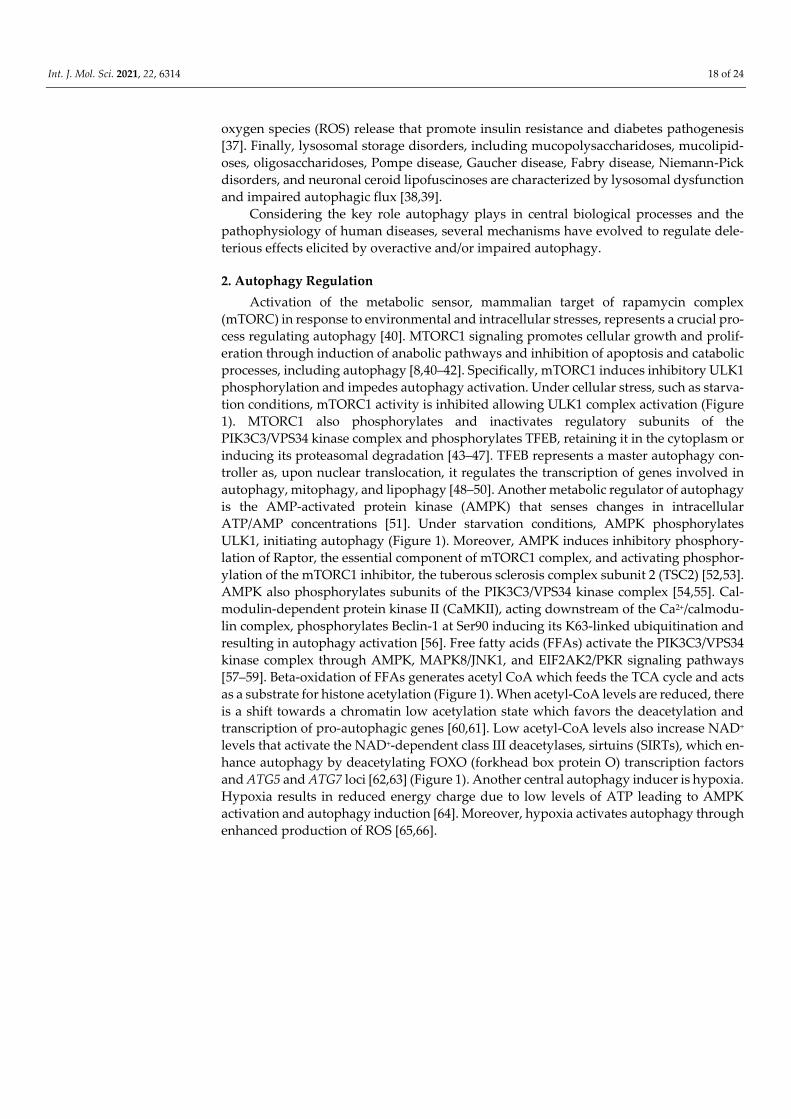
Figure 1. Mechanisms of autophagy regulation. MTORC1 induces inhibitory ULK1 phosphorylation and impedes autoph-
agy activation. Under cellular stress, such as starvation conditions, mTORC1 activity is inhibited allowing ULK1 complex
activation. MTORC1 also phosphorylates and inactivates regulatory subunits of the PIK3C3/VPS34 kinase complex and
phosphorylates TFEB. AMPK senses changes in intracellular ATP/AMP concentrations and under starvation conditions,
AMPK phosphorylates ULK1, initiating autophagy. Moreover, AMPK induces inhibitory phosphorylation of Raptor. Free
fatty acids (FFAs) activate the PIK3C3/VPS34 kinase complex through AMPK. Beta-oxidation of FFAs generates acetyl
CoA which feeds the TCA cycle and acts as a substrate for histone acetylation. SIRTs enhance autophagy by deacetylating
FOXO, ATG5, and ATG7. Activation of TFEB drives its nuclear translocation wherein it promotes the transcription of
autophagy and lysosome genes. Under glucose starvation, AMPK activation promotes TFEB nuclear translocation,
through the phosphorylation of ACSS2. ACSS2 binds to TFEB and initiates transcription of lysosome biosynthesis and
autophagy genes. ACSS2 also generates acetyl-CoA that is used for histone H3 acetylation and autophagy gene induction.
The purinergic receptor P2X7 induces TFEB nuclear translocation through AMPK activation. FOXO3 represents a key
transcriptional regulator of autophagy genes. P53 controls the expression and activity of FOXO3 and promotes TFEB/TFE3
nuclear translocation. Epigenetic modifications, including histone H3K9 dimethylation, H3K27 trimethylation, and H4K16
acetylation play an essential role in the regulation of autophagic responses. SIRT1-induced deacetylation of ATG proteins,
such as ATG5, ATG7, and LC3, and the FOXO family of transcription factors are also involved in autophagy induction.

Int. J. Mol. Sci. 2021, 22, 6314 20 of 24
Apart from metabolic signals, immune mediators, including cytokines, are critical
controllers of autophagic responses. Signaling downstream of the anti-inflammatory cy-
tokine IL-10 and its receptor IL-10R inhibits starvation-induced autophagy in murine mac-
rophages by activating the phosphatidylinositol 3-kinase (PI3K)/protein kinase B (AKT)
signaling pathway [67]. In contrast, other studies have shown that IL-10 promotes autoph-
agy in lipopolysaccharide (LPS)-stimulated bone marrow-derived macrophages and pre-
vents the accumulation of dysfunctional mitochondria and ROS release through suppres-
sion of mTORC1 functions [68]. The Th1 cell-associated cytokine IFN-γ stimulates autoph-
agy in macrophages [68,69]. In fact, in Mycobacterium tuberculosis-stimulated peripheral
blood mononuclear cells (PBMCs), high IFN-γ production, correlates with increased LC3-
II levels, proposing a protective role for IFN-γ-induced autophagy during bacterial infec-
tions [70]. In contrast, the Th2 cell-associated cytokines, IL-4 and IL-13 inhibit autophagy
in murine and human macrophages, under starvation or IFN-γ stimulation [69]. However,
IL-4 activates autophagy in CD4+ T cells, B cells, and dendritic cells (DCs), while IL-13
enhances autophagy and mucus secretion in airway epithelial cells, pointing to cell-type
specific effects of these cytokines on autophagy activation [71–73]. IL-21 suppresses au-
tophagy in activated CD4+ T cells, associated with defective differentiation and effector
function [74]. Remarkably, autophagy induction in Th2 polarized cells prevents TCR ac-
tivation through targeting Bcl10 for degradation and inhibiting NF-kB activation [75]. IL-
2 upregulates LC3-II expression and autophagosome formation in mouse embryonic and
primary lung fibroblasts, leading to enhanced proliferation and survival [76]. In lung ep-
ithelial cells, IL-17A stimulation inhibits BCL2 phosphorylation, preventing its degrada-
tion, and thus, allowing BCL2 and Beclin-1 interaction and autophagy attenuation [77].
IFN-γ enhances annexin A2 exosomal release by lung epithelial cells through autophagy
(exophagy) induction [78].
Altogether, these studies highlight distinct and often opposing effects of autophagy
on cell metabolism and effector functions that depend on the environmental stimulus, the
context of the response, the cell-type and its activation status, and suggest that close mon-
itoring of autophagic processes is essential for the protection against pathologic sequelae
that drive human diseases.
3. Transcriptional Control of Autophagy
Autophagic responses are also controlled at the transcriptional level. Transcription
factors documented to regulate autophagy gene expression, include TFEB, FOXO3, and
p53 [79–81]. TFEB belongs to the microphthalmia/transcription factor E (MiT/TFE) family
of transcription factors that include melanogenesis associated transcription factor (MITF),
Transcription Factor EC (TFEC), and Transcription Factor Binding To IGHM Enhancer 3
(TFE3) proteins that are characterized by the recognition of coordinated lysosomal expres-
sion and regulation (CLEAR) motifs [82–84]. CLEAR motifs characterize the promoter re-
gion of lysosomal genes [33] (Figure 1). Studies in Tfeb and Tfe3 knockout macrophages
demonstrated that these transcription factors promote autophagy gene expression inde-
pendently of each other, indicative of partially redundant functions [85]. Activation of
TFEB drives its nuclear translocation wherein it promotes the transcription of autophagy
and lysosome genes. Under nutrient-rich conditions, guanosine triphosphate (GTP)-
bound heterodimeric RagGTPases (RagA) recruit mTORC1 to the lysosomal membrane,
where it phosphorylates TFEB at serine/threonine residues, creating a binding site for the
cytosolic chaperone-like protein 14-3-3 phospho-serine/phospho-threonine binding pro-
tein and resulting in TFEB cytosolic sequestration [47,84,86–88]. MTORC1 also impedes
the nuclear localization of TFE3 and MITF [89]. ERK2 phosphorylates TFEB at Ser142, also
inhibiting its nuclear translocation [88]. Under glucose starvation, AMPK activation pro-
motes TFEB nuclear translocation, through the phosphorylation of ACSS2 (acetyl-CoA
synthetase short-chain family member 2) [90] (Figure 1). ACSS2 binds to TFEB and initi-
ates transcription of lysosome biosynthesis and autophagy genes. ACSS2 also generates
acetyl-CoA that is used for histone H3 acetylation and autophagy gene induction [90]. The

Int. J. Mol. Sci. 2021, 22, 6314 21 of 24
purinergic receptor P2 × 7 induces TFEB nuclear translocation through AMPK activation
[91]. Notably, activation of the calcium and calmodulin-dependent serine/threonine phos-
phatase, calcineurin, dephosphorylates TFEB and triggers its nuclear translocation, point-
ing to a role for calcium signaling in autophagy induction [92]. Recent studies also demon-
strated that Protein Kinase C Alpha (PRKCA) inhibits Glycogen Synthase Kinase 3 Beta
(GSK3β) signaling, resulting in decreased TFEB phosphorylation and enhancement of its
nuclear translocation [93].
FOXO3 represents a key transcriptional regulator of autophagy genes, including
ATG4, BECN1, LC3, ULK1, and VPS34 [80,94,95]. In response to growth factors and insulin
stimulation, the activity of FOXO3 is inhibited by AKT, an upstream inducer of mTORC1,
resulting in its cytoplasmic retention and attenuation of autophagy genes transcription
(Figure 1). A recent study in Caenorhabditis elegans demonstrated that DAF16 (FOXO in
mammals) cooperates with HLH30 (TFEB in mammals) to ensure appropriate expression
of target genes during organismal responses to stressors, pointing to potential transac-
tivating functions of FOXO and TFEB on autophagy gene induction [96].
P53, a tumor suppressor protein, inhibits mTORC1 lysosomal recruitment, through
transcriptional induction of Sestrin proteins which activate AMPK [97,98]. Moreover, P53
induces the expression of a lysosomal protein called Damaged-regulated- modulator
(DRAM) that enhances autophagy through an unknown mechanism [99]. P53 also con-
trols the expression and activity of FOXO3 [100–102], and upon DNA damage, promotes
TFEB/TFE3 nuclear translocation [103] (Figure 1). On the other hand, cytoplasmic p53 may
act as a negative regulator of autophagy; however, the precise molecular mechanisms me-
diating this inhibitory effect remain elusive [81,104]. BCL2 Interacting Protein 3 (BNIP3)
is another activator of autophagy, induced under hypoxia that disrupts the inhibitory
binding of BCL-2 to Beclin1, promoting autophagosome biogenesis. BNIP3 is regulated
by the transcription factors E2F1 and NF-kB; under normoxia, NF-kB binds to the BNIP3
promoter, suppressing its expression. Under hypoxic conditions, reduced NF-kB binding
to the BNIP3 gene allows E2F1 binding to BNIP3 regulatory elements promoting gene
transcription [105]. E2F1 also controls the expression of other autophagy genes, such as
ULK1, LC3, and ATG5 [106].
Epigenetic modifications, including histone H3K9 dimethylation, H3K27 trimethyla-
tion, and H4K16 acetylation play an essential role in the regulation of autophagic re-
sponses [107] (Figure 1). H3K27 trimethylation, catalyzed by Enhancer of Zeste Homolog
2 (EZH2), suppresses the expression of negative regulators of mTORC1, leading to au-
tophagy inhibition [108]. Bromodomain-containing protein 4 (BRD4) hinders autophagy
and lysosomal gene transcription through the recruitment of the histone lysine methyl-
transferase G9a, which places a suppressive H3K9 dimethylation mark on their promoters
[109]. In contrast, AMPK inhibits BRD4 activation, allowing lysosomal and autophagic
gene induction. Notably, low amino acid or glucose concentrations enhance co-activator-
associated histone arginine methyltransferase 1 (CARM1)-mediated dimethylation of H3
Arg17 on the promoters of autophagy and lysosomal genes, activating their transcription
[110]. In fact, CARM1 exerts transcriptional co-activator function on autophagy-related
and lysosomal genes through interactions with TFEB. Interestingly, recent studies have
demonstrated that H4K16 acetylation controls autophagy gene expression associated with
degradation of human Males absent On the First (hMOF/KAT8/MYST1), an H4K16 acetyl-
transferase [111]. More specifically, upon nutrient starvation, histone acetyltransferase
hMOF/KAT8/MYST1 activity is reduced, leading to decreased acetylation of H4K16,
which results in the transcriptional activation of autophagy genes. SIRT1-induced
deacetylation of ATG proteins, such as ATG5, ATG7, and LC3, and the FOXO family of
transcription factors are also involved in autophagy induction [112]. SIRT1 also deacety-
lates the Tumor suppressor serine/threonine-protein kinase LKB1 that activates AMPK
signaling, increasing autophagy induction [113] (Figure 1). Finally, under nutrient-rich
conditions, Forkhead box K (FOXK) drives the transcriptional repression of autophagy
gene expression by binding to promoter regions of early-stage autophagy genes (e.g., ULK

Int. J. Mol. Sci. 2021, 22, 6314 22 of 24
complex) and recruiting the SIN3A-Histone deacetylase (HDAC) repressor complex to
these regions [114].
Collectively, the aforementioned paragraphs highlight the pleiotropic effects of met-
abolic, transcriptional, and epigenetic mechanisms on the regulation of autophagy and
illuminate the existence of complex networks that are activated or inhibited in each cell to
ensure cell survival and maintenance of its homeostasis during encounters with intracel-
lular and extracellular stressors.
4. Allergic Asthma Immunopathogenesis
Asthma is a chronic heterogeneous lung disease characterized by airway hyperre-
sponsiveness (AHR) to innocuous inhaled allergens and pulmonary inflammation [115].
Asthma encompasses complex and multiple clinical phenotypes that incorporate persis-
tent symptoms and acute disease exacerbations [115–117]. Certain asthmatics exhibit se-
vere asthma (SA) that is poorly controlled and represents a major health and socio-eco-
nomic burden [116,117]. SA patients are characterized by frequent, and sometimes life-
threatening, disease exacerbations and more severe symptoms, including shortness of
breath, wheeze, cough, and increased mucus production [118,119]. These individuals re-
quire treatment with high-dose inhaled (or systemic) corticosteroids (CS) in combination
with a second long-term (controller) medication and exhibit low lung function and per-
sistent eosinophilia in the bronchoalveolar lavage fluid (BAL), along with high levels of
neutrophils and exhaled nitric oxide [120]. Depending on the type of immune cells in-
volved in disease pathogenesis, asthma endotypes are mainly categorized as type 2
asthma, characterized by Th2 cell-mediated and eosinophilic inflammation, and non-type
2 asthma, associated with Th1 and/or Th17 cell and neutrophilic inflammation [120–122].
Another type of asthma is the paucigranulocytic asthma (PGA) characterized by low-
grade airway inflammation and low numbers of eosinophil or neutrophil numbers in the
airways, compared to other asthma endotypes [123,124]. PGA is associated with airway
smooth muscle (ASM) dysfunction, persistent airflow limitation, and AHR. The lack of an
effective therapeutic regime for individuals with SA represents a serious clinical need
without an obvious solution at present.
Inflammatory cells infiltrating the allergic airways, including DCs, eosinophils, neu-
trophils, mast cells, and lymphocytes, play a crucial role in the initiation and propagation
of asthmatic responses (Figure S1). DCs in the lung mucosa take up allergens, reach the
mediastinal lymph nodes, and present allergen components to naive T cells which differ-
entiate into Th1, Th2, Th9, or Th17 cell subsets and initiate inflammatory responses upon
relocalization to the airways [125]. The production of Th2 cell-associated cytokines, such
as IL-4, IL-5, and IL-13, by Th2 cells enhances mucus production, bronchial remodeling,
and the recruitment of innate effector cells, including mast cells, basophils, and eosino-
phils [126,127]. A group of asthmatics is characterized by increased neutrophilic inflam-
mation, along with Th17 and Th1 cell infiltration that produce high levels of IFN-γ and
IL-17 in the allergic airways that correlate with asthma severity [128]. Th9 cells and type
2 innate lymphoid cells (ILC2s) enhance the production of IL-2 by mast cells, leading to
further expansion of ILC2s, which in turn activate Th9 cells in a positive feedback loop
[129]. ILC2s are activated in response to alarmins released by airway epithelial cells, in-
cluding IL-25, IL-33, and thymic stromal lymphopoietin (TSLP), and represent the first
producers of Th2 cytokines that activate B cell, T cell, and granulocyte infiltration in the
allergic lung (Figure S1) [129]. Apart from airway inflammation, SA is characterized by
extensive airway remodeling and narrowing thickened epithelium, and fixed airflow ob-
struction (Supplementary Figure S1) [130,131].
Emerging clinical, epidemiological, and experimental evidence has illuminated
dysregulated autophagy as a principal mechanism underlying asthma pathogenesis with
conflicting reports showing both detrimental and beneficial effects [131–134]. Hence, de-
lineation of the precise role of autophagy in the regulation of asthmatic responses is es-

Int. J. Mol. Sci. 2021, 22, 6314 23 of 24
sential for the restoration of lung homeostasis and the development of more effective ther-
apeutic interventions. In the next section, we will delineate current knowledge on the role
of autophagy in the attenuation or propagation of allergic airway inflammation and
linked human asthma.
5. Role of Autophagy in Allergic Airway Inflammation In Vivo
The role of autophagy in allergen-driven inflammatory responses in the airways re-
mains poorly understood. Studies using ovalbumin (OVA)-induced allergic airway in-
flammation (AAI) mouse models documented decreased expression of Atg5, Lc3, and Be-
clin1 in lung homogenates and BAL macrophages, accompanied by reduced protein levels
in OVA-treated mice, compared to PBS-control mice [135]. In contrast, other reports
demonstrated increased expression of Lc3b by airway epithelial cells and elevated Atg5
levels in lung homogenates in a mouse model of cockroach-allergen induced AAI [136].
Certain studies report suppressive effects of autophagy associated with protection against
AAI (Table 1). Indeed, treatment with simvastatin reduced airway inflammation and re-
modeling, attenuated AHR, and decreased the levels of IL-4, IL-5, IL-13, and IFN-γ in the
BAL in OVA-challenged mice and this was associated with increased expression of au-
tophagy proteins, such as Atg5, LC3B and Beclin1 in lung homogenates, as well as, en-
hanced autophagosome formation in the lung parenchyma [135] (Table 1). Notably, ad-
ministration of the autophagy inhibitor, 3-Methyladenine (3-MA), reversed simvastatin-
induced suppression of AAI and remodeling. Seminal studies in Atg5 deficient mice
demonstrated increased neutrophilic influx, AHR, airway inflammation, and goblet cell
accumulation, concomitant with elevated IL-1β and IL-17A levels in whole lung lysates
upon house dust mite (HDM) sensitization and challenge [137]. Using CD11c-specific
Atg5-/- mice the authors further showed that impaired autophagy in DCs resulted in in-
creased IL-1 and IL-23 release, spontaneous AHR, severe neutrophilic and Th17 cell-me-
diated airway inflammation, and glucocorticoid resistance, while adoptive transfer of
Atg5-/- CD11c+ DCs aggravated lung inflammation and increased IL-17 release in the aller-
gic airways (Table 2, Figure S1). Interestingly, using bone marrow chimeras, these studies
revealed that autophagy deficiency in non-hematopoietic cells did not ameliorate allergic
airway disease phenotype, excluding a detrimental role for autophagy activation in lung
tissue-resident cells [137]. In agreement, in another mouse model of acute AAI induced
by intravenous transfer of in vitro generated OVA-specific Th17 cells, treatment with ra-
pamycin, an autophagy inducer, reduced pulmonary inflammation accompanied by in-
creased recruitment of plasmacytoid dendritic cells and reduction of neutrophilic infiltra-
tion and CXCL-1 levels in the BAL [138] (Table 1, Supplementary Figure S1). Remarkably,
mice lacking Atg7 in myeloid cells exhibited enhanced sterile lung inflammation, accom-
panied by submucosal thickening, goblet cell metaplasia, increased collagen content,
heightened IL-1β, IL-18, and IL-17 levels in the lungs and serum and increased mortality
following intraperitoneal LPS injection, while bleomycin administration aggravated pul-
monary inflammation and induced severe fibrosis (Table 2, Supplementary Figure S1)
[139]. Similarly, other investigators using myeloid-specific Atg7-deficient mice demon-
strated spontaneous pulmonary inflammation, elevated expression of Tnfα, Il6, Ccl2,
Cxcl1, Cxcl2 genes, and myeloid cell infiltration in the lung [140]. These mice also exhibited
increased mitochondrial ROS production and heightened sensitivity of alveolar macro-
phages (AMs) to TLR4 ligands, including low doses of LPS. In a model of eosinophilic
chronic rhinosinusitis (ECRS), mice with myeloid cell-specific deletion of Atg7 exhibited
aggravated eosinophilic and mast cell infiltration, mucosal thickening, and increased pro-
duction of prostaglandin D2 [141]. Macrophage activation in Atg7−/− mice was associated
with increased IL-1β levels, while macrophage depletion or IL1β receptor blockade alle-
viated eosinophilic inflammation. Overall, these studies propose that baseline autophagy
activation in myeloid cells contributes to the maintenance of lung homeostasis and the
control of aberrant inflammatory AM responses.

Int. J. Mol. Sci. 2021, 22, 6314 24 of 24
Table 1. Activation of autophagy and its effect on Allergic Airway Disease Outcome.
Autophagy Activation
Treatment Allergic Airway Disease Outcome
Mtor-/- bronchial epithelial cells
in HDM and/or OVA chal-
lenged mice
Increased recruitment of inflammatory cells and eosinophils in the BAL
Enhanced mucus accumulation
Exacerbated AHR
Heightened IL-25 levels
ILC2-specific TfebTG mice in IL-
33-induced AAI
Enhanced ILC2s cell infiltration in the lungs
Increased survival and proliferation of ILC2s
Enhanced Th2 cytokine release
Simvastatin administration in
OVA-challenged mice
Reduced airway inflammation and remodeling through autophagy activation
Attenuated AHR
Decreased IL-4, IL-5, IL-13, and IFN-γ levels in the BAL
Rapamycin administration in
acute AAI induced by intrave-
nous transfer of in vitro gener-
ated OVA-specific Th17 cells
Reduced pulmonary inflammation
Increased recruitment of plasmacytoid dendritic cells
Reduction of neutrophilic infiltration in the BAL
Reduced CXCL-1 levels in the BAL
Table 2. Deficiency of autophagy and its impact on Allergic Airway Disease Outcome.
Autophagy Deficiency
Treatment Allergic Airway Disease Outcome
CD11c-specific Atg5-/- mice (HDM mouse
model)
Increased IL-1β and IL-23 release
Increased AHR
Severe neutrophilic and Th17 cell-mediated airway inflammation
Glucocorticoid resistance
Myeloid specific Atg7-/- mice (LPS or ble-
omycin)
Increased IL-1β, IL-18, and IL-17 levels in the lungs and serum
Increased mortality
Atg7-/- airway epithelial-specific mice
Swelling of bronchial epithelial cells
Increased AHR
Enhanced airway wall thickening
Increased p62 accumulation
Atg16l1-/- mice
(intranasal IL-33 administration) Attenuated mucus secretion
3-MA or Atg5-/- mice
(OVA-induced
severe eosinophilic AAI)
Attenuated AHR
Decreased inflammatory cell recruitment in the BAL and lung
Reduced IL-5 levels in the BAL
Lc3-b-/- mice
(HDM and/or OVA AAI)
Reduced airway inflammation and mucus production
Increased AHR
Atg5-/- ILC2s specific mice
(IL-33-AAI)
Decreased Th2 cytokine release
Impaired fatty acid oxidation
Attenuated pulmonary inflammation and AHR
Atg5-/- B cell-specific mice
(OVA-induced AAI)
Reduced levels of IL-4, IL-13, and inflammatory cell numbers in the BAL
Decreased OVA-specific IgE production
Reduced mucus production
3-MA administration
(mouse model of cockroach allergen-in-
duced AAI)
Decreased lung inflammation and mucus production
Attenuated AHR
Reduced ROS release
Decreased Th2 cell-associated cytokines
3-MA administration
(OVA-induced AAI) Attenuated pulmonary inflammation

Int. J. Mol. Sci. 2021, 22, 6314 25 of 24
Reduced eosinophil numbers, eosinophil peroxidase activity, and extracellu-
lar DNA concentrations in the BAL
Reduced ROS levels
Decreased goblet cell hyperplasia
CQ administration
(HDM-induced acute and chronic AAI)
Decreased inflammatory cell infiltration
Reduced TGF-β production in the BAL
Attenuated AHR
Decreased collagen deposition and mucus production
Reduced airway remodeling
Astraglin administration
(OVA-induced AAI)
Decreased the subepithelial deposition of collagen fibers through autophagy
inhibition
Luteolin administration (OVA-induced
AAI)
Decreased inflammatory cell infiltration
Reduced IL-4, IL-5, IL-13 levels in the BAL
Decreased collagen deposition and mucus production
EX-527 administration
(OVA-induced AAI)
Decreased airway inflammation
Reduced IL-4, IL-13, and IFN-γ levels in the BAL
JTE-013 administration
(OVA-induced chronic AAI)
Decreased inflammatory cell recruitment
Reduced IL-1, IL-4, IL-5 levels in BAL
Reduced mucus production
Attenuated collagen deposition and smooth muscle cell activation
Emerging evidence also highlights a prominent role for autophagy in airway epithe-
lial cell responses. Using mice with an inducible epithelial cell-specific (under the Clara
cell promoter) disruption of Atg7, recent studies documented that loss of autophagy in
airway epithelial cells resulted in swelling of bronchial epithelial cells, accompanied by
p62 accumulation and increased AHR to methacholine due to enhanced airway thicken-
ing [142] (Table 2, Supplementary Figure S1). Mechanistically, bronchial epithelial cells
from Atg7 deficient mice exhibited enhanced expression of the cytoprotective genes Nqo1,
Txnrd1, and activation of the Keap-Nrf2 pathway that was associated with p62 accumula-
tion.
Contradictory data propose a detrimental role for autophagy in the initiation and
progression of AAI. For example, in a mouse model of OVA-induced severe eosinophilic
AAI, treatment with 3-MA or Atg5 knockdown reduced LC3-II expression in lung-infil-
trating eosinophils, attenuated AHR, and decreased inflammatory cell recruitment in the
BAL and lung [143] (Table 2). Moreover, the levels of IL-5 but not IL-4, IL-13, and IFN-γ
were decreased in the BAL, while in vivo anti-IL-5 administration reduced LC3II expres-
sion in allergic lungs. In agreement, other investigators showed that treatment with 3-MA
right before the OVA challenge, attenuated pulmonary inflammation associated with de-
creased eosinophil extracellular trap formation [144] (Table 2, Supplementary Figure S1).
In fact, it was demonstrated that 3-MA treatment reduced eosinophil numbers, eosinophil
peroxidase activity and goblet cell hyperplasia, extracellular DNA concentrations in the
BAL, and dampened IL-5, IL-13, IFN-γ, TNF-α, IL-1β, nuclear factor kappaB (NFκB) p65
and ROS levels in the lung. Notably, luteolin administration inhibited OVA-induced air-
way inflammation, accompanied by decreased Beclin-1-PI2KC3 protein expression and
enhanced PI3K/Akt/mTOR activation [145]. Interestingly, other studies demonstrated that
knockdown of Mtor in airway epithelial cells resulted in increased recruitment of inflam-
matory cells and eosinophils in the BAL, enhanced mucus accumulation, exacerbated
AHR and heightened IL-25 levels, following HDM and/or OVA challenge (Table 1, Sup-
plementary Figure S1) [146]. In support, the administration of EX-527, a SIRT1 inhibitor,
suppressed airway inflammation and reduced IL-4, IL-13, and IFN-γ levels in the BAL,
associated with enhanced mTOR activation and decreased autophagy induction, effects
that were reversed by concomitant rapamycin treatment [147]. Further support of a path-

Int. J. Mol. Sci. 2021, 22, 6314 26 of 24
ogenic role for autophagy in AAI came from studies in Atg16l1-deficient mice which ex-
hibited attenuated mucus secretion upon intranasal IL-33 administration [75]. In support,
3-MA administration in a mouse model of cockroach allergen-induced AAI dampened
lung inflammation, mucus production, AHR, ROS release, and Th2 cell-associated cyto-
kines [136], (Table 2). Interestingly, miR-135-5p negatively regulated p62 expression and
decreased inflammatory cytokine and chemokine release in a rat basophil cell line [148],
highlighting miR-mediated antagonism as a novel mechanism of autophagy regulation.
Lc3-b deficient mice were also characterized by reduced allergen-induced airway inflam-
mation and mucus production [146]. Still, AHR was enhanced in Lc3-b deficient mice,
pointing to differential effects of autophagy on AHR and pulmonary inflammation (Table
2).
Autophagy also controls pro-allergic responses elicited by innate immune cells. Pio-
neering studies revealed that Atg5 deficiency in ILC2s decreased Th2 cytokine release,
impaired fatty acid oxidation, and induced the accumulation of dysfunctional mitochon-
dria [149] (Table 2, Supplementary Figure S1). Remarkably, adoptive transfer of Atg5-/-
ILC2 cells attenuated IL-33-induced pulmonary inflammation and AHR, while autophagy
activation in ILC2-specific TfebTG mice enhanced Th2 cytokine release, highlighting a cen-
tral role for autophagy in the effector function and metabolic responses of ILC2s. B cell
responses are also controlled by autophagy in the context of AAI. More specifically, mice
lacking Atg5 in B cells exhibit lower levels of IL-4, IL-13, and inflammatory cell numbers
in the BAL, decreased OVA-specific IgE production in the serum, attenuated mucus pro-
duction and antigen-presenting functions, concomitant with increased apoptosis and gly-
colysis [72] (Table 2, Supplementary Figure S1). Notably, IL-4 induced JAK signaling was
essential for autophagy activation in B cells.
In a chronic asthma model, deficient autophagic flux and increased p62 expression
were detected in lung homogenates and were associated with exacerbated airway remod-
eling [150]. Interestingly, a recent study using a mouse model of HDM-induced acute and
chronic AAI revealed that autophagy inhibition via intranasal administration of chloro-
quine (CQ) decreased inflammatory cell infiltration and TGF-β production in the BAL and
attenuated AHR [151]. Moreover, CQ administration decreased collagen and mucus dep-
osition, reduced airway remodeling, and lowered BECLIN-1 and ATG-5 protein levels in
the allergic lungs (Table 2, Supplementary Figure S1). A detrimental role for autophagy
in airway remodeling was also noted in studies involving oral administration of
astragalin, a kaempferol-3-O-glucoside, in OVA-sensitized mice (Table 2, Supplementary
Figure S1) [152]. Indeed, the investigators reported that astragalin treatment prevented
the subepithelial deposition of collagen fibers and was associated with reduced beclin-1
and LC3 expression in airway epithelial cells and lung tissue. Remarkably, administration
of JTE-013, a Sphingosine-1-phosphate receptor 2 (S1PR2) antagonist, in a mouse model
of OVA-induced chronic airway inflammation and remodeling, decreased inflammatory
cell recruitment and mucus production, reduced BAL IL-1, IL-4, IL-5, and serum IgE levels
and attenuated collagen deposition and smooth muscle cell-activating proteins in the
lungs. JTE-013 effects were accompanied by decreased Beclin-1 levels and LC3II/LC3I con-
version and increased p62 accumulation, indicative of dampened autophagy [153].
Autophagy is critically involved in host defense mechanisms against invading path-
ogens, including respiratory tract infections. In fact, bacteria and viruses attack autophagy
(xenophagy) early on the following infection, to avoid autophagosome formation and
linked destruction [154]. In this respect, Atg7 deficiency following Pseudomonas aeruginosa
infection impaired pathogen clearance increased neutrophilic inflammation, and resulted
in elevated IL-1β production [155]. Furthermore, in a mouse model of Human Rhinovirus
1B (HRV1B) infection, the anti-viral effects of budesonide administration were associated
with autophagy activation [156]. In sharp contrast, baf-A1-induced autophagy blockade
impeded influenza A virus replication in lung epithelial cells [157], while autophagy in-
duction was essential for the formation of double-membrane vesicle-bound major histo-
compatibility complex (MHC) replication complexes in a mouse model of hepatitis virus

Int. J. Mol. Sci. 2021, 22, 6314 27 of 24
infection [158]. Notably, respiratory syncytial virus (RSV)-infected Map1lceb-/- mice exhib-
ited aggravated IL-17A-dependent lung pathology, while Becn1+/- mice displayed de-
creased Th2 cytokine release, mucus secretion, and lung inflammation, illuminating im-
portant tissue-targeting effects of autophagy in the context of certain viral infections [159].
Still, further mechanistic studies are needed to explore the precise role of autophagy in
respiratory tract infections and pathogen-induced asthma exacerbations.
6. Activation of Autophagy in Human Asthma
Early studies discovered genetic polymorphisms of the ATG5 and ATG7 genes in in-
dividuals with pediatric and adult asthma that were linked to airway remodeling and
impairment in respiratory system mechanics [23,24,160]. Still, subsequent studies did not
detect an association of ATG5 gene polymorphisms with asthma severity but only with
higher sputum neutrophil numbers [161]. Increased autophagosome formation was ob-
served in fibroblasts and airway epithelial cells, and heightened expression of LC3B and
ATG5 was detected in lung biopsies from asthmatic patients compared to healthy controls
[24]. Other studies did not report activation of autophagy in large airway epithelial cells
in asthmatics, as monitored by ATG5, Beclin-1, and p62 expression in lung sections, while
enhanced ATG5 and Beclin-1 levels, accompanied by reduced p62 expression, were de-
tected in large ASMs [151,160]. In sharp contrast, recent studies showed that ATG5 ex-
pression in airway epithelium, ASM, and inflammatory cells was not increased in asth-
matics and did not correlate with asthma severity or lung function [161]. These contradic-
tory findings may be due to the distinct methodological approaches utilized, as well as,
due to differences in patient cohorts or the timing of the analyses. Hence, further studies
are needed to delineate the activation patterns of autophagy in the asthmatic lung and its
potential correlations with disease onset and/or severity.
Interestingly, a protective role was identified for the complement regulatory protein
CD46 in nasal airway epithelial cells from asthmatic individuals through enhancing au-
tophagy [162] (Supplementary Figure S1). More specifically, it was demonstrated that air-
way epithelial cells from asthmatics exposed in vitro to Dermatophagoides pteronyssinus ex-
hibited increased autophagosome formation, LC3II expression, decreased apoptosis, and
lower pro-IL-1β and NLRP3 levels following CD46 mAb crosslinking, while treatment
with 3-MA reversed the anti-inflammatory effects of the CD46 mAb on these cells. Inter-
estingly, upon exposure of human bronchial epithelial cells (BECs) to particulate matter,
autophagy induction enhanced mucus secretion, protecting these particles [163]. In con-
trast, ATG5 and ATG14 knockdown in primary human tracheal epithelial cells stimulated
with IL-13 decreased MUC5AC secretion and CCL26 (eotaxin-3) and ROS release, sug-
gesting that autophagy activation in this setting is pathogenic [73] (Supplementary Figure
S1). Furthermore, in vitro stimulation of human BECs with Alternaria extract enhanced
LC3 conversion and p62 degradation associated with elevated IL-18 levels [164]. Notably,
treatment with 3-MA and Baf-A1, suppressed IL-18 release by Altenaria-stimulated BECs,
highlighting an autophagy-induced unconventional mechanism of IL-18 secretion (Sup-
plementary Figure S1). In other studies, IL-1β increased LC3-II expression and IL-8 pro-
duction by human small airway epithelial cells, while 3-MA, CQ, the PI3K inhibitor
LY294002 or knockdown of ATG5 and Beclin-1 reversed IL-1β effects [165]. IL-13 or IL-33
treatment also enhanced autophagy in human airway epithelial cells (HAECs) from asth-
matics, concomitant with a decrease in mTOR activity [146]. Remarkably, a protective role
was proposed for autophagy in preventing ferroptotic cell death induced by Th2 inflam-
matory conditions on HAECs from asthmatics, while increased LC3II levels in HAECs
correlated with lower mitochondrial DNA in the BAL fluid [166]. Notably, cockroach al-
lergen-stimulated HAECs displayed decreased mitochondrial ROS release upon 3-MA
treatment, while mechanistic studies demonstrated that oxidized CaMKII activation was
essential for cockroach allergen-mediated autophagy activation [136].
Pertinent to human inflammatory cells, Ban et al. (2015) detected elevated LC3 puncta
formation in the cytoplasm of sputum granulocytes and peripheral blood cells in patients

Int. J. Mol. Sci. 2021, 22, 6314 28 of 24
with SA, compared to non-severe asthma patients and healthy controls [167]. More spe-
cifically, peripheral blood eosinophils and other PBMCs from SA patients exhibited higher
autophagy levels, as evidenced by increased LC3B expression at baseline, which were fur-
ther upregulated upon IL-5 or inhibited by 3-MA treatment. Other studies showed that
peripheral blood neutrophils (PBNs) from individuals with SA had elevated LC3-II levels
and NET production both at baseline and following ex vivo IL-8 stimulation [168] (Sup-
plementary Figure S1Increased NET production by PBNs correlated with autophagy,
while IL-8-induced NET formation negatively correlated with lung function measure-
ment, such as FEV1/FVC. Moreover, treatment with CQ decreased PBNs migration to-
wards IL-8 (Supplementary Figure S1). Still, ATG5 knockout did not compromise the abil-
ity of neutrophils or eosinophils to form extracellular traps [169]. Notably, activation of
autophagy has been shown to elicit caspase-independent cell death in eosinophils and
neutrophils under inflammatory conditions, emphasizing potential protective effects of
autophagy against the presence of activated granulocytes [169,170].
Human bronchial fibroblasts (HBF) from individuals with SA exhibit enhanced pro-
tein expression of LC3-ΙΙ, LAMP2A, Pink1, and Sirt1 and mitochondrial damage, com-
pared to cells from healthy volunteers [171]. Moreover, in vitro stimulation of HBF from
asthmatics with IL-17 further increased mitochondrial dysfunction, collagen and fibron-
ectin mRNA levels, and expression of LC3-ΙΙ and p62. Inhibition of autophagy using
bafilomycin-A1 restored IL-17-mediated increase in PINK1 protein levels in HBFs from
asthmatics and decreased pro-fibrotic gene expression, illuminating a role for IL-17-in-
duced autophagy in promoting HBF fibrotic responses (Supplementary Figure S1). Addi-
tionally, HBF from asthmatics exhibited enhanced mitochondrial depolarization and in-
creased mRNA expression of Pink/Parkin pro-fibrotic and pro-inflammatory cytokines,
such as IL-6, IL-8, COL1a1, COL3A1, and FN1, while treatment with 3-MA decreased pro-
fibrotic and pro-inflammatory gene expression [172] (Supplementary Figure S1). Interest-
ingly, in vitro stimulation of human ASM cells with TGF-β1 increased Beclin-1 and LC3II
levels, concomitant with enhanced collagen I expression, while the addition of CQ re-
versed TGF-β1 effects, pointing to pro-fibrotic effects of autophagy activation in human
ASM cells [151] (Supplementary Figure S1). In contrast, a protective role of autophagy
was detected in bronchial smooth muscle cells from asthmatics, as evidenced by enhanced
survival, concomitant with decreased Th2 cytokine release in simvastatin-treated cells co-
administered with rapamycin (Supplementary Figure S1) [135]. In support, 3-MA treat-
ment reversed simvastatin-induced anti-inflammatory effects on human bronchial
smooth muscle cells. Further studies showed that knockdown of p62 inhibited the in vitro
proliferation and migration of bronchial smooth muscle cells from asthmatics, associated
with decreased glucose consumption and lactate production, whereas p62 overexpression
had the opposite effects, highlighting a cross-regulation between autophagy and meta-
bolic responses in human ASM cells [150] (Supplementary Figure S1).
The increase in autophagosome formation and p62 levels in allergic airways reported
in certain studies could be due either to enhanced autophagic flux and autophagy activa-
tion or to inhibition of autophagosome-lysosome fusion, indicating impaired autophagy.
As such, studies showing increased expression of autophagy proteins in lung-resident
and inflammatory immune cells in asthmatics should be carefully evaluated and accom-
panied by detailed time course, functional and mechanistic analyses to delineate the pre-
cise role of autophagy in these cells.
7. Concluding Remarks and Future Perspectives
Autophagy displays both protective and detrimental roles in allergic airway inflam-
mation and linked asthma depending on the cell type, the lung micromilieu, and the cell-
intrinsic bioenergetic requirements. In fact, autophagy seems to play a balancing role in-
tended to avoid excessive lung tissue damage, while ensuring a protective anti-pathogen
response. For example, at baseline, activation of autophagy maintains homeostasis of

Int. J. Mol. Sci. 2021, 22, 6314 29 of 24
lung-resident cells, while during respiratory tract infections, autophagy induction in in-
filtrating immune cells, such as macrophages and DCs, is essential for the elimination of
pathogens and the activation of pathogen-specific T effector responses. Still, impaired au-
tophagy or persistent autophagy activation during the chronic phase of the allergic re-
sponse can enhance autophagosome accumulation and activate lung-infiltrating innate
immune cells, as well as, airway epithelial cells, leading to a decline in lung function. Au-
tophagy also represents a critical regulator of fibrosis and can enhance extracellular matrix
(ECM) production (e.g., collagen, fibronectin) in ASM and mesenchymal cells, leading to
airway wall thickening and rigidity [151,171]. Conversely, autophagy has been shown to
mitigate fibrosis through its involvement in the degradation of ECM proteins [173]. In this
respect, the documented genetic correlation of polymorphisms in ATG genes with asthma
may be an inherent protective mechanism to reduce chronic airway inflammation and
remodeling and therefore, correlations or associations of autophagy genes with lung func-
tion changes should not be associated with causal mechanisms [23,24,160].
Apart from autophagy, the role of mitophagy in allergic responses remains poorly
defined and should be further explored. Mitochondrial depolarization, along with ele-
vated ROS production, are associated with atopy, atopic dermatitis, and asthma [174–177].
Dysregulated mitochondrial bioenergetics may weaken airway epithelial cell integrity,
enhance their fragility and lead to impaired secretion [178]. Considering that mitophagy
may be essential for the restoration of allergen-induced mitochondrial dysfunction and
linked phenotypic changes in asthma, targeting mitophagy may possess therapeutic po-
tential. For example, rapamycin and metformin, as general autophagy-inducing drugs,
preserve energy metabolism through regulating mitophagy and mitochondrial biogenesis
stimulation [179,180]. Moreover, naturally occurring compounds, such as spermidine,
resveratrol, and urolithin A, enhance mitochondrial integrity through mitophagy activa-
tion and exert potent anti-inflammatory effects [181]. Investigation of the role of these mi-
tophagy-inducing substances in the protection against mitochondrial dysfunction and
linked AAI is timely and needed.
Several questions regarding the precise role of autophagy in the pathophysiology of
human asthma remain opaque. Considering that autophagy represents a versatile im-
mune modulator, a better understanding of the interplay between autophagy and im-
mune responses in the allergic airways is needed and expected to have important thera-
peutic applications for asthma. Still, targeting autophagy therapeutically, using autoph-
agy activators or inhibitors, when the autophagic response is different in distinct cellular
compartments of the lung is challenging. Additionally, although selective autophagy sub-
strates have been identified, the physiological significance of degradation of each sub-
strate in lung-resident and airway-infiltrating immune cells needs to be further explored.
Critical questions also relate to the successful identification of autophagy biomarkers that
determine autophagic activity in allergic diseases, particularly when monitoring drug ef-
fectiveness. Moreover, currently available autophagy-modulating drugs are not specific
and the development of more specific autophagy activators and inhibitors is required be-
fore their use in preclinical and clinical studies. Future studies should also consider air-
way-targeted autophagy regulators that can be administered locally, such as, the devel-
opment of a novel inhaler, or nanoparticle-based cell-targeted methods to avoid non-spe-
cific potentially detrimental systemic side effects. Interestingly, administration of certain
autophagy inducers, such as trehalose, has been shown to inhibit cytomegalovirus infec-
tion, suggesting that autophagy could be also exploited as a therapeutic regime towards
virus-induced asthma exacerbations [132]. Considering that currently utilized asthma im-
munotherapies, including montelukast, anti-IL-5, and anti-IgE antibody, can inhibit au-
tophagy, their potentially detrimental effects on cell types wherein autophagy is protec-
tive should be also carefully evaluated [178]. Finally, the specific asthma endotype and
stage of the disease should be taken under consideration when targeting autophagy sys-
temically or in specific cell types.

Int. J. Mol. Sci. 2021, 22, 6314 30 of 24
In conclusion, studies using both animal models and clinical samples from asthmatic
individuals, along with advanced transcriptomics, proteomics, and metabolomic analyses
at the single-cell level, are imperative to delineate the precise mechanisms underlying au-
tophagy activation and inhibition, particularly after exposure to environmental pollutants
and allergens, and will ultimately help identify appropriate therapeutic targets that can
effectively control severe treatment-refractory asthmatic responses.
Supplementary Materials: The following are available online at www.mdpi.com/arti-
cle/10.3390/ijms22126314/s1, Figure S1: Autophagy effects on allergic airway inflammation and re-
modeling.
Author Contributions: E.T. searched the literature, wrote the manuscript, and designed the figures;
G.X. wrote the manuscript. All authors have read and agreed to the published version of the man-
uscript.
Funding: E.T. Hellenic Foundation for Research and Innovation (HFRI) under the fellowship grant
(fellowship number: 330) and the General Secretariat for Research and Technology (GSRT). G.X. is
supported by the General Secretariat for Research and Technology (GSRT) and the Hellenic Foun-
dation for Research and Innovation (HFRI) Grant (# 3303, Reaction).
Acknowledgments: The authors would like to thank Maria Semitekolou and Gina Papadopoulou
for critical reviewing of the manuscript. The figures presented in the manuscript were created with
Biorender.com.
Conflicts of Interest: The authors declare no conflict of interest.
Abbreviations
ATG autophagy-related genes
LC3 ATG8/microtubule-associated protein 1 light chain 3
TFEB transcription factor EB
ROS reactive oxygen species
mTORC mammalian target of rapamycin complex
AMPK AMP-activated protein kinase
SIRTs sirtuins
FOXO forkhead box protein O
PI3K phosphatidylinositol 3-kinase
AKT protein kinase B
LPS lipopolysaccharide
PBMCs peripheral blood mononuclear cells
DCs dendritic cells
BCL2 B-cell CLL/lymphoma 2
AHR Hyperresponsiveness
BAL bronchoalveolar lavage fluid
CS corticosteroids
FeNO nitric oxide
ASM airway smooth muscle
PGA paucigranulocytic asthma
ILC2s type 2 innate lymphoid cells
TSLP thymic stromal lymphopoietin
OVA ovalbumin
AAI allergic airway inflammation
3-MA 3-Methyladenine
HDM house dust mite
AMs alveolar macrophages
ECRS eosinophilic chronic rhinosinusitis
CQ chloroquine
Int. J. Mol. Sci. 2021, 22, 6314 31 of 24
HRV1B Human Rhinovirus 1B
MHC major histocompatibility complex
RSV respiratory syncytial virus
BECs bronchial epithelial cells
HAECs airway epithelial cells
PBNs peripheral blood neutrophils
HBF Human bronchial fibroblasts
ECM extracellular matrix
PE phosphatidylethanolamine
p62 Sequestosome 1
LIR LC3-interacting region
TSC2 tuberous sclerosis complex subunit 2
CaMKII Calmodulin-dependent protein kinase II
FFAs Free fatty acids
MiT/TFE microphthalmia/transcription factor E
MITF melanogenesis associated transcription factor
TFEC Transcription Factor EC
TFE3 Transcription Factor Binding To IGHM Enhancer 3
CLEAR coordinated lysosomal expression and regulation
GTP guanosine triphosphate
RagA heterodimeric RagGTPases
YWHA/14-3-3 14-3-3 phospho-serine/phospho-treonine binding protein
ACSS2 acetyl-CoA synthetase short-chain family member 2
NFAT nuclear factors of activated T cells
PRKCA Protein Kinase C Alpha
GSK3β Glycogen Synthase Kinase 3 Beta
DRAM Damaged-regulated- modulator
BNIP3 BCL2 Interacting Protein 3
EZH2 Enhancer of Zeste Homolog 2
BRD4 Bromodomain-containing protein 4
CARM1 co-activator associated histone arginine methyltransferase 1
hMOF Males absent On the First
FOXK Forkhead box K
HDAC Histone deacetylase
SA severe asthma
NFκB nuclear factor kappaB
S1PR2 Sphingosine-1-phosphate receptor 2
References
1. Dikic, I.; Elazar, Z. Mechanism and medical implications of mammalian autophagy. Nat. Rev. Mol. Cell Biol. 2018, 19, 349–
364, doi:10.1038/s41580-018-0003-4.
2. Gatica, D.; Lahiri, V.; Klionsky, D.J. Cargo recognition and degradation by selective autophagy. Nat. Cell Biol. 2018, 20, 233–
242, doi:10.1038/s41556-018-0037-z.
3. Li, W.; He, P.; Huang, Y.; Li, Y.-F.; Lu, J.; Li, M.; Kurihara, H.; Luo, Z.; Meng, T.; Onishi, M.; et al. Selective autophagy of
intracellular organelles: Recent research advances. Theranostics 2021, 11, 222–256, doi:10.7150/thno.49860.
4. Mizushima, N.; Komatsu, M. Autophagy: Renovation of Cells and Tissues. Cell 2011, 147, 728–741,
doi:10.1016/j.cell.2011.10.026.
5. Mizumura, K.; Cloonan, S.M.; Haspel, J.A.; Choi, A.M. The Emerging Importance of Autophagy in Pulmonary Diseases.
Chest 2012, 142, 1289–1299, doi:10.1378/chest.12-0809.
6. Kaur, J.; Debnath, J. Autophagy at the crossroads of catabolism and anabolism. Nat. Rev. Mol. Cell Biol. 2015, 16, 461–472,
doi:10.1038/nrm4024.

Int. J. Mol. Sci. 2021, 22, 6314 2 of 24
7. Choi, A.M.; Ryter, S.W.; Levine, B. Autophagy in Human Health and Disease. N. Engl. J. Med. 2013, 368, 651–662,
doi:10.1056/nejmra1205406.
8. Galluzzi, L.; Baehrecke, E.H.; Ballabio, A.; Boya, P.; Pedro, J.M.B.-S.; Cecconi, F.; Choi, A.M.; Chu, C.T.; Codogno, P.; Co-
lombo, M.I.; et al. Molecular definitions of autophagy and related processes. EMBO J. 2017, 36, 1811–1836,
doi:10.15252/embj.201796697.
9. Ohsumi, Y. Historical landmarks of autophagy research. Cell Res. 2014, 24, 9–23, doi:10.1038/cr.2013.169.
10. Xie, Z.; Klionsky, D.J. Autophagosome formation: Core machinery and adaptations. Nat. Cell Biol. 2007, 9, 1102–1109,
doi:10.1038/ncb1007-1102.
11. Axe, E.L.; Walker, S.A.; Manifava, M.; Chandra, P.; Roderick, H.L.; Habermann, A.; Griffiths, G.; Ktistakis, N.T. Autopha-
gosome formation from membrane compartments enriched in phosphatidylinositol 3-phosphate and dynamically con-
nected to the endoplasmic reticulum. J. Cell Biol. 2008, 182, 685–701, doi:10.1083/jcb.200803137.
12. Tasdemir, E.; Galluzzi, L.; Maiuri, M.C.; Criollo, A.; Vitale, I.; Hangen, E.; Modjtahedi, N.; Kroemer, G. Methods for As-
sessing Autophagy and Autophagic Cell Death. Methods Mol. Biol. 2008, 445, 29–76, doi:10.1007/978-1-59745-157-4_3.
13. Yoshii, S.R.; Mizushima, N. Monitoring and Measuring Autophagy. Int. J. Mol. Sci. 2017, 18, 1865, doi:10.3390/ijms18091865.
14. Qian, M.; Fang, X.; Wang, X. Autophagy and inflammation. Clin. Transl. Med. 2017, 6, 24, doi:10.1186/s40169-017-0154-5.
15. Harris, H.; Rubinsztein, D.C. Control of autophagy as a therapy for neurodegenerative disease. Nat. Rev. Neurol. 2011, 8,
108–117, doi:10.1038/nrneurol.2011.200.
16. Deretic, V.; Singh, S.; Master, S.; Harris, J.; Roberts, E.; Kyei, G.; Davis, A.; de Haro, S.; Naylor, J.; Lee, H.H.; et al. My-
cobacterium tuberculosis inhibition of phagolysosome biogenesis and autophagy as a host defence mechanism. Cell. Micro-
biol. 2006, 8, 719–727, doi:10.1111/j.1462-5822.2006.00705.x.
17. Mizushima, N.; Levine, B. Autophagy in Human Diseases. N. Engl. J. Med. 2020, 383, 1564–1576, doi:10.1056/nejmra2022774.
18. White, E. The role for autophagy in cancer. J. Clin. Investig. 2015, 125, 42–46, doi:10.1172/jci73941.
19. Trinh, J.; Farrer, M. Advances in the genetics of Parkinson disease. Nat. Rev. Neurol. 2013, 9, 445–454, doi:10.1038/nrneu-
rol.2013.132.
20. Kitada, T.; Asakawa, S.; Hattori, N.; Matsumine, H.; Yamamura, Y.; Minoshima, S.; Yokochi, M.; Mizuno, Y.; Shimizu, N.
Mutations in the parkin gene cause autosomal recessive juvenile parkinsonism. Nat. Cell Biol. 1998, 392, 605–608,
doi:10.1038/33416.
21. Valente, E.M.; Abou-Sleiman, P.M.; Caputo, V.; Muqit, M.M.; Harvey, K.; Gispert S.; Ali, Z.; Del Turco, D.; Bentivoglio, A.R.;
Healy, D.G.; et al. Hereditary early-onset Parkinson’s disease caused by mutations in PINK. Science 2004, 304, 1158–1160,
doi:10.1126/science.1096284.
22. Hampe, J.; Franke, A.; Rosenstiel, P.; Till, A.; Teuber, M.; Huse, K.; Albrecht, M.; Mayr, G.; De La Vega, F.M.; Briggs, J.; et al.
A genome-wide association scan of nonsynonymous SNPs identifies a susceptibility variant for Crohn disease in ATG16L.
Nat. Genet. 2006, 39, 207–211, doi:10.1038/ng1954.
23. Rioux, J.D.; Xavier, R.J.; Taylor, K.D.; Silverberg, M.S.; Goyette, P.; Huett, A.; Green, T.; Kuballa, P.; Barmada, M.M.; Datta,
L.W.; et al. Genome-wide association study identifies new susceptibility loci for Crohn disease and implicates autophagy in
disease pathogenesis. Nat. Genet. 2007, 39, 596–604, doi:10.1038/ng2032.
24. Barrett, J.C.; Hansoulm, S.; Nicolaem, D.L. Genome-wide association defines more than 30 distinct susceptibility loci for
Crohn’s disease. Nat. Genet. 2008, 40, 955–962, doi:10.1038/ng.175.
25. Laurin, N.; Brown, J.P.; Morissette, J.; Raymond, V. Recurrent Mutation of the Gene Encoding sequestosome 1
(SQSTM1/p62) in Paget Disease of Bone. Am. J. Hum. Genet. 2002, 70, 1582–1588, doi:10.1086/340731.
26. Martin, L.J.; Gupta, J.; Jyothula, S.S.S.K.; Kovacic, M.B.; Myers, J.M.B.; Patterson, T.L.; Ericksen, M.B.; He, H.; Gibson, A.M.;
Baye, T.M.; et al. Functional Variant in the Autophagy-Related 5 Gene Promotor is Associated with Childhood Asthma.
PLoS ONE 2012, 7, e33454, doi:10.1371/journal.pone.0033454.
27. Poon, A.; Eidelman, D.; Laprise, C.; Hamid, Q. ATG5, autophagy and lung function in asthma. Autophagy 2012, 8, 694–695,
doi:10.4161/auto.19315.
28. Zhou, X.-J.; Lu, X.-L.; Lv, J.-C.; Yang, H.-Z.; Qin, L.-X.; Zhao, M.-H.; Su, Y.; Li, Z.-G.; Zhang, H. Genetic association of
PRDM1-ATG5 intergenic region and autophagy with systemic lupus erythematosus in a Chinese population. Ann. Rheum.
Dis. 2011, 70, 1330–1337, doi:10.1136/ard.2010.140111.
29. Pierdominici, M.; Vomero, M.; Barbati, C.; Colasanti, T.; Maselli, A.; Vacirca, D.; Giovannetti, A.; Malorni, W.; Ortona, E.
Role of autophagy in immunity and autoimmunity, with a special focus on systemic lupus erythematosus. FASEB J. 2011,
26, 1400–1412, doi:10.1096/fj.11-194175.
30. Kilpatrick, K.; Zeng, Y.; Hancock, T.; Segatori, L. Genetic and Chemical Activation of TFEB Mediates Clearance of Aggre-
gated α-Synuclein. PLoS ONE 2015, 10, e0120819, doi:10.1371/journal.pone.0120819.
31. Decressac, M.; Mattsson, B.; Weikop, P.; Lundblad, M.; Jakobsson, J.; Bjorklund, A. TFEB-mediated autophagy rescues mid-
brain dopamine neurons from -synuclein toxicity. Proc. Natl. Acad. Sci. USA 2013, 110, E1817–E1826,
doi:10.1073/pnas.1305623110.
32. Tsunemi, T.; Ashe, T.D.; Morrison, B.E.; Soriano, K.R.; Au, J.; Roque, R.A.; Lazarowski, E.R.; Damian, V.A.; Masliah, E.; La
Spada, A.R. PGC-1α rescues Huntington’s disease proteotoxicity by preventing oxidative stress and promoting TFEB func-
tion. Sci. Transl. Med. 2012, 4, 142ra97, doi:10.1126/scitranslmed.3003799.

Int. J. Mol. Sci. 2021, 22, 6314 3 of 24
33. Sardiello, M.; Palmieri, M.; Di Ronza, A.; Medina, D.L.; Valenza, M.; Gennarino, V.A.; Di Malta, C.; Donaudy, F.; Embrione,
V.; Polishchuk, R.S.; et al. A Gene Network Regulating Lysosomal Biogenesis and Function. Science 2009, 325, 473–477,
doi:10.1126/science.1174447.
34. Chauhan, S.; Ahmed, Z.; Bradfute, S.B.; Arko-Mensah, J.; Mandell, M.A.; Choi, S.W.; Kimura, T.; Blanchet, F.; Waller, A.;
Mudd, M.H.; et al. Pharmaceutical screen identifies novel target processes for activation of autophagy with a broad trans-
lational potential. Nat. Commun. 2015, 6, 8620, doi:10.1038/ncomms9620.
35. A Polito, V.; Li, H.; Martini-Stoica, H.; Wang, B.; Yang, L.; Xu, Y.; Swartzlander, D.B.; Palmieri, M.; Di Ronza, A.; Lee, V.M.;
et al. Selective clearance of aberrant tau proteins and rescue of neurotoxicity by transcription factor EB. EMBO Mol. Med.
2014, 6, 1142–1160, doi:10.15252/emmm.201303671.
36. Sergin, I.; Evans, T.D.; Zhang, X.; Bhattacharya, S.; Stokes, C.J.; Song, E.; Ali, S.; Dehestani, B.; Holloway, K.B.; Micevych, P.;
et al. Exploiting macrophage autophagy-lysosomal biogenesis as a therapy for atherosclerosis. Nat. Commun. 2017, 8, 15750,
doi:10.1038/ncomms15750.
37. Gonzalez, C.D.; Lee, M.-S.; Marchetti, P.; Pietropaolo, M.; Towns, R.; Vaccaro, M.I.; Watada, H.; Wiley, J.W. The emerging
role of autophagy in the pathophysiology of diabetes mellitus. Autophagy 2011, 7, 2–11, doi:10.4161/auto.7.1.13044.
38. Sun, A. Lysosomal storage disease overview. Ann. Transl. Med. 2018, 6, 476, doi:10.21037/atm.2018.11.39.
39. Boustany, R.-M.N. Lysosomal storage diseases—the horizon expands. Nat. Rev. Neurol. 2013, 9, 583–598, doi:10.1038/nrneu-
rol.2013.163.
40. James, H.H.; Lindsey, N.Y. Mechanisms of Autophagy Initiation. Annu Rev. Biochem. 2017, 86, 225–244, doi:10.1146/annurev-
biochem-061516-044820.
41. Egan, D.F.; Kim, J.; Shaw, R.J.; Guan, K.-L. The autophagy initiating kinase ULK1 is regulated via opposing phosphorylation
by AMPK and mTOR. Autophagy 2011, 7, 643–644, doi:10.4161/auto.7.6.15123.
42. Hosokawa, N.; Hara, T.; Kaizuka, T.; Kishi, C.; Takamura, A.; Miura, Y.; Iemura, S.; Natsume, T.; Takehana, K.; Yamada, N.;
et al. Nutrient-dependent MTORC1 association with the ULK1-Atg13-FIP200 complex re-quired for autophagy. Mol. Biol.
Cell. 2009, 20, 1981–1991, doi:10.1091/mbc.e08-12-1248.
43. Nazio, F.; Strappazzon, F.; Antonioli, M.; Bielli, P.; Cianfanelli, V.; Bordi, M.; Gretzmeier, C.; Dengjel, J.; Piacentini, M.; Fimia,
G.M.; et al. mTOR inhibits autophagy by controlling ULK1 ubiquitylation, self-association and function through AMBRA1
and TRAF. Nat. Cell Biol. 2013, 15, 406–416, doi:10.1038/ncb2708.
44. Kim, Y.-M.; Jung, C.H.; Seo, M.; Kim, E.K.; Park, J.-M.; Bae, S.S.; Kim, D.-H. mTORC1 Phosphorylates UVRAG to Negatively
Regulate Autophagosome and Endosome Maturation. Mol. Cell 2015, 57, 207–218, doi:10.1016/j.molcel.2014.11.013.
45. Yuan, H.-X.; Russell, R.C.; Guan, K.-L. Regulation of PIK3C3/VPS34 complexes by MTOR in nutrient stress-induced autoph-
agy. Autophagy 2013, 9, 1983–1995, doi:10.4161/auto.26058.
46. Martina, J.A.; Chen, Y.; Gucek, M.; Puertollano, R. MTORC1 functions as a transcriptional regulator of autophagy by pre-
venting nuclear transport of TFEB. Autophagy 2012, 8, 903–914, doi:10.4161/auto.19653.
47. Sha, Y.; Rao, L.; Settembre, C.; Ballabio, A.; Eissa, N.T. STUB1 regulates TFEB-induced autophagy-lysosome pathway. EMBO
J. 2017, 36, 2544–2552, doi:10.15252/embj.201796699.
48. Nezich, C.L.; Wang, C.; Fogel, A.I.; Youle, R.J. MiT/TFE transcription factors are activated during mitophagy downstream
of Parkin and Atg. J. Cell Biol. 2015, 210, 435–450, doi:10.1083/jcb.201501002.
49. Settembre, C.; De, Cegli, R.; Mansueto, G.; Saha, P.K.; Vetrini, F.; Visvikis, O.; Huynh, T.; Carissimo, A.; Palmer, D.; Klisch,
T.J.; et al. TFEB controls cellular lipid metabolism through a starvation-induced autoregulatory loop. Nat. Cell Biol. 2013, 15,
647–658, doi:10.1038/ncb2718.
50. Medina, D.L.; Fraldi, A.; Bouche, V.; Annunziata, F.; Mansueto, G.; Spampanato, C.; Puri, C.; Pignata, A.; Martina, J.A.;
Sardiello, M.; et al. Transcriptional Activation of Lysosomal Exocytosis Promotes Cellular Clearance. Dev. Cell 2011, 21, 421–
430, doi:10.1016/j.devcel.2011.07.016.
51. Hardie, D.G. AMP-activated protein Kinase-An energy sensor that regulates all aspects of cell function. Genes Dev. 2011, 25,
1895–1908, doi:10.1101/gad.17420111.
52. Gwinn, D.M.; Shackelford, D.B.; Egan, D.F.; Mihaylova, M.M.; Mery, A.; Vasquez, D.S.; Turk, B.E.; Shaw, R.J. AMPK phos-
phorylation of raptor mediates a metabolic checkpoint. Mol. Cell. 2008, 30, 214–226, doi:10.1016/j.molcel.2008.03.003.
53. Inoki, K.; Zhu, T.; Guan, K.-L. TSC2 Mediates Cellular Energy Response to Control Cell Growth and Survival. Cell 2003, 115,
577–590, doi:10.1016/s0092-867400929-2.
54. He, C.; Zhu, H.; Li, H.; Zou, MH.; Xie, Z. Dissociation of Bcl-2-Beclin1 complex by activated AMPK enhances cardiac au-
tophagy and protects against cardiomyocyte apoptosis in diabetes. Diabetes 2013, 62, 1270–1281, doi:10.2337/db12-0533.
55. Kim, J.; Kim, Y.C.; Fang, C.; Russell, R.C.; Kim, J.H.; Fan, W.; Liu, R.; Zhong, Q.; Guan, K.-L. Differential Regulation of
Distinct Vps34 Complexes by AMPK in Nutrient Stress and Autophagy. Cell 2013, 152, 290–303,
doi:10.1016/j.cell.2012.12.016.
56. Li, X.; Wu, X.Q.; Deng, R.; Li, D.D.; Tang, J.; Chen, W.D.; Chen, J.H.; Ji, J.; Jiao, L.; Jiang, S.; et al. CaMKII-mediated Beclin 1
phosphorylation regulates autophagy that promotes degradation of Id and neuro-blastoma cell differentiation. Nat. Com-
mun. 2017, 8, 1159, doi:10.1038/s41467-017-01272-2.
57. Komiya, K.; Uchida, T.; Ueno, T.; Koike, M.; Abe, H.; Hirose, T.; Kawamori, R.; Uchiyama, Y.; Kominami, E.; Fujitani, Y.; et
al. Free fatty acids stimulate autophagy in pancreatic β-cells via JNK pathway. Biochem. Biophys. Res. Commun. 2010, 401,
561–567, doi:10.1016/j.bbrc.2010.09.101.

Int. J. Mol. Sci. 2021, 22, 6314 4 of 24
58. Shen, S.; Niso-Santano, M.; Adjemian, S.; Takehara, T.; Malik, S.A.; Minoux, H.; Souquere, S.; Mariño, G.; Lachkar, S.; Seno-
villa, L.; et al. Cytoplasmic STAT3 Represses Autophagy by Inhibiting PKR Activity. Mol. Cell 2012, 48, 667–680,
doi:10.1016/j.molcel.2012.09.013.
59. Niso-Santano, M.; Malik, S.A.; Pietrocola, F.; Pedro, J.M.B.-S.; Mariño, G.; Cianfanelli, V.; Ben-Younès, A.; Troncoso, R.;
Markaki, M.; Sica, V.; et al. Unsaturated fatty acids induce non-canonical autophagy. EMBO J. 2015, 34, 1025–1041,
doi:10.15252/embj.201489363.
60. Eisenberg, T.; Schroeder, S.; Andryushkova, A.; Pendl, T.; Küttner, V.; Bhukel, A.; Mariño, G.; Pietrocola, F.; Harger, A.;
Zimmermann, A.; et al. Nucleocytosolic Depletion of the Energy Metabolite Acetyl-Coenzyme A Stimulates Autophagy and
Prolongs Lifespan. Cell Metab. 2014, 19, 431–444, doi:10.1016/j.cmet.2014.02.010.
61. Mariño, G.; Pietrocola, F.; Eisenberg, T.; Kong, Y.; Malik, S.A.; Andryushkova, A.; Schroeder, S.; Pendl, T.; Harger, A.; Niso-
Santano, M.; et al. Regulation of Autophagy by Cytosolic Acetyl-Coenzyme A. Mol. Cell 2014, 53, 710–725, doi:10.1016/j.mol-
cel.2014.01.016.
62. Lee, I.H.; Cao, L.; Mostoslavsky, R.; Lombard, D.; Liu, J.; Bruns, N.E.; Tsokos, M.; Alt, F.W.; Finkel, T. A role for the NAD-
dependent deacetylase Sirt1 in the regulation of autophagy. Proc. Natl. Acad. Sci. USA 2008, 105, 3374–3379,
doi:10.1073/pnas.0712145105.
63. Hariharan, N.; Maejima, Y.; Nakae, J.; Paik, J.; Depinho, R.A.; Sadoshima, J. Deacetylation of FoxO by Sirt1 Plays an Essential
Role in Mediating Starvation-Induced Autophagy in Cardiac Myocytes. Circ. Res. 2010, 107, 1470–1482,
doi:10.1161/circresaha.110.227371.
64. Dengler, F. Activation of AMPK under Hypoxia: Many Roads Leading to Rome. Int. J. Mol. Sci. 2020, 21, 2428,
doi:10.3390/ijms21072428.
65. Scherz-Shouval, R.; Shvets, E.; Fass, E.; Shorer, H.; Gil, L.; Elazar, Z. Reactive oxygen species are essential for autophagy and
specifically regulate the activity of Atg. EMBO J. 2007, 26, 1749–1760, doi:10.1038/sj.emboj.7601623.
66. Chen, Y.; Azad, M.; Gibson, S.B. Superoxide is the major reactive oxygen species regulating autophagy. Cell Death Differ.
2009, 16, 1040–1052, doi:10.1038/cdd.2009.49.
67. Hun-Jung, P.; Suk Jun, L.; Sang-Hoon, K.; Jihye, H.; Joonbeom, B.; Sang Joon, K.; Chung-Gyu, P.; Taehoon, C. IL-10 inhibits
the starvation induced autophagy in macrophages via class I phosphatidylinositol 3-kinase (PI3K) pathway. Mol. Immunol.
2011, 48, 720–727.
68. Eddie, W.K.I.; Namiko, H.; Dror, S.; Snapper, S.; Medzhitov, R. Anti-inflammatory effect of IL-10 mediated by metabolic
reprogramming of macrophages. Science 2017, 6337, 513–519.
69. Harris, J.; De Haro, S.A.; Master, S.S.; Keane, J.; Roberts, E.A.; Delgado, M.; Deretic, V. T Helper 2 Cytokines Inhibit Au-
tophagic Control of Intracellular Mycobacterium tuberculosis. Immunulogy 2007, 27, 505–517, doi:10.1016/j.im-
muni.2007.07.022.
70. Rovetta, A.I.; Peña, D.; Del Pino, R.E.H.; Recalde, G.M.; Pellegrini, J.; Bigi, F.; Musella, R.M.; Palmero, D.J.; Gutierrez, M.;
Colombo, M.I.; et al. IFNG-mediated immune responses enhance autophagy against Mycobacterium tuberculosis antigens
in patients with active tuberculosis. Autophagy 2014, 10, 2109–2121, doi:10.4161/15548627.2014.981791.
71. Harris, J. Autophagy and cytokines. Cytokine 2011, 56, 140–144, doi:10.1016/j.cyto.2011.08.022.
72. Xia, F.; Deng, C.; Jiang, Y.; Qu, Y.; Deng, J.; Cai, Z.; Ding, Y.; Guo, Z.; Wang, J. IL4 (interleukin 4) induces autophagy in B
cells leading to exacerbated asthma. Autophagy 2018, 14, 450–464, doi:10.1080/15548627.2017.1421884.
73. Dickinson, J.D.; Alevy, Y.; Malvin, N.P.; Patel, K.K.; Gunsten, S.P.; Holtzman, M.J.; Stappenbeck, T.S.; Brody, S.L. IL13 acti-
vates autophagy to regulate secretion in airway epithelial cells. Autophagy 2016, 12, 397–409,
doi:10.1080/15548627.2015.1056967.
74. Kato, H.; Perl, A. Blockade of Treg Cell Differentiation and Function by the Interleukin-21-Mechanistic Target of Rapamycin
Axis Via Suppression of Autophagy in Patients with Systemic Lupus Erythematosus. Arthritis Rheumatol. 2018, 70, 427–438,
doi:10.1002/art.40380.
75. Paul, S.; Kashyap, A.K.; Jia, W.; He, Y.-W.; Schaefer, B.C. Selective autophagy of the adaptor protein Bcl10 modulates T cell
receptor activation of NF-κB. Immunity 2012, 36, 947–958, doi:10.1016/j.immuni.2012.04.008.
76. Kang, R.; Tang, D.; Lotze, M.T.; Iii, H.J.Z. Autophagy is required for IL-2-mediated fibroblast growth. Exp. Cell Res. 2013,
319, 556–565, doi:10.1016/j.yexcr.2012.11.012.
77. Liu, H.; Mi, S.; Li, Z.; Hua, F.; Hu, Z.-W. Interleukin 17A inhibits autophagy through activation of PIK3CA to interrupt the
GSK3B-mediated degradation of BCL2 in lung epithelial cells. Autophagy 2013, 9, 730–742, doi:10.4161/auto.24039.
78. Chen, Y.-D.; Fang, Y.-T.; Cheng, Y.-L.; Lin, C.-F.; Hsu, L.-J.; Wang, S.-Y.; Anderson, R.; Chang, C.-P.; Lin, Y.-S. Exophagy of
annexin A2 via RAB11, RAB8A and RAB27A in IFN-γ-stimulated lung epithelial cells. Sci. Rep. 2017, 7, 1–13,
doi:10.1038/s41598-017-06076-4.
79. Settembre, C.; Di Malta, C.; Polito, V.A.; Arencibia, M.G.; Vetrini, F.; Serkan, E.; Erdin, S.U.; Huynh, T.; Medina, D.; Colella,
P.; et al. TFEB Links Autophagy to Lysosomal Biogenesis. Science 2011, 332, 1429–1433, doi:10.1126/science.1204592.
80. Zhao, J.; Brault, J.J.; Schild, A.; Cao, P.; Sandri, M.; Schiaffino, S.; Lecker, S.H.; Goldberg, A.L. FoxO3 Coordinately Activates
Protein Degradation by the Autophagic/Lysosomal and Proteasomal Pathways in Atrophying Muscle Cells. Cell Metab.
2007, 6, 472–483, doi:10.1016/j.cmet.2007.11.004.
81. Green, D.R.; Kroemer, G. Cytoplasmic functions of the tumour suppressor p53. Nature 2009, 458, 1127–1130, doi:10.1038/na-
ture07986.

Int. J. Mol. Sci. 2021, 22, 6314 5 of 24
82. Hemesath, T.J.; Steingrímsson, E.; McGill, G.; Hansen, M.J.; Vaught, J.; Hodgkinson, C.A.; Arnheiter, H.; Copeland, N.G.;
Jenkins, N.A.; Fisher, D.E. Microphthalmia, a critical factor in melanocyte development, defines a discrete transcription
factor family. Genes Dev. 1994, 8, 2770–2780, doi:10.1101/gad.8.22.2770.
83. Steingrimsson, E.; Tessarollo, L.; Pathak, B.; Hou, L.; Arnheiter, H.; Copeland, N.G.; Jenkins, N.A. Mitf and Tfe3, two mem-
bers of the Mitf-Tfe family of bHLH-Zip transcription factors, have important but functionally redundant roles in osteoclast
de-velopment. Proc. Natl. Acad. Sci. USA 2002, 99, 4477–4482, doi:10.1073/pnas.072071099.
84. Martina, J.A.; Diab, H.I.; Lishu, L.; Jeong-A, L.; Patange, S.; Raben, N.; Puertollano, R. The Nutrient-Responsive Transcrip-
tion Factor TFE3 Promotes Autophagy, Lysosomal Biogenesis, and Clearance of Cellular Debris. Sci. Signal. 2014, 7, ra9,
doi:10.1126/scisignal.2004754.
85. Pastore, N.; Brady, O.A.; Diab, H.I.; Martina, J.A.; Sun, L.; Huynh, T.; Lim, J.-A.; Zare, H.; Raben, N.; Ballabio, A.; et al. TFEB
and TFE3 cooperate in the regulation of the innate immune response in activated macrophages. Autophagy 2016, 12, 1240–
1258, doi:10.1080/15548627.2016.1179405.
86. Martina, J.A.; Puertollano, R. Rag GTPases mediate amino acid–dependent recruitment of TFEB and MITF to lysosomes. J.
Cell Biol. 2013, 200, 475–491, doi:10.1083/jcb.201209135.
87. Roczniak-Ferguson, A.; Petit, C.S.; Froehlich, F.; Qian, S.; Ky. J.; Angarola, B.; Walther, T.C.; Ferguson, S.M. The transcription
factor TFEB links mTORC1 signaling to transcriptional control of lysosome homeostasis. Sci. Signal 2012, 5, ra42,
doi:10.1126/scisignal.2002790.
88. Settembre, C.; Zoncu, R.; Medina, D.L.; Vetrini, F.; Erdin, S.; Erdin, S.; Huynh, T.; Ferron, M.; Karsenty, G.; Vellard, M.C.; et
al. A lysosome-to-nucleus signalling mechanism senses and regulates the lysosome via mTOR and TFEB. EMBO J. 2012, 31,
1095–1108, doi:10.1038/emboj.2012.32.
89. Napolitano, G.; Esposito, A.; Choi, H.; Matarese, M.; Benedetti, V.; Di Malta, C.; Monfregola, J.; Medina, D.L.; Lip-pincott-
Schwartz, J.; Ballabio, A. mTOR-dependent phosphorylation controls TFEB nuclear export. Nat. Commun. 2018, 9, 3312,
doi:10.1038/s41467-018-05862-6.
90. Li, X.; Qian, X.; Lu, Z. Local histone acetylation by ACSS2 promotes gene transcription for lysosomal biogenesis and au-
tophagy. Autophagy 2017, 13, 1790–1791, doi:10.1080/15548627.2017.1349581.
91. Sekar, P.; Huang, D.-Y.; Hsieh, S.-L.; Chang, S.-F.; Lin, W.-W. AMPK-dependent and independent actions of P2X7 in regu-
lation of mitochondrial and lysosomal functions in microglia. Cell Commun. Signal. 2018, 16, 83, doi:10.1186/s12964-018-0293-
3.
92. Yang, J.; Hu, G.; Wang, S.-W.; Li, Y.; Martin, R.; Li, K.; Yao, Z. Calcineurin/Nuclear Factors of Activated T Cells (NFAT)-
activating and Immunoreceptor Tyrosine-based Activation Motif (ITAM)-containing Protein (CNAIP), a Novel ITAM-con-
taining Protein That Activates the Calcineurin/NFAT-signaling Pathway. J. Biol. Chem. 2003, 278, 16797–16801,
doi:10.1074/jbc.m211060200.
93. Li, Y.; Xu, M.; Ding, X.; Yan, C.; Song, Z.; Chen, L.; Huang, X.; Wang, X.; Jian, Y.; Tang, G.; et al. Protein kinase C controls
lysosome biogenesis independently of mtorc. Nat. Cell Biol. 2016, 18, 1065–1077, doi:10.1038/ncb3407.
94. Mammucari, C.; Milan, G.; Romanello, V.; Masiero, E.; Rudolf, R.; Del Piccolo, P.; Burden, S.J.; Di Lisi, R.; Sandri, C.; Zhao,
J.; et al. FoxO3 Controls Autophagy in Skeletal Muscle In Vivo. Cell Metab. 2007, 6, 458–471, doi:10.1016/j.cmet.2007.11.001.
95. Sanchez, A.M.; Csibi, A.; Raibon, A.; Cornille, K.; Gay, S.; Bernardi, H.; Candau, R. AMPK promotes skeletal muscle autoph-
agy through activation of forkhead FoxO3a and interaction with Ulk. J. Cell. Biochem. 2012, 113, 695–710,
doi:10.1002/jcb.23399.
96. Lin, X.X.; Sen, I.; Janssens, G.E.; Zhou, X.; Fonslow, B.R.; Edgar, D.; Stroustrup, N.; Swoboda, P.; Yates, J.R., 3rd.; Ruvkun,
G.; et al. C.G. DAF-16/FOXO and HLH-30/TFEB Function as combinatorial transcription factors to promote stress resistance
and longevity. Nat. Comm. 2018, 9, 4400, doi:10.1038/s41467-018-06624-0.
97. Budanov, A.V.; Karin, M. p53 Target Genes Sestrin1 and Sestrin2 Connect Genotoxic Stress and mTOR Signaling. Cell 2008,
134, 451–460, doi:10.1016/j.cell.2008.06.028.
98. Chantranupong, L.; Wolfson, R.L.; Orozco, J.M.; Saxton, R.A.; Scaria, S.M.; Bar-Peled, L.; Spooner, E.; Isasa, M.; Gygi, S.P.;
Sabatini, D.M. The Sestrins Interact with GATOR2 to Negatively Regulate the Amino-Acid-Sensing Pathway Upstream of
mTORC. Cell Rep. 2014, 9, 1–8, doi:10.1016/j.celrep.2014.09.014.
99. Crighton, D.; Wilkinson, S.; O’Prey, J.; Syed, N.; Smith, P.; Harrison, P.R.; Gasco, M.; Garrone, O.; Crook, T.; Ryan, K.M.
DRAM, a p53-Induced Modulator of Autophagy, Is Critical for Apoptosis. Cell 2006, 126, 121–134,
doi:10.1016/j.cell.2006.05.034.
100. You, H.; Jang, Y.; You-Ten, A.I.; Okada, H.; Liepa, J.; Wakeham, A.; Zaugg, K.; Mak, T.W. p53-dependent inhibition of
FKHRL1 in response to DNA damage through protein kinase SGK. Proc. Natl. Acad. Sci. USA 2004, 101, 14057–14062,
doi:10.1073/pnas.0406286101.
101. Fu, W.; Ma, Q.; Chen, L.; Li, P.; Zhang, M.; Ramamoorthy, S.; Nawaz, Z.; Shimojima, T.; Wang, H.; Yang, Y.; et al. MDM2
Acts Downstream of p53 as an E3 Ligase to Promote FOXO Ubiquitination and Degradation. J. Biol. Chem. 2009, 284, 13987–
14000, doi:10.1074/jbc.m901758200.
102. Miyaguchi, Y.; Tsuchiya, K.; Sakamoto, K. P53 negatively regulates the transcriptional activity of FOXO3a under oxidative
stress. Cell Biol. Int. 2009, 33, 853–860, doi:10.1016/j.cellbi.2009.04.017.
103. Jeong, E.; Brady, A.O.; A Martina, J.; Pirooznia, M.; Tunc, I.; Puertollano, R. The transcription factors TFE3 and TFEB amplify
p53 dependent transcriptional programs in response to DNA damage. eLife 2018, 7, 40856, doi:10.7554/elife.40856.

Int. J. Mol. Sci. 2021, 22, 6314 6 of 24
104. Comel, A.; Sorrentino, G.; Capaci, V.; Del Sal, G. The cytoplasmic side of p53’s oncosuppressive activities. FEBS Lett. 2014,
588, 2600–2609, doi:10.1016/j.febslet.2014.04.015.
105. Shaw, J.; Yurkova, N.; Zhang, T.; Gang, H.; Aguilar, F.; Weidman, D.; Scramstad, C.; Weisman, H.; Kirshenbaum, L.A. An-
tagonism of E2F-1 regulated Bnip3 transcription by NF-kappaB is essential for basal cell survival. Proc. Natl. Acad. Sci. USA
2008, 105, 20734–20739, doi:10.1073/pnas.0807735105.
106. Polager, S.; Ofir, M.; Ginsberg, D. E2F1 regulates autophagy and the transcription of autophagy genes. Oncogene 2008, 27,
4860–4864, doi:10.1038/onc.2008.117.
107. Baek, S.H.; Kim, K.I. Epigenetic Control of Autophagy: Nuclear Events Gain More Attention. Mol. Cell 2017, 65, 781–785,
doi:10.1016/j.molcel.2016.12.027.
108. Wei, F.-Z.; Cao, Z.; Wang, X.; Wang, H.; Cai, M.-Y.; Li, T.; Hattori, N.; Wang, D.; Du, Y.; Song, B.; et al. Epigenetic regulation
of autophagy by the methyltransferase EZH2 through an MTOR-dependent pathway. Autophagy 2015, 11, 2309–2322,
doi:10.1080/15548627.2015.1117734.
109. Sakamaki, J.I.; Wilkinson, S.; Hahn, M.; Tasdemir, N.; O’Prey, J.; Clark, W.; Hedley, A.; Nixon, C.; Long, J.S.; New, M.; Van
et al. Bromodomain Protein BRD4 Is a Transcriptional Repressor of Au-tophagy and Lysosomal Function. Mol. Cell. 2017,
66, 517–532.e9, doi:10.1016/j.molcel.2017.04.027.
110. Shin, H.-J.R.; Kim, H.; Oh, S.; Lee, J.-G.; Kee, M.; Ko, H.-J.; Kweon, M.-N.; Won, K.-J.; Baek, S.H. AMPK–SKP2–CARM1
signalling cascade in transcriptional regulation of autophagy. Nature 2016, 534, 553–557, doi:10.1038/nature18014.
111. Füllgrabe, J.; Lynch-Day, M.A.; Heldring, N.; Li, W.; Struijk, R.; Ma, Q.; Hermanson, O.; Rosenfeld, M.G.; Klionsky, D.J.;
Joseph, B. The histone H4 lysine 16 acetyltransferase hMOF regulates the outcome of autophagy. Nat. Cell Biol. 2013, 500,
468–471, doi:10.1038/nature12313.
112. Brunet, A.; Sweeney, L.B.; Sturgill, J.F.; Chua, K.F.; Greer, P.L.; Lin, Y.; Tran, H.; Ross, S.E.; Mostoslavsky, R.; Cohen, H.Y.;
et al. Stress-Dependent Regulation of FOXO Transcription Factors by the SIRT1 Deacetylase. Science 2004, 303, 2011–2015,
doi:10.1126/science.1094637.
113. Lan, F.; Cacicedo, J.M.; Ruderman, N.; Ido, Y. SIRT1 modulation of the acetylation status, cytosolic localization, and activity
of LKBPossible role in AMP-activated protein kinase activation. J. Biol Chem. 2008, 283, 27628–27635,
doi:10.1074/jbc.M805711200.
114. Bowman, C.J.; Ayer, D.; Dynlacht, B.D. Foxk proteins repress the initiation of starvation-induced atrophy and autophagy
programs. Nat. Cell Biol. 2014, 16, 1202–1214, doi:10.1038/ncb3062.
115. Holgate, S.T.; Holloway, J.; Wilson S.; Howarth, P.H.; Haitchi, H.M.; Babu, S.; Davies, D.E. Understanding the pathophysi-
ology of severe asthma to generate new therapeutic opportunities. JACI 2006, 117, 496–506, doi:10.1016/j.jaci.2006.01.039.
116. Wenzel, S. Severe asthma: From characteristics to phenotypes to endotypes. Clin. Exp. Allergy 2012, 42, 650–658,
doi:10.1111/j.1365-2222.2011.03929.x.
117. Wenzel, S.E. Asthma phenotypes: The evolution from clinical to molecular approaches. Nat. Med. 2012, 18, 716–725,
doi:10.1038/nm.2678.
118. Edwards, M.R.; Saglani, S.; Schwarze, J.; Skevaki, C.; Smith, J.A.; Ainsworth, B.; Almond, M.; Andreakos, E.; Belvisi, M.G.;
Chung, K.F.; et al. Addressing unmet needs in un-derstanding asthma mechanisms: From the European Asthma Research
and Innovation Partnership (EARIP) Work Package (WP)2 collaborators. Eur Respir J. 2017, 49, 1602448,
doi:10.1183/13993003.02448-2016.
119. Ray, A.; Raundhal, M.; Oriss, T.B.; Ray, P.; Wenzel, S.E. Current concepts of severe asthma. J. Clin. Investig. 2016, 126, 2394–
2403, doi:10.1172/jci84144.
120. Santus, P.; Saad, M.; Damiani, G.; Patella, V.; Radovanovic, D. Current and future targeted therapies for severe asthma:
Managing treatment with biologics based on phenotypes and biomarkers. Pharmacol. Res. 2019, 146, 104296,
doi:10.1016/j.phrs.2019.104296.
121. Robinson, D.; Humbert, M.; Buhl, R.; Cruz, A.A.; Inoue, H.; Korom, S.; Hanania, N.A.; Nair, P. Revisiting Type 2-high and
Type 2-low airway inflammation in asthma: Current knowledge and therapeutic implications. Clin. Exp. Allergy 2017, 47,
161–175, doi:10.1111/cea.12880.
122. Samitas, K.; Delimpoura, V.; Zervas, E.; Gaga, M. Anti-IgE treatment, airway inflammation and remodelling in severe aller-
gic asthma: Current knowledge and future perspectives. Eur. Respir. Rev. 2015, 24, 594–601, doi:10.1183/16000617.00001715.
123. Ntontsi, P.; Loukides, S.; Bakakos, P.; Kostikas, K.; Papatheodorou, G.; Papathanassiou, E.; Hillas, G.; Koulouris, N.; Papiris,
S.; Papaioannou, A.I. Clinical, functional and inflammatory characteristics in patients with paucigranulocytic stable asthma:
Comparison with different sputum phenotypes. Allergy 2017, 72, 1761–1767, doi:10.1111/all.13184.
124. Haldar, P.; Pavord, I.D. Noneosinophilic asthma: A distinct clinical and pathologic phenotype. J. Allergy Clin. Immunol. 2007,
119, 1043–1052, doi:10.1016/j.jaci.2007.02.042.
125. Kubo, M. Innate and adaptive type 2 immunity in lung allergic inflammation. Immunol. Rev. 2017, 278, 162–172,
doi:10.1111/imr.12557.
126. Lloyd, C.M.; Saglani, S. Epithelial cytokines and pulmonary allergic inflammation. Curr. Opin. Immunol. 2015, 34, 52–58,
doi:10.1016/j.coi.2015.02.001.
127. Angkasekwinai, P.; Chang, S.H.; Thapa, M.; Watarai, H.; Dong, C. Regulation of IL-9 expression by IL-25 signaling. Nat.
Immunol. 2010, 11, 250–256, doi:10.1038/ni.1846.

Int. J. Mol. Sci. 2021, 22, 6314 7 of 24
128. Hastie, A.T.; Moore, W.C.; Li, H.; Rector, B.M.; Ortega, V.E.; Pascual, R.M.; Peters, S.P.; Meyers, D.A.; Bleecker, E.R. Bi-
omarker surrogates do not accurately predict sputum eosinophil and neutrophil percentages in asthmatic subjects. J. Allergy
Clin. Immunol. 2013, 132, 72–80.e12, doi:10.1016/j.jaci.2013.03.044.
129. Barlow, J.L.; McKenzie, A.N. Type-2 innate lymphoid cells in human allergic disease. Curr. Opin. Allergy Clin. Immunol. 2014,
14, 397–403, doi:10.1097/ACI.0000000000000090.
130. Chung, K.F. Managing severe asthma in adults: Lessons from the ERS/ATS guidelines. Curr. Opin. Pulm. Med. 2015, 21, 8–
15, doi:10.1097/MCP.0000000000000116.
131. King, G.G.; James, A.; Harkness, L.; Wark, P. Pathophysiology of severe asthma: We’ve only just started. Respirology 2018,
23, 262–271, doi:10.1111/resp.13251.
132. Painter, J.D.; Galle-Treger, L.; Akbari, O. Role of Autophagy in Lung Inflammation. Front. Immunol. 2020, 11, 1337,
doi:10.3389/fimmu.2020.01337.
133. Zeki, A.A.; Yeganeh, B.; Kenyon, N.J.; Post, M.; Ghavami, S. Autophagy in airway diseases: A new frontier in human
asthma? Allergy 2016, 71, 5–14, doi:10.1111/all.12761.
134. Racanelli, A.C.; Kikkers, S.A.; Choi, A.M.; Cloonan, S.M. Autophagy and inflammation in chronic respiratory disease. Au-
tophagy 2018, 14, 221–232, doi:10.1080/15548627.2017.1389823.
135. Gu, W.; Cui, R.; Ding, T.; Li, X.; Peng, J.; Xu, W.; Han, F.; Guo, X. Simvastatin alleviates airway inflammation and remodel-
ling through up-regulation of autophagy in mouse models of asthma. Respirology 2017, 22, 533–541, doi:10.1111/resp.12926.
136. Zhang, Y.; Do, D.C.; Hu, X.; Wang, J.; Zhao, Y.; Mishra, S.; Zhang, X.; Wan, M.; Gao, P. CaMKII oxidation regulates cockroach
allergen–induced mitophagy in asthma. J. Allergy Clin. Immunol. 2021, 147, 1464–1477.e11, doi:10.1016/j.jaci.2020.08.033.
137. Suzuki, Y.; Maazi, H.; Sankaranarayanan, I.; Lam, J.; Khoo, B.; Soroosh, P.; Barbers, R.G.; Ou, J.-H.J.; Jung, J.U.; Akbari, O.
Lack of autophagy induces steroid-resistant airway inflammation. J. Allergy Clin. Immunol. 2016, 137, 1382–1389.e9,
doi:10.1016/j.jaci.2015.09.033.
138. Joean, O.; Hueber, A.; Feller, F.; Jirmo, A.C.; Lochner, M.; Dittrich, A.-M.; Albrecht, M. Suppression of Th17-polarized airway
inflammation by rapamycin. Sci. Rep. 2017, 7, 1–13, doi:10.1038/s41598-017-15750-6.
139. Fattah, E.A.; Bhattacharya, A.; Herron, A.; Safdar, Z.; Eissa, N.T. Critical Role for IL-18 in Spontaneous Lung Inflammation
Caused by Autophagy Deficiency. J. Immunol. 2015, 194, 5407–5416, doi:10.4049/jimmunol.1402277.
140. Kanayama, M.; He, Y.-W.; Shinohara, M.L. The Lung Is Protected from Spontaneous Inflammation by Autophagy in Mye-
loid Cells. J. Immunol. 2015, 194, 5465–5471, doi:10.4049/jimmunol.1403249.
141. Choi, G.E.; Yoon, S.-Y.; Kim, J.-Y.; Kang, D.-Y.; Jang, Y.J.; Kim, H.S. Autophagy deficiency in myeloid cells exacerbates
eosinophilic inflammation in chronic rhinosinusitis. J. Allergy Clin. Immunol. 2018, 141, 938–950.e12,
doi:10.1016/j.jaci.2017.10.038.
142. Inoue, D.; Kubo, H.; Taguchi, K.; Suzuki, T.; Komatsu, M.; Motohashi, H.; Yamamoto, M. Inducible disruption of autophagy
in the lung causes airway hyper-responsiveness. Biochem. Biophys. Res. Commun. 2011, 405, 13–18,
doi:10.1016/j.bbrc.2010.12.092.
143. Liu, J.-N.; Suh, D.-H.; Trinh, H.K.T.; Chwae, Y.-J.; Park, H.-S.; Shin, Y.S. The role of autophagy in allergic inflammation: A
new target for severe asthma. Exp. Mol. Med. 2016, 48, e243, doi:10.1038/emm.2016.38.
144. Silveira, J.S.; Antunes, G.L.; Kaiber, D.B.; Da Costa, M.S.; Ferreira, F.S.; Marques, E.P.; Schmitz, F.; Gassen, R.B.; Breda, R.V.;
Wyse, A.T.S.; et al. Autophagy induces eosinophil extracellular traps formation and allergic airway inflammation in a mu-
rine asthma model. J. Cell. Physiol. 2020, 235, 267–280, doi:10.1002/jcp.28966.
145. Wang, S.; Wuniqiemu, T.; Tang, W.; Teng, F.; Bian, Q.; Yi, L.; Qin, J.; Zhu, X.; Wei, Y.; Dong, J. Luteolin inhibits autophagy
in allergic asthma by activating PI3K/Akt/mTOR signaling and inhibiting Beclin-1-PI3KC3 complex. Int. Immunopharmacol.
2021, 94, 107460, doi:10.1016/j.intimp.2021.107460.
146. Li, W.; Wu, Y.; Zhao, Y.; Li, Z.; Chen, H.; Dong, L.; Liu, H.; Zhang, M.; Wu, Y.; Zhou, J.; et al. MTOR suppresses autophagy-
mediated production of IL25 in allergic airway inflammation. Thorax 2020, 75, 1047–1057, doi:10.1136/thoraxjnl-2019-213771.
147. Wu, Y.; Li, W.; Hu, Y.; Liu, Y.; Sun, X. Suppression of sirtuin 1 alleviates airway inflammation through mTOR mediated au-
tophagy. Mol. Med. Rep. 2020, 22, 2219–2226, doi:10.3892/mmr.2020.11338.
148. Kim, M.; Park, Y.; Kwon, Y.; Kim, Y.; Byun, J.; Jeong, M.S.; Kim, H.-U.; Jung, H.S.; Mun, J.Y.; Jeoung, D. MiR-135-5p-p62
Axis Regulates Autophagic Flux, Tumorigenic Potential, and Cellular Interactions Mediated by Extracellular Vesicles Dur-
ing Allergic Inflammation. Front. Immunol. 2019, 10, 738, doi:10.3389/fimmu.2019.00738.
149. Galle-Treger, L.; Hurrell, B.P.; Lewis, G.; Howard, E.; Jahani, P.S.; Banie, H.; Razani, B.; Soroosh, P.; Akbari, O. Autophagy
is critical for group 2 innate lymphoid cell metabolic homeostasis and effector function. J. Allergy Clin. Immunol. 2020, 145,
502–517.e5, doi:10.1016/j.jaci.2019.10.035.
150. Yu, H.; Cheng, Y.; Zhang, G.; Wang, X.; Gu, W.; Guo, X. p62-dependent autophagy in airway smooth muscle cells regulates
metabolic reprogramming and promotes airway remodeling. Life Sci. 2021, 266, 118884, doi:10.1016/j.lfs.2020.118884.
151. McAlinden, K.D.; Deshpande, D.A.; Ghavami, S.; Xenaki, D.; Sohal, S.S.; Oliver, B.G.; Haghi, M.; Sharma, P. Autophagy
Activation in Asthma Airways Remodeling. Am. J. Respir. Cell Mol. Biol. 2019, 60, 541–553, doi:10.1165/rcmb.2018-0169oc.
152. Cho, I.-H.; Choi, Y.-J.; Gong, J.-H.; Shin, D.; Kang, M.-K.; Kang, Y.-H. Astragalin inhibits autophagy-associated airway epi-
thelial fibrosis. Respir. Res. 2015, 16, 1–13, doi:10.1186/s12931-015-0211-9.
153. Liu, H.; Li, L.; Chen, Z.; Song, Y.; Liu, W.; Gao, G.; Li, L.; Jiang, J.; Xu, C.; Yan, G.; et al. S1PR2 Inhibition Attenuates Allergic
Asthma Possibly by Regulating Autophagy. Front. Pharmacol. 2021, 11, 598007, doi:10.3389/fphar.2020.598007.

Int. J. Mol. Sci. 2021, 22, 6314 8 of 24
154. Abdoli, A.; Alirezaei, M.; Mehrbod, P.; Forouzanfar, F. Autophagy: The multi-purpose bridge in viral infections and host
cells. Rev. Med. Virol. 2018, 28, e1973, doi:10.1002/rmv.1973.
155. Pu, Q.; Gan, C.; Li, R.; Li, Y.; Tan, S.; Li, X.; Wei, Y.; Lan, L.; Deng, X.; Liang, H.; et al. Atg7 Deficiency Intensifies Inflam-
masome Activation and Pyroptosis inPseudomonasSepsis. J. Immunol. 2017, 198, 3205–3213, doi:10.4049/jimmunol.1601196.
156. Kim, S.-R.; Song, J.-H.; Ahn, J.-H.; Lee, G.-S.; Ahn, H.; Yoon, S.-I.; Kang, S.G.; Kim, P.-H.; Jeon, S.-M.; Choi, E.-J.; et al. Anti-
viral and anti-inflammatory activity of budesonide against human rhinovirus infection mediated via autophagy activation.
Antivir. Res. 2018, 151, 87–96, doi:10.1016/j.antiviral.2018.01.012.
157. Yeganeh, B.; Ghavami, S.; Kroeker, A.L.; Mahood, T.H.; Stelmack, G.L.; Klonisch, T.; Coombs, K.; Halayko, A.J. Suppression
of influenza A virus replication in human lung epithelial cells by noncytotoxic concentrations bafilomycin A1. Am. J. Physiol.
Cell. Mol. Physiol. 2015, 308, L270–L286, doi:10.1152/ajplung.00011.2014.
158. Prentice, E.; Jerome, W.G.; Yoshimori, T.; Mizushima, N.; Denison, M.R. Coronavirus Replication Complex Formation Uti-
lizes Components of Cellular Autophagy. J. Biol. Chem. 2004, 279, 10136–10141, doi:10.1074/jbc.m306124200.
159. Mukherjee, S.; Lindell, D.M.; Berlin, A.A.; Morris, S.B.; Shanley, T.P.; Hershenson, M.B.; Lukacs, N.W. IL-17–Induced Pul-
monary Pathogenesis during Respiratory Viral Infection and Exacerbation of Allergic Disease. Am. J. Pathol. 2011, 179, 248–
258, doi:10.1016/j.ajpath.2011.03.003.
160. Poon, A.H.; Choy, D.F.; Chouiali, F.; Ramakrishnan, R.K.; Mahboub, B.; Audusseau, S.; Mogas, A.; Harris, J.M.; Arron, J.R.;
Laprise, C.; et al. Increased Autophagy-Related 5 Gene Expression Is Associated with Collagen Expression in the Airways
of Refractory Asthmatics. Front. Immunol. 2017, 8, 355, doi:10.3389/fimmu.2017.00355.
161. Pham, D.L.; Kim, S.H.; Losol, P.; Yang, E.M.; Shin, Y.S.; Ye, Y.M.; Park, H.S. Association of autophagy related gene poly-
morphisms with neutrophilic airway inflammation in adult asthma. Korean J. Intern. Med. 2016, 31, 375–385,
doi:10.3904/kjim.2014.390.
162. Tsai, Y.-G.; Wen, Y.-S.; Wang, J.-Y.; Yang, K.D.; Sun, H.-L.; Liou, J.-H.; Lin, C.-Y. Complement regulatory protein CD46
induces autophagy against oxidative stress-mediated apoptosis in normal and asthmatic airway epithelium. Sci. Rep. 2018,
8, 12973, doi:10.1038/s41598-018-31317-5.
163. Chen, Z.-H.; Wu, Y.-F.; Wang, P.-L.; Wu, Y.-P.; Li, Z.; Zhao, Y.; Zhou, J.-S.; Zhu, C.; Cao, C.; Mao, Y.-Y.; et al. Autophagy is
essential for ultrafine particle-induced inflammation and mucus hyperproduction in airway epithelium. Autophagy 2016, 12,
297–311, doi:10.1080/15548627.2015.1124224.
164. Murai, H.; Okazaki, S.; Hayashi, H.; Kawakita, A.; Hosoki, K.; Yasutomi, M.; Sur, S.; Ohshima, Y. Alternaria extract activates
autophagy that induces IL-18 release from airway epithelial cells. Biochem. Biophys. Res. Commun. 2015, 464, 969–974,
doi:10.1016/j.bbrc.2015.05.076.
165. Ban, G.-Y.; Pham, D.L.; Trinh, T.H.K.; Lee, S.-I.; Suh, D.-H.; Yang, E.-M.; Ye, Y.-M.; Shin, Y.S.; Chwae, Y.-J.; Park, H.-S.
Autophagy mechanisms in sputum and peripheral blood cells of patients with severe asthma: A new therapeutic target.
Clin. Exp. Allergy 2015, 46, 48–59, doi:10.1111/cea.12585.
166. Zhao, J.; Dar, H.H.; Deng, Y.; Croix, C.M.S.; Li, Z.; Minami, Y.; Shrivastava, I.H.; Tyurina, Y.Y.; Etling, E.; Rosenbaum, J.C.;
et al. PEBP1 acts as a rheostat between prosurvival autophagy and ferroptotic death in asthmatic epithelial cells. Proc. Natl.
Acad. Sci. USA 2020, 117, 14376–14385, doi:10.1073/pnas.1921618117.
167. Le Pham, D.; Ban, G.; Kim, S.; Shin, Y.S.; Ye, Y.; Chwae, Y.; Park, H. Neutrophil autophagy and extracellular DNA traps
contribute to airway inflammation in severe asthma. Clin. Exp. Allergy 2017, 47, 57–70, doi:10.1111/cea.12859.
168. Germic, N.; Stojkov, D.; Oberson, K.; Yousefi, S.; Simon, H. Neither eosinophils nor neutrophils require ATG5-dependent
autophagy for extracellular DNA trap formation. Immunology 2017, 152, 517–525, doi:10.1111/imm.12790.
169. Mihalache, C.C.; Yousefi, S.; Conus, S.; Villiger, P.M.; Schneider, E.M.; Simon, H.-U. Inflammation-Associated Autophagy-
Related Programmed Necrotic Death of Human Neutrophils Characterized by Organelle Fusion Events. J. Immunol. 2011,
186, 6532–6542, doi:10.4049/jimmunol.1004055.
170. Radonjic-Hoesli, S.; Wang, X.; de Graauw, E.; Stoeckle, C.; Styp-Rekowska, B.; Hlushchuk, R.; Simon, D.; Spaeth, P.J.;
Yousefi, S.; Simon, H.-U. Adhesion-induced eo-sinophil cytolysis requires the receptor-interacting protein kinase 3 (RIPK3)-
mixed lineage kinase-like (MLKL) signaling pathway, which is counterregulated by autophagy. J. Allergy Clin. Immunol.
2017, 140, 1632–1642, doi:10.1016/j.jaci.2017.01.044.
171. Ramakrishnan, R.K.; Bajbouj, K.; Al Heialy, S.; Mahboub, B.; Ansari, A.W.; Hachim, I.Y.; Rawat, S.; Salameh, L.; Hachim,
M.Y.; Olivenstein, R.; et al. IL-17 Induced Autophagy Regulates Mitochondrial Dysfunction and Fibrosis in Severe Asth-
matic Bronchial Fibroblasts. Front. Immunol. 2020, 11, 1002, doi:10.3389/fimmu.2020.01002.
172. Ramakrishnan, R.K.; Bajbouj, K.; Hachim, M.Y.; Mogas, A.K.; Mahboub, B.; Olivenstein, R.; Hamoudi, R.; Halwani, R.; Ha-
mid, Q. Enhanced mitophagy in bronchial fibroblasts from severe asthmatic patients. PLoS ONE 2020, 15, e0242695,
doi:10.1371/journal.pone.0242695.
173. Herrera, J.; Henke, C.A.; Bitterman, P. Extracellular matrix as a driver of progressive fibrosis. J. Clin. Investig. 2018, 128, 45–
53, doi:10.1172/jci93557.
174. Ghavami, S.; Yeganeh, B.; Serebrin, A.; Mutawe, M.M.; Sharma, P.; McNeill, K.D.; Stelmack, G.; Kashani, H.; Dixon, I.M.;
Klonisch, T.; et al. Autophagy Regulates Tgf-Beta1 Induced Fibrosis In Human Airway Smooth Muscle Cells. Surviv. RCMB
Autophagy Senescence Surviv. 2011, 183, 2110, doi:10.1164/ajrccm-conference.2011.183.1_meetingabstracts.a2110.
175. Aguilera-Aguirre, L.; Bacsi, A.; Saavedra-Molina, A.; Kurosky, A.; Sur, S.; Boldogh, I. Mitochondrial Dysfunction Increases
Allergic Airway Inflammation. J. Immunol. 2009, 183, 5379–5387, doi:10.4049/jimmunol.0900228.

Int. J. Mol. Sci. 2021, 22, 6314 9 of 24
176. Iyer, D.; Mishra, N.; Agrawal, A. Mitochondrial Function in Allergic Disease. Curr. Allergy Asthma Rep. 2017, 17, 865,
doi:10.1007/s11882-017-0695-0.
177. Pattnaik, B.; Bodas, M.; Bhatraju, N.K.; Ahmad, T.; Pant, R.; Guleria, R.; Ghosh, B.; Agrawal, A. IL-4 promotes asymmetric
dimethylarginine accumulation, oxo-nitrative stress, and hypoxic response–induced mitochondrial loss in airway epithelial
cells. J. Allergy Clin. Immunol. 2016, 138, 130–141.e9, doi:10.1016/j.jaci.2015.11.036.
178. Reddy, P.H. Mitochondrial Dysfunction and Oxidative Stress in Asthma: Implications for Mitochondria-Targeted Antioxi-
dant Therapeutics. Pharmacy 2011, 4, 429–456, doi:10.3390/ph4030429.
179. Hardie, D.G. AMPK: A Target for Drugs and Natural Products with Effects on Both Diabetes and Cancer. Diabetes 2013, 62,
2164–2172, doi:10.2337/db13-0368.
180. Kim, J.; Yang, G.; Kim, Y.; Kim, J.; Ha, J. AMPK activators: Mechanisms of action and physiological activities. Exp. Mol. Med.
2016, 48, e224, doi:10.1038/emm.2016.16.
181. Palikaras, K.; Lionaki, E.; Tavernarakis, N. Mechanisms of mitophagy in cellular homeostasis, physiology and pathology.
Nat. Cell Biol. 2018, 20, 1013–1022, doi:10.1038/s41556-018-0176-2.