evolving insights on metabolism, autophagy, and ... et al. liver myofibroblasts: metabolism,...
TRANSCRIPT
REVIEWpublished: 01 June 2016
doi: 10.3389/fphys.2016.00191
Frontiers in Physiology | www.frontiersin.org 1 June 2016 | Volume 7 | Article 191
Edited by:
Jiri Kanta,
Charles University Faculty of Medicine
Hradec Kralove, Czech Republic
Reviewed by:
Matthias J. Bahr,
Sana Kliniken Lübeck, Germany
Wing-Kin Syn,
The Institute of Hepatology,
Foundation for Liver Research, UK
*Correspondence:
Steven Dooley
steven.dooley@medma.
uni-heidelberg.de
†Present Address:
Yan Liu,
Boehringer Ingelheim Pharma GmbH
& Co.KG, Biberach, Germany
Specialty section:
This article was submitted to
Gastrointestinal Sciences,
a section of the journal
Frontiers in Physiology
Received: 30 December 2015
Accepted: 12 May 2016
Published: 01 June 2016
Citation:
Nwosu ZC, Alborzinia H, Wölfl S,
Dooley S and Liu Y (2016) Evolving
Insights on Metabolism, Autophagy,
and Epigenetics in Liver
Myofibroblasts. Front. Physiol. 7:191.
doi: 10.3389/fphys.2016.00191
Evolving Insights on Metabolism,Autophagy, and Epigenetics in LiverMyofibroblastsZeribe C. Nwosu 1, Hamed Alborzinia 2, Stefan Wölfl 2, Steven Dooley 1* and Yan Liu 1 †
1Molecular Hepatology Section, Department of Medicine II, Medical Faculty Mannheim, University of Heidelberg, Mannheim,
Germany, 2 Institute of Pharmacy and Molecular Biotechnology, University of Heidelberg, Heidelberg, Germany
Liver myofibroblasts (MFB) are crucial mediators of extracellular matrix (ECM) deposition
in liver fibrosis. They arise mainly from hepatic stellate cells (HSCs) upon a process
termed “activation.” To a lesser extent, and depending on the cause of liver damage,
portal fibroblasts, mesothelial cells, and fibrocytes may also contribute to the MFB
population. Targeting MFB to reduce liver fibrosis is currently an area of intense research.
Unfortunately, a clog in the wheel of antifibrotic therapies is the fact that although MFB
are known to mediate scar formation, and participate in liver inflammatory response,
many of their molecular portraits are currently unknown. In this review, we discuss
recent understanding of MFB in health and diseases, focusing specifically on three
evolving research fields: metabolism, autophagy, and epigenetics. We have emphasized
on therapeutic prospects where applicable and mentioned techniques for use in MFB
studies. Subsequently, we highlighted uncharted territories in MFB research to help direct
future efforts aimed at bridging gaps in current knowledge.
Keywords: liver myofibroblasts, metabolism, autophagy, epigenetics, fibrosis, hepatic stellate cells
INTRODUCTION
Long-term exposure of the liver to injurious xenobiotic insults is a major cause of liver fibrosisand its sequelae, notably cirrhosis, acute liver failure, and liver cancer. Fibrosis is characterized bythe net accumulation of extracellular matrix (ECM) and scar formation. This process is driven bya heterogeneous population of liver myofibroblasts (MFB) that are recruited to and accumulateat the site of injury. Hepatic stellate cells (HSCs) are widely accepted as the major source of liverMFB. Studies have consistently shown that upon activation to MFB, HSCs play a crucial role inthe development of liver fibrosis. The activation process is induced by various stimulatory factors,
Abbreviations: 3PO, Cell-permeable inhibitor of PFKFB3; ACC, Acyl CoA carboxylase; ACLY, ATP citrate lyase; ALD,
Alcoholic liver disease; ADRP, Adipose differentiation-related protein; BDL, Bile duct ligation; CCl4, Carbon tetrachloride;
CTGF, Connective tissue growth factor; DHA, Docosahexaenoic acid; ECM, Extracellular matrix; FA, Fatty acid; FASN, Fatty
acid synthase complex; FDFT1, Farnesyl-diphosphate farnesyltransferase 1; GLUT1, Glucose transporter 1; GSK3β, Glycogen
synthase kinase 3 beta; H3K9me2, Histone H3 dimethyl Lys9; HCC, Hepatocellular carcinoma; HDAC, Histone deacetylase;
HK2, Hexokinase 2 (muscle isoform); HMGCR, 3-hydroxy-3-methylglutaryl-Coenzyme A reductase; HMGCS, 3-hydroxy-3-
methylglutaryl-Coenzyme A synthase 1; HSCs, Hepatic stellate cells; IL, Interleukin; LDs, Lipid droplets; LX-2, immortalized
human hepatic stellate cells; MCT4, Monocarboxylate transporter 4; MFB, Myofibroblasts; MMP, Matrix metalloproteinase;
NASH, Non-alcoholic steatohepatitis; OA, Oleic acid; PA, Palmitic acid; PPARγ, Peroxisome proliferator-activated receptor
gamma; PFKFB3, 6-phosphofructo-2-kinase/fructose-2,6-biphosphatase 3; PKM2, Pyruvate kinase M2 (muscle isoform);
PUFAs, Polyunsaturated fatty acids; ROS, Reactive oxygen species; TCA, Tricarboxylic acid; TGF-β, Transforming growth
factor beta; VPA, Valproic acid; α-SMA, alpha smooth muscle actin.
Nwosu et al. Liver Myofibroblasts: Metabolism, Autophagy and Epigenetics
including transforming growth factor beta (TGF-β) andinflammatory cytokines (Dooley et al., 2003; Gabbiani, 2003;Mederacke et al., 2013; Seki and Schwabe, 2015). Besides HSCs,other cell types such as portal fibroblasts, bone marrow-derivedfibrocytes and mesothelial cells may also contribute to liver MFBin response to chronic injury (Iwaisako et al., 2014; Xu et al.,2015).
Regardless of their origin, MFB are highly contractile,proliferative, and produce ECM components such as collagentypes I, III, and fibronectin (Bataller and Brenner, 2005).MFB mediate reconstruction of connective tissues upon injury(Gabbiani, 2003; Swiderska-Syn et al., 2014). For example,following partial hepatectomy, MFB not only accumulate at thesite of injury to initiate liver regeneration, but also activateliver progenitor cells, and subsequently induce proliferation ofhepatocytes and cholangiocytes via hedgehog signaling pathway(Swiderska-Syn et al., 2014). It is therefore conceivable thatextremely sophisticated mechanisms are responsible for thetimely activation, recruitment, homing, and perpetuation ofMFBfunctions at injured sites. There is also evidence that activatedHSCs may undergo a coordinated reversion to quiescence once“their job” is done (Pellicoro et al., 2014).
In the last decade, new understanding of cellular metabolismarose especially with regards to cancer cells. This followedconsistent in vitro and in vivo experimental proofs thattumor cells reprogram their metabolism to ensure continualsurvival (Vander Heiden et al., 2009; Hanahan and Weinberg,2011). However, quite contrary to prevailing views, metabolicalterations or reprogramming are not exclusive to cancercells. In fact, many other cell types, including dendritic cells,macrophages, T-cells, myeloid derived suppressor cells, corticalastrocytes, microglia, and skeletal muscle cells may also undergometabolic changes under a variety of initiating factors (Bentaibet al., 2014; Gimeno-Bayón et al., 2014; Kelly and O’Neill, 2015;Maekawa et al., 2015; Pallett et al., 2015; Ryall et al., 2015; Shiet al., 2015; Xu et al., 2015). Hence, after years of focus oncell signaling, it is time to refocus efforts on how metabolicperturbations might influence the activity of MFB, including anytherapeutic prospects it holds.
Closely linked to metabolism is autophagy (Galluzzi et al.,2014; Filomeni et al., 2015), and in many contexts, both processeshave the same goal—energy generation. In autophagy, cells “eatup” their cellular components to produce sufficient energy tomeet other immediate needs; however, autophagy could also be acell death process (Elmore, 2007; Green and Levine, 2014). Such adynamic system could be pivotal in MFB homeostasis. Metabolicalterations and autophagic responses may have epigenetic twists,e.g., via the transcriptional switch of critical gene networks(Hanley et al., 2010). Thus, epigenetic processes could enhanceor suppress gene functions as the need arises during HSC-MFBtransdifferentiation.
In this review, we have highlighted current knowledge onmetabolism, autophagy and epigenetics in liver MFB. We alsobriefly mention recent technical advances that could help unravelnew insights on the three topics in discourse. Finally, weoffer perspectives to stimulate further questions on the role ofmetabolism, autophagy, and epigenetics in liver MFB.
METABOLIC ALTERATIONS IN LIVERMYOFIBROBLASTS
There is a growing knowledge of metabolic alterations invarious types of cells. Despite paucity of experimental evidences,it is plausible that metabolic alterations are critical in thetransdifferentiation of HSCs toMFB. Key intermediarymetabolicpathways previously implicated in malignant transformation andcell survival, may be intricately involved in the maintenance ofmembrane integrity, morphology, energy production, signalingamong other functions in MFB. Thus, metabolism could controlthe balance between MFB and the reversal to quiescent HSCs(Figure 1).
GlycolysisThe role of glycolysis in MFB origin or function is currentlyunderstudied. HSCs gain a glycolytic phenotype upon activation(Chen et al., 2012). Specifically, several glycolytic targetsincluding GLUT1, HK2, PKM2, and lactate transporter MCT4were simultaneously upregulated with alpha smoothmuscle actin(α-SMA) during culture activation of HSCs and in animal liverfibrosis models (Chen et al., 2012). The glycolytic feature wasmediated via Hedgehog (Hh) signaling and strongly correlatedwith expression of hypoxia inducible factor 1α (HIF1α), a knowntranscriptional regulator of glycolytic genes (Chen et al., 2012).Mechanistically, damaged hepatocytes release Hedgehog ligands,which activate HSCs via Hh signaling mediator Smoothened(SMO), and HIF1α induction. Deletion of SMO in quiescentHSCs suppressed basal mRNA expression of Glut1, Hk2, Pkm2,and HIF1α, while the opposite effect was observed with SMOagonist (SAG). Hence, the authors confirmed a direct linkbetween MFB glycolytic activity and progression of liver fibrosissince inhibition of Hh signaling, HIF1α, glycolysis, or lactateaccumulation all converted MFB to quiescent HSCs. In addition,the number of glycolytic stromal cells, as determined by PKM2expression, also correlated with the severity of fibrosis in diseasedlivers of animals and patients (Chen et al., 2012). Consistentwith the above findings, Hh signaling inhibitors with potentantifibrotic effects (i.e., cyclopamine and curcumin) were recentlyshown to decrease intracellular levels of adenosine triphosphate(ATP), lactate, and the expression of glycolytic targets HK,PFK2, and Glut4 in HSCs (Lian et al., 2015). These evidencessupport the role of glycolysis in HSC activation and highlightthe possibilities of targeting this metabolic pathway towardameliorating fibrosis. Obviously, more studies are required toinvestigate the direct effect of modulating glycolytic targetsin HSCs. Specifically, findings from MFB of other cellularorigin could be tested in liver MFB. For example, glycolyticalterations are observed during MFB differentiation in thelung and prostate. Lung MFB at their early activation stagehave increased and sustained expression of glycolytic enzymesPFK1, HK2, and notably PFKFB3. Inhibition of PFKFB3 with3PO suppressed fibroblast differentiation to MFB (Xie et al.,2015). In the prostate, however, TGF-β1-induced fibroblast-to-MFB transdifferentiation led to suppression of pyruvate kinase,PKM2 (Untergasser et al., 2005). In cancer-associated fibroblasts,the glycolytic product lactate is associated with increased
Frontiers in Physiology | www.frontiersin.org 2 June 2016 | Volume 7 | Article 191
Nwosu et al. Liver Myofibroblasts: Metabolism, Autophagy and Epigenetics
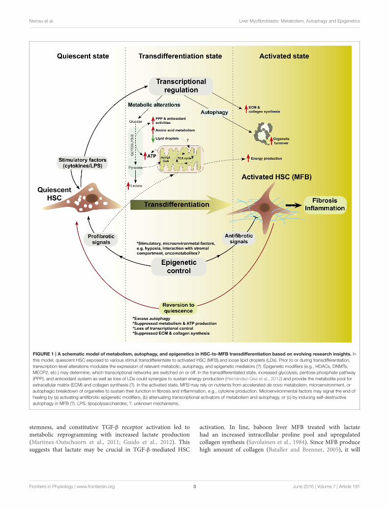
FIGURE 1 | A schematic model of metabolism, autophagy, and epigenetics in HSC-to-MFB transdifferentiation based on evolving research insights. In
this model, quiescent HSC exposed to various stimuli transdifferentiate to activated HSC (MFB) and loose lipid droplets (LDs). Prior to or during transdifferentiation,
transcription-level alterations modulate the expression of relevant metabolic, autophagy, and epigenetic mediators (?). Epigenetic modifiers (e.g., HDACs, DNMTs,
MECP2, etc.) may determine, which transcriptional networks are switched on or off. In the transdifferentiated state, increased glycolysis, pentose phosphate pathway
(PPP), and antioxidant system as well as loss of LDs could synergize to sustain energy production (Hernández-Gea et al., 2012) and provide the metabolite pool for
extracellular matrix (ECM) and collagen synthesis (?). In the activated state, MFB may rely on nutrients from accelerated de novo metabolism, microenvironment, or
autophagic breakdown of organelles to sustain their function in fibrosis and inflammation, e.g., cytokine production. Microenvironmental factors may signal the end of
healing by (a) activating antifibrotic epigenetic modifiers, (b) attenuating transcriptional activators of metabolism and autophagy, or (c) by inducing self-destructive
autophagy in MFB (?). LPS, lipopolysaccharides; ?, unknown mechanisms.
stemness, and constitutive TGF-β receptor activation led tometabolic reprogramming with increased lactate production(Martinez-Outschoorn et al., 2011; Guido et al., 2012). Thissuggests that lactate may be crucial in TGF-β-mediated HSC
activation. In line, baboon liver MFB treated with lactatehad an increased intracellular proline pool and upregulatedcollagen synthesis (Savolainen et al., 1984). Since MFB producehigh amount of collagen (Bataller and Brenner, 2005), it will
Frontiers in Physiology | www.frontiersin.org 3 June 2016 | Volume 7 | Article 191
Nwosu et al. Liver Myofibroblasts: Metabolism, Autophagy and Epigenetics
be interesting to test whether glucose-derived lactate couldmodulate human or murine liver MFB, including analysis onthe role of lactate dehydrogenases in this context. Together,the currently available information suggests that glycolysisis critical in MFB physiology and offers hints for furtherinvestigations.
Tricarboxylic Acid (TCA) CycleThe role of TCA in HSC activation or MFB function is stillunclear and the relevance of most TCA enzymes is yet to bedelineated. It is also unclear if loss of lipid droplets (LDs),which occurs during HSC activation (Blaner et al., 2009; Kluweet al., 2011), is aimed at supplying acetyl-CoA for TCA viaβ-oxidation. The sole evidence of TCA involvement in HSCactivation stems from a recent study showing that succinateinduces G protein-coupled receptor 91 (GPR91) to increasethe production of TGF-β, collagen type I, and α-SMA (Liet al., 2015). This link between succinate and HSC activationsuggests a likely relevance of TCA intermediates in metabolismand signaling regulation during HSC-MFB transdifferentiation.Noteworthy, succinate level is significantly increased in lungMFB and fibrotic lungs. Succinate accumulation enhancedTGF-β1-induced HIF-1α stabilization and MFB differentiation(Xie et al., 2015). Thus, it will be interesting to furtherinvestigate the effect of other TCA intermediates, including α-ketoglutarate that was recently shown to maintain pluripotencyin embryonic stem cells via epigenetic control (Carey et al.,2015). Other interesting intermediates are fumarate and 2-hydroxyglutarate, which are called “oncometabolites” due totheir oncogenic effect on rapidly proliferating cells (Xu et al.,2011; Sullivan et al., 2013; Nowicki and Gottlieb, 2015).Whether these “oncometabolites” exert profibrotic effects onMFB is yet to be explored. On a broader perspective,any abnormal accumulation of TCA intermediates may altertranscriptomic or signaling networks to initiate or sustain MFBphenotype.
Glutamine MetabolismGlutamine is a very abundant amino acid and a highly energy-rich metabolite in humans. Research on mechanisms of cellularglutamine flux have rapidly evolved in recent years to elucidateits relevance in cell metabolism—e.g., in sustaining nucleotidebiosynthesis, TCA, and lipogenesis (DeBerardinis et al., 2007;Metallo et al., 2012; Mullen et al., 2012; Son et al., 2013). InHSCs, the mechanism of glutamine utilization or its relevanceto activation is currently unknown. Nevertheless, activated HSCsare long known to express high glutamine synthetase (GS; Bodeet al., 1998). GS expression and glutamine metabolism have beenseverally linked to theWnt/β-catenin signaling pathway (Cadoretet al., 2002; Austinat et al., 2008; Schmidt et al., 2011; Karner et al.,2015). Thus, the evidence that activation of the Wnt pathwayelevates GS expression, while suppressing HSC activation markerα-SMA (Kordes et al., 2008) suggests that Wnt may controlHSC fate by modulating glutamine metabolism. However, sincethe authors measured neither the intracellular nor extracellularglutamine level in their experiment, it is hard to discuss the effect
of the elevated GS on glutamine metabolism. Therefore, furtherstudies are needed to clarify the role of glutamine in activationand MFB bioenergetics, including any prospects of targetingglutamine utilization in MFB.
Fatty Acid MetabolismFatty acids (FA) are important in liver physiology, notablyin the maintenance of membrane integrity, signaling, energyproduction, and regulation of inflammation in various cellularand tissue compartments (Freigang et al., 2013; Bazinet and Layé,2014; Serhan, 2014). When deregulated, FA metabolism accountsfor several liver diseases such as hepatic steatosis, steatohepatitis,and cirrhosis (Rinella, 2015). Specifically, polyunsaturated fattyacids (PUFAs) as substrates for the cyclooxygenase pathwayvitally regulate initiation and resolution of inflammation(Alhouayek and Muccioli, 2014; Buckley et al., 2014), and havebeen linked to HSC activation. Rat HSCs at early activation stagereplace retinyl esters with PUFAs in LDs (Testerink et al., 2012).However, the mechanism by which the incorporated PUFAslater contribute to HSC activation is not yet known, especiallygiven that HSCs loose LDs during activation (Blaner et al.,2009).
Saturated FA such as oleic acid (OA) and palmitic acids (PA)also participate in MFB activity (Lee et al., 2010, 2014). Further,palmitate and retinol supplementation suppress activation ofhuman immortalized LX-2 (Xu et al., 2005) and primary humanHSCs (Lee et al., 2010). Lee et al. found that palmitate andretinol induced adipose differentiation-related protein (ADRP),which regulates the formation of LDs. Mechanistically, ADRPinduction led to LDs formation, and the suppression of activationand fibrogenic targets including α-SMA, collagen, and MMP1(Lee et al., 2010). The effect of OA and PA on increasing lipidstorage was also corroborated by a recent study, in which theseFA were reported to further synergize with natural compounds,such as rutin and curcumin, to increase LDs and suppressproliferation of HSCs (Lee et al., 2014). On the contrary,OA treatment induced TGF-β, which ostensibly promoted theMFB phenotype in mesangial cells by inducing the expressionof collagen I, fibronectin,and α-SMA (Mishra and Simonson,2008); whether OA similarly induces TGF-β in HSCs is yetunknown. Besides activating MFB, lipids may participate inMFB-mediated inflammatory functions. For instance, humanliver MFB were found to trigger activation of monocytesby secreting prostaglandin-E2 (PGE2) in vitro. Accordingly,blocking PGE2 production with cyclooxygenase 2 inhibitor (NS-398) reduced the expression of the monocyte marker CD163(Zhang et al., 2014).
Evidences also suggest that FA regulates MFB activationand function in other tissues. For instance, arachidonicacid and docosahexaenoic acid (DHA) reversed the MFBphenotype of valvular interstitial cells from porcine aorticvalves by decreasing contractility and expression of α-SMA viaa mechanism involving suppression of RhoA/G-actin/MRTFsignaling (Witt et al., 2014). In the prostate, DHA alsosuppressed fibroblast to MFB differentiation. Specifically DHAprevented TGF-β-induced differentiation, α-SMA expression,
Frontiers in Physiology | www.frontiersin.org 4 June 2016 | Volume 7 | Article 191
Nwosu et al. Liver Myofibroblasts: Metabolism, Autophagy and Epigenetics
and migration of prostate associated fibroblasts (Bianchini et al.,2012). Furthermore, DHA suppressed matrix metalloproteinase2 (MMP2) release and reversed the myofibroblast phenotypeof prostate adenocarcinoma-associated fibroblasts (Bianchiniet al., 2012). In other studies, dietary supplementation with fishoil blocked cardiac fibroblast activation and prevented cardiacfibrosis. Accordingly, eicosapentaenoic acid and DHA increasedcyclic GMP levels, attenuated cardiac fibroblast transformation,proliferation, and collagen synthesis, and also blunted TGF-β1-induced phospho-Smad2/3 nuclear translocation throughactivation of cyclic GMP/protein kinase G pathway (Chenet al., 2011). Nitrated fatty acids (NFAs), formed when nitricoxide (NO) and NO-derived species react with unsaturatedFA, are critical mediators of signaling and inflammation-relatedfunctions (reviewed by Trostchansky and Rubbo, 2008). NFAsupregulated PPARγ and blocked TGF-β signaling/activity inhuman lung fibroblasts (Reddy et al., 2014). In vivo, NFAtreatment led to reduction of disease severity and reversal ofexisting MFB numbers and collagen deposition in a mousemodel of pulmonary fibrosis (Reddy et al., 2014). Furthermore,resolvins, a family of lipid mediators derived from omega-3PUFA, and known to have anti-inflammatory potency (Xu et al.,2010), inhibited interstitial fibrosis by blocking proliferation ofresident fibroblasts (Qu et al., 2012). Despite these evidencesthat fatty acids influence the MFB phenotype, no study hasdirectly interrogated the role of key enzymes in fatty acidmetabolism, including FASN, ACLY, ACC in HSC activation.Hence, further studies will help to resolve the exact molecularregulation and relevance of fatty acid metabolism in HSCbiology.
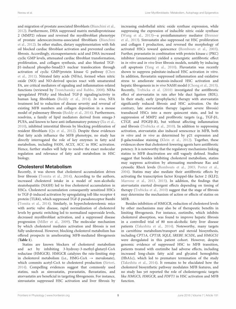
Cholesterol MetabolismRecently, it was shown that cholesterol accumulation drivesliver fibrosis (Tomita et al., 2014). According to the authors,increased cholesterol intake in a model of non-alcoholicsteatohepatitis (NASH) led to free cholesterol accumulation inHSCs. Cholesterol accumulation consequently sensitized HSCsto TGF-β-induced activation by upregulating toll-like receptor 4protein (TLR4), which suppressed TGF-β pseudoreceptor Bambi(Tomita et al., 2014). Similarly, in hypercholesterolemic micewith aortic valve disease, rapid normalization of cholesterollevels by genetic switching led to normalized superoxide levels,decreased myofibroblast activation, and a suppressed diseaseprogression (Miller et al., 2009). The molecular mechanismby which cholesterol mediates activation and fibrosis is notfully understood. However, blocking cholesterol metabolism hasoffered prospects in ameliorating MFB-mediated fibrogenesis(Table 1).
Statins are known blockers of cholesterol metabolismand act by inhibiting 3-hydroxy-3-methyl-glutaryl-CoAreductase (HMGCR). HMGCR catalyzes the rate-limiting stepin cholesterol metabolism (i.e., HMG-CoA → mevalonate),which commits acetyl-CoA to cholesterol production (Sirtori,2014). Compelling evidences suggest that commonly usedstatins, such as simvastatin, pravastatin, fluvastatins, andatorvastatin are beneficial in targeting fibrogenesis. For instance,simvastatin suppressed HSC activation and liver fibrosis by
increasing endothelial nitric oxide synthase expression, whilesuppressing the expression of inducible nitric oxide synthase(Wang et al., 2013)—a proinflammatory mediator (Brenneret al., 2013). Simvastatin also suppressed rat HSC proliferationand collagen I production, and reversed the morphology ofactivated HSCs toward quiescence (Rombouts et al., 2003).Further, pravastatin in combination with protein kinase c (PKC)inhibitor (enzastaurin) yielded a synergistic antifibrotic effectin in vitro and in vivo liver fibrosis models, notably by inducingcell apoptosis (Yang et al., 2010). Fluvastatin was recentlyshown to suppress palmitate-induced HSC activation in vitro.In addition, fluvastatin suppressed inflammation and oxidativestress to ameliorate steatosis-induced HSC activation andhepatic fibrogenesis in in vivo NASH model (Chong et al., 2015).Recently, Trebicka et al. (2010) investigated the antifibroticeffect of atorvastatin in rats after bile duct ligation (BDL).In their study, early (prophylactic) therapy with atorvastatinsignificantly reduced fibrosis and HSC activation. On thecontrary, late atorvastatin therapy (against severe fibrosis)transduced HSCs into a more quiescent state, and led tosuppression of MMP2 and profibrotic targets (e.g., TGF-β1,CTGF, and PDGFβ-R), but without affecting inflammationand fibrosis (Trebicka et al., 2010). In addition to suppressingactivation, atorvastatin also induced senescence in MFB, bothin vitro and in vivo as determined by p21 expression andβ-galactosidase staining (Klein et al., 2012). Together, theseevidences show that cholesterol-lowering agents have antifibroticpotency. It is noteworthy that the regulatory mechanisms linkingstatins to MFB deactivation are still vaguely defined. Studiessuggest that besides inhibiting cholesterol metabolism, statinsmay suppress activation by attenuating membrane Ras andcytosolic RhoA levels (Rombouts et al., 2003; Porter et al.,2004). Statins may also mediate their antifibrotic effects byactivating the transcription factor Kruppel-like factor 2 (KLF2;Marrone et al., 2013, 2015). In addition, the findings thatatorvastatin exerted divergent effects depending on timing oftherapy (Trebicka et al., 2010) suggest that the stage of fibrosismay determine the mechanism of action or effects of statins onMFB.
Besides inhibition of HMGCR, reduction of cholesterol levelsby other mechanisms may also be of therapeutic benefits inlimiting fibrogenesis. For instance, ezetimibe, which inhibitscholesterol absorption, was found to improve hepatic fibrosisin a controlled trial of 80 non-alcoholic fatty liver diseasepatients (Takeshita et al., 2014). Noteworthy, many targetsin carnithine metabolism/transport and steroid biosynthesis,including CPT1A, CPT1B, SQLE, SREBF, SC5DL, and HMGCS1were deregulated in this patient cohort. However, despitegenomic evidence of suppressed HSC to MFB transition,patients treated with ezetimibe had adverse effects, includingincreased long-chain fatty acid and glycated hemoglobin(HbA1c), which led to premature termination of the study(Takeshita et al., 2014). It remains to be elucidated how thecholesterol biosynthetic pathway modulates MFB features, andno study has yet reported the role of cholesterogenic targetslike HMGCS, HMGCR, and FDFT1 in HSC activation and MFBfunction.
Frontiers in Physiology | www.frontiersin.org 5 June 2016 | Volume 7 | Article 191
Nwosu et al. Liver Myofibroblasts: Metabolism, Autophagy and Epigenetics
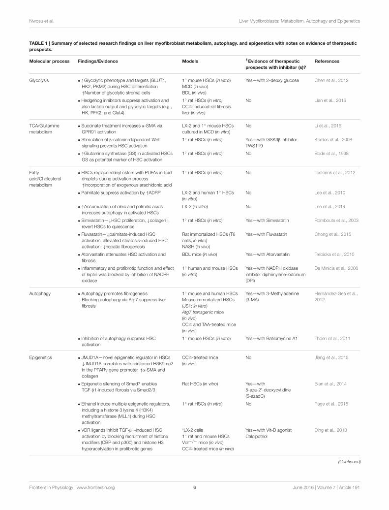
TABLE 1 | Summary of selected research findings on liver myofibroblast metabolism, autophagy. and epigenetics with notes on evidence of therapeutic
prospects.
Molecular process Findings/Evidence Models 1Evidence of therapeutic
prospects with inhibitor (s)?
References
Glycolysis • ↑Glycolytic phenotype and targets (GLUT1,
HK2, PKM2) during HSC differentiation
↑Number of glycolytic stromal cells
1◦ mouse HSCs (in vitro)
MCD (in vivo)
BDL (in vivo)
Yes—with 2-deoxy glucose Chen et al., 2012
• Hedgehog inhibitors suppress activation and
also lactate output and glycolytic targets (e.g.,
HK, PFK2, and Glut4)
1◦ rat HSCs (in vitro)
CCl4-induced rat fibrosis
liver (in vivo)
No Lian et al., 2015
TCA/Glutamine
metabolism
• Succinate treatment increases α-SMA via
GPR91 activation
LX-2 and 1◦ mouse HSCs
cultured in MCD (in vitro)
No Li et al., 2015
• Stimulation of β-catenin-dependent Wnt
signaling prevents HSC activation
1◦ rat HSCs (in vitro) Yes—with GSK3β inhibitor
TWS119
Kordes et al., 2008
• ↑Glutamine synthetase (GS) in activated HSCs
GS as potential marker of HSC activation
1◦ rat HSCs (in vitro) No Bode et al., 1998
Fatty
acid/Cholesterol
metabolism
• HSCs replace retinyl esters with PUFAs in lipid
droplets during activation process
↑Incorporation of exogenous arachidonic acid
1◦ rat HSCs (in vitro) No Testerink et al., 2012
• Palmitate suppress activation by ↑ADRP LX-2 and human 1◦ HSCs
(in vitro)
No Lee et al., 2010
• ↑Accumulation of oleic and palmitic acids
increases autophagy in activated HSCs
LX-2 (in vitro) No Lee et al., 2014
• Simvastatin—↓HSC proliferation, ↓collagen I,
revert HSCs to quiescence
1◦ rat HSCs (in vitro) Yes—with Simvastatin Rombouts et al., 2003
• Fluvastatin—↓palmitate-induced HSC
activation; alleviated steatosis-induced HSC
activation; ↓hepatic fibrogenesis
Rat immortalized HSCs (T6
cells; in vitro)
NASH (in vivo)
Yes—with Fluvastatin Chong et al., 2015
• Atorvastatin attenuates HSC activation and
fibrosis
BDL mice (in vivo) Yes—with Atorvastatin Trebicka et al., 2010
• Inflammatory and profibrotic function and effect
of leptin was blocked by inhibition of NADPH
oxidase
1◦ human and mouse HSCs
(in vitro)
Yes—with NADPH oxidase
inhibitor diphenylene-iodonium
(DPI)
De Minicis et al., 2008
Autophagy • Autophagy promotes fibrogenesis
Blocking autophagy via Atg7 suppress liver
fibrosis
1◦ mouse and human HSCs
Mouse immortalized HSCs
(JS1; in vitro)
Atg7 transgenic mice
(in vivo)
CCl4 and TAA-treated mice
(in vivo)
Yes—with 3-Methyladenine
(3-MA)
Hernández-Gea et al.,
2012
• Inhibition of autophagy suppress HSC
activation
1◦ mouse HSCs (in vitro) Yes—with Bafilomycine A1 Thoen et al., 2011
Epigenetics • JMJD1A—novel epigenetic regulator in HSCs
↓JMJD1A correlates with reinforced H3K9me2
in the PPARγ gene promoter, ↑α-SMA and
collagen
CCl4-treated mice
(in vivo)
No Jiang et al., 2015
• Epigenetic silencing of Smad7 enables
TGF-β1-induced fibrosis via Smad2/3
Rat HSCs (in vitro) Yes—with
5-aza-2’-deoxycytidine
(5-azadC)
Bian et al., 2014
• Ethanol induce multiple epigenetic regulators,
including a histone 3 lysine 4 (H3K4)
methyltransferase (MLL1) during HSC
activation
1◦ rat HSCs (in vitro) No Page et al., 2015
• VDR ligands inhibit TGF-β1-induced HSC
activation by blocking recruitment of histone
modifiers (CBP and p300) and histone H3
hyperacetylation in profibrotic genes
*LX-2 cells
1◦ rat and mouse HSCs
Vdr−/− mice (in vivo)
CCl4-treated mice (in vivo)
Yes—with Vit-D agonist
Calcipotriol
Ding et al., 2013
(Continued)
Frontiers in Physiology | www.frontiersin.org 6 June 2016 | Volume 7 | Article 191
Nwosu et al. Liver Myofibroblasts: Metabolism, Autophagy and Epigenetics
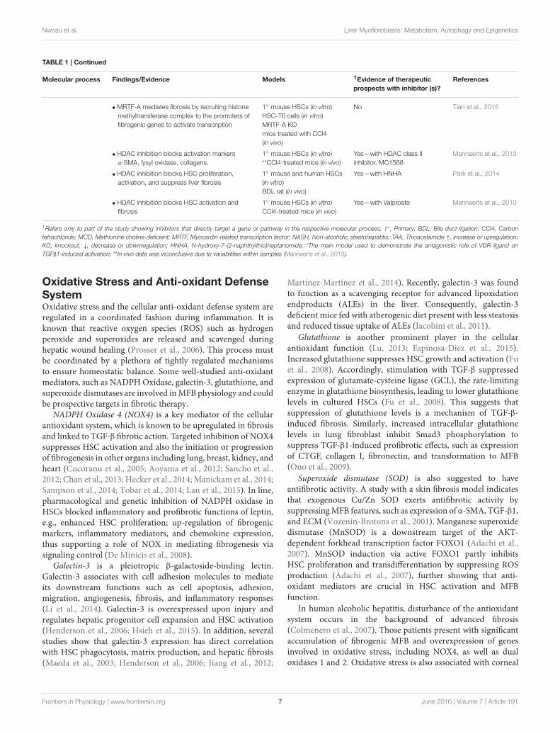
TABLE 1 | Continued
Molecular process Findings/Evidence Models 1Evidence of therapeutic
prospects with inhibitor (s)?
References
• MRTF-A mediates fibrosis by recruiting histone
methyltransferase complex to the promoters of
fibrogenic genes to activate transcription
1◦ mouse HSCs (in vitro)
HSC-T6 cells (in vitro)
MRTF-A KO
mice treated with CCl4
(in vivo)
No Tian et al., 2015
• HDAC inhibition blocks activation markers
α-SMA, lysyl oxidase, collagens.
1◦ mouse HSCs (in vitro)
**CCl4-treated mice (in vivo)
Yes—with HDAC class II
inhibitor, MC1568
Mannaerts et al., 2013
• HDAC inhibition blocks HSC proliferation,
activation, and suppress liver fibrosis
1◦ mouse and human HSCs
(in vitro)
BDL rat (in vivo)
Yes—with HNHA Park et al., 2014
• HDAC inhibition blocks HSC activation and
fibrosis
1◦ mouse HSCs (in vitro)
CCl4-treated mice (in vivo)
Yes—with Valproate Mannaerts et al., 2010
1Refers only to part of the study showing inhibitors that directly target a gene or pathway in the respective molecular process; 1◦, Primary; BDL, Bile duct ligation; CCl4, Carbon
tetrachloride; MCD, Methionine choline-deficient; MRTF, Myocardin-related transcription factor; NASH, Non-alcoholic steatohepatitis; TAA, Thioacetamide ↑, increase or upregulation;
KO, knockout; ↓, decrease or downregulation; HNHA, N-hydroxy-7-(2-naphthylthio)heptanomide; *The main model used to demonstrate the antagonistic role of VDR ligand on
TGFβ1-induced activation; **In vivo data was inconclusive due to variabilities within samples (Mannaerts et al., 2013).
Oxidative Stress and Anti-oxidant DefenseSystemOxidative stress and the cellular anti-oxidant defense system areregulated in a coordinated fashion during inflammation. It isknown that reactive oxygen species (ROS) such as hydrogenperoxide and superoxides are released and scavenged duringhepatic wound healing (Prosser et al., 2006). This process mustbe coordinated by a plethora of tightly regulated mechanismsto ensure homeostatic balance. Some well-studied anti-oxidantmediators, such as NADPH Oxidase, galectin-3, glutathione, andsuperoxide dismutases are involved inMFB physiology and couldbe prospective targets in fibrotic therapy.
NADPH Oxidase 4 (NOX4) is a key mediator of the cellularantioxidant system, which is known to be upregulated in fibrosisand linked to TGF-β fibrotic action. Targeted inhibition of NOX4suppresses HSC activation and also the initiation or progressionof fibrogenesis in other organs including lung, breast, kidney, andheart (Cucoranu et al., 2005; Aoyama et al., 2012; Sancho et al.,2012; Chan et al., 2013; Hecker et al., 2014; Manickam et al., 2014;Sampson et al., 2014; Tobar et al., 2014; Lan et al., 2015). In line,pharmacological and genetic inhibition of NADPH oxidase inHSCs blocked inflammatory and profibrotic functions of leptin,e.g., enhanced HSC proliferation; up-regulation of fibrogenicmarkers, inflammatory mediators, and chemokine expression,thus supporting a role of NOX in mediating fibrogenesis viasignaling control (De Minicis et al., 2008).
Galectin-3 is a pleiotropic β-galactoside-binding lectin.Galectin-3 associates with cell adhesion molecules to mediateits downstream functions such as cell apoptosis, adhesion,migration, angiogenesis, fibrosis, and inflammatory responses(Li et al., 2014). Galectin-3 is overexpressed upon injury andregulates hepatic progenitor cell expansion and HSC activation(Henderson et al., 2006; Hsieh et al., 2015). In addition, severalstudies show that galectin-3 expression has direct correlationwith HSC phagocytosis, matrix production, and hepatic fibrosis(Maeda et al., 2003; Henderson et al., 2006; Jiang et al., 2012;
Martínez-Martínez et al., 2014). Recently, galectin-3 was foundto function as a scavenging receptor for advanced lipoxidationendproducts (ALEs) in the liver. Consequently, galectin-3deficient mice fed with atherogenic diet present with less steatosisand reduced tissue uptake of ALEs (Iacobini et al., 2011).
Glutathione is another prominent player in the cellularantioxidant function (Lu, 2013; Espinosa-Diez et al., 2015).Increased glutathione suppresses HSC growth and activation (Fuet al., 2008). Accordingly, stimulation with TGF-β suppressedexpression of glutamate-cysteine ligase (GCL), the rate-limitingenzyme in glutathione biosynthesis, leading to lower glutathionelevels in cultured HSCs (Fu et al., 2008). This suggests thatsuppression of glutathione levels is a mechanism of TGF-β-induced fibrosis. Similarly, increased intracellular glutathionelevels in lung fibroblast inhibit Smad3 phosphorylation tosuppress TGF-β1-induced profibrotic effects, such as expressionof CTGF, collagen I, fibronectin, and transformation to MFB(Ono et al., 2009).
Superoxide dismutase (SOD) is also suggested to haveantifibrotic activity. A study with a skin fibrosis model indicatesthat exogenous Cu/Zn SOD exerts antifibrotic activity bysuppressingMFB features, such as expression of α-SMA, TGF-β1,and ECM (Vozenin-Brotons et al., 2001). Manganese superoxidedismutase (MnSOD) is a downstream target of the AKT-dependent forkhead transcription factor FOXO1 (Adachi et al.,2007). MnSOD induction via active FOXO1 partly inhibitsHSC proliferation and transdifferentiation by suppressing ROSproduction (Adachi et al., 2007), further showing that anti-oxidant mediators are crucial in HSC activation and MFBfunction.
In human alcoholic hepatitis, disturbance of the antioxidantsystem occurs in the background of advanced fibrosis(Colmenero et al., 2007). Those patients present with significantaccumulation of fibrogenic MFB and overexpression of genesinvolved in oxidative stress, including NOX4, as well as dualoxidases 1 and 2. Oxidative stress is also associated with corneal
Frontiers in Physiology | www.frontiersin.org 7 June 2016 | Volume 7 | Article 191
Nwosu et al. Liver Myofibroblasts: Metabolism, Autophagy and Epigenetics
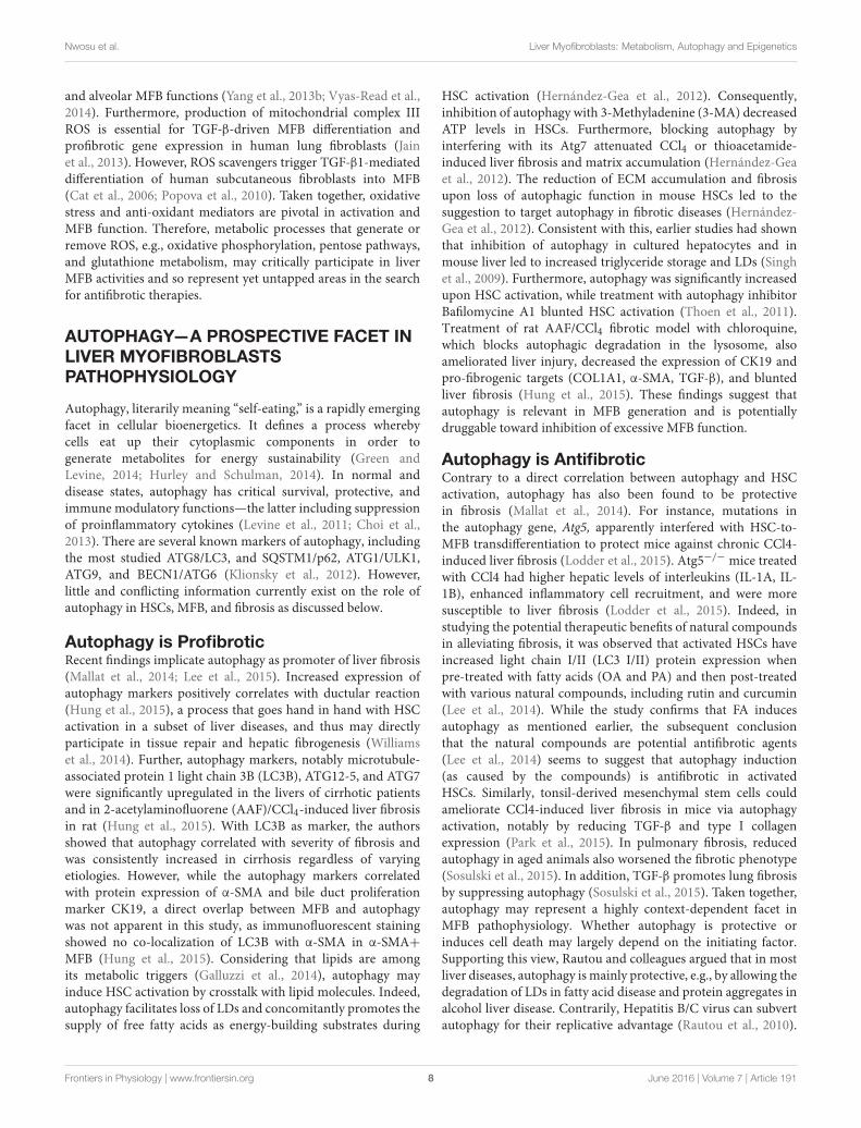
and alveolar MFB functions (Yang et al., 2013b; Vyas-Read et al.,2014). Furthermore, production of mitochondrial complex IIIROS is essential for TGF-β-driven MFB differentiation andprofibrotic gene expression in human lung fibroblasts (Jainet al., 2013). However, ROS scavengers trigger TGF-β1-mediateddifferentiation of human subcutaneous fibroblasts into MFB(Cat et al., 2006; Popova et al., 2010). Taken together, oxidativestress and anti-oxidant mediators are pivotal in activation andMFB function. Therefore, metabolic processes that generate orremove ROS, e.g., oxidative phosphorylation, pentose pathways,and glutathione metabolism, may critically participate in liverMFB activities and so represent yet untapped areas in the searchfor antifibrotic therapies.
AUTOPHAGY—A PROSPECTIVE FACET INLIVER MYOFIBROBLASTSPATHOPHYSIOLOGY
Autophagy, literarily meaning “self-eating,” is a rapidly emergingfacet in cellular bioenergetics. It defines a process wherebycells eat up their cytoplasmic components in order togenerate metabolites for energy sustainability (Green andLevine, 2014; Hurley and Schulman, 2014). In normal anddisease states, autophagy has critical survival, protective, andimmune modulatory functions—the latter including suppressionof proinflammatory cytokines (Levine et al., 2011; Choi et al.,2013). There are several known markers of autophagy, includingthe most studied ATG8/LC3, and SQSTM1/p62, ATG1/ULK1,ATG9, and BECN1/ATG6 (Klionsky et al., 2012). However,little and conflicting information currently exist on the role ofautophagy in HSCs, MFB, and fibrosis as discussed below.
Autophagy is ProfibroticRecent findings implicate autophagy as promoter of liver fibrosis(Mallat et al., 2014; Lee et al., 2015). Increased expression ofautophagy markers positively correlates with ductular reaction(Hung et al., 2015), a process that goes hand in hand with HSCactivation in a subset of liver diseases, and thus may directlyparticipate in tissue repair and hepatic fibrogenesis (Williamset al., 2014). Further, autophagy markers, notably microtubule-associated protein 1 light chain 3B (LC3B), ATG12-5, and ATG7were significantly upregulated in the livers of cirrhotic patientsand in 2-acetylaminofluorene (AAF)/CCl4-induced liver fibrosisin rat (Hung et al., 2015). With LC3B as marker, the authorsshowed that autophagy correlated with severity of fibrosis andwas consistently increased in cirrhosis regardless of varyingetiologies. However, while the autophagy markers correlatedwith protein expression of α-SMA and bile duct proliferationmarker CK19, a direct overlap between MFB and autophagywas not apparent in this study, as immunofluorescent stainingshowed no co-localization of LC3B with α-SMA in α-SMA+MFB (Hung et al., 2015). Considering that lipids are amongits metabolic triggers (Galluzzi et al., 2014), autophagy mayinduce HSC activation by crosstalk with lipid molecules. Indeed,autophagy facilitates loss of LDs and concomitantly promotes thesupply of free fatty acids as energy-building substrates during
HSC activation (Hernández-Gea et al., 2012). Consequently,inhibition of autophagy with 3-Methyladenine (3-MA) decreasedATP levels in HSCs. Furthermore, blocking autophagy byinterfering with its Atg7 attenuated CCl4 or thioacetamide-induced liver fibrosis and matrix accumulation (Hernández-Geaet al., 2012). The reduction of ECM accumulation and fibrosisupon loss of autophagic function in mouse HSCs led to thesuggestion to target autophagy in fibrotic diseases (Hernández-Gea et al., 2012). Consistent with this, earlier studies had shownthat inhibition of autophagy in cultured hepatocytes and inmouse liver led to increased triglyceride storage and LDs (Singhet al., 2009). Furthermore, autophagy was significantly increasedupon HSC activation, while treatment with autophagy inhibitorBafilomycine A1 blunted HSC activation (Thoen et al., 2011).Treatment of rat AAF/CCl4 fibrotic model with chloroquine,which blocks autophagic degradation in the lysosome, alsoameliorated liver injury, decreased the expression of CK19 andpro-fibrogenic targets (COL1A1, α-SMA, TGF-β), and bluntedliver fibrosis (Hung et al., 2015). These findings suggest thatautophagy is relevant in MFB generation and is potentiallydruggable toward inhibition of excessive MFB function.
Autophagy is AntifibroticContrary to a direct correlation between autophagy and HSCactivation, autophagy has also been found to be protectivein fibrosis (Mallat et al., 2014). For instance, mutations inthe autophagy gene, Atg5, apparently interfered with HSC-to-MFB transdifferentiation to protect mice against chronic CCl4-induced liver fibrosis (Lodder et al., 2015). Atg5−/− mice treatedwith CCl4 had higher hepatic levels of interleukins (IL-1A, IL-1B), enhanced inflammatory cell recruitment, and were moresusceptible to liver fibrosis (Lodder et al., 2015). Indeed, instudying the potential therapeutic benefits of natural compoundsin alleviating fibrosis, it was observed that activated HSCs haveincreased light chain I/II (LC3 I/II) protein expression whenpre-treated with fatty acids (OA and PA) and then post-treatedwith various natural compounds, including rutin and curcumin(Lee et al., 2014). While the study confirms that FA inducesautophagy as mentioned earlier, the subsequent conclusionthat the natural compounds are potential antifibrotic agents(Lee et al., 2014) seems to suggest that autophagy induction(as caused by the compounds) is antifibrotic in activatedHSCs. Similarly, tonsil-derived mesenchymal stem cells couldameliorate CCl4-induced liver fibrosis in mice via autophagyactivation, notably by reducing TGF-β and type I collagenexpression (Park et al., 2015). In pulmonary fibrosis, reducedautophagy in aged animals also worsened the fibrotic phenotype(Sosulski et al., 2015). In addition, TGF-β promotes lung fibrosisby suppressing autophagy (Sosulski et al., 2015). Taken together,autophagy may represent a highly context-dependent facet inMFB pathophysiology. Whether autophagy is protective orinduces cell death may largely depend on the initiating factor.Supporting this view, Rautou and colleagues argued that in mostliver diseases, autophagy is mainly protective, e.g., by allowing thedegradation of LDs in fatty acid disease and protein aggregates inalcohol liver disease. Contrarily, Hepatitis B/C virus can subvertautophagy for their replicative advantage (Rautou et al., 2010).
Frontiers in Physiology | www.frontiersin.org 8 June 2016 | Volume 7 | Article 191
Nwosu et al. Liver Myofibroblasts: Metabolism, Autophagy and Epigenetics
Here, it is worthy to highlight that the loss of LDs attributed to“protective” autophagy (Rautou et al., 2010) is also a mechanismthrough which autophagy provides energy substrates to promoteHSC activation and fibrosis (Hernández-Gea et al., 2012).Therefore, it will be of interest to further interrogate protectiveand detrimental autophagy in HSC activation, MFB functions,and in the switch between activation and dedifferentiation toquiescence (Figure 1).
EPIGENETIC ALTERATIONS IN LIVERMYOFIBROBLASTS
Epigenetics refer to heritable traits resulting from chromosomalalterations that do not alter DNA sequence. Epigeneticalterations maintain cell identity and include DNA methylation,histone modifications, chromatin remodeling, transcriptionalcontrol, and post-translational modification of non-coding RNA(Berger et al., 2009; Cedar and Bergman, 2009; Portela andEsteller, 2010; Mann, 2014). Interestingly, several epigenetictargets, including DNA methyltransferases (DNMTs), histonedeacetylases (HDACs), histone methyltransferases (DOT1L,EZH2, G9A), histone demethylases (JmjC-domain proteins,LSD1), and binding domains (BET, BAZ2B, L3MBTL1) aredruggable in human diseases (Helin and Dhanak, 2013).Epigenetic alterations occur in liver fibrosis and chronic liverdiseases (Mann, 2014; Atta, 2015; Lleo et al., 2015) and arerelevant in HSC activation (Kang et al., 2015).
DNA MethylationDuring activation, HSCs accumulate methylation changes thatsignificantly modulate the expression of genes involved in cellactivation and inflammation (Götze et al., 2015). Specifically,expression of DNAmethyltransferases, DNMT3A andDNMT3B,increased with HSC activation (Götze et al., 2015). Oneconsequence of hypermethylation is gene silencing. For instance,transcriptional silencing of PPARγ, which occurs during HSCactivation (Hazra et al., 2004), has been attributed to methylationbased epigenetic control (Mann et al., 2007; Yang et al.,2012). Recently, the Jumonji Domain-Containing Protein 1A(JMJD1A)—a histone H3K9 demethylase—was found to regulateHSC activation and liver fibrosis by targeting PPARγ geneexpression (Jiang et al., 2015). Knockdown of JMJD1A inHSCs correlated with reinforced H3K9me2 in the PPARγ
gene promoter; increased α-SMA and collagen expression, andenhanced necrosis in the CCl4 mouse fibrosis model (Jianget al., 2015). Consistent with this finding, blocking CpGmethylation with the nucleotide analog 5-aza-2′-deoxycytidine(5-azadC) prevented loss of PPARγ expression (Mann et al.,2007). Methylation-based control in HSCs is also evidentfrom methyl-CpG-binding protein (MECP2), known to represschromatin structures. MECP2 is induced during HSC activation,correlates with α-SMA expression and contributes to MFBtransdifferentiation by regulating fibrogenic targets (Mannet al., 2007; Yang et al., 2013a). Mechanistically, MECP2repressed Patched (PTCH1), whose loss upon hypermethylationis necessary for sustained fibroblast activation and liver
fibrosis (Yang et al., 2013a). Furthermore, DNA methylationis responsible for epigenetic silencing of Smad7, whichenables fibrogenic TGF-β effects via Smad2 and Smad3phosphorylation (Bian et al., 2014). Hence, RNA interference and5-azadC-mediated inhibition of the methylation gene DNMT1prevented TGF-β-induced proliferation and upregulation ofactivation markers in HSCs (Bian et al., 2014). More studiesare required to further validate methylation switches atvarious MFB differentiation stages under normal and perturbedmicroenvironments.
Histone ModificationHistone modification is another active epigenetic alterationduring HSC activation. For instance, HSCs in a mouse modelof acute liver failure secrete IL-1, which induces high MMP9levels, leading to collagen IV degradation (Yan et al., 2008). Ifuncontrolled, MMP9 expression could oppose MFB-mediatedaccumulation of ECM. Hence maintenance of appropriatebalance is necessary during HSC activation or MFB function.Interestingly, epigenetic repression of MMPs has been suggestedas a mechanism that controls HSC transdifferentiation (Qinand Han, 2010). Consequently, MMP9 and MMP13 promotersin MFB display impaired histone acetylation and assemblyof transcription machinery. These alterations blocked dockingof transcription factor c-Jun on the MMP promoters (Qinand Han, 2010). Similarly, ectopic expression of HDAC4 inquiescent HSCs suppressed intrinsic and IL-1-induced MMPpromoter activity and repressed MMP9 expression. Thesefindings implicate accumulation of HDACs at MMP promoters,specifically HDAC4, as an epigenetic mechanism to repressMMP expression during HSC activation (Qin and Han, 2010).Similar regulation is provided via HDAC7, which represseshepatocyte growth factor (HGF) and thus increases susceptibilityto hepatocellular damage, inflammation, and fibrosis in liverinjury (Pannem et al., 2014). HDAC7-mediated repression ofHGF in HSCs is antagonized by the tumor suppressor genecylindromatosis (CYLD). Accordingly, CYLD interacts with andremoves HDAC7 from the HGF promoter, hence enablingHGF induction, which subsequently is secreted and protectsagainst hepatocellular injury and fibrosis (Pannem et al., 2014).In cultured human skin fibroblasts, HDAC6, HDAC8, butmost potently HDAC4 were identified as crucial epigeneticregulators of TGF-β-induced MFB differentiation, ostensiblyby blocking the expression of TGF-β signaling repressors 5′-TG-3′-Interacting Factor (TGIF) and TGIF2 (Glenisson et al.,2007).
Recently, the myocardin-related transcription factor (MRTF),ethanol and vitamin D receptor (VDR) have been identified asepigenomic modifiers during HSC activation. MRTF promotesMFB differentiation, fibrosis, and TGF-β-inducedHSC activation(Crider et al., 2011; O’Connor and Gomez, 2013; Velasquezet al., 2013; O’Connor et al., 2015; Sisson et al., 2015).Mechanistically, MRTF-A mediates fibrosis via recruitment ofthe histone methyltransferase complex to the promoters offibrogenic genes and subsequent transcriptional activation (Tianet al., 2015). Ethanol exposure was found to promote ratHSC transdifferentiation by inducing global changes in histone
Frontiers in Physiology | www.frontiersin.org 9 June 2016 | Volume 7 | Article 191

Nwosu et al. Liver Myofibroblasts: Metabolism, Autophagy and Epigenetics
modifying enzymes that upregulate ECM components elastin(ELN) and collagens (Page et al., 2015). The authors foundthat ethanol induced the expression of histone 3 lysine 4(H3K4) methyltransferases, mainly MLL1. MLL1 binding wasenriched on ELN gene promoter and consequently inducedELN expression in transitioning HSCs. In addition, MLL1expression also correlated with ELN and collagens in ALDliver explants further confirming that ethanol induced pro-fibrogenic processes via epigenetic regulators (Page et al.,2015). VDR ligands also induce chromatin remodeling as amechanism to counteract TGF-β-driven HSC activation (Dinget al., 2013). TGF-β induced activation by promoting therecruitment of histone-modifying cofactors, p300 and CBP, andby promoting histone H3 hyperacetylation at a VDR/SMAD co-occupied regulatory region of COL1A1. Treatment with VDRligands antagonized activation by disrupting TGF-β-mediatedSMAD/VDR interaction. Consequently, synthetic VDR agonistCalcipotriol reduced collagen deposition and fibrotic geneexpression in vitro and in vivo (Ding et al., 2013). These studiesunderscore the critical role of histone modification in HSCtransdifferentiation (Figure 1).
Indeed, evolving links between epigenetics and MFB functionjustifies targeting histone modifiers in antifibrotic therapies.In line, various HDAC inhibitors are effective against TGF-β-induced MFB generation (Glenisson et al., 2007; Guo et al.,2009; Liu et al., 2013), including MC1568, valproic acid (VPA),trichostatin A (TSA), and butyrate. MC1568 inhibited HSCactivation markers, such as type I/III collagen, SMA, and lysyloxidase. In addition, MC1568 induced antifibrotic microRNA-29 and also suppressed the proliferation of freshly isolatedmouse HSCs (Mannaerts et al., 2013). Unfortunately, the authorswere unable to reproduce the result in CCl4 fibrosis modelowing to technical issues, probably due to inefficient delivery orfast metabolization of the drug (Mannaerts et al., 2013). VPAalso suppresses liver fibrosis and HSC activation in vitro andin vivo (Mannaerts et al., 2010; Aher et al., 2015). However,given the pivotal role of TGF-β in HSC activation, it isimportant to mention that VPA did not interfere with earlyTGF-β targets, SMAD 6 and 7 (Mannaerts et al., 2010), thusraising further questions on the exact mechanism, by whichepigenetics influences HSC activation by TGF-β. Furthermore,TSA and RNA interference against HDAC4 prevented MFBdifferentiation as measured by α-SMA expression (Glenissonet al., 2007). Nilotinib, a tyrosine kinase inhibitor, selectivelyinduces apoptotic and autophagic cell death in HSCs byblocking HDAC 1, 2, and 4 (Shaker et al., 2013). Other HDACinhibitors that suppress HSC activation include a chalconederivative 2’,4’,6’-tris(methoxymethoxy) chalcone (TMMC; (Leeet al., 2011)), and N-hydroxy-7-(2-naphthylthio)heptanomide(HNHA; (Park et al., 2014)). HNHA not only suppressed HSCproliferation, activation and liver fibrosis, but also restoredliver function and prolonged survival in the BDL rat model(Park et al., 2014). Further evidence for targeting HDACsin fibrotic diseases have been shown in other settings, e.g.,cardiac and lung fibroblasts (Zhang et al., 2013; Sanders et al.,2014; Schuetze et al., 2014). Together, these data highlight therelevance of epigenetics in HSC activation and encourage the
exploitation of epigenetic targets in the control of MFB-mediatedfibrogenesis.
TECHNICAL ADVANCES THAT COULDHELP DELINEATE METABOLISM,AUTOPHAGY, AND EPIGENETICS IN LIVERMYOFIBROBLASTS
Within the last few decades, several cutting edge techniques haveemerged for the study of complex biological processes.
In metabolism, mass spectrometry based metabolomicstechniques have been developed for measuring metabolic flux.Consequently, it is now possible to precisely determine utilityof metabolites with a very high degree of precision (Zamboniet al., 2009; Hiller and Metallo, 2013). Thus, metabolic fluxanalyses could enable (a) holistic and simultaneous quantificationof labeled and unlabeled metabolites derived from parent carbonsources (e.g., glucose or glutamine), (b) help to delineate themetabolic properties of MFB at different differentiation stages,and (c) offer hints on prospective metabolic pathways oftherapeutic relevance in HSC activation or MFB function. Inaddition, cellular respiration can be easily measured by fiberoptic oxygen sensors, clark electrode, and extracellular fluxanalyzers—the latter offering the advantage of assessing oxygenconsumption and extracellular acidification rates in living cells(Zhang et al., 2012; Perry et al., 2013). Furthermore, in vivomeasurement of metabolism using hyperpolarized, (13)C-labeledcells has been successfully applied (Rodrigues et al., 2014; Brindle,2015). The latter in vivo approach could enhance accuracy giventhe challenges of potential artifacts from cell cultures.
For autophagy studies, techniques such as time-lapsemicroscopy (Muzzey and van Oudenaarden, 2009) andtransmission electron microscopy could be adopted withmolecular biology methods (Klionsky et al., 2012) to dissecthow autophagy affects the evolution of fibrosis. This couldhelp identify novel potential mediators of MFB autophagicmechanisms for therapeutic purposes. Also, research on MFBautophagy will benefit from other advances in microscopy,e.g., super-resolution microscopy that allow high-resolutionimaging and protein tracking in living cells (Bergner et al., 2013;Barden et al., 2015; Chéreau et al., 2015). Tools for measuringautophagy based on biochemical features, e.g., quenching ofGFP fluorescent signals in the lysosome at low pH (Bamptonet al., 2005), could enable a more improved understanding ofautophagy during HSC transdifferention (Figure 1).
Delineating epigenetic alterations in HSCs will begreatly enhanced by bisulfite conversion, chromatinimmunoprecipitation and high throughput DNA methylomeanalyses (Shull et al., 2015; Tang et al., 2015). Other techniques,including single cell analysis, sequencing techniques, andmulti-color fluorescence activated cell sorting could be appliedto uncover yet unknown epigenetic alterations specific to HSCactivation and MFB functions (Figure 1).
To accelerate understanding of MFB function, it is importantto consider and possibly tackle the fact that in vivo changes ingene expression during HSC activationmay differ markedly from
Frontiers in Physiology | www.frontiersin.org 10 June 2016 | Volume 7 | Article 191
Nwosu et al. Liver Myofibroblasts: Metabolism, Autophagy and Epigenetics
those that occur in in vitro culture (De Minicis et al., 2007). Suchdifferences may be due to a plethora of factors, including but notlimited to cell culture conditions, contamination by other livercell populations, and sample handling. Where possible, a strategyto overcome this challenge would be to interface the assaysmentioned above with lineage tracing. Lineage tracing has beensuccessfully applied as a powerful innovative tool for trackingMFB origin (Mederacke et al., 2013; Lua et al., 2014; Swiderska-Syn et al., 2014). Hence, lineage tracing opens up the feasibilityof in vivo studies of alterations in HSCs at quiescent, transitory,and activated states, and will highly complement genomic andfunctional assays.
Finally, optimal application of novel gene editing techniques,such as CRISPR/Cas9, TALENs, etc. (Cho et al., 2014;Jamal et al., 2015; LaFountaine et al., 2015; Laufer andSingh, 2015) will hugely accelerates the identification andunderstanding of metabolic, autophagic, and epigenetic targetsin MFB, especially when complemented with proteomicand transcriptomic profilings. Ultimately, perhaps the mostimportant technical step in understanding MFB physiology,especially in the context of metabolism, autophagy, andepigenetics, is to explore all possible strategies to eliminateanalytical variables that distort results.
UNCHARTED TERRITORIES IN THESTUDY OF METABOLISM, AUTOPHAGYAND EPIGENETICS IN LIVERMYOFIBROBLASTS
Currently, few studies have focused on how MFB feed,regulate survival via autophagy or via epigenetic alterationsthat activate or silence key genes in spatio-temporal cell fatedecisions. Consequently, knowledge of metabolism, autophagy,and epigenetics in MFB is still at a very nascent stage withmany convoluted parts worth further clarifications (Figure 1).The studies that so far focused on the above subjects have offeredexciting platforms for further questions. However, more effortsshould be dedicated to delineate their molecular relevance toMFB origin and function in health and disease. Lessons canalso be learned from other settings, e.g., cancer. For example,metabolism has evolved as a potentially druggable process incancer entities and many metabolic targets are in preclinical andclinical trials (Galluzzi et al., 2013). Whether those therapeuticstrategies would find application in MFB origin/liver fibrosisremains an open question. It is also unknown which metabolicpriorities are exploited by MFB, e.g., particular substrates thatare indispensable for their survival. The timing of metabolicalterations is also critical to any studies, as changes in mRNAtranscripts occur within hours in cultured HSCs (Chen et al.,2012). Unresolved questions also abound in the area of nutrientexchange between MFB and other liver cell populations inthe microenvironment, including the extent of their capacityto sustain de novo anabolism. Regarding autophagy, scientificefforts should clarify boundaries between “self-eating” autophagyto provide energy building substrates and autophagy with aself-destructive consequence. Questions like “how autophagy
markers contribute to MFB status” remain hugely unanswered.Similar questions in metabolism and autophagy also applyto epigenetics. Many epigenomic targets are druggable andseveral epigenomic drugs yield a beneficial response in MFBand fibrosis. Hence, understanding how DNA methylationand histone modification control HSC transdifferentiationcould substantially improve the prospects of better therapeuticinterventions. Furthermore, questions also arise on possiblecrosstalks or feedback loops or overarching control by potentialregulators that are chiefly responsible for MFB physiology.These may include transcription factors, microRNAs, long non-coding RNAs, and other regulators of the genome. Hence, itis yet unclear under which circumstances certain regulatorsswitch on/off metabolism, autophagy, or epigenetic modifiers inMFB (Figure 1). Ultimately, there are currently no establishedmetabolic, autophagy, or epigenetics markers in MFB. Dissectingthese uncharted territories will substantially open new windowsfor therapeutic interventions in MFB-mediated fibrosis.
CONCLUSION
In the light of evolving molecular insights, metabolism,autophagy, and epigenetics are critical players in HSC activationand MFB functions. Currently available data lead us to proposethat transcriptional and epigenetic controls likely coordinatemetabolism and autophagy in HSC to MFB transdifferentiation(Figure 1). In the context of the discussed molecular processes,more studies are required to deepen understanding of MFBorigin and function in liver fibrogenesis. We suggest that ongoingand future MFB research should interrogate the relevance ofkey metabolic enzymes, autophagy markers, and epigeneticmodifiers, including but not limited to those mentioned hereand already investigated in cancer (Claus and Lübbert, 2003;Cheong et al., 2012; Popovic and Licht, 2012; Galluzzi et al.,2013; Helin and Dhanak, 2013; Table 1). We also recommendthat researchers should critically consider the time pointsselected for MFB studies since unforeseen switch from activationto quiescence or vice versa could obscure molecular details.In the end, a broad-spectrum integration of cutting edgetools that enable simultaneous measurements, such as “omics”technologies, will enable better understanding of MFB andfurther expose novel regulators or biomarkers of MFB activity.We conclude that a detailed understanding of metabolism,autophagy and epigenetics in liverMFBwill inspire a new frontierin the development of antifibrotic therapy.
AUTHOR CONTRIBUTIONS
ZCN, HA, and YL conceived and wrote the manuscript, whileSW, SD, and YL provided comments, revised, and corrected themanuscript.
FUNDING
SD is supported by funds from the DeutscheForschungsgemeinschaft (DFG) Do373/13-1, the BMBF
Frontiers in Physiology | www.frontiersin.org 11 June 2016 | Volume 7 | Article 191
Nwosu et al. Liver Myofibroblasts: Metabolism, Autophagy and Epigenetics
programs “Virtual Liver” (Grants 0315755, 0315764), andLiSyM (Grant PTJ-FKZ: 031L0043), as well as from MarieCurie Actions of the European Union’s Seventh FrameworkProgramme (FP7/2007-2013) Grant PITN-GA-2012-316549(IT LIVER: Inhibiting TGF-beta in liver diseases). SW is
supported by BMBF SysTox Grant (FKZ 031A303E). ZCNis a recipient of Ph.D. Scholarship from the Niger DeltaDevelopment Commission, Nigeria, and appreciates thegenerous supports from HBIGS, University of Heidelberg,Germany.
REFERENCES
Adachi, M., Osawa, Y., Uchinami, H., Kitamura, T., Accili, D., and Brenner, D.
A. (2007). The forkhead transcription factor FoxO1 regulates proliferation and
transdifferentiation of hepatic stellate cells. Gastroenterology 132, 1434–1446.
doi: 10.1053/j.gastro.2007.01.033
Aher, J. S., Khan, S., Jain, S., Tikoo, K., and Jena, G. (2015). Valproate ameliorates
thioacetamide-induced fibrosis by hepatic stellate cell inactivation. Hum. Exp.
Toxicol. 34, 44–55. doi: 10.1177/0960327114531992
Alhouayek, M., and Muccioli, G. G. (2014). COX-2-derived endocannabinoid
metabolites as novel inflammatory mediators. Trends Pharmacol. Sci. 35,
284–292. doi: 10.1016/j.tips.2014.03.001
Aoyama, T., Paik, Y. H., Watanabe, S., Laleu, B., Gaggini, F., Fioraso-Cartier,
L., et al. (2012). Nicotinamide adenine dinucleotide phosphate oxidase in
experimental liver fibrosis: GKT137831 as a novel potential therapeutic agent.
Hepatology 56, 2316–2327. doi: 10.1002/hep.25938
Atta, H. M. (2015). Reversibility and heritability of liver fibrosis: implications
for research and therapy. World J. Gastroenterol. 21, 5138–5148. doi:
10.3748/wjg.v21.i17.5138
Austinat, M., Dunsch, R., Wittekind, C., Tannapfel, A., Gebhardt, R., and Gaunitz,
F. (2008). Correlation between beta-catenin mutations and expression of Wnt-
signaling target genes in hepatocellular carcinoma. Mol. Cancer 7:21. doi:
10.1186/1476-4598-7-21
Bampton, E. T., Goemans, C. G., Niranjan, D., Mizushima, N., and Tolkovsky,
A. M. (2005). The dynamics of autophagy visualized in live cells: from
autophagosome formation to fusion with endo/lysosomes. Autophagy 1, 23–36.
doi: 10.4161/auto.1.1.1495
Barden, A. O., Goler, A. S., Humphreys, S. C., Tabatabaei, S., Lochner, M.,
Ruepp, M. D., et al. (2015). Tracking individual membrane proteins and their
biochemistry: The power of direct observation. Neuropharmacology 98, 22–30.
doi: 10.1016/j.neuropharm.2015.05.003
Bataller, R., and Brenner, D. A. (2005). Liver fibrosis. J. Clin. Invest. 115, 209–218.
doi: 10.1172/JCI24282
Bazinet, R. P., and Layé, S. (2014). Polyunsaturated fatty acids and their
metabolites in brain function and disease. Nat. Rev. Neurosci. 15, 771–785. doi:
10.1038/nrn3820
Bentaib, A., De Tullio, P., Chneiweiss, H., Hermans, E., Junier, M. P.,
and Leprince, P. (2014). Metabolic reprogramming in transformed mouse
cortical astrocytes: a proteomic study. J. Proteomics 113C, 292–314. doi:
10.1016/j.jprot.2014.09.019
Berger, S. L., Kouzarides, T., Shiekhattar, R., and Shilatifard, A. (2009).
An operational definition of epigenetics. Genes Dev. 23, 781–783. doi:
10.1101/gad.1787609
Bergner, S., Vatsyayan, P., and Matysik, F. M. (2013). Recent advances in high
resolution scanning electrochemical microscopy of living cells–a review. Anal.
Chim. Acta 775, 1–13. doi: 10.1016/j.aca.2012.12.042
Bian, E. B., Huang, C., Wang, H., Chen, X. X., Zhang, L., Lv, X. W., et al.
(2014). Repression of Smad7 mediated by DNMT1 determines hepatic stellate
cell activation and liver fibrosis in rats. Toxicol. Lett. 224, 175–185. doi:
10.1016/j.toxlet.2013.10.038
Bianchini, F., Giannoni, E., Serni, S., Chiarugi, P., and Calorini, L. (2012). 22: 6n-
3 DHA inhibits differentiation of prostate fibroblasts into myofibroblasts and
tumorigenesis. Br. J. Nutr. 108, 2129–2137. doi: 10.1017/S0007114512000359
Blaner, W. S., O’Byrne, S. M., Wongsiriroj, N., Kluwe, J., D’Ambrosio, D. M.,
Jiang, H., et al. (2009). Hepatic stellate cell lipid droplets: a specialized
lipid droplet for retinoid storage. Biochim. Biophys. Acta 1791, 467–473. doi:
10.1016/j.bbalip.2008.11.001
Bode, J. G., Peters-Regehr, T., Gressner, A. M., and Häussinger, D. (1998). De
novo expression of glutamine synthetase during transformation of hepatic
stellate cells into myofibroblast-like cells. Biochem. J. 335(Pt 3), 697–700. doi:
10.1042/bj3350697
Brenner, C., Galluzzi, L., Kepp, O., and Kroemer, G. (2013). Decoding
cell death signals in liver inflammation. J. Hepatol. 59, 583–594. doi:
10.1016/j.jhep.2013.03.033
Brindle, K. M. (2015). Imaging metabolism with hyperpolarized. (13)C-labeled cell
substrates. J. Am. Chem. Soc. 137, 6418–6427. doi: 10.1021/jacs.5b03300
Buckley, C. D., Gilroy, D. W., and Serhan, C. N. (2014). Proresolving lipid
mediators and mechanisms in the resolution of acute inflammation. Immunity
40, 315–327. doi: 10.1016/j.immuni.2014.02.009
Cadoret, A., Ovejero, C., Terris, B., Souil, E., Lévy, L., Lamers, W. H., et al. (2002).
New targets of beta-catenin signaling in the liver are involved in the glutamine
metabolism. Oncogene 21, 8293–8301. doi: 10.1038/sj.onc.1206118
Carey, B. W., Finley, L. W., Cross, J. R., Allis, C. D., and Thompson, C. B. (2015).
Intracellular α-ketoglutarate maintains the pluripotency of embryonic stem
cells. Nature 518, 413–416. doi: 10.1038/nature13981
Cat, B., Stuhlmann, D., Steinbrenner, H., Alili, L., Holtkötter, O., Sies, H., et al.
(2006). Enhancement of tumor invasion depends on transdifferentiation of
skin fibroblasts mediated by reactive oxygen species. J. Cell Sci. 119(Pt 13),
2727–2738. doi: 10.1242/jcs.03011
Cedar, H., and Bergman, Y. (2009). Linking DNA methylation and histone
modification: patterns and paradigms. Nat. Rev. Genet. 10, 295–304. doi:
10.1038/nrg2540
Chan, E. C., Peshavariya, H. M., Liu, G. S., Jiang, F., Lim, S. Y., and Dusting,
G. J. (2013). Nox4 modulates collagen production stimulated by transforming
growth factor β1 in vivo and in vitro. Biochem. Biophys. Res. Commun. 430,
918–925. doi: 10.1016/j.bbrc.2012.11.138
Chen, J., Shearer, G. C., Chen, Q., Healy, C. L., Beyer, A. J., Nareddy,
V. B., et al. (2011). Omega-3 fatty acids prevent pressure overload-
induced cardiac fibrosis through activation of cyclic GMP/protein
kinase G signaling in cardiac fibroblasts. Circulation 123, 584–593. doi:
10.1161/CIRCULATIONAHA.110.971853
Chen, Y., Choi, S. S., Michelotti, G. A., Chan, I. S., Swiderska-Syn, M.,
Karaca, G. F., et al. (2012). Hedgehog controls hepatic stellate cell fate by
regulating metabolism. Gastroenterology 143, 1319-29.e1–1319-29.e11. doi:
10.1053/j.gastro.2012.07.115
Cheong, H., Lu, C., Lindsten, T., and Thompson, C. B. (2012). Therapeutic targets
in cancer cell metabolism and autophagy. Nat. Biotechnol. 30, 671–678. doi:
10.1038/nbt.2285
Chéreau, R., Tønnesen, J., and Nägerl, U. V. (2015). STED microscopy
for nanoscale imaging in living brain slices. Methods 88, 57–66. doi:
10.1016/j.ymeth.2015.06.006
Cho, S. W., Kim, S., Kim, Y., Kweon, J., Kim, H. S., Bae, S., et al. (2014). Analysis
of off-target effects of CRISPR/Cas-derived RNA-guided endonucleases and
nickases. Genome Res. 24, 132–141. doi: 10.1101/gr.162339.113
Choi, A. M., Ryter, S. W., and Levine, B. (2013). Autophagy in human health and
disease. N. Engl. J. Med. 368, 651–662. doi: 10.1056/NEJMra1205406
Chong, L. W., Hsu, Y. C., Lee, T. F., Lin, Y., Chiu, Y. T., Yang, K. C., et al.
(2015). Fluvastatin attenuates hepatic steatosis-induced fibrogenesis in rats
through inhibiting paracrine effect of hepatocyte on hepatic stellate cells. BMC
Gastroenterol. 15:22. doi: 10.1186/s12876-015-0248-8
Claus, R., and Lübbert, M. (2003). Epigenetic targets in hematopoietic
malignancies. Oncogene 22, 6489–6496. doi: 10.1038/sj.onc.1206814
Colmenero, J., Bataller, R., Sancho-Bru, P., Bellot, P., Miquel, R., Moreno, M.,
et al. (2007). Hepatic expression of candidate genes in patients with alcoholic
hepatitis: correlation with disease severity. Gastroenterology 132, 687–697. doi:
10.1053/j.gastro.2006.12.036
Crider, B. J., Risinger, G. M. Jr, Haaksma, C. J., Howard, E. W., and Tomasek, J. J.
(2011). Myocardin-related transcription factors A and B are key regulators of
Frontiers in Physiology | www.frontiersin.org 12 June 2016 | Volume 7 | Article 191

Nwosu et al. Liver Myofibroblasts: Metabolism, Autophagy and Epigenetics
TGF-β1-induced fibroblast to myofibroblast differentiation. J. Invest. Dermatol.
131, 2378–2385. doi: 10.1038/jid.2011.219
Cucoranu, I., Clempus, R., Dikalova, A., Phelan, P. J., Ariyan, S., Dikalov, S.,
et al. (2005). NAD(P)H oxidase 4 mediates transforming growth factor-beta1-
induced differentiation of cardiac fibroblasts into myofibroblasts. Circ. Res. 97,
900–907. doi: 10.1161/01.RES.0000187457.24338.3D
DeBerardinis, R. J., Mancuso, A., Daikhin, E., Nissim, I., Yudkoff, M., Wehrli,
S., et al. (2007). Beyond aerobic glycolysis: transformed cells can engage
in glutamine metabolism that exceeds the requirement for protein and
nucleotide synthesis. Proc. Natl. Acad. Sci. U.S.A. 104, 19345–19350. doi:
10.1073/pnas.0709747104
De Minicis, S., Seki, E., Oesterreicher, C., Schnabl, B., Schwabe, R. F., and Brenner,
D. A. (2008). Reduced nicotinamide adenine dinucleotide phosphate oxidase
mediates fibrotic and inflammatory effects of leptin on hepatic stellate cells.
Hepatology 48, 2016–2026. doi: 10.1002/hep.22560
De Minicis, S., Seki, E., Uchinami, H., Kluwe, J., Zhang, Y., Brenner, D. A., et al.
(2007). Gene expression profiles during hepatic stellate cell activation in culture
and in vivo.Gastroenterology 132, 1937–1946. doi: 10.1053/j.gastro.2007.02.033
Ding, N., Yu, R. T., Subramaniam, N., Sherman, M. H., Wilson, C., Rao, R., et al.
(2013). A vitamin D receptor/SMAD genomic circuit gates hepatic fibrotic
response. Cell 153, 601–613. doi: 10.1016/j.cell.2013.03.028
Dooley, S., Hamzavi, J., Breitkopf, K., Wiercinska, E., Said, H. M., Lorenzen, J.,
et al. (2003). Smad7 prevents activation of hepatic stellate cells and liver fibrosis
in rats. Gastroenterology 125, 178–191. doi: 10.1016/S0016-5085(03)00666-8
Elmore, S. (2007). Apoptosis: a review of programmed cell death. Toxicol Pathol.
35, 495–516. doi: 10.1080/01926230701320337
Espinosa-Diez, C., Miguel, V., Mennerich, D., Kietzmann, T., Sánchez-Pérez, P.,
Cadenas, S., et al. (2015). Antioxidant responses and cellular adjustments to
oxidative stress. Redox Biol. 6, 183–197. doi: 10.1016/j.redox.2015.07.008
Filomeni, G., De Zio, D., and Cecconi, F. (2015). Oxidative stress and autophagy:
the clash between damage and metabolic needs. Cell Death Differ. 22, 377–388.
doi: 10.1038/cdd.2014.150
Freigang, S., Ampenberger, F., Weiss, A., Kanneganti, T. D., Iwakura, Y.,
Hersberger, M., et al. (2013). Fatty acid-induced mitochondrial uncoupling
elicits inflammasome-independent IL-1α and sterile vascular inflammation in
atherosclerosis. Nat. Immunol. 14, 1045–1053. doi: 10.1038/ni.2704
Fu, Y., Zheng, S., Lu, S. C., and Chen, A. (2008). Epigallocatechin-3-
gallate inhibits growth of activated hepatic stellate cells by enhancing the
capacity of glutathione synthesis. Mol. Pharmacol. 73, 1465–1473. doi:
10.1124/mol.107.040634
Gabbiani, G. (2003). The myofibroblast in wound healing and fibrocontractive
diseases. J. Pathol. 200, 500–503. doi: 10.1002/path.1427
Galluzzi, L., Kepp, O., Vander Heiden, M. G., and Kroemer, G. (2013).
Metabolic targets for cancer therapy. Nat. Rev. Drug Discov. 12, 829–846. doi:
10.1038/nrd4145
Galluzzi, L., Pietrocola, F., Levine, B., and Kroemer, G. (2014). Metabolic control
of autophagy. Cell 159, 1263–1276. doi: 10.1016/j.cell.2014.11.006
Gimeno-Bayón, J., López-López, A., Rodríguez, M. J., and Mahy, N. (2014).
Glucose pathways adaptation supports acquisition of activated microglia
phenotype. J. Neurosci. Res. 92, 723–731. doi: 10.1002/jnr.23356
Glenisson, W., Castronovo, V., and Waltregny, D. (2007). Histone deacetylase
4 is required for TGFbeta1-induced myofibroblastic differentiation. Biochim.
Biophys. Acta 1773, 1572–1582. doi: 10.1016/j.bbamcr.2007.05.016
Götze, S., Schumacher, E. C., Kordes, C., and Häussinger, D. (2015). Epigenetic
changes during hepatic stellate cell activation. PLoS ONE 10:e0128745. doi:
10.1371/journal.pone.0128745
Green, D. R., and Levine, B. (2014). To be or not to be? How selective autophagy
and cell death govern cell fate. Cell. 157, 65–75. doi: 10.1016/j.cell.2014.
02.049
Guido, C., Whitaker-Menezes, D., Capparelli, C., Balliet, R., Lin, Z., Pestell, R.
G., et al. (2012). Metabolic reprogramming of cancer-associated fibroblasts by
TGF-β drives tumor growth: connecting TGF-β signaling with “Warburg-like”
cancer metabolism and L-lactate production. Cell Cycle 11, 3019–3035. doi:
10.4161/cc.21384
Guo, W., Shan, B., Klingsberg, R. C., Qin, X., and Lasky, J. A. (2009).
Abrogation of TGF-beta1-induced fibroblast-myofibroblast differentiation by
histone deacetylase inhibition. Am. J. Physiol. Lung Cell. Mol. Physiol. 297,
L864–L870. doi: 10.1152/ajplung.00128.2009
Hanahan, D., and Weinberg, R. A. (2011). Hallmarks of cancer: the next
generation. Cell 144, 646–674. doi: 10.1016/j.cell.2011.02.013
Hanley, B., Dijane, J., Fewtrell, M., Grynberg, A., Hummel, S., Junien, C., et al.
(2010). Metabolic imprinting, programming and epigenetics – a review of
present priorities and future opportunities. Br. J. Nutr. 104(Suppl. 1), S1–S25.
doi: 10.1017/S0007114510003338
Hazra, S., Xiong, S., Wang, J., Rippe, R. A., Krishna, V., Chatterjee, K.,
et al. (2004). Peroxisome proliferator-activated receptor gamma induces a
phenotypic switch from activated to quiescent hepatic stellate cells. J. Biol.
Chem. 279, 11392–11401. doi: 10.1074/jbc.M310284200
Hecker, L., Logsdon, N. J., Kurundkar, D., Kurundkar, A., Bernard, K.,
Hock, T., et al. (2014). Reversal of persistent fibrosis in aging by
targeting Nox4-Nrf2 redox imbalance. Sci. Transl. Med. 6, 231ra47. doi:
10.1126/scitranslmed.3008182
Helin, K., and Dhanak, D. (2013). Chromatin proteins and modifications as drug
targets. Nature 502, 480–488. doi: 10.1038/nature12751
Henderson, N. C., Mackinnon, A. C., Farnworth, S. L., Poirier, F., Russo, F.
P., Iredale, J. P., et al. (2006). Galectin-3 regulates myofibroblasts activation
and hepatic fibrosis. Proc. Natl. Acad. Sci. U.S.A. 103, 5060–5065. doi:
10.1073/pnas.0511167103
Hernández-Gea, V., Ghiassi-Nejad, Z., Rozenfeld, R., Gordon, R., Fiel, M. I.,
Yue, Z., et al. (2012). Autophagy releases lipid that promotes fibrogenesis by
activated hepatic stellate cells in mice and in human tissues. Gastroenterology
142, 938–946. doi: 10.1053/j.gastro.2011.12.044
Hiller, K., and Metallo, C. M. (2013). Profiling metabolic networks to
study cancer metabolism. Curr. Opin. Biotechnol. 24, 60–68. doi:
10.1016/j.copbio.2012.11.001
Hsieh, W. C., Mackinnon, A. C., Lu, W. Y., Jung, J., Boulter, L., Henderson, N. C.,
et al. (2015). Galectin-3 regulates hepatic progenitor cell expansion during liver
injury. Gut 64, 312–321. doi: 10.1136/gutjnl-2013-306290
Hung, T. M., Yuan, R. H., Huang, W. P., Chen, Y. H., Lin, Y. C., Lin, C.
W., et al. (2015). Increased autophagy markers are associated with ductular
reaction during the development of cirrhosis. Am. J. Pathol. 185, 2454–2467.
doi: 10.1016/j.ajpath.2015.05.010
Hurley, J. H., and Schulman, B. A. (2014). Atomistic autophagy: the structures of
cellular self-digestion. Cell 157, 300–311. doi: 10.1016/j.cell.2014.01.070
Iacobini, C., Menini, S., Ricci, C., Blasetti Fantauzzi, C., Scipioni, A., Salvi, L.,
et al. (2011). Galectin-3 ablation protects mice from diet-induced NASH: a
major scavenging role for galectin-3 in liver. J. Hepatol. 54, 975–983. doi:
10.1016/j.jhep.2010.09.020
Iwaisako, K., Jiang, C., Zhang, M., Cong, M., Moore-Morris, T. J., Park, T. J., et al.
(2014). Origin of myofibroblasts in the fibrotic liver in mice. Proc. Natl. Acad.
Sci. U.S.A. 111, E3297–E3305. doi: 10.1073/pnas.1400062111
Jain, M., Rivera, S., Monclus, E. A., Synenki, L., Zirk, A., Eisenbart, J., et al. (2013).
Mitochondrial reactive oxygen species regulate transforming growth factor-β
signaling. J. Biol. Chem. 288, 770–777. doi: 10.1074/jbc.M112.431973
Jamal, M., Khan, F. A., Da, L., Habib, Z., Dai, J., and Cao, G. (2015). Keeping
CRISPR/Cas on-Target. Curr. Issues Mol. Biol. 20, 1–20.
Jiang, J. X., Chen, X., Hsu, D. K., Baghy, K., Serizawa, N., Scott, F., et al. (2012).
Galectin-3 modulates phagocytosis-induced stellate cell activation and liver
fibrosis in vivo. Am. J. Physiol. Gastrointest. Liver Physiol. 302, G439–G446. doi:
10.1152/ajpgi.00257.2011
Jiang, Y., Wang, S., Zhao, Y., Lin, C., Zhong, F., Jin, L., et al. (2015). Histone
H3K9 demethylase JMJD1Amodulates hepatic stellate cells activation and liver
fibrosis by epigenetically regulating peroxisome proliferator-activated receptor
γ. FASEB J. 29, 1830–1841. doi: 10.1096/fj.14-251751
Kang, N., Shah, V. H., and Urrutia, R. (2015). Membrane-to-nucleus signals and
epigeneticmechanisms formyofibroblastic activation and desmoplastic stroma:
potential therapeutic targets for liver metastasis?Mol. Cancer Res. 13, 604–612.
doi: 10.1158/1541-7786.MCR-14-0542
Karner, C. M., Esen, E., Okunade, A. L., Patterson, B. W., and Long, F. (2015).
Increased glutamine catabolism mediates bone anabolism in response to WNT
signaling. J. Clin. Invest. 125, 551–562. doi: 10.1172/JCI78470
Kelly, B., and O’Neill, L. A. (2015). Metabolic reprogramming in macrophages
and dendritic cells in innate immunity. Cell Res. 25, 771–784. doi:
10.1038/cr.2015.68
Klein, S., Klösel, J., Schierwagen, R., Körner, C., Granzow,M., Huss, S., et al. (2012).
Atorvastatin inhibits proliferation and apoptosis, but induces senescence in
Frontiers in Physiology | www.frontiersin.org 13 June 2016 | Volume 7 | Article 191
Nwosu et al. Liver Myofibroblasts: Metabolism, Autophagy and Epigenetics
hepatic myofibroblasts and thereby attenuates hepatic fibrosis in rats. Lab.
Invest. 92, 1440–1450. doi: 10.1038/labinvest.2012.106
Klionsky, D. J., Abdalla, F. C., Abeliovich, H., Abraham, R. T., Acevedo-Arozena,
A., Adeli, K., et al. (2012). Guidelines for the use and interpretation of assays
for monitoring autophagy. Autophagy 8, 445–544. doi: 10.4161/auto.19496
Kluwe, J., Wongsiriroj, N., Troeger, J. S., Gwak, G. Y., Dapito, D. H., Pradere, J.
P., et al. (2011). Absence of hepatic stellate cell retinoid lipid droplets does
not enhance hepatic fibrosis but decreases hepatic carcinogenesis. Gut 60,
1260–1268. doi: 10.1136/gut.2010.209551
Kordes, C., Sawitza, I., and Häussinger, D. (2008). Canonical Wnt signaling
maintains the quiescent stage of hepatic stellate cells. Biochem. Biophys. Res.
Commun. 367, 116–123. doi: 10.1016/j.bbrc.2007.12.085
LaFountaine, J. S., Fathe, K., and Smyth, H. D. (2015). Delivery and therapeutic
applications of gene editing technologies ZFNs, TALENs, and CRISPR/Cas9.
Int. J. Pharm. 494, 180–194. doi: 10.1016/j.ijpharm.2015.08.029
Lan, T., Kisseleva, T., and Brenner, D. A. (2015). Deficiency of NOX1
or NOX4 prevents liver inflammation and fibrosis in mice through
inhibition of hepatic stellate cell activation. PLoS ONE 10:e0129743. doi:
10.1371/journal.pone.0129743
Laufer, B. I., and Singh, S. M. (2015). Strategies for precision modulation of gene
expression by epigenome editing: an overview. Epigenetics Chromatin 8, 34. doi:
10.1186/s13072-015-0023-7
Lee, K. W., Thiyagarajan, V., Sie, H. W., Cheng, M. F., Tsai, M. J., Chia, Y. C.,
et al. (2014). Synergistic effect of natural compounds on the fatty acid-induced
autophagy of activated hepatic stellate cells. J. Nutr. Biochem. 25, 903–913. doi:
10.1016/j.jnutbio.2014.04.001
Lee, S. H., Zhao, Y. Z., Park, E. J., Che, X. H., Seo, G. S., and Sohn, D. H. (2011).
2′,4′,6′-Tris(methoxymethoxy) chalcone induces apoptosis by enhancing Fas-
ligand in activated hepatic stellate cells. Eur. J. Pharmacol. 658, 9–15. doi:
10.1016/j.ejphar.2011.01.067
Lee, T. F., Mak, K. M., Rackovsky, O., Lin, Y. L., Kwong, A. J., Loke, J. C.,
et al. (2010). Downregulation of hepatic stellate cell activation by retinol and
palmitate mediated by adipose differentiation-related protein. (ADRP). J. Cell
Physiol. 223, 648–657. doi: 10.1002/jcp.22063
Lee, Y. A.,Wallace, M. C., and Friedman, S. L. (2015). Pathobiology of liver fibrosis:
a translational success story. Gut 64, 830–841. doi: 10.1136/gutjnl-2014-306842
Levine, B., Mizushima, N., and Virgin, H. W. (2011). Autophagy in immunity and
inflammation. Nature 469, 323–335. doi: 10.1038/nature09782
Li, L. C., Li, J., and Gao, J. (2014). Functions of galectin-3 and its role in fibrotic
diseases. J. Pharmacol. Exp. Ther. 351, 336–343. doi: 10.1124/jpet.114.218370
Li, Y. H., Woo, S. H., Choi, D. H., and Cho, E. H. (2015). Succinate causes α-
SMA production through GPR91 activation in hepatic stellate cells. Biochem.
Biophys. Res. Commun. 463, 853–858. doi: 10.1016/j.bbrc.2015.06.023
Lian, N., Jiang, Y., Zhang, F., Jin, H., Lu, C., Wu, X., et al. (2015). Curcumin
regulates cell fate and metabolism by inhibiting hedgehog signaling in hepatic
stellate cells. Lab. Invest. 95, 790–803. doi: 10.1038/labinvest.2015.59
Liu, N., He, S., Ma, L., Ponnusamy, M., Tang, J., Tolbert, E., et al. (2013). Blocking
the class I histone deacetylase ameliorates renal fibrosis and inhibits renal
fibroblast activation via modulating TGF-beta and EGFR signaling. PLoS ONE.
8:e54001. doi: 10.1371/journal.pone.0054001
Lleo, A., Zhang, W., Zhao, M., Tan, Y., Bernuzzi, F., Zhu, B., et al. (2015). DNA
methylation profiling of the X chromosome reveals an aberrant demethylation
on CXCR3 promoter in primary biliary cirrhosis. Clin. Epigenetics 7, 61. doi:
10.1186/s13148-015-0098-9
Lodder, J., Denaës, T., Chobert, M. N., Wan, J., El-Benna, J., Pawlotsky, J. M.,
et al. (2015). Macrophage autophagy protects against liver fibrosis in mice.
Autophagy 11, 1280–1292. doi: 10.1080/15548627.2015.1058473
Lu, S. C. (2013). Glutathione synthesis. Biochim. Biophys. Acta 1830, 3143–3153.
doi: 10.1016/j.bbagen.2012.09.008
Lua, I., James, D., Wang, J., Wang, K. S., and Asahina, K. (2014). Mesodermal
mesenchymal cells give rise to myofibroblasts, but not epithelial cells, in mouse
liver injury. Hepatology 60, 311–322. doi: 10.1002/hep.27035
Maeda, N., Kawada, N., Seki, S., Arakawa, T., Ikeda, K., Iwao, H., et al. (2003).
Stimulation of proliferation of rat hepatic stellate cells by galectin-1 and
galectin-3 through different intracellular signaling pathways. J. Biol. Chem. 278,
18938–18944. doi: 10.1074/jbc.M209673200
Maekawa, Y., Ishifune, C., Tsukumo, S., Hozumi, K., Yagita, H., and
Yasutomo, K. (2015). Notch controls the survival of memory CD4+ T
cells by regulating glucose uptake. Nat. Med. 21, 55–61. doi: 10.1038/
nm.3758
Mallat, A., Lodder, J., Teixeira-Clerc, F., Moreau, R., Codogno, P., and Lotersztajn,
S. (2014). Autophagy: a multifaceted partner in liver fibrosis. Biomed Res. Int.
2014:869390. doi: 10.1155/2014/869390
Manickam, N., Patel, M., Griendling, K. K., Gorin, Y., and Barnes, J. L. (2014).
RhoA/Rho kinase mediates TGF-β1-induced kidney myofibroblast activation
through Poldip2/Nox4-derived reactive oxygen species. Am. J. Physiol. Renal
Physiol. 307, F159–F171. doi: 10.1152/ajprenal.00546.2013
Mann, D. A. (2014). Epigenetics in liver disease. Hepatology 60, 1418–1425. doi:
10.1002/hep.27131
Mann, J., Oakley, F., Akiboye, F., Elsharkawy, A., Thorne, A. W., and Mann, D. A.
(2007). Regulation of myofibroblast transdifferentiation by DNA methylation
andMeCP2: implications for wound healing and fibrogenesis.Cell Death Differ.
14, 275–285. doi: 10.1038/sj.cdd.4401979
Mannaerts, I., Eysackers, N., Onyema, O. O., Van Beneden, K., Valente, S.,
Mai, A., et al. (2013). Class II HDAC inhibition hampers hepatic stellate
cell activation by induction of microRNA-29. PLoS ONE 8:e55786. doi:
10.1371/journal.pone.0055786
Mannaerts, I., Nuytten, N. R., Rogiers, V., Vanderkerken, K., van Grunsven, L.
A., and Geerts, A. (2010). Chronic administration of valproic acid inhibits
activation of mouse hepatic stellate cells in vitro and in vivo. Hepatology 51,
603–614. doi: 10.1002/hep.23334
Marrone, G., Maeso-Díaz, R., García-Cardena, G., Abraldes, J. G., García-Pagán, J.
C., Bosch, J., et al. (2015). KLF2 exerts antifibrotic and vasoprotective effects
in cirrhotic rat livers: behind the molecular mechanisms of statins. Gut 64,
1434–1443. doi: 10.1136/gutjnl-2014-308338
Marrone, G., Russo, L., Rosado, E., Hide, D., García-Cardeña, G., García-Pagán, J.
C., et al. (2013). The transcription factor KLF2 mediates hepatic endothelial
protection and paracrine endothelial-stellate cell deactivation induced by
statins. J. Hepatol. 58, 98–103. doi: 10.1016/j.jhep.2012.08.026
Martínez-Martínez, E., Jurado-López, R., Valero-Muñoz, M., Bartolomé, M. V.,
Ballesteros, S., Luaces, M., et al. (2014). Leptin induces cardiac fibrosis through
galectin-3, mTOR and oxidative stress: potential role in obesity. J. Hypertens.
32, 1104–1114; discussion 1114. doi: 10.1097/HJH.0000000000000149
Martinez-Outschoorn, U. E., Prisco, M., Ertel, A., Tsirigos, A., Lin, Z., Pavlides,
S., et al. (2011). Ketones and lactate increase cancer cell “stemness,” driving
recurrence, metastasis and poor clinical outcome in breast cancer: achieving
personalized medicine via metabolo-genomics. Cell Cycle 10, 1271–1286. doi:
10.4161/cc.10.8.15330
Mederacke, I., Hsu, C. C., Troeger, J. S., Huebener, P., Mu, X., Dapito, D. H.,
et al. (2013). Fate tracing reveals hepatic stellate cells as dominant contributors
to liver fibrosis independent of its aetiology. Nat. Commun. 4:2823. doi:
10.1038/ncomms3823
Metallo, C. M., Gameiro, P. A., Bell, E. L., Mattaini, K. R., Yang, J., Hiller, K., et al.
(2012). Reductive glutamine metabolism by IDH1 mediates lipogenesis under
hypoxia. Nature 481, 380–384. doi: 10.1038/nature10602
Miller, J. D., Weiss, R. M., Serrano, K. M., Brooks, R. M. II, Berry, C. J.,
Zimmerman, K., et al. (2009). Lowering plasma cholesterol levels halts
progression of aortic valve disease in mice. Circulation 119, 2693–2701. doi:
10.1161/CIRCULATIONAHA.108.834614
Mishra, R., and Simonson, M. S. (2008). Oleate induces a myofibroblast-like
phenotype in mesangial cells.Arterioscler. Thromb. Vasc. Biol. 28, 541–547. doi:
10.1161/ATVBAHA.107.157339
Mullen, A. R., Wheaton, W. W., Jin, E. S., Chen, P. H., Sullivan, L. B.,
Cheng, T., et al. (2012). Reductive carboxylation supports growth in tumour
cells with defective mitochondria. Nature 481, 385–388. doi: 10.1038/nature
10642
Muzzey, D., and vanOudenaarden, A. (2009). Quantitative time-lapse fluorescence
microscopy in single cells. Annu. Rev. Cell Dev. Biol. 25, 301–327. doi:
10.1146/annurev.cellbio.042308.113408
Nowicki, S., and Gottlieb, E. (2015). Oncometabolites: tailoring our genes. FEBS J.
282, 2796–2805. doi: 10.1111/febs.13295
O’Connor, J. W., and Gomez, E. W. (2013). Cell adhesion and shape regulate TGF-
beta1-induced epithelial-myofibroblast transition via MRTF-A signaling. PLoS
ONE 8:e83188. doi: 10.1371/journal.pone.0083188
O’Connor, J. W., Riley, P. N., Nalluri, S. M., Ashar, P. K., and Gomez, E. W. (2015).
Matrix rigidity mediates TGFβ1-induced epithelial-myofibroblast transition by
Frontiers in Physiology | www.frontiersin.org 14 June 2016 | Volume 7 | Article 191
Nwosu et al. Liver Myofibroblasts: Metabolism, Autophagy and Epigenetics
controlling cytoskeletal organization and MRTF-A localization. J. Cell. Physiol.
230, 1829–1839. doi: 10.1002/jcp.24895
Ono, A., Utsugi, M., Masubuchi, K., Ishizuka, T., Kawata, T., Shimizu, Y.,
et al. (2009). Glutathione redox regulates TGF-beta-induced fibrogenic
effects through Smad3 activation. FEBS Lett. 583, 357–362. doi:
10.1016/j.febslet.2008.12.021
Page, A., Paoli, P. P., Hill, S. J., Howarth, R., Wu, R., Kweon, S. M., et al. (2015).
Alcohol directly stimulates epigenetic modifications in hepatic stellate cells.
J. Hepatol. 62, 388–397. doi: 10.1016/j.jhep.2014.09.033
Pallett, L. J., Gill, U. S., Quaglia, A., Sinclair, L. V., Jover-Cobos, M., Schurich,
A., et al. (2015). Metabolic regulation of hepatitis B immunopathology by
myeloid-derived suppressor cells.Nat. Med. 21, 591–600. doi: 10.1038/nm.3856
Pannem, R. R., Dorn, C., Hellerbrand, C., and Massoumi, R. (2014).
Cylindromatosis gene CYLD regulates hepatocyte growth factor expression in
hepatic stellate cells through interaction with histone deacetylase 7. Hepatology
60, 1066–1081. doi: 10.1002/hep.27209
Park, K. C., Park, J. H., Jeon, J. Y., Kim, S. Y., Kim, J. M., Lim, C. Y., et al. (2014).
A new histone deacetylase inhibitor improves liver fibrosis in BDL rats through
suppression of hepatic stellate cells. Br. J. Pharmacol. 171, 4820–4830. doi:
10.1111/bph.12590
Park, M., Kim, Y. H., Woo, S. Y., Lee, H. J., Yu, Y., Kim, H. S., et al. (2015). Tonsil-
derived mesenchymal stem cells ameliorate CCl4-induced liver fibrosis in mice
via autophagy activation. Sci. Rep. 5:8616. doi: 10.1038/srep08616
Pellicoro, A., Ramachandran, P., Iredale, J. P., and Fallowfield, J. A. (2014). Liver
fibrosis and repair: immune regulation of wound healing in a solid organ. Nat.
Rev. Immunol. 14, 181–194. doi: 10.1038/nri3623
Perry, C. G., Kane, D. A., Lanza, I. R., and Neufer, P. D. (2013). Methods for
assessing mitochondrial function in diabetes. Diabetes 62, 1041–1053. doi:
10.2337/db12-1219
Popova, E. N., Pletjushkina, O. Y., Dugina, V. B., Domnina, L. V., Ivanova, O.
Y., Izyumov, D. S., et al. (2010). Scavenging of reactive oxygen species in
mitochondria induces myofibroblast differentiation.Antioxid. Redox Signal. 13,
1297–1307. doi: 10.1089/ars.2009.2949
Popovic, R., and Licht, J. D. (2012). Emerging epigenetic targets and therapies in
cancer medicine. Cancer Discov. 2, 405–413. doi: 10.1158/2159-8290.CD-12-
0076
Portela, A., and Esteller, M. (2010). Epigenetic modifications and human disease.
Nat. Biotechnol. 28, 1057–1068. doi: 10.1038/nbt.1685
Porter, K. E., Turner, N. A., O’Regan, D. J., Balmforth, A. J., and Ball, S. G. (2004).
Simvastatin reduces human atrial myofibroblast proliferation independently of
cholesterol lowering via inhibition of RhoA. Cardiovasc. Res. 61, 745–755. doi:
10.1016/j.cardiores.2003.11.032
Prosser, C. C., Yen, R. D., and Wu, J. (2006). Molecular therapy for hepatic
injury and fibrosis: where are we? World J. Gastroenterol. 12, 509–515. doi:
10.3748/wjg.v12.i4.509
Qin, L., and Han, Y. P. (2010). Epigenetic repression of matrix metalloproteinases
in myofibroblastic hepatic stellate cells through histone deacetylases
4: implication in tissue fibrosis. Am. J. Pathol. 177, 1915–1928. doi:
10.2353/ajpath.2010.100011
Qu, X., Zhang, X., Yao, J., Song, J., Nikolic-Paterson, D. J., and Li, J. (2012).
Resolvins E1 and D1 inhibit interstitial fibrosis in the obstructed kidney
via inhibition of local fibroblast proliferation. J. Pathol. 228, 506–519. doi:
10.1002/path.4050
Rautou, P. E., Mansouri, A., Lebrec, D., Durand, F., Valla, D., and Moreau,
R. (2010). Autophagy in liver diseases. J. Hepatol. 53, 1123–1134. doi:
10.1016/j.jhep.2010.07.006
Reddy, A. T., Lakshmi, S. P., Zhang, Y., and Reddy, R. C. (2014). Nitrated
fatty acids reverse pulmonary fibrosis by dedifferentiating myofibroblasts and
promoting collagen uptake by alveolar macrophages. FASEB J. 28, 5299–5310.
doi: 10.1096/fj.14-256263
Rinella, M. E. (2015). Nonalcoholic fatty liver disease: a systematic review. JAMA
313, 2263–2273. doi: 10.1001/jama.2015.5370
Rodrigues, T. B., Serrao, E. M., Kennedy, B. W., Hu, D. E., Kettunen, M. I., and
Brindle, K. M. (2014). Magnetic resonance imaging of tumor glycolysis using
hyperpolarized 13C-labeled glucose.Nat.Med. 20, 93–97. doi: 10.1038/nm.3416
Rombouts, K., Kisanga, E., Hellemans, K., Wielant, A., Schuppan, D., and Geerts,
A. (2003). Effect of HMG-CoA reductase inhibitors on proliferation and
protein synthesis by rat hepatic stellate cells. J. Hepatol. 38, 564–572. doi:
10.1016/S0168-8278(03)00051-5
Ryall, J. G., Dell’Orso, S., Derfoul, A., Juan, A., Zare, H., Feng, X., et al. (2015).
The NAD(+)-dependent SIRT1 deacetylase translates a metabolic switch into
regulatory epigenetics in skeletal muscle stem cells. Cell Stem Cell. 16, 171–183.
doi: 10.1016/j.stem.2014.12.004
Sampson, N., Berger, P., and Zenzmaier, C. (2014). Redox signaling as a therapeutic
target to inhibit myofibroblast activation in degenerative fibrotic disease.
Biomed. Res. Int. 2014:131737. doi: 10.1155/2014/131737
Sancho, P., Mainez, J., Crosas-Molist, E., Roncero, C., Fernández-Rodriguez, C.
M., Pinedo, F., et al. (2012). NADPH oxidase NOX4 mediates stellate cell
activation and hepatocyte cell death during liver fibrosis development. PLoS
ONE 7:e45285. doi: 10.1371/journal.pone.0045285
Sanders, Y. Y., Hagood, J. S., Liu, H., Zhang,W., Ambalavanan, N., and Thannickal,
V. J. (2014). Histone deacetylase inhibition promotes fibroblast apoptosis and
ameliorates pulmonary fibrosis in mice. Eur. Respir. J. 43, 1448–1458. doi:
10.1183/09031936.00095113
Savolainen, E. R., Leo, M. A., Timpl, R., and Lieber, C. S. (1984). Acetaldehyde
and lactate stimulate collagen synthesis of cultured baboon livermyofibroblasts.
Gastroenterology 87, 777–787.
Schmidt, A., Braeuning, A., Ruck, P., Seitz, G., Armeanu-Ebinger, S., Fuchs,
J., et al. (2011). Differential expression of glutamine synthetase and
cytochrome P450 isoforms in human hepatoblastoma. Toxicology 281, 7–14.
doi: 10.1016/j.tox.2011.01.006
Schuetze, K. B., McKinsey, T. A., and Long, C. S. (2014). Targeting cardiac
fibroblasts to treat fibrosis of the heart: focus on HDACs. J. Mol. Cell. Cardiol.
70, 100–107. doi: 10.1016/j.yjmcc.2014.02.015
Seki, E., and Schwabe, R. F. (2015). Hepatic inflammation and fibrosis: functional
links and key pathways. Hepatology 61, 1066–1079. doi: 10.1002/hep.27332
Serhan, C. N. (2014). Pro-resolving lipid mediators are leads for resolution
physiology. Nature 510, 92–101. doi: 10.1038/nature13479
Shaker, M. E., Ghani, A., Shiha, G. E., Ibrahim, T. M., and Mehal, W. Z. (2013).
Nilotinib induces apoptosis and autophagic cell death of activated hepatic
stellate cells via inhibition of histone deacetylases. Biochim. Biophys. Acta 1833,
1992–2003. doi: 10.1016/j.bbamcr.2013.02.033
Shi, S. Y., Lu, S. Y., Sivasubramaniyam, T., Revelo, X. S., Cai, E. P., Luk,
C. T., et al. (2015). DJ-1 links muscle ROS production with metabolic
reprogramming and systemic energy homeostasis in mice. Nat. Commun.
6:7415. doi: 10.1038/ncomms8415
Shull, A. Y., Noonepalle, S. K., Lee, E. J., Choi, J. H., and Shi, H. (2015). Sequencing
the cancer methylome. Methods Mol. Biol. 1238, 627–651. doi: 10.1007/978-1-
4939-1804-1_33
Singh, R., Kaushik, S., Wang, Y., Xiang, Y., Novak, I., Komatsu, M., et al.
(2009). Autophagy regulates lipid metabolism. Nature 458, 1131–1135. doi:
10.1038/nature07976
Sirtori, C. R. (2014). The pharmacology of statins. Pharmacol. Res. 88, 3–11. doi:
10.1016/j.phrs.2014.03.002
Sisson, T. H., Ajayi, I. O., Subbotina, N., Dodi, A. E., Rodansky, E. S., Chibucos,
L. N., et al. (2015). Inhibition of myocardin-related transcription factor/serum
response factor signaling decreases lung fibrosis and promotes mesenchymal
cell apoptosis. Am. J. Pathol. 185, 969–986. doi: 10.1016/j.ajpath.2014.12.005
Son, J., Lyssiotis, C. A., Ying, H., Wang, X., Hua, S., Ligorio, M., et al. (2013).
Glutamine supports pancreatic cancer growth through a KRAS-regulated
metabolic pathway. Nature 496, 101–105. doi: 10.1038/nature12040
Sosulski, M. L., Gongora, R., Danchuk, S., Dong, C., Luo, F., and Sanchez, C.
G. (2015). Deregulation of selective autophagy during aging and pulmonary
fibrosis: the role of TGFβ1. Aging Cell. 14, 774–783. doi: 10.1111/acel.12357
Sullivan, L. B., Martinez-Garcia, E., Nguyen, H., Mullen, A. R., Dufour, E.,
Sudarshan, S., et al. (2013). The proto-oncometabolite fumarate binds
glutathione to amplify ROS-dependent signaling. Mol. Cell 51, 236–248. doi:
10.1016/j.molcel.2013.05.003
Swiderska-Syn, M., Syn, W. K., Xie, G., Krüger, L., Machado, M. V., Karaca, G.,
et al. (2014). Myofibroblastic cells function as progenitors to regenerate murine
livers after partial hepatectomy. Gut 63, 1333–1344. doi: 10.1136/gutjnl-2013-
305962
Takeshita, Y., Takamura, T., Honda,M., Kita, Y., Zen, Y., Kato, K., et al. (2014). The
effects of ezetimibe on non-alcoholic fatty liver disease and glucosemetabolism:
Frontiers in Physiology | www.frontiersin.org 15 June 2016 | Volume 7 | Article 191
Nwosu et al. Liver Myofibroblasts: Metabolism, Autophagy and Epigenetics
a randomised controlled trial. Diabetologia 57, 878–890. doi: 10.1007/s00125-
013-3149-9
Tang, J., Fang, F., Miller, D. F., Pilrose, J. M., Matei, D., Huang, T. H., et al.
(2015). Global DNA methylation profiling technologies and the ovarian cancer
methylome. Methods Mol. Biol. 1238, 653–675. doi: 10.1007/978-1-4939-1804-
1_34
Testerink, N., Ajat, M., Houweling, M., Brouwers, J. F., Pully, V. V., vanManen, H.
J., et al. (2012). Replacement of retinyl esters by polyunsaturated triacylglycerol
species in lipid droplets of hepatic stellate cells during activation. PLoS ONE
7:e34945. doi: 10.1371/journal.pone.0034945
Thoen, L. F., Guimarães, E. L., Dollé, L., Mannaerts, I., Najimi, M., Sokal, E., et al.
(2011). A role for autophagy during hepatic stellate cell activation. J. Hepatol.
55, 1353–1360. doi: 10.1016/j.jhep.2011.07.010
Tian, W., Hao, C., Fan, Z., Weng, X., Qin, H., Wu, X., et al. (2015). Myocardin
related transcription factor A programs epigenetic activation of hepatic stellate
cells. J. Hepatol. 62, 165–174. doi: 10.1016/j.jhep.2014.07.029
Tobar, N., Toyos, M., Urra, C., Méndez, N., Arancibia, R., Smith, P. C., et al.
(2014). c-Jun N terminal kinase modulates NOX-4 derived ROS production
and myofibroblasts differentiation in human breast stromal cells. BMC Cancer
14:640. doi: 10.1186/1471-2407-14-640
Tomita, K., Teratani, T., Suzuki, T., Shimizu, M., Sato, H., Narimatsu, K., et al.
(2014). Free cholesterol accumulation in hepatic stellate cells: mechanism of
liver fibrosis aggravation in nonalcoholic steatohepatitis in mice. Hepatology
59, 154–169. doi: 10.1002/hep.26604
Trebicka, J., Hennenberg, M., Odenthal, M., Shir, K., Klein, S., Granzow, M., et al.
(2010). Atorvastatin attenuates hepatic fibrosis in rats after bile duct ligation
via decreased turnover of hepatic stellate cells. J. Hepatol. 53, 702–712. doi:
10.1016/j.jhep.2010.04.025
Trostchansky, A., and Rubbo, H. (2008). Nitrated fatty acids: mechanisms of
formation, chemical characterization, and biological properties. Free Radic.
Biol. Med. 44, 1887–1896. doi: 10.1016/j.freeradbiomed.2008.03.006
Untergasser, G., Gander, R., Lilg, C., Lepperdinger, G., Plas, E., and Berger,
P. (2005). Profiling molecular targets of TGF-beta1 in prostate fibroblast-
to-myofibroblast transdifferentiation. Mech. Ageing Dev. 126, 59–69. doi:
10.1016/j.mad.2004.09.023
Vander Heiden, M. G., Cantley, L. C., and Thompson, C. B. (2009). Understanding
the Warburg effect: the metabolic requirements of cell proliferation. Science
324, 1029–1033. doi: 10.1126/science.1160809
Velasquez, L. S., Sutherland, L. B., Liu, Z., Grinnell, F., Kamm, K. E., Schneider,
J. W., et al. (2013). Activation of MRTF-A-dependent gene expression with
a small molecule promotes myofibroblast differentiation and wound healing.
Proc. Natl. Acad. Sci. U.S.A. 110, 16850–16855. doi: 10.1073/pnas.13167
64110
Vozenin-Brotons, M. C., Sivan, V., Gault, N., Renard, C., Geffrotin, C., Delanian,
S., et al. (2001). Antifibrotic action of Cu/Zn SOD is mediated by TGF-beta1
repression and phenotypic reversion of myofibroblasts. Free Radic. Biol. Med.
30, 30–42. doi: 10.1016/S0891-5849(00)00431-7
Vyas-Read, S., Wang, W., Kato, S., Colvocoresses-Dodds, J., Fifadara, N. H.,
Gauthier, T. W., et al. (2014). Hyperoxia induces alveolar epithelial-to-
mesenchymal cell transition. Am. J. Physiol. Lung Cell Mol. Physiol. 306,
L326–L340. doi: 10.1152/ajplung.00074.2013
Wang, W., Zhao, C., Zhou, J., Zhen, Z., Wang, Y., and Shen, C. (2013). Simvastatin
ameliorates liver fibrosis via mediating nitric oxide synthase in rats with
non-alcoholic steatohepatitis-related liver fibrosis. PLoS ONE 8:e76538. doi:
10.1371/journal.pone.0076538
Williams, M. J., Clouston, A. D., and Forbes, S. J. (2014). Links between hepatic
fibrosis, ductular reaction, and progenitor cell expansion.Gastroenterology 146,
349–356. doi: 10.1053/j.gastro.2013.11.034
Witt, W., Büttner, P., Jannasch, A., Matschke, K., and Waldow, T. (2014).
Reversal of myofibroblastic activation by polyunsaturated fatty acids in valvular
interstitial cells from aortic valves. Role of RhoA/G-actin/MRTF signalling. J.
Mol. Cell Cardiol. 74, 127–138. doi: 10.1016/j.yjmcc.2014.05.008
Xie, N., Tan, Z., Banerjee, S., Cui, H., Ge, J., Liu, R. M., et al. (2015). Glycolytic
reprogramming in myofibroblast differentiation and lung fibrosis. Am. J.
Respir. Crit. Care Med. 192, 1462–1474. doi: 10.1164/rccm.201504-0780OC
Xu, J., Cong, M., Park, T. J., Scholten, D., Brenner, D. A., and Kisseleva, T. (2015).
Contribution of bone marrow-derived fibrocytes to liver fibrosis.Hepatobiliary
Surg. Nutr. 4, 34–47. doi: 10.3978/j.issn.2304-3881
Xu, L., Hui, A. Y., Albanis, E., Arthur, M. J., O’Byrne, S. M., Blaner, W. S., et al.
(2005). Human hepatic stellate cell lines, LX-1 and LX-2: new tools for analysis
of hepatic fibrosis. Gut 54, 142–151. doi: 10.1136/gut.2004.042127
Xu, W., Yang, H., Liu, Y., Yang, Y., Wang, P., Kim, S. H., et al. (2011).
Oncometabolite 2-hydroxyglutarate is a competitive inhibitor of α-
ketoglutarate-dependent dioxygenases. Cancer Cell 19, 17–30. doi:
10.1016/j.ccr.2010.12.014
Xu, Z. Z., Zhang, L., Liu, T., Park, J. Y., Berta, T., Yang, R., et al. (2010). Resolvins
RvE1 and RvD1 attenuate inflammatory pain via central and peripheral actions.
Nat Med. 16, 592–597, 1p following 597. doi: 10.1038/nm.2123
Yan, C., Zhou, L., and Han, Y. P. (2008). Contribution of hepatic stellate cells and
matrix metalloproteinase 9 in acute liver failure. Liver Int. 28, 959–971. doi:
10.1111/j.1478-3231.2008.01775.x
Yang, J. I., Yoon, J. H., Bang, Y. J., Lee, S. H., Lee, S. M., Byun, H. J., et al.
(2010). Synergistic antifibrotic efficacy of statin and protein kinase C inhibitor
in hepatic fibrosis. Am. J. Physiol. Gastrointest. Liver Physiol. 298, G126–G132.
doi: 10.1152/ajpgi.00299.2009
Yang, J. J., Tao, H., Huang, C., Shi, K. H., Ma, T. T., Bian, E. B., et al. (2013a). DNA
methylation and MeCP2 regulation of PTCH1 expression during rats hepatic
fibrosis. Cell Signal. 25, 1202–1211. doi: 10.1016/j.cellsig.2013.01.005
Yang, M. D., Chiang, Y. M., Higashiyama, R., Asahina, K., Mann, D. A., Mann, J.,
et al. (2012). Rosmarinic acid and baicalin epigenetically derepress peroxisomal
proliferator-activated receptor γ in hepatic stellate cells for their antifibrotic
effect. Hepatology 55, 1271–1281. doi: 10.1002/hep.24792
Yang, Y., Wang, Z., Yang, H., Wang, L., Gillespie, S. R., Wolosin, J. M.,
et al. (2013b). TRPV1 potentiates TGFβ-induction of corneal myofibroblast
development through an oxidative stress-mediated p38-SMAD2 signaling loop.
PLoS ONE 8:e77300. doi: 10.1371/journal.pone.0077300
Zamboni, N., Fendt, S. M., Rühl, M., and Sauer, U. (2009). (13)C-based metabolic
flux analysis. Nat. Protoc. 4, 878–892. doi: 10.1038/nprot.2009.58
Zhang, J., Nuebel, E., Wisidagama, D. R., Setoguchi, K., Hong, J. S., Van Horn,
C. M., et al. (2012). Measuring energy metabolism in cultured cells, including
human pluripotent stem cells and differentiated cells.Nat. Protoc. 7, 1068–1085.
doi: 10.1038/nprot.2012.048
Zhang, M., Ye, Y., Wang, F., Zhu, J., Zhao, Q., Zheng, Y., et al. (2014). Liver
myofibroblasts up-regulate monocyte CD163 expression via PGE2 during
hepatitis B induced liver failure. J. Transl. Med. 12:60. doi: 10.1186/1479-5876-
12-60
Zhang, X., Liu, H., Hock, T., Thannickal, V. J., and Sanders, Y. Y. (2013). Histone
deacetylase inhibition downregulates collagen 3A1 in fibrotic lung fibroblasts.
Int. J. Mol. Sci. 14, 19605–19617. doi: 10.3390/ijms141019605
Conflict of Interest Statement: The authors declare that the research was
conducted in the absence of any commercial or financial relationships that could
be construed as a potential conflict of interest.
Copyright © 2016 Nwosu, Alborzinia, Wölfl, Dooley and Liu. This is an open-access
article distributed under the terms of the Creative Commons Attribution License (CC
BY). The use, distribution or reproduction in other forums is permitted, provided the
original author(s) or licensor are credited and that the original publication in this
journal is cited, in accordance with accepted academic practice. No use, distribution
or reproduction is permitted which does not comply with these terms.
Frontiers in Physiology | www.frontiersin.org 16 June 2016 | Volume 7 | Article 191