review article renal stem cells: fact or science … · review article renal stem cells: fact or...
TRANSCRIPT

Biochem. J. (2012) 444, 153–168 (Printed in Great Britain) doi:10.1042/BJ20120176 153
REVIEW ARTICLERenal stem cells: fact or science fiction?Kristen K. MCCAMPBELL and Rebecca A. WINGERT1
Department of Biological Sciences, University of Notre Dame, Notre Dame, IN 46556, U.S.A.
The kidney is widely regarded as an organ without regenerativeabilities. However, in recent years this dogma has been challengedon the basis of observations of kidney recovery followingacute injury, and the identification of renal populations thatdemonstrate stem cell characteristics in various species. It iscurrently speculated that the human kidney can regenerate insome contexts, but the mechanisms of renal regeneration remainpoorly understood. Numerous controversies surround the potency,
behaviour and origins of the cell types that are proposed toperform kidney regeneration. The present review explores thecurrent understanding of renal stem cells and kidney regenerationevents, and examines the future challenges in using these insightsto create new clinical treatments for kidney disease.
Key words: kidney, kidney development, kidney disease,neonephrogenesis, regeneration, repair, stem cell.
INTRODUCTION
The kidney has several essential roles that include metabolic wasteexcretion and the maintenance of fluid and electrolyte balance.Kidney diseases originate from congenital, acute and chroniccauses that eliminate renal function. Kidney diseases affectepidemic numbers worldwide and have risen in incidence overrecent years, thus representing a burgeoning global healthcareburden [1,2]. Cellular damage to the functional units of the kidney,known as nephrons, is a common attribute among diverse kidneydiseases. Progressive destruction of nephrons culminates inkidney failure. Fortunately, dialysis treatments provide life-savingrenal replacement therapy to patients with abrogated kidneyfunction. Although effective, dialysis is grueling, expensive andthe only medical option for patients during the lengthy wait(sometimes >10 years) to obtain an organ transplant. Patientsthat receive a kidney transplant can face numerous healthcomplications that require ongoing medical care. Thus thelimitations to current treatments for kidney disease are not trivialand pose challenges in terms of managing medical resources andhigh economic costs [1,2]. There is an urgent need to find newtherapies to promote kidney health in the wake of continuedescalations in renal disease. Research aimed at finding ways tofacilitate renal regeneration has recently gained significant interest[3].
Historically, the kidney numbers among those body partsthought to lack regenerative powers. This notion is certainlysupported by the prevalence and dire outcomes of kidney diseases.Scientific observations about the events of kidney developmentand rates of cellular turnover in the adult organ have supportedthe idea that the mammalian kidney is deficient in regenerativeproperties. For example, nephrogenesis (nephron production)
ceases during human gestation (at approximately week 36),whereas mouse nephrogenesis continues until birth and thenrapidly attenuates [4,5]. Examinations of uninjured adult mousekidneys hinted at the presence of some endogenous cellproliferation [6], but these findings were interpreted to representa negligible contribution to organ homoeostasis. Reports thatmammalian nephrons exhibited extensive cell regeneration afterinjury were published at the turn of the 20th century [7]. Thesefindings were not integrated into mainstream knowledge aboutthe kidney, and surprisingly little attention was given to thisphenomenon for another century [7]. As a result, the long-standingdogma has been that kidney organs are endowed with a set numberof nephrons that can only decline in activity from injury/diseaseand cannot be repaired during the lifespan of an individual.
The phenomenon of kidney regeneration has been increasinglyre-evaluated over the past two decades for several reasons. First,a number of studies observed cell proliferation and restorationof kidney function in mouse and rat models of renal injuryfollowing ischaemia or the exposure to a chemical toxin [8–11].Secondly, both the scientific and medical communities have cometo a new appreciation for the role of adult stem cells in humanbody homoeostasis, heralded by the ongoing identification ofresident stem cells in organs that were long believed to lacksubstantial cell production during adulthood. Taken together,these findings have spurred a search for endogenous renal stemcells, igniting an intense reappraisal of adult kidney cells andtheir properties. In addition, increased attention has been paidto delineating the molecular programme of kidney development,with the connection being that knowledge about how renallineages arise from mesoderm progenitors may provide cluesabout the characteristics of renal stem cells that will facilitate theiridentification. There is pervading excitement about these recent
Abbreviations used: ACEi, angiotensin-converting enzyme inhibitor(s); AKI, acute kidney injury; ALDH, aldehyde dehydrogenase; BMSC, bone marrowstem cell; BrdU, bromodeoxyuridine; CAKUT, congenital and acquired diseases of the kidney and urinary tract; CM, cap mesenchyme; eGFP, enhancedgreen fluorescent protein; EMT, epithelial–mesenchymal transition; ES, embryonic stem; ESRD, end-stage renal disease; GBM, glomerular basementmembrane; GFP, green fluorescent protein; Lhx1, Lim homeobox 1; LRC, label-retaining cell; MET, mesenchymal–epithelial transition; MKPC, mousekidney progenitor cell; MM, metanephric mesenchyme; MRPC, multipotent renal progenitor cell; MSC, mesenchymal stem cell; Myh9, myosin heavy chain9; NFATc1, nuclear factor of activated T-cells cytoplasmic 1; Oct4, octamer-binding transcription factor 4; Osr1, odd-skipped-related 1; PA, pretubularaggregate; Pax2, paired box gene 2; PDX, podocalyxin; PEC, parietal epithelial cell; RV, renal vesicle; Sca-1, stem cell antigen-1; SM, stromal mesenchyme;SP, side population; UB, ureteric bud; VIM, vimentin; Wt1, wilms tumour 1.
1 To whom correspondence should be addressed (email [email protected]).
c© The Authors Journal compilation c© 2012 Biochemical Society

154 K. K. McCampbell and R. A. Wingert
Figure 1 Composition of the adult mammalian kidney
(A) The mammalian kidney is comprised of an outer cortex and inner medulla, and urinary wastefrom the collecting ducts is drained into the respective calyxes and then funneled through therenal pelvis to the ureter. (B) The functional units of the kidney are situated throughout variousstrata in the cortex, and many tubules elongate throughout the medulla region. Each nephronconsist of three main components: a renal corpuscle (1), a tubule with many discrete functionalsegments in respective proximal, intermediate and distal regions (2), and lastly a collectingduct (3). Sites that have been proposed to house adult renal stem cells are indicated with anasterisk (*).
trends in kidney research, founded in the hope that such studieswill eventually lead to the creation of innovative treatments forkidney diseases [12].
The search for renal stem cells has produced controversialfindings in many regards [13–18]. To date, several intrarenalcell populations have been found that demonstrate stem cell-likecharacteristics. There is also evidence that differentiated tubularepithelial cells in the nephron undergo a dedifferentiation processand then proliferate to replace damaged neighbouring cells.The potency and activities of these different renal regenerationcell sources remain debatable. Interestingly, the adult kidney inseveral lower vertebrates houses renal stem cells that producenew nephrons in response to damage [19,20], which furtherbegs the question of whether analogous cells (or residual stemcell properties) are conserved in humans. The present reviewexamines the current state of knowledge about the renal stem cellpopulations that exist during kidney organogenesis and adulthood,and the mechanisms of kidney regeneration in various damagesettings. Finally, we address the future outlook and challenges inthe search for reparative treatments for kidney disease.
KIDNEY COMPOSITION AND DISEASE
The mammalian kidney performs numerous physiologicalfunctions: it collects metabolic waste for excretion by filteringthe circulation, maintains fluid homoeostasis by co-ordinatingsalt and water levels, regulates acid/base balance and secreteshormones that serve numerous endocrine functions [21,22]. Assuch, it is unsurprising that this organ is architecturally complex.The human kidney is organized into an outer cortex and innermedullary pyramids that culminate in renal papilla which drain tothe bladder (Figure 1A) [23]. The nephron functional units are spe-cialized epithelial tubes located in the cortex and medulla, packedin tiered arrays that enable them to interface at opposing ends to
Figure 2 Kidney dysfunction: disruptions to the embryonic and adult kidney
Kidney disorders that interfere with healthy kidney functions can be generally characterizedinto CAKUT conditions with affect renal development (left) and conditions that injurethe normally developed organ (right). Left: kidney development defects can lead to theabsence of one or both kidneys, termed agenesis (top), a significantly smaller kidneytermed hypoplastic (middle), or a kidney with malformed or cystic (fluid-filled and enlarged)kidneys (bottom). Right: post-natal to adult disruptions in kidney function arise from acuteinjuries (top), from which complete or partial function can be restored through to regeneration andchronic injuries (bottom) which progressively scar the organ and are thought to be irreparable.
a capillary and the central drainage system (Figure 1B) [23]. Thenumber of nephrons varies between mammals, ranging from manythousands (in rodents such as the mouse) to millions (in humans).Within a species, the number of nephrons varies widely amongindividuals; for example, humans can possess from 200000 toupwards of 1.8 million nephrons in a given kidney [24–26].
Kidney health depends on the net functionality of the nephronsand their component parts. Nephrons are organized into threemajor segments, a renal corpuscle, a tubule and a collecting duct,which are conserved among vertebrates [27]. The renal corpuscleis the site of blood filtration and consists of a glomerulus that filtersthe blood, and the Bowman’s capsule that collects the filtrate. Thefiltrate passes from the capsule into the tubule and later intothe collecting duct. The tubule is comprised of multiple segmentsthat are specialized for different secretion and/or reabsorptiontasks: for example, the proximal segments reabsorb amino acidsand electrolytes, whereas distal segments make fine adjustmentsin urinary salt content [21]. Overall, the daily volume of filtrationand fluid regulation performed by the kidney is immense: thekidneys in a healthy adult human filter on the order of 170 litres ofblood each day, typically excreting between 1 and 2 litres of fluid[28]. Because of this high workload, the abrogation of nephronactivity has dramatic consequences on body homoeostasis.
Kidney diseases affect millions of individuals worldwide,and arise from conditions that alter nephron development ortrigger nephron damage during neonatal, juvenile and adult stages(Figure 2). CAKUT (congenital and acquired diseases of thekidney and urinary tract) conditions are anomalies that lead toabsent kidneys (agenesis), reduced kidney size (hypoplastic) ormalformed nephrons [29–32]. The nature of the developmentaldefect dictates the physiological deficiency, with the mostsevere being renal failure and premature death [33]. Increasingevidence has also supported a link between nephron endowmentand long-term health: reduced nephron numbers correlate withthe development of hypertension, chronic renal failure andpredisposition to heart disease [34]. In addition, AKI (acute kidneyinjury) or chronic disease can disrupt nephron function. Particularnephron cell types tend to incur the highest rates of damage andare a common site of primary insult that can lead to nephron
c© The Authors Journal compilation c© 2012 Biochemical Society
Renal stem cells: fact or science fiction? 155
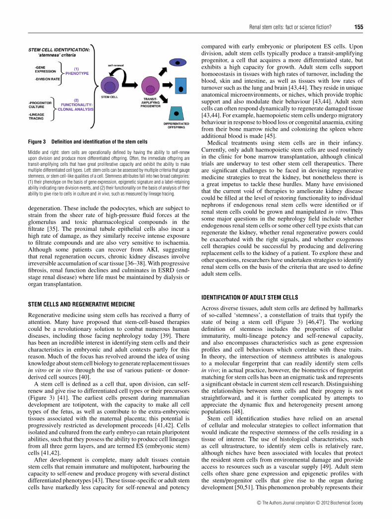
Figure 3 Definition and identification of the stem cells
Middle and right: stem cells are operationally defined by having the ability to self-renewupon division and produce more differentiated offspring. Often, the immediate offspring aretransit-amplifying cells that have great proliferative capacity and exhibit the ability to makemultiple differentiated cell types. Left: stem cells can be assessed by multiple criteria that gaugestemness, or stem cell-like qualities of a cell. Stemness attributes fall into two broad categories:(1) their phenotype on the basis of gene expression, epigenetic signature and a label-retainingability indicating rare division events, and (2) their functionality on the basis of analysis of theirability to give rise to cells in culture and in vivo, such as measured by lineage tracing.
degeneration. These include the podocytes, which are subject tostrain from the sheer rate of high-pressure fluid forces at theglomerulus and toxic pharmacological compounds in thefiltrate [35]. The proximal tubule epithelial cells also incur ahigh rate of damage, as they similarly receive intense exposureto filtrate compounds and are also very sensitive to ischaemia.Although some patients can recover from AKI, suggestingthat renal regeneration occurs, chronic kidney diseases involveirreversible accumulation of scar tissue [36–38]. With progressivefibrosis, renal function declines and culminates in ESRD (end-stage renal disease) where life must be maintained by dialysis ororgan transplantation.
STEM CELLS AND REGENERATIVE MEDICINE
Regenerative medicine using stem cells has received a flurry ofattention. Many have proposed that stem-cell-based therapiescould be a revolutionary solution to combat numerous humandiseases, including those facing nephrology today [39]. Therehas been an incredible interest in identifying stem cells and theircharacteristics in embryonic and adult contexts partly for thisreason. Much of the focus has revolved around the idea of usingknowledge about stem cell biology to generate replacement tissuesin vitro or in vivo through the use of various patient- or donor-derived cell sources [40].
A stem cell is defined as a cell that, upon division, can self-renew and give rise to differentiated cell types or their precursors(Figure 3) [41]. The earliest cells present during mammaliandevelopment are totipotent, with the capacity to make all celltypes of the fetus, as well as contribute to the extra-embryonictissues associated with the maternal placenta; this potential isprogressively restricted as development proceeds [41,42]. Cellsisolated and cultured from the early embryo can retain pluripotentabilities, such that they possess the ability to produce cell lineagesfrom all three germ layers, and are termed ES (embryonic stem)cells [41,42].
After development is complete, many adult tissues containstem cells that remain immature and multipotent, harbouring thecapacity to self-renew and produce progeny with several distinctdifferentiated phenotypes [43]. These tissue-specific or adult stemcells have markedly less capacity for self-renewal and potency
compared with early embryonic or pluripotent ES cells. Upondivision, adult stem cells typically produce a transit-amplifyingprogenitor, a cell that acquires a more differentiated state, butexhibits a high capacity for growth. Adult stem cells supporthomoeostasis in tissues with high rates of turnover, including theblood, skin and intestine, as well as tissues with low rates ofturnover such as the lung and brain [43,44]. They reside in uniqueanatomical microenvironments, or niches, which provide trophicsupport and also modulate their behaviour [43,44]. Adult stemcells can often respond dynamically to regenerate damaged tissue[43,44]. For example, haemopoietic stem cells undergo migratorybehaviour in response to blood loss or congenital anaemia, exitingfrom their bone marrow niche and colonizing the spleen whereadditional blood is made [45].
Medical treatments using stem cells are in their infancy.Currently, only adult haemopoietic stem cells are used routinelyin the clinic for bone marrow transplantation, although clinicaltrials are underway to test other stem cell therapeutics. Thereare significant challenges to be faced in devising regenerativemedicine strategies to treat the kidney, but nonetheless there isa great impetus to tackle these hurdles. Many have envisionedthat the current void of therapies to ameliorate kidney diseasecould be filled at the level of restoring functionality to individualnephrons if endogenous renal stem cells were identified or ifrenal stem cells could be grown and manipulated in vitro. Thussome major questions in the nephrology field include whetherendogenous renal stem cells or some other cell type exists that canregenerate the kidney, whether renal regenerative powers couldbe exacerbated with the right signals, and whether exogenouscell therapies could be successful by producing and deliveringreplacement cells to the kidney of a patient. To explore these andother questions, researchers have undertaken strategies to identifyrenal stem cells on the basis of the criteria that are used to defineadult stem cells.
IDENTIFICATION OF ADULT STEM CELLS
Across diverse tissues, adult stem cells are defined by hallmarksof so-called ‘stemness’, a constellation of traits that typify thestate of being a stem cell (Figure 3) [46,47]. The workingdefinition of stemness includes the properties of cellularimmaturity, multi-lineage potency and self-renewal capacity,and also encompasses characteristics such as gene expressionprofiles and cell behaviours which correlate with these traits.In theory, the intersection of stemness attributes is analogousto a molecular fingerprint that can readily identify stem cellsin vivo; in actual practice, however, the biometrics of fingerprintmatching for stem cells has been an enigmatic task and representsa significant obstacle in current stem cell research. Distinguishingthe relationships between stem cells and their progeny is notstraightforward, and it is further complicated by attempts toappreciate the dynamic flux and heterogeneity present amongpopulations [48].
Stem cell identification studies have relied on an arsenalof cellular and molecular strategies to collect information thatwould indicate the respective stemness of the cells residing in atissue of interest. The use of histological characteristics, suchas cell ultrastructure, to identify stem cells is relatively rare,although niches have been associated with locales that protectthe resident stem cells from environmental damage and provideaccess to resources such as a vascular supply [49]. Adult stemcells often share gene expression and epigenetic profiles withthe stem/progenitor cells that give rise to the organ duringdevelopment [50,51]. This phenomenon probably represents their
c© The Authors Journal compilation c© 2012 Biochemical Society

156 K. K. McCampbell and R. A. Wingert
Table 1 Proposed renal stem, progenitor and regenerative cell types in the mammalian kidney
Cells exhibiting one or more stemness attributes have been characterized in both the developing and adult mammalian kidney. The cells have been found in mesenchymal and epithelial compartments,and the origins of several isolated by fractionation techniques have yet to be precisely determined.
Contribution in kidneyLocation Molecular and cell characteristics Potency in vitro Potency in vivo injury or disease model Reference(s)
Embryonic kidneyCM Six2+ , self-renewing Nephron [74,75]SM FoxD1+ Interstitium [72,73]UB Collecting duct [62–64]
Adult kidneyNephron
PEC stem cell CD24+ CD133 + , PDX− Proximal anddistal tubule
Podocyte Yes; gave rise to bothpodocytes and tubulecells
[96,97,103,104]
PEC progenitor CD24+ CD133 + , PDX+ Podocyte Limited; rare podocytecontribution
[103]
Tubule differentiated epithelia Tubule cells marked by eGFP or Six2-reporter+ Tubule [127,128]Tubular stem cell-proximal VIM+ , from proximal tubule (S3 segment) [133]Tubular stem cell-proximal NFATc1− lacZ + , proximal tubule [134]Tubular stem cell CD24+ CD133 + Multipotent [135]Tubular stem cell CD24+ CD133 + Aldhhigh from renal cortex Multipotent [136]Tubular stem cell CD133+ , heterogenous Multipotent Tubule Yes [138]
Renal papillaPapilla stem cell BrdU label retention in tubules and interstitium [129,133,139]Papilla stem cell Nestin+ CD133 + Multipotent Multipotent [142]
Whole kidney fractionationFractionation by antigens, behaviour Sca-1+ Lin − Multipotent Tubule [143]Fractionation by side population Sca-1+ , musculin + , noted to reside in interstitium Yes [144]Fractionation by side population Sca-1+ Multipotent Yes [145,146]Long-term proliferation in culture Oct4, Pax2 Multipotent Tubule Yes [148]Long-term proliferation in culture Myh9− GFP+ , Pax2, Oct4, Wt1 Multipotent Tubule Yes [149]
shared common ontological origins and the congruous usageof genetic and molecular processes to direct the production ofcell types in a specific tissue or organ. Interestingly, there aremany markers common to diverse lineages that approximatea category of universal stem cell traits. For example, adultstem cells express similar cohorts of cell surface proteins, suchas c-Kit, Sca-1 (stem cell antigen-1), CD34 and CD133; thepresence of these antigens is used to isolate stem cell fractionswith flow cytometry [52]. Stem cells also exhibit low vitaldye staining due to the expression of membrane pumps of theABC (ATP-binding cassette) transporter family [52]. Becauseof these transporter proteins, stem cells actively efflux highlevels of the fluorescent DNA-staining dyes Hoeschst 33342 andRhodamine 123 compared with other cells, and distribute in a sidepopulation upon flow cytometry [52]. These efflux properties arehypothesized to be a mechanism to protect stem cell integrity fromcytotoxic compounds, thus promoting long-term survival [52].
Adult stem cells share archetypal behaviours that are closelytied to their potency and self-renewal. For example, many adultstem cells are quiescent, sharing the property of infrequentcell division over long periods of time. This may representa mechanism to limit the accrual of mutations during DNAreplication, thus staving off the potentially deleterious con-sequences of replicative aging [53]. Cell turnover can be gaugedby providing a pulse of a nucleotide analogue, such as BrdU(bromodeoxyuridine), which is incorporated into dividing cells.Over time, adult stem cells tend to be LRCs (label-retainingcells) because they do not proliferate (in normal conditions) ata rate that dilutes the label, whereas transit-amplifying progenyrapidly dilute such labels.
Regardless of its gene expression and proliferation dynamics, astem cell assignation is only indisputably determined by assessing
functionality, i.e. by operational tests that can reveal whichprogeny can be produced and over what period of time [54].Functionality can be probed through many assays, which canbe broadly categorized into in vitro or in vivo tests. Putativestem cells are isolated, typically on the basis of complementsof cell-surface markers, then cultured in vitro to observe theiractivities in different conditions, namely to perform clonal assaysto assess self-renewal and find out which other cell types canbe produced [43]. The operational activity of a putative stemcell is most stringently evaluated by tracking its progeny in vivo.Some tissues are amenable to transplantation techniques whereprospective stem cells are isolated from a donor, re-introducedinto a genetically distinct (but compatible) recipient, and thentracked by various methods [55]. Serial transplantation enablesthe assessment of long-term self-renewal and is necessary tofunctionally distinguish stem cells from their transit-amplifyingoffspring. Lineage tracing using genetic fate mapping in the mousemodel has been invaluable to assess stem cell progeny production[56]. Genetic fate mapping is performed by creating transgenicanimals in which subsets of cells (on the basis of tissue-specificpromoter activity) can be marked at a desired time by inducingthe stable genetic expression of a reporter such as GFP (greenfluorescent protein). The offspring of the labelled cells inherit thereporter expression, enabling their fate to be tracked over time.
To date, the search to find renal stem cells and discoverif/how kidney regeneration works has occurred on two relatedfronts that have implemented the scientific tools that gaugestemness parameters. Research to identify renal stem cellsduring development has provided a new understanding of lineagerelationships in the kidney, and delineated a multipotent renalstem cell that generates the nephrons. Meanwhile, adult kidneypopulations have received intense scrutiny to re-evaluate their
c© The Authors Journal compilation c© 2012 Biochemical Society

Renal stem cells: fact or science fiction? 157
Figure 4 The mammalian kidney develops from distinct pools of renal progenitors
(A) Top left: the mammalian embryo will form a series of three kidney structures from the intermediate mesoderm (indicated in grey and purple) in a caudal region of the trunk. Enlargement: the threekidneys from along the rostral–caudal axis in an archetypal order, with the pronephros first, followed by the mesonephros and finally the metanephros. The pronephros and mesonephros consist ofsimple tiered arrays of nephrons. The pronephros is vestigial, whereas the mesonephros functions for a short time and then degenerates upon metanephros formation. (B) The metanephros developswhen the nephric duct (grey) is induced to form the UB outgrowth by the MM (purple). Branching morphogenesis of the UB generates a highly branched collecting duct system. (C) The MM givesrise to two mesenchymal compartments, the CM which caps the UB branch points, and the SM, which is loosely distributed in the vicinity. The CM is a Six2+ self-renewing stem cell compartment,and adjacent to each UB branch points forms a PA, which initially maintains Six2+ and will become a nephron. The PA progresses to form an epithelial RV that is Six2− Wnt4 + , and grows to makevarious shapes, including an S-shaped body that will eventually proliferate and elongate to make nascent nephrons that connect to the UB ductal network.
properties in the context of normal homoeostasis and disease,identifying a cast of stem and progenitor cells that are the subjectof ongoing research and debate. Putative adult renal stem cellshave been proposed to exist in several adult kidney sites (shownby an asterisk in Figure 1B, and see Table 1). We first considerrenal stem cells in development and then discuss the evidence foradult renal stem cells.
RENAL STEM CELLS AND MAMMALIAN KIDNEY ONTOGENY
Kidney organogenesis is a unique developmental phenomenonthat progresses through a sequence of stages wherein a renalstructure is formed and later replaced with a more intricate organconfiguration [57,58]. Each kidney derives from the mesodermand is comprised of nephron tubules, although whether thenephrons become functional and how long they persist variesby species. Mammals form a pronephros, then a mesonephros
and finally a metanephros that becomes the adult kidney(Figure 4A). The mammalian pronephros is a rudimentaryvestigial structure. The mesonephros exists for a short timeand has limited functionality, degenerating with the onset ofmetanephros formation. Nephron arrangements in pro- and meso-nephric kidneys are simple, with nephrons few in number. Theseearly kidneys have a pair of nephric (or Wolffian) ducts, derivedfrom mesoderm that coalesces to form an epithelial tube, and formnephrons from surrounding mesenchyme called the nephrogeniccord. The pronephros forms anteriorly, with the mesonephrictubules located more posteriorly. The nephric ducts later growcaudally along the trunk, where the bilateral metanephric kidneyswill be induced.
The characterization of renal stem cells during mammaliandevelopment has been focused on the metanephros because itbecomes the permanent organ and is much more substantialin size. Many questions remain about the genetic programmesand signalling pathways that direct pro- and meso-nephros
c© The Authors Journal compilation c© 2012 Biochemical Society

158 K. K. McCampbell and R. A. Wingert
specification, and the molecular relationships between renalprogenitors of these and metanephric kidneys. Duringmetanephros formation, discrete populations of mesodermalderivatives interact to generate the arborized arrangement ofnephrons and collecting system [59,60]. The collecting ductsystem originates from the nephric duct, which in the courseof its caudal growth will be induced to evaginate by a specializedMM (metanephric mesenchyme) and form an outgrowth knownas the UB (ureteric bud) (Figure 4B) [61]. Classic cell associationand retroviral labelling studies demonstrated that the MM couldgenerate nephrons, whereas the UB epithelium could makecollecting ducts [62–64]. The UB undergoes a reiterative processof branching morphogenesis during which nephrons are inducedfrom the MM located adjacent to the UB branch points, generatingan arborized nephron array. Over time, these events beget a denseperiphery of nephrogenesis activity, called the nephrogenic zone.Elaborate reciprocal signalling between the MM and UB underliesthe repeated cycles of UB branching and growth.
As the MM and UB interact, the MM is subdivided into the CM(cap mesenchyme) and SM (stromal mesenchyme), representinga bifurcation in kidney lineages (Figure 4C and Table 1) [65,66].The CM is a condensed mesenchyme that encapsulates the UB andis broadly defined by expression of the homeobox transcriptionfactor Six2 (sine oculis-related homeobox 2 homologue) afterUB invasion [67,68]. A subset of the CM that flanks eachbranch point will become a PA (pretubular aggregate) thatmaintains Six2 expression and is induced to undergo an epithelialtransition, forming a circular RV (renal vesicle) in whichSix2 expression is lost (Figure 4C). Each RV proliferatesto make a nephron, expanding into a comma-shape, next anS-shape and then elongates further to make convolutions and fusewith the collecting duct system [69–71]. The elongating nephronis polarized along its proximal–distal axis such that podocytesalways arise from the nephron progenitors furthest from the pointof fusion, and the intervening progenitors develop into the tubule[69–71]. In stark contrast with the CM, the SM is a loose cellulararray situated between the UB branches and growing nephrons.The SM is defined by expression of the forkhead transcriptionfactor FoxD1 (forkhead box D1) and these cells will contribute tothe interstitial population [72,73].
Previous studies have demonstrated that the CM contains aself-renewing group of nephrogenic stem cells. Genetic fatemapping has shown that the CM is specified early, when the UBfirst enters the MM [74,75]. On the basis of gene expressionand functional studies, CM specification probably involves aseries of cues originating from the signalling of BMPs (bonemorphogenetic protein) acting through the Alk3 (activin-likekinase 3) receptor, and transcriptional activities mediated bythe Hox11 paralogues, Osr1 (odd-skipped-related 1) and Pax2(paired box gene 2), all of which guide MM patterning [65,76–78]. CM cells are defined by the continued expression of theHox11 paralogues, Osr1 and Pax2, as well as the transcriptionfactors Cited1 {CBP [CREB (cAMP-response-element-bindingprotein)-binding protein]/p300-interacting transactivator, withGlu/Asp-rich carboxy-terminal domain 1}, Eya1 (eyes absenthomologue 1), Sall1 (sal-like 1), Six2 and Wt1 (Wilms tumour 1)[59,60,69–71]. Following its specification, the CM is not believedto receive additional cell contributions, but rather self-renews toperpetuate the expanding nephrogenic zone [74,75]. Among thegenes associated with CM identity, Six2 activity is essentialto propagate the stem cell population. In the absence of Six2,nephrogenesis terminates rapidly after it is initiated becausethe CM compartment is not maintained [75]. Clonal analysisof Six2-expressing CM has demonstrated that at least a subsetof these cells can contribute to multiple nephron segments,
suggesting that the CM contains a multipotent population [75].During the progressive rounds of nephrogenesis, the CM isheterogenous, containing a self-renewing pool of Six2+ cellsand a subgroup in transit to the nephron fate that is marked byexpression of Wnt4 (wingless-type MMTV integrator site family,member 4) [75]. Recent findings suggest that Wnt9b (wingless-type MMTV integrator site family, member 9b)/β-cateninsignals positively regulate Six2+ CM self-renewal, althoughthe precise molecular relationship of these factors has yetto be resolved [79]. The phenomenon of CM self-renewal istransient, and the cessation of nephrogenesis is associated withCM disappearance. Further work is needed to address how the CMcompartment is terminated, as it is unclear whether this is dueto the active commitment of cells to nephrogenesis and/or anexhaustion of self-renewal properties [75]. Elucidation of howthe CM is regulated could provide valuable insights into the cell-autonomous and environmental signalling pathways that influencekidney lineage self-renewal.
OVERVIEW: THE HUNT FOR ADULT RENAL STEM CELLS
Searches for renal stem cells in the adult mammalian kidneyhave not identified a multipotent cell that can self-renew andmake the >20 specialized renal cell types. However, distinctlocations within the nephron and elsewhere in the kidney appearto house cells that exhibit various degrees of potential to makedifferentiated cell types during normal turnover and followinginjury. These sites include the renal corpuscle, the nephron tubuleand the renal papilla. The spectrum of regenerative cells includesimmature stem-like cells and mature differentiated cells. In thefollowing sections, we discuss renal regeneration at these distinctsites and the features of the cells that are purportedly involved.
EVIDENCE FOR A PODOCYTE STEM CELL IN THE RENALCORPUSCLE
The renal corpuscle is made up of several cell types: fenestratedendothelial cells that form the capillary network, mesangial cellsinterspersed between the capillary loops, glomerular podocytes(or visceral epithelial cells) and the PECs (parietal epithelial cells)of Bowman’s capsule (Figure 5) [80]. Of these cell types, the latterthree derive from kidney mesoderm during development, withthe origin of angioblasts not yet resolved [80]. The endothelialcells and podocytes sit in apposition, attaching on either side ofthe GBM (glomerular basement membrane) to form the bloodfilter. The podocytes are octopus-shaped cells with long axonal-like extensions. They connect to neighbouring podocytes withinterdigitating projections (called foot processes) that create smallspaces for fluid to pass through. Foot processes are joined by theslit diaphragm, a dense velcro of elaborate protein complexes atthe cell membrane that are harnessed to the interior cytoskeleton[81]. As such, podocytes are highly specialized differentiatedcells, and are thought to be mitotically quiescent, except in certainpathological conditions [82–84]. The integrity of the blood filterdepends on the ability of podocytes to retain their connections withthe GBM and adjacent podocytes. This intact barrier prevents theleakage of high-molecular-mass proteins and cells into the tubule.
Proper development and survival of podocytes is essentialfor kidney health [35,82]. Podocyte disruption has catastrophicconsequences, for the majority of patients who develop ESRD,the primary pathological insult is glomerular disruption thatcauses proteinuria and progresses to scarring and loss of nephronfunction [35,82]. Surprisingly, podocytes are shed at a lowfrequency in the urine of healthy individuals [85]. They can be
c© The Authors Journal compilation c© 2012 Biochemical Society

Renal stem cells: fact or science fiction? 159
Figure 5 Glomerular injury and source(s) of replacement cells
The renal corpuscle is composed of (1) a glomerulus, which is a ball of capillaries made of fenestrated epithelial cells that are surrounded by a GBM and podocytes, and (2) the Bowman’scapsule, which is an epithelium that encapsulates the glomerulus and serves as a collecting space for fluid filtered through the glomerulus. Current evidence is consistent with the epithelium at theurinary pole, the so-called PECs, containing a podocyte stem cell that is CD24+ CD133 + PDX− . The PEC stem cell gives rise to transitional offspring that are podocyte progenitor cells; thesetransitional cells express CD24+ CD133 + PDX+ and are speculated to migrate around the capsule and later enter the glomerulus (pathway indicated by green arrows), where they differentiate intoCD24 − CD133 − PDX+ podocytes.
recovered from the urine and even cultured, where they displayhallmarks typical of normal podocytes [85]. It is thought thatthe mechanical forces of fluid flow disrupt podocyte attachments,leading to their effacement even though they are otherwise viable.Podocyte excretion is dramatically elevated in the setting of manyrenal diseases, probably reflecting a harmful environment [85].There are some data suggesting that compensatory podocytehypertrophy can occur to counteract shedding, and thatpodocytes hypertrophy in a number of disease states [86–88];in vitro, podocytes have been observed to undergo hypertrophyin response to glucose and mechanical stretch [89,90]. Theobservations of podocyte loss and absence of proliferation areat odds with the ability to maintain sufficient podocyte numbersand renal function with age. In addition, a reversal of glomerularscarring and regression of renal disease was reported in patientswith diabetes that had pancreatic transplants and in patients withchronic nephropathy that received ACEi (angiotensin-convertingenzyme inhibitors), suggestive of podocyte regeneration [90–94].
A re-evaluation of the cells at the renal corpuscle revealed thatsome PECs showed stem cell characteristics [95–97]. In biopsiesfrom normal adult human kidneys, researchers detected a subsetof PECs that express the surface antigens CD24 and CD133,which mark several adult stem cell types [96]. The PEC subsetwas purified and grown in culture, where individual cells exhibitedclonogenic self-renewal and could generate multiple progeny withcharacteristics of proximal and distal tubule epithelia [96]. In amouse model of AKI, tubular regeneration occurred in animalsthat received an injection of the human CD24+ CD133+ PECfraction, but not with CD24− CD133− PECs [96]. Interestingly, indeveloping human kidneys CD24+ CD133+ cells were identifiedin the renal vesicles and S-shaped bodies, and were later restrictedto the urinary pole of Bowman’s capsule [97]. The embryonicCD24+ CD133+ PECs gave rise to cells with phenotypescomparable with the adult PEC source when grown in culture oradministered to the same mouse AKI model [97]. Taken together,these results suggested that the CD24+ CD133+ PECs could bemultipotent nephron progenitor descendents.
Interestingly, previous studies had reported morphologicalheterogeneity in the PEC of Bowman’s capsule on the basisof histological analyses. In human glomeruli, cells close tothe urinary pole have a flat epithelial shape, whereas cellsclose to the vascular pole exhibit a podocyte-like appearancewith long cell processes, and were termed parietal podocytes[98,99]. Examinations of Bowman’s capsule in sheep describedheterogeneity as well, reporting a unique cell type located nearthe vascular pole on the basis of prominent cytoplasmic granules,and these were called peripolar cells [100,101]. The mixtureof morphological phenotypes in Bowmans’ capsule, along withmultipotency of CD24+ CD133+ PECs, are suggestive that thePEC population is comprised of several cell types with variousroles, some of which might serve to replace podocytes. Thishypothesis has been supported by clonogenic analysis and lineagestudies [102–104].
Further molecular characterization of CD24+ CD133+ humanPECs revealed that this group is heterogenous, and includescells that express markers typical of differentiated podocytes,like nestin and PDX (podocalyxin) [103]. Human PECs arespatially organized in a continuum along the capsule, withCD24+ CD133+ PDX− cells present closest to the urinarypole, CD24+ CD133+ PDX+ cells concentrated towards thevascular pole, and differentiated podocytes exhibiting aCD24− CE133− PDX+ character (Figure 5) [103]. After sortingof CD24+ CD133+ PECs into PDX− and PDX+ fractions, onlythe individual PDX− cells exhibit multipotency in vitro on thebasis of the ability to generate tubular cells and podocytes [103].Furthermore, injection of the CD24+ CD133+ PDX− fractionproduced podocytes and tubular cells in mice with adriamycin-induced renal injury (a model of the human podocyte diseaseknown as focal segmental glomerulosclerosis), and was associatedwith reduced proteinuria [103]. A similar infusion with theCD24+ CD133+ PDX+ fraction, in contrast, only gave rise torare podocytes [103]. These findings support the notion thatCD24+ CD133+ PDX+ cells are transitional cells with limitedproliferative capacity that express progenitor and podocyte
c© The Authors Journal compilation c© 2012 Biochemical Society
160 K. K. McCampbell and R. A. Wingert
markers, and do not replace injured podocytes [103]. Geneticlabelling has been used to mark the PECs and irreversibly tracktheir progeny in newborn and adolescent mice, demonstratingthat PEC offspring become podocytes during the completion ofnephrogenesis in the postnatal period and during rapid kidneygrowth in juveniles [104].
These findings provide compelling evidence that a podocytestem cell resides among the PECs. The ability of cultured PECsto contribute to the tubule could indicate a broader potency ofPECs, but future experiments are needed to determine whetherthe PEC is multipotent in vivo. Genetic tracking is also neededto assess the extent to which the PEC progenitor pool can self-renew in adults over the long-term. Furthermore, the mechanismsof progenitor migration are unresolved. The gradient of cellphenotypes along the capsule is consistent with a cycle whereinpodocytes gradually shift towards the glomerulus as sheddingoccurs (an example pathway is indicated by green arrows inFigure 5) [95,96,103,104]. Differentiating podocytes might alsotake a more direct route to the glomerulus [95,104], on the basisof the observation of cell bridges or so-called tuft-to-capsuleadhesions, where chains of cells link the glomerulus andcapsule [105]. Finally, the developmental pathways essentialto specify the PEC podocyte progenitor are largely obscure.In a recent study, the removal of β-catenin signalling duringthe late S-shaped body stage of nephrogenesis resulted in theabrogation of PECs and a replacement with parietal podocytes[106]. This phenotype could reflect a number of possibilities,such as a requirement for β-catenin in PEC self-renewal,PEC survival or a lineage conversion event. Understandingthe signals that modulate PEC development will be vital inunderstanding disease pathology, as exemplified by reports thatPEC progenitors contribute to cell lesions in several glomerulardiseases [107,108] and can be enhanced by activating Notchsignalling [109]. Interestingly, a recent report provided evidencethat ACEi moderates renal progenitor dynamics using a ratmodel of progressive glomerular injury [110]. In this setting,treatment with ACEi was associated with improved glomerulararchitecture and linked to moderated progenitor activation [110].These findings suggest that moderating the cellular response torenal damage may confer renoprotective effects.
NEPHRON TUBULAR REGENERATION AND STEM CELLS?
Cellular regeneration in the nephron tubules was noted as farback as the late 19th century, and was suggested to arise fromsurviving cells within injured tubules [7]. Since this time, bothintra- and extra-renal cell sources have been proposed to supportnephron tubule regeneration (Figure 6). Genetic fate-mappingstudies have shown that intratubular cells restore nephron integrityfollowing acute injury, although the origin of the cells is disputed.Putative stem cells have been located in the tubules themselves,and the collecting ducts and interstitium of the renal papilla,but the identities and behaviours of these cells are controversial.Although extrarenal cells such as BMSCs (bone marrow stemcells) and MSCs (mesenchymal stem cells) were once thought tomake direct cell contributions to renal regeneration, they are nowthought to provide humoral support. In the following sections weaddress the lines of evidence concerning the proposed contributorsto tubule regeneration.
Intratubular cells and maintenance of nephron integrity
Nephron tubule epithelia residents are highly specialized anddifferentiated, with distinct cuboidal or columnar shapes, and are
polarized along their apical–basal axis [21]. As such, a reasonableexpectation is that these cells are quiescent, similar to podocytes;however, several lines of evidence indicate that tubule cells havea low basal turnover rate. Early cell division studies using apulse–chase of tritiated thymidine reported incorporation of thelabel throughout the proximal, distal and collecting tubules ofhealthy male rats [6]. Furthermore, antibody staining with thecell-division markers PCNA (proliferating-cell nuclear antigen)and Ki67 detected positive tubule cells in adult human kidneys[111]. Later, proliferation was documented using mitotic figures,BrdU pulse–chase experiments, and cell-cycle markers in juvenilerat kidneys (as the rate of cell division was noted to range between2 and 4%, higher than the 0.4% seen in adults) [112–114].Proliferating cells were reported in both proximal and distaltubules [111]. This investigation found that mitotic cells in theproximal tubule had differentiated traits, as they co-expressedmarkers of the brush border and showed proper distributionof several polarity proteins, suggesting that dividing cells weredifferentiated epithelia [112]. Interestingly, a high incidence ofcells in both juvenile and adult tubules were positive for cyclinD1, indicating that many tubular cells were poised in G1-phase[114].
Over the past decades, studies of AKI from ischaemia ornephrotoxin exposure have chronicled a prototypical sequence ofcellular changes that precede a full restoration of nephron tubuleintegrity [113–123]. The descriptions have primarily centredon the proximal tubule because the prominent feature of theseparticular acute injuries is damage to the proximal epithelialcells. Following injury, damaged and apoptotic tubule cells sloughinto the lumen, denuding the basement membrane. The exposedbasement membrane is then progressively covered by cells withmesenchymal features, including a flattened elongated appearanceand the expression of proteins that are characteristic of motilemesenchyme. High rates of intratubular proliferation ensue soonafter injury (within 12–36 h), and several studies have found thatthe dividing cells have features of differentiated cells, as in thecycling cells of healthy kidneys [112,113]. Eventually, tubularintegrity is re-established as the mesenchymal cells transitionto become epithelial, with regeneration complete in as fast as2 weeks. The regenerating cells express renal development genes,including Pax2, NCAM (neural cell adhesion molecule) andLhx1 (Lim homeobox 1), supporting the hypothesis that tubuleregeneration may recapitulate some developmental processes[124–126].
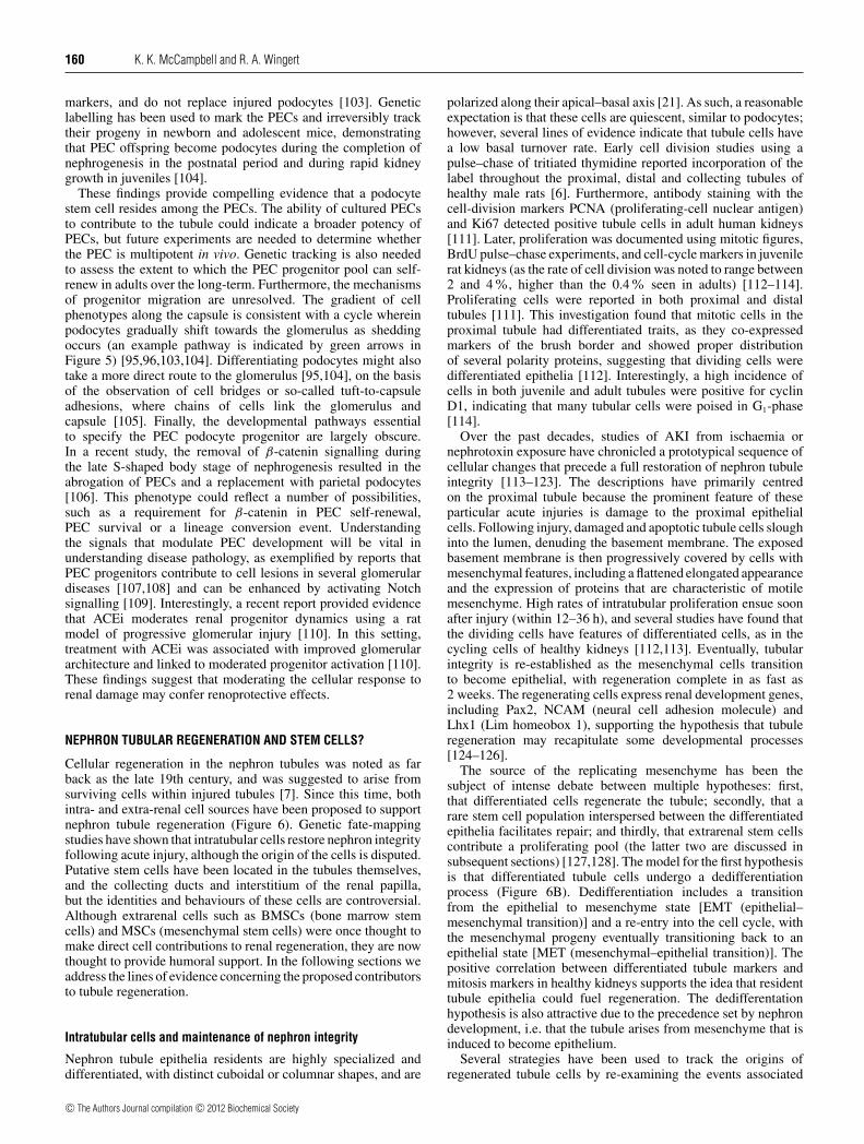
The source of the replicating mesenchyme has been thesubject of intense debate between multiple hypotheses: first,that differentiated cells regenerate the tubule; secondly, that arare stem cell population interspersed between the differentiatedepithelia facilitates repair; and thirdly, that extrarenal stem cellscontribute a proliferating pool (the latter two are discussed insubsequent sections) [127,128]. The model for the first hypothesisis that differentiated tubule cells undergo a dedifferentiationprocess (Figure 6B). Dedifferentiation includes a transitionfrom the epithelial to mesenchyme state [EMT (epithelial–mesenchymal transition)] and a re-entry into the cell cycle, withthe mesenchymal progeny eventually transitioning back to anepithelial state [MET (mesenchymal–epithelial transition)]. Thepositive correlation between differentiated tubule markers andmitosis markers in healthy kidneys supports the idea that residenttubule epithelia could fuel regeneration. The dedifferentationhypothesis is also attractive due to the precedence set by nephrondevelopment, i.e. that the tubule arises from mesenchyme that isinduced to become epithelium.
Several strategies have been used to track the origins ofregenerated tubule cells by re-examining the events associated
c© The Authors Journal compilation c© 2012 Biochemical Society

Renal stem cells: fact or science fiction? 161
Figure 6 Tubular injury and source(s) of replacement cells
(A) Injury models that damage the proximal tubule by ischaemia/reperfusion or nephrotoxins have been most extensively used to study tubular regeneration. These analyses have led to a currentdebate between several cellular sources of regeneration that have converged to focus on (1) differentiated tubule epithelial cells (labelled with Six2-reporter expression) and (2) intratubular stem cells(labeled by BrdU and CD24+ CD133 + Aldhhigh in various studies). A third cell source, the extratubular compartment, which over the years has been speculated to include kidney-resident interstitialfibroblasts, BMSCs and MSCs, has been negated by evidence of intratubular sources of regeneration. (B) The scenarios of tubular regeneration are proposed to involve either a dedifferentiation(1, top panel) or a stem cell mechanism (2, bottom panel). Dedifferentiation includes events through which the tubular epithelium oscillates between a mesenchymal phenotype (EMT and then MET),with the mesenchyme purported to show migration and division to replace lost tubular cells. A tubular stem cell has been proposed to share hallmarks with its differentiated neighbours, and replacelost cells through division that may or may not involve transit-amplifying progenitors.
with ischaemia/reperfusion injury. In mice where tubule epitheliawere mosaically marked with eGFP (enhanced GFP) usingthe Ksp (kidney-specific)-cadherin promoter, eGFP+ cellsincorporated BrdU and co-stained with differentiated markersduring regeneration, suggesting an intratubular source ofregeneration [127]. Another study used the Six2 promoter tostably mark the entire differentiated tubular epithelium with areporter, and then determined whether regenerated tubules hada ubiquitous or diluted reporter pattern [128]. In this transgenicsystem, the Six2 reporter marks the tubule cells that arisefrom the CM during nephrogenesis. Following kidney repair, nodilution of the Six2 marker was observed, evidence that Six2descendents were the primary regeneration source. Interpretationof this data does hinge on the transgenic marking strategy: anyre-expression of Six2 during regeneration would trigger stablereporter expression in those cells and their descendents thatwould be indistinguishable from the adult tubule population. Byseveral measures, the authors did not detect Six2 re-expression atthe timepoints they examined, although a rapid pulse of Six2activity could always be possible [128]. Overall, these dataprovide strong evidence in favour of an intratubular regenerationsource.
In a subsequent study, proximal tubule regeneration was trackedin mice with a two-step sequence of nucleotide analogue pulsesafter ischaemia [first CldU (5-chloro-2-deoxyuridine), and thenIdU (5-iodo-2-deoxyuridine)] that were administered close in timeto one another, with the hypothesis being that the offspring of astem cell that divided to make a transit-amplifying progenitorwould retain both labels [129]. The researchers observed lowco-labelling, concluding that these data illustrate a stochasticprocess of tubular proliferation among differentiated cell types.These findings provide an intriguing snapshot into tubular celldynamics during the time window defined by the labelling pulses(between 24 and 45 h post-injury). Additional analysis of earliertime windows could potentially be informative, since they noted arise in tubular proliferation starting at 12 h post-injury [129], andsimilar rapid escalations in division were observed in studies of ratischaemic injury [130]. Future studies tracking individual cells areneeded in order to definitively resolve cell dynamics during tubuleregeneration. Taken together, the cell-tracking studies performedto date are consistent with the model that an intratubular cellsource fuels regeneration, and that this source has differentiatedtraits. However, the remaining questions revolve around how tointerpret this combination of the differentiated traits: are they
c© The Authors Journal compilation c© 2012 Biochemical Society

162 K. K. McCampbell and R. A. Wingert
the sign of mature cells, or are tubular stem cells scattered innephrons?
Evidence for tubular stem cells
Several groups have proposed the existence of rare tubular stemcells on the basis of the observation of stemness attributes ina minor subset of the resident tubule population. For example,cell-cycling differences among tubular cells have been suggestedby a cohort of pulse–chase experiments. Individual rat kidneytubule cells were shown to incorporate BrdU and retained the labelduring a short (2 week) chase [130]. When a viable subset of theBrdU+ cells was isolated, they retained low Hoeschst staining;in addition, they could be cultured in vitro to generate tubule-like structures, and contributed to nephrons and collecting ductswhen transplanted to a growing metanephros [131]. These studiescharacterized cells that retained BrdU over just a short chase, andthus may represent a pool of transit-amplifying cells and/or stemcells. In a much longer pulse–chase study, infrequent BrdU+
proximal tubule cells were detected after 35 weeks followingBrdU administration in normal newborn rats [112]. Rare corticalnephron labelling was reported in a similar chase, but with 3-day-old rats examined after 2 months [132]. The low number ofBrdU+ cells observed after these much longer chases is consistentwith the existence of a tubule stem cell that divides relativelyinfrequently.
Prospective tubule stem cells in the rodent kidney have alsobeen identified from gene expression and functional assays.One group microdissected a single nephron from an adult ratkidney and established a cell line from the proximal segmentwith expansive growth potential, designated rKS56 [133]. rKS56cells co-expressed mesenchymal and epithelial markers, such asthe intermediate filament VIM (vimentin) and the water channelaquaporin respectively, a phenotype likening them to immaturetubule cells. rKS56 cells also expressed c-Kit and Sca-1, markersassociated with an immature progenitor state. Consistent withthis comparison, rKS56 differentiated into mature epitheliumin vitro and after transplant into the post-ischaemic adult kidney.In the mouse, a proximal tubule progenitor-like population wasidentified through studies of NFATc1 (nuclear factor of activatedT-cells cytoplasmic 1), a transcription factor expressed in corticalnephron tubules [134]. An NFATc1–LacZ reporter labelled asubset of proximal tubule epithelia that expanded in the dayfollowing toxin exposure, and then contracted in number withinseveral days. NFATc1-labelled cells were resistant to apoptosis,suggesting that they were fated to survive the renal injury. Lineageanalysis of these cells using an NFATc1–Cre reporter revealed asubpopulation of proximal tubule that divided after renal injury,with the progeny reconstituting large tubule stretches [134].
Corroborating findings from several reports have suggestedthat a multipotent CD24+ CD133+ tubule cell is present in thehuman kidney. This nephron population was first identified bysearching for CD133+ cells in tubular fractions on the basis ofthe correlation between this antigen and the PEC glomerular stemcell [135]. A subset of the CD133+ tubular cells was found to co-express CD24 and, by clonogenic analysis, the CD24+ CD133+
tubule population could differentiate into multiple cell typesin vivo. This tubule population also had a similar gene signatureto CD24+ CD133+ glomerular cells by microarray. Polarizedtubule cells expressing both CD24 and CD133 were found inproximal and distal segments, although the frequency was notquantified. More recently, another research group discovered andcharacterized a human CD24+ CD133+ multipotent tubule cellusing an entirely different regime of stem cell traits [136]. The
initial isolation strategy in this case was on the basis of theprecedence that high ALDH (aldehyde dehydrogenase) activityis associated with stemness [137]. Kidney cells with ALDHhigh
activity were isolated from the renal cortex and had an increasedcapacity to form sphere-like clusters of epithelial cells in culture,and were capable of anchorage-independent growth features seenin several types of multipotent stem cells. By whole-genomeexpression profiling, the ALDHhigh cells showed high levels ofCD24, CD133, VIM, and cytokeratins 7 and 19. Localizationstudies showed that CD24+ CD133+ cells were rare tubule cells(only occasionally found in pairs), and cells in the parietalregion of the renal corpuscle, the latter in keeping with previousstudies [96,103,104]. CD133+ VIM+ co-expressing cells wereinterspersed in a similar fashion throughout the healthy tubules;in human acute renal damage biopsies, these were expandedinto long stretches of CD133+ VIM+ cells, suggesting that theyexpanded during regeneration. Interestingly, a previous studyisolated CD133+ cells from the adult human kidney cortex thatalso expressed Pax2 [138]. The CD133+ cells produced bothrenal epithelial and endothelial cells in culture, and integrated intonephron tubules in mice with AKI. The expression of CD133 isa characteristic of haemopoietic stem cells and endothelial cells.Thus the isolated CD133+ fraction in this case may representa heterogenous population that has been suggested by some toinclude interstitial cells [14].
Taken together, these findings support the hypothesis ofa tubular stem cell, but the evidence remains incompleteand requires much more investigation. The very idea of thetubular stem cell hypothesis is at odds with a long historyof studies that examined tubule cells by ultrastructural andmolecular methods, and never reported a minor populationof ‘different’ cells in tubular segments. To reconcile all ofthese findings, one must reason that tubular stem cells arevery rare, and have been overlooked by many methods. Tubulestem cells could also look like differentiated cells, possessingtraits of apical–basal polarity that are actually needed forthem to physically reside in the tubule, but which make themhard to discriminate from bona fide differentiated neighbourswith the typically used repertoire of markers. The analysisof NFATc1+ cells in the mouse and CD24+ CD133+ cellsin humans has now delineated several unique markers thatset some tubule cells apart from their neighbours. Futurecomparative studies will be valuable to learn more about theidentity and correspondence between these cell types. Movingforward, more markers are absolutely essential. Genetic trackingusing validated indicators of differentiated epithelium or tubulesubtypes is vital to unequivocally distinguish the mechanisms oftubular regeneration. Finally, it will be interesting to determinewhether a generic tubular stem cell type possesses ‘tubule-wide’multipotency and can make epithelial cells of multiple segments,or whether different nephron segments house more specializedtubular stem cell residents.
STEM CELLS OUTSIDE THE NEPHRON PROPER: STUDIES OF THERENAL PAPILLA
The renal papilla, or inner medulla, is the apex of the medullaryzones and contains tracks of collecting ducts, intermediatenephron segments (the loops of Henle) and intervening interstitialcells (Figure 7) [21]. The renal papilla is a hypoxic hyperosmotictissue where the business of water conservation is transactedthrough a countercurrent exchange system made possible bythis special local environment [21]. The existence of stem cellsin the renal papilla was first proposed after the discovery of
c© The Authors Journal compilation c© 2012 Biochemical Society

Renal stem cells: fact or science fiction? 163
Figure 7 The renal papilla and its proliferative compartments
The renal papilla is a zoneof the kidney that contains collecting ducts, nephron tubule portions that include the intermediate and distal segments, and interstitial fibroblast cells. BrdU-labelled LRCshave been found in all three cellular compartments, although whether these represent self-renewing stem or progenitor cells is not known. In addition, interstitial cells that are Nestin+ CD133 + havebeen detected and are speculated to represent a potential stem cell compartment.
label-retaining populations in this region. Pulse–chase studies us-ing BrdU in the healthy rat kidney showed that LRCs were presentin the papilla after 2 months, mostly in the interstitium, but also intubules [132]. Independent pulse–chase experiments have shownthat the tubular fraction includes the collecting duct and loopof Henle cells [129]. Following ischaemia, proliferating cellswere found concentrated in the upper-most papillary regions,and then, several months later, these regions were largely devoidof LRCs [132,139]. These findings were surprising becausetransient ischaemia was not damaging to the cells within therenal papilla, although of course is well-established to triggerdestruction of the nephron proximal tubule cells. On the basisof these observations, the renal papilla was hypothesized to bea source for tubular regeneration, and cells generated from thepapilla region were proposed to migrate into damaged nephronsthroughout the medulla and cortex [132,139].
Currently, there are several conflicting reports surrounding theidea of papillary migration. Evidence in favour of renal papillamigration was provided by labelling mouse papillary cells with apulse–chase of a vital dye and then inducing an ischaemic injury;post-ischaemia the labelled cells were found in a redistributedpattern, and some were even associated with unlabelled tubules,suggesting that local movement and nephron integration occurredover time [139]. Cell redistributions toward the cortex followingischaemia were also observed in a Nestin− GFP+ transgenicmouse, although Nestin+ cells probably included a widelyheterogenous population of labelled cell types in the papilla[140]. In contrast, other time-course studies have failed to detecta change in the distribution of nucleotide analogue label-retainingpapillary cells after injury [128]. Genetic tracing of papillaepithelial cells marked using mTert (mouse telomerase reversetranscriptase), an enzyme expressed by ES cells and severaladult stem cells, also failed to detect cells of papillary originmoving to the outer medulla or cortex following ischaemia [141].In this study the label-retaining papilla interstitial cells were
not marked, therefore migrating interstitial cells could not beevaluated [141].
Given these sets of conflicting data and the strong geneticevidence that intratubular cells regenerate nephrons, it seemsunlikely that the renal papilla serves as the primary sourcefor the bulk of cortical nephron regeneration. However, thisdoes not preclude the existence of a renal papilla stem cellpool, especially when one considers the label-retention capacityand the in vitro behaviours attributed to subsets of papillarycells. In particular, more research is needed to resolve theidentity and true potency of the papillary interstitial cells. Forexample, a recent study found a fraction of Nestin+ CD133+ co-expressing interstitial cells in the human renal papilla, which isintriguing given the traits associated with CD133+ cells in therenal corpuscle [142]. These Nestin+ CD133+ interstitial cellsshowed expression of mesenchymal and ES cell markers, andcontributed to tubulogenesis in a three-dimensional culture assayand when transplanted into developing mouse kidneys. Futuregenetic-tracking studies will be critical to determine the fatesof this interstitial compartment after renal injury, and additionalwork is needed to delineate the identity and phenotypes of theheterogenous interstitial population.
ISOLATION OF OTHER ENDOGENOUS PUTATIVE RENAL STEMCELLS
Several groups have sought to identify renal stem cells ‘at large’without focusing on any one area of the kidney. A commonapproach has been to isolate particular cell fractions usinga stemness marker(s) and then ascertain the multipotency ofthe isolated fractions in vitro. On the basis of the precedencethat Sca-1 marks adult stem cells across several mesodermalderivatives, one group isolated and characterized a Sca-1+ Lin−
fraction from the adult mouse kidney [143]. Clonally derived
c© The Authors Journal compilation c© 2012 Biochemical Society

164 K. K. McCampbell and R. A. Wingert
lines of Sca-1+ Lin− cells were capable of differentiating intomyogenic, osteogenic, adipogenic and neural lineages in vitro,and adopted a tubular phenotype when injected into the renalparenchyma following ischaemic injury. Immunohistochemistrystudies detected Sca-1 on numerous cell types in the kidney,which included tubule and renal papilla interstitium, suggestingthat the clonal lines could represent heterogenous origins withinthe kidney.
A second stemness trait used to fractionate the kidney has beenthe SP (side population) assay. Several groups have reported theisolation and characterization of SP cells from mouse andhuman kidneys [144–147]. The SP isolated from adult mousekidneys was initially reported to be Sca-1+ with >95% ofthe SP expressing the basic helix–loop–helix transcription factormusculin, which has been linked to promoting a dedifferentiatedfate [144]. When mice with AKI from the nephrotoxin cisplatinwere given an injection of the kidney SP, they displayed renalfunction improvement on the basis of creatinine excretion levels.Interestingly, musculin+ cells were shown to reside in theinterstitial space of the kidney. Subsequent expression profilingof kidney SP from embryonic and adult mouse kidneys reportedsimilar Sca-1, but not musculin, positivity [145,146]. Functionalassays have found that the kidney SP can exhibit multipotencyin vitro, contribute to UB and MM structures in metanephricorgan culture, and engraft into adriamycin-damaged kidneys ata low frequency [145]. Molecular and functional analyses of thehuman kidney SP have not yet been reported.
Yet another strategy used to locate renal stem cells has been toselect them on the basis of their ability to propagate in long-termculture. One group dissociated adult rat kidneys and grew thecells for several weeks, mimicking the culture conditions usedto previously isolate adult stem cells from the bone marrow[148]. The surviving cells were termed MRPCs (multipotentrenal progenitor cells), and were defined by co-expressionof the transcription factors Pax2 and Oct4 (octamer-bindingtranscription factor 4). The MRPCs could produce multiplelineages in vitro, ranging from hepatocytes to endothelial cells andneurons, and contributed to tubular cells when injected into normaland ischaemically injured kidneys. The researchers also identifiedrare Oct4+ cells in tubules of the adult kidney, suggesting apossible tubular origin of the MRPCs. A recent study examiningmouse kidneys took a similar long-term culturing tactic using atransgenic line in which the Myh9 (myosin heavy chain 9) genewas disrupted with a GFP cassette, marking several differentiatedcell types in nephrons, as well as the interstitium [149]. Afterculturing the GFP+ cells for 8 weeks, the surviving cells adopteda similar morphology and showed self-renewal characteristics,and were designated MKPCs (mouse kidney progenitor cells).Traits shared by the MKPCs included expression of Pax2, Oct4,Wt1 and VIM, suggesting parallels with pluripotency and renalprogenitors during development.
Taken together, these studies could indicate that the kidneycould house one or more multipotent cell types. However, thereare several caveats and future questions to consider with regardto these findings. One consideration is whether an isolated celltype/line is representative of a cell that normally resides in thekidney. Some of the procedures used to isolate renal cells couldcapture rare (stem) cells from other tissues that are able to survivein culture. Some procedures could also favour the selection andexpansion of genetically altered cells that are not representativeof the normal kidney. The relationships between multipotent cellsfrom these various reports have not been defined, nor has theirrelationship to cells such as the PEC-podocyte stem cell. Forexample, a multipotent cell line might actually be the derivativeof PECs. Another consideration is whether the cells underwent
genetic or epigenetic changes in the course of their manipulation.Thus althogh these findings are intriguing, the contribution ofthese cells to renal homoeostasis and their impact on renal diseaseremains unclear.
CONCLUSIONS AND FUTURE PERSPECTIVES
Research to find stem cells in the kidney suggests that a numberof previously unappreciated renal cell types exist, thereby raisinga multitude of questions for future work. Ongoing researchis needed to further delineate the respective activities andrelationships between the PEC-podocyte stem cell, the tubularregeneration source(s), papillary cells and the growing categoryof ‘other’ renal stem cell-like types. It is particularly intriguingthat the cells for which there is the strongest evidence ofregeneration, the podocytes and the proximal tubule, are the mostsusceptible and most frequently damaged in human kidneys. Theobservation of regeneration in these renal compartments begsthe question as to why such mechanisms are remiss in obviatinginjuries to these cell types. To begin to understand this puzzle,we will need to know more about the long-term capacities forregeneration of the various renal stem cell compartments, as thereis very little data on the mechanisms that control self-renewalproperties of each cell type. Knowledge about these mechanismscould provide insights into understanding the parameters of stemcell exhaustion, such as how the aged environment might effectstem cell maintenance and behaviour. Tools for genetic fatemapping will be essential to evaluate the progeny of these cells andthe long-term homoeostasis of the stem cell compartment(s). Forexample, an explanation for different AKI outcomes in patientsmay reside with their past history of renal damage (over a lifetimeof damage/regeneration cycles) and its cumulative effect on thereplicative capacity of the resident kidney stem cells. In addition,there is a poor understanding of the niches that are inhabitedby these cells, and an increased understanding of their variousmicroenvironments would provide valuable information.
One additional source of insight into renal stem cell propertiesmay come from research in non-mammalian species [150,151].Interestingly, multipotent renal stem cells with seemingly highreplicative potential throughout adult life have been describedin several vertebrate species, including the elasmobranchs andteleost fish [152,153]. In these species, the adult renal stem cellsfunction in a unique regard: they make entirely new nephrons inthe adult through a process termed neonephrogenesis [152,153].For example, in a model of AKI in the adult zebrafish, widespreadproximal tubule injury was rapidly followed by the generationof new nephrons [153]. Neonephrogenesis was induced fromrenal stem cell clusters that were defined by the expression ofthe transcription factors Pax2, Lhx1 and Wt1, genes that markthe kidney lineage during development [153]. Renal stem cellfunctionality was assessed using serial transplantation, whichrevealed that these clusters could sequentially generate newnephrons up to three recipients, suggesting that they possessedself-renewal properties [153]. While the adult fish kidney isa multi-nephron mesonephros that differs from mammals inits architectural arrangement of nephrons and collecting ducts,these findings add further weight to the notion that vertebratekidney cells can possess striking regenerative powers. There arefundamental differences, however, between the physiology offish and humans: species such as the zebrafish are characterizedby continual growth in adulthood, thus the lifelong growth ofthe kidney probably enables renal functions to keep pace with theincreasing demands of the biomass. Nonetheless, future studiesthat uncover the workings of the renal stem cell properties in other
c© The Authors Journal compilation c© 2012 Biochemical Society

Renal stem cells: fact or science fiction? 165
species could provide clues for how to enhance stem cell activityin the human kidney.
There are clearly numerous challenges to surmount in orderto apply knowledge about renal stem cells towards clinicaltherapies. The renal disease(s) amenable to stem cell therapeuticsprobably depend on the nature of the disease and its stage/severity.Patients with AKI may be more amenable to treatment comparedwith those with chronic kidney disease, who may be refractoryto cellular interventions due to long-term inflammation, thedeposition of widespread fibrotic lesions and other tissuepathologies. To circumvent this issue, the identification ofbiomarkers that can diagnose early stages of chronic conditionsis crucial to better facilitate early diagnosis and enable successfulintervention(s). In treating these kidney conditions, the ability totrigger reparative behaviours with endogenous renal cells (stemor other) using small molecules would be ideal, as this eliminatesthe complications in generating and delivering cells. Exogenouscell sources, such as those derived from pluri-or multi-potentembryonic or adult cell lines, may prove viable if safety concernscan be met, such as ensuring the quality and identity of the cells.Interestingly, there are data to suggest that the adult kidney willbe a permissible environment to receive exogenous renal cells.Experiments in mice showed that the metanephros could continueto grow when transplanted into the renal cortex post-development,suggesting that the adult kidney may be able to receive and/orsupport cellular growth [154].
In conclusion, the discovery of renal cells with stemnessattributes has heralded a new and exciting chapter in kidneybiology. The continued study of these renal populations may oneday lead to the creation of regenerative medicine treatments forthe kidney.
ACKNOWLEDGEMENTS
We thank Gary F. Gerlach and Yue Li for their critical review of the paper prior to submission,and the other members of our laboratory for providing helpful discussions and support.
FUNDING
The work of our laboratory is supported by the National Institutes of Health National Instituteof Diabetes and Digestive and Kidney Disease [grant number K01DK083512], a NationalInstitutes of Health Director’s New Innovator award [grant number DP2OD008470], aMarch of Dimes Basil O’Connor Starter Scholar award, and funds from the University ofNotre Dame College of Science and Department of Biological Sciences.
REFERENCES
1 Levey, A. S., Atkins, R., Coresh, J., Cohen, E. P., Collins, A. J., Eckardt, K. U., Nahas,M. E., Jaber, B. L., Jadoul, M., Levin, A. et al. (2007) Chronic kidney disease as a globalpublic health problem: approaches and initiatives - a position statement from KidneyDisease Improving Global Outcomes. Kidney Int. 72, 247–259
2 Meguid El Nahas, A. and Bello, A. K. (2005) Chronic kidney disease: the globalchallenge. Lancet 365, 331–340
3 Little, M. H. (2006) Regrow or repair: potential regenerative therapies for the kidney.J. Am. Soc. Nephrol. 17, 2390–2401
4 Potter, E. L. and Thierstein, S. T. (1943) Glomerular development in the kidney as anindex of fetal maturity. J. Pediatr. 22, 695–706
5 Hartman, H. A., Lai, H. L. and Patterson, L. T. (2007) Cessation of renal morphogenesisin mice. Dev. Biol. 310, 379–387
6 Messier, B. and Leblond, C. P. (1960) Cell proliferation and migration as revealed byradioautography after injection of thymidine-H3 into male rats and mice. Am. J. Anat.106, 247–285
7 Romagnani, P. (2011) Family portrait: renal progenitor of Bowman’s capsule and itstubular brothers. Am. J. Pathol. 178, 490–493
8 Toback, F. G. (1992) Regeneration after acute tubular necrosis. Kidney Int. 41, 226–2469 Thadhani, R., Pascual, M. and Bonventre, J. V. (1996) Acute renal failure. N. Engl. J.
Med. 334, 1448–1460
10 Bonventre, J. V. (2003) Dedifferentiation and proliferation of surviving epithelial cells inacute renal failure. J. Am. Soc. Nephrol. 14, S55–S61
11 Romagnani, P. and Kalluri, R. (2009) Possible mechanisms of kidney repair. Fibrog.Tissue Repair 2, 3
12 Chhabra, P. and Brayman, K. L. (2009) The use of stem cells in kidney disease. Curr.Opin. Organ Transplant. 14, 72–78
13 Guo, J. K. and Cantley, L. G. (2010) Cellular maintenance and repair of the kidney. Annu.Rev. Physiol. 72, 357–376
14 Hopkins, C., Li, J., Rae, F. and Little, M. H. (2009) Stem cell options for kidney disease.J. Pathol. 217, 265–281
15 Little, M. H. and Bertram, J. F. (2009) Is there such a thing as a renal stem cell? J. Am.Soc. Nephrol. 20, 2112–2117
16 Pleniceanu, O., Harari-Steinberg, O. and Dekel, B. (2010) Concise review: kidneystem/progenitor cells: differentiate, sort out, or reprogram? Stem Cells 28, 1649–1660
17 Sagrinati, C., Ronconi, E., Lazzeri, E., Lasagni, L. and Romagnani, P. (2008) Stem-cellapproaches for kidney repair: choosing the right cells. Trends Mol. Med. 14, 277–285
18 Zubko, R. and Frishman, W. (2009) Stem cell therapy for the kidney? Am. J. Ther. 16,247–256
19 Benigni, A., Morigi, M. and Remuzzi, G. (2010) Kidney regeneration. Lancet 375,1310–1317
20 Davidson, A. J. (2011) Uncharted waters: nephrogenesis and renal regeneration in fishand mammals. Pediatr. Nephrol. 26, 1435–1443
21 Reilly, R. F., Bulger, R. E. and Kriz, W. (2007) Structural-functional relationships in thekidney. In Diseases of the Kidney and Urinary Tract (Schrier, R. W., ed.), pp. 2–53,Lippincott Williams & Wilkins, Philadelphia, PA
22 Khairallah, W., Merheb, M., Chaiban, J. and Badr, K. F. (2007) Hormones and the kidney.In Diseases of the Kidney and Urinary Tract (Schrier, R. W., ed.), pp. 234–284, LippincottWilliams & Wilkins, Philadelphia, PA
23 Hallgrimsson, B., Benediktsson, H. and Vize, P. D. (2003) Anatomy and histology of thehuman urinary system. In The Kidney, From Normal Development to Congenital Disease(Vize, P. D., Woolf, A. S. and Bard, J.B.L., eds), pp. 149–164, Academic Press, London
24 Hoy, W. E., Douglas-Denton, R. N., Hughson, M. D., Cass, A., Johnson, K. and Bertram,J. F. (2003) A stereological study of glomerular number and volume: preliminaryfindings in a multiracial study of kidneys at autopsy. Kidney Int. Suppl. 83, S31–S37
25 Hughson, M., Farris, III, A. B., Douglas-Denton, R., Hoy, W. E. and Bertram, J. F. (2003)Glomerular number and size in autopsy kidneys: the relationship to birth weight. KidneyInt. 63, 2113–2122
26 Nyengaard, J. R. and Bendtsen, T. F. (1992) Glomerular number and size in relation toage, kidney weight, and body surface in normal man. Anat. Rec. 232, 194–201
27 Wingert, R. A. and Davidson, A. J. (2008) The zebrafish pronephros: a model to studynephron segmentation. Kidney Int. 73, 1120–1127
28 Arendshorst, W. J. and Navar, L. G. (2007) Renal circulation and glomerularhemodynamics. In Diseases of the Kidney and Urinary Tract (Schrier, R. W., ed.),pp. 54–95, Lippincott Williams & Wilkins, Philadelphia, PA
29 Schedl, A. (2007) Renal abnormalities and their developmental origin. Nat. Rev. Genet.8, 791–802
30 Kerecuk, L., Schreuder, M. F. and Woolf, A. S. (2008) Renal tract malformations:perspectives for nephrologists. Nat. Clin. Pract. Nephrol. 4, 312–325
31 Song, R. and Yosypiv, I. V. (2011) Genetics of congenital anomalies of the kidney andurinary tract. Pediatr. Nephrol. 26, 353–364
32 Woolf, A. S. (2000) A molecular and genetic view of human renal and urinary tractmalformations. Kidney Int. 58, 500–512
33 Renkema, K. Y., Winyard, P. J., Skovorodkin, I. N., Levtchenko, E., Hindryckx, A.,Jeanpierre, C., Weber, S., Salomon, R., Antignac, C., Vainio, S. et al. (2011) Novelperspectives for investigating congenital anomalies of the kidney and urinary tract(CAKUT). Nephrol. Dial. Transplant. 26, 3843–3851
34 Cain, J. E., Di Giovanni, V., Smeeton, J. and Rosenblum, N. D. (2010) Genetics of renalhypoplasia: insights into the mechanisms controlling nephron endowment. Pediatr. Res.68, 91–98
35 Wiggins, R. C. (2007) The spectrum of podocytopathies: a unifying view of glomerulardiseases. Kidney Int. 71, 1205–1214
36 Murugan, R. and Kellum, J. A. (2011) Acute kidney injury: what’s the prognosis? Nat.Rev. Nephrol. 7, 209–217
37 Ricci, Z., Cruz, D. N. and Ronco, C. (2011) Classification and staging of acute kidneyinjury: beyond the RIFLE and AKIN criteria. Nat. Rev. Nephrol. 7, 201–208
38 Liu, Y. (2011) Cellular and molecular mechanisms of renal fibrosis. Nat. Rev. Nephrol. 7,684–696
39 Teo, A. K. and Vallier, L. (2010) Emerging use of stem cells in regenerative medicine.Biochem. J. 428, 11–23
40 Hipp, J. and Atala, A. (2008) Sources of stem cells for regenerative medicine. Stem CellRev. 4, 3–11
c© The Authors Journal compilation c© 2012 Biochemical Society

166 K. K. McCampbell and R. A. Wingert
41 Jaenisch, R. and Young, R. (2008) Stem cells, the molecular circuitry of pluripotencyand nuclear reprogramming. Cell 132, 567–582
42 Hanna, J. H., Saha, K. and Jaenisch, R. (2010) Pluripotency and cellularreprogramming: facts, hypotheses, unresolved issues. Cell 143, 508–525
43 Morrison, S. J. and Spradling, A. C. (2008) Stem cells and niches: mechanisms thatpromote stem cell maintenance throughout life. Cell 132, 598–611
44 Simons, B. D. and Clevers, H. (2011) Strategies for homeostatic stem cell self-renewalin adult tissues. Cell 145, 851–862
45 Trumpp, A., Essers, M. and Wilson, A. (2010) Awakening dormant haematopoietic stemcells. Nat. Rev. Immunol. 10, 201–209
46 Eckfeldt, C. E., Mendenhall, E. M. and Verfaillie, C. M. (2005) The molecular repertoireof the ‘almighty’ stem cell. Nat. Rev. Mol. Cell Biol. 6, 726–737
47 Mikkers, H. and Frisen, J. (2005) Deconstructing stemness. EMBO J. 24, 2715–271948 Graf, T. and Stadtfeld, M. (2008) Heterogeneity of embryonic and adult stem cells. Cell
Stem Cell 3, 480–48349 Al-Awqati, Q. and Oliver, J. A. (2002) Stem cells in the kidney. Kidney Int. 61, 387–39550 He, S., Nakada, D. and Morrison, S. J. (2009) Mechanisms of stem cell self-renewal.
Annu. Rev. Cell Dev. Biol. 25, 377–40651 Bloushtain-Qimron, N., Yao, J., Shipitsin, M., Maruyama, R. and Polyak, K. (2009)
Epigenetic patterns of embryonic and adult stem cells. Cell Cycle 8, 809–81752 Challen, G. A. and Little, M. H. (2006) A side order of stem cells: the SP phenotype.
Stem Cells 24, 3–1253 Orford, K. W. and Scadden, D. S. (2008) Deconstructing stem cell self-renewal: genetic
insights into cell-cycle regulation. Nat. Rev. Genet. 9, 115–12854 Orkin, S. H. and Zon, L. I. (2008) Hematopoiesis: an evolving paradigm for stem cell
biology. Cell 132, 631–64455 Lee, Z., Dennis, J. E. and Gerson, S. L. (2008) Imaging stem cell implant for
cellular-based therapies. Exp. Biol. Med. 233, 930–94056 Duffield, J. S. and Humphreys, B. D. (2011) Origin of new cells in the adult kidney:
results from genetic labeling techniques. Kidney Int. 79, 494–50157 Dressler, G. R. (2006) The cellular basis of kidney development. Annu. Rev. Cell Dev.
Biol. 22, 509–52958 Dressler, G. R. (2009) Advances in early kidney specification, development, and
patterning. Development 136, 3863–387459 Costantini, F. and Kopan, R. (2010) Patterning a complex organ: branching
morphogenesis and nephron segmentation in kidney development. Dev. Cell 18,698–712
60 Hendry, C., Rumballe, B., Moritz, K. and Little, M. H. (2010) Defining and redefining thenephron progenitor population. Pediatr. Nephrol. 26, 1395–1406
61 Saxen, L. and Sariola, H. (1987) Early organogenesis of the kidney. Pediatr. Nephrol. 1,385–392
62 Auerbach, R. and Grobstein, C. (1958) Inductive interaction of embryonic tissues afterdissociation and reaggregation. Exp. Cell Res. 15, 384–397
63 Herzlinger, D., Koseki, C., Mikawa, T. and Al-Awqati, Q. (1992) Metanephricmesenchyme contains multipotent stem cells whose fate is restricted after induction.Development 114, 565–572
64 Qiao, J., Cohen, D. and Herzlinger, D. (1995) The metanephric blastema differentiatesinto collecting system and nephron epithelia in vitro. Development 121, 3207–3214
65 Mugford, J. W., Sipila, P., McMahon, J. A. and McMahon, A. P. (2008) Osr1 expressiondemarcates a multi-potent population of intermediate mesoderm that undergoesprogressive restriction to an Osr1-dependent nephron progenitor compartment withinthe mammalian kidney. Dev. Biol. 324, 88–98
66 Mugford, J. W., Yu, J., Kobayashi, A. and McMahon, A. P. (2009) High-resolution geneexpression analysis of the developing mouse kidney defines novel cellularcompartments within the nephron progenitor population. Dev. Biol. 333, 312–323
67 Oliver, G., Wehr, R., Jenkins, N. A., Copeland, N. G., Cheyette, B. N. R., Hartenstein, V.,Zipursky, S. L. and Gruss, P. (1995) Homeobox genes and connective tissue patterning.Development 121, 693–705
68 Self, M., Lagutin, O. V., Bowling, B., Hendrix, J., Cai, Y., Dressler, G. R. and Oliver, G.(2006) Six2 is required for suppression of nephrogenesis and progenitor renewal in thedeveloping kidney. EMBO J. 25, 5214–5228
69 Georgas, K., Rumballe, B., Valerius, M. T., Chiu, H. S., Thiagarajan, R. D., Lesieur, E.,Aronow, B. J., Brunskill, E. W., Combes, A. N., Tang, D. et al. (2009) Analysis of earlynephron patterning reveals a role for distal RV proliferation in fusion to the ureteric tipvia a cap mesenchyme-derived connecting segment. Dev. Biol. 332, 273–286
70 Rumballe, B. A., Georgas, K. M., Combes, A. N., Ju, A. L., Gilbert, T. and Little, M. H.(2011) Nephron formation adopts a novel spatial topology at cessation ofnephrogenesis. Dev. Biol. 360, 110–122
71 Thiagarajan, R. D., Georgas, K. M., Rumballe, B. A., Lesieur, E., Chiu, H. S., Taylor, D.,Tang, D.T.P., Grimmond, S. M. and Little, M. H. (2011) Identification of anchor genesduring kidney development defines ontological relationships, molecularsubcompartments and regulatory pathways. PLoS ONE 6, e17286
72 Hatini, V., Huh, S. O., Herzlinger, D., Soares, V. C. and Lai, E. (1996) Essential role ofstromal mesenchyme in kidney morphogenesis revealed by targeted disruption ofWinged Helix transcription factor BF-2. Genes Dev. 10, 1467–1478
73 Levinson, R. S., Batourina, E., Choi, C., Vorontchikhina, M., Kitajewski, J. andMendelsohn, C. L. (2005) Foxd1-dependent signals control cellularity in the renalcapsule, a structure required for normal renal development. Development 132, 529–539
74 Boyle, S., Misfeldt, A., Chandler, K. J., Deal, K. K., Southard-Smith, E. M., Mortlock,D. P., Baldwin, H. S. and de Caestecker, M. (2008) Fate mapping using Cited1-CreERT2mice demonstrates that the cap mesenchyme contains self-renewing progenitor cellsand gives rise exclusively to nephronic epithelia. Dev. Biol. 313, 234–245
75 Kobayashi, A., Valerius, M. T., Mugford, J. W., Carroll, T. J., Self, M., Oliver, G. andMcMahon, A. P. (2008) Six2 defines and regulates a multipotent self-renewing nephronprogenitor population throughout mammalian kidney development. Cell Stem Cell 3,169–181
76 Di Giovanni, V., Alday, A., Chi, L., Mishina, Y. and Rosenblum, N. D. (2011) Alk3controls nephron number and androgen production via lineage-specific effects inintermediate mesoderm. Development 138, 2717–2727
77 Mugford, J. W., Sipila, P., Kobayashi, A., Behringer, R. R. and McMahon, A. P. (2008)Hoxd11 specifies a program of metanephric kidney development within the intermediatemesoderm of the mouse embryo. Dev. Biol. 319, 396–405
78 Dressler, G. R., Deutsch, U., Chowdhury, K., Nornes, H. O. and Gruss, P. (1990) Pax2, anew murine paired-box-containing gene and its expression in the developing excretorysystem. Development 109, 787–795
79 Karner, C. M., Das, A., Ma, Z., Self, M., Chen, C., Lum, L., Oliver, G. and Carroll, T. J.(2011) Canonical Wnt9b signaling balances progenitor cell expansion anddifferentiation during kidney development. Development 138, 1247–1257
80 Vaughan, M. R. and Quaggin, S. E. (2008) How do mesangial and endothelial cells formthe glomerular tuft? J. Am. Soc. Nephrol. 19, 24–33
81 Quaggin, S. E. and Kreidberg, J. A. (2008) Development of the renal glomerulus: goodneighbors and good fences. Development 135, 609–620
82 Shankland, S. J. (2006) The podocyte’s response to injury: role in proteinuria andglomerulosclerosis. Kidney Int. 69, 2131–2147
83 He, J. C., Husain, M., Sunamoto, M., D’Agati, V. D., Klotman, M. E., Iyengar, R. andKlotman, P. E. (2004) Nef stimulates proliferation of glomerular podocytes throughactivation of Src-dependent Stat3 and MAPK1,2 pathways. J. Clin. Invest. 114, 643–651
84 Moeller, M. J., Soofi, A., Hartmann, I., Le Hir, M., Wiggins, R., Kriz, W. and Holzman,L. B. (2004) Podocytes populate cellular crescents in a murine model of inflammatoryglomerulonephritis. J. Am. Soc. Nephrol. 15, 61–67
85 Vogelmann, S. U., Nelson, W. J., Myers, B. D. and Lemley, K. V. (2003) Urinary excretionof viable podocytes in health and renal disease. Am. J. Physiol. Renal Physiol. 285,F40–F48
86 Bhathena, D. B. (2003) Glomerular basement membrane length to podocyte ratio inhuman nephronopenia: implications for focal segmental glomerulosclerosis. Am. J.Kidney Dis. 41, 1179–1188
87 Gross, M. L., Ritz, E., Schoof, A., Adamczak, M., Koch, A., Tulp, O., Parkman, A.,El-Shakmak, A., Szabo, A. and Amann, K. (2004) Comparison of renal morphology inthe Streptozotocin and the SHR/N-cp models of diabetes. Lab. Invest. 84, 452–464
88 Gross, M. L., Heiss, N., Weckbach, M., Hansen, A., El-Shakmak, A., Szabo, A., Munter,K., Ritz, E. and Amann, K. (2004) ACE-inhibition is superior to endothelin A receptorblockade in preventing abnormal capillary supply and fibrosis of the heart inexperimental diabetes. Diabetologia 47, 316–324
89 Xu, Z. G., Yoo, T. H., Ryu, D. R., Cheon Park, H., Ha, S. K., Han, D. S., Adler, S. G.,Natarajan, R. and Kang, S. W. (2005) Angiotensin II receptor blocker inhibits p27Kip1expression in glucose-stimulated podocytes and in diabetic glomeruli. Kidney Int. 67,944–952
90 Petermann, A. T., Pippin, J., Durvasula, R., Pichler, R., Hiromura, K., Monkawa, T.,Couser, W. G. and Shankland, S. J. (2005) Mechanical stretch induces podocytehypertrophy in vitro. Kidney Int. 67, 157–166
91 Fioretto, P., Steffes, M. W., Sutherland, D. E., Goetz, F. C. and Mauer, M. (1998) Reversalof lesions of diabetic nephropathy after pancreas transplantation. N. Engl. J. Med. 339,69–75
92 Ruggenenti, P., Perna, A, Benini, R., Bertani, T., Zoccali, C., Maggiore, Q., Salvadori, M.and Remuzzi, G. (1999) In chronic nephropathies prolonged ACE inhibition can induceremission: dynamics of time-dependent changes in GFR. J. Am. Soc. Nephrol. 10,997–1006
93 Remuzzi, G., Benigni, A. and Remuzzi, A. (2006) Mechanisms of progression andregression of renal lesions of chronic nephropathies and diabetes. J. Clin. Invest. 116,288–296
94 Macconi, D., Sangalli, F., Bonomelli, M., Conti, S., Condorelli, L., Gagliardini, E.,Remuzzi, G. and Remuzzi, A. (2009) Podocyte repopulation contributes to regression ofglomerular injury induced by ACE inhibition. Am. J. Pathol. 174, 797–807
c© The Authors Journal compilation c© 2012 Biochemical Society

Renal stem cells: fact or science fiction? 167
95 Lasagni, L. and Romagnani, P. (2010) Glomerular epithelial stem cells: the good, thebad, and the ugly. J. Am. Soc. Nephrol. 21, 1612–1619
96 Sagrinati, C., Netti, G. S., Mazzinghi, B., Lazzeri, E., Liotta, F., Frosali, F., Ronconi, E.,Meini, C., Gacci, M., Squecco, R. et al. (2006) Isolation and characterization ofmultipotent progenitor cells from the Bowman’s capsule of adult human kidneys. J. Am.Soc. Nephrol. 17, 2443–2456
97 Lazzeri, E., Crescioli, C., Ronconi, E., Mazzinghi, B., Sagrinati, C., Netti, G. S., Angelotti,M. L., Parente, E., Ballerini, L., Cosmi, L. et al. (2007) Regenerative potential ofembryonic renal multipotent progenitors in acute renal failure. J. Am. Soc. Nephrol. 18,3128–3138
98 Gibson, I. W., Downie, I., Downie, T. T., Han, S. W., More, I.A.R. and Lindop, G.B.M.(1992) The parietal podocyte: a study of the vascular pole of the human glomerulus.Kidney Int. 41, 211–214
99 Bariety, J., Mandet, C., Hill, G. S. and Bruneval, P. (2006) Parietal podocytes in normalhuman glomeruli. J. Am. Soc. Nephrol. 17, 2770–2780
100 Ryan, G. B., Coghlan, J. P. and Scoggins, B. A. (1979) The granulated peripolarepithelial cell: a potential secretory component of the renal juxtaglomerular complex.Nature 277, 655–656
101 Kelly, G., Downie, I., Gardiner, D. S., More, I.A.R. and Lindop, G.B.M. (1990) Theperipolar cell: a distinctive cell type in the mammalian glomerulus. Morphologicalevidence from a study of sheep. J. Anat. 168, 217–227
102 Poulsom, R. and Little, M. H. (2009) Parietal epithelial cells regenerate podocytes.J. Am. Soc. Nephrol. 20, 231–233
103 Ronconi, E., Sagrinati, C., Angelotti, M. L., Lazzeri, E., Mazzinghi, B., Ballerini, L.,Parente, E., Becherucci, F., Gacci, M., Carini, M. et al. (2009) Regeneration of glomerularpodocytes by human renal progenitors. J. Am. Soc. Nephrol. 20, 322–332
104 Appel, D., Kershaw, D. B., Smeets, B., Yuan, G., Fuss, A., Frye, B., Elger, M., Kriz, W.,Floege, J. and Moeller, M. J. (2009) Recruitment of podocytes from glomerular parietalepithelial cells. J. Am. Soc. Nephrol. 20, 333–343
105 Gibson, I. W., Downie, T. T., More, I.A.R. and Lindop, G.B.M. (1998) Tuft-to-capsuleadhesions and their precursors: differences between the vascular and tubular poles ofthe human glomerulus. J. Pathol. 184, 430–435
106 Grouls, S., Iglesias, D. M., Wentzensen, N., Moeller, M. J., Bouchard, M., Kemler, R.,Goodyer, P., Niggli, F., Grone, H. J., Kriz, W. and Koesters, R. (2012) Lineagespecification of parietal epithelial cells requires β-catenin/Wnt signaling. J. Am. Soc.Nephrol. 23, 63–72
107 Smeets, B., Angelotti, M. L., Rizzo, P., Dijkman, H., Lazzeri, E., Mooren, F., Ballerini, L.,Parente, E., Sagrinati, C., Mazzinghi, B. et al. (2009) Renal progenitor cells contribute tohyperplastic lesions of podocytopathies and crescentic glomerulonephritis. J. Am. Soc.Nephrol. 20, 2593–2603
108 Smeets, B., Uhlig, S., Fuss, A., Mooren, F., Wetzels, J.F. M., Floege, J. and Moeller,M. J. (2009) Tracing the origin of glomerular extracapillary lesions from parietalepithelial cells. J. Am. Soc. Nephrol. 20, 2604–2615
109 Lasagni, L., Ballerini, L., Angelotti, M. L., Parente, E., Sagrinati, C., Mazzinghi, B.,Peired, A., Ronconi, E., Becherucci, F., Bani, D. et al. (2010) Notch activationdifferentially regulates renal progenitors proliferation and differentiation toward thepodocyte lineage in glomerular disorders. Stem Cells 28, 1674–1685
110 Benigni, A., Morigi, M., Rizzo, P., Gagliardini, E., Rota, C., Abbate, M., Ghezzi, S.,Remuzzi, A. and Remuzzi, G. (2011) Inhibiting angiotensin-converting enzyme promotesrenal repair by limiting progenitor cell proliferation and restoring the glomerulararchitecture. Am. J. Pathol. 179, 628–638
111 Nadasdy, T., Laszik, Z., Blick, K. E., Johnson, L. D. and Silva, F. G. (1994) Proliferativeactivity of intrinsic cell populations in the normal human kidney. J. Am. Soc. Nephrol. 4,2032–2039
112 Vogetseder, A., Karadeniz, A., Kaissling, B. and Le Hir, M. (2005) Tubular cellproliferation in the healthy rat kidney. Histochem. Cell Biol. 124, 97–104
113 Vogetseder, A., Palan, T., Bacic, D., Kaissling, B. and Le Hir, M. (2007) Proximal tubularepithelial cells are generated by division of differentiated cells in the healthy kidney. Am.J. Physiol. Cell Physiol. 292, C807–C813
114 Vogetseder, A., Picard, N., Gaspert, A., Walch, M., Kaissling, B. and Le Hir, M. (2008)Proliferation capacity of the renal proximal tubule involves the bulk of differentiatedepithelial cells. Am. J. Physiol. Cell Physiol. 294, C22–C28
115 Oliver, J., MacDowell, M. and Tracy, A. (1951) The pathogenesis of acute renal failureassociated with traumatic and toxic injury; renal ischemia, nephrotoxic damage, and theischemic episode. J. Clin. Invest. 30, 1307–1439
116 Cuppage, F. E. and Tate, A. (1967) Repair of the nephron following injury with mercuricchloride. Am. J. Pathol. 51, 405–429
117 Houghton, D. C., Hartnett, M., Campbell-Boswell, M., Porter, G. and Bennett, W. (1976)A light and electron microscopic analysis of gentamicin nephrotoxicity in rats. Am. J.Pathol. 82, 589–612
118 Venkatachalam, M. A., Bernard, D. B., Donohoe, J. F. and Levinsky, N. G. (1978)Ischemic damage and repair in the rat proximal tubule: differences among the S1, S2,and S3 segments. Kidney Int. 14, 31–49
119 Ledda-Columbano, G. M., Columbano, A., Coni, P., Curto, M., Faa, G. and Pani, P.(1987) Cell proliferation in rat kidney induced by 1, 2-dibromoethane. Toxicol. Lett. 37,85–90
120 Grone, H. J., Weber, K., Grone, E., Helmchen, U. and Osborn, M. (1987) Coexpressionof keratin and vimentin in damaged and regenerating tubular epithelia of the kidney. Am.J. Pathol. 129, 1–8
121 Moll, R., Hage, C. and Thoenes, W. (1991) Expression of intermediate filament proteinsin fetal and adult human kidney: modulations of intermediate filament patterns duringdevelopment and in damaged tissue. Lab. Invest. 65, 74–86
122 Witzgall, R., Brown, D., Schwarz, C. and Bonventre, J. V. (1994) Localization ofproliferating cell nuclear antigen, vimentin, c-Fos, and clusterin in the postischemickidney. J. Clin. Invest. 93, 2175–2188
123 Zuk, A., Bonventre, J. V., Brown, D. and Matlin, K. S. (1998) Polarity, integrin, andextracellular matrix dynamics in the postischemic rat kidney. Am. J. Physiol. 275,C711–C731
124 Imgrund, M., Grone, E., Grone, H. J., Kretzler, M., Holzman, L., Schlondorff, D. andRothenpieler, U. W. (1999) Re-expression of the developmental gene Pax-2 duringexperimental acute tubular necrosis in mice. Kidney Int. 56, 1423–1431
125 Abbate, M., Brown, D. and Bonventre, J. V. (1999) Expression of NCAM recapitulatestubulogenic development in kidneys recovering from acute ischemia. Am. J. Physiol.277, F454–F463
126 Villanueva, S., Cespedes, C. and Vio, C. P. (2006) Ischemic acute renal failure inducesthe expression of a wide range of nephrogenic proteins. Am. J. Physiol. Regul. Integr.Comp. Physiol. 290, R861–R870
127 Lin, F., Moran, A. and Igarashi, P. (2005) Intrarenal cells, not bone marrow-derived cells,are the major source for regeneration in postischemic kidney. J. Clin. Invest. 115,1756–1764
128 Humphreys, B. D., Valerius, M. T., Kobayashi, A., Mugford, J. W., Soeung, S., Duffield,J. S., McMahon, A. P. and Bonventre, J. V. (2008) Intrinsic epithelial cells repair thekidney after injury. Cell Stem Cell 2, 284–291
129 Humphreys, B. D., Czerniak, S., DiRocco, D. P., Hasnain, W., Cheema, R. and Bonventre,J. V. (2011) Repair of injured proximal tubule does not involve specialized progenitors.Proc. Natl. Acad. Sci. U.S.A. 108, 9226–9231
130 Maeshima, A., Yamashita, S. and Nojima, Y. (2003) Identification of renal progenitor-liketubular cells that participate in the regeneration processes of the kidney. J. Am. Soc.Nephrol. 14, 3138–3146
131 Maeshima, A., Sakurai, H. and Nigam, S. K. (2006) Adult kidney tubular cell populationshowing phenotypic plasticity, tubulogenic capacity, and integration capability intodeveloping kidney. J. Am. Soc. Nephrol. 17, 188–198
132 Oliver, J. A., Maarouf, O., Cheema, F. H., Martens, T. P. and Al-Awqati, Q. (2004) Therenal papilla is a niche for adult kidney stem cells. J. Clin. Invest. 114, 795–804
133 Kitamura, S., Yamasaki, Y., Kinomura, M., Sugaya, T., Sugiyama, H., Maeshima, Y. andMakino, H. (2005) Establishment and characterization of renal progenitor like cells fromS3 segment of nephron in rat adult kidney. FASEB J. 19, 1789–1797
134 Langworthy, M., Zhou, B., de Caestecker, M., Moeckel, G. and Baldwin, H. S. (2009)NFATc1 identifies a population of proximal tubule cell progenitors. J. Am. Soc. Nephrol.20, 311–321
135 Sallustio, F., De Benedictis, L., Castellano, G., Zaza, G., Loverre, A., Costantino, V.,Grandaliano, G. and Schena, F. P. (2010) TLR2 plays a role in the activation of humanresident renal stem/progenitor cells. FASEB J. 24, 514–525
136 Lindgren, D., Bostrom, A. K., Nilsson, K., Hansson, J., Sjolund, J., Moller, C., Jirstrom,K., Nilsson, E., Landberg, G., Axelson, H. et al. (2011) Isolation and characterization ofprogenitor-like cells from human renal proximal tubules. Am. J. Pathol. 178, 828–837
137 Ma, I. and Allan, A. L. (2011) The role of human aldehyde dehydrogenase in normal andcancer stem cells. Stem Cell Rev. 7, 292–306
138 Bussolati, B., Bruno, S., Grange, C., Buttiglieri, S., Deregibus, M. C., Cantino, D. andCamussi, G. (2005) Isolation of renal progenitor cells from adult human kidney. Am. J.Pathol. 166, 545–555
139 Oliver, J. A., Klinakis, A., Cheema, F. H., Friedlander, J., Sampogna, R. V., Martens, T. P.,Liu, C., Efstratiadis, A. and Al-Awqati, Q. (2009) Proliferation and migration oflabel-retaining cells of the kidney papilla. J. Am. Soc. Nephrol. 20, 2315–2327
140 Patschan, D., Michurina, T., Shi, H. K., Dolff, S., Brodsky, S. V., Vasilieva, T.,Cohen-Gould, L., Winaver, J., Chander, P. N., Enikolopov, G. and Goligorsky, M. S.(2007) Normal distribution and medullary-to-cortical shift of Nestin-expressing cells inacute renal ischemia. Kidney Int. 71, 744–754
141 Song, J., Czerniak, S., Wang, T., Ying, W., Carlone, D. L., Breault, D. T. and Humphreys,B. D. (2011) Characterization and fate of telomerase-expressing epithelia during kidneyrepair. J. Am. Soc. Nephrol. 22, 2256–2265
142 Ward, H. H., Romero, E., Welford, A., Pickett, G., Bacallao, R., Gattone, 2nd, V. H., Ness,S. A., Wandinger-Ness, A. and Roitbak, T. (2011) Adult human CD133/1 + kidney cellsisolated from papilla integrate into developing kidney tubules. Biochim. Biophys. Acta1812, 1344–1357
c© The Authors Journal compilation c© 2012 Biochemical Society

168 K. K. McCampbell and R. A. Wingert
143 Dekel, B., Zangi, L., Shezen, E., Reich-Zeliger, S., Eventov-Friedman, S., Katchman, H.,Jacob-Hirsch, J., Amariglio, N., Rechavi, G., Margalit, R. and Reisner, Y. (2006) Isolationand characterization of non-tubular Sca-1 + Lin − multipotent stem/progenitor cellsfrom adult mouse kidney. J. Am. Soc. Nephrol. 17, 3300–3314
144 Hishikawa, K., Marumo, T., Miura, S., Nakanishi, A., Matsuzaki, Y., Shibata, K.,Ichiyanagi, T., Kohike, H., Komori, T., Takahashi, I. et al. (2005) Musculin/MyoR isexpressed in kidney side population cells and can regulate their function. J. Cell Biol.169, 921–928
145 Challen, G. A., Bertoncello, I., Deane, J. A., Ricardo, S. D. and Little, M. H. (2006)Kidney side population reveals multilineage potential and renal functional capacity butalso cellular heterogeneity. J. Am. Soc. Nephrol. 17, 1896–1912
146 Imai, N., Hishikawa, K., Marumo, T., Hirahashi, J., Inowa, T., Matsuzaki, Y., Okano, H.,Kitamura, T., Salant, D. and Fujita, T. (2007) Inhibition of histone deacetylase activatesside population cells in kidney and partially reverses chronic renal injury. Stem Cells25, 2469–2475
147 Inowa, T., Hishikawa, K., Takeuchi, T., Kitamura, T. and Fujita, T. (2008) Isolation andpotential existence of side population cells in adult human kidney. Int. J. Urol. 15,272–274
148 Gupta, S., Verfaillie, C., Chmielewski, D., Kren, S., Eidman, K., Connaire, J., Heremans,Y., Lund, T., Blackstad, M., Jiang, Y. et al. (2006) Isolation and characterization ofkidney-derived stem cells. J. Am. Soc. Nephrol. 17, 3028–3040
149 Lee, P. T., Lin, H. H., Jiang, S. T., Lu, P. J., Chou, K. J., Fang, H. C., Chiou, Y. Y. andTang, M. J. (2010) Mouse kidney progenitor cells accelerate renal regeneration andprolong survival after ischemic injury. Stem Cells 28, 573–584
150 Benigni, A., Morigi, M. and Remuzzi, G. (2010) Kidney regeneration. Lancet 375,1310–1317
151 Reimschuessel, R. (2001) A fish model of renal regeneration and development. ILAR J.42, 285–291
152 Elger, M., Hentschel, H., Litteral, J., Wellner, M., Kirsch, T., Luft, F. C. and Haller, H.(2003) Nephrogenesis is induced by partial nephrectomy in the elasmobranch Leucorajaerinacea. J. Am. Soc. Nephrol. 14, 1506–1518
153 Diep, C. Q., Ma, D., Deo, R. C., Holm, T. M., Naylor, R. W., Arora, N., Wingert, R. A.,Bollig, F., Djordjevic, G., Lichman, B. et al. (2011) Identification of adult nephronprogenitors capable of kidney regeneration in zebrafish. Nature 470, 95–100
154 Woolf, A. S., Palmer, S. J., Snow, M. L. and Fine, L. G. (1990) Creation of a functioningchimeric mammalian kidney. Kidney Int. 38, 991–997
Received 30 January 2012/21 March 2012; accepted 21 March 2012Published on the Internet 11 May 2012, doi:10.1042/BJ20120176
c© The Authors Journal compilation c© 2012 Biochemical Society