isolation of stem cells from adult rat kidneys · isolation of stem cells from adult rat kidneys...
TRANSCRIPT
Isolation of stem cells from adult rat kidneys
YOUSOF GHEISARI1, 2, MASOUD SOLEIMANI3*, SIROUS ZEINALI2, EHSAN AREFIAN1, AMIR ATASHI1, 3, AND MAHIN
NIKOUGOFTAR ZARIF4
1. Stem Cell Technology Company, Tehran, Iran2. Department of Molecular Medicine, Pasteur Institute of Iran, Tehran, Iran3. Department of Hematology, Tarbiat Modares University, Tehran, Iran4. Research Center, Iranian Blood Transfusion Organization, Tehran, Iran
Key words: kidney failure, regeneration, cell differentiation, renal replacement therapy.
ABSTRACT: The kidney has an inherent ability for recovery and regeneration following acute damage.However, there has been much contention as to the source of regenerating renal cells. The aim of this studywas to isolate and characterize these cells. Normal rat kidneys were minced and cells were isolated withcollagenase I and were cultured in an expansion medium. Adherent cells were isolated and expanded for morethan 120 days in vitro. These cells had the potential of trans-lineage differentiation into neural cells, adipocytesand osteocytes. These cells also expressed Nucleostemin, Cyclin D1, Notch1 and Survivin which are com-monly expressed in stem cells. The results of the current work show that the adult kidney contains a popula-tion of multipotent stem cells.
BIOCELL2009, 33(1): 33-38
ISSN 0327 - 9545PRINTED IN ARGENTINA
Introduction
The suboptimal current treatments for debilitatingdisorders such as acute and chronic renal failure haveled to the search for enhanced therapeutic options. Stemcell based approaches seems to be promising and thepotential of bone morrow stem cells and embryonic stemcells for the treatment of renal injury and kidney tissueengineering has been vastly investigated (Lazzeri et al.,2007; Guillot et al., 2008; Morigi et al., 2004; Herreraet al., 2004).
Tissue specific stem cells have been found in dif-ferent organs and there are several proofs for the exist-ence of such cells in the kidney. Renal cells have the
potential for proliferation and differentiation after in-jury. These proliferating cells express vimentin, a mes-enchymal marker, and Pax-2, a transcription factor es-sential for embryonic development of the kidney, whichsuggests their immature phenotype (Maeshima et al.,2002; Lin et al., 2005). Considering the slow cyclingproperty of stem cells, Maeshima et al. used abromodeoxyuridine retaining method, to show that adultkidneys possess progenitor cells (Maeshima et al.,2003). The renal papilla was suggested to be a niche forthese cells (Oliver et al., 2004).
Renal stem cells are probably the main source ofhealing after ischemia (Lin et al., 2005; Duffield et al.,2005). Therefore, they are a promising target for de-signing new therapeutic approaches. Different studieshave shown the presence of stem cells in kidneys (Chenet al., 2008; Plotkin and Goligorsky, 2006; da SilvaMeirelles et al., 2006). However, primary methods forisolating these cells and studying their characteristicsshould be further developed.
*Address correspondence to: Masoud Soleimani. Departmentof Hematology, Tarbiat Modares University, Gisha Bridge,Tehran, IRAN. E-mail: [email protected]: March 25, 2008. Final version received: January 23,2009. Accepted: February 2, 2009.
YOUSOF GHEISARI et al.34
The purpose of this work was to isolate and char-acterize adult rat kidney stem cells. We developed amethod for isolation of these cells and evaluated theirdifferentiation potential and transcription profile.
Materials and methods
Animals
Normal female Wistar rats weighting 180-230 gwere purchased from Pasteur Institute of Iran. Animalcare and experiments were in accordance with the Na-tional Institute of Health Guide for the Care and Use ofLaboratory Animals.
Isolation of renal cells
Rats were anesthetized with intraperitoneal injec-tion of Ketamin (80 mg/kg) and Xylazine (8 mg/kg).Kidneys were harvested and the rats were euthanized.After removal of perinephric fat and renal capsule, thekidneys were minced and incubated in collagenase I at37ºC for 45 minutes. The dispersed cells were then col-lected by centrifugation.
Cell Culture
The isolated cells were disseminated in Dulbecco’smodif ied Eagle medium (DMEM) (GIBCO-BRL,Grand Island, NY, USA) containing 20% fetal bovineserum (GIBCO-BRL), stem cell factor (20 ng/ml) (R&DSystems, Inc. Minneapolis, Minn., USA), basic fibro-blastic growth factor (bFGF) (25 ng/ml) (PeproTec,Rocky Hill, New Jersey, USA) as well as penicillin andstreptomycin (GIBCO-BRL). After 24 hours non-adher-ent cells and debris were discarded and fresh mediumwas added to the adherent cells. Cultures were main-tained at 37ºC and 5% CO2.
Differentiation
Cells were deposited in 24-well plates with DMEMculture medium containing 20% FBS. Neural differen-tiation was obtained in the presence of 3-isobutyl-1-methylxanthine (isobutylmethylxanthine, 0.5 mM)(Sigma), dibutyryl cyclic AMP (1mM) (Sigma) andretinoic acid 10-6 M. Immunostaining with neuron-spe-cific enolase antibody (DAKO, Copenhagen, Denmark)was used to evaluate neural differentiation. For osteo-cyte differentiation, the cells were treated with 10 mM
beta-glycero-phosphate (Merck, Darmstadt, Germany),50 g/ml ascorbic acid 2-phosphate, and 10-7 M dexam-ethasone (Sigma). The presence of calcium deposits wasassessed by staining with Alizarin Red S (Sigma) for10 min at 4ºC, after fixing the cells with parafor-maldehyde 4%. Adipocyte differentiation was achievedin the presence of dexamethasone (10-7 M) andisobutylmethylxanthine (0.5 mM). Oil Red O stainingwas used to assay the accumulation of oil droplets inthe vacuoles.
Immunocytochemistry
Immunostaining was performed on the cellsfixed in 4% paraformaldehyde for 15 minutes andpermeabilized with 0.4% triton X-100 for 20 minutesat room temperature. After inactivation of endogenousperoxidase with 0.1% H
2O
2 for 20 minutes, the cells
were preincubated with 1% bovine serum albumin inphosphate-buffered saline (pH: 7.4) for 30 minutes.The cells were incubated with HRP-labeled anti neu-ron-specific enolase antibody at 4°C overnight, thenwashed with PBS-Tween 0.1% three times and incu-bated with diaminobenzidine solution (Sigma) for 10minutes.
Gene Expression Analysis
Total RNA was isolated and random hexamerprimed cDNA synthesis was carried out using revertaid f irst strand cDNA synthesis kit (Fermen-tas, Ontario, Canada). For PCR amplif ication,primer sequences were as follows: Survivin forward:TCTACACCTTCAAGAACTGGC; Survivin reverse:TTCTTCCACCTGCTT CTTGAC; Cyclin D1 forward:ATGTTCGTGGCCTCTAAGATG; CyclinD1 reverse:TGCGGATGATCTGCTTGTTC; Nucleostemin forward:TCCGAAGTCCAGCAAGTA TTG; Nucleostemin re-verse: AATGAGGCACCTGTCCACTC; Notch1 for-ward: AGAT GCTCCCAGCCAAGTG; Notch1 reverse:CCATGGTCCACAACATAGCAC; Oct4 forward:AAGCTGCTGAAACAGAAGAGG; Oct4 reverse:ACACGGTTCTCAATGC TAGTC; Beta 2 microglobinforward: CCGTGATCTTTCTGGTGCTT; Beta 2microglobin reverse: TTT TGGGCTCCTTCAGAGTG.The reaction mixture was heated at 94ºC for 2 minutesand then subjected to 30 cycles of denaturation (94ºC, 30sec), annealing (55ºC, 40 sec) and extension (72ºC, 40sec), followed by one cycle of 72ºC, 4 minutes. PCR prod-ucts were separated on a 2% agarose gel and visualizedby staining the gel on ethidium bromide.
35ISOLATION OF STEM CELLS FROM ADULT RAT KIDNEYS
Isolation and characterization of bone marrow mesen-chymal stem cells
Rat bone marrow mesenchymal stem cells were iso-lated and characterized as previously described (Gheisariet al., 2008).
Results
Isolation of adherent fibroblast-like cells from adultkidney
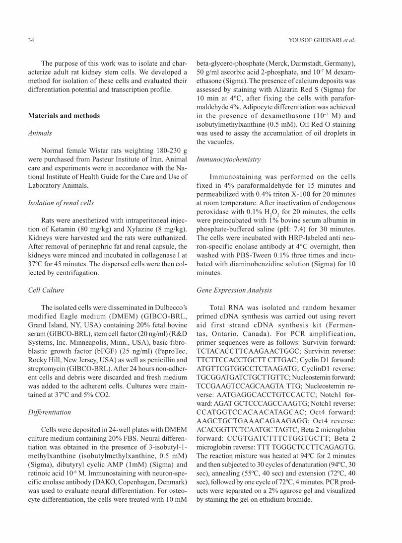
About 3-5 days after isolation of renal cells, colo-nies displaying adherent fibroblast-like morphologywere visible. Fresh medium was added to the cells after3 days. Non-adherent cells and debris were discardedafter 1 week by exchanging the medium completely.Adherent cells proliferated and formed a monolayer ofabout %80 confluence after about 1 month (Fig. 1).These adherent cells could be readily expanded by suc-cessive cycles of trypsinization, seeding and culture formore than 120 days in vitro.
Differentiation potential of isolated Cells
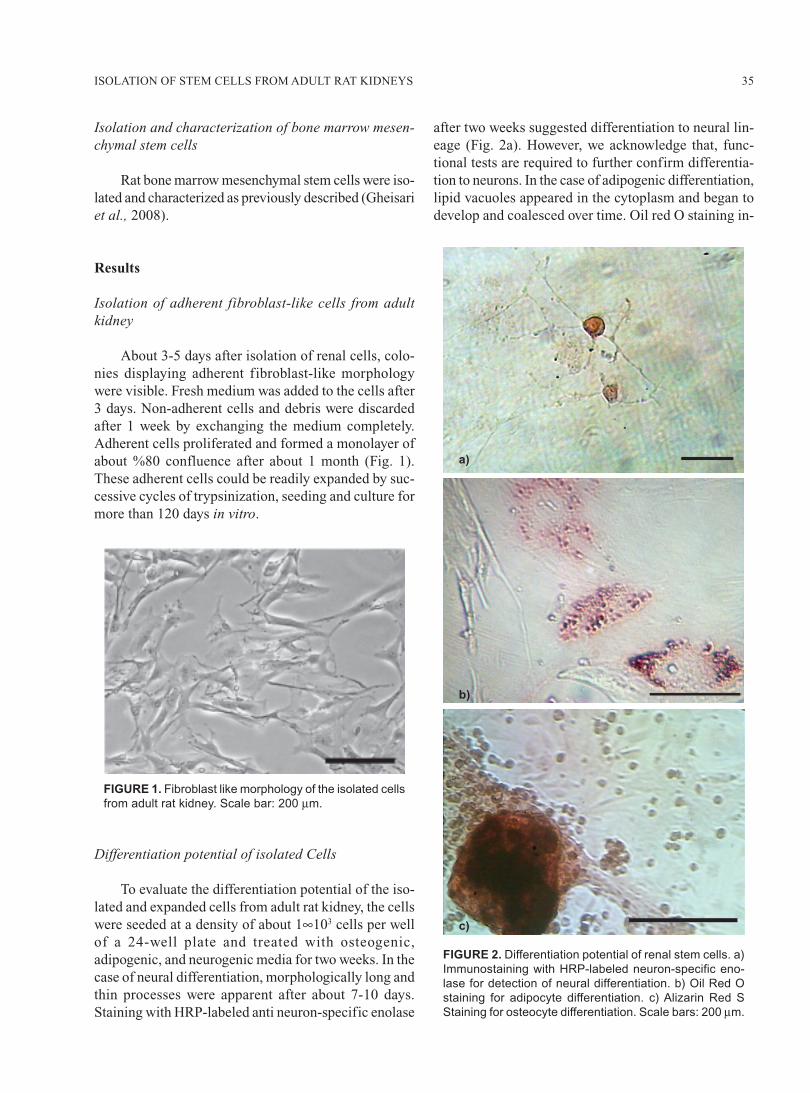
To evaluate the differentiation potential of the iso-lated and expanded cells from adult rat kidney, the cellswere seeded at a density of about 1∞103 cells per wellof a 24-well plate and treated with osteogenic,adipogenic, and neurogenic media for two weeks. In thecase of neural differentiation, morphologically long andthin processes were apparent after about 7-10 days.Staining with HRP-labeled anti neuron-specific enolase
after two weeks suggested differentiation to neural lin-eage (Fig. 2a). However, we acknowledge that, func-tional tests are required to further confirm differentia-tion to neurons. In the case of adipogenic differentiation,lipid vacuoles appeared in the cytoplasm and began todevelop and coalesced over time. Oil red O staining in-
FIGURE 1. Fibroblast like morphology of the isolated cells
from adult rat kidney. Scale bar: 200 μm.
FIGURE 2. Differentiation potential of renal stem cells. a)
Immunostaining with HRP-labeled neuron-specific eno-
lase for detection of neural differentiation. b) Oil Red O
staining for adipocyte differentiation. c) Alizarin Red S
Staining for osteocyte differentiation. Scale bars: 200 μm.
b)
c)
a)
YOUSOF GHEISARI et al.36
dicated adipocyte differentiation in these cells (Fig. 2b).These cells also underwent osteogenic differentiation.Calcium deposits, were stained with Alizarin red S (Fig.2c).
Gene Expression Patterns
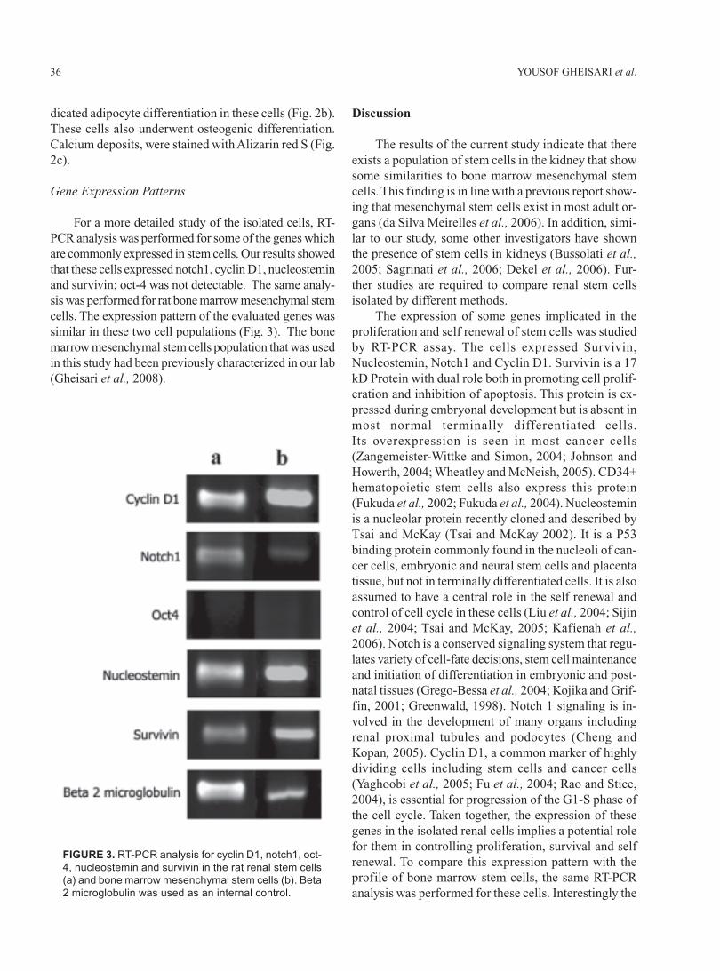
For a more detailed study of the isolated cells, RT-PCR analysis was performed for some of the genes whichare commonly expressed in stem cells. Our results showedthat these cells expressed notch1, cyclin D1, nucleosteminand survivin; oct-4 was not detectable. The same analy-sis was performed for rat bone marrow mesenchymal stemcells. The expression pattern of the evaluated genes wassimilar in these two cell populations (Fig. 3). The bonemarrow mesenchymal stem cells population that was usedin this study had been previously characterized in our lab(Gheisari et al., 2008).
Discussion
The results of the current study indicate that thereexists a population of stem cells in the kidney that showsome similarities to bone marrow mesenchymal stemcells. This finding is in line with a previous report show-ing that mesenchymal stem cells exist in most adult or-gans (da Silva Meirelles et al., 2006). In addition, simi-lar to our study, some other investigators have shownthe presence of stem cells in kidneys (Bussolati et al.,2005; Sagrinati et al., 2006; Dekel et al., 2006). Fur-ther studies are required to compare renal stem cellsisolated by different methods.
The expression of some genes implicated in theproliferation and self renewal of stem cells was studiedby RT-PCR assay. The cells expressed Survivin,Nucleostemin, Notch1 and Cyclin D1. Survivin is a 17kD Protein with dual role both in promoting cell prolif-eration and inhibition of apoptosis. This protein is ex-pressed during embryonal development but is absent inmost normal terminally differentiated cells.Its overexpression is seen in most cancer cells(Zangemeister-Wittke and Simon, 2004; Johnson andHowerth, 2004; Wheatley and McNeish, 2005). CD34+hematopoietic stem cells also express this protein(Fukuda et al., 2002; Fukuda et al., 2004). Nucleosteminis a nucleolar protein recently cloned and described byTsai and McKay (Tsai and McKay 2002). It is a P53binding protein commonly found in the nucleoli of can-cer cells, embryonic and neural stem cells and placentatissue, but not in terminally differentiated cells. It is alsoassumed to have a central role in the self renewal andcontrol of cell cycle in these cells (Liu et al., 2004; Sijinet al., 2004; Tsai and McKay, 2005; Kafienah et al.,2006). Notch is a conserved signaling system that regu-lates variety of cell-fate decisions, stem cell maintenanceand initiation of differentiation in embryonic and post-natal tissues (Grego-Bessa et al., 2004; Kojika and Grif-fin, 2001; Greenwald, 1998). Notch 1 signaling is in-volved in the development of many organs includingrenal proximal tubules and podocytes (Cheng andKopan, 2005). Cyclin D1, a common marker of highlydividing cells including stem cells and cancer cells(Yaghoobi et al., 2005; Fu et al., 2004; Rao and Stice,2004), is essential for progression of the G1-S phase ofthe cell cycle. Taken together, the expression of thesegenes in the isolated renal cells implies a potential rolefor them in controlling proliferation, survival and selfrenewal. To compare this expression pattern with theprofile of bone marrow stem cells, the same RT-PCRanalysis was performed for these cells. Interestingly the
FIGURE 3. RT-PCR analysis for cyclin D1, notch1, oct-
4, nucleostemin and survivin in the rat renal stem cells
(a) and bone marrow mesenchymal stem cells (b). Beta
2 microglobulin was used as an internal control.
37ISOLATION OF STEM CELLS FROM ADULT RAT KIDNEYS
pattern of expression of the evaluated genes was thesame in the isolated renal stem cells and bone marrowstem cells. Although the number of analyzed genes isinadequate for an absolute assessment, it could be hy-pothesized that similar pathways are used by the iso-lated renal cells and bone marrow stem cells to controlthe properties related to “stemness”.
These cells have a gene expression pattern similarto undifferentiated stem cells in vitro. However we don’tknow if these cells are in a similar undifferentiated statein vivo. It has been suggested that the role of stem cellsin the kidney could be played by some of the differenti-ated cells (Vogetseder et al., 2005).
Tissue specific stem cells are commonly expectedto differentiate into cell types of the organ of origin, butour results show that renal stem cells are also capableof differentiating into cells that are not commonly foundin the kidney such as osteocytes. This implies that thesecells probably have high plasticity.
One of the pitfalls of our stem cell isolation proto-col is that it is a simple method based on the adherenceproperty of the cells. Therefore, it is possible that theisolated cells are heterogeneous and the above-men-tioned features could not be necessarily attributed tothe whole cell population. In addition, it is possible thatthere exist other kinds of stem cells in the kidney thatwe were not able to detect by this method. The kidney ishistologicaly a complex organ with at least 26 differenttypes of cells. It seems unlikely that a single adult stemcell may be capable of forming all these cells (Zerbiniet al., 2006; Lin, 2006). We hypothesize that there ex-ists more than one type of stem cells in the kidney anddifferent methods of isolation could result in differentcell types.
Gupta et al. have developed a method for isolationof renal stem cells named multipotent renal progenitorcells (Gupta et al., 2006). Their method is similar to ourmethod, except that they used different cytokines andmediators in the process of cell isolation and expan-sion. It seems that multipotent renal progenitor cells andthe cells isolated in this study are in some way similar;however, the effect of these different combinations ofcytokines on the type of isolated cells remains to bestudied.
The adult kidney has the ability to regenerate fol-lowing injury. The potential sources of regenerating cellsinclude surviving epithelial cells, bone marrow cells andrenal stem cells (Lin et al., 2006). It has been shownthat 28 day after injury, the majority of remaining cellsare descendants of either surviving epithelial cells orrenal stem cells, suggesting that intra renal cells are the
main source of recovery following kidney injury (Linet al., 2005). Similarly, Duffield et al. (2005) used threedifferent methods for cell labeling to evaluate the par-ticipation of bone marrow cells in renal injury. Theyconcluded that these cells do not make a significantcontribution to epithelial repair. Instead intrinsic tubu-lar cell proliferation accounts for the replacement ofmost renal tubular cells following ischemia (Duffieldet al., 2005). The characteristics of these intrinsic renalcells that are capable of regeneration has not been yetclearly defined.
Successful isolation of renal progenitor cells wouldbe essential for replacement and regeneration of dam-aged cells. The renal stem cells are potentially clini-cally useful because these cells, at variance with bonemarrow stem cells are probably more committed to re-nal cells and they have similar properties to differenti-ated renal cells.
In conclusion the results of the current study indi-cate that there is a population of stem cells in the adultkidney. These cells are probably one of the sources ofregeneration after kidney injury. They could be valu-able in cell based therapeutic approaches for the treat-ment of renal failure.
Acknowledgement
This work was supported by a grant from Stem CellTechnology Company, Tehran, Iran. Special thanks toMinoo Saeidi for assistance in this study.
References
Bussolati B, Bruno S, Grange C, Buttiglieri S, Deregibus MC,Cantino D, et al (2005). Isolation of renal progenitor cellsfrom adult human kidney. American Journal of Pathology 166:545-555.
Chen J, Park HC, Addabbo F, Ni J, Pelger E, Li H et al (2008).Kidney-derived mesenchymal stem cells contribute tovasculogenesis, angiogenesis and endothelial repair. KidneyInternational 74: 879-89.
Cheng HT, Kopan R (2005). The role of Notch signaling in specifi-cation of podocyte and proximal tubules within the develop-ing mouse kidney. Kidney International 68: 1951-1952.
da Silva ML, Chagastelles PC, Nardi NB (2006). Mesenchymalstem cells reside in virtually all post-natal organs and tissues.Journal of Cell Science 119: 2204-13.
Dekel B, Zangi L, Shezen E, Reich-Zeliger S, Eventov-FriedmanS, Katchman H, et al (2006). Isolation and characterizationof nontubular sca-1+lin- multipotent stem/progenitor cellsfrom adult mouse kidney. Journal of the American Society ofNephrology 17: 3300-3314.

YOUSOF GHEISARI et al.38
Duffield JS, Park KM, Hsiao LL, Kelley VR, Scadden DT, IchimuraT, et al (2005). Restoration of tubular epithelial cells duringrepair of the postischemic kidney occurs independently of bonemarrow-derived stem cells. Journal of Clinical Investigation115: 1743-1755.
Fu M, Wang C, Li Z, Sakamaki T, Pestell RG (2004). Minireview:Cyclin D1: normal and abnormal functions. Endocrinology145: 5439-5447.
Fukuda S, Foster RG, Porter SB, Pelus LM (2002). The antiapoptosisprotein survivin is associated with cell cycle entry of normalcord blood CD34(+) cells and modulates cell cycle and pro-liferation of mouse hematopoietic progenitor cells. Blood 100:2463-2471.
Fukuda S, Mantel CR, Pelus LM (2004). Survivin regulates he-matopoietic progenitor cell proliferation through p21WAF1/Cip1-dependent and -independent pathways. Blood 103: 120-127.
Gheisari Y, Soleimani M, Azadmanesh K, Zeinali S (2008).Multipotent mesenchymal stromal cells: optimization andcomparison of five cationic polymer-based gene deliverymethods. Cytotherapy 10: 815-23.
Greenwald I (1998). LIN-12/Notch signaling: lessons from wormsand flies. Genes and Development 12: 1751-1762.
Grego-Bessa J, Diez J, Timmerman L, de la Pompa JL (2004). Notchand epithelial-mesenchyme transition in development and tu-mor progression: another turn of the screw. Cell Cycle 3: 718-721.
Guillot PV, Cook HT, Pusey CD, Fisk NM, Harten S, Moss J, et al(2008). Transplantation of human fetal mesenchymal stemcells improves glomerulopathy in a collagen type I alpha 2-deficient mouse. Journal of Pathology 214: 627-636.
Gupta S, Verfaillie C, Chmielewski D, Kren S, Eidman K, ConnaireJ, et al (2006). Isolation and characterization of kidney-de-rived stem cells. Journal of the American Society of Nephrol-ogy 17: 3028-3040.
Herrera MB, Bussolati B, Bruno S, Fonsato V, Romanazzi GM,Camussi G (2004). Mesenchymal stem cells contribute to therenal repair of acute tubular epithelial injury. InternationalJournal of Molecular Medicine 14: 1035-1041.
Johnson ME, Howerth EW (2004). Survivin: a bifunctional inhibi-tor of apoptosis protein. Veterinary Pathology 41: 599-607.
Kaf ienah W, Mistry S, Williams C, Hollander AP (2006).Nucleostemin is a marker of proliferating stromal stem cellsin adult human bone marrow. Stem Cells 24: 1113-1120.
Kojika S, Griffin JD (2001). Notch receptors and hematopoiesis.Experimental Hematology 29: 1041-1052.
Lazzeri E, Crescioli C, Ronconi E, Mazzinghi B, Sagrinati C, NettiGS, et al (2007). Regenerative potential of embryonic renalmultipotent progenitors in acute renal failure. Journal of theAmerican Society of Nephrology 18: 3128-3138.
Lin F, Moran A, Igarashi P (2005). Intrarenal cells, not bone mar-row-derived cells, are the major source for regeneration inpostischemic kidney. Journal of Clinical Investigation 115:1756-1764.
Lin F (2006). Stem cells in kidney regeneration following acuterenal injury. Pediatric Research 59: 74R-78R.
Liu SJ, Cai ZW, Liu YJ, Dong MY, Sun LQ, Hu GF, et al (2004).Role of nucleostemin in growth regulation of gastric cancer,
liver cancer and other malignancies. World Journal of Gas-troenterology 10: 1246-1249.
Maeshima A, Maeshima K, Nojima Y, Kojima I (2002). Involve-ment of Pax-2 in the action of activin A on tubular cell regen-eration. Journal of the American Society of Nephrology 13:2850-2859.
Maeshima A, Yamashita S, Nojima Y (2003). Identification of re-nal progenitor-like tubular cells that participate in the regen-eration processes of the kidney. Journal of the American So-ciety of Nephrology 14: 3138-3146.
Morigi M, Imberti B, Zoja C, Corna D, Tomasoni S, Abbate M, etal (2004). Mesenchymal stem cells are renotropic, helping torepair the kidney and improve function in acute renal failure.Journal of the American Society of Nephrology 15: 1794-1804.
Oliver JA, Maarouf O, Cheema FH, Martens TP, Al-Awqati Q(2004). The renal papilla is a niche for adult kidney stem cells.Journal of Clinical Investigation 114: 795-804.
Plotkin MD, Goligorsky MS (2006). Mesenchymal cells from adultkidney support angiogenesis and differentiate into multipleinterstitial cell types including erythropoietin-producing fi-broblasts. American Journal of Physiology, Renal Physiology291: F902-F912.
Rao RR, Stice SL (2004). Gene expression profiling of embryonicstem cells leads to greater understanding of pluripotency andearly developmental events. Biology of Reproduction 71: 1772-1778.
Sagrinati C, Netti GS, Mazzinghi B, Lazzeri E, Liotta F, Frosali F,et al (2006): Isolation and characterization of multipotent pro-genitor cells from the Bowman’s capsule of adult human kid-neys. Journal of the American Society of Nephrology 17: 2443-2456.
Sijin L, Ziwei C, Yajun L, Meiyu D, Hongwei Z, Guofa H, et al(2004). The effect of knocking-down nucleostemin gene ex-pression on the in vitro proliferation and in vivo tumorigen-esis of HeLa cells. Journal of Experimental and Clinical Can-cer Research 23: 529-538.
Tsai RY, McKay RD (2002). A nucleolar mechanism controllingcell proliferation in stem cells and cancer cells. Genes andDevelopment 16: 2991-3003.
Tsai RY, McKay RD (2005). A multistep, GTP-driven mechanismcontrolling the dynamic cycling of nucleostemin. Journal ofCell Biology 168: 179-184.
Vogetseder A, Karadeniz A, Kaissling B, Le Hir M (2005). Tubularcell proliferation in the healthy rat kidney. Histochemistry andCell Biology 124: 97-104.
Wheatley SP, McNeish IA (2005). Survivin: a protein with dualroles in mitosis and apoptosis. International Review of Cytol-ogy 247: 35-88.
Yaghoobi MM, Mowla SJ, Tiraihi T (2005). Nucleostemin, a coor-dinator of self-renewal, is expressed in rat marrow stromalcells and turns off after induction of neural differentiation.Neuroscience Letters 390: 81-86.
Zangemeister-Wittke U, Simon HU (2004). An IAP in action: themultiple roles of survivin in differentiation, immunity andmalignancy. Cell Cycle 3: 1121-1123.
Zerbini G, Piemonti L, Maestroni A, Dell’Antonio G, Bianchi G(2006). Stem cells and the kidney: a new therapeutic tool?Journal of the Amercan Society of Nephrology 17: S123-126.