review article mammalian myc proteins and cancerdownloads.hindawi.com/archive/2014/757534.pdf ·...
TRANSCRIPT

Review ArticleMammalian MYC Proteins and Cancer
William P. Tansey
Department of Cell and Developmental Biology, Vanderbilt University School of Medicine, 465 21st Avenue South,Nashville, TN 37232, USA
Correspondence should be addressed to William P. Tansey; [email protected]
Received 29 August 2013; Accepted 1 November 2013; Published 2 February 2014
Academic Editor: Ygal Haupt
Copyright © 2014 William P. Tansey. This is an open access article distributed under the Creative Commons Attribution License,which permits unrestricted use, distribution, and reproduction in any medium, provided the original work is properly cited.
The MYC family of proteins is a group of basic-helix-loop-helix-leucine zipper transcription factors that feature prominently incancer. Overexpression of MYC is observed in the vast majority of human malignancies and promotes an extraordinary set ofchanges that impact cell proliferation, growth, metabolism, DNA replication, cell cycle progression, cell adhesion, differentiation,and metastasis. The purpose of this review is to introduce the reader to the mammalian family of MYC proteins, highlightimportant functional properties that endow them with their potent oncogenic potential, describe their mechanisms of action andof deregulation in cancer cells, and discuss efforts to target the unique properties of MYC, and of MYC-driven tumors, to treatcancer.
1. Introduction
MYC is an oncoprotein transcription factor that featuresin the cancer-related deaths of an estimated 100,000 peoplein the United States—as well as millions worldwide—everyyear. Its formidable oncogenic reputation stems from itsfrequent deregulation in a host of human cancers and froma suite of activities that place MYC at the nexus of cellgrowth, proliferation, metabolism, and genome stability. Thepurpose of this review is to introduce readers to the basicfeatures ofmammalianMYCproteins and distill key conceptsand general themes in what is known about the molecularprocesses through which MYC drives tumorigenesis. Alongthe way, we will also discuss controversial or unresolvedissues that limit current understanding of MYC proteins andhighlight areas that are ripe for exploration in the future.My intention is to leave the reader with the sense that MYCpossesses a unique and expansive set of activities that underlieits place as amajor human oncoprotein andmaywell hold thekey to development of broadly effective anticancer therapies.
2. MYC: The Early Years
MYC is arguably one of the most well-studied proteins inhuman history.More than 22,000 primary and review articleshave been written on this topic, which in recent years havebeen appearing at an average rate of almost three publications
per day (Figure 1). MYC has been studied by virologists,geneticists, molecular biologists, structural biologists, cellbiologists, biochemists, and theoretical biologists, and count-less millions of dollars have been spent interrogating both itsnormal and its cancer-relevant functions. In any subject asexpansive and as lasting as this, it is impossible to providea comprehensive review of all of the major discoveries thathave shaped current thinking on the MYC proteins. Butas the origins of this field are fading quickly into history,it is worthwhile to reflect briefly on the early studies thatgerminated the MYC phenomenon.
The discovery of MYC stemmed directly from experi-ments begun in the early 1960s, but the crucial context forthose experiments was established a half a century beforeby Peyton Rous, who showed that cell-free filtrates from achicken sarcoma could be used to transmit disease to sus-ceptible animals [2]. This ground-breaking work ultimatelyled to the discovery of retroviruses and viral oncogenes(and their cellular counterparts) [3], but crucially it spurredresearchers to exploit avian leukosis viruses as a way tounlock the basis of cancer. Framed in this setting was theisolation in 1964 ofMC29, a strain of virus propagated from aRhode Island Red chicken in Sofia, the capital city of Bulgaria[4]. The hen had succumbed to a spontaneously developedhematological disease that included anemia and solid tumorsof promyelocytic character. Subsequent isolation and pas-saging of the virus in animals revealed that MC29 induces
Hindawi Publishing CorporationNew Journal of ScienceVolume 2014, Article ID 757534, 27 pageshttp://dx.doi.org/10.1155/2014/757534

2 New Journal of Science
0
375
750
1125
1500
1964
1967
1970
1973
1976
1979
1982
1985
1988
1991
1994
1997
2000
2003
2006
2009
2012
Year
identified
Cellular MYC
MYC and RAS
Overexpression in cancer
MAX
MYC and
MYC target genes
MYC regulates
MC29 isolation and characterization
“Amplifier”
Genomic analyses of MYC
MYC inhibitors
MYC and iPS cells
v-myc
Publ
icat
ions
per
yea
r
model
characterized
cooperation apoptosis cell growth
identifieddiscoveredgenes cloned
Figure 1: Timeline of studying MYC. The figure shows the annual number of publications on MYC proteins from 1964 to 2012, as listed onthe NCBI’s PubMed website, along with approximate timing of key discoveries in the MYC field during that time. Citations from the period1964–1980 were recovered using the search term “MC29.”The search excludes instances where MYC refers to use of the MYC epitope tag. Seetext for more detailed description of key discoveries.
neoplasia predominantly in the hematopoietic compartmentof recipient fowl [4, 5] but differs from other avian leukosisviruses in that it does not typically result in developmentof leukemia. Instead, MC29 transforms myeloid cells toform either diffuse growths—myelocytomatosis—or solidtumors—myelocytomas. It was this unique disease spectrumthat ultimately gave MYC its name (myelocytomatosis).
Following the initial characterization of the virus, thehunt began for identification of the genetic basis of itstumorigenicity.This was not a trivial feat, as early researchersdid not have access to rapid assays of tumorigenesis oradvanced molecular biology tools nor did they formallyknow thatMC29 carried a bona fide oncogene—its oncogenicproperties, for example, could have resulted from its influ-ence on the expression of a cellular oncogene. Ultimately,dissection of the oncogenic activity of MC29 was facilitatedby a number of crucial technological developments and byinherent characteristics of the virus itself. The demonstrationthat MC29 could act in a variety of isolated cell types toinduce transformation [6–10]—the acquisition of phenotypicchanges that resemble those displayed by tumor cells invivo—provided researchers with a rapid and reliable proxyfor assaying its tumorigenic function. The virus itself wasfairly easy to grow to high titer in tissue culture, and as wenow know carries just a single functional gene, the gag-mycfusion [11–13]. Importantly, development of molecular biol-ogy approaches, especially the ability to radiolabel specificnucleic acid sequences and to interrogate genome structureby solution hybridization, gave researchers the tools theyneeded tomolecularly characterize the virus. Indeed, in just ashort period of time in the late 1970s the candidate v-myc genewas identified [13, 14], shown to be distinct from the src onco-gene present in Rous’s original virus [15], and found to have acellular homolog in uninfected vertebrate cells [16, 17], con-sistent with Bishop andVarmus’s model for the cellular originof retroviral oncogenes [18].The presence of v-myc sequences
in related oncogenic retroviruses [19], together with findingsthat expression of a cellular v-myc homolog is greatly stim-ulated by retroviral promoter insertion of a distinct avianleukosis virus [20–22], cemented the idea that MYC is thebona fide oncogene within MC29. Finally, in 1982, the c-mycgenewas cloned and characterized [23], an event that usheredin a new era of exploration of the molecular mechanisms oftumorigenesis and triggered a massive wave of research intoMYC’s regulation, structure, and functions (Figure 1).
As indicated in Figure 1, the MYC boom of the 1980sresulted in fundamental new insight into MYC, and thesestudies in turn led to development of key concepts intumorigenesis, such as oncogene cooperation [24] and theimportance of apoptosis [25–27] as a tumor surveillancemechanism. Many of these discoveries form the basis ofmaterial discussed throughout this review. Looking at thepace of MYC research over recent years, it is interesting tonote that we are currently in the midst of a second “MYCrush” that began around 2006, undoubtedly triggered by theadvent of genomic approaches towards studying MYC andfueled by discoveries that MYC is one of the “magic four”[28, 29] factors that can reprogram differentiated cells to theinduced pluripotent stem cell (iPS) state. Given the intenseinterest in MYC and the ever-expanding universe of MYCfunctions, it seems certain that the current rush will continueif not accelerate for years to come.
3. The MYC Family of Proteins
The c-myc gene first identified in 1982 is the prototype fora family of related proteins (Figure 2) that are not onlyconserved across metazoan life [30] but can be found insimilar form and function in premetazoan organisms suchas choanoflagellates [31]. In mammalian cells, MYC pro-teins arise from three distinct gene family members—c-Myc,N-myc, and L-myc—which function in a similar manner

New Journal of Science 3
NMYC
IIIbIIIaIII IV BR-HLH-LZC-MYC
IIIbIIIaIII IV BR-HLH-LZN-MYC
IIIbIII IV BR-HLH-LZL-MYC
Transcriptional activation Central portion DNA binding
Figure 2: Architecture of the MYC family. The image at the top shows a generic representation of a mammalian MYC protein, indicatingthe transcriptional activation domain, the central portion, the canonical nuclear localization sequence (N), and the region involved in DNAbinding via interaction with MAX. Below is a representation of conserved sequences present in C-, N-, and L-MYC family members. c-MYCis drawn to scale at 439 amino acids. N- and L-MYC proteins are different lengths (464 and 364 amino acids, resp.) due to differences in thelength of nonconserved sequences but are drawn to highlight the conservation and relative location of Myc boxes.
but display notable differences in potency [32, 33] andpatterns of expression [34–36]. Rodents also express a B-myc variant that shares homology with the amino-terminiof MYC proteins [37] but this has yet to be characterizedin other mammals. The presence of multiple MYC fam-ily members with distinct expression patterns undoubtedlyreflects different spatial and temporal requirements for MYCactivity, both in development and in the adult animal [38],and is most frankly seen in the particular way each gene isoverexpressed in specific cancer types [34, 39, 40]. c-Myc isbroadly overexpressed in both blood-borne and solid tumors.N-myc is most frequently overexpressed in solid cancers ofneural origin, such as neuroblastoma and glioma. And L-mycis most often overexpressed in small cell lung carcinomas.Given the conservation of these proteins, it is reasonableto assume that the mechanisms through which they drivetumorigenesis are similar, although it is also important torecall that most functional studies of MYC to date havefocused on the archetypal c-Myc product.
As well as multiple family members, a number of c-Mycprotein variants can be generated bymechanisms that includealternative start codon usage (referred to as “p67” and “p64”[41, 42]), downstream translation initiation within the MYCmRNA (“Myc-S;” [43]), and site-specific proteolysis (“MYC-nick;” [44]). Although these variants are intriguing, they havereceived comparatively little attention and hence their signif-icance to cancer, for the most part, is not well understood.
3.1. The Anatomy of MYC. The general architecture of mam-malianMYC proteins resembles those of a typical sequence-specific DNA-binding transcriptional regulator (Figure 2). Atthe amino-terminus ofMYC lies its transcriptional activationdomain (TAD), a region that is sufficient for transcriptionalactivation when fused to a heterologous DNA bindingdomain (DBD) [45] and is required for MYC’s transformingactivity in vitro [46]. This region is accepted to be theprimary element within MYC that contacts RNA polymeraseII-associated proteins to stimulate gene induction. LikemanyTADs, the MYC TAD is intrinsically unstructured in the
absence of partner proteins [47], is enriched in acidic, proline,and glutamine residues, and retains its ability to stimulatetranscription in organisms that do not have MYC proteins,such as the yeast Saccharomyces cerevisiae [48, 49]. Also likeother TADS [50], this region ofMYC is a potent “degron” [51]and is the primary domain responsible for signaling the rapiddestruction ofMYC by ubiquitin- (Ub-)mediated proteolysis[52].
It is interesting to note that although full-length MYC isa notoriously weak activator of pol II genes [53], the TADitself, when fused to a heterologous DBD, is exceptionallypotent—comparable in magnitude to the canonical VP16activation domain [45, 54]. Although the apparent strengthof the isolated MYC TAD could be due to peculiarities ofthe assay systems involved, these assays do faithfully reflectdifferences between the C- and L-MYC TADs that underlietheir different tumorigenic potentials [33], making it difficultto dismiss the results as entirely artifactual. One possibility isthat processes operate within full-length MYC to temper itstransactivation potential, allowing for bursts of MYC activityat specific points in development or in response to specificsignals but otherwise keeping its activity in check.The abilityof cells to tightly limit MYC expression and activity is arecurring theme in MYC biology and one that we will returnto later in this review.
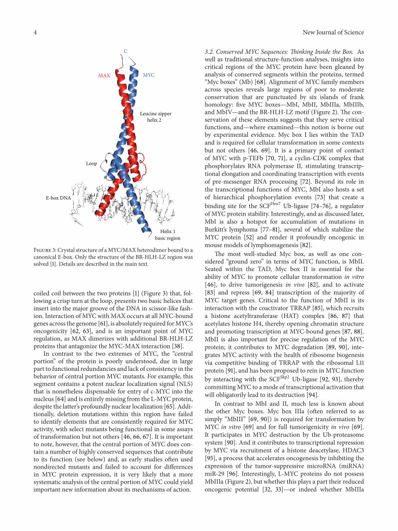
At the carboxy terminus of MYC is a ∼100 amino acidbasic helix-loop-helix-leucine zipper (BR-HLH-LZ) regionthat functions as its DNA-binding domain [55, 56]. This typeof DNAbinding domain is not unique toMYC but is found inother sequence-specific transcriptional regulators includingthe Pho4 activator in yeast [57] and the hypoxia-inducibleHIF-1𝛼 factor inmammalian cells [58]. BR-HLH-LZ proteinsbind DNA as obligate dimers and recognize a consensussequence “CACGTG,” which is termed the “Enhancer box”(E-box) [59]. At physiological concentrations, MYC does nothomodimerize [56] but instead interacts with the small BR-HLH-LZ proteinMAX to form a heterodimer that constitutesa coreDNA-bindingmodule [60]. Dimerization ofMYCwithMAX is driven by the leucine zipperwhich forms an extended
4 New Journal of Science
MAX MYC
C
Leucine zipper helix 2
Loop
E-box DNA
Helix 1
basic region
MAX MYC
Leucine zipper helix 2
Loop
A
Helix 1
basic regio
Figure 3: Crystal structure of aMYC/MAX heterodimer bound to acanonical E-box. Only the structure of the BR-HLH-LZ region wassolved [1]. Details are described in the main text.
coiled coil between the two proteins [1] (Figure 3) that, fol-lowing a crisp turn at the loop, presents two basic helices thatinsert into the major groove of the DNA in scissor-like fash-ion. Interaction ofMYCwithMAX occurs at all MYC-boundgenes across the genome [61], is absolutely required forMYC’soncogenicity [62, 63], and is an important point of MYCregulation, as MAX dimerizes with additional BR-HLH-LZproteins that antagonize the MYC-MAX interaction [38].
In contrast to the two extremes of MYC, the “centralportion” of the protein is poorly understood, due in largepart to functional redundancies and lack of consistency in thebehavior of central portion MYC mutants. For example, thissegment contains a potent nuclear localization signal (NLS)that is nonetheless dispensable for entry of c-MYC into thenucleus [64] and is entirely missing from the L-MYC protein,despite the latter’s profoundly nuclear localization [65]. Addi-tionally, deletion mutations within this region have failedto identify elements that are consistently required for MYCactivity, with select mutants being functional in some assaysof transformation but not others [46, 66, 67]. It is importantto note, however, that the central portion of MYC does con-tain a number of highly conserved sequences that contributeto its function (see below) and, as early studies often usednondirected mutants and failed to account for differencesin MYC protein expression, it is very likely that a moresystematic analysis of the central portion of MYC could yieldimportant new information about its mechanisms of action.
3.2. Conserved MYC Sequences: Thinking Inside the Box. Aswell as traditional structure-function analyses, insights intocritical regions of the MYC protein have been gleaned byanalysis of conserved segments within the proteins, termed“Myc boxes” (Mb) [68]. Alignment of MYC family membersacross species reveals large regions of poor to moderateconservation that are punctuated by six islands of frankhomology: five MYC boxes—MbI, MbII, MbIIIa, MbIIIb,and MbIV—and the BR-HLH-LZ motif (Figure 2). The con-servation of these elements suggests that they serve criticalfunctions, and—where examined—this notion is borne outby experimental evidence. Myc box I lies within the TADand is required for cellular transformation in some contextsbut not others [46, 69]. It is a primary point of contactof MYC with p-TEFb [70, 71], a cyclin-CDK complex thatphosphorylates RNA polymerase II, stimulating transcrip-tional elongation and coordinating transcription with eventsof pre-messenger RNA processing [72]. Beyond its role inthe transcriptional functions of MYC, MbI also hosts a setof hierarchical phosphorylation events [73] that create abinding site for the SCFFbw7 Ub-ligase [74–76], a regulatorof MYC protein stability. Interestingly, and as discussed later,MbI is also a hotspot for accumulation of mutations inBurkitt’s lymphoma [77–81], several of which stabilize theMYC protein [52] and render it profoundly oncogenic inmouse models of lymphomagenesis [82].
The most well-studied Myc box, as well as one con-sidered “ground zero” in terms of MYC function, is MbII.Seated within the TAD, Myc box II is essential for theability of MYC to promote cellular transformation in vitro[46], to drive tumorigenesis in vivo [82], and to activate[83] and repress [69, 84] transcription of the majority ofMYC target genes. Critical to the function of MbII is itsinteraction with the coactivator TRRAP [85], which recruitsa histone acetyltransferase (HAT) complex [86, 87] thatacetylates histone H4, thereby opening chromatin structureand promoting transcription at MYC-bound genes [87, 88].MbII is also important for precise regulation of the MYCprotein; it contributes to MYC degradation [89, 90], inte-grates MYC activity with the health of ribosome biogenesisvia competitive binding of TRRAP with the ribosomal L11protein [91], and has been proposed to rein in MYC functionby interacting with the SCFSkp2 Ub-ligase [92, 93], therebycommittingMYC to amode of transcriptional activation thatwill obligatorily lead to its destruction [94].
In contrast to MbI and II, much less is known aboutthe other Myc boxes. Myc box IIIa (often referred to assimply “MbIII” [69, 90]) is required for transformation byMYC in vitro [69] and for full tumorigenicity in vivo [69].It participates in MYC destruction by the Ub-proteasomesystem [90]. And it contributes to transcriptional repressionby MYC via recruitment of a histone deacetylase, HDAC3[95], a process that accelerates oncogenesis by inhibiting theexpression of the tumor-suppressive microRNA (miRNA)miR-29 [96]. Interestingly, L-MYC proteins do not possessMbIIIa (Figure 2), but whether this plays a part their reducedoncogenic potential [32, 33]—or indeed whether MbIIIa

New Journal of Science 5
MYCDNA
MYCRNA
PTMs TXNinitiation
mRNAexport
Proteindestruction
MYC MYC
Pausedpolymerase
TLNblockade
mRNAdestruction
Functional inhibition
activityprotein
Figure 4: Restricting MYC expression and activity. Each stage in MYC expression, from transcription of the MYC gene through to afunctionally activeMYC protein, are presented in terms of the central dogma. Events that control or suppress these stages are indicated by redboxes. Many of these processes are perturbed in cancer. See text for details. TXN, transcription; TLN, translation; PTMs, post-translationalmodifications.
has other functions and binding partners besides HDAC3—remains to be determined.
Myc box IIIb has not, to my knowledge, been system-atically studied in any mammalian system; a remarkableoversight given its presence in every earthly MYC protein.
Finally, Myc box IV has been studied in just one publi-cation [97]. It is required for the full proapoptotic functionsof MYC but not its ability to induce cell proliferation, and itcontributes to both transcriptional activation and repressionof MYC target genes. Despite lying outside of the BR-HLH-LZ, deletion of MbIV renders MYC less able to bindDNA both in vitro and in cells, suggesting that it somehowcontributes to target gene recognition. It is not knownwhether this effect is due to MbIV making contact withDNA, or recruiting additional factors, or whether deletion ofthis element perturbs structural aspects of MYC that preventefficient binding by MYC/MAX heterodimers.
3.3. Is the Map of MYC Complete? Given the volume of workon the subject, one might expect that we understand the con-tribution of almost every amino acid withinMYC to its func-tions as a transcriptional regulator and oncoprotein. And yet,except for the two extremes of the protein, this is not the case.The central portion of MYC is largely unchartered territory,particularly with respect to Myc boxes IIIa, IIIb, and IV. Theconservation of these elements implies an important com-mon role and leads to the notion that they must be involvedin protein-protein (or perhaps protein-DNA) interactionscritical to the intrinsic functions of MYC. As the search fornewways to targetMYC in cancer accelerates, uncovering thefunction of these elements seems fertile territory that couldlead to new therapeutic routes of intervention. Additionally,beyondMyc boxes, we know very little about the segments inMYC that are not universally conserved acrossMYC proteinsbut are conserved within individual members of the MYCfamily. The payoff for understanding the function of thesesequences could be tremendous. For example, learning how
sequences unique to L-MYC contribute to small cell lungcancers could very well lead to insight into unique oncogenicprocesses that occur within that specific tumor environmentand lead to development of therapies that are more effectiveand induce less collateral damage on other cell and tissuetypes. It may not be the most glamorous work, but morecareful investigation into how the entire sequence of MYCproteins dictates their function is one major gap that needsto be filled in the MYC arena.
4. Mechanisms of MYC Regulation andIts Deregulation in Cancer
A little bit of MYC is a good thing. Normal fibroblasts inculture typically express just a few thousand molecules ofMYC protein per cell [98], yet this can be over two ordersof magnitude higher in cancer cell lines growing undersimilar conditions [61]. Before reviewing the broad impactof increased MYC burden on cellular processes, it is worthdiscussing how normal cells control MYC and how theseprocesses go awry in cancer.
4.1. Regulating MYC: Just Say “No”. One important princi-ple to emerge from studies over the last 30 years is thatmammalian cells have evolved a sophisticated network ofprocesses that constantly impede MYC action. Indeed, ifone considers MYC expression and activity in terms of thecentral dogma (Figure 4), it is clear that MYC proteins aresubject to some kind of stringent control at every step oftheir life. A primary point of regulating MYC is at the levelof transcription. MYC is an “immediate early” gene [99]—not transcribed in quiescent cells but rapidly induced inresponse to growth factor signaling [100, 101]. Regulation ofMYC transcription is exerted at both the level of initiation[102] and of release of paused RNA polymerase II [103, 104]and integrates both positive and negative inputs to insurethat MYC genes are transcribed only in the presence of

6 New Journal of Science
appropriate “go” signals within the cellular milieu. Followingtranscription, the MYC mRNA itself is subject to severalrestrictive processes that are also tuned to the growth statusof the cell. Export of MYC mRNA to the cytoplasm iscoordinated by the translation initiation factor eIF4E [105],which is under direct control by mitogenic signals [106]and binds the MYC message early during the transcriptionprocess, leaving little chance that errant MYC mRNAs canescape the nucleus without appropriate restraint. Once in thecytoplasm, translation of the MYC mRNA is suppressed byfactors that mediate productive ribosome engagement [107]and is temporally limited by the extraordinarily short half-life of the mRNA [108]. And following synthesis, the proteinitself is under intense scrutiny and control. MYC is subject toa bevy of posttranslational modifications that include phos-phorylation, acetylation, glycosylation, and ubiquitylation[109–111], many of which interact to establish particular statesof MYC expression or activity. The ubiquitylation of MYC,in particular, appears to be an important point of control[112]. Not only is the rapid Ub-mediated proteolysis of MYCcrucial for keepingMYC levels low and tied to early processesthat restrict MYC synthesis, but ubiquitylation of MYC—as well as its deubiquitylation [113, 114]—acts to modulatethe inherent transcriptional properties of the protein [92, 93,115, 116], providing a “point of action” mode of control thatmicromanages MYC activity. Finally, MYC activity is alsorestricted by the action of specific proteins mentioned abovethat either interact directly with MYC to limit its function[91] or interact with MAX to prevent MYC from finding itsessential partner protein [38].
Much in the same way as multilayered systems are inplace to allow nuclear reactors to safely harness the power ofnuclear fission, the processes that restrict MYC conspire toallow cells to efficiently manage its function for their normalactivities without stepping on the path to tumorigenesis.As discussed below, however, and analogous to a nuclearmeltdown, failure of these processes at any point can havecatastrophic outcomes.
4.2. MYC Deregulation in Cancer. One of the key conceptsin understanding how MYC is deregulated in cancer is thefact that—unlike oncoproteins such as RAS [117]—the codingsequence of MYC does not need to be changed in orderfor its oncogenic potential to be unleashed. As discussedin the next section, mutant forms of MYC are prevalentin Burkitt’s lymphoma but are not widely seen in othercancers, and all evidence from cell- and animal-based studiesis that expression of a pristine form of MYC is sufficientto promote tumorigenesis [24, 118–121]. The most commonroute to a MYC-driven cancer, therefore, is changes that leadto overexpression of MYC and disconnect it from the criticalsignaling processes (above) that normally keep it in check.
On average, 50% of human cancers show increasedexpression of MYC (for a comprehensive presentation of thestatistics, see [122]), a characteristic that usually correlateswith poor patient survival [101]. The percentage of cancersfunctionally overexpressing MYC is certainly much higher,as most studies to date have focused on MYC gene and RNA
dosage, which are invisible to perturbations that increase thesynthesis of MYC proteins or slow their rate of degradation,and do not account for functional deregulation of the protein.Although the range of cancers prompted by individual MYCproteins is fairly well delineated (Section 3), the MYC familyas a whole are equal opportunity oncoproteins and areoverexpressed in cancers as diverse as lymphoma,melanoma,multiple myeloma, and neuroblastoma, as well as colon,breast, and lung cancers [39, 122]. It is these statistics that giveMYC its formidable reputation but also inspire researcherswith the promise that therapeutic strategies to target MYCcould have broad impact in reducing or eliminating manycancer fatalities.
There are many interesting ways in which cancer cellsderegulate MYC, but they typically fall into one of twocategories: changes to theMYC loci themselves that stimulateincreased MYC mRNA production, or changes extrinsicto MYC that disarm critical regulatory mechanisms. Asmentioned early in this review, retroviral promoter insertionwas the first mechanism recognized to lead to enhancedMYC transcription [20–22] and was a seminal discovery interms of cementing the role ofMYC as a bona fide oncogene.This was soon followed by the observation that the c-MYClocus is translocated in Burkitt’s lymphomas and mouseplasmacytomas [123, 124], an event that places MYC codingsequences under the control of the immunoglobulin 𝜇 heavychain enhancer, driving very high levels of mRNA synthesis.This translocation, as well as others of similar nature, arefound in 100% of Burkitt’s lymphoma patients and indeed areused (together with other approaches) to diagnose the disease[125].
Despite their prevalence in Burkitt’s lymphoma, suchrearrangements are not common in other cancers, wherethe typical mode of increasing MYC synthesis via genomicmodification is gene amplification [122]. This manner ofMYC induction is fairly common in cancer, affects all MYCfamily members, and can take the form of small focal ampli-fications [126, 127], large amplifications [128], and double-minute chromosomes [129]. Although increasing the numberof copies of MYC does not always correlate with MYCoverexpression [130, 131], the frequency with which MYCgene amplification occurs and the correlation of this eventwith metastatic disease and poor prognosis [132] stronglyindicate a direct role for MYC gene amplification in manyhuman malignancies.
It is important to note thatMYCmRNAsynthesis can alsobe affected by genomic changes that, at first blush, have littleto do with theMYC gene itself. An inherited nucleotide vari-ant, rs6983267, was recently described as conferring increasedrisk to colorectal and prostate cancer [133]. Subsequent stud-ies showed that—although this polymorphism occurs morethan one megabase away from the c-MYC gene—its tumor-promoting properties manifest by stimulating binding of theTCF-4 activator to a previously unrecognized distal MYCenhancer, thereby leading to increased MYC transcription[134, 135]. The remoteness of this enhancer from the MYCgene raises the distinct possibility that other, apparentlyunconnected, genomic changes that occur in cancer couldresult in increased MYC transcription.

New Journal of Science 7
IIIbIIIaIII IV BR-HLH-LZ
ND
20
10
0Num
ber o
f ind
epen
dent
repo
rts
TAD/degron Pest DNA binding
Figure 5: Frequency and location of lymphoma-associated point mutations in c-MYC. The histogram shows the number of times thatindependent point mutations altering the coding sequence of c-MYC have been reported in the literature, isolated from either patient samplesor cultured cell lines. Duplicate identifications (e.g., those occurring in the same cell line but reported by different investigators) have beenremoved.The analysis did not differentiate between conservative and nonconservative amino acid changes.The histogram is fit to scale atop acartoon ofMYC, showing conserved sequences, functional elements, and regions involved in controlling themetabolic stability of the protein.
External to MYC, it is reasonable to assume that anyof the processes depicted in Figure 4 could be subject toderegulation in tumor cells. Indeed, uncovering which eventsare frequently perturbed in tumors provides valuable insightinto those mechanisms that are most important for thenormal control of MYC. A few prominent examples illustratethe diversity of ways MYC can run amok in cancer cells. Atthe level of transcription, it is clear thatMYC lies at the end ofa set of signal transduction pathways that become ectopicallyactivated in cancer. Loss of the adenomatous polyposiscoli (APC) tumor suppressor, for example, which occurs inalmost all colorectal cancers [136], leads to the accumulationof 𝛽-catenin, which in turn binds to the TCF-4 activator(mentioned above) to stimulate constitutive and high-leveltranscription ofMYC [137]. Similar scenarios result in potentinduction of MYC transcription in response to perturbationsof Sonic hedgehog [138] and Notch signaling pathways [139,140]. At the level of mRNA dynamics, stability of the MYCmessage can be increased in cancer cells [141]. And the eIF4Etranslation factor that exports MYCmRNA from the nucleusis overexpressed in a slew of malignancies [106], functioningas an oncogene by stimulating the translation of a cohort ofgrowth-promoting factors, including MYC [142]. Finally, atthe level of protein destruction, MYC can be stabilized byviral oncogenes [143] and by loss of critical regulators suchas the SCFFbw7 ubiquitin ligase [144] that is inactivated in anestimated 6% of all human cancers [145]. Given the plethoraof ways that tumor cells can increase their MYC burden,it is not unrealistic to conclude that loss of appropriatecontrol over MYC contributes in a substantive way to allmalignancies.
4.3. MYC Mutations in Cancer. Tumor-associated mutationsthat alter the c-MYC coding sequence were reported as earlyas 1983 [77] but have received surprisingly little attention inthe last 30 years—and not without reason. Such mutationsare found in a very narrow spectrum of tumors—principallyBurkitt’s lymphoma and a smattering of AIDS-associated
hematologic malignancies [78–82, 146–151]—and are alwaysassociated with some other genomic rearrangement thatitself leads to a massive increase in MYC expression,such as translocation proximal to the immunoglobulin 𝜇enhancer. As such rearrangements are clearly sufficient todrive lymphomagenesis independent of any changes in theMYC protein, the likelihood that these mutations contributemeaningfully to the pathophysiology of the disease is small.Moreover, because the translocated MYC allele is placedwithin a hypermutable region of the genome [152], it istempting to view these mutations as “collateral damage” andnoninformative to understand MYC function in this setting.
Nonetheless, changes to the coding sequence of MYC arefound in nearly 50% of Burkitt’s lymphomas [78] and clusterin specific and recurring places on the protein (Figure 5),suggesting that they are not neutral events. And, whereexamined, these mutations do impact MYC regulation andfunction. A common theme in this area is that tumor-derivedmutations subvert the rapid destruction of MYC by Ub-mediated proteolysis. As shown in Figure 5, these mutationstypically cluster within elements that controlMYC stability—the TAD/degron [52], the “D-region” [90], and the “PESTelement” [153] (for review of these elements see [112])—and several of the most common recurring mutations havebeen shown to stabilize MYC in cultured cells [52]. Indeed,the most frequently mutated residue in the tumor collectionis threonine 58 (T58), which lies within the heart of thephosphodegron that is recognized by SCFFbw7 to promoteMYC destruction [74–76]. The fact that the MYC—SCFFbw7interaction is disturbed in trans by loss of Fbw7 [144] andin cis by recurring tumor-associated MYC mutations [52]provides compelling support for the idea that breaking Ub-mediated turnover of MYC is advantageous to cancer cells.
The impact of tumor-derived mutations on MYC func-tion has been controversial. Early reports indicated thatmutations within MbI (e.g., T58A) disrupt interaction ofMYC with the Rb family member p107 [154, 155], but itremains unclear whether p107 is a physiologically relevant

8 New Journal of Science
MYC partner protein. Moreover, it is difficult to tease apartdirect effects on MYC function from effects that occur as aresult of increasing MYC protein levels. The T58A mutationincreases the transforming ability of MYC in vitro [156, 157]and renders MYC aggressively oncogenic in mouse systemsof lymphomagenesis [82] and mammary cancer [158], asmight be expected from a perturbation that increases MYCprotein expression.What is curious, however, is that theT58Amutant protein achieves its enhanced tumorigenic activityby selectively disabling the proapoptotic function of MYC[82, 156], a remarkable feat given that enhancedMYC burdenis associated with increased apoptotic potential [159]. Onepossibility is that mutations such as those at T58 not onlydisturb the levels of MYC but also qualitatively alter itsfunction, either by disrupting an entirely different process orby perturbing its normal patterns of ubiquitylation, which areknown to modulate MYC activity through both proteolyticand nonproteolytic means [92, 93, 115, 160, 161]. Furtherinvestigation will be required to fully understand the impactof these mutations on MYC.
4.4. Losing Control of MYC: Is There More to Learn? Reflect-ing on the history of MYC, it is astounding to see that a basictenet of theMYCfield—that enhancedMYC expression leadsto cancer—was articulated in the very earliest days of MYCresearch [21]. Over the intervening years, we have certainlycome to appreciate themyriad of ways this can occur, but thatfundamental concept has not changed. With so much effortalready having been spent on understanding how MYC isderegulated in cancer, is there anymore valuable informationto be learned from continuing to study this aspect of MYCbiology?
Understanding how cancer cells lose control of MYC andwhen this occurs in the process of disease progression isimmensely important in terms of staging cancers, predictingoutcomes, and designing therapies. In the emerging worldof “precision medicine” [162], matching the molecular causewith the pharmacological cure is paramount, meaning thatif MYC expression is to be targeted we need to knowthe basis and significance of elevated MYC levels in eachpatient. Outside of gene amplification and translocations,however, this can be challenging. The increasingly pow-erful genomic approaches used to interrogate tumor cellshold great potential for unlocking common transcriptionalpathways, mutations, and epigenetic changes that lead toectopic MYC production in each cancer type but will needto be married with functional studies to dissect underlyingmechanisms and contributions to oncogenesis. The relativelyrecent discovery of the distalMYC enhancer discussed above,for example, illustrates howmuch we have to learn about justone aspect of MYC regulation. And recurring tumor-derivedmutations in MYC are a neglected area, with only a handfulof mutants being analyzed and key questions remaining asto how these mutations truly impact tumorigenesis. Givenall that has been learned about important tumor moleculessuch as APC, RAS, p53, and VHL from studying their tumor-associated mutations, it seems that our understanding ofMYC would benefit from taking advantage of cancer’s abilityto shine a spotlight on critical regulatory processes.
5. Tumor-Relevant Actions of MYC
What does MYC do to cells that pushes them on thepath to tumorigenesis? As described in Section 6, MYCalmost certainly acts by regulating the expression of genesthat promote transformation, but there is still considerablecontroversy over precisely which genes are important, therole of transcriptional activation versus repression, and howthe modest transcriptional output of MYC [53] leads to suchdramatic changes in cell behavior. Before delving into themolecular processes that lie at the heart of MYC’s oncogenicactivity, therefore, it is useful to step back and examinethe phenotypic consequences of ectopic MYC function onthe cell. The overarching theme here is that MYC takesa holistic approach towards transformation by modulatinga broad range of cellular events relevant to tumorigenesis(Figure 6). Below I discuss several of the more high-profileand illuminating consequences of unrestrainedMYC activityon cell comportment. Note that although these consequencesare discussed separately, there is likely to be significantoverlap in the molecular events that contribute to each ofthese outcomes and significant interactions between them.
5.1. Pushing the Cell Cycle. MYC proteins are not classiccell-cycle regulators, in that their expression or functionis not typically modulated during the course of a normalcell cycle [163–165]. MYC proteins do, however, profoundlyinfluence the capacity of cells to enter the cell cycle, as well asaccelerating the rate of key stages in cell cycle progression.As mentioned, MYC is an immediate early gene that isone of the first to be induced upon exposure of quiescentcells to growth factors [99] and in fact is required for arobust cell cycle response to mitogenic signals [166, 167].MYC also expedites both the G1 and G2 phases of the cellcycle [166, 168, 169], causing cells to cycle more rapidly, andhave reduced requirements for growth factors to maintainthe cycling state. Most notable, however, are findings thatforced MYC expression is sufficient to drive quiescent cellsto re-enter the cell cycle [170, 171], independent of anygrowth stimuli. Direct activation of cyclin/CDK expressionand inhibition of cell cycle checkpoints [172] appear to bethe mechanism through which MYC works in this capacity.The ability of MYC to take a cell that has responded to thelack of mitogens appropriately and exited the cycle and forcethat cell to replicate is undoubtedly one of its most prominenttumorigenic actions.
5.2. Apoptosis, a Key Tumor-Defense Mechanism. In theabsence of survival factors, forced expression of high levelsof MYC in otherwise normal cells promotes apoptosis [25,26]—a process by which cells rapidly and systematicallydeconstruct their internal anatomy prior to engulfment bytheir neighbors [173]. It may seem antithetical to discussapoptosis among the list of tumor-relevant MYC activities,but the ability of MYC to drive programmed cell deathis one of the important integrated safeguards to preventtumorigenesis and one that must almost always be subvertedfor a MYC-overexpressing cell to become cancerous. Indeed,the ability of oncogenes such as MYC to promote apoptosis

New Journal of Science 9
Regulate apoptotic factorsInduce ARF-MDM2-p53
MYC
Cell cycle
Disconnect from GF signals
Tumorenvironment
Promote angiogenesisPromote metastasis
(EMT)
Genomestability
Induce DNA damageCause replicative stress
Growth andmetabolism
Enhance protein synthesisReprogram metabolism
Shorten G1 and G2
Apoptosis
Figure 6: Key tumor-relevant functions of MYC proteins. The cartoon illustrates five of the most high-profile tumor relevant processes thatare influenced by ectopic MYC expression. This image reflects an aggregate of MYC activities, and is not meant to imply that all functionsnecessary contribute to any one specific cancer setting.
and the need for this process to be overcome in cancerunderlies the phenomenon of oncogene cooperation [24] andthe now familiar concept that multiple genetic perturbationsare required for tumor development [174]. Apoptosis is alsoa key to understanding the action of chemotherapy agentsand the acquisition of chemotherapy resistance [175], and asa result much effort has been spent on unraveling how MYCtriggers this process.
Like many oncogenes [176], MYC proteins induce apop-tosis through a number of mechanisms, some of which aredependent on cellular context. MYC can function by disturb-ing the equilibrium between pro- and antiapoptotic proteins,particularly those in the BCL-2/BH3-only category [177].During lymphomagenesis, for example, MYC suppressesexpression of the antiapoptotic Bcl-2 and Bcl-X(L) proteins[178], while at the same time stimulating expression of theproapoptotic BH3-only protein (and Bcl-2 antagonist) Bim[82, 179], thereby priming the mitochondria for cytochromec release and induction of the apoptotic program [177]. MYCcan also act directly on the same process by activating Bax[180], the mitochondrial protein responsible for inducing theevents that lead to caspase activation. AndMYC can promoteapoptosis by inducing the expression of other proteins thatthemselves trigger apoptotic tumor-defense mechanisms,
such as the E2F family of transcriptional regulators [181].By far and away the most pervasive process through whichMYC induces apoptosis, however, is via the ARF-MDM2-p53axis [182]. In this scenario, MYC increases expression of theARF tumor suppressor [183], which in turn destabilizes andinactivates the MDM2 Ub-ligase [184–186], thereby leadinga rapid induction of p53 and activation of its broad tumor-suppressive apoptotic response [187]. Interestingly, ARF alsobinds directly to MYC [188], a process that both redirectsMYC to activate novel proapoptotic genes [161] and stabilizesthe ARF protein [189], the latter of which has been suggestedto be an important mechanism through which cells discrim-inate between low (normal) and high (oncogenic) levels ofMYC. The role of ARF-MDM2-p53-mediated apoptosis incombatting the tumorigenic potential of MYC is reflected inthe frequent loss of p53 in human cancers [190] and evidencedby data from mouse model systems showing that loss of thispathway collaborates with MYC to drive oncogenesis [191,192] and that certain tumor-derived mutations in MYC thatprevent it from tripping p53-dependent tumor surveillancemechanisms (e.g., T58A) are profoundly oncogenic [82].
5.3. Cell Growth andMetabolism. Rapidly dividing cells needa steady stream of nutrients, energy, and proteins to sustain

10 New Journal of Science
a high rate of duplication and division. Given the capacityof MYC to promote cell cycle progression, it is perhaps notsurprising that its signals to the cell to “go” are backed up byan impressive array of growth-promoting properties that fuelcell expansion.
It was recognized as early as the mid-1990s that MYCstimulates cell growth—the accumulation of cell mass byenhanced protein synthesis [193]. Cells forcibly expressingMYC grow to twice the size [194], make twice as manyproteins [194], and carry twice the total RNA content [61,195] of comparable cells with normal MYC levels. As seenthroughout this review, MYC achieves this feat through amassively parallel set of activities that, in this case, targetjust about every step in protein catabolism. MYC broadlyactivates the expression of protein-coding genes involvedin ribosome biogenesis [196, 197] and protein translation[198] and can stimulate translation of individual mRNAs bypromoting their capping [199]. In addition to these processes,MYC also increases the translational capacity of cells byactivating transcription by RNA polymerases I [200, 201] andIII [202], which increases synthesis of rRNAs and tRNAs,respectively. AndMYC collaborates with mTOR—the masterregulator of protein synthesis—to directly stimulate the activ-ity of factors (notably an eIF4E-binding protein) that rampup the efficiency with which mRNAs productively engagethe ribosome [203]. The importance of enhanced proteinsynthesis to MYC function is dramatically demonstratedby the finding that reducing the number of ribosomes ina precancerous cell overexpressing MYC is sufficient tosuppress the transition of that cell to the tumorigenic state[204].
Hand in hand with its electrification of protein synthesis,MYC also intensifies and reprograms the metabolic capacityof cells to support their rapid expansion (for review see [205]).Although there are many ways this occurs, two of the mostillustrative are by inducing changes in glycolysis and changesin glutamine metabolism. It has been known for almost 90years that cancer cells typically display enhanced glycolysisand glucose utilization [206] and that this metabolic repro-gramming offers a set of advantages to tumor cells, helpingthem grow, invade, and survive under conditions of waveringoxygen tension [206]. MYC stimulates the expression of abroad set of genes involved in glucose uptake and glycolysis[207], and key glycolytic regulators such as LDH-A areactivated by MYC in a variety of cell types [208]. Moreover,cells transformed by MYC become “glucose addicts,” rapidlyundergoing apoptosis in response to glucose deprivation orexposure to glucose antimetabolites [209]. In a similar vein,MYC-transformed cells also have a profound appetite forglutamine and enhanced glutamine metabolism [210], againmediated by MYC-driven changes in the expression of keymetabolic enzymes [210, 211] and again converting cells to astate in which they become addicted to glutamine for theirsurvival [211]. The sweeping changes that MYC implementsto cellular metabolic processes and the requirement of eachof these changes for tumor cell persistence has generatedconsiderable excitement over the prospect that metabolicinhibitors could hold the key to selective destruction ofcancer cells in the clinic [206].
5.4. Genomic Instability. Changes to the genetic makeup ofa cell are the fuel that drives the onset and progression oftumorigenesis. It is clear that aberrant proliferation inducedby oncogenes disconnects the events ofDNA replication fromother processes that must normally be tightly coordinated toinsure faithful chromosome duplication. This process resultsin (among other things) a phenomenon termed “replicationstress” [212]—the collapse of DNA replication forks at fragilesites in the genome [213] that, when resolved, leads todouble-stranded DNA breaks, loss of heterozygosity, andother abnormalities. MYC is no exception here [214]. Theeffects of MYC on DNA replication were noted as earlyas 1987 [215], and within a few years it became apparentthat MYC promotes amplifications and rearrangements atselect loci [216] and induces genome-wide chromosomalabnormalities [217]. Forced expression ofMYC induces DNAdamage [218], likely via induction of harmful reactive oxygenspecies (ROS; [218, 219]), although ROS-independent, MYC-driven DNA breaks have also been reported [220]. MYC alsotriggers premature origin firing and disrupts the symmetryof the DNA replication fork [221], leading directly to forkcollapse and its resultant impact on DNA integrity. Finally,MYC acts to uncouple the events of S-phase from those thatoccur in mitosis [222], causing endoreduplication and thusaneuploidy [222, 223].
The effects of MYC on genomic integrity is intuitivelyaligned with our thinking of how cancer cells evolve. Therewas some initial skepticism as to whether the effects ofMYC on DNA replicative processes were important for itstumorigenic functions, or indeed whether ongoing genomicinstability occurs in cancer and truly drives progression ofthe disease [224]. It should be noted, however, that replicativestress at least is now generally recognized as a common andrelevant oncogene function [212], and it is hard to imaginehow the impact of MYC on genomic integrity could notcontribute in some way to cancer onset or progression. Inter-estingly, the actions of MYC on genome maintenance mayvery well be one place where nontranscriptional mechanismsare at work, and we shall return to discuss these possiblemechanisms and their consequences in Section 7.
5.5. Influencing the Tumor Environment. AlthoughMYC firstsurfaced within the context of blood-borne cancers, it is clearthat MYC is a common driver of solid tumors, where thechallenges of forming a “malignant organ” [225] demand ablood supply and extensive interactions with the surroundingextracellular matrix and connective tissue. To wreak havoc,malignant tumors must also escape their primary site andmetastasize, an event of extreme clinical significance becauseit leads to over 90% of all cancer fatalities [226]. MYC meetsthese demands in several ways. MYC increases expression ofvascular endothelial growth factor [227] and decreases theexpression of thrombospondin-1 [228] to trip the “angiogenicswitch”—that point in tumor developmentwhere events alignto allow nascent tumor masses to develop their own vascula-ture. MYC protein stability is fine-tuned to allow tumor cellsthat are distant from the nutrient and oxygen supply of bloodvessels to decrease their MYC levels, avoiding the inevitabledeath that would occur as a result of nutrient deficiency in the

New Journal of Science 11
core of the tumor mass [229]. In models of pancreatic cancerdevelopment, forced expression of MYC in 𝛽-cells leads torelease of the inflammatory cytokine interleukin-𝛽, which inturn coaxes adjacent endothelial cells to proliferate and formthe vascular network [230], a function further stimulated byMYC-dependent activation of inflammatory responses thatlead to stromal remodeling [231]. And finally, MYC alsoappears to play both direct and indirect roles in inducingtumor metastasis. Indirectly, the effects of MYC on angio-genesis could certainly promote metastasis, as hematologicspread is a significantmechanism for transportation of tumorcells to distant tissues. This process could also be facilitatedby the ability of MYC to promote cell “stemness” [28, 232,233], which in turn leaves migrated cells in a better state tothrive in distal, foreign, tissues. Directly, MYC stimulates theexpression of factors that enhance tumor invasion [234] andreduce cell adhesion [235], and regulates key micro-RNAsto promote the epithelial mesenchymal transition [236–238].As the switch of polarized epithelial cells to the motilemesenchymal phenotype is recognized as amajor contributorto tumor invasion and metastasis [239], the ability of MYCto tap into this process arguably contributes to the all-too-frequent poor prognoses of cancer patients with frank MYCoverexpression [132].
5.6. Will the Real Mechanism of Tumorigenesis by MYCPlease Stand-Up? Because of its impressive array of functionsconnected to cancer onset and progression, MYC has beendubbed “the oncogene from hell” [224]. And it is certainlytrue that each of the activities described here (and others)impinge on the very fundamental characteristics that definecancer [240]. With so many ways that MYC can drive cancerat its disposal, which is most important? Are all of thesefunctions required, or are some less relevant than others?
Across the entire spectrum of all human malignancy,it is easy to imagine how each of these functions of MYCcould contribute to either the initiation, maintenance, orprogression of the tumorigenic state. But for individualcancers, which evolve from distinct cell types in specificniches, it is likely that not all of the tumorigenic functionsof MYC operate or even confer an advantage to individualcancer cells. In considering the relationship of these activitiesto human malignancy, it is important to remember that few,if any, studies have examined whether the MYC activitiesdescribed here manifest simultaneously, influence distinctphases in tumor progression, or are restricted by cell identityor environment or even the mechanism with which MYCexpression is activated. And recall that most of what weknow about MYC comes either from cells grown in tissueculture or from mouse models in which cancers are drivenunder rarefied conditions where MYC is the manipulated tobe the primary oncogenic lesion. Understanding how all ofthese functions apply to the complex equation of spontaneoushuman tumors is a key question thatwill require developmentof more complex models that better mimic human disease.
In a somewhat counterintuitive way, the vast array of pro-tumorigenic functions of MYC may actually be beneficialto design of effective ways to treat cancers where MYC is
induced. One of the recurring themes in this arena is that,in model systems at least, many of the individual functions ofMYC are each required for tumor maintenance. Cancer cellsare highly dysfunctional and achieve that state by pushingthe processes of growth, proliferation, metabolism, and cellarchitecture to their limits. As such, cancer cells are morelikely to be susceptible to perturbations that restrain any oneof these processes than normal cells that tend to “buffer”their critical functions. In therapeutic terms, therefore, it maynot matter which MYC-driven process is more important totumorigenesis but rather which is more amenable to selectiveinhibition in specific cancer cell types.
6. Transcriptional Functions of MYC
Reflecting on the history of MYC research, it is interesting tosee how views on its function as a transcriptional regulatorhave evolved. After the discovery ofMAX [60] and definitionof its transcriptional activation domain [33, 45], the hunt wason for discovering “the” critical target genes that are activatedby MYC [241, 242]. As MYC was interrogated in more detail,the number of MYC target genes expanded dramatically(into many thousands; [243–245]), MYC was shown to haverepressive as well as stimulatory roles in transcriptionalregulation [246], and eventually the concept emerged thatMYC controls the expression of every active gene in a givencell type [61, 195]. Because this area is still evolving and aconsensus view is yet to emerge, I will discuss some of thechallenges in understanding the transcriptional properties ofMYC, highlight common themes in this area, and share somehope on what future years of research in this field may bring.
6.1. Defining MYC Target Genes. Conceptually, defining atarget gene for any transcription factor should be a fairlystraightforward exercise. Important criteria include the pres-ence of that factor’s cognateDNAbinding sequence in criticalregulatory elements within a gene, the physical associationof the transcription factor with those segments in vivo,and a change in the transcriptional output of the gene inresponse to the appearance, or disappearance, of the factor.Once these criteria are satisfied on a genome-wide scale, a“transcriptional signature” can be developed that allows forinference of the impact of that factor on transcriptional eventswithin a given cell, which in turn can be used for purposesof diagnosis of disease states, or dissection of downstreammechanisms that exert the physiological actions of that factor.For MYC, however, this process has not been easy, and it isstill difficult to make definitive statements about sets of genesthat are directly controlled by MYC across all cell and cancertypes [247] and that are central to its oncogenic properties.The reasons that have stymied this undertaking, however, arealmost as informative as the results of the studies themselvesand provide a useful framework for analyzing the action ofMYC in gene regulation, particularly the ways it selects targetsites in the genome (Figure 7).
First, canonical E-boxes are prevalent throughout thegenome, occurring on average once every 4 kb [248]. More-over, MYC/MAX dimers are also able to bind degenerate E-boxes [61] and to promoters bereft of this element [249–252],

12 New Journal of Science
E-box Target geneMAXMYC
(a)
E-box Target geneMAXMYC
CpGH3-K4
me
“Active chromatin”
(b)
EiE E Target geneMYC
EiE E Target geneMYCMYCMYC
“Low MYC”
“High MYC”
(c)
RARE Target gene
MAXMYC
RAR
(d)
Figure 7: Targeting MYC to chromatin. The figure shows four waysthat MYC can be targeted to its genomic sites in vivo. (a) Theclassic view. In this model, binding of MYC/MAX dimers to DNA issignaled primarily through sequence-specific recognition of the E-box by the BR-HLH-LZ regions of each protein (Figure 4). (b) Effectof chromatin context. In this model, MYC/MAX dimers will onlybind E-boxes within a permissive chromatin environment, markedby the presence of CpG islands and active histone modificationssuch as H3-K4 methylation (me). (c) Effect of MYC dosage. Atlow levels of MYC (top panel), MYC/MAX dimers bind primarilyto promoter proximal canonical E-boxes. At high levels of MYC,MYC/MAX dimers bind E-boxes at distal enhancer sequences andalso recognize imperfect E-box sequences (“iE”). (d) Recruitmentby other transcription factors. In this image, MYC/MAX dimers arerecruited by the retinoic acid receptor-𝛼 (RAR) to its cognate DNAelement (RARE) to regulate entirely new sets of genes. Note thatinteraction with RAR𝛼 does not account for all or likely most of theE-box independent binding of MYC that has been reported. Alsonote that these mechanisms are not mutually exclusive.
and it is important to recall that E-boxes themselves bindnot just MYC, but also an entire set of other BR-HLH-LZproteins [59]. The frequency with which E-boxes occur andthe ability ofMYC to bind chromatin in their absencemake itdifficult to predict whether any genewill functionally respondto MYC simply by inspection of its primary DNA sequence.And examining MYC binding by techniques such as ChIP-seq may not help either, as these approaches typically showa pervasive level of interaction of MYC across thousands ofsites on chromatin [61, 195, 243–245], with no robust way to
assess a functional outcome. Placing MYC at “the scene ofthe crime,” therefore, may not be stringent-enough criteria,especially considering that MYC activates typical reportergene constructs only weakly [242], meaning that one of thepast gold standards for establishing a transcription factor-target gene relationship—a requirement for the presence ofthe cognate cis-DNA element for transcriptional response[253]—is rarely met for MYC.
Second, binding of MYC to sites on chromatin is notsimply dependent on the presence of an E-box or otherspecific DNA sequences. The impact of genomic approachesto resolving not only the global binding of MYC but alsoits correlation with a host of chromatin characteristics hasrevealed that MYC has a fondness for certain chromatinstates, and that not all E-boxes are in an environment thatcan capture MYC. MYC binds with strong preference to E-boxes that are located proximal to CpG islands [252, 254],which are known to define regions of open, active, chromatin[255]. Similarly, MYC binding is also heavily influenced byposttranslational modifications on nucleosomes, avoiding E-boxes buried in heterochromatin but associating with thosethat carry “active” marks such as histone H3 methylation atlysine residues 4 and 79 (H3-K4/79; [61, 250]). Because thesemarks are inherited epigenetically and depend on the originand history of the cell, precisely whereMYC is able to bind inany particular cell is likely to be unique. With this in mind,defining a consensus set of MYC target genes is extremelydifficult, as every cancer—or even individual cells within agiven cancer population—could have different patterns ofepigenetic marks that dictate MYC binding.
Third, precisely where MYC interacts with the genomeis governed by how much MYC is present in the cell. Thisis a particularly important issue when comparing normalwith tumorigenic states. If targets genes for MYC are rigidlydefined according to the criteria stated above, there simplyis not enough MYC in a normal cell to bind to all of thetargets that have been described. Either MYC has to act ina highly dynamic way, flitting from one gene to another, orwe have to redefine targets to parse out those that respondto MYC at physiological versus pathophysiological levels ofMYC expression. Careful dosing of MYC has shown that,at low levels, the protein occupies primarily active E-box-containing promoters, but as MYC levels are increased itsbinding spreads to include enhancers and segments thatcontain degenerate E-box sequences [61]. Experimentally, thedose-dependent binding of MYC to chromatin means thatsites mapped in one study may not necessarily correlate withthose in another, where the levels of MYC, or the thresholdvalues that discriminate promoter versus enhancer/variant E-binding, could be very different.
Fourth, MYC can be coerced to new target genes byinteraction with other DNA binding transcription factors.Co-binding of transcription factors is emerging as a generaltheme in eukaryotic transcriptional regulation [256] andrecently it was reported thatMYC/MAX dimers are recruitedby the retinoic acid receptor-𝛼 (RAR𝛼; [257]) to a set of thelatter’s target genes (which lack E-boxes). In this way, MYCfunctions together with RAR𝛼, and in a hormone-dependentmanner, to integrate signals that control the balance between

New Journal of Science 13
proliferation and differentiation in leukemia cells. If thisphenomenon proves to be widespread—and if MYC can bebrought to new sites on chromatin by teamingwith additionaltranscription factors—our ability to flag any given gene as aMYC target will depend precisely on the cellular context inwhich the studies are performed.
Fifth, as mentioned earlier (Section 3.1), MYC is (underexperimental conditions at least) a “wimpy” transcriptionfactor, eliciting only modest changes in transcript levels fromeven its most accepted target genes. The timidity of effectsof MYC on transcriptional patterns creates problems teasingbona fide responses out of fundamentally noisy biologicaldata and raises the problem of how to discriminate directfrom indirect effects. In the past, inhibition of proteinsynthesis was often used to ask whether transcriptionalchanges were a direct or indirect consequence of MYCexpression [258], but, as this treatment will also cause rapidclearance of virtually every unstable protein from the cell,the data obtained with this method is unlikely to reflectsolely the direct actions of MYC on the transcriptome. Theconundrum created by the subtlety of MYC’s transcriptionaloutput is further deepened by the prospect that MYC couldenhance transcription globally, either by broad effects on thephosphorylation status of RNA polymerase II [259] or bysomething as seemingly unrelated as increasing cell size [194],which in turn leads to general increases in transcriptionalactivity [260].
For the reasons described, therefore, there are as yetno unified and robust criteria for distilling with certaintya set of MYC target genes that underpin its tumorigenicfunctions. But all is not lost. By analogy to the tumor-relevantproperties of MYC discussed earlier (Section 5), the wealthof information on MYC’s transcriptional behavior provides avital foundation for understanding how MYC can functionin toto, and elucidation of the individual parameters thatdetermine where MYC acts will—in the context of deeperknowledge of how these parameters are set in a given tumorcell—ultimately lead to an appreciation of how MYC iscontributing to tumorigenesis across the spectrum of humanmalignancies. And it is possible to draw at least one criticalconsensus point from all of this discussion:MYCbinds to andregulates lots of genes. This realization is nontrivial becauseit informs us that searching for ways to target MYC itself islikely to be more efficacious in cancer therapy than seekingthe elusive one gene that exerts all of the relevant downstreamactions of MYC proteins.
6.2. The “Amplifier” Model. Very recently, two papersemerged [61, 195] that offer a new way of thinking aboutthe actions of MYC on transcriptional processes—the“amplifier” model. In a nutshell, this model states that MYCdoes not act as a sequence-specific transcriptional activatorof specific gene programs but rather works by binding to andamplifying the expression of every gene that is already “on”in a given cell type. In this way, MYC drives tumorigenesis by“amping up” everything a cell does, creating a chaotic stateof flux through every biochemical pathway and producingchaos by massive acceleration of cellular processes. This viewis very different from past held beliefs that MYC initiates a
discrete set of gene expression changes that push cells on thepath to tumorigenesis and provides an intuitive explanationfor the wealth of MYC activities that have been described.The model is supported by solid data from a small numberof systems, explains how the output of increased MYCburden can be cell and context dependent, and is entirelyaligned with previous observations linking MYC binding topreexisting marks of active chromatin [61, 250]. Because itreconciles many of the observations that have confoundedpast thinking on the genomic actions of MYC, the amplifiermodel has gained considerable attention in the past year. Butit is not without its limitations [261].
One concern with this model is that, at its heart, it isfundamentally very difficult to test. If MYCworks by increas-ing the expression of everything, how can we ever hope toresolve whether it is the totality of this transcriptional assaultthat is important, or whether select genes are particularlyimportant in a given setting? Another concern is that themodel is based on data that correlates widespread MYCbinding with increases in transcriptional output but fails toshow a connection between these processes. In other words,we are still unclear as to whether all of the MYC-boundgenes are responding in a direct way to the transcriptionalactivities of MYC, or whether the more global effects of MYCdescribed above (e.g., increase in cell size) result, indirectly,in the global amplification of transcription. Additionally,because of the recent nature of these reports, we are yet tosee whether transcriptional amplification is a general modelof MYC action in all cell and tumor types. And finally, theamplifier model fails to account for the actions of MYC inrepressing transcription. Indeed, in this model, it is arguedthat past methods for normalizing transcriptomic data haveled to the appearance of a set of MYC-repressed genes [262],but these are in fact nonresponding genes that merely seemto be repressed against the many thousands that are induced.Although concerns over data normalization are certainlyvalid, this conclusion is hard to reconcile with evidence show-ing that MYC does repress transcription of select genes andexposing the molecular processes through which this occurs(Section 6.4). It appears, therefore, that while the amplifiermodel is important and has many appealing characteristics,it needs to be viewed as one of many stepping stones inthe evolution of our understanding of MYC. Future effortsto refine the model to include transcription repression andreduce it to a point where clear molecular predictions can betested in a range of contexts are clearly needed.
6.3. Transcriptional Activation by MYC: A Brief Summary.Many of the important aspects of transcriptional activationby MYC proteins have already been described throughoutthis review, but a few common points are worth repeatingto distill a general view of transcriptional activation, MYC,and cancer. First,MYC binds to and stimulates the expressionof many genes—somewhere betweenmany thousand and theentire collection of active loci. Second, genomic targeting byMYC is influenced by many factors, the most important ofwhich appear to be its expression levels and the presence ofan E-box within a permissive chromatin environment.Third,MYC stimulates transcription by all three RNA polymerase

14 New Journal of Science
MIZ-1 Target MAX
E-boxMYC
Ac
Ac
INRMIZ-1
Ac
Ac
Me
p300
Dnmt3a
(a)
E-box Target geneMAXMYC
Ac
Ac
Ac
Ac
HDAC
(b)
Figure 8: Mechanisms of transcriptional repression by MYC. (a) Antiactivation. In this scenario, MYC represses transcription by blockingthe activating functions of another transcription factor; in this case MIZ-1. Association of MYC with MIZ-1 leads to its recruitment to MIZ-1responsive genes, blocks interaction of MIZ-1 with the p300 histone acetyltransferase, and brings in the DNA methyltransferase Dnmt3a.The effect is to deacetylate histones at the gene (Ac) and cause promoter DNA methylation (Me), thereby shutting down transcription. (b)Repression by HDAC recruitment. In this scenario, MYC recruits HDACs to a set of its target genes, directly leading to histone deacetylationand compaction of chromatin structure to inhibit gene activation. Note that the two scenarios may not be independent (i.e., it may be thatHDACs are recruited to MIZ-1 target genes in the presence of MYC).
molecules, leading to increases in mRNA, rRNA, tRNA, andmiRNA expression. Fourth, MYC achieves gene activationby a number of mechanisms that include promoting theformation of active chromatin via histone acetylation andstimulating the activity of paused polymerasemolecules. Andfinally, MYC activates each of its target genes only modestly.These common themes in transcriptional activation by MYClead to the very clear impression that MYC takes a “shot-gun” approach towards genome activation—activating lots ofgenes a little bit—particularly in cancer cells where its levelsare unusually high. There are obviously many details to workout and controversies to be resolved, in this area. But the ideathat the appearance of high levels of MYC in a burgeoningtumor cell results inwidespread butmodest activation of geneexpression, rather than potent activation of a specific andlimited program, is a useful way of reconciling decades of dataon MYC function and is a concept that—for now at least—ishere to stay.
6.4. Turning MYC Target Genes Off: A Repressed Concept?The idea that MYC can repress gene transcription hasreceived considerably less attention than its activation poten-tial and as mentioned [262] is now the center of a controversyover whether results from genomic analyses are skewed tooverreport the extent with which this activity influences thetranscriptome. It must be remembered, however, that thenotion that MYC proteins repress transcription is firmlyrooted in theMYCfield [246] and alignswell with the broaderappreciation that many transcriptional regulators functionas both activators and repressors (e.g., [263, 264]). Whileits significance and breadth are under debate, it is worth
reviewing a few of the things we know about MYC-mediatedtranscriptional repression.
Ironically, the first MYC-repressed gene to be identifiedwas c-MYC itself [265, 266], a process that leads to repressionof the untranslocated c-MYC allele in Burkitt’s lymphoma[267] and one that can be subverted in cancer cells [268].Since that time, MYC proteins have been shown to repressa host of genes that—not surprisingly—tend to be thosewith antiproliferative or anticancer properties, such as cellcycle inhibitors [269], tumor-suppressive miRNAs [96], andcell adhesion molecules [270]. But how does this occur?The most prominent mechanism through which MYC canrepress transcription is via its association with MIZ-1 [271](Figure 8(a)).MIZ-1 is amutli-zinc-finger-containing proteinthat—in the absence of MYC—binds “initiator” elementssurrounding the transcription start site of select genes (e.g.,the CDK inhibitor p15 [272]) and stimulates their expression,resulting in a potent growth arrest [271]. When complexedwith MYC, however, MIZ-1 undergoes a set of changesthat include relocalization of MIZ-1 in the nucleus [271],loss of interaction with the transcriptional coactivator andhistone acetyltransferase p300 [272], and new associationwith the DNA methyltransferase Dnmt3a, which methylatesthe promoters of MIZ-1 target genes [273]. In this way, MYCcan be thought to act by reprogramming the transcriptionalproperties of MIZ-1, preventing recruitment of activatingfactors (p300) and inducing formation of inhibitory marks(promoter DNA methylation) that conspire to repress MIZ-1targets. The importance of MIZ-1 to the actions of MYC hasbeen observed in a number of different contexts [246, 270,272–276] and is perhaps most strikingly demonstrated by

New Journal of Science 15
the fact that a single point mutation in MYC that disruptsinteraction with MIZ-1 attenuates the oncogenic potential ofMYC in vivo [275]. Interestingly, the “antiactivation” schemethrough which MYC acts on MIZ-1 may ultimately prove tobe a common mechanism in MYC-mediated repression, assimilar interactions have been reported between MYC andthe SP1 [277, 278] and the C/EBP𝛼 [279] transactivators.
Additionally,MYCproteins can also directly repress tran-scription via recruitment of histone deacetylases (HDACs)to chromatin (Figure 8(b)). Targeted recruitment of HDACsby MYC is associated with loss of acetylation of histones[95], a process that leads to nucleosome compaction andestablishment of a chromatin environment that is refractoryto transcription. As mentioned, MbIII recruits HDAC3 torepress transcription of select MYC target genes [95], andthis process is important for repression of tumor-inhibitorymiRNAs [96, 280]. MYC proteins also appear to repress tran-scription by recruitment of HDAC1 [281] and HDAC5 [282],but how and when this occurs is poorly defined at present.
Although large numbers of MYC-repressed genes havebeen described [283], defining genes that are directlyrepressed by MYC is as challenging as defining those thatare directly activated. And the issue is particularly dauntinggiven the normalization issues mentioned above. What isclear, however, is that MYC associates with proteins thatmediate transcriptional repression and that these activitiesare important in the cases that have been examined. Giventhe problems noted with simply looking at which genes areturned on and off in response to MYC, we suggest that theserepressive interaction partners offer the best entry point fordelineating the MYC “repressome.” By tracking how andwhere MYC associates with the proteins it uses to represstranscription, and correlating these associationswith changesin the expression of linked target genes, it will be possible tobegin to understand fully which genes are truly repressed byMYC and describe their influence on MYC function.
7. Nontranscriptional Functions of MYC
The issue of whether MYC’s functions depend entirely on itsaction as a transcriptional regulator, or whether it has activi-ties outside of modulating transcription, is hotly debated. Onthe one hand, inductive reasoning can be used to make theargument that MYC is a transcriptional regulator, and givenits breadth of influence on gene expression there is no barrierfrom preventingMYC from regulating any biological processvia the induction (or repression) of select target genes. Onthe other hand, MYC does appear to influence some eventswithout the need to act through intermediary gene products,and there is no reason to imagine why it could not beacting through both transcriptional and nontranscriptionalmechanisms in both normal and cancerous settings.
Three nontranscriptional actions of MYC have beenproposed. The first is based on studies showing that forcedexpression of the amino-terminus of MYC results in globalincreases in RNA polymerase II phosphorylation that, inturn, promotes mRNA cap methylation, polysome loading,and mRNA translation [259]. Although the ability of MYCto stimulate RNA polymerase phosphorylation, capping, and
mRNA translation is an inherent function of the wild-typeprotein [199], the fact that this can occur in the absenceof its DNA-binding domain formally establishes the notionthat MYC can act outside of sequence-specific DNA bindingto influence cellular processes. How this phenomenon con-tributes to MYC function is presently unclear, but it couldfeature in cancers where MYC levels are particularly high,and genomic binding sites for the protein are saturated.Perhaps, under these conditions, “excess” MYC is not simplynonfunctional, but rather contributes to gene deregulation byacting globally on RNA polymerase II.
As described in Section 5.4, the ability of MYC to inducereplication stress is another process that, in part, is mediatedvia nontranscriptional mechanisms. Supporting this notion,MYC binds directly to DNA replication factors [284] andassociates with origins of DNA replication in vivo [284].Mostimportantly, however, the effects of MYC on origin activity[284] and DNA replication stress [221] can be reproducedin cell-free settings that are transcriptionally inert, providingcompelling biochemical support for the idea that its functionsin replication are not dependent on its ability to regulate theexpression of genomemaintenance genes. At themoment, themechanisms through which this occur are opaque, but thedevelopment of robust in vitro assays for studying MYC inthis context should allow the mechanism to be deciphered,selectively inhibited, and their consequences forMYC-driventumorigenesis understood.
Finally, a recent study [44] showed that calpain-inducedcleavage of MYC at lysine 298 (betweenMbIIIb andMbIV inFigure 2) results in an N-terminal fragment of MYC, “MYC-nick,” that lacks the BR-HLH-LZ and nuclear localizationregions of MYC and thus is found in the cytoplasm. Thisform of MYC recruits the histone acetyltransferase GCN5to microtubules, where it promotes 𝛼-tubulin acetylation,stabilizes microtubule structure, and cooperates with MyoDto induce myogenic differentiation—the opposite of full-length MYC [285]. MYC-nick is widely distributed in tissueculture cell types and abundant in the skeletal muscle and thebrain, and its discovery reconciles previously inconvenientfindings of MYC in the cytoplasm and its ability to associatewith microtubular proteins [286]. It remains to be seenhow MYC-nick contributes to—or interferes with—MYCfunctions in cancer.
Nontranscriptional functions of MYC are understudiedand their significance is not well understood. And to manytheir actionmay seem foreign and quite disparate frommech-anisms of transcriptional regulation. What is particularlyinteresting, however, is the prospect that nontranscriptionalfunctions of MYC are rooted in the same biochemical activi-ties that allow MYC to regulate gene expression. Stimulationof mRNA cap methylation occurs through TFIIH and P-TEFb—molecules that MYC uses to release paused poly-merases and to promote cotranscriptional mRNA capping.Acetylation of 𝛼-tubulin by MYC-nick occurs via interactionwith GCN5, which is used by MYC to activate transcriptionthrough histone acetylation [86]. And although the mecha-nisms by which MYC influences DNA replicative propertiesare unknown, stimulation of DNA replication is a commonfeature of transcriptional regulators that does not depend on

16 New Journal of Science
transcription per se [287] but rather is linked to the inherentability of these proteins to promote an open chromatinenvironment [288]. It is possible, therefore, that nontran-scriptional function ofMYCwill resolve to be as familiar as itstranscriptional properties, and that any action that freesMYCof its frank transcriptional functions—such as overwhelmingthe number of cognate binding sites in the genome ormodifyingMYC to alter its subcellular localization—can leadto these typically transcriptional biochemical functions beingput to new work.
8. Targeting MYC in Cancer
The role that MYC plays in regulating so many pro-tumorigenic functions, together with its extensive—and per-haps absolute—deregulation in human cancer, makes MYCproteins prime targets in the quest to cure cancer. Indeed,it can be argued that broadly effective strategies to treat orcure cancer cannot be realized until therapeutic means toattenuate MYC, or to take advantage of unique propertiesconferred by MYC on cancer cells, are developed. The topicof targeting MYC in cancer has gained considerable tractionin recent years, and a number of excellent reviews havebeen written on this subject (e.g., [289–292]). Rather thanextensively discussing this topic, therefore, we will insteaddescribe some of the key observations that fuel the notionthat MYC is a tractable target in cancer and highlight somestrategies that are being pursued.
In the past, we typically imagined cancer cells as beingakin to an out of control roller coaster, pushed from the apexof normal cell division by a set of collaborating oncogenicevents, and propelled downward by accumulated genomicchange to metastatic lethal disease. In this way, oncogenessuch asMYC act through a “hit and run” mechanism, placingcells in a state where they are irreversibly free to evolveinto tumors, but—in well-developed cancers at least— farremoved from the initiating events in the cancer cell. Apartfrom cancer prevention, it is very difficult to see how targetingMYC in this context as a way to treat cancers could work. Butfortunately this view is wrong.
In the last decade, the hit and run model for tumorige-nesis has largely been replaced by the concept of oncogeneaddiction—the notion that cancer cells remain physiologi-cally dependent on activated oncogenes for their malignantstate [293]. And this is certainly the case for MYC. In numer-ous mouse model systems, researchers have found that eventransient inactivation of MYC leads to tumor collapse [294–297] and even in settings where MYC activation is not theprimary driving mutation [298]. Moreover, effective killingof cancer cells in such models does not require completeblockade ofMYC activity, butmerely attenuatingMYC belowa certain threshold [299], raising the distinct possibility thatmoderately-effective MYC inhibitors could have tremendousvalue in the clinic, and creating a therapeutic window thatcould kill tumors but leave normal cells unharmed. Althoughwe are yet to see whether spontaneous human tumors areaddicted to MYC, the finding that tumor cells continuallyneedMYC inspires hope that strategies designed to target thisfamily could have real clinical value in the future.
In principle, the ways that MYC-driven cancers canbe targeted are fairly intuitive: either target MYC itself—atthe level of expression or activity—or target some uniqueproperty that MYC confers on cancer cells and is requiredfor maintenance of their tumorigenic state. Ideally, thesestrategies would involve small molecules that can evolveinto drugs, they would discriminate between normal andtumorigenic states, and theywould exploit a property ofMYCthat manifests across tumor types, making them as broadlyeffective as possible. Developing these strategies requires acombination of knowledge of how MYC functions, the weakpoints in cancer cells, and plain luck.
Theoretically, any of the processes depicted in Figure 4could be used to interfere withMYC expression in select can-cer types. At the level ofMYC gene transcription, compoundshave been developed that attenuate MYC transcription byinducing unique and inhibitory DNA structures at the MYCpromoter [300]. And much excitement has recently beengenerated over generation of BET bromodomain inhibitors[301–303] that repress MYC transcription by disruptingchromatin-bound events at a distal MYC “superenhancer”[304] and show tremendous promise in a number of pre-clinical models of cancer. At the level of MYC function,a suite of molecules have been developed that target theMYC-MAX interaction (universally acknowledged as beingrequired for MYC activity) [289, 305, 306], although thisfeat is arguably more difficult than small molecule inhibitionof enzymatic function, and important preclinical proof ofconcept for thesemolecules is yet to come. Finally, at the otherend of the spectrum, the Ub-mediated proteolysis of MYChas gained considerable attention as an attractive target incancer, spurred by the fact that the Ub-proteasome systemrelies on enzymatic (i.e., druggable) activities and is alreadyestablished as a bona fide way to kill cancer cells [307].Developing molecules that either accelerate the rate of MYCdestruction (e.g., by inhibiting deubiquitylating enzymesthat stabilize MYC [113, 114]) or massively stabilize MYC tokill cancer stem cells [308] may ultimately prove effectivein tumor settings, although these concepts have yet to bereduced to practice.
In parallel with MYC inhibition, a body of literature hasemerged exploring the feasibility of targeting the downstreamcellular processes that are controlled by MYC and are uniqueand important for tumor cell maintenance. In the past, con-siderable effort has been placed on antagonizing the actionof specific MYC target genes believed to be central to itsfunction, and, although some successes in this area have beennoted [309], the evolution of thinking aboutMYC targets hasled to concerns that this strategy may be too narrow to bebroadly effective. Instead, researchers have looked towardscommon phenotypic characteristics induced by MYC, suchas its ability to cause cancer cells to become addicted toglucose and glutamine (Section 5.3), which leads to interestin metabolic therapies (e.g., [310]), or its ability to inducereplicative stress, which points to the suitability of replicationstress response inhibitors for MYC-overexpressing tumors[311]. The wealth of knowledge of how MYC changes cellsprovide ample opportunities for targeting MYC in cancer,providing that suitable strategies and effectors can be defined.

New Journal of Science 17
Perhaps most exciting in this regard, however, is thepossibility that we may no longer have to make educatedguesses about which processes are the Achilles Heel of anyone cancer type. Development of high throughput RNAiscreening technologies now makes it possible to screen theentire genome for genes, that, when knocked down, result inthe selective killing ofMYC-driven cancer cells.The approachof “synthetic lethality” screening allows for an unbiased sur-vey of gene products uniquely required for survival of cancercells and is theoretically adoptable to any cancer type thatcan be grown in culture (and eventually presumably inmousemodels of tumorigenesis). The promise of this approach wasrecently highlighted by the results of a synthetic lethal screenin MYC-driven mammary cell tumors, which were foundto be particularly dependent on enzymes in the Ub-relatedSUMO pathway [312]. The requirement of SUMO for MYC-dependent cancer cell survival was completely unexpectedand highlights both the applicability of the method and theextent of what we still have to learn about what MYC doeswithin a cancer cell.
9. Five Simple Rules for Understanding MYC
MYCproteins are complicated.This review has just scratchedthe surface of a vast wealth of MYC literature and yet pre-sented a complex set of activities, behaviors, and uncertaintiesin our understanding of MYC. To aid theMYC novice, I havedistilled five common “rules” regarding MYC proteins thatare not particularly sophisticated but capturemuch of the loresurroundingMYC and are a useful framework fromwhich toconceptualize MYC and its relationship to malignancy.
Rule no. 1. MYC Is Involved in All Cancers Unless ProvenOtherwise. The properties of MYC we have discussed heremake it especially beneficial to aspiring cancer cells. Frankchanges that alter MYC expression (e.g., amplificationsand translocations) occur in many malignancies, but MYCis also silently activated by point mutations and—mostcommonly—by signaling events that lie downstream of otheroncogenic pathways. It is very likely, therefore, that the vastmajority of human tumors have lost their ability to controlMYC.
Rule no. 2.MYCDoes Everything.The litany of effects of forcedMYC expression on cells is breathtaking. MYC regulatescellular processes as diverse as growth, metabolism, DNAreplication, cell cycle progression, cell adhesion, differenti-ation, and apoptosis. It can activate virtually every gene. Itfunctions as a transcriptional repressor. And it has actionsoutside of its canonical transcriptional roles. Whether thisimpressive repertoire is truly unique to MYC, or simplyreflects the intense and sustained scrutiny these proteins havereceived over 30 years, is debatable. It should also be notedthat just becauseMYC can do everything, it is highly unlikelythat all of its functions are acting in every cancer cell all of thetime.
Rule no. 3. Most MYC Functions Are Exerted via Effectson Gene Expression. Although debate continues as to the
extent to which MYC directly regulates gene expressionand the contribution of activation versus repression, theoverwhelming body of evidence demonstrates that MYC actsas a transcriptional regulator. Most of the downstream effectsof MYC, therefore, can be assumed to result from alterationsin the expression of direct and indirect MYC target genes.
Rule no. 4. When it Comes to MYC, Details Matter. Itwould be useful to be able to distill a sweeping descriptionof MYC behavior that applies irrespective of cell contextor experimental conditions, but this is not possible. MYCproteins are finely tuned to the state of the cell. They bindchromatin according to the epigenetic environment in whichthey exist, exert different effects depending on their levelsof expression, and may also behave differently depending onhow they have been activated in tumor cells. Experimentally,this means that the outputs we measure depend on the celltype and context, the precise level of MYC proteins in thenucleus, the manner of overexpression, and perhaps factorsas innocuous as the presence or absence of epitope tags. Itis wise, therefore, to recognize that seemingly incompatibleresults from different systems may reflect peculiarities of theexperimental setup and to seek ways to standardize how weinterrogate MYC protein function.
Rule no. 5. MYC Can Be Targeted to Treat and Cure Cancer.Two important facts support this rule: we know that cancercells remain addicted toMYC, and we know that forcedMYCexpression alters tumor cells in ways that make them distinctfrom their normal counterparts (e.g., metabolic reprogram-ming). Strategies to target either MYC or its downstreameffects offer tremendous hope for development of broadlyeffective cancer therapies and are being led by our wealth ofknowledge on MYC and new approaches such as syntheticlethality screening.
10. Future Perspectives
What does the future of MYC research hold? Efforts todevelop therapies designed to exploit aspects ofMYC biologywill surely accelerate and have been flagged byNobel LaureateJames Watson as a top priority in the war on cancer [313].These efforts will likely involve refining existing moleculesand determining their suitability in different cancer types, aswell as identifying and validating new targets and developingways to attack them with drug-like molecules. If nationsare intelligent about how they spend their research funds,these efforts will continue to be met with basic researchinto the MYC proteins themselves, as there is much thatremains to learn about MYC. We still do not know howMYC activity is fundamentally different in cancer versusnormal cells. Does forcing MYC expression simply lead to aquantitative change in what MYC does, or do new featuresemerge (this latter scenario seemsmost likely)?We still do notunderstand the function ofmost ofMYCprotein, particularlyregions outside of the TAD and BR-HLH-LZ. We have yetto fully address functional differences between MYC familymembers that may be relevant to their restricted cancerassociations.Wedonot totally understandhowMYC targeted

18 New Journal of Science
to its sites on chromatin, particularly its recruitment tononcanonical E-boxes and its specific requirements in termsof nucleosome modifications. We have no clear mechanisticunderstanding of how MYC stimulates DNA replication andreplication stress in the absence of transcription and lackfull appreciation of how other nontranscriptional processesfeature in cancer. And, perhaps most dauntingly, we have noreal perception of howMYCacts in the complex environmentof spontaneous human cancers, where a myriad of oncogenicchanges are likely to impact how MYC behaves and how itcan be reigned in to block tumorigenesis. Another 30 yearsshould sort these things out.
Conflict of Interests
The author declares that there is no conflict of interestsregarding the publication of this paper.
Acknowledgments
The author is grateful to Drs. Lance Thomas and StephenHann for stimulating discussions and reading of the paper.
References
[1] S. K. Nair and S. K. Burley, “X-ray structures of Myc-Max andMad-Max recognizing DNA: molecular bases of regulation byproto-oncogenic transcription factors,” Cell, vol. 112, no. 2, pp.193–205, 2003.
[2] P. Rous, “A sarcoma of the fowl transmissible by an agentseparable from the tumor cells,” The Journal of ExperimentalMedicine, vol. 13, no. 4, pp. 397–411, 1911.
[3] H. E. Varmus, “The molecular genetics of cellular oncogenes,”Annual Review of Genetics, vol. 18, pp. 553–612, 1984.
[4] X. Ivanov, Z. Mladenov, S. Nedyalkov, and T. G. Todorov,“Experimental investigations into avian leucoses. Transmission,haematology and morphology of avian myelocytomatosis,”Bulletin de Institut de Pathology Comparative Animaux, vol. 10,pp. 5–38, 1964.
[5] Z.Mladenov, U. Heine, D. Beard, and J.W. Beard, “StrainMC29avian leukosis virus. Myelocytoma, endothelioma, and renalgrowths: pathomorphological and ultrastructural aspects,” Jour-nal of the National Cancer Institute, vol. 38, no. 3, pp. 251–285,1967.
[6] D. P. Bolognesi, A. J. Langlois, L. Sverak, R. A. Bonar, and J.W. Beard, “In vitro chick embryo cell response to strain MC29avian leukosis virus,” Journal of Virology, vol. 2, no. 6, pp. 576–586, 1968.
[7] A. J. Langlois, S. Sankaran, P. H. Hsiung, and J. W. Beard,“Massive direct conversion of chick embryo cells by strainMC29 avian leukosis virus,” Journal of Virology, vol. 1, no. 5, pp.1082–1084, 1967.
[8] A. J. Langlois, R. B. Fritz, U. Heine, D. Beard, D. P. Bolognesi,and J. W. Beard, “Response of bone marrow to MC29 avianleukosis virus in vitro,”Cancer Research, vol. 29, no. 11, pp. 2056–2074, 1969.
[9] T. Graf, “Two types of target cells for transformation with avianmyelocytomatosis virus,” Virology, vol. 54, no. 2, pp. 398–413,1973.
[10] B. Royer Pokora, H. Beug, and M. Claviez, “Transformationparameters in chicken fibroblasts transformed by AEV andMC29 avian leukemia viruses,” Cell, vol. 13, no. 4, pp. 751–760,1978.
[11] P. H. Duesberg, K. Bister, and P. K. Vogt, “The RNA of avianacute leukemia virus MC29,” Proceedings of the National Acad-emy of Sciences of the United States of America, vol. 74, no. 10,pp. 4320–4324, 1977.
[12] S. S. F. Hu, M.M. C. Lai, and P. K. Vogt, “Genome of avian mye-locytomatosis virus MC29: analysis by heteroduplex mapping,”Proceedings of the National Academy of Sciences of the UnitedStates of America, vol. 76, no. 3, pp. 1265–1268, 1979.
[13] D. Sheiness, L. Fanshier, and J. M. Bishop, “Identification ofnucleotide sequences whichmay encode the oncogenic capacityof avian retrovirus MC29,” Journal of Virology, vol. 28, no. 2, pp.600–610, 1978.
[14] P. Mellon, A. Pawson, and K. Bister, “Specific RNA sequencesand gene products of MC29 avian acute leukemia virus,” Pro-ceedings of the National Academy of Sciences of the United Statesof America, vol. 75, no. 12, pp. 5874–5878, 1978.
[15] D. Stehelin and T. Graf, “Avianmyelocytomatosis and erythrob-lastosis viruses lack the transforming gene src of avian sarcomaviruses,” Cell, vol. 13, no. 4, pp. 745–750, 1978.
[16] D. Sheiness and J. M. Bishop, “DNA and RNA from uninfectedvertebrate cells contain nucleotide sequences related to theputative transforming gene of avian myelocytomatosis virus,”Journal of Virology, vol. 31, no. 2, pp. 514–521, 1979.
[17] M. Roussel, S. Saule, and C. Lagrou, “Three new types of viraloncogene of cellular origin specific for haematopoietic celltransformation,” Nature, vol. 281, no. 5731, pp. 452–455, 1979.
[18] D. Stehelin, H. E. Varmus, J. M. Bishop, and P. K. Vogt, “DNArelated to the transforming gene(s) of avian sarcoma viruses ispresent in normal avian DNA,” Nature, vol. 260, no. 5547, pp.170–173, 1976.
[19] T. Graf and H. Beug, “Avian leukemia viruses. Interaction withtheir target cells in vivo and in vitro,” Biochimica et BiophysicaActa, vol. 516, no. 3, pp. 269–299, 1978.
[20] B. G. Neel, W. S. Hayward, and H. L. Robinson, “Avian leukosisvirus-induced tumors have common proviral integration sitesand synthesize discrete new RNAs: oncogenesis by promoterinsertion,” Cell, vol. 23, no. 2, pp. 323–334, 1981.
[21] W. S.Hayward, B. G.Neel, and S.M.Astrin, “Activation of a cell-ular onc gene by promoter insertion in ALV-induced lymphoidleukosis,” Nature, vol. 290, no. 5806, pp. 475–480, 1981.
[22] G. S. Payne, S. A. Courtneidge, and L. B. Crittenden, “Analysisof avian leukosis virus DNA and RNA in bursal tumors: viralgene expression is not required for maintenance of the tumorstate,” Cell, vol. 23, no. 2, pp. 311–322, 1981.
[23] B. Vennstrom, D. Sheiness, J. Zabielski, and J. M. Bishop,“Isolation and characterization of c-myc, a cellular homolog ofthe oncogene (v-myc) of avian myelocytomatosis virus strain29,” Journal of Virology, vol. 42, no. 3, pp. 773–779, 1982.
[24] H. Land, L. F. Parada, and R. A. Weinberg, “Tumorigenicconversion of primary embryo fibroblasts requires at least twocooperating oncogenes,”Nature, vol. 304, no. 5927, pp. 596–602,1983.
[25] D. S. Askew, R. A. Ashmun, B. C. Simmons, and J. L. Cleveland,“Constitutive c-myc expression in an IL-3-dependent myeloidcell line suppresses cell cycle arrest and accelerates apoptosis,”Oncogene, vol. 6, no. 10, pp. 1915–1922, 1991.

New Journal of Science 19
[26] G. I. Evan, A. H. Wyllie, C. S. Gilbert et al., “Induction of apop-tosis in fibroblasts by c-myc protein,” Cell, vol. 69, no. 1, pp. 119–128, 1992.
[27] Y. Shi, J. M. Glynn, L. J. Guilbert, T. G. Cotter, R. P. Bissonnette,and D. R. Green, “Role for c-myc in activation-induced apop-totic cell death in T cell hybridomas,” Science, vol. 257, no. 5067,pp. 212–214, 1992.
[28] K. Takahashi and S. Yamanaka, “Induction of pluripotent stemcells from mouse embryonic and adult fibroblast cultures bydefined factors,” Cell, vol. 126, no. 4, pp. 663–676, 2006.
[29] A. Meissner, M. Wernig, and R. Jaenisch, “Direct reprogram-ming of genetically unmodified fibroblasts into pluripotentstem cells,” Nature Biotechnology, vol. 25, no. 10, pp. 1177–1181,2007.
[30] M. Hartl, A.-M. Mitterstiller, T. Valovka, K. Breuker, B. Hob-mayer, and K. Bister, “Stem cell-specific activation of an ances-tral myc protooncogene with conserved basic functions in theearly metazoan Hydra,” Proceedings of the National Academy ofSciences of the United States of America, vol. 107, no. 9, pp. 4051–4056, 2010.
[31] S. L. Young, D. Diolaiti, M. Conacci-Sorrell, I. Ruiz-Trillo, R.N. Eisenman, and N. King, “Premetazoan ancestry of the Myc-Max network,”Molecular Biology and Evolution, vol. 28, no. 10,pp. 2961–2971, 2011.
[32] C. E. Nesbit, L. E. Grove, X. Yin, and E. V. Prochownik,“Differential apoptotic behaviors of c-myc, N-myc, and L-myconcoproteins,” Cell Growth and Differentiation, vol. 9, no. 9, pp.731–741, 1998.
[33] J. Barrett, M. J. Birrer, G. J. Kato, H. Dosaka-Akita, and C.V. Dang, “Activation domains of L-Myc and c-Myc determinetheir transforming potencies in rat embryo cells,”Molecular andCellular Biology, vol. 12, no. 7, pp. 3130–3137, 1992.
[34] V. Strieder and W. Lutz, “Regulation of N-myc expression indevelopment and disease,” Cancer Letters, vol. 180, no. 2, pp.107–119, 2002.
[35] E. Legouy, R. DePinho, K. Zimmerman et al., “Structure andexpression of the murine L-myc gene,”The EMBO Journal, vol.6, no. 11, pp. 3359–3366, 1987.
[36] K. Zimmerman, E. Legouy, V. Stewart, R. Depinho, and F. W.Alt, “Differential regulation of the N-myc gene in transfectedcells and transgenic mice,” Molecular and Cellular Biology, vol.10, no. 5, pp. 2096–2103, 1990.
[37] S. Ingvarsson, C. Asker, H. Axelson, G. Klein, and J. Sumegi,“Structure and expression of B-myc, a new member of the mycgene family,” Molecular and Cellular Biology, vol. 8, no. 8, pp.3168–3174, 1988.
[38] C. Grandori, S. M. Cowley, L. P. James, and R. N. Eisenman,“TheMyc/Max/Mad network and the transcriptional control ofcell behavior,”Annual Review of Cell andDevelopmental Biology,vol. 16, pp. 653–699, 2000.
[39] C. E. Nesbit, J. M. Tersak, and E. V. Prochownik, “MYC onco-genes and human neoplastic disease,” Oncogene, vol. 18, no. 19,pp. 3004–3016, 1999.
[40] M. Schwab, “MYCN in neuronal tumours,” Cancer Letters, vol.204, no. 2, pp. 179–187, 2004.
[41] T. A. Stewart, A. R. Bellve, and P. Leder, “Transcription andpromoter usage of the myc gene in normal somatic andspermatogenic cells,” Science, vol. 226, no. 4675, pp. 707–710,1984.
[42] S. R. Hann and R. N. Eisenman, “Proteins encoded by thehuman c-myc oncogene: differential expression in neoplastic
cells,” Molecular and Cellular Biology, vol. 4, no. 11, pp. 2486–2497, 1984.
[43] G. D. Spotts, S. V. Patel, Q. Xiao, and S. R. Hann, “Identificationof downstream-initiated c-Myc proteins which are dominant-negative inhibitors of transactivation by full-length c-Mycproteins,”Molecular and Cellular Biology, vol. 17, no. 3, pp. 1459–1468, 1997.
[44] M. Conacci-Sorrell, C. Ngouenet, and R. N. Eisenman, “Myc-nick: a cytoplasmic cleavage product of Myc that promotes 𝛼-tubulin acetylation and cell differentiation,” Cell, vol. 142, no. 3,pp. 480–493, 2010.
[45] G. J. Kato, J. Barrett, M. Villa-Garcia, and C. V. Dang, “Anamino-terminal c-Myc domain required for neoplastic transfor-mation activates transcription,”Molecular and Cellular Biology,vol. 10, no. 11, pp. 5914–5920, 1990.
[46] J. Stone, T. De Lange, and G. Ramsay, “Definition of regions inhuman c-myc that are involved in transformation and nuclearlocalization,” Molecular and Cellular Biology, vol. 7, no. 5, pp.1697–1709, 1987.
[47] C. Andresen, S. Helander, A. Lemak et al., “Transient structureand dynamics in the disordered c-Myc transactivation domainaffect Bin1 binding,” Nucleic Acids Research, vol. 40, no. 13, pp.6353–6366, 2012.
[48] K. Lech, K. Anderson, and R. Brent, “DNA-bound fos proteinsactivate transcription in yeast,” Cell, vol. 52, no. 2, pp. 179–184,1988.
[49] M. Muratani, C. Kung, K. M. Shokat, and W. P. Tansey, “TheF box protein Dsg1/Mdm30 is a transcriptional coactivator thatstimulates Gal4 turnover and cotranscriptional mRNAprocess-ing,” Cell, vol. 120, no. 6, pp. 887–899, 2005.
[50] S. E. Salghetti, M. Muratani, H. Wijnen, B. Futcher, and W. P.Tansey, “Functional overlap of sequences that activate trans-cription and signal ubiquitin-mediated proteolysis,”Proceedingsof the National Academy of Sciences of the United States ofAmerica, vol. 97, no. 7, pp. 3118–3123, 2000.
[51] A. Varshavsky, “The ubiquitin system,” Trends in BiochemicalSciences, vol. 22, no. 10, pp. 383–387, 1997.
[52] S. E. Salghetti, S. Y. Kim, andW. P. Tansey, “Destruction of Mycby ubiquitin-mediated proteolysis: cancer-associated and trans-forming mutations stabilize Myc,” The EMBO Journal, vol. 18,no. 3, pp. 717–726, 1999.
[53] D. Levens, “Disentangling the MYC web,” Proceedings of theNational Academy of Sciences of the United States of America,vol. 99, no. 9, pp. 5757–5759, 2002.
[54] S. E. Salghetti, A. A. Caudy, J. G. Chenoweth, and W. P. Tansey,“Regulation of transcriptional activation domain function byubiquitin,” Science, vol. 293, no. 5535, pp. 1651–1653, 2001.
[55] T. K. Blackwell, L. Kretzner, E. M. Blackwood, R. N. Eisenman,and H. Weintraub, “Sequence-specific DNA binding by the c-Myc protein,” Science, vol. 250, no. 4984, pp. 1149–1151, 1990.
[56] G. C. Prendergast and E. B. Ziff, “Methylation-sensitivesequence-specific DNA binding by the c-Myc basic region,”Science, vol. 251, no. 4990, pp. 186–189, 1991.
[57] K. A. Robinson and J. M. Lopes, “SURVEY AND SUMMARY:saccharomyces cerevisiae basic helix-loop-helix proteins regu-late diverse biological processes,”Nucleic Acids Research, vol. 28,no. 7, pp. 1499–1505, 2000.
[58] G. L. Wang, B.-H. Jiang, E. A. Rue, and G. L. Semenza,“Hypoxia-inducible factor 1 is a basic-helix-loop-helix-PASheterodimer regulated by cellular O
2tension,” Proceedings of the
National Academy of Sciences of theUnited States of America, vol.92, no. 12, pp. 5510–5514, 1995.

20 New Journal of Science
[59] S. Jones, “An overview of the basic helix-loop-helix proteins,”Genome Biology, vol. 5, no. 6, article 226, 2004.
[60] E. M. Blackwood and R. N. Eisenman, “Max: a helix-loop-helixzipper protein that forms a sequence-specific DNA-bindingcomplex with Myc,” Science, vol. 251, no. 4998, pp. 1211–1217,1991.
[61] C. Y. Lin, J. Loven, P. B. Rahl et al., “Transcriptional amplifica-tion in tumor cells with elevated c-Myc,” Cell, vol. 151, no. 1, pp.56–67, 2012.
[62] B. Amati, T. D. Littlewood, G. I. Evan, and H. Land, “The c-Mycprotein induces cell cycle progression and apoptosis throughdimerization with Max,” The EMBO Journal, vol. 12, no. 13, pp.5083–5087, 1993.
[63] B. Amati, M. W. Brooks, N. Levy, T. D. Littlewood, G. I. Evan,and H. Land, “Oncogenic activity of the c-Myc protein requiresdimerization with Max,” Cell, vol. 72, no. 2, pp. 233–245, 1993.
[64] C. V. Dang andW.M. F. Lee, “Identification of the human c-mycprotein nuclear translocation signal,” Molecular and CellularBiology, vol. 8, no. 10, pp. 4048–4054, 1988.
[65] J. De Greve, J. Battey, J. Fedorko et al., “The human L-myc geneencodes multiple nuclear phosphoproteins from alternativelyprocessed mRNAs,” Molecular and Cellular Biology, vol. 8, no.10, pp. 4381–4388, 1988.
[66] M. L. Heaney, J. Pierce, and J. T. Parsons, “Site-directedmutage-nesis of the gag-myc gene of avian myelocytomatosis virus 29:biological activity and intracellular localization of structurallyaltered proteins,” Journal of Virology, vol. 60, no. 1, pp. 167–176,1986.
[67] B. J. Biegalke, M. L. Heaney, and A. Bouton, “MC29 deletionmutants which fail to transform chicken macrophages arecompetent for transformation of quail macrophages,” Journal ofVirology, vol. 61, no. 7, pp. 2138–2142, 1987.
[68] W. R. Atchley and W. M. Fitch, “Myc and Max: molecularevolution of a family of proto-oncogene products and theirdimerization partner,” Proceedings of the National Academy ofSciences of theUnited States of America, vol. 92, no. 22, pp. 10217–10221, 1995.
[69] A. Herbst, M. T. Hemann, K. A. Tworkowski, S. E. Salghetti, S.W. Lowe, and W. P. Tansey, “A conserved element in Myc thatnegatively regulates its proapoptotic activity,” EMBO Reports,vol. 6, no. 2, pp. 177–183, 2005.
[70] S. R. Eberhardy and P. J. Farnham, “c-Myc mediates activationof the cad promoter via a Post-RNA polymerase II recruitmentmechanism,” Journal of Biological Chemistry, vol. 276, no. 51, pp.48562–48571, 2001.
[71] S. R. Eberhardy and P. J. Farnham, “Myc recruits P-TEFb tomediate the final step in the transcriptional activation of the cadpromoter,” Journal of Biological Chemistry, vol. 277, no. 42, pp.40156–40162, 2002.
[72] V. Bres, S. M. Yoh, and K. A. Jones, “The multi-tasking P-TEFbcomplex,”CurrentOpinion inCell Biology, vol. 20, no. 3, pp. 334–340, 2008.
[73] B. Lutterbach and S. R. Hann, “Hierarchical phosphorylationat N-terminal transformation-sensitive sites in c-Myc protein isregulated by mitogens and in mitosis,” Molecular and CellularBiology, vol. 14, no. 8, pp. 5510–5522, 1994.
[74] M. Welcker, J. Singer, K. R. Loeb et al., “Multisite phospho-rylation by Cdk2 and GSK3 controls cyclin E degradation,”Molecular Cell, vol. 12, no. 2, pp. 381–392, 2003.
[75] M. Welcker, A. Orian, J. Jin et al., “The Fbw7 tumor sup-pressor regulates glycogen synthase kinase 3 phosphorylation-dependent c-Myc protein degradation,” Proceedings of the
National Academy of Sciences of the United States of America,vol. 101, no. 24, pp. 9085–9090, 2004.
[76] M. Welcker, A. Orian, J. A. Grim, R. N. Eisenman, and B. E.Clurman, “A nucleolar isoform of the Fbw7 ubiquitin ligaseregulates c-Myc and cell size,” Current Biology, vol. 14, no. 20,pp. 1852–1857, 2004.
[77] T. H. Rabbitts, P. H. Hamlyn, and R. Baer, “Altered nucleotidesequences of a translocated c-myc gene in Burkitt lymphoma,”Nature, vol. 306, no. 5945, pp. 760–765, 1983.
[78] K. Bhatia, K. Huppi, G. Spangler, D. Siwarski, R. Iyer, and I.Magrath, “Pointmutations in the c-myc transactivation domainare common in Burkitt’s lymphoma and mouse plasmacy-tomas,” Nature Genetics, vol. 5, no. 1, pp. 56–61, 1993.
[79] K. Bhatia, G. Spangler, G. Gaidano, N. Hamdy, R. Dalla-Favera,and I. Magrath, “Mutations in the coding region of c-myc occurfrequently in acquired immunodeficiency syndrome-associatedlymphomas,” Blood, vol. 84, no. 3, pp. 883–888, 1994.
[80] C. Love et al., “The genetic landscape of mutations in Burkittlymphoma,”Nature Genetics, vol. 44, no. 12, pp. 1321–1325, 2012.
[81] R. Schmitz, R. M. Young, M. Ceribelli et al., “Burkitt lymphomapathogenesis and therapeutic targets from structural and func-tional genomics,” Nature, vol. 490, no. 7418, pp. 116–120, 2012.
[82] M. T. Hemann, A. Bric, J. Teruya-Feldstein et al., “Evasion ofthe p53 tumour surveillance network by tumour-derived MYCmutants,” Nature, vol. 436, no. 7052, pp. 807–811, 2005.
[83] X.-Y. Zhang, L. M. DeSalle, and S. B. McMahon, “Identificationof novel targets ofMYCwhose transcription requires the essen-tial MbII domain,” Cell Cycle, vol. 5, no. 3, pp. 238–241, 2006.
[84] L.-H. Li, C.Nerlov,G. Prendergast, D.MacGregor, andE. B. Ziff,“C-Myc represses transcription in vivo by a novel mechanismdependent on the initiator element andMyc box II,”The EMBOJournal, vol. 13, no. 17, pp. 4070–4079, 1994.
[85] S. B. McMahon, H. A. Van Buskirk, K. A. Dugan, T. D.Copeland, and M. D. Cole, “The novel ATM-related proteinTRRAP is an essential cofactor for the c- Myc and E2Foncoproteins,” Cell, vol. 94, no. 3, pp. 363–374, 1998.
[86] S. B. McMahon, M. A. Wood, and M. D. Cole, “The essentialcofactor TRRAP recruits the histone acetyltransferase hGCN5to c-Myc,”Molecular andCellular Biology, vol. 20, no. 2, pp. 556–562, 2000.
[87] S. R. Frank, T. Parisi, S. Taubert et al., “MYC recruits theTIP60 histone acetyltransferase complex to chromatin,” EMBOReports, vol. 4, no. 6, pp. 575–580, 2003.
[88] M. A. Nikiforov, S. Chandriani, J. Park et al., “TRRAP-dependent and TRRAP-independent transcriptional activationby Myc family oncoproteins,” Molecular and Cellular Biology,vol. 22, no. 14, pp. 5054–5063, 2002.
[89] E. M. Flinn, C. M. C. Busch, and A. P. H. Wright, “myc boxes,which are conserved in myc family proteins, are signals forprotein degradation via the proteasome,”Molecular andCellularBiology, vol. 18, no. 10, pp. 5961–5969, 1998.
[90] A. Herbst, S. E. Salghetti, S. Y. Kim, and W. P. Tansey, “Mul-tiple cell-type-specific elements regulate Myc protein stability,”Oncogene, vol. 23, no. 21, pp. 3863–3871, 2004.
[91] M.-S. Dai, H. Arnold, X.-X. Sun, R. Sears, andH. Lu, “Inhibitionof c-Myc activity by ribosomal protein L11,”The EMBO Journal,vol. 26, no. 14, pp. 3332–3345, 2007.
[92] N. von der Lehr, S. Johansson, S. Wu et al., “The F-box proteinSkp2 participates in c-Myc proteosomal degradation and actsas a cofactor for c-Myc-regulated transcription,”Molecular Cell,vol. 11, no. 5, pp. 1189–1200, 2003.
New Journal of Science 21
[93] S. Y. Kim, A. Herbst, K. A. Tworkowski, S. E. Salghetti, and W.P. Tansey, “Skp2 regulates Myc protein stability and activity,”Molecular Cell, vol. 11, no. 5, pp. 1177–1188, 2003.
[94] F. Geng, S. Wenzel, and W. P. Tansey, “Ubiquitin and protea-somes in transcription,” Annual Review of Biochemistry, vol. 81,pp. 177–201, 2012.
[95] J. F. Kurland and W. P. Tansey, “Myc-mediated transcrip-tional repression by recruitment of histone deacetylase,” CancerResearch, vol. 68, no. 10, pp. 3624–3629, 2008.
[96] X. Zhang, X. Zhao, W. Fiskus et al., “Coordinated silencing ofMYC-mediated miR-29 by HDAC3 and EZH2 as a therapeutictarget of histonemodification in aggressive B-Cell lymphomas,”Cancer Cell, vol. 22, no. 4, pp. 506–523, 2012.
[97] V. H. Cowling, S. Chandriani, M. L. Whitfield, and M. D.Cole, “A conserved Myc protein domain, MBIV, regulates DNAbinding, apoptosis, transformation, and G2 arrest,” Molecularand Cellular Biology, vol. 26, no. 11, pp. 4226–4239, 2006.
[98] C. M. Waters, T. D. Littlewood, D. C. Hancock, J. P. Moore,and G. I. Evan, “c-myc protein expression in untransformedfibroblasts,” Oncogene, vol. 6, no. 5, pp. 797–805, supplied bythe addition of growth factors. In vitro recombinant cdc25astrongly activates the 120 kDa, but only poorly activates the250 kDa cyclin E-cdk2 complex. Our data show that twodistinct signals, one of which is supplied by Myc, are necessaryfor consecutive steps during growth factor-induced formationof active cyclin E-cdk2 complexes in G(o)-arrested rodentfibroblasts, 1991.
[99] K. Kelly, B. H. Cochran, C. D. Stiles, and P. Leder, “Cell-specificregulation of the c-myc gene by lymphocyte mitogens andplatelet-derived growth factor,” Cell, vol. 35, no. 3, part 2, pp.603–610, 1983.
[100] K. B. Marcu, “Regulation of expression of the c-myc proto-oncogene,” BioEssays, vol. 6, no. 1, pp. 28–32, 1987.
[101] C. A. Spencer andM.Groudine, “Control of c-myc regulation innormal and neoplastic cells,” Advances in Cancer Research, vol.56, pp. 1–48, 1991.
[102] G. Krystal, M. Birrer, J. Way et al., “Multiple mechanisms fortranscriptional regulation of the myc gene family in small-celllung cancer,” Molecular and Cellular Biology, vol. 8, no. 8, pp.3373–3381, 1988.
[103] D. L. Bentley and M. Groudine, “A block to elongation islargely responsible for decreased transcription of c-myc indifferentiated HL60 cells,” Nature, vol. 321, no. 6071, pp. 702–706, 1986.
[104] D. Eick and G. W. Bornkamm, “Transcriptional arrest withinthe first exon is a fast control mechanism in c-myc gene expres-sion,”Nucleic Acids Research, vol. 14, no. 21, pp. 8331–8346, 1986.
[105] B. Culjkovic, I. Topisirovic, L. Skrabanek, M. Ruiz-Gutierrez,and K. L. B. Borden, “eIF4E is a central node of an RNA regulonthat governs cellular proliferation,” Journal of Cell Biology, vol.175, no. 3, pp. 415–426, 2006.
[106] M. Carroll and K. L. Borden, “The oncogene eIF4E: usingbiochemical insights to target cancer,” Journal of Interferon &Cytokine Research, vol. 33, no. 5, pp. 227–238, 2013.
[107] K. Mazan-Mamczarz, A. Lal, J. L. Martindale, T. Kawai, andM. Gorospe, “Translational repression by RNA-binding proteinTIAR,” Molecular and Cellular Biology, vol. 26, no. 7, pp. 2716–2727, 2006.
[108] D. C. Dani Ch., J. M. Blanchard, and M. Piechaczyk, “Extremeinstability of myc mRNA in normal and transformed humancells,” Proceedings of the National Academy of Sciences of theUnited States of America, vol. 81, no. 22 I, pp. 7046–7050, 1984.
[109] J. Vervoorts, J. M. Luscher-Firzlaff, S. Rottmann et al., “Stim-ulation of c-MYC transcriptional activity and acetylation byrecruitment of the cofactor CBP,” EMBO Reports, vol. 4, no. 5,pp. 484–490, 2003.
[110] J. Vervoorts, J. Luscher-Firzlaff, and B. Luscher, “The ins andouts of MYC regulation by posttranslational mechanisms,”Journal of Biological Chemistry, vol. 281, no. 46, pp. 34725–34729, 2006.
[111] T.-Y. Chou, G. W. Hart, and C. V. Dang, “c-Myc is glycosylatedat threonine 58, a known phosphorylation site and amutationalhot spot in lymphomas,” Journal of Biological Chemistry, vol.270, no. 32, pp. 18961–18965, 1995.
[112] L. R.Thomas andW. P. Tansey, “Proteolytic control of the onco-protein transcription factor Myc,” Advances in Cancer Research,vol. 110, pp. 77–106, 2011.
[113] N. Popov, M. Wanzel, M. Madiredjo et al., “The ubiquitin-specific protease USP28 is required for MYC stability,” NatureCell Biology, vol. 9, no. 7, pp. 765–774, 2007.
[114] N. Popov, S. Herold, M. Llamazares, C. Schulein, and M. Eilers,“Fbw7 and Usp28 regulate Myc protein stability in response toDNA damage,” Cell Cycle, vol. 6, no. 19, pp. 2327–2331, 2007.
[115] S. Adhikary, F. Marinoni, A. Hock et al., “The ubiquitin ligaseHectH9 regulates transcriptional activation by Myc and isessential for tumor cell proliferation,” Cell, vol. 123, no. 3, pp.409–421, 2005.
[116] X. Zhao, J. I.-T. Heng, D. Guardavaccaro, R. Jiang, M. Pagano,and F. Guillemot, “The HECT-domain ubiquitin ligase Huwe1controls neural differentiation and proliferation by destabilizingthe N-Myc oncoprotein,” Nature Cell Biology, vol. 10, no. 6, pp.643–653, 2008.
[117] K. A. Rauen, “The RASopathies,” Annual Review of Genomicsand Human Genetics, vol. 14, pp. 355–369, 2013.
[118] W. Y. Langdon, A. W. Harris, S. Cory, and J. M. Adams, “The c-myc oncogene perturbs B lymphocyte development in E𝜇-myctransgenic mice,” Cell, vol. 47, no. 1, pp. 11–18, 1986.
[119] L. Zhan, A. Rosenberg, K. C. Bergami et al., “Deregulation ofscribble promotes mammary tumorigenesis and reveals a rolefor cell polarity in carcinoma,” Cell, vol. 135, no. 5, pp. 865–878,2008.
[120] C. Arvanitis and D.W. Felsher, “Conditional transgenic modelsdefine how MYC initiates and maintains tumorigenesis,” Semi-nars in Cancer Biology, vol. 16, no. 4, pp. 313–317, 2006.
[121] H. Rosenbaum, E. Webb, J. M. Adams, S. Cory, and A. W.Harris, “N-myc transgene promotes B lymphoid proliferation,elicits lymphomas and reveals cross-regulationwith c-myc,”TheEMBO Journal, vol. 8, no. 3, pp. 749–755, 1989.
[122] M. Vita and M. Henriksson, “The Myc oncoprotein as a ther-apeutic target for human cancer,” Seminars in Cancer Biology,vol. 16, no. 4, pp. 318–330, 2006.
[123] R. Taub, I. Kirsch, and C. Morton, “Translocation of the c-myc gene into the immunoglobulin heavy chain locus inhuman Burkitt lymphoma and murine plasmacytoma cells,”Proceedings of the National Academy of Sciences of the UnitedStates of America, vol. 79, no. 24 I, pp. 7837–7841, 1982.
[124] R. Dalla-Favera, M. Bregni, and J. Erikson, “Human c-myc oncgene is located on the region of chromosome 8 that is trans-located in Burkitt lymphoma cells,” Proceedings of the NationalAcademy of Sciences of the United States of America, vol. 79, no.24 I, pp. 7824–7827, 1982.
[125] S. S. Dave, K. Fu, G. W. Wright et al., “Molecular diagnosis ofBurkitt’s lymphoma,”TheNew England Journal of Medicine, vol.354, no. 23, pp. 2431–2442, 2006.

22 New Journal of Science
[126] P. A. Northcott, D. J. Shih, J. Peacock et al., “Subgroup-specificstructural variation across 1,000 medulloblastoma genomes,”Nature, vol. 488, no. 7409, pp. 49–56, 2012.
[127] Cancer Genome Atlas Network, “Comprehensive molecularcharacterization of human colon and rectal cancer,”Nature, vol.487, no. 7407, pp. 330–337, 2012.
[128] M. L. Cher, G. S. Bova, D. H. Moore et al., “Genetic alterationsin untreated metastases and androgen-independent prostatecancer detected by comparative genomic hybridization andallelotyping,” Cancer Research, vol. 56, no. 13, pp. 3091–3102,1996.
[129] L. Thomas, J. Stamberg, I. Gojo, Y. Ning, and A. P. Rapoport,“Doubleminute chromosomes inmonoblastic (M5) andmyelo-blastic (M2) acute myeloid leukemia: two case reports and areview of literature,” American Journal of Hematology, vol. 77,no. 1, pp. 55–61, 2004.
[130] C. Escot, C.Theillet, and R. Lidereau, “Genetic alteration of thec-myc protooncogene (MYC) in human primary breast carci-nomas,” Proceedings of the National Academy of Sciences of theUnited States of America, vol. 83, no. 13, pp. 4834–4838, 1986.
[131] R. Mariani-Costantini, C. Escot, C.Theillet et al., “In situ c-mycexpression and genomic status of the c-myc locus in infiltratingductal carcinomas of the breast,” Cancer Research, vol. 48, no. 1,pp. 199–205, 1988.
[132] A. D. Singhi, A. Cimino-Mathews, R. B. Jenkins et al., “MYCgene amplification is often acquired in lethal distant breastcancer metastases of unamplified primary tumors,” ModernPathology, vol. 25, no. 3, pp. 378–387, 2012.
[133] C. A. Haiman, L. Le Marchand, J. Yamamato et al., “A commongenetic risk factor for colorectal and prostate cancer,” NatureGenetics, vol. 39, no. 8, pp. 954–956, 2007.
[134] M. M. Pomerantz, N. Ahmadiyeh, L. Jia et al., “The 8q24 cancerrisk variant rs6983267 shows long-range interaction with MYCin colorectal cancer,”Nature Genetics, vol. 41, no. 8, pp. 882–884,2009.
[135] J. B. Wright, S. J. Brown, and M. D. Cole, “Upregulation of c-MYC in cis through a large chromatin loop linked to a cancerrisk-associated single-nucleotide polymorphism in colorectalcancer cells,” Molecular and Cellular Biology, vol. 30, no. 6, pp.1411–1420, 2010.
[136] J. Groden, A. Thliveris, W. Samowitz et al., “Identification andcharacterization of the familial adenomatous polyposis coligene,” Cell, vol. 66, no. 3, pp. 589–600, 1991.
[137] T.-C. He, A. B. Sparks, C. Rago et al., “Identification of c-MYCas a target of the APC pathway,” Science, vol. 281, no. 5382, pp.1509–1512, 1998.
[138] T. G. Oliver, L. L. Grasfeder, A. L. Carroll et al., “Transcriptionalprofiling of the Sonic hedgehog response: a critical role for N-myc in proliferation of neuronal precursors,” Proceedings of theNational Academy of Sciences of the United States of America,vol. 100, no. 12, pp. 7331–7336, 2003.
[139] A. P. Weng, J. M. Millholland, Y. Yashiro-Ohtani et al., “c-Mycis an important direct target of Notch1 in T-cell acute lympho-blastic leukemia/lymphoma,” Genes and Development, vol. 20,no. 15, pp. 2096–2109, 2006.
[140] T. Palomero, K. L. Wei, D. T. Odom et al., “NOTCH1 directlyregulates c-MYC and activates a feed-forward-loop transcrip-tional network promoting leukemic cell growth,” Proceedings ofthe National Academy of Sciences of the United States of America,vol. 103, no. 48, pp. 18261–18266, 2006.
[141] S. C. Schiavi, J. G. Belasco, andM. E. Greenberg, “Regulation ofproto-oncogenemRNA stability,”Biochimica et Biophysica Acta,vol. 1114, no. 2-3, pp. 95–106, 1992.
[142] A. De Benedetti and J. R. Graff, “eIF-4E expression and its rolein malignancies and metastases,” Oncogene, vol. 23, no. 18, pp.3189–3199, 2004.
[143] K. A. Tworkowski, A. A. Chakraborty, A. V. Samuelson et al.,“Adenovirus E1A targets p400 to induce the cellular oncoproteinMyc,” Proceedings of the National Academy of Sciences of theUnited States of America, vol. 105, no. 16, pp. 6103–6108, 2008.
[144] Z. Wang, H. Inuzuka, J. Zhong et al., “Tumor suppressor func-tions of FBW7 in cancer development and progression,” FEBSLetters, vol. 586, no. 10, pp. 1409–1418, 2012.
[145] S. Akhoondi, D. Sun, N. von der Lehr et al., “FBXW7/hCDC4 isa general tumor suppressor in human cancer,” Cancer Research,vol. 67, no. 19, pp. 9006–9012, 2007.
[146] L. C. Showe, M. Ballantine, and K. Nishikura, “Cloning andsequencing of a c-myc oncogene in a Burkitt’s lymphoma cellline that is translocated to a germ line alpha switch region,”Molecular and Cellular Biology, vol. 5, no. 3, pp. 501–509, 1985.
[147] W. Murphy, J. Sarid, and R. Taub, “A translocated human c-myc oncogene is altered in a conserved coding sequence,”Proceedings of the National Academy of Sciences of the UnitedStates of America, vol. 83, no. 9, pp. 2939–2943, 1986.
[148] J. M. Johnston and W. L. Carroll, “c-myc hypermutation inBurkitt’s lymphoma,” Leukemia and Lymphoma, vol. 8, no. 6, pp.431–439, 1992.
[149] H. M. Clark, T. Yano, T. Otsuki, E. S. Jaffe, D. Shibata, and M.Raffeld, “Mutations in the coding region of c-MYC in AIDS-associated and other aggressive lymphomas,” Cancer Research,vol. 54, no. 13, pp. 3383–3386, 1994.
[150] H. Axelson, M. Henriksson, Y. Wang, K. P. Magnusson, and G.Klein, “The amino-terminal phosphorylation sites of C-MYCare frequently mutated in Burkitt’s lymphoma lines but notin mouse plasmacytomas and rat immunocytomas,” EuropeanJournal of Cancer A, vol. 31, no. 12, pp. 2099–2014, 1995.
[151] T. Albert, B. Urlbauer, F. Kohlhuber, B. Hammersen, and D.Eick, “Ongoing mutations in the N-terminal domain of c-Myc affect transactivation in Burkitt’s lymphoma cell lines,”Oncogene, vol. 9, no. 3, pp. 759–763, 1994.
[152] T. Shen, K. B. Horwitz, and C. A. Lange, “Transcriptionalhyperactivity of human progesterone receptors is coupled totheir ligand-dependent down-regulation by mitogen-activatedprotein kinase-dependent phosphorylation of serine 294,”Molecular and Cellular Biology, vol. 21, no. 18, pp. 6122–6131,2001.
[153] M. A. Gregory and S. R. Hann, “c-Myc proteolysis by theubiquitin-proteasome pathway: stabilization of c-Myc in Burk-itt’s lymphoma cells,”Molecular and Cellular Biology, vol. 20, no.7, pp. 2423–2435, 2000.
[154] A. T. Hoang, B. Lutterbach, B. C. Lewis et al., “A link betweenincreased transforming activity of lymphoma-derived MYCmutant alleles, their defective regulation by p107, and alteredphosphorylation of the c-myc transactivation domain,” Molec-ular and Cellular Biology, vol. 15, no. 8, pp. 4031–4042, 1995.
[155] B. Smith-Sørensen, E. M. Hijmans, R. L. Beijersbergen, and R.Bernards, “Functional analysis of Burkitt’s lymphomamutant c-Myc proteins,” Journal of Biological Chemistry, vol. 271, no. 10,pp. 5513–5518, 1996.
[156] S. D. Conzen, K. Gottlob, E. S. Kandel et al., “Induction ofcell cycle progression and acceleration of apoptosis are two

New Journal of Science 23
separable functions of c-Myc: transrepression correlates withacceleration of apoptosis,” Molecular and Cellular Biology, vol.20, no. 16, pp. 6008–6018, 2000.
[157] E. Yeh, M. Cunningham, H. Arnold et al., “A signalling pathwaycontrolling c-Myc degradation that impacts oncogenic transfor-mation of human cells,” Nature Cell Biology, vol. 6, no. 4, pp.308–318, 2004.
[158] X. Wang, M. Cunningham, X. Zhang et al., “Phosphorylationregulates c-Myc’s oncogenic activity in the mammary gland,”Cancer Research, vol. 71, no. 3, pp. 925–936, 2011.
[159] D. J.Murphy,M. R. Junttila, L. Pouyet et al., “Distinct thresholdsgovern Myc’s biological output in vivo,” Cancer Cell, vol. 14, no.6, pp. 447–457, 2008.
[160] N. Popov, C. Schulein, L. A. Jaenicke, and M. Eilers, “Ubiquity-lation of the amino terminus ofMyc by SCF𝛽-TrCP antagonizesSCFFbw7-mediated turnover,” Nature Cell Biology, vol. 12, no.10, pp. 973–981, 2010.
[161] Q. Zhang, E. Spears, D. N. Boone, Z. Li, M. A. Gregory, andS. R. Hann, “Domain-specific c-Myc ubiquitylation controls c-Myc transcriptional and apoptotic activity,” Proceedings of theNational Academy of Sciences of the United States of America,vol. 110, no. 3, pp. 978–983, 2013.
[162] D. M. Roden and R. F. Tyndale, “Genomic medicine, precisionmedicine, personalized medicine: what’s in a name?” ClinicalPharmacology &Therapeutics, vol. 94, no. 2, pp. 169–172, 2013.
[163] S. R. Hann, C. B.Thompson, and R. N. Eisenman, “c-myc onco-gene protein synthesis is independent of the cell cycle in humanand avian cells,” Nature, vol. 314, no. 6009, pp. 366–369, 1985.
[164] C. B. Thompson, P. B. Challoner, P. E. Neiman, and M.Groudine, “Levels of c-myc oncogene mRNA are invariantthroughout the cell cycle,” Nature, vol. 314, no. 6009, pp. 363–366, 1985.
[165] A. A. Chakraborty and W. P. Tansey, “Inference of cell cycle-dependent proteolysis by laser scanning cytometry,”Experimen-tal Cell Research, vol. 315, no. 10, pp. 1772–1778, 2009.
[166] M. K. Mateyak, A. J. Obaya, S. Adachi, and J. M. Sedivy, “Phe-notypes of c-Myc-deficient rat fibroblasts isolated by targetedhomologous recombination,” Cell Growth and Differentiation,vol. 8, no. 10, pp. 1039–1048, 1997.
[167] I. M. de Alboran, R. C. O’Hagan, F. Gartner et al., “Analysis ofc-Myc function in normal cells via conditional gene-targetedmutation,” Immunity, vol. 14, no. 1, pp. 45–55, 2001.
[168] J. Karn, J. V. Watson, A. D. Lowe, S. M. Green, andW. Vedeckis,“Regulation of cell cycle duration by c-myc levels,” Oncogene,vol. 4, no. 6, pp. 773–787, 1989.
[169] H. Shibuya, M. Yoneyama, J. Ninomiya-Tsuji, K. Matsumoto,and T. Taniguchi, “IL-2 and EGF receptors stimulate the hema-topoietic cell cycle via different signaling pathways: demonstra-tion of a novel role for c-myc,”Cell, vol. 70, no. 1, pp. 57–67, 1992.
[170] M. Eilers, D. Picard, K. R. Yamamoto, and J. M. Bishop, “Chim-aeras ofMyc oncoprotein and steroid receptors cause hormone-dependent transformation of cells,” Nature, vol. 340, no. 6228,pp. 66–68, 1989.
[171] L. Kaczmarek, J. K. Hyland, and R. Watt, “Microinjected c-mycas a competence factor,” Science, vol. 228, no. 4705, pp. 1313–1315,1985.
[172] S. K. Oster, C. S. Ho, E. L. Soucie, and L. Z. Penn, “The myconcogene: marvelously complex,” Advances in Cancer Research,vol. 84, pp. 81–154, 2002.
[173] S. Elmore, “Apoptosis: a review of programmed cell death,”Toxicologic Pathology, vol. 35, no. 4, pp. 495–516, 2007.
[174] H. E. Ruley, “Adenovirus early region 1A enables viral andcellular transforming genes to transform primary cells inculture,” Nature, vol. 304, no. 5927, pp. 602–606, 1983.
[175] H.-G. Wendel and S. W. Lowe, “Reversing drug resistance invivo,” Cell Cycle, vol. 3, no. 7, pp. 847–849, 2004.
[176] S. W. Lowe, E. Cepero, and G. Evan, “Intrinsic tumour suppres-sion,” Nature, vol. 432, no. 7015, pp. 307–315, 2004.
[177] R. J. Youle andA. Strasser, “The BCL-2 protein family: opposingactivities thatmediate cell death,”Nature ReviewsMolecular CellBiology, vol. 9, no. 1, pp. 47–59, 2008.
[178] C. M. Eischen, D. Woo, M. F. Roussel, and J. L. Cleveland,“Apoptosis triggered by Myc-induced suppression of Bcl-XLor Bcl-2 is bypassed during lymphomagenesis,” Molecular andCellular Biology, vol. 21, no. 15, pp. 5063–5070, 2001.
[179] A. Egle, A.W. Harris, P. Bouillet, and S. Cory, “Bim is a suppres-sor of Myc-induced mouse B cell leukemia,” Proceedings of theNational Academy of Sciences of the United States of America,vol. 101, no. 16, pp. 6164–6169, 2004.
[180] E. L. Soucie, M. G. Annis, J. Sedivy et al., “Myc potentiatesapoptosis by stimulating bax activity at the mitochondria,”Molecular and Cellular Biology, vol. 21, no. 14, pp. 4725–4736,2001.
[181] G. Leone, R. Sears, E. Huang et al., “Myc requires distinct E2Factivities to induce S phase and apoptosis,” Molecular Cell, vol.8, no. 1, pp. 105–113, 2001.
[182] Z. Li and S. R. Hann, “The Myc-nucleophosmin-ARF network:a complex web unveiled,”Cell Cycle, vol. 8, no. 17, pp. 2703–2707,2009.
[183] F. Zindy, C. M. Eischen, D. H. Randle et al., “Myc signaling viathe ARF tumor suppressor regulates p53-dependent apoptosisand immortalization,” Genes and Development, vol. 12, no. 15,pp. 2424–2433, 1998.
[184] Y. Zhang, Y. Xiong, and W. G. Yarbrough, “ARF promotesMDM2 degradation and stabilizes p53: ARF-INK4a locus dele-tion impairs both the Rb and p53 tumor suppression pathways,”Cell, vol. 92, no. 6, pp. 725–734, 1998.
[185] R. Honda and H. Yasuda, “Association of p19(ARF) withMdm2inhibits ubiquitin ligase activity of Mdm2 for tumor suppressorp53,”The EMBO Journal, vol. 18, no. 1, pp. 22–27, 1999.
[186] W. Tao and A. J. Levine, “P19(ARF) stabilizes p53 by blockingnucleo-cytoplasmic shuttling of Mdm2,” Proceedings of theNational Academy of Sciences of the United States of America,vol. 96, no. 12, pp. 6937–6941, 1999.
[187] M.Henriksson,G. Selivanova,M. Lindstrom, andK.G.Wiman,“Inactivation of Myc-induced p53-dependent apoptosis inhuman tumors,” Apoptosis, vol. 6, no. 1-2, pp. 133–137, 2001.
[188] Y. Qi,M. A. Gregory, Z. Li, J. P. Brousal, K.West, and S. R. Hann,“p19ARF directly and differentially controls the functions of c-Myc independently of p53,” Nature, vol. 431, no. 7009, pp. 712–717, 2004.
[189] D. Chen, N. Kon, J. Zhong, P. Zhang, L. Yu, andW. Gu, “Differ-ential effects onARF stability by normal versus oncogenic levelsof c-Myc expression,” Molecular Cell, vol. 51, no. 1, pp. 46–56,2013.
[190] M. Hollstein, D. Sidransky, B. Vogelstein, and C. C. Harris, “p53Mutations in human cancers,” Science, vol. 253, no. 5015, pp. 49–53, 1991.
[191] C. M. Eischen, J. D. Weber, M. F. Roussel, C. J. Sherr, andJ. L. Cleveland, “Disruption of the ARF-Mdm2-p53 tumorsuppressor pathway in Myc-induced lymphomagenesis,” Genesand Development, vol. 13, no. 20, pp. 2658–2669, 1999.

24 New Journal of Science
[192] C.A. Schmitt,M. E.McCurrach, E.De Stanchina, R. R.Wallace-Brodeur, and S. W. Lowe, “INK4a/ARF mutations acceleratelymphomagenesis and promote chemoresistance by disablingp53,” Genes and Development, vol. 13, no. 20, pp. 2670–2677,1999.
[193] I. B. Rosenwald, “Upregulated expression of the genes encodingtranslation initiation factors eIF-4E and eIF-2𝛼 in transformedcells,” Cancer Letters, vol. 102, no. 1-2, pp. 113–123, 1996.
[194] B. M. Iritani and R. N. Eisenman, “c-Myc enhances proteinsynthesis and cell size during B lymphocyte development,”Proceedings of the National Academy of Sciences of the UnitedStates of America, vol. 96, no. 23, pp. 13180–13185, 1999.
[195] Z. Nie, G. Hu, G. Wei et al., “c-Myc is a universal amplifier ofexpressed genes in lymphocytes and embryonic stem cells,”Cell,vol. 151, no. 1, pp. 68–79, 2012.
[196] K. Boon, H. N. Caron, R. Van Asperen et al., “N-myc enhancesthe expression of a large set of genes functioning in ribosomebiogenesis and protein synthesis,” The EMBO Journal, vol. 20,no. 6, pp. 1383–1393, 2001.
[197] I. Schlosser, M. Holzel, R. Hoffmann et al., “Dissection oftranscriptional programmes in response to serum and c-Mycin a human B-cell line,” Oncogene, vol. 24, no. 3, pp. 520–524,2005.
[198] E. V. Schmidt, “The role of c-myc in cellular growth control,”Oncogene, vol. 18, no. 19, pp. 2988–2996, 1999.
[199] M. D. Cole and V. H. Cowling, “Specific regulation of mRNAcap methylation by the c-Myc and E2F1 transcription factors,”Oncogene, vol. 28, no. 9, pp. 1169–1175, 2009.
[200] A. Arabi, S. Wu, K. Ridderstrale et al., “c-Myc associates withribosomal DNA and activates RNA polymerase I transcription,”Nature Cell Biology, vol. 7, no. 3, pp. 303–310, 2005.
[201] C. Grandori, N. Gomez-Roman, Z. A. Felton-Edkins et al., “c-Myc binds to human ribosomal DNA and stimulates transcrip-tion of rRNA genes by RNA polymerase I,” Nature Cell Biology,vol. 7, no. 3, pp. 311–318, 2005.
[202] N. Gomez-Roman, C. Grandori, R. N. Eisenman, and R. J.White, “Direct activation of RNA polymerase III transcriptionby c-Myc,” Nature, vol. 421, no. 6920, pp. 290–294, 2003.
[203] M. Pourdehnad, M. L. Truitta, I. N. Siddiqic et al., “Mycand mTOR converge on a common node in protein synthesiscontrol that confers synthetic lethality in Myc-driven cancers,”Proceedings of the National Academy of Sciences of the UnitedStates of America, vol. 110, no. 29, pp. 11988–11993, 2013.
[204] M. Barna, A. Pusic, O. Zollo et al., “Suppression of Myc onco-genic activity by ribosomal protein haploinsufficiency,” Nature,vol. 456, no. 7224, pp. 971–975, 2008.
[205] C. V. Dang, “MYC, metabolism, cell growth, and tumorigene-sis,” Cold Spring Harbor Perspectives in Medicine, vol. 3, no. 8,Article ID a014217, 2013.
[206] G. Kroemer and J. Pouyssegur, “Tumor cell metabolism: can-cer’s Achilles’ heel,”Cancer Cell, vol. 13, no. 6, pp. 472–482, 2008.
[207] S. Hu, A. Balakrishnan, R. A. Bok et al., “13C-pyruvate imagingreveals alterations in glycolysis that precede c-Myc-inducedtumor formation and regression,” Cell Metabolism, vol. 14, no.1, pp. 131–142, 2011.
[208] H. Shim, C. Dolde, B. C. Lewis et al., “c-Myc transactivation ofLDH-A: implications for tumormetabolism and growth,”Proce-edings of the National Academy of Sciences of the United States ofAmerica, vol. 94, no. 13, pp. 6658–6663, 1997.
[209] H. Shim, Y. S. Chun, B. C. Lewis, and C. V. Dang, “A uniqueglucose-dependent apoptotic pathway induced by c-Myc,” Pro-ceedings of the National Academy of Sciences of the United Statesof America, vol. 95, no. 4, pp. 1511–1516, 1998.
[210] P. Gao, I. Tchernyshyov, T.-C. Chang et al., “C-Myc suppressionof miR-23a/b enhances mitochondrial glutaminase expressionand glutamine metabolism,”Nature, vol. 458, no. 7239, pp. 762–765, 2009.
[211] D. R. Wise, R. J. Deberardinis, A. Mancuso et al., “Myc regu-lates a transcriptional program that stimulates mitochondrialglutaminolysis and leads to glutamine addiction,” Proceedings ofthe National Academy of Sciences of the United States of America,vol. 105, no. 48, pp. 18782–18787, 2008.
[212] T. D. Halazonetis, V. G. Gorgoulis, and J. Bartek, “An oncogene-induced DNA damage model for cancer development,” Science,vol. 319, no. 5868, pp. 1352–1355, 2008.
[213] A. Dereli-Oz, G. Versini, and T. D. Halazonetis, “Studiesof genomic copy number changes in human cancers revealsignatures of DNA replication stress,” Molecular Oncology, vol.5, no. 4, pp. 308–314, 2011.
[214] S. Campaner and B. Amati, “Two sides of the Myc-inducedDNA damage response: from tumor suppression to tumormaintenance,” Cell Division, vol. 7, article 6, 2012.
[215] M. Classon, M. Henriksson, J. Sumegi, G. Klein, and M.-L. Hammaskjold, “Elevated c-myc expression facilitates thereplication of SV40 DNA in human lymphoma cells,” Nature,vol. 330, no. 6145, pp. 272–274, 1987.
[216] S. Mai, J. Hanley-Hyde, and M. Fluri, “c-Myc overexpressionassociated DHFR gene amplification in hamster, rat, mouse andhuman cell lines,” Oncogene, vol. 12, no. 2, pp. 277–288, 1996.
[217] D. W. Felsher and J. M. Bishop, “Transient excess of MYCactivity can elicit genomic instability and tumorigenesis,” Pro-ceedings of the National Academy of Sciences of the United Statesof America, vol. 96, no. 7, pp. 3940–3944, 1999.
[218] O. Vafa, M. Wade, S. Kern et al., “c-Myc can induce DNAdamage, increase reactive oxygen species, and mitigate p53function: a mechanism for oncogene-induced genetic instabil-ity,”Molecular Cell, vol. 9, no. 5, pp. 1031–1044, 2002.
[219] S. S. Thorgeirsson, V. M. Factor, and E. G. Snyderwine, “Trans-genic mouse models in carcinogenesis research and testing,”Toxicology Letters, vol. 112-113, pp. 553–555, 2000.
[220] S. Ray, K. R. Atkuri, D. Deb-Basu et al., “MYC can induceDNAbreaks in vivo and in vitro independent of reactive oxygenspecies,” Cancer Research, vol. 66, no. 13, pp. 6598–6605, 2006.
[221] S. V. Srinivasan, D. Dominguez-Sola, L. C. Wang, O. Hyrien,and J. Gautier, “Cdc45 is a critical effector of myc-dependentDNA replication stress,”Cell Reports, vol. 3, no. 5, pp. 1629–1639,2013.
[222] Q. Li and C. V. Dang, “c-myc overexpression uncouples DNAreplication from mitosis,” Molecular and Cellular Biology, vol.19, no. 8, pp. 5339–5351, 1999.
[223] X. Y. Yin, L. Grove, N. S. Datta, M. W. Long, and E. V.Prochownik, “C-myc overexpression and p53 loss cooperate topromote genomic instability,” Oncogene, vol. 18, no. 5, pp. 1177–1184, 1999.
[224] L. Soucek and G. Evan, “Myc—is this the oncogene fromHell?”Cancer Cell, vol. 1, no. 5, pp. 406–408, 2002.
[225] H. Fang and Y. A. Declerck, “Targeting the tumor microen-vironment: from understanding pathways to effective clinicaltrials,” Cancer Research, vol. 73, no. 16, pp. 4965–4977, 2013.
New Journal of Science 25
[226] P. Mehlen and A. Puisieux, “Metastasis: a question of life ordeath,” Nature Reviews Cancer, vol. 6, no. 6, pp. 449–458, 2006.
[227] T. A. Baudino, C. McKay, H. Pendeville-Samain et al., “c-Myc is essential for vasculogenesis and angiogenesis duringdevelopment and tumor progression,” Genes and Development,vol. 16, no. 19, pp. 2530–2543, 2002.
[228] M. Dews, A. Homayouni, D. Yu et al., “Augmentation of tumorangiogenesis by a Myc-activated microRNA cluster,” NatureGenetics, vol. 38, no. 9, pp. 1060–1065, 2006.
[229] H. Okuyama, H. Endo, T. Akashika, K. Kato, and M. Inoue,“Downregulation of c-MYC protein levels contributes to cancercell survival under dual deficiency of oxygen and glucose,”Cancer Research, vol. 70, no. 24, pp. 10213–10223, 2010.
[230] K. Shchors, E. Shchors, F. Rostker, E. R. Lawlor, L. Brown-Swigart, and G. I. Evan, “The Myc-dependent angiogenicswitch in tumors is mediated by interleukin 1𝛽,” Genes andDevelopment, vol. 20, no. 18, pp. 2527–2538, 2006.
[231] L. Soucek, E. R. Lawlor, D. Soto, K. Shchors, L. B. Swigart,and G. I. Evan, “Mast cells are required for angiogenesisand macroscopic expansion of Myc-induced pancreatic islettumors,” Nature Medicine, vol. 13, no. 10, pp. 1211–1218, 2007.
[232] E. Laurenti, B.Varnum-Finney,A.Wilson et al., “Hematopoieticstem cell function and survival depend on c-Myc and N-Mycactivity,” Cell Stem Cell, vol. 3, no. 6, pp. 611–624, 2008.
[233] D. J. Wong, H. Liu, T. W. Ridky, D. Cassarino, E. Segal, and H.Y. Chang, “Module map of stem cell genes guides creation ofepithelial cancer stem cells,”Cell Stem Cell, vol. 2, no. 4, pp. 333–344, 2008.
[234] S. Yan, C. Zhou, X. Lou et al., “PTTG overexpression promoteslymph node metastasis in Human esophageal squamous cellcarcinoma,”Cancer Research, vol. 69, no. 8, pp. 3283–3290, 2009.
[235] M. Frye, C. Gardner, E. R. Li, I. Arnold, and F. M. Watt,“Evidence that Myc activation depletes the epidermal stemcell compartment by modulating adhesive interactions withthe local microenvironment,” Development, vol. 130, no. 12, pp.2793–2808, 2003.
[236] A. P. Smith, A. Verrecchia, G. Faga et al., “A positive role forMyc in TGFbeta-induced Snail transcription and epithelial-to-mesenchymal transition,”Oncogene, vol. 28, no. 3, pp. 422–430,2009.
[237] K. B. Cho, M. K. Cho, W. Y. Lee, and K. W. Kang, “Overex-pression of c-myc induces epithelial mesenchymal transition inmammary epithelial cells,” Cancer Letters, vol. 293, no. 2, pp.230–239, 2010.
[238] L. Ma, J. Young, H. Prabhala et al., “MiR-9, a MYC/MYCN-activated microRNA, regulates E-cadherin and cancer metas-tasis,” Nature Cell Biology, vol. 12, no. 3, pp. 247–256, 2010.
[239] M. Guarino, B. Rubino, and G. Ballabio, “The role of epithelial-mesenchymal transition in cancer pathology,”Pathology, vol. 39,no. 3, pp. 305–318, 2007.
[240] D. Hanahan and R. A.Weinberg, “Hallmarks of cancer: the nextgeneration,” Cell, vol. 144, no. 5, pp. 646–674, 2011.
[241] A. J. Wagner, A. J. Wagner, C. Meyers, L. A. Laimins, and N.Hay, “c-Myc induces the expression and activity of ornithinedecarboxylase,” Cell Growth and Differentiation, vol. 4, no. 11,pp. 879–883, 1993.
[242] S. Gaubatz, A. Meichle, andM. Eilers, “An E-box element local-ized in the first intron mediates regulation of the prothymosin𝛼 gene by c-myc,”Molecular and Cellular Biology, vol. 14, no. 6,pp. 3853–3862, 1994.
[243] D. Y. L. Mao, J. D. Watson, P. S. Yan et al., “Analysis of MYCbound loci identified by CpG island arrays shows that Max isessential for MYC-dependent repression,” Current Biology, vol.13, no. 10, pp. 882–886, 2003.
[244] J. Kim, J.-H. Lee, and V. R. Iyer, “Global identification of Myctarget genes reveals its direct role in mitochondrial biogenesisand its E-box usage in vivo,” PLoS ONE, vol. 3, no. 3, Article IDe1798, 2008.
[245] V. Seitz, P. Butzhammer, B. Hirsch et al., “Deep sequencing ofMYCDNA-Binding sites in Burkitt lymphoma,” PLoS ONE, vol.6, no. 11, Article ID e26837, 2011.
[246] B. Herkert and M. Eilers, “Transcriptional repression: the darkside of Myc,” Genes and Cancer, vol. 1, no. 6, pp. 580–586, 2010.
[247] S. Chandriani, E. Frengen, V. H. Cowling et al., “A core MYCgene expression signature is prominent in basal-like breastcancer but only partially overlaps the core serum response,”PLoS ONE, vol. 4, no. 8, Article ID e6693, 2009.
[248] M. Eilers and R. N. Eisenman, “Myc’s broad reach,” Genes andDevelopment, vol. 22, no. 20, pp. 2755–2766, 2008.
[249] D. N. Boone, Y. Qi, Z. Li, and S. R. Hann, “Egr1 mediatesp53-independent c-Myc-induced apoptosis via a noncanonicalARF-dependent transcriptional mechanism,” Proceedings of theNational Academy of Sciences of the United States of America,vol. 108, no. 2, pp. 632–637, 2011.
[250] E. Guccione, F. Martinato, G. Finocchiaro et al., “Myc-binding-site recognition in the human genome is determined by chro-matin context,” Nature Cell Biology, vol. 8, no. 7, pp. 764–770,2006.
[251] K. I. Zeller, A. G. Jegga, B. J. Aronow, K. A. O’Donnell, and C. V.Dang, “An integrated database of genes responsive to the Myconcogenic transcription factor: identification of direct genomictargets,” Genome Biology, vol. 4, no. 10, p. R69, 2003.
[252] K. I. Zeller, X. Zhao, C. W. H. Lee et al., “Global mapping of c-Myc binding sites and target gene networks in human B cells,”Proceedings of the National Academy of Sciences of the UnitedStates of America, vol. 103, no. 47, pp. 17834–17839, 2006.
[253] M. Molenaar, M. Van De Wetering, M. Oosterwegel et al.,“XTcf-3 transcription factor mediates 𝛽-catenin-induced axisformation in xenopus embryos,” Cell, vol. 86, no. 3, pp. 391–399,1996.
[254] P. C. Fernandez, S. R. Frank, L. Wang et al., “Genomic targets ofthe human c-Myc protein,” Genes and Development, vol. 17, no.9, pp. 1115–1129, 2003.
[255] T. K. Kundu and M. R. S. Rao, “CpG islands in chromatinorganization and gene expression,” Journal of Biochemistry, vol.125, no. 2, pp. 217–222, 1999.
[256] M. B. Gerstein, A. Kundaje, M. Hariharan et al., “Architectureof the human regulatory network derived from ENCODE data,”Nature, vol. 489, no. 7414, pp. 91–100, 2012.
[257] I. Uribesalgo, M. Buschbeck, A. Gutierrez et al., “E-box-independent regulation of transcription and differentiation byMYC,” Nature Cell Biology, vol. 13, no. 12, pp. 1443–1449, 2011.
[258] C. V. Dang, “c-Myc target genes involved in cell growth,apoptosis, andmetabolism,”Molecular and Cellular Biology, vol.19, no. 1, pp. 1–11, 1999.
[259] V. H. Cowling and M. D. Cole, “The Myc transactivationdomain promotes global phosphorylation of the RNA poly-merase II carboxy-terminal domain independently of directDNA binding,”Molecular and Cellular Biology, vol. 27, no. 6, pp.2059–2073, 2007.

26 New Journal of Science
[260] J. M. Mitchison, “Growth during the cell cycle,” InternationalReview of Cytology, vol. 226, pp. 165–258, 2003.
[261] “Unlocking the mysterious mechanisms of Myc,” NatureMedicine, vol. 19, no. 1, pp. 26–27, 2013.
[262] J. Loven, D. A. Orlando, A. A. Sigova et al., “Revisiting globalgene expression analysis,” Cell, vol. 151, no. 3, pp. 476–482, 2012.
[263] D. Koludrovic and I. Davidson, “MITF, the Janus transcriptionfactor of melanoma,” Future Oncology, vol. 9, no. 2, pp. 235–244,2013.
[264] S. Ross, J. L. Best, L. I. Zon, and G. Gill, “SUMO-1 modificationrepresses Sp3 transcriptional activation and modulates its sub-nuclear localization,”Molecular Cell, vol. 10, no. 4, pp. 831–842,2002.
[265] J. L. Cleveland, M. Huleihel, P. Bressler et al., “Negativeregulation of c-myc transcription involvesmyc family proteins,”Oncogene Research, vol. 3, no. 4, pp. 357–375, 1988.
[266] L. J. Z. Penn,M.W. Brooks, E.M. Laufer, andH. Land, “Negativeautoregulation of c-myc transcription,”The EMBO Journal, vol.9, no. 4, pp. 1113–1121, 1990.
[267] P. Leder, J. Battey, and G. Lenoir, “Translocations amongantibody genes in human cancer,” Science, vol. 222, no. 4625,pp. 765–771, 1983.
[268] F. Grignani, L. Lombardi, G. Inghirami, L. Sternas, K. Cechova,and R. Dalla-Favera, “Negative autoregulation of c-myc geneexpression is inactivated in transformed cells,” The EMBOJournal, vol. 9, no. 12, pp. 3913–3922, 1990.
[269] J. Seoane, H.-V. Le, and J. Massague, “Myc suppression ofthe p21Cip1 Cdk inhibitor influences the outcome of the p53response to DNA damage,” Nature, vol. 419, no. 6908, pp. 729–734, 2002.
[270] A. Gebhardt, M. Frye, S. Herold et al., “Myc regulates ker-atinocyte adhesion and differentiation via complex formationwith Miz1,” Journal of Cell Biology, vol. 172, no. 1, pp. 139–149,2006.
[271] K. Peukert, P. Staller, A. Schneider, G. Carmichael, F. Hanel, andM. Eilers, “An alternative pathway for gene regulation by Myc,”The EMBO Journal, vol. 16, no. 18, pp. 5672–5686, 1997.
[272] P. Staller, K. Peukert, A. Kiermaier et al., “Repression ofp15INK4b expression by Myc through association with Miz-1,”Nature Cell Biology, vol. 3, no. 4, pp. 392–399, 2001.
[273] C. Brenner, R. Deplus, C. Didelot et al., “Myc repressestranscription through recruitment of DNA methyltransferasecorepressor,” The EMBO Journal, vol. 24, no. 2, pp. 336–346,2005.
[274] A. Schneider, K. Peukert,M. Eilers, and F.Hanel, “Association ofMyc with the zinc-finger protein Miz-1 defines a novel pathwayfor gene regulation byMyc,” Current Topics in Microbiology andImmunology, vol. 224, pp. 137–146, 1997.
[275] J. van Riggelen, J. Muller, T. Otto et al., “The interactionbetween Myc and Miz1 is required to antagonize TGF𝛽-dependent autocrine signaling during lymphoma formationand maintenance,” Genes and Development, vol. 24, no. 12, pp.1281–1294, 2010.
[276] J. H. Patel and S. B. McMahon, “Targeting of Miz-1 is essentialfor Myc-mediated apoptosis,” The Journal of Biological Chem-istry, vol. 281, no. 6, pp. 3283–3289, 2006.
[277] A. L. Gartel, X. Ye, E. Goufman et al., “Myc represses the p21(WAF1/CIP1) promoter and interacts with Sp1/Sp3,” Proceedingsof the National Academy of Sciences of the United States ofAmerica, vol. 98, no. 8, pp. 4510–4515, 2001.
[278] A. L. Gartel and K. Shchors, “Mechanisms of c-myc-mediatedtranscriptional repression of growth arrest genes,” ExperimentalCell Research, vol. 283, no. 1, pp. 17–21, 2003.
[279] K.-H. Klempnauer, S. Steinmann, K. Schulte, K. Beck, S.Chachra, and T. Bujnicki, “v-myc inhibits c/ebp𝛽 activity bypreventing c/ebp𝛽-Induced phosphorylation of the co-activatorp300,” Oncogene, vol. 28, no. 26, pp. 2446–2455, 2009.
[280] X. Zhang, X. Chen, J. Lin et al., “Myc represses miR-15a/miR-16-1 expression through recruitment of HDAC3 in mantle celland other non-Hodgkin B-cell lymphomas,” Oncogene, vol. 31,no. 24, pp. 3002–3008, 2012.
[281] G. Jiang, A. Espeseth, D. J. Hazuda, and D. M. Margolis, “c-Myc and Sp1 contribute to proviral latency by recruiting histonedeacetylase 1 to the human immunodeficiency virus type 1promoter,” Journal of Virology, vol. 81, no. 20, pp. 10914–10923,2007.
[282] Y. Sun, P. Y. Liu, C. J. Scarlett et al., “Histone deacetylase 5 blocksneuroblastoma cell differentiation by interacting with N-Myc,”Oncogene, 2013.
[283] B. C. O’Connell, A. F. Cheung, C. P. Simkevich et al., “Alarge scale genetic analysis of c-Myc-regulated gene expressionpatterns,” Journal of Biological Chemistry, vol. 278, no. 14, pp.12563–12573, 2003.
[284] D. Dominguez-Sola, C. Y. Ying, C. Grandori et al., “Non-transcriptional control of DNA replication by c-Myc,” Nature,vol. 448, no. 7152, pp. 445–451, 2007.
[285] J. H. Miner and B. J. Wold, “c-myc Inhibition of MyoD andmyogenin-initiated myogenic differentiation,” Molecular andCellular Biology, vol. 11, no. 5, pp. 2842–2851, 1991.
[286] K. Mousavi and V. Sartorelli, “Myc-nick: the force behind c-Myc,” Science Signaling, vol. 3, no. 152, article pe49, 2010.
[287] H. Kohzaki and Y. Murakami, “Transcription factors and DNAreplication origin selection,” BioEssays, vol. 27, no. 11, pp. 1107–1116, 2005.
[288] N. Rhind, “DNA replication timing: random thoughts aboutorigin firing,” Nature Cell Biology, vol. 8, no. 12, pp. 1313–1316,2006.
[289] C. V. Dang, “MYC on the path to cancer,” Cell, vol. 149, no. 1, pp.22–35, 2012.
[290] N. M. Sodir and G. I. Evan, “Finding cancer’s weakest link,”Oncotarget, vol. 2, no. 12, pp. 1307–1313, 2011.
[291] C. V. Dang, “Therapeutic targeting of Myc-reprogrammedcancer cell metabolism,” Cold Spring Harbor Symposia onQuantitative Biology, vol. 76, pp. 369–374, 2011.
[292] A. Albihn, J. I. Johnsen, and M. A. Henriksson, “MYC in onco-genesis and as a target for cancer therapies,”Advances in CancerResearch, vol. 107, pp. 163–224, 2010.
[293] I. B. Weinstein, “Cancer: addiction to oncogenes—the Achillesheal of cancer,” Science, vol. 297, no. 5578, pp. 63–64, 2002.
[294] M. Jain, C. Arvanitis, K. Chu et al., “Sustained loss of a neoplas-tic phenotype by brief inactivation of MYC,” Science, vol. 297,no. 5578, pp. 102–104, 2002.
[295] C.-H.Wu, J. Van Riggelen, A. Yetil, A. C. Fan, P. Bachireddy, andD. W. Felsher, “Cellular senescence is an important mechanismof tumor regression upon c-Myc inactivation,”Proceedings of theNational Academy of Sciences of theUnited States of America, vol.104, no. 32, pp. 13028–13033, 2007.
[296] S. Giuriato, S. Ryeom, A. C. Fan et al., “Sustained regressionof tumors upon MYC inactivation requires p53 or thrombo-spondin-1 to reverse the angiogenic switch,” Proceedings of theNational Academy of Sciences of the United States of America,vol. 103, no. 44, pp. 16266–16271, 2006.

New Journal of Science 27
[297] C. M. Shachaf, A. M. Kopelman, C. Arvanitis et al., “MYCinactivation uncovers pluripotent differentiation and tumourdormancy in hepatocellular cancer,” Nature, vol. 431, no. 7012,pp. 1112–1117, 2004.
[298] L. Soucek, J.Whitfield, C. P.Martins et al., “ModellingMyc inhi-bition as a cancer therapy,” Nature, vol. 455, no. 7213, pp. 679–683, 2008.
[299] C.M. Shachaf, A. J. Gentles, S. Elchuri et al., “Genomic and pro-teomic analysis reveals a threshold level of MYC required fortumor maintenance,” Cancer Research, vol. 68, no. 13, pp. 5132–5142, 2008.
[300] R. V. Brown, F. L. Danford, V. Gokhale, L. H. Hurley, and T. A.Brooks, “Demonstration that drug-targeted down-regulation ofMYC in non-Hodgkins lymphoma is directlymediated throughthe promoter G-quadruplex,” Journal of Biological Chemistry,vol. 286, no. 47, pp. 41018–41027, 2011.
[301] J. E. Delmore, G. C. Issa, M. E. Lemieux et al., “BET bromod-omain inhibition as a therapeutic strategy to target c-Myc,”Cell,vol. 146, no. 6, pp. 904–917, 2011.
[302] J. A. Mertz, A. R. Conery, B. M. Bryant et al., “TargetingMYC dependence in cancer by inhibiting BET bromodomains,”Proceedings of the National Academy of Sciences of the UnitedStates of America, vol. 108, no. 40, pp. 16669–16674, 2011.
[303] A. Puissant, S. M. Frumm, G. Alexe et al., “Targeting MYCNin neuroblastoma by BET bromodomain inhibition,” CancerDiscovery, vol. 3, no. 3, pp. 308–323, 2013.
[304] J. Loven et al., “Selective inhibition of tumor oncogenes bydisruption of super-enhancers,”Cell, vol. 153, no. 2, pp. 320–334,2013.
[305] E. V. Prochownik andP. K.Vogt, “Therapeutic targeting ofMyc,”Genes & Cancer, vol. 1, no. 6, pp. 650–659, 2010.
[306] T. Berg, “Small-molecule modulators of c-Myc/Max and Max/Max interactions,”Current Topics inMicrobiology and Immunol-ogy, vol. 348, pp. 139–149, 2011.
[307] M. Frezza, S. Schmitt, and Q. P. Dou, “Targeting the ubiquitin-proteasome pathway: an emerging concept in cancer therapy,”Current Topics in Medicinal Chemistry, vol. 11, no. 23, pp. 2888–2905, 2011.
[308] L. Reavie, S. M. Buckley, E. Loizou et al., “Regulation of c-Myc ubiquitination controls chronic myelogenous leukemiainitiation and progression,” Cancer Cell, vol. 23, no. 3, pp. 362–375, 2013.
[309] A. Le, C. R. Cooper, A. M. Gouw et al., “Inhibition of lactatedehydrogenase A induces oxidative stress and inhibits tumorprogression,” Proceedings of the National Academy of Sciences ofthe United States of America, vol. 107, no. 5, pp. 2037–2042, 2010.
[310] J. R. Dorr, Y. Yu, M.Milanovic et al., “Synthetic lethal metabolictargeting of cellular senescence in cancer therapy,”Nature, 2013.
[311] M. Murga, S. Campaner, A. J. Lopez-Contreras et al., “Exploit-ing oncogene-induced replicative stress for the selective killingof Myc-driven tumors,” Nature Structural and Molecular Biol-ogy, vol. 18, no. 12, pp. 1331–1335, 2011.
[312] J. D. Kessler, K. T. Kahle, T. Sun et al., “A SUMOylation-dependent transcriptional subprogram is required for Myc-driven tumorigenesis,” Science, vol. 335, no. 6066, pp. 348–353,2012.
[313] J. Watson, “Oxidants, antioxidants and the current incurabilityof metastatic cancers,” Open Biology, vol. 3, no. 1, Article ID120144, 2013.

Submit your manuscripts athttp://www.hindawi.com
Hindawi Publishing Corporationhttp://www.hindawi.com Volume 2014
Anatomy Research International
PeptidesInternational Journal of
Hindawi Publishing Corporationhttp://www.hindawi.com Volume 2014
Hindawi Publishing Corporation http://www.hindawi.com
International Journal of
Volume 2014
Zoology
Hindawi Publishing Corporationhttp://www.hindawi.com Volume 2014
Molecular Biology International
GenomicsInternational Journal of
Hindawi Publishing Corporationhttp://www.hindawi.com Volume 2014
The Scientific World JournalHindawi Publishing Corporation http://www.hindawi.com Volume 2014
Hindawi Publishing Corporationhttp://www.hindawi.com Volume 2014
BioinformaticsAdvances in
Marine BiologyJournal of
Hindawi Publishing Corporationhttp://www.hindawi.com Volume 2014
Hindawi Publishing Corporationhttp://www.hindawi.com Volume 2014
Signal TransductionJournal of
Hindawi Publishing Corporationhttp://www.hindawi.com Volume 2014
BioMed Research International
Evolutionary BiologyInternational Journal of
Hindawi Publishing Corporationhttp://www.hindawi.com Volume 2014
Hindawi Publishing Corporationhttp://www.hindawi.com Volume 2014
Biochemistry Research International
ArchaeaHindawi Publishing Corporationhttp://www.hindawi.com Volume 2014
Hindawi Publishing Corporationhttp://www.hindawi.com Volume 2014
Genetics Research International
Hindawi Publishing Corporationhttp://www.hindawi.com Volume 2014
Advances in
Virolog y
Hindawi Publishing Corporationhttp://www.hindawi.com
Nucleic AcidsJournal of
Volume 2014
Stem CellsInternational
Hindawi Publishing Corporationhttp://www.hindawi.com Volume 2014
Hindawi Publishing Corporationhttp://www.hindawi.com Volume 2014
Enzyme Research
Hindawi Publishing Corporationhttp://www.hindawi.com Volume 2014
International Journal of
Microbiology