review article evaluating ph in the extracellular tumor...
TRANSCRIPT

Review ArticleEvaluating pH in the Extracellular Tumor MicroenvironmentUsing CEST MRI and Other Imaging Methods
Liu Qi Chen1 and Mark D Pagel1234
1Department of Chemistry and Biochemistry University of Arizona Tucson AZ 85721 USA2Department of Biomedical Engineering University of Arizona Tucson AZ 85721 USA3Department of Medical Imaging University of Arizona Tucson AZ 85724 USA4University of Arizona Cancer Center University of Arizona Tucson AZ 85724 USA
Correspondence should be addressed to Mark D Pagel mpageluarizonaedu
Received 22 October 2014 Revised 8 February 2015 Accepted 8 February 2015
Academic Editor Orazio Schillaci
Copyright copy 2015 L Q Chen and M D Pagel This is an open access article distributed under the Creative Commons AttributionLicense which permits unrestricted use distribution and reproduction in any medium provided the original work is properlycited
Tumor acidosis is a consequence of altered metabolism which can lead to chemoresistance and can be a target of alkalinizingtherapies Noninvasive measurements of the extracellular pH (pHe) of the tumor microenvironment can improve diagnoses andtreatment decisionsA variety of noninvasive imagingmethods have beendeveloped formeasuring tumor pHeThis reviewprovidesa detailed description of the advantages and limitations of each method providing many examples from previous research reportsA substantial emphasis is placed on methods that use MR spectroscopy and MR imaging including recently developed methodsthat use chemical exchange saturation transfer MRI that combines some advantages of MR spectroscopy and imaging Togetherthis review provides a comprehensive overview of methods for measuring tumor pHe which may facilitate additional creativeapproaches in this research field
1 Introduction
An emerging hallmark of cancer is the dysregulation ofcellular energetics which involves reprograming cellularenergy metabolism to most effectively support neoplasticproliferation [1] Under aerobic conditions normal cells con-vert glucose to pyruvate via glycolysis in the cytoplasm andthereafter dispatch the pyruvate to the oxygen-consumingmitochondria to produce carbon dioxide and ATP (Figure 1)[2] Under anaerobic conditions pyruvate is converted to lac-tic acid in the cytosolThis anaerobicmetabolism is inefficientas it produces sim18-fold fewer ATPmolecules relative to mito-chondrial oxidative phosphorylation Warburg first reportedthat cancer cells limit their energy metabolism largely toglycolysis even in the presence of oxygen [3] Despite lowerenergy production efficiency increased glycolysis allows thediversion of glycolytic intermediates into various biosyn-thetic pathways including those generating nucleosides andamino acids [4] This in turn facilitates the biosynthesis ofthe macromolecules and organelles required for assembling
new cells supporting the active cell proliferation in neoplasticdisease
The consequence of increased intracellular productionof lactic acid is extracellular tumor acidosis To maintainan intracellular pH (pHi) that is slightly alkaline (simpH 74)tumor cells upregulate several proton extrusion mechanismssuch as the Na+H+ exchanger (NHE) HCO3
minus transportercarbonic anhydrase IX vacuolar-ATPase and the H+K+ATPase [5 6] Excess protons are excreted into the extracel-lular matrix causing the extracellular pH (pHe) of the tumormicroenvironment to become acidic In certain tumor typessuch as human MCF-7 mammary carcinoma the pHe hasbeenmeasured to be as low as pH 644 [6] Chronic exposureto acidic pHe has been reported to promote invasiveness andmetastatic behavior in several tumor types [7ndash9]
Most chemotherapeutic agents enter cancer cells viapassive diffusion across the cell membrane which requiresthe agent to be in a nonionized form Thus the cell toxicitiesof weak-base drugs such as daunorubicin doxorubicin andmitoxantrone are greatly reduced under acidic conditions
Hindawi Publishing CorporationAdvances in RadiologyVolume 2015 Article ID 206405 25 pageshttpdxdoiorg1011552015206405
2 Advances in Radiology
Glucose
Glucose-6PPentose phosphate pathway
LactoseLactose synthesis
Glyceraldehyde-3P
Pyruvate
Lactate
Xylulose-5Pand ribose-5P
Citric acid cycle
Citric acid
Fatty acidsynthesis
Triglyceride synthesisTriglycerols
Fattyacids
ATP
Nucleic acids
Amino acids
Figure 1 Schematic of glycolysis and associatedmetabolic pathwaysthat create biomolecules for tumor growth Adapted from [2]
[10ndash12] (Figure 2) When the pH drops below the pKa ofthese weak bases they become predominantly protonatedand positively charged and are therefore less permeable to cellmembranes resulting in cellular drug resistance Converselyweak acid chemotherapeutic drugs such as chlorambucilcyclophosphamide and 5-fluorouracil have higher cytotox-icity at lower pH [12] Hence knowing the pHe of the tumormicroenvironment can enable physicians to select the bestchemotherapy based on tumor pHe and provide personalizedchemotherapy for each individual patient
Research has been conducted to investigate alkalinizingtherapy as an option to increase tumor pHe that can enhancechemotherapy and counteract acid-mediated invasion andmetastasis In vitro results have shown that the cytotoxicity ofdoxorubicin has 225-fold enhancement when the pH of thecell culture media was raised from 68 to 74 [13] In the samestudy 200mMbicarbonate in drinking water was introducedad libitum to amousemodel ofMCF-7mammary carcinomaIn vivo results showed that the tumor volume of the MCF-7tumors in mice treated with both bicarbonate and doxoru-bicin were significantly smaller than mice treated with onlydoxorubicin (119875 lt 003) In another study bicarbonate treat-ment was also shown to reduce the formation of spontaneousmetastases with fewer metastatic lung lesions and longersurvival times in mouse models of MDA-MB-231 metastaticbreast cancer [14] (Figure 3) Computer simulations showedthat the addition of a moderate amount of bicarbonate inblood that is sim40 higher than normal serum concentrationcan reduce the amount of intratumoral and peritumoralacidosis and can almost completely eliminate tumor invasion[15] Therefore being able to accurately monitor pHe intumors and normal tissues will greatly aid in evaluating theutility of alkalinizing treatment for cancer care
2 Imaging Methods to Measure In Vivo pH
A variety of biomedical imaging methods have been devel-oped in an attempt to measure tumor pHe (Table 1) Eachmethod has shown a remarkable ability to produce quan-titative measurements during in vivo studies although theaccuracies of these quantitative measurements have been
difficult to confirm due to a lack of a ldquogold standardrdquo Perhapsmore importantly most of these methods have disadvantageswhich complicate or eliminate the possibility that themethodcan be translated to the radiology clinic to diagnose cancerpatients The following descriptions provide a summary ofeach method including advantages and disadvantages
3 pH Measurements withMethods other than MRI
31 pHElectrode The traditional ion-selective glass electrodeis one of the first types of instrumentation that was usedto measure pH in a human tumor Although the use of anelectrode is not an imaging method pH measurements withan electrode are useful to compare to measurements madewith noninvasive imagingmethodsThepHof surface tumorssuch as malignant melanomatosis can be measured using apH electrode because these tumors are easily accessible tothe percutaneous technique [16] At the completion of eachprocedure the nodule has to be excised to avoid the possi-bility of tumor fungating More recently a needle-shaped pHelectrode has been developed to minimize the invasivenessand improve the speed of this technique (Figure 4) The pHmeasurement is assumed to be accurate because this micro-electrode can be accurately calibrated with external buffersolutions The spatial resolution depends on the number ofinsertions and the spacing between each insertion into thesolid tumor which has ranged between 4 and 20 insertions at05ndash10 cm intervals in tumor studies with canines [17]Whenthe tumor is not on the surface an additional localizer stepsuch as MRI is required to locate the tumor and assist theplacement of the pH electrode into the tumor and to directthe electrodes away from necrotic areas in the tumor Dueto inevitable tissue damage during electrode insertion intotissue the measured pH must be a weighted average of pHiand pHe and may also be affected by potential damage tomicrovasculature [18]
32 Optical Imaging with Fluorescence Optical imaging witha fluorescence dye is a low cost imaging tool with greatversatility A fluorescence agent can be engineered to couplewith a peptide antibody or other biomolecule which canbind to a tumor biomarker and enhance selectivity fortumor tissue [25] The pH can be measured independent ofconcentration by assessing the ratio of fluorescence signalsat different emission wavelengths (Figure 5) [19ndash21 26] orat different fluorescence lifetimes [27] Near-infrared (NIR)light at 700ndash900 nm wavelength is preferred for in vivoimaging because NIR light can propagate through tissue toa depth of approximately one centimeter and still be ade-quately detected relative to background signals due to lowertissue absorption and autofluorescence in this wavelengthregime [25] Optical imaging has high sensitivity and candetect nanomolar concentrations of agents It also has goodtemporal resolution on the order of seconds [14 26 28 29]Surface-accessible tumors can be imaged at excellent micron-scale spatial resolution Based on these merits preclinicaloptical imaging has been applied to surface-accessible tumors
Advances in Radiology 3
20
15
10
05
00
Mito
xant
rone
(120583g
g w
et w
eigh
t)
0 4 8 12 16 20
Time (hours)ControlNaHCO3
A mitoxantrone-C3H tumor
(a)
Day
s to
reac
h1000
mm3
12m
gkg
2times6
mg
kg
20
15
10
5
0
(b)
100
80
60
40
20
0
Surv
ival
()
0 5 10 15 20 25 30
Days posttreatment
Mitox 14mgkgMitox 16mgkgMitox 19mgkgMitox 24mgkgMitox 29mgkgMitox 34mgkg
Mitox + bicarb 14mgkgMitox + bicarb 16mgkgMitox + bicarb 19mgkgMitox + bicarb 24mgkgMitox + bicarb 29mgkgMitox + bicarb 29mgkg
(c)
Figure 2 Tumor acidosis causes chemoresistance against weak-base drugs (a) Uptake and retention of mitoxantrone a weak-base drugwas greater in C3H tumor tissue that was neutralized with sodium bicarbonate (b) Mitoxantrone treatment (1 dose of 12mgkg or 2 dosesof 6mgkg) caused a greater growth delay when sodium bicarbonate neutralized the tumor acidosis (c) Sodium bicarbonate also improvedsurvival with mitoxantrone treatment Reproduced with permission from [10]
4 Advances in Radiology
Table 1 Summary of pH measurement methods with various instruments
MethodSampling timeSpatial resolutionAgent [119862]
Advantagesanddisadvantages
Reference
MicroelectrodeFast secLocalized measurementNo agent required
Fast accurate if calibrated with an external bufferLimited to surface-accessible tumors requires MRI to guide electrodes intotumors
[16ndash18]
Fluorescenceimaging
Fast sec5 120583m120583MndashnM
Sensitive low cost can be used during clinical fluorescence guided surgeryLimited to surface-accessible tumors
[14 19ndash21 25ndash29]
PET10min2mmnM
Fast whole body imagingRequires radioactive isotope coarse resolution and limited accuracy [22ndash24 31]
1H MRSgt30min1mm3
mM
Simultaneous measurement of pHe and detection of metabolitesPoor sensitivity some agents are pH buffers that change tissue pHe [34 39 40]
31P MRS40min1 cm3
mM
Can simultaneously measure pHi and pHeRequires a 31119875MRI transceiver coil [41 43ndash46]
19F MRS5min1 cm3
120583MndashmM
Fast good sensitivityRequires a 19119865MRI transceiver coil [42 47 49]
Hyperpolarized13C MRS
5 sec0375mm3
mM
Very fastRequires a 13119862MRI transceiver coil requires a hyperpolarizer instrumentShort hyperpolarized 13119862 life timeMeasures pHi and pHe
[48]
pH dependent119879
1relaxation
lt1min01mm3
mM
Fast high resolutionRequires a cocktail of contrast agents [50ndash53]
CEST MRIsim5min120583mndashmmmM
Good specificityPoor sensitivity [37]
Untreated UntreatedFluorescenceLight
NaHCO3NaHCO3
(a)
Untreated0
100200300400500600700800900
1000
2000
2100
Lesio
n pi
xels
per a
nim
al
NaHCO3
(b)
Figure 3 Treatment of a mouse model of 120573-galactosidase-labeled MDA-MB-231 mammary carcinoma with 200mM NaHCO3ad libitum
for 60 days resulted in lower lung metastases as evidenced by (a) 120573-galactosidase-induced fluorescence staining of lung lesions and (b) thenumber of lesion pixels per animal Reproduced with permission from [14]
Advances in Radiology 5
Platinum wire
Electrolyte
pH sensitive tip
0 1 2 3 4
(cm)
(a) (b)
Figure 4 A pH microelectrode for measuring in vivo tumor pH (a) A photo of an angled glass electrode with a platinum wire and 01 Nhydrochloric acid electrolyte (b)Themicroelectrodewas inserted into amelanoma nodule of a patient stabilizedwith sponge rubber betweenthe skin and electrode shaft and secured with adhesive tape A calomel reference electrode was secured to the skin Adapted from [16] withpermission
such as the rabbit ear chamber [29] and window chambermodels of mammary carcinoma [14] and optical imaginghas been used during image-guided surgery that providesaccess to the surface of tumors [30]However optical imagingcannot interrogate tumors that are located in deep tissueswhich severely limits the utility of this imaging method formeasuring tumor pHe Furthermore lower optical signalsgenerated from deeper regions of surface-accessible tumorscan weigh the measurement to the characteristics of thetumor surface For these reasons pHe measurements withoptical imaging have not been translated to routine clinicaluse
33 Positron Emission Tomography Positron emission to-mography (PET) is a widely used molecular imagingmodality in both clinical and research settings Whole-bodyPET imaging of mice can be performed in less than 10minutes To image tumor pHe with PET a 64Cu radioactivenuclide has been conjugated to the pH low insertion peptide(pHLIP) which folds to form an 120572-helix and inserts itselfinto cell membranes when the tumor pHe is acidic (Figure 6)[22ndash24 31] This method has high sensitivity and thereforecan produce images with an administration of agent as lowas sim001 ngkg of mouse weight Higher retention of theagent within the tumor has been shown to correlate withlower acidity [22] However this method depends on anequilibrium between the peptide conformations that can andcannot insert into a cell membrane and therefore the fractionof membrane-inserted peptide has a sigmoidal relationshipwith pH This causes the pH measurement to be semiqual-itative instead of quantitative Furthermore PET has spatialresolution of sim2mm [32] and may not be as applicable tomeasure pH in small tumors such as tumors in lymph nodesandmay not be able to adequately assess tumor heterogeneity
34 Electron Paramagnetic Resonance Electron paramag-netic resonance (EPR) spectroscopy is a technique for study-ing material with unpaired electrons Because most stablemolecules have all of their electrons paired only a fewmolecules are sensitive to EPR This limitation also meansthat EPR offers great specificity and there is no competingbackground signal [35] The use of pH-sensitive nitroxidesoffers a unique opportunity for noninvasive assessment of pHvalues from0 to 14 in living animals with sensitivity in the120583Mregime [36]The pH can be determined bymeasuring the fre-quency separation of the spectral peaks or by quenching theEPR signal within a pH-dependent polymer (Figure 7) [33]Unfortunately tissues must be irradiated with high power toperform EPR studies which limits this technique to the studyof small animal models and peripheral human tissues
4 Magnetic Resonance BasedMethods other than CEST MRI
Magnetic resonance imaging (MRI) and MR spectroscopy(MRS) are excellent whole-body imaging tools that pro-vide excellent soft tissue contrast with little radiation expo-sure Beyond anatomical information recent MRI andMRS developments have focused on providing environmen-tal biomarker evaluations (pH temperature and oxygen)and molecular information (proteins enzyme activity geneexpression metabolites and metal ions) [37] In recentyears innovations in MR instrumentation have drasticallyimproved spatial and temporal resolution and it is now pos-sible to image in vivo tissues with sim01mm spatial resolution
41 Magnetic Resonance Spectroscopy Magnetic resonancespectroscopy (MRS) has been used for more than threedecades to examine metabolite distributions among living
6 Advances in Radiology
Fluo
resc
ence
(AU
)
Wavelength (nm)
1000
3000
866833800766733700666633
2000
0540 590 640
pH
690 740
(a) (b)
pH74
66
(c)
Figure 5 Fluorescence imaging of tumor pHe (a) The emission spectrum of SNARF-1 dye shows that pH is correlated with the ratio of thefluorescence signal at 570650 nm (b) A window chamber model can access a solid tumor growing under a coverslip in a skinfold (c) Theparametric map of pHe was determined from the ratio of fluorescence signals of SNARF-1 dye in an in vivo window chamber model of aHCT116-GFP tumor Black and purple arrows indicate acidic environment toward which the tumor is growing The pHe values of the tumoredge are listed near short red lines Reproduced with permission from [19ndash21]
cells and tissues The most common applications of in vivoMRS are used to detect endogenous signals from 1H 31Por 23Na [38] 1H MRS is the most favorable spectroscopicmethod because 1H has the highest inherent sensitivitywith 9998 natural abundance and the highest gyromag-netic ratio of stable isotopes [39] and most clinical MRinstruments have the capability to detect 1H MR signalsFurthermore the most atoms in the human body are hydro-gen atoms 67 by atomic percent Tumor pHe can bemeasured by detecting endogenous metabolites that havechemical shifts that are sensitive to pH For example pHis correlated with the chemical shift difference between thetwo protons on histidine both of which are pH-sensitiveSuch analyses do not require known concentrations ofhistidine [40] Another example is (plusmn)2-imidazole-1-yl-3-ethoxycarbonylpropionic acid (IEPA) which can measure
pH by comparing the pH-dependent chemical shift of oneproton with the pH-independent chemical shift of a secondproton in the same molecule (Figure 8) [34] Thus a ratio ofthe two chemical shifts correlates with pH in a concentrationindependent manner However the use of IEPA to measurepHe is questionable because IEPA is a pH buffer and canalter the pHe by interfering with the clearance of extracellularproton to the blood stream31P MRS has been used to measure the chemical shift of
endogenous inorganic phosphate (Pi) However this chemi-cal shift is weighted to reporting the pHi rather than the pHebecause sim2-3mM of Pi is in the intracellular compartmentand only sim1mM is in the extracellular compartment [39]The exogenous contrast agent 3-aminopropylphosphonate(3-APP) can be used to measure pHe via 31P MRS (Figure 9)[41]The chemical shift of 3-APP is referenced to the chemical
Advances in Radiology 7
State I State II State III
pH lt 70
pH gt 70
pH lt 60
pH gt 60
(a)
Normalized
120582max
3 4 5 6 7 8
pH
pKa = 618
(b)
1h 4h 24h
LNCa
PPC
-3
Min
Max
(c)
Figure 6 PET imaging of tumor pHe (a) A pHLIP peptide inserts into cell membranes in low pHe conditions (b) PET imaging of 64Cu-DOTA-pHLIP showed higher uptake and retention in a subcutaneous mouse model of LNCaP relative to PC-3 indicating that the LNCaPtumor model had lower pHe MR spectroscopy confirmed that the average pHe values of the LNCap and PC-3 tumor models were 678 plusmn029 and 723 plusmn 010 respectively White circles show the locations of the subcutaneous tumors (c) A membrane-insertion of a variation ofpHLIP shows a sigmoidal dependence on pH Reproduced with permission from [22ndash24]
shift of the alpha peak of APT or GPC and therefore infor-mation about the concentration of 3-APP is not requiredFurthermore 3-APP has low toxicity and can measure pHewithin a physiological range of pH 6 to 8 This method ofanalysis allows simultaneous pHe and pHi measurementswith 3-APP and Pi [43 44] and has been used to studyalkalosis and acidification of tumor models [45 46] Thismethod has been a popular choice as a cross-reference whendeveloping new methods to measure pHe [22 47 48]
Even though 31P MRI has 100 natural abundance itssignal sensitivity relative to 1H MRI signals is only 66For comparison 19F is 100 naturally abundant and has aMR sensitivity of 83 relative to 1H [39] Furthermore 19FMRS has a relatively large chemical shift difference and analmost lack of endogenous MR signal in normal tissuesThe fluorinated compound ZK-150471 emits two 19F MRsignals that have a frequency difference which is dependenton pH (Figure 10) [42] This 19F agent has been used to
8 Advances in Radiology
pH 56
(a)
5mT
pH 74
(b)
pH 56 74
(c)
Figure 7 EPR imaging of pH The nitroxide radical TEMPO shows strong EPR signals in solution which are quenched when TEMPO isencapsulated in a nanoparticle (a) Degradation of the nanoparticle at low pH dequenches the EPR signals from TEMPO (b) No change inthe nanoparticle at neutral pH retains the EPR-quenched state (c) Phantom images demonstrate that the difference in pH can be spatiallylocalized Reproduced with permission from [33]
detect changes in tumor pHe in response to treatment withhydralazine andor heating In a study that compared 19Fand 31P MRS at a 1 cm3 spatial resolution 31P MRS requiredsim40min of acquisition time while its counterpart 19FMRS only required sim5min [47] However pH-sensitive 19Fcontrast agents have practical problems that limit in vivo usesuch as the instability of some fluorinated compounds andnonspecific accumulation in normal tissues which can resultin low sensitivity in the tumor tissue To improve stabilityand specificity 19F contrast agents can be encapsulated intonanogels that specifically target a tumor The diameter of thenanogel is pH-sensitive and indirectly measuring a changein diameter via 19F MRS provides a pH measurement [49]In addition the accuracy of pHe measurements with 19Fagents may be questionable partly due to many potentialbiomolecular interactions with the agent that can change thechemical shift of the 19F nucleus
Despite the promise of 31P and 19F MRS for measuringtumor pHe these MRS methods have many detriments forclinical use Most clinical MRI instruments do not havethe capability to measure isotopes other than 1H A typical1H MRS result requires a long acquisition time greaterthan 30min and provides only coarse resolution of approx-imately 1 cm3 Due to the low signal to noise ratio (SNR)MRS requires a high degree of magnetic field homogeneityand minimal movement [39] Over the years strategieshave been developed to reduce acquisition time andor toincrease spatial resolution For example point resolved spec-troscopy (PRESS) [69] and stimulated echo acquisitionmode(STEAM) [70] have been developed to enable simultaneousacquisition of spectra frommultiple volumes at 1mm3 spatialresolution However the relationship between SNR spatialresolution and acquisition time are dictated by the physics ofthe nuclear spins and electronicsHence improved resolutionor reduced acquisition time is always associated with reduc-tions in SNR For example a 40mL voxel interrogated witha clinical 15 T MRI instrument requires 30min acquisitiontime while a 105mL voxel requires only 6min of acquisitiontime with a 15 T MRI instrument [40]
42 Hyperpolarized 13C Spectroscopy The 13C isotope has11 natural abundance and a MRS sensitivity of 0016relative to 1H MRS at 37∘C Dynamic nuclear polarization(DNP) can be used to increase the sensitivity of 13C MRS[71] This technique involves cooling a 13C labeled moleculetosim1 K and then transferring polarization from electron spinsto 13C nuclei with microwave irradiation The sample is thenwarmed rapidly to body temperature while retaining a highlevel of nuclear spin polarization A ratio of H13CO
3
minus and13CO2from injected 13C labeled bicarbonate is correlated
with pH (Figure 11) [48] Each acquisition is sim5 sec and yieldsa resolution of 0375mm3 per voxel Despite the ultrafastacquisition hyperpolarized 13C has a rapid decay of sim20 secand requires a special transceiver coil and hyperpolarizerFurthermore 13C labeled bicarbonate can only measure aweighted average of pHi and pHe
43 1198791Relaxivity MRI The use of a paramagnetic metal
complex as a MRI contrast agent is now widely accepted indiagnostic radiology Agents currently approved for clinicaluse are based on low-molecularweight chelates of gadoliniumthat partition throughout all extracellular space and enhancethe 1198791MR relaxation of nearby water protons The 119879
1relax-
ivities of some MRI contrast agents are pH-dependent suchas Gd(III) chelates that have a pH-sensitive ligand whichcan block water from accessing the Gd(III) ion only undercertain pH conditions [37 72ndash80] Other agents currentlyapproved for clinical use are based on superparamagneticiron oxide nanoparticles that enhance the119879
2MRrelaxation of
nearby water protons For example iron oxide nanoparticlesencapsulated within pH-responsive nanocapsules [81] andpH-sensitive hydrogels [82] exhibit different 119879
2relaxation
properties under different pH conditionsAmong these relaxation-based MRI contrast agents Gd-
DOTA-4Amp has been applied to measure in vivo pHe withthe greatest success [85] Knowledge on the concentrationof Gd-DOTA-4Amp is required to convert the 119879
1-weighted
MRI signal to a pHe measurement because the MRI signalis dependent on both concentration and the 119879
1relaxation
Advances in Radiology 9
9 8 7 6 5(ppm)
(a) (c)
(b) (d)
Chem
ical
shift
(ppm
)
80
85
90
5 986 74pH
75
128
0
pHe
75
74
73
72
71
70
69
68
67
66
Figure 8 1HMR spectroscopic imaging of tumor pHe (a)The 1H spectrum from within a glioma showed the chemical shift of the IEPA H2resonance (arrow) (b) which is correlated with pH as shown by an in vitro titration (c) A parametric map of IEPA signal amplitudes showedaccumulation of the agent in the glioma (d) A parametric map of pHe was determined from the chemical shift of the IEPA H2 resonanceand the correlation shown in (b) Reproduced with permission from [34]
effect of the agent A pH-unresponsive contrast agent withan analogous chemical structure can be used to account forthe concentration of pH-responsive agent [37] For exampletwo serial boluses of Gd-DOTA-4AmP and Gd-DOTP havebeen used to measure tumor pHe in C6 glioma and renalcarcinoma [50ndash52] The 119879
1-weighted MRI signal from Gd-
DOTP was used to determine the temporally dynamic con-centration of this agent in the tumor which was assumed tobe identical to the temporally dynamic concentration of Gd-DOTA-4AmP Based on this concentration the 119879
1-weighted
MRI signal of Gd-DOTA-4AmP was used to determine thepHe However this method with serial injections makes the
risky assumption that the biodistributions are identical fortwo MRI contrast agents that are administered at differenttimes To avoid this assumption a single cocktail of Gd-DOTA-4AmP and Dy-DOTP has been administered to amouse model of C6 glioma (Figure 12) [53] The Gd-DOTA-4Amp dominated the 119879
1relaxation process generated by
the agents whereas Dy-DOTP dominated the 1198792
lowast relaxationprocess of the agents The 119879
2
lowast relaxation effect was thenused to determine the pixelwise concentration of Dy(III)which was assumed to be identical to Gd(III) This value wasused to convert the pixelwise 119879
1-weighted MRI signals into
pHe maps In a similar in vitro study the ratio of 1198791and
10 Advances in Radiology
30 20 10 0 minus10 minus20
3-APP = 2481ppm
pHe = 702
Pi = 5056ppm
pHi = 740
PCr =0ppm
(a)
6 875 9pH
20
21
22
23
24
Chem
ical
shift
(ppm
)15∘
22∘
37∘
(b)
(ppm)20 10 0 minus10 minus20
120573
120574 120572
PME
3-APP
Pi = 678
Pi = 731
3-APP = 645
Pi = 726
3-APP = 674
PCr
Pi
(c)
Figure 9 31P MR spectroscopy of tumor pHe (a) The 31P spectrum from mouse leg muscle injected with 3-APP can measure extracellularpH (pHe) from the 3-APP chemical shift and can measure intracellular pH (pHi) from the chemical shift of inorganic phosphate (Pi) (b)The31P chemical shift of 3-APP is correlated with pH as shown by an in vitro titration (c) The pHe and pHi measured with 3-APP and Pi in atumor before (bottom) and after (middle) injection of 3-APP and after euthanasia (top) Reproduced with permission from [41]
Advances in Radiology 11
minus80 minus60 minus40 0 40minus20 20 60 80
ParaF
(ppm)
ParaF
CF3
(a)
90
SalinePlasma
65
60
55
(ppm
)
50 60 70pH
8050
716
736
(b)
Time after treatment (min)
58
60
62
64
66
68
70
72
Before 20 40
pH 19
F-M
RS
HDZ only
HT 15minHDZ + HT 15min
(c)
Figure 10 19F MR spectroscopy of tumor pHe (a)The 19F MR spectrum of ZK-150471 shows two peaks (b)The chemical shift difference ofthese two peaks is correlated with pH (c)The average pHemeasured with 19FMRS showed a decrease after heating with and without 5mgkghydralazine Reproduced with permission from [42]
119879
2relaxation processes of a single agent [(GdDOTAam)
33minus
Orn205
] was shown to be correlated with pH [86] Howeverthis large-molecule contrast agent has not been demonstratedwith in vivo studies and may not be clinically translatablebecause large-molecule Gd(III) chelates are potentially toxic
5 Chemical Exchange SaturationTransfer MRI
A chemical exchange saturation transfer (CEST) MRI exper-iment can selectively detect multiple proton exchanging siteswithin the same contrast agent This provides a great advan-tage relative to relaxivity-basedMRImethods because a ratio
of CEST effects from the same CEST agent is independentof concentration If one or both of the CEST effects aredependent on pH then this ratio can be used to accuratelymeasure pH CEST MRI has advantages relative to otherimaging methods that measure pH CEST MRI does notrequire special instrumentation such as hyperpolarizer for13C MRS or a detector coil for 19F 31P or 13C MRS CESTMRI allows full body imaging and it is not limited by depth ofview as with fluorescence imaging CESTMRI uses relativelylow energy radio frequency and does not raise safety concernslike EPR or PET imaging Most importantly CEST MRI isnoninvasive which is a great improvement relative to invasiveelectrodes and microsensors
12 Advances in Radiology
180 160 140 120Chemical shift (ppm)
HCOminus3
CO2
(a)
70 74 78pH from electrode
70
74
78
6666
pH fr
om13
CM
RS
y = 101x + 001
(b)
(c)
13C-
MRS
pH
73
71
69
67
60
(d)
Figure 11 Hyperpolarized 13C MR spectroscopic imaging of tumor pHe (a) 13C MR spectroscopy was used to measure the ratio of[H13CO
3][13CO
2] which was used to calculate pH based on the Henderson-Hasselbalch equation pH = pKa + log
10([H13CO
3][13CO
2])
(b) A solid line shows the correlation of the pH values determined from 13C MRS and an electrode The dashed line representing equalmeasurements is provided as a visual reference (c) A subcutaneously implanted EL4 tumor in amouse is outlined in red in an axialMR image(d)The pHmap of the same mouse calculated from 13CMR spectroscopic imagingThe tumor margin is outlined in white Reproduced withpermission from [48]
51 The CEST Mechanism CEST MRI employs an endoge-nous or exogenous chemical agent that has a proton whichcan exchange with water at a slow-to-moderate rate of100ndash5000Hz (Figure 13(a)) The protocol for CEST MRIstarts with saturating a specific resonance frequency of anexchangeable proton on the CEST agent (Figure 13(b)) Satu-ration is a conditionwherein the number of nuclear spinmag-netic moments (ldquospinsrdquo) aligned against the field is increasedat the expense of spins aligned with an external magneticfieldThis soft irradiation pulse temporary disrupts the Boltz-mann distribution of spins and leads to a decrease in MRIsignal amplitude of the exchangeable proton in NMR spectrawhich depends on the net difference of spins alignedwith and
against the magnetic field [87] Due to the natural processof chemical exchange of labile protons between moleculesthis proton on the CEST agent is transferred to a nearbywater molecule (Figure 13(c)) The spin aligned against thefield from the exchangeable proton is transferred to thepopulation of spins for water in exchange for a spin alignedwith the field that is transferred to the population of spins forthe agent This transfer of saturation from the exchangeableproton to the water proton reduces the MRI signal amplitudeof water In practice the CEST MRI protocol starts witha series of selective saturation RF pulses (Figure 13(d))followed by a standard MRI acquisition sequence that canmeasure the amplitude of water signal throughout the image
Advances in Radiology 13
minus08minus0400408(ppm)
minus08minus0400408(ppm)
0
1
2
3
4
5
6
Inte
nsity
(au
)
Inte
nsity
(au
)
8
4
2
0
6
10times105 times104
(a)
2
3
4
5
6
7
8
r 1(sminus1mM
minus1)
(b)
4 5 6 7 8 9pH
35
4
45
5
55
r 1(m
Mminus1sminus1)
(c)
8
7
6
5
(d)
Figure 12 Relaxation-based MRI of Gd-DOTA-4Amp and DyDOTP can measure tumor pHe (a) The change in water linewidth beforeinjection (left) and after injection (right) is used to estimate the concentration of the agent (b) A parametric map of the 119903
1relaxivity of the
agent in a glioma model is obtained from a 1198791-weighted MR images and the concentration of the agent (c) The 119903
1relaxivity of the agent is
pH-dependent (d) which can be used to convert the 1199031relaxivity map to a pH map (color scale bar shows pH units) Reproduced from [53]
with permission
14 Advances in Radiology
lfloorlceil rceil
rfloorm
nn
Saturation
Acquisition
Time (s)0 50 542 10 5 0 minus5 minus10
Saturation frequency (ppm)
MR image
Saturation of amide
MRI frequency
OHminus
Chemical exchange
(a) (b) (c)
(d) (e) (f)
Figure 13 Chemical exchange saturation transfer (a) The number of magnetic moments aligned with the B0 static magnetic field is greaterthan the number aligned against the B0 field for an amide proton and awater proton (b) Selective saturation of theMR frequency of the amideproton causes the magnetic moments to equilibrate between states (c) Subsequent chemical exchange of the amide proton and water protontransfers some of the saturation to the water protons causing a partial equilibration of the states for the water protons (d) The CEST-FISPMRI protocol consists of a series of Gaussian-shaped saturation pulses repeated ldquo119898rdquo times followed by a FISP MRI acquisition sequenceThe entire process is repeated for a series of ldquo119899rdquo saturation frequencies (e) Fourier transformation of the frequency-domain signals createsa series of ldquo119899rdquo MR images at each saturation frequency (f) The integral of the MR signal of the tumor is plotted as a function of saturationfrequency for the ldquo119899rdquo images creating a CEST spectrum A sum of four Lorentzian line shapes was fit to the experimental CEST spectrum toquantify the CEST effects at 56 42 and 08 ppm of the CEST agent used in this example (iopromide Ultravist Bayer Healthcare Inc) andalso account for the direct saturation of water at 0 ppm (black line raised 10 above the CEST spectral peaks to improve the view)
(Figures 13(d) and 13(e)) A CEST spectrum is obtained byiterating a series of saturation frequencies and recoding thenormalized water signal amplitudes (Figures 13(e) and 13(f))
The CEST agent typically has a low concentration in the1 to 10mM range The MR spectroscopic resonances of theagent are not directly observable during a standard MRSacquisition due to the low concentration of agent relativeto the concentration of water However with continuousRF irradiation (Figure 13(d)) the saturation from the agentis continuously transferred to the bulk water (Figure 13(c))resulting in a reduction of water intensity that is observableby MRI (Figures 13(e) and 13(f)) Hence this continuoustransfer of saturation serves as amplification for the agent andallows for indirect observation of the CEST agent at low mMconcentrations [88]
52 Endogenous CEST MRI An ingenious application ofCEST is to detect exchanging groups that are already presentin the tissue such as hydroxyl amide and amine groups in
proteins and peptides Endogenous CEST avoids the need ofan exogenous contrast agent resulting in high impact in bothin vivo applications and clinical translatability For exampleglycoCEST detects hydroxyls (05ndash15 ppm) from glycogenin liver and muscle [89] gagCEST detects hydroxyls (09ndash15 ppm) from glycosaminoglycan in cartilage [90] gluCESTdetects amines (30 ppm) from glutamate in brain [91] andamide proton transfer (APT) detects amides (35 ppm) fromproteins and peptides in brain [59]
Among the established endogenous CESTmethods stud-ied only APT can measure differences in pH The chemicalexchange between the amide protons and the bulk wateris base-catalyzed so that the exchange rate decreases withdecreasing pH The pH-weighted APT imaging method hasbeen widely used to study acute ischemic stroke in an animalmodel (Figure 14) [64 83 84 92ndash94]
When using APT to evaluate pH the generated con-trast depends on many parameters which compromises themeasurement of pH These other parameters include water
Advances in Radiology 15
5 4 3 2 1 0Offset (ppm)
minus4
minus2
0
2
4
MTR
asym
()
(a)
IschaemiaMild ischaemiaControl
pHi
minus003
minus002
minus001
055 656 7
ΔA
PTR
(b)
minus10
0
10
MTRasym ()
(c)
pH6
65
7
75
(d)
Figure 14 Endogenous CEST MRI of tumor pHi (a) The asymmetry of the magnetization transfer ratio (MTRasym) a measure of CESTdecreases after ischemia is induced in a ratmodel (black ischemic region green nonischemic contralateral region data represents the averageand standard deviation of results with 7 rats) (b) This decrease in CEST also represented as a change in the amide proton transfer ration(ΔAPTR) has been correlated with intracellular pH (pHi) as measured with 31P MR spectroscopy (c) The MTRasym can be mapped in a ratmodel of ischemia (d) which can be converted into a pH map Reproduced with permission from [59 83 84]
proton concentration amide proton concentration spin-lattice relaxation rate and saturation time when measuringCEST via MTRasym at 35 ppm (Equation (1)) [59] For thisreason the pH-weighted APT image can measure relativechanges in pH but no absolute pH value can be calculatedIn the past pH-weighted APT images have been correlatedwith apparent diffusion coefficient (ADC) images [64 8494] isotropic diffusion-weighted images [59 95] and lacticacid content using nonimaging methods [83 94] Alternativequantificationmethods have been applied such as calibratingwith various pH solutions of creatine [60] cross-reference
with 31PMRS [59] and employing QUESTQUESP to estab-lish a relationship between the amide proton transfer ratio(APTR) and pH [96] Consider
APTR (35 ppm) = MTRasym (35 ppm)
+
[amide proton][water proton]
sdot
(1 minus 119890
minus1198771119908119905sat)
119877
1119908
sdot 119896base sdot 10pHminuspK
119908
(1)
16 Advances in Radiology
Sign
al in
tens
ity (a
u)
minus100 0 1000
05
1
Saturation offset (ppm)
Z-Spectra
BeforeAfter
92ppm
66ppm
(a)Ra
tiom
etric
val
ues
3
2
1
50 55 60 65 70pH
pH = 606
Calibration curve at 33∘C
(b)
T2w post 10998400 slice 2
(c)
65
53
55
60
63
ge67
le50
pHmap
(d)
Figure 15 CESTMRI of tumor pHe with Yb-HPDO3A (a) An in vivoCEST spectrum of the tumor mass before and after iv injection of theagent into a mouse model of B16-F10 melanoma (b) A ratio of the two CEST effects is calibrated with pH at 33∘C Although the calibration isdependent on temperature the temperature can be determined from the chemical shifts of the CEST effects (c) An anatomical image showsthe location of the subcutaneous tumor (d) The pixelwise pHe map of the tumor shows a heterogenous distribution of pHe values with anaverage pHe of 58 Reproduced with permission from [61]
Due to the high concentration of mobile proteins andpeptides in the cytoplasmAPTmainlymeasures intracellularpH [59] Furthermore as shown in (1) an increase in APTRcan potentially be caused by an increase in cytosolic proteinand peptide content rather than intracellular pH In factthe increased protein and peptide content in the tumor isthe most likely explanation for changes in APT contrast intumors because the intracellular pH is highly regulated byactive proton exporting systems [5] and there is usually onlya small difference (lt01 pH unit) between a malignant tumorand normal tissue Thus APT imaging would be a more
appropriate tool to provide visual information about thepresence and grade of tumor based on increased content ofmobile proteins and peptides as shown in past clinical studies[60 97]
53 Exogenous PARACEST Agents Paramagnetic Gd(III) isthe most commonly used contrast agent for MRI because itis the most efficient at relaxing bulk water protons providingexcellent contrast in a 119879
1weighted image However having
such a rapid water relaxation does not allow for sufficienttime to generate CEST between an agent and water which
Advances in Radiology 17
WaterAmideAmine
Experimental dataSum of fittings
15 10 5 0 minus5 minus10 minus20minus15Saturation offset (ppm)
1
08
06
04
02
020
MsM
0
(a)
minus04
0
04
08
6 65 7 75 8pH
R2 = 081
Log 10(M
0M
sminus1)
amin
e(M
0M
sminus1)
amid
e
(b)
CEST
ratio
8
6
4
2
0
(c) (d)pH
6
7
8
(e)
Figure 16 CESTMRI of tumor pHewith Yb-DO3A-oAA (a)TheCEST spectrumof Yb-DO3A-oAAwas fittedwith Lorentzian line shapes tomeasure two CEST effects (b) A ratio of the CEST effects was linearly correlated with pH (c)The same CEST ratio was measured in aMCF-7mammary carcinoma model after direct injection of the agent into the tumor tissue (d) A map of extracellular pH (pHe) was determinedfrom the map of the CEST ratio (e) The pHe map was filtered to only retain results from pixels that had two statistically significant CESTamplitudes resulting in a pHe map of the tumor and tube containing the agent (the other tube contained only water) Reproduced withpermission from [57]
makes it unsuitable for CEST detection On the contraryother lanthanide ions have slower exchange rates and aremore suitable as PARACEST agents [99]
PARACEST agents contain a lanthanide ion that greatlyshifts theMR resonance frequency of the exchangeable amideprotons from the MR frequency of water which expands therange of MR frequencies that can generate a CEST effectaway from the direct saturation of water experienced in the
APT experiment This expanded frequency range facilitatesthe development of an agent with two CEST effects thathave different MR frequencies Due to the variety of thechelation chemistry PARACEST agents can be designedto possess both pH-responsive and pH-unresponsive CESTeffects within a single agent A ratio of the two CESTeffects can then be used to measure pHe without complica-tions of the concentration term (Equation (2)) [100] unlike
18 Advances in Radiology
35
30
25
20
15
10
5
0
PreinjectionPostinjection
0 20 40 60Saturation offset (ppm)
MzM
0(
)
minus60 minus40 minus20
(a)
8757656pH
Experimental Linear fit
0
02
04
06
08
1
12
log(
linew
idth
minus 3
59)
minus02
minus04
y = 069 lowast x minus 45
R2 = 099
(b)
5
6
7
8
9
85
75
65
55
(c)
39
38
37
36
35
34
33
40
(∘C)
(d)
Figure 17 CESTMRI of leg pHewith Tm-DOTAM-Gly-Lys (a) An in vivoCEST spectrumof the tumormass before and after direct injectionof the agent into the left mouse leg (b) The linewidth of the CEST effect is calibrated with pH (c) The in vivo pHe map and (d) the in vivotemperaturemap are superimposed onto a preinjection anatomical imageThe temperature is determined from the chemical shift of the CESTeffect Reproduced with permission from [62 63]
approaches with Gd(III) pH-responsive relaxivity agent orAPT Consider
[(1198720 minus119872119904) 119872119904]CA1[(1198720 minus119872119904) 119872119904]CA2
=
119899CA2119896CA2119899CA1119896CA1
(2)
Aime and colleagues were the first to design a series ofpH-responsive ratiometric PARACEST agents (Ln-DOTAM-Gly Ln = Pr Nd Eu) [54ndash56] The CEST effect from anamide is pH-responsive while CEST from the metal-boundwater is pH-unresponsive The ratio of CEST effects fromPr-DOTAM-Gly gave the most sensitive pH response over
a range of pH 55ndash75 Unfortunately owing to fast exchangerate of metal-bound water at physiologic temperature highsaturation of 876120583T power was required [56] Such highsaturation power exceeds the specific absorption rate (SAR)and limits the agentrsquos applicability for in vivo tumor pHemeasurements
To avoid detecting the CEST effect from a metal-boundwater the same groupmeasured the CEST effects of hydroxylgroups in Yb-HPDO3A an analogue to FDA approvedProHance (Gd-HPDO3A) (Figure 15) [61 101] The ratio ofhydroxyl CEST effects arises from the two isomeric forms
Advances in Radiology 19
090807060904030201
24 0 minus2 minus4 minus66
Zss
(Δ120596
)
Δ120596 (ppm)
Analytical solutionkb = 5Hz kc = 30Hz
kb = 50Hz kc = 300Hzkb = 100Hz kc = 600Hz
(a)
875765655Experimental pH
Calc
ulat
ed p
H
600
650
700
750 In vitro iopamidol pH dependence
5500
550
(b)
(c)
8
75
7
75
55
5
6
(d)
Figure 18 CESTMRI of tumor pHe with iopamidol (a) A CEST spectrum the agent shows two CEST effects at 42 and 55 ppm Both CESTeffects are dependent on saturation conditions which indicates that CEST MRI measurements of pH should be performed with optimizedsaturation conditions for best results (b)The calculated pH based on a ratio of the CEST effects is correlated with experimental pHmeasuredwith an electrode (c) An anatomical image shows the locations of the kidneys (d)The pixelwise pHemap of the kidneys shows a homogenousdistribution of pHe values with an average pHe of 58 Reproduced with permission from [66 98]
and was linearly correlated within the pH range of 52ndash67Using this agent the pHe of early and late stage murinemelanoma flank tumors were measured to be pH 61 and58 respectively Although this approach is promising thesepHe values are far below the typical range of acidic tumorswhich ranges from pH 64 to 72 Therefore this result mayhave a systematic error that produces pH measurementsthat are too low Another limitation is that Yb-HPDO3Acan only measure pHe below pH 67 which is less than thephysiologic pH range for tumors and normal tissues Aninteresting variation of pHmeasurements with Yb-HPDO3Aused a PAMAM dendrimer to carry multiple Yb-HPDO3Amonomers [57]
Our research group has previously developed Yb-DO3A-oAA that has an aryl amine and amide (Figure 16) [65 100]The chemical exchange rate of an aryl amine is sufficiently
slower due to the hydrogen bonding to proximal carboxy-lates This allows the use of lower saturation powers (20120583T[102]) that are safer for in vivo preclinical measurementsof tumor pHe In addition the base-catalyzed chemicalexchange rates of an amide and aryl amine are differentand a ratio of these CEST effects is linearly correlated withpH within the range of 635ndash757 Using this agent thepHe measured in a mammary carcinoma flank tumor waspH 68 plusmn 021 which is well within the physiologic pHrange [65] Nonetheless the use of the lanthanide ion and20120583T saturation power is not considered safe for clinicaltranslatability
An alternative to measuring the magnitude of two CESTpeaks is measuring a ratio of CEST at two frequencies Wuand colleagues took a ratio of CEST signals at 55 and 49 ppmof Eu-DO3A-tris(amide) which only has one CEST effect
20 Advances in Radiology
42ppm
56ppm
minus5 minus10
Saturation frequency (ppm)
pH 633
pH 669
pH 696
10 5 0
(a)
63 65 67 69 71 73
pH
2
15
1
05
0
minus05
R2 = 095
log 10[(M
0minusM
s)M
s]42pp
m
[(M
0minusM
s)M
s]56pp
m
(b)
Before MIBG
Tube Tumor
70
69
68
67
66
65
64
63
62
pH
(c)
After MIBG
pH
Tumor
Bladder
Tube
70
69
68
67
66
65
64
63
62
(d)
Figure 19 Tumor pHe measured with acidoCEST MRI using iopromide (a) The CEST spectra of the agent show that the two CESTeffects at 42 and 56 ppm are dependent on pH (b) A log
10ratio of the two CEST effects is linearly correlated with pH as measured
with a microelectrode (c d) The pixelwise pHe map of a mouse bearing a Raji xenograft tumor before and after treatment with MIBG amidrocondrial poison that causes acidification Colored pixels have acidic pHe values le 70 that correspond to the color-bar White pixelsrepresent tumor regions with only a single CEST effect at 42 ppm which were considered to have neutral pHe values gt 70 Reproduced withpermission from [68]
that varies with pH between these ppm values [103] A ratioof CEST at 55 and 49 ppm is linearly correlated with pHwithin the range of 60 and 76 and is independent of agentconcentration
An alternative to ratiometric PARACEST is the use oflinewidth analysis of a single CEST peak (Figure 17) [62 63]The linewidth of the CEST effect of Tm-DOTAM-Gly-Lys isindependent of agent concentration for given saturation pulseconditions Within the physiological range of pH 60ndash75linewidth analysis is less sensitive to temperature The agentwas directly injected into mousersquos leg muscle and a pH valueof 72 plusmn 02 was measured This example also provides thepossibility of conjugating DOTAM with peptides to improvepharmacokinetic properties such as high cellular uptake andlonger intracellular retention [62 63 104] However thismethod of analysis is not suitable for overlapping CESTeffects
54 Exogenous DIACEST Agents The first documentedCEST MRI contrast agent was urea which decreased thewater proton signal in ex vivo kidney tissue [105] As thechemical exchange of an amide proton with water is base-catalyzed the CEST effect of urea was shown to be dependenton pH [106]This CEST effect is characterized as diamagneticCEST (DIACEST) as opposed to PARACEST agent withmetal ions
DIACEST agents typically have exchangeable protonswith a chemical shift that is less than 10 ppm from the waterresonance For example a hydroxyl proton has a chemicalshift of sim1 ppm an amine proton resonates at sim2-3 ppm andan amide proton typically has a MR frequency greater than25 ppm with the basic and aromatic proton shifted moredown field [107] The first reported DIACEST agent 56-dihydrouracil has a ratio of CEST effects from the two amidesthat follows a sigmoidal relationship with pH [105]
Advances in Radiology 21
Table 2 Summary of CEST methods to detect pH
Agent Range Admin Spatial rescm3 Temporal res CEST
acquisition Mouse model Reference
Yb-DOTAM-Gly ampEu-DOTAM-Gly 65ndash80 mdash mdash mdash mdash In vitro [54]
Pr-DOTAM-Gly 55ndash75 mdash mdash mdash mdash In vitro [55 56]Nd-DOTAM-Gly 6ndash75 mdash mdash mdash mdash In vitro [55]Eu-DOTAM-Gly 6ndash75 mdash mdash mdash mdash In vitro [55]DendrimerYb-DOTAM 5ndash7 mdash mdash mdash mdash In vitro [57]
56-Dihydrouracil 65ndash70 mdash mdash mdash mdash In vitro [58]
APT pH gt 50 Endogenous 156 times 10minus3 4 + 10 s(2 offset)
CEST-EPI06 120583T
Ischemic rat brain652 plusmn 032 [59]
APT withcreatine 52ndash64 Endogenous 25 times 10minus3 75 + 15 s
(2 offset)CEST-EPI06 120583T Healthy human brain [60]
Yb-HPDO3A 52ndash67 iv 12mmolkg 22 times 10minus3 5min CEST-RARE24 120583T
MelanomaEarly 2mm dia 61Late 10mm dia 58
[61]
Tm-DOTAM-Gly-Lys 60ndash80 Direct inj 50 120583L
100mM 128 times 10minus3 26min 36 s CEST-RARE14 120583T
Leg muscle72 plusmn 02 [62 63]
Yb-DO3A-oAA61ndash775635ndash757
634ndash782 plusmn 021
50 120583L of 60mMdirect injectioninto flank tumor
iv 50 120583L100mM
117 times 10minus3 52min CEST-FISP20120583T
MCF-7pHe lt 65
37∘CMDA-MB-231 ROI
682 plusmn 021Pixel 68 plusmn 04
[64 65]
Iopamidol 55ndash77 20mMkg 439 times 10minus4 4min 18 s CEST-RARE3 120583T
Acute kidney injury ofmouse healthy kidney671 plusmn 016 after injury
pH 709 plusmn 010
[66 67]
Iopromide 62ndash72 12mMkg 125 times 10minus4 4min 50 s CEST-FISP28 120583T
MDA-MB-231pH 65ndash72 [68]
Many pH dependent DIACEST agents have been inves-tigated including poly-l-lysine (single amide 375 ppm)and polyamidoamine dendrimer (three amides 34ndash36 ppm)[96] poly-l-arginine (guanidyl amide and amide 18 and36 ppm resp) and poly-l-threonine (hydroxyl and amide06 and 35 ppm resp) [58] The DIACEST agents glycogenl-arginine and poly-l-lysine have also been encapsulated inliposomes for lymph node mapping in vivo No pH depen-dence on the CEST effect was reported for these liposome-encapsulated agents but these pioneering studies offer newapproaches for studying the spatial and temporal dynamics ofcomplex biological systems As another intriguing approachthe hydroxyl peak of poly-l-threonine has been shown todrop as a function of pH because the exchange rate becomestoo fast compared to the chemical shift difference betweenwater and the hydroxyl group at high pH [58]
Iopamidol (Isovue Bracco Imaging SpA) is a FDAapproved contrast agent for CTX-ray imaging that has beenshown to be a DIACEST agent (Figure 18) [98 108] Thisagent has five hydroxyl protons two amide protons thatshare the same MR frequency and an additional amideproton with a different MR frequency Due to the iodinatedaryl ring the amide protons have MR frequencies thatare significantly shifted from the water frequency which
facilitates the detection of the CEST effects from these amideprotons The ratio of the CEST effects from these amideprotons is correlated with pH in the range of 55ndash74 viaa third-order polynomial function [66] In vivo kidney pHmappingwas performedwith healthymice that were acidifiedand alkalinized with ammonium chloride and bicarbonateand with mice that had acute kidney injury [67] No pHmapping of a tumor was performed presumably because theagent could not reach a sufficient concentration in the tumorfor statistically significant CEST detection
We have developed a similar CEST MRI method thatuses a similar FDA approved CTX-ray contrast agentiopromide (Ultravist BayerHealthcare Inc) (Figure 19) [68]The measurement of tumor acidosis with this method isknown as ldquoacidoCESTMRIrdquoThis approach uses a rapid FISPMRI acquisition protocol to compensate for the potentiallyrapid in vivo pharmacokinetics of the agent and processes theresults using signal averaging Gaussian filtering cubic splinesmoothing and Lorentzian line shape fitting to detect bothCEST effects from this agent A log
10ratio of these CEST
signals is linearly correlated with pHe without influencefrom the agentrsquos concentration or other environmental effectssuch as the endogenous 119879
1relaxation time of the tissue
This approach can be used to measure the average pHe
22 Advances in Radiology
of the tumor and can also be used to construct pixelwisemaps of tumor pHe To date we have used acidoCEST MRIto measure the tumor pHe in MCF-7 and MDA-MB-231breast cancermodels Raji Ramos andGranta 519 lymphomamodels and orthotopic and xenograft models of SKOV3ovarian cancer which demonstrates the broad applicabilityof this method In addition iopromide is clinically approvedfor CT diagnoses saturation power and durations needed foracidoCESTMRI have already been implemented in the clinicand most clinical MRI instruments can perform FISP MRIstudies Therefore acidoCEST MRI is poised for immediatetranslation to the clinic (Table 2)
6 Conclusions
The noninvasive measurement of pH has evolved fromoptical imaging PET EPR imaging MR spectroscopy and119879
1-weighted MRI towards the newer technique of CEST
MRI Improvements in CEST MRI pulse sequences andimage analysis methods have provided the ability to performrelatively routine research studies Methods using endoge-nous proteins to generate CEST PARACEST agents andDIACEST agents are each under continued developmentTheapproaches thatmeasure endogenous CEST signals or signalsfrom DIACEST agents that are already clinically approvedhave great potential for clinical translation These inevitableclinical studies may provide the foundation for providingimproved diagnoses and rapid evaluations of early responsesto anticancer therapies and treatments
Conflict of Interests
The authors declare that there is no conflict of interestsregarding the publication of this paper
Acknowledgments
The authors would like to acknowledge the support fromthe Phoenix Friends of the Arizona Cancer Center theCommunity Foundation of Southern Arizona the Betterthan Ever Program R01CA167183-01 and P50 CA95060 LiuQi Chen was supported through the Anne Rita MonahanFoundation
References
[1] D Hanahan and R AWeinberg ldquoHallmarks of cancer the nextgenerationrdquo Cell vol 144 no 5 pp 646ndash674 2011
[2] A J Beddek P Rawson L Peng et al ldquoProfiling the metabolicproteome of bovine mammary tissuerdquo Proteomics vol 8 no 7pp 1502ndash1515 2008
[3] OWarburg ldquoOn the origin of cancer cellsrdquo Science vol 123 no3191 pp 309ndash314 1956
[4] M G V Heiden L C Cantley and C B Thompson ldquoUnder-standing the Warburg effect the metabolic requirements of cellproliferationrdquo Science vol 324 no 5930 pp 1029ndash1033 2009
[5] R J Gillies N Raghunand M L Garcia-Martin and R AGatenby ldquopH imagingrdquo IEEE Engineering in Medicine andBiology Magazine vol 23 no 5 pp 57ndash64 2004
[6] R J GilliesN RaghunandG S Karczmar andZMBhujwallaldquoMRI of the tumor microenvironmentrdquo Journal of MagneticResonance Imaging vol 16 no 4 pp 430ndash450 2002
[7] R Martınez-Zaguilan E A Seftor R E B Seftor Y W Chu RJ Gillies andM J CHendrix ldquoAcidic pH enhances the invasivebehavior of human melanoma cellsrdquo Clinical and ExperimentalMetastasis vol 14 no 2 pp 176ndash186 1996
[8] R J Gillies and R A Gatenby ldquoHypoxia and adaptive land-scapes in the evolution of carcinogenesisrdquo Cancer and Metas-tasis Reviews vol 26 no 2 pp 311ndash317 2007
[9] M L Marino F Lozupone S Fais and A de Milito ldquoTumoracidity and malignancy novel aspects in the design of anti-tumor therapyrdquo Cancer vol 6 pp 55ndash66 2008
[10] N Raghunand B P Mahoney and R J Gillies ldquoTumoracidity ion trapping and chemotherapeutics II pH-dependentpartition coefficients predict importance of ion trapping onpharmacokinetics of weakly basic chemotherapeutic agentsrdquoBiochemical Pharmacology vol 66 no 7 pp 1219ndash1229 2003
[11] N Raghunand R Martınez-Zaguilan S H Wright and R JGillies ldquopH and drug resistance II Turnover of acidic vesiclesand resistance to weakly basic chemotherapeutic drugsrdquo Bio-chemical Pharmacology vol 57 no 9 pp 1047ndash1058 1999
[12] B P Mahoney N Raghunand B Baggett and R J GilliesldquoTumor acidity ion trapping and chemotherapeutics I AcidpH affects the distribution of chemotherapeutic agents in vitrordquoBiochemical Pharmacology vol 66 no 7 pp 1207ndash1218 2003
[13] N Raghunand X He R van Sluis et al ldquoEnhancement ofchemotherapy by manipulation of tumour pHrdquo British Journalof Cancer vol 80 no 7 pp 1005ndash1011 1999
[14] I F Robey B K Baggett N D Kirkpatrick et al ldquoBicarbonateincreases tumor pH and inhibits spontaneous metastasesrdquoCancer Research vol 69 no 6 pp 2260ndash2268 2009
[15] A S Silva J A Yunes R J Gillies and R A Gatenby ldquoThepotential role of systemic buffers in reducing intratumoralextracellular pH and acid-mediated invasionrdquo Cancer Researchvol 69 no 6 pp 2677ndash2684 2009
[16] B S Ashby and M B Cantab ldquopH studies in human malignanttumoursrdquoThe Lancet vol 288 no 7458 pp 312ndash315 1966
[17] DM Prescott H C Charles J M Poulson et al ldquoThe relation-ship between intracellular and extracellular pH in spontaneouscanine tumorsrdquoClinical Cancer Research vol 6 no 6 pp 2501ndash2505 2000
[18] J L Wike-Hooley A P van den Berg J van der Zee and HS Reinhold ldquoHuman tumour pH and its variationrdquo EuropeanJournal of Cancer and Clinical Oncology vol 21 no 7 pp 785ndash791 1985
[19] R A Gatenby E T Gawlinski A F Gmitro B Kaylor and RJ Gillies ldquoAcid-mediated tumor invasion a multidisciplinarystudyrdquo Cancer Research vol 66 no 10 pp 5216ndash5223 2006
[20] Y Lin T-YWu and A F Gmitro ldquoError analysis of ratiometricimaging of extracellular pH in a window chamber modelrdquoJournal of Biomedical Optics vol 17 no 4 Article ID 0460042012
[21] V Estrella T Chen M Lloyd et al ldquoAcidity generated bythe tumor microenvironment drives local invasionrdquo CancerResearch vol 73 no 5 pp 1524ndash1535 2013
[22] Y K Reshetnyak M Segala O A Andreev and D MEngelman ldquoA monomeric membrane peptide that lives inthree worlds in solution attached to and inserted across lipidbilayersrdquo Biophysical Journal vol 93 no 7 pp 2363ndash2372 2007
Advances in Radiology 23
[23] A L Vavere G B Biddlecombe W M Spees et al ldquoAnovel technology for the imaging of acidic prostate tumors bypositron emission tomographyrdquoCancer Research vol 69 no 10pp 4510ndash4516 2009
[24] D Weerakkody A Moshnikova M S Thakur et al ldquoFamily ofpH (low) insertion peptides for tumor targetingrdquo Proceedings ofthe National Academy of Sciences of the United States of Americavol 110 no 15 pp 5834ndash5839 2013
[25] C Li J Xia XWei H Yan Z Si and S Ju ldquoPH-Activated near-infrared fluorescence nanoprobe imaging tumors by sensing theacidic microenvironmentrdquo Advanced Functional Materials vol20 no 14 pp 2222ndash2230 2010
[26] MDellianGHelmlinger F Yuan andRK Jain ldquoFluorescenceratio imaging of interstitial pH in solid tumours effect ofglucose on spatial and temporal gradientsrdquo British Journal ofCancer vol 74 no 8 pp 1206ndash1215 1996
[27] M Hassan J Riley V Chernomordik et al ldquoFluorescencelifetime imaging system for in vivo studiesrdquoMolecular Imagingvol 6 no 4 pp 229ndash236 2007
[28] G R Martin and R K Jain ldquoNoninvasive measurement ofinterstitial pH profiles in normal and neoplastic tissue usingfluorescence ratio imaging microscopyrdquo Cancer Research vol54 no 21 pp 5670ndash5674 1994
[29] R A Gatenby E T Gawlinski A F Gmitro B Kaylor and RJ Gillies ldquoAcid-mediated tumor invasion a multidisciplinarystudyrdquo Cancer Research vol 66 no 10 pp 5216ndash5223 2006
[30] W Stummer U Pichlmeier T Meinel O D Wiestler FZanella and H-J Reulen ldquoFluorescence-guided surgery with5-aminolevulinic acid for resection of malignant glioma arandomised controlled multicentre phase III trialrdquo The LancetOncology vol 7 no 5 pp 392ndash401 2006
[31] O A Andreev A D Dupuy M Segala et al ldquoMechanism anduses of amembrane peptide that targets tumors and other acidictissues in vivordquo Proceedings of the National Academy of Sciencesof the United States of America vol 104 no 19 pp 7893ndash78982007
[32] E P Visser J A Disselhorst M Brom et al ldquoSpatial resolutionand sensitivity of the Inveon small-animal PET scannerrdquo Jour-nal of Nuclear Medicine vol 50 no 1 pp 139ndash147 2009
[33] T Yoshitomi R Suzuki T Mamiya H Matsui A Hirayamaand Y Nagasaki ldquopH-sensitive radical-containing-nanoparticle(RNP) for the L-band-EPR imaging of low pH circumstancesrdquoBioconjugate Chemistry vol 20 no 9 pp 1792ndash1798 2009
[34] M-L Garcıa-Martın G Herigualt C Remy et al ldquoMappingextracellular pH in rat brain gliomas in vivo by 1H magneticresonance spectroscopic imaging comparison with maps ofmetabolitesrdquo Cancer Research vol 61 no 17 pp 6524ndash65312001
[35] V V Khramtsov I A Grigorrsquoev M A Foster D J Lurie and INicholson ldquoBiological applications of spin pH probesrdquo Cellularand Molecular Biology vol 46 no 8 pp 1361ndash1374 2000
[36] A Sotgiu K Mader G Placidi S Colacicchi C L Ursiniand M Alecci ldquopH-Sensitive imaging by low-frequency EPRa model study for biological applicationsrdquo Physics in Medicineand Biology vol 43 no 7 pp 1921ndash1930 1998
[37] B Yoo and M D Pagel ldquoAn overview of responsive MRIcontrast agents for molecular imagingrdquo Frontiers in Biosciencevol 13 no 5 pp 1733ndash1752 2008
[38] W Golder ldquoMagnetic resonance spectroscopy in clinical oncol-ogyrdquo Onkologie vol 27 no 3 pp 304ndash309 2004
[39] R J Gillies and D L Morse ldquoIn vivo magnetic resonance spec-troscopy in cancerrdquo Annual Review of Biomedical Engineeringvol 7 pp 287ndash326 2005
[40] P Vermathen A A Capizzano and A A Maudsley ldquoAdmin-istration and 1H MRS detection of histidine in human brainapplication to in vivo pHmeasurementrdquoMagnetic Resonance inMedicine vol 43 no 5 pp 665ndash675 2000
[41] R J Gillies Z Liu and Z Bhujwalla ldquo 31P-MRS measure-ments of extracellular pH of tumors using 3-aminopropylpho-sphonaterdquo The American Journal of Physiology vol 267 no 1pp C195ndashC203 1994
[42] Y Aoki K Akagi Y Tanaka J Kawai and M TakahashildquoMeasurement of intratumor pH by pH indicator used in 19F-magnetic resonance spectroscopy measurement of extracel-lular pH decrease caused by hyperthermia combined withhydralazinerdquo Investigative Radiology vol 31 no 11 pp 680ndash6891996
[43] Z M Bhujwalla C L McCoy J D Glickson R J Gillies andM Stubbs ldquoEstimations of intra- and extracellular volume andpH by 31P magnetic resonance spectroscopy effect of therapyon RIF-1 tumoursrdquo British Journal of Cancer vol 78 no 5 pp606ndash611 1998
[44] N W Lutz Y L Fur J Chiche J Pouysse and P J CozzoneldquoQuantitative in vivo characterization of intracellular and extra-cellular pH profiles in heterogeneous tumors a novel methodenablingmultiparametric pH analysisrdquoCancer Research vol 73no 15 pp 4616ndash4628 2013
[45] R Zhou N Bansal D B Leeper and J D Glickson ldquoIntracel-lular acidification of human melanoma xenografts by the respi-ratory inhibitor m-iodobenzylguanidine plus hyperglycemia a31P magnetic resonance spectroscopy studyrdquo Cancer Researchvol 60 no 13 pp 3532ndash3536 2000
[46] N Raghunand B Mahoney R van Sluis B Baggett andR J Gillies ldquoAcute metabolic alkalosis enhances response ofC3Hmouse mammary tumors to the weak base mitoxantronerdquo
Neoplasia vol 3 no 3 pp 227ndash235 2001[47] A S E Ojugo P M J McSheehy D J O McIntyre et al
ldquoMeasurement of the extracellular pH of solid tumours inmice by magnetic resonance spectroscopy a comparison ofexogenous 19F and 31P probesrdquoNMR in Biomedicine vol 12 no8 pp 495ndash504 1999
[48] F A Gallagher M I Kettunen S E Day et al ldquoMagneticresonance imaging of pH in vivo using hyperpolarized 13C-labelled bicarbonaterdquo Nature vol 453 no 7197 pp 940ndash9432008
[49] MOishi S Sumitani andYNagasaki ldquoOn-off regulation of 19Fmagnetic resonance signals based on pH-sensitive PEGylatednanogels for potential tumor-specific smart 19F MRI probesrdquoBioconjugate Chemistry vol 18 no 5 pp 1379ndash1382 2007
[50] M L Garcia-Martin G V Martinez N Raghunand A DSherry S Zhang and R J Gillies ldquoHigh resolution pHeimaging of rat glioma using pH-dependent relaxivityrdquoMagneticResonance in Medicine vol 55 no 2 pp 309ndash315 2006
[51] N Raghunand C Howison A D Sherry S Zhang and R JGillies ldquoRenal and systemic pH imaging by contrast-enhancedMRIrdquo Magnetic Resonance in Medicine vol 49 no 2 pp 249ndash257 2003
[52] N Raghunand S Zhang A D Sherry and R J Gillies ldquoIn vivomagnetic resonance imaging of tissue pH using a novel pH-sensitive contrast agent GdDOTA-4AmPrdquoAcademic Radiologyvol 9 no 2 pp S481ndashS483 2002
24 Advances in Radiology
[53] G V Martinez X Zhang M L Garcıa-Martın et al ldquoImagingthe extracellular pH of tumors byMRI after injection of a singlecocktail of T
1and T
2contrast agentsrdquoNMR in Biomedicine vol
24 no 10 pp 1380ndash1391 2011[54] S Aime A Barge D D Castelli et al ldquoParamagnetic lan-
thanide(III) complexes as pH-sensitive chemical exchange sat-uration transfer (CEST) contrast agents for MRI applicationsrdquoMagnetic Resonance in Medicine vol 47 no 4 pp 639ndash6482002
[55] S Aime D Delli Castelli and E Terreno ldquoNovel pH-reporterMRI contrast agentsrdquo Angewandte Chemie International Edi-tion vol 41 no 22 pp 4334ndash4336 2002
[56] E Terreno D D Castelli G Cravotto L Milone and S AimeldquoLn(III)-DOTAMGly complexes a versatile series to assess thedeterminants of the efficacy of paramagnetic chemical exchangesaturation transfer agents for magnetic resonance imagingapplicationsrdquo Investigative Radiology vol 39 no 4 pp 235ndash2432004
[57] J A Pikkemaat R T Wegh R Lamerichs et al ldquoDendriticPARACEST contrast agents for magnetic resonance imagingrdquoContrast Media and Molecular Imaging vol 2 no 5 pp 229ndash239 2007
[58] M T McMahon A A Gilad M A DeLiso S M CromerBerman J W M Bulte and P C M van Zijl ldquoNew lsquomulticolorrsquopolypeptide diamagnetic chemical exchange saturation transfer(DIACEST) contrast agents for MRIrdquo Magnetic Resonance inMedicine vol 60 no 4 pp 803ndash812 2008
[59] J Zhou J-F Payen D A Wilson R J Traystman and P CM Van Zijl ldquoUsing the amide proton signals of intracellularproteins and peptides to detect pH effects in MRIrdquo NatureMedicine vol 9 no 8 pp 1085ndash1090 2003
[60] P Z Sun T Benner A Kumar and A G Sorensen ldquoInves-tigation of optimizing and translating pH-sensitive pulsed-chemical exchange saturation transfer (CEST) imaging to a 3Tclinical scannerrdquoMagnetic Resonance inMedicine vol 60 no 4pp 834ndash841 2008
[61] D Delli Castelli G Ferrauto J C Cutrin E Terreno and SAime ldquoIn vivo maps of extracellular pH in murine melanomaby CEST-MRIrdquo Magnetic Resonance in Medicine vol 71 no 1pp 326ndash332 2014
[62] N McVicar A X Li M Suchy R H E Hudson R S Menonand R Bartha ldquoSimultaneous in vivo pH and temperaturemapping using a PARACEST-MRI contrast agentrdquo MagneticResonance in Medicine vol 70 no 4 pp 1016ndash1025 2013
[63] F Wojciechowski M Suchy A X Li H A Azab R Barthaand R H E Hudson ldquoA robust and convergent synthesis ofdipeptide-DOTAM conjugates as chelators for lanthanide ionsnew PARACEST MRI agentsrdquo Bioconjugate Chemistry vol 18no 5 pp 1625ndash1636 2007
[64] P Z Sun E Wang J S Cheung X Zhang T Benner andA G Sorensen ldquoSimulation and optimization of pulsed radiofrequency irradiation scheme for chemical exchange saturationtransfer (CEST) MRI-demonstration of pH-weighted pulsed-amide proton CEST MRI in an animal model of acute cerebralischemiardquo Magnetic Resonance in Medicine vol 66 no 4 pp1042ndash1048 2011
[65] G Liu Y Li V R Sheth and M D Pagel ldquoImaging in vivoextracellular pH with a single paramagnetic chemical exchangesaturation transfermagnetic resonance imaging contrast agentrdquoMolecular Imaging vol 11 no 1 pp 47ndash57 2012
[66] D L Longo W Dastru G Digilio et al ldquoIopamidol as aresponsiveMRI-chemical exchange saturation transfer contrast
agent for pHmapping of kidneys in vivo studies in mice at 7 TrdquoMagnetic Resonance inMedicine vol 65 no 1 pp 202ndash211 2011
[67] D L Longo A Busato S Lanzardo F Antico and S AimeldquoImaging the pH evolution of an acute kidney injury modelby means of iopamidol a MRI-CEST pH-responsive contrastagentrdquoMagnetic Resonance in Medicine vol 70 no 3 pp 859ndash864 2013
[68] L Q Chen C M Howison J J Jeffery I F Robey P H Kuoand M D Pagel ldquoEvaluations of extracellular pH within invivo tumors using acidoCEST MRIrdquo Magnetic Resonance inMedicine vol 72 no 5 pp 1408ndash1417 2014
[69] P A Bottomley ldquoSpatial localization in NMR spectroscopy invivordquo Annals of the New York Academy of Sciences vol 508 pp333ndash348 1987
[70] J Frahm K-D Merboldt and W Hanicke ldquoLocalized protonspectroscopy using stimulated echoesrdquo Journal of MagneticResonance vol 72 no 3 pp 502ndash508 1987
[71] J H Ardenkjaeligr-Larsen B Fridlund A Gram et al ldquoIncreasein signal-to-noise ratio of gt10000 times in liquid-state NMRrdquoProceedings of the National Academy of Sciences of the UnitedStates of America vol 100 no 18 pp 10158ndash10163 2003
[72] S Aime M Botta S G Crich G Giovenzana G Palmisanoand M Sisti ldquoA macromolecular Gd(III) complex as pH-responsive relaxometric probe for MRI applicationsrdquo ChemicalCommunications no 16 pp 1577ndash1578 1999
[73] R Hovland C Gloslashgard A J Aasen and J Klaveness ldquoGadolin-ium DO3A derivatives mimicking phospholipids preparationand in vitro evaluation as pH responsive MRI contrast agentsrdquoJournal of the Chemical Society Perkin Transactions 2 no 6 pp929ndash933 2001
[74] S Laus A Sour R Ruloff E Toth and A E Merhachldquootational dynamics account for pH-dependent relaxivitiesof PAMAM dendrimeric Gd-based potential MRI contrastagentsrdquo ChemistrymdashA European Journal vol 11 no 10 pp3064ndash3076 2005
[75] K B Hartman S Laus R D Bolskar et al ldquoGadonanotubesas ultrasensitive pH-smart probes for magnetic resonanceimagingrdquo Nano Letters vol 8 no 2 pp 415ndash419 2008
[76] E Toth R D Bolskar A Borel et al ldquoWater-soluble gado-fullerenes toward high-relaxivity pH-responsive MRI contrastagentsrdquo Journal of the American Chemical Society vol 127 no 2pp 799ndash805 2005
[77] K-E Lokling S L Fossheim R Skurtveit A Bjornerud andJ Klaveness ldquopH-sensitive paramagnetic liposomes as MRIcontrast agents in vitro feasibility studiesrdquoMagnetic ResonanceImaging vol 19 no 5 pp 731ndash738 2001
[78] M P Lowe D Parker O Reany et al ldquopH-dependent modula-tion of relaxivity and luminescence in macrocyclic gadoliniumand europium complexes based on reversible intramolecularsulfonamide ligationrdquo Journal of the American Chemical Societyvol 123 no 31 pp 7601ndash7609 2001
[79] M Woods G E Kiefer S Bott et al ldquoSynthesis relaxometricand photophysical properties of a new pH-responsive MRIcontrast agent the effect of other ligating groups on dissociationof a p-nitrophenolic pendant armrdquo Journal of the AmericanChemical Society vol 126 no 30 pp 9248ndash9256 2004
[80] J Hall R Haner S Aime et al ldquoRelaxometric and lumi-nescence behaviour of triaquahexaazamacrocyclic complexesthe gadolinium complex displaying a high relaxivity with apronounced pHdependencerdquoNew Journal of Chemistry vol 22no 6 pp 627ndash631 1998
Advances in Radiology 25
[81] S Lecommandoux O Sandre F Checot and R PerzynskildquoSmart hybrid magnetic self-assembled micelles and hollowcapsulesrdquo Progress in Solid State Chemistry vol 34 no 2ndash4 pp171ndash179 2006
[82] F M Goycoolea M E Fernandez-Valle I Aranaz and AHeras ldquoPH- and temperature-sensitive chitosan hydrogelsswelling and MRI studiesrdquo Macromolecular Chemistry andPhysics vol 212 no 9 pp 887ndash895 2011
[83] K T Jokivarsi H I Grohn O H Grohn and R A KauppinenldquoProton transfer ratio lactate and intracellular pH in acutecerebral ischemiardquoMagnetic Resonance in Medicine vol 57 no4 pp 647ndash653 2007
[84] P Z Sun E Wang and J S Cheung ldquoImaging acute ischemictissue acidosis with pH-sensitive endogenous amide protontransfer (APT) MRI-Correction of tissue relaxation and con-comitant RF irradiation effects toward mapping quantitativecerebral tissue pHrdquo NeuroImage vol 60 no 1 pp 1ndash6 2012
[85] S Zhang K Wu and A D Sherry ldquoA novel pH-sensitive MRIcontrast agentrdquo Angewandte ChemiemdashInternational Editionvol 38 no 21 pp 3192ndash3194 1999
[86] S Aime F Fedeli A Sanino and E Terreno ldquoA R2R1ratiometric procedure for a concentration-independent pH-responsive Gd(III)-based MRI agentrdquo Journal of the AmericanChemical Society vol 128 no 35 pp 11326ndash11327 2006
[87] A D Sherry and M Woods ldquoChemical exchange saturationtransfer contrast agents for magnetic resonance imagingrdquoAnnual Review of Biomedical Engineering vol 10 pp 391ndash4112008
[88] E Vinogradov A D Sherry and R E Lenkinski ldquoCEST frombasic principles to applications challenges and opportunitiesrdquoJournal of Magnetic Resonance vol 229 pp 155ndash172 2013
[89] P C M van Zijl C K Jones J Ren C R Malloy andA D Sherry ldquoMRI detection of glycogen in vivo by usingchemical exchange saturation transfer imaging (glycoCEST)rdquoProceedings of the National Academy of Sciences of the UnitedStates of America vol 104 no 11 pp 4359ndash4364 2007
[90] W Ling R R Regatte G Navon and A Jerschow ldquoAssess-ment of glycosaminoglycan concentration in vivo by chemicalexchange-dependent saturation transfer (gagCEST)rdquo Proceed-ings of the National Academy of Sciences of the United States ofAmerica vol 105 no 7 pp 2266ndash2270 2008
[91] K Cai M Haris A Singh et al ldquoMagnetic resonance imagingof glutamaterdquoNature Medicine vol 18 no 2 pp 302ndash306 2012
[92] J Zhou B Lal D A Wilson J Laterra and P C M van ZijlldquoAmide proton transfer (APT) contrast for imaging of braintumorsrdquo Magnetic Resonance in Medicine vol 50 no 6 pp1120ndash1126 2003
[93] P Z Sun J Zhou J Huang and PVanZijl ldquoSimplified quantita-tive description of amide proton transfer (APT) imaging duringacute ischemiardquoMagnetic Resonance in Medicine vol 57 no 2pp 405ndash410 2007
[94] P Z Sun J S Cheung E Wang and E H Lo ldquoAssociationbetween pH-weighted endogenous amide proton chemicalexchange saturation transfer MRI and tissue lactic acidosisduring acute ischemic strokerdquo Journal of Cerebral Blood Flowand Metabolism vol 31 no 8 pp 1743ndash1750 2011
[95] J Zhou D A Wilson P Z Sun J A Klaus and P C M VanZijl ldquoQuantitative description of proton exchange processesbetweenwater and endogenous and exogenous agents forWEXCEST and APT experimentsrdquoMagnetic Resonance in Medicinevol 51 no 5 pp 945ndash952 2004
[96] M T McMahon A A Gilad J Zhou P Z Sun J W M Bulteand P C M van Zijl ldquoQuantifying exchange rates in chemicalexchange saturation transfer agents using the saturation timeand saturation power dependencies of the magnetization trans-fer effect on the magnetic resonance imaging signal (QUESTand QUESP) pH calibration for poly-L-lysine and a starburstdendrimerrdquoMagnetic Resonance in Medicine vol 55 no 4 pp836ndash847 2006
[97] P Z Sun YMurata J Lu XWang EH Lo andAG SorensenldquoRelaxation-compensated fast multislice amide proton transfer(APT) imaging of acute ischemic strokerdquo Magnetic Resonancein Medicine vol 59 no 5 pp 1175ndash1182 2008
[98] M Zaiss and P Bachert ldquoChemical exchange saturation transfer(CEST) and MR Z-spectroscopy in vivo a review of theoreticalapproaches and methodsrdquo Physics in Medicine and Biology vol58 no 22 pp R221ndashR269 2013
[99] S Zhang MMerritt D EWoessner R E Lenkinski and A DSherry ldquoPARACEST agents modulatingMRI contrast via waterproton exchangerdquoAccounts of Chemical Research vol 36 no 10pp 783ndash790 2003
[100] V R Sheth G Liu Y Li and M D Pagel ldquoImproved pHmeasurements with a single PARACEST MRI contrast agentrdquoContrast Media and Molecular Imaging vol 7 no 1 pp 26ndash342012
[101] D Dellicastelli E Terreno and S Aime ldquoYb119868119868119868-HPDO3Aa dual pH- and temperature-responsive CEST agentrdquo Ange-wandte ChemiemdashInternational Edition vol 50 no 8 pp 1798ndash1800 2011
[102] V R Sheth Y Li L Q Chen C M Howison C A Flask andM D Pagel ldquoMeasuring in vivo tumor pHe with CEST-FISPMRIrdquo Magnetic Resonance in Medicine vol 67 no 3 pp 760ndash768 2012
[103] Y Wu T C Soesbe G E Kiefer P Zhao and A D Sherry ldquoAresponsive europium(III) chelate that provides a direct readoutof pH by MRIrdquo Journal of the American Chemical Society vol132 no 40 pp 14002ndash14003 2010
[104] M Suchy A X Li M Milne R Bartha and R H E HudsonldquoDOTAM-type ligands possessing arginine pendant groupsfor use in PARACEST MRIrdquo Contrast Media and MolecularImaging vol 7 no 5 pp 441ndash449 2012
[105] K M Ward and R S Balaban ldquoDetermination of pH usingwater protons and chemical exchange dependent saturationtransfer (CEST)rdquo Magnetic Resonance in Medicine vol 44 no5 pp 799ndash802 2000
[106] S D Wolff and R S Balaban ldquoMagnetization transfer contrast(MTC) and tissue water proton relaxation in vivordquo MagneticResonance in Medicine vol 10 no 1 pp 135ndash144 1989
[107] K M Ward A H Aletras and R S Balaban ldquoA new class ofcontrast agents for MRI based on proton chemical exchangedependent saturation transfer (CEST)rdquo Journal of MagneticResonance vol 143 no 1 pp 79ndash87 2000
[108] S Aime L Calabi L Biondi et al ldquoIopamidol exploring thepotential use of a well-established x-ray contrast agent forMRIrdquoMagnetic Resonance in Medicine vol 53 no 4 pp 830ndash8342005
Submit your manuscripts athttpwwwhindawicom
Stem CellsInternational
Hindawi Publishing Corporationhttpwwwhindawicom Volume 2014
Hindawi Publishing Corporationhttpwwwhindawicom Volume 2014
MEDIATORSINFLAMMATION
of
Hindawi Publishing Corporationhttpwwwhindawicom Volume 2014
Behavioural Neurology
EndocrinologyInternational Journal of
Hindawi Publishing Corporationhttpwwwhindawicom Volume 2014
Hindawi Publishing Corporationhttpwwwhindawicom Volume 2014
Disease Markers
Hindawi Publishing Corporationhttpwwwhindawicom Volume 2014
BioMed Research International
OncologyJournal of
Hindawi Publishing Corporationhttpwwwhindawicom Volume 2014
Hindawi Publishing Corporationhttpwwwhindawicom Volume 2014
Oxidative Medicine and Cellular Longevity
Hindawi Publishing Corporationhttpwwwhindawicom Volume 2014
PPAR Research
The Scientific World JournalHindawi Publishing Corporation httpwwwhindawicom Volume 2014
Immunology ResearchHindawi Publishing Corporationhttpwwwhindawicom Volume 2014
Journal of
ObesityJournal of
Hindawi Publishing Corporationhttpwwwhindawicom Volume 2014
Hindawi Publishing Corporationhttpwwwhindawicom Volume 2014
Computational and Mathematical Methods in Medicine
OphthalmologyJournal of
Hindawi Publishing Corporationhttpwwwhindawicom Volume 2014
Diabetes ResearchJournal of
Hindawi Publishing Corporationhttpwwwhindawicom Volume 2014
Hindawi Publishing Corporationhttpwwwhindawicom Volume 2014
Research and TreatmentAIDS
Hindawi Publishing Corporationhttpwwwhindawicom Volume 2014
Gastroenterology Research and Practice
Hindawi Publishing Corporationhttpwwwhindawicom Volume 2014
Parkinsonrsquos Disease
Evidence-Based Complementary and Alternative Medicine
Volume 2014Hindawi Publishing Corporationhttpwwwhindawicom
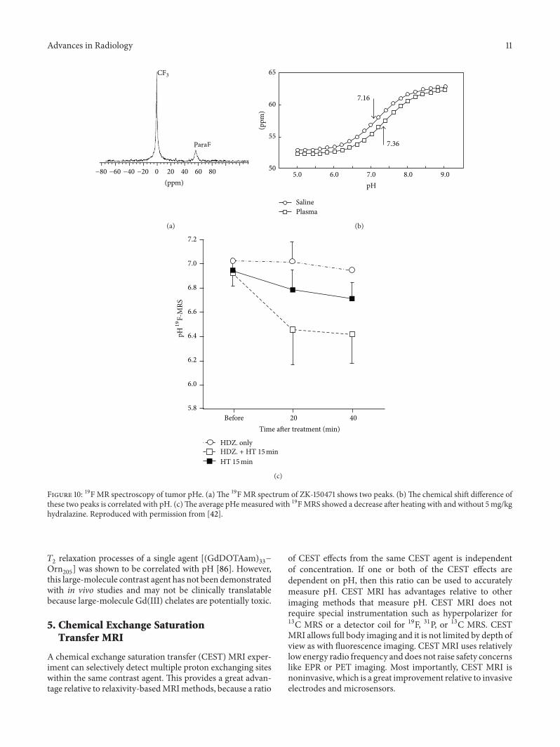
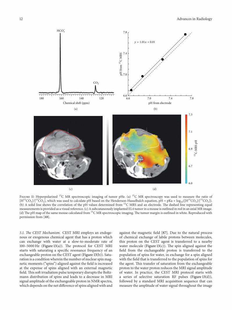
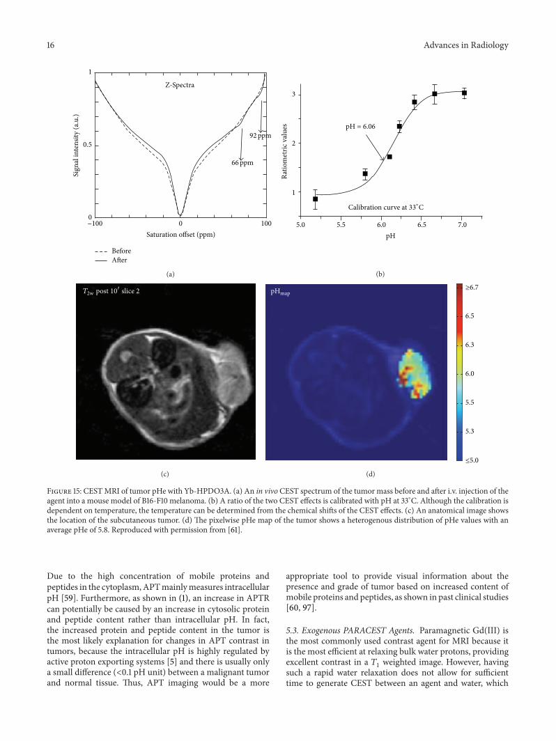
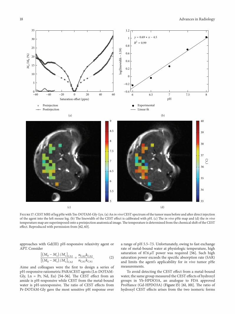
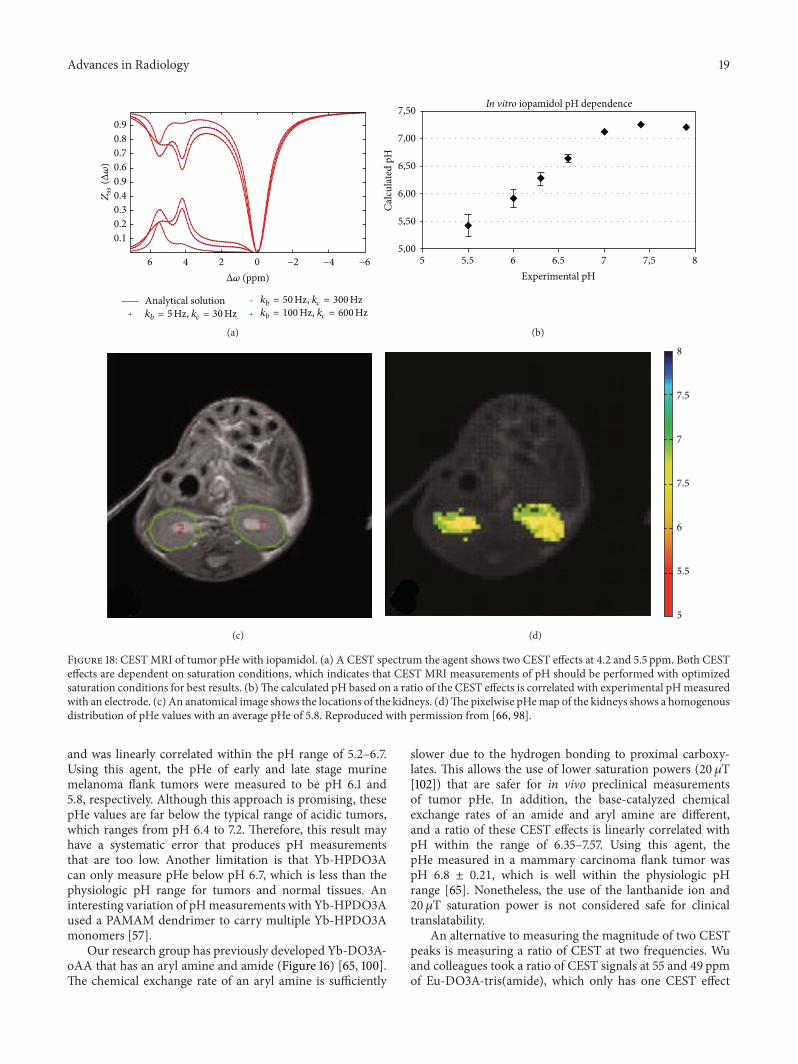
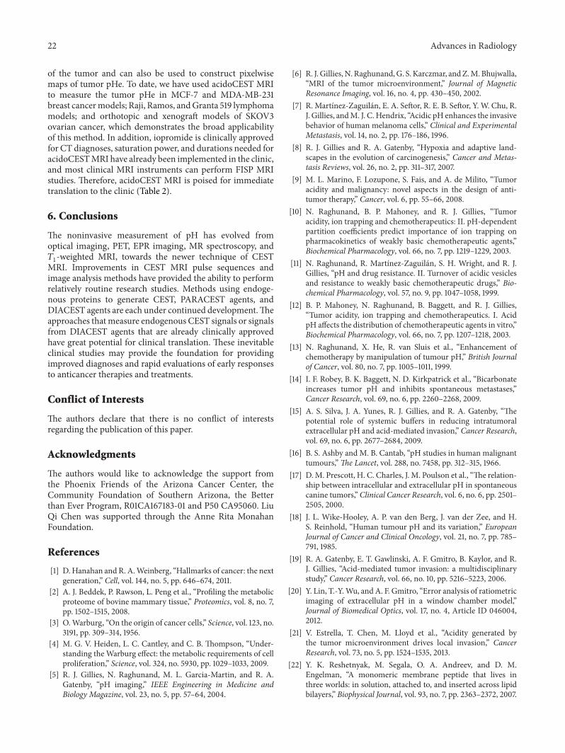