efficient production and enhanced tumor delivery of ... · efficient production and enhanced tumor...
TRANSCRIPT

lable at ScienceDirect
Biomaterials 105 (2016) 195e205
Contents lists avai
Biomaterials
journal homepage: www.elsevier .com/locate/biomater ia ls
Efficient production and enhanced tumor delivery of engineeredextracellular vesicles
Dionysios C. Watson a, Defne Bayik b, c, 1, Avinash Srivatsan d, 1, Cristina Bergamaschi e,Antonio Valentin a, Gang Niu d, Jenifer Bear e, Mitchell Monninger f, Mei Sun f,Aizea Morales-Kastresana g, Jennifer C. Jones g, Barbara K. Felber e, Xiaoyuan Chen d,Ihsan Gursel c, George N. Pavlakis a, *
a Human Retrovirus Section, Vaccine Branch, Center for Cancer Research, National Cancer Institute at Frederick, Frederick, MD 21702, United Statesb Cancer and Inflammation Program, Center for Cancer Research, National Cancer Institute at Frederick, Frederick, MD 21702, United Statesc Department of Molecular Biology and Genetics, Bilkent University, Ankara, 06800 Turkeyd Laboratory of Molecular Imaging and Nanomedicine, National Institute of Biomedical Imaging and Bioengineering, National Institutes of Health, Bethesda,MD 20892, United Statese Human Retrovirus Pathogenesis Section, Vaccine Branch, Center for Cancer Research, National Cancer Institute at Frederick, Frederick, MD 21702, UnitedStatesf Pathology Division, United States Army Medical Research Institute of Infectious Diseases, Frederick, MD 21702, United Statesg Vaccine Branch, Center for Cancer Research, National Cancer Institute, Bethesda, MD 20892, United States
a r t i c l e i n f o
Article history:Received 8 March 2016Received in revised form1 July 2016Accepted 5 July 2016Available online 6 July 2016
Keywords:ExosomesDrug deliveryBiodistributionScavenger receptorReticuloendothelial systemDextran sulfate
* Corresponding author.E-mail address: [email protected] (G.N. Pav
1 These authors contributed equally to the work.
http://dx.doi.org/10.1016/j.biomaterials.2016.07.0030142-9612/Published by Elsevier Ltd. This is an open
a b s t r a c t
Extracellular vesicles (EV), including exosomes and microvesicles, are nano-sized intercellular commu-nication vehicles that participate in a multitude of physiological processes. Due to their biologicalproperties, they are also promising candidates for the systemic delivery of therapeutic compounds, suchas cytokines, chemotherapeutic drugs, siRNAs and viral vectors. However, low EV production yield andrapid clearance of administered EV by liver macrophages limit their potential use as therapeutic vehicles.We have used a hollow-fiber bioreactor for the efficient production of bioactive EV bearing the heter-odimeric cytokine complex Interleukin-15:Interleukin-15 receptor alpha. Bioreactor culture yielded ~40-fold more EV per mL conditioned medium, as compared to conventional cell culture. Biophysical analysisand comparative proteomics suggested a more diverse population of EV in the bioreactor preparations,while serum protein contaminants were detectable only in conventional culture EV preparations. Wealso identified the Scavenger Receptor Class A family (SR-A) as a novel monocyte/macrophage uptakereceptor for EV. In vivo blockade of SR-A with dextran sulfate dramatically decreased EV liver clearance inmice, while enhancing tumor accumulation. These findings facilitate development of EV therapeuticmethods.
Published by Elsevier Ltd. This is an open access article under the CC BY-NC-ND license (http://creativecommons.org/licenses/by-nc-nd/4.0/).
1. Introduction
Extracellular vesicles (EV), including exosomes and micro-vesicles, are nano-sized membrane vesicles secreted by most celltypes. Given their intrinsic properties, e.g. immunomodulation [1,2]and their ability to distribute systemically, EV are being developedas biocompatible, targeted therapeutic particles [3,4]. Techniques
lakis).
access article under the CC BY-NC
to load bioactive cargo, such as protein [5,6], siRNA [7], viral vectors[8], and chemotherapeutics [9] are also currently investigated inseveral settings.
EV for therapeutic applications are typically purified from cellculture conditioned media of cell lines or primary cells. Purificationmethods are comprised of combinations of techniques, includingultracentrifugation, filtration, precipitation, and chromatography[10e12], each resulting in somewhat different mixtures of EVspecies and non-EV contaminants. Reported EV yields range from 1to 10 mg/mL culture supernatant, even from “high-yield” systems,such as the Integra CELLine bioreactor [7,13,14]. Thus, obtainingsufficient material for in vivo studies currently comprises a
-ND license (http://creativecommons.org/licenses/by-nc-nd/4.0/).

D.C. Watson et al. / Biomaterials 105 (2016) 195e205196
technical bottleneck for therapeutic EV development, irrespectiveof the purification method.
With regard to systemic delivery of EV, modifying surface pro-teins with targeting ligands has enabled increased delivery to theCNS (via the RGD targeting peptide) [3] and to EGFR-expressingtumors (via the GE11 peptide) [15]. However, systemically deliv-ered EV are rapidly cleared by the monocyte/macrophage orreticuloendothelial system (RES), resulting in minimal accumula-tion within the desired target sites [4,15e19], which is a majorhurdle for EV-based systemic therapeutic approaches. Depletion ofmonocytes/macrophages was shown to significantly prolong EVsystemic half-life in mice, suggesting uptake by these cells as animportant mechanism of EV clearance in vivo [17]. Clearance ofsynthetic nanoparticles by the RES was shown to be largely medi-ated by the Scavenger Receptor Class A family (SR-A), which rec-ognizes a variety of negatively charged ligands, includingphosphatidylserine [20], a phospholipid shown to be enriched onEV. Indeed, masking phosphatidylserine with annexin-V decreasedEV uptake in vitro [21].
Herein, we describe approaches to overcome obstacles of EVproduction and delivery, which will facilitate further developmentof this therapeutic platform. We compare EV production using anefficient hollow-fiber culture system to that of conventional culturemethods. We show that this method produces bioactive EVretaining surface proteins and can be used for the production ofbioactive, EV associated heterodimeric interleukin-15. We alsoidentify SR-A as a major receptor for the clearance of EV bymonocyte/macrophages, and assess the applicability of SR-Ablockade to achieve tumor delivery of administered EV in mice.
2. Materials and methods
2.1. Cells
HEK293 and all mouse cell lines (RAW264.7, 4T1, B16, LLC1,MC38 and EG.7) were obtained fromATCC. HEK293 cells expressinghigh levels of hetIL-15 were previously described [22,23]. NK92cells were kindly provided by Dr. Howard A. Young (Cancer andInflammation Program, National Cancer Institute, USA). Bloodsamples from healthy blood donors were collected in acid-citrate-dextrose tubes, under approved protocols for human subjects'research by the National Cancer Institute Investigational ReviewBoard. Peripheral bloodmononuclear cells (PBMC) were purified bygradient centrifugation over Histopaque-1077 (Sigma-Aldrich),according to the manufacturer's protocol.
4T1 cells were cultured in RPMI 1640 medium supplementedwith 10% fetal calf serum. RAW264.7 and E.G7 cells were cultured inRPMI 1640 medium, supplemented with 5% fetal calf serum. Theremaining cell lines were cultured in DMEM medium, supple-mented with 10% fetal calf serum.
Human PBMC were cultured overnight in RPMI medium sup-plemented with 10% fetal calf serum and 100 U/mL penicillin/streptomycin. The cells were used within 24 h of purification fromwhole blood.
2.2. Cell culture method for EV production
EV/protein aggregate depleted cell culture medium was ob-tained by ultrafiltration of complete medium through a 500 kDacommercial hollow fiber ultrafiltration module (mPES MidiKros500 kDa filter module, Spectrum Laboratories; Rancho Dominquez,CA), as previously described [14,24]. Specifically, a peristaltic pumpwas used to slowly circulate culture medium through the filtermodule, and filtrate was collected to be used as EV/protein aggre-gate depleted medium. The entire procedure was carried out using
sterile materials within a biosafety cabinet. Ultrafiltered superna-tant was filtered a second time through a 0.22 mm filter device toensure sterility.
To obtain conditioned medium from conventional cultures, 3million cells were seeded in 175 cm2 tissue culture flasks in DMEMsupplemented with 10% fetal calf serum and 100 U/mL penicillin/streptomycin. After overnight incubation, cell monolayer wasgently washed with PBS, and 15 mL of fresh EV/protein aggregate-depleted medium was added to each flask. 48 h later, conditionedmedium was harvested and pooled for immediate EV purification.After conditionedmediumwas removed, cells were collected in PBSand pelleted by centrifugation at 300 � g. Cell lysates were pre-pared by addition of N1 lysis buffer to cell pellet, incubation on icefor 1 h, and two rounds of sonication for 6 s.
2.3. Fibercell hollow-fiber bioreactor culture
The HEK293 cell clone stably expressing hetIL-15 (clone 19.7)was expanded in conventional culture flasks and used to seed amedium-sized, hollow-fiber culture cartridge, with a 20 kDa mo-lecular weight cut-off (Fibercell Systems; Frederick, MD). Cells wereadapted over two weeks to bioreactor culture conditions bygradually increasing the proportion of protein-free medium(DMEM þ 10% Fibercell Systems CDMHD protein-freesupplement þ 100 U/mL penicillin/streptomycin). Bioreactorconditioned medium (20 mL) was collected for each harvest threetimes per week. Harvests were cleared of cells by 300 � g centri-fugation, and supernatants stored at�80 �C for further purification.
2.4. EV purification
After removing large cell debris by centrifugation at 3000 � gfor 15 min, the supernatants were carefully moved to poly-carbonate tubes, and spun for 45 min at 20,000 � g in a type 45Tirotor (Beckman-Coulter; Brea, CA). Supernatants were then filteredthrough 0.22 mm Stericup device (EMD Millipore; Billerica, MA),moved to Snakeskin 10 kDa MWCO dialysis tubing (Thermo-Scientific; Grand Island, NY), and dialyzed overnight in >30 vol-umes of Tris-buffered saline (TBS). Dialyzed supernatants werecentrifuged for 2 h at 110,000 � g in a type 70.1Ti rotor (Beckman-Coulter) to pellet EV. Pellets were resuspended to the originalvolume in TBS, by passing through a 27G needle approximately 5times (until aggregates were no longer visible), and centrifugedagain at 110,000 � g to wash away contaminating soluble proteins.EV pellets were resuspended in 1/50 original volume of TBSfollowing the procedure described above. Finally, EV were addi-tionally cleared of residual aggregation by 3 min centrifugation at20,000 � g in a microfuge, and the supernatants containing the EVwere transferred to a clean Lobind protein tube (Eppendorf;Hauppauge, NY), and stored at �80 �C for downstream applica-tions. All purification steps were conducted at 4 �C.
For experiments directly comparing conventional culture tobioreactor EV, conditioned cell culture media were concentrated byultrafiltration (Centricon-70, 100 kDa MWCO; EMDMillipore) priorto 110,000 � g centrifugation. This allowed for a larger volume ofconventional flask-derived supernatants to be pooled prior to ul-tracentrifugation. Furthermore, initial EV pellet was not washed,but rather immediately resuspended in 100 mL TBS. These protocolmodifications were implemented to allow for sufficient EV yieldsfrom conventional flask supernatants for downstream comparisonanalyses.
2.5. Biophysical characterization and imaging
Nanoparticle tracking analysis (NTA) was performed on fresh
D.C. Watson et al. / Biomaterials 105 (2016) 195e205 197
and freeze-thawed EV samples in triplicate using a Nanosight LM10(Malvern Instruments; Malvern, United Kingdom) to estimate sizedistribution (hydrodynamic diameter), particle concentration, andeffects of freeze-thawing (Fig. S1). Samples were diluted to 1e2 mg/mL in PBS, and 3 videos of 30 s were acquired for each triplicate.Dynamic light scattering (DLS) was performed on a nanoparticleanalyzer SZ-100 (Horiba Scientific) at the same dilution.
For transmission electronmicroscopy (TEM) sample preparationand imaging, EV suspensions were fixed with 2% glutaraldehyde(Electron Microscopy Sciences, Cat#16020) and then adsorbed toformvar/carbon coated TEM copper grids (SPI, Cat#3420C-MB).Samples were then negative stained with 1% Uranyl Acetate(Electron Microscopy Sciences, Cat# 22400) for 10 s. The sampleswere evaluated on a JEOL 1011 transmission electron microscope at80 kV, and digital images were acquired using AMT camera system.
2.6. EV protein composition determination
Purified EV preparations were monitored for protein content bythe Bradford assay, using bovine gamma-globulin as a standard.Specifically, 5 mL of EV preparation was added to 250 mL QuickstartBradford reagent (Bio-Rad; Hercules, CA), and incubated for15 min at room temperature. Protein concentration was quantifiedby measuring absorbance at 595 nm on a microplate reader.
For Western blots, EV were lysed by adding 5� RIPA buffer andincubating on ice for 45 min. Antibodies used to probe the blotsincluded: aCD63 (System Biosciences; Mountain View, CA), aAlix(clone 3A9, LifeSpan Biosciences; Seattle, WA), aIL-15 (AF315, R&DSystems) and aIL-15Ra (AF247, R&D Systems). All blots were pro-bed overnight at 4 �C.
Detection of EV-associated IL-15 was performed by flowcytometry using the ExoFlow kit (System Biosciences) according tomanufacturer's protocol. Briefly, 9.1 mm streptavidin-coated mag-netic beads were loaded with biotinylated anti-CD63 exosomecapture antibody. Next, 100 mg of purified EV were incubatedovernight at 4 �C with antibody-loaded beads in a rotating micro-tube holder. EV-loaded magnetic beads were then washed using amagnetic tube stand, and divided for staining with individualfluorophore-conjugated antibodies: FITC-lectin (provided in kit)and PE-conjugated anti-IL-15 (clone 34559; R&D Systems, Minne-apolis, MN). Incubation with antibodies was for 2 h on ice, gentlyflicking the tube to resuspend beads every 30min. Finally, unboundantibody was washed from beads using a magnetic tube stand, andEV-loaded beads were acquired on a flow cytometer (LSR-II; BDBiosciences, Franklin Lakes, New Jersey).
2.7. Comparative proteomics study
5� RIPA buffer was added to EV samples (~20 mg), and incubatedfor 30 min on ice to lyse EV. Samples were then mixed with LDS gelloading buffer and NuPAGE sample reducing agent (Invitrogen),heated at 96 �C for 10 min, then, loaded on a 4e12% NuPAGE gel(Invitrogen). After two hours of running at a constant voltage(100 V), the gel was stained with SimplyBlue Safestain (Invitrogen).Each gel lane was excised into 12 gel slices. The gel slices were in-gel digested individually with Trypsin (Promega) at 37 �C over-night. Each digested peptide sample was desalted by C18 ZipTip(Millipore), lyophilized and re-suspended in 0.1% formic acid forLC-MS analysis. Each sample (6 mL) was loaded on an Easy nLC IInano-capillary HPLC system (Thermo Scientific) with a C18 NanoTrap Column, (Acclaim PepMap100 C18, 2 cm, nanoViper, ThermoScientific) and an analytical column (Acclaim PepMap RSLC C18,15 cm, nanoViper, Thermo Scientific) connected with a stainlesssteel emitter, coupled online with a Q Exactive hybrid OrbiTrapmass spectrometer (Thermo Scientific) for RPLC-MS/MS analysis.
Peptides were eluted using a linear gradient of 2% mobile phase B(acetonitrile with 0.1% formic acid) to 42% mobile phase B within70 min at a constant flow rate of 200 nL/min. The twelve mostintense molecular ions in the MS scan were sequentially selectedfor high-energy collisional dissociation (HCD) using a normalizedcollision energy of 30%. The mass spectrawere acquired at the massrange of m/z 300e2000. The Easy Nano Spray ion source (ThermoScientific) capillary voltage and temperature were set at 1.7 kV and275 �C, respectively. The dynamic exclusion function on the massspectrometer was enabled during the MS2 data acquisition. The MSdata were searched against the UniProt Homo sapiens databasedownloaded from the European Bioinformatics Institute website(http://www.ebi.ac.uk/integr8, January, 2016) utilizing ProteomeDiscoverer 1.4 (Thermo Scientific). Up to two missed trypticcleavage sites was allowed during the database search. Oxidation(þ15.9949 Da) of methionyl residue was included as dynamicmodifications. The data was searched with a precursor ion toler-ance of 20 ppm and a fragment ion tolerance of 50 ppm. Thepeptide identifications are filtered through protein percolator withthe cutoff of a false peptide discover rate (FDR) less than 1% for allpeptide identified. “Strict Maximum Parsimony Principle” wasapplied during the data compiling.
For comparative analysis of bioreactor vs. flask EV, peak area ofpeptide mappings (grouped by corresponding gene) were consid-ered. Data were normalized to total peak area, using a normaliza-tion factor of 1.15.
2.8. Bioactivity assay
NK92 cells used for the IL-15 bioactivity assay were cultured inRPMI 1640medium supplemented with 10% fetal calf serum,100 U/mL penicillin/streptomycin, 200 U/mL recombinant IL-2 (NationalCancer Institute), and 10 ng/mL hetIL-15 (Admune Therapeutics,Danvers, MA). Cell density was adjusted three times a week to aconcentration of 3e4 � 105 cells/ml.
Purified EV were mixed with an equal volume of lysis buffer (1%Triton-X 100/200 mM Tris-HCl, pH7.4) and lysed by five freeze/thaw cycles. The amount of EV-associated IL-15 was assessed byELISA (Human IL-15 Quantikine ELISA kit; R&D Systems, Minne-apolis, MN) according to the manufacturer's instructions.
The bioactivity of EV-associated hetIL-15 was assessed on thehuman NK-92 cell line, which responds to IL-15 treatment byproliferating in a dose-dependent manner. NK-92 cells werecultured overnight at a concentration of 4 � 105 cells/mL incytokine-free media prior to assay use. 50 mL of cytokine-starvedNK-92 cells (4 � 105 cells/mL) were seeded in each assay well of96-well plates. hetIL-15 purified protein standard (Admune Ther-apeutics) and purified EV samples were used to prepare solutionscontaining 0.05e2 ng/mL IL-15 (measured as single chain content)in complete cell culture medium; 50 mL of each IL-15 protein or EVsolution was then added to the corresponding cell culture wells.Equal amounts of EV lacking IL-15 were used as negative control.After 72 h incubation, 25 mL MTT labeling reagent (Roche, Indian-apolis, IN) was added to each well, and plates incubated for 5 h at37 �C. Next, 100 mL solubilization buffer (10% SDS in 0.01 M HCl;Roche) was added to each well, and plates incubated for 24 h at37 �C. Optical density of samples was measured at 570/690 nm,using a SpectraMax Plus 384 microplate reader (Molecular Devices,Sunnyvale, CA).
2.9. EV labeling for in vitro and in vivo tracking
Two different labeling dyes were used to monitor EV uptake: agreen RNA dye (Syto RNASelect Green; Invitrogen, Grand Island,NY), and a near-infrared lipid dye (DiOC18(7) or DiR; Invitrogen).

D.C. Watson et al. / Biomaterials 105 (2016) 195e205198
For Syto RNASelect Green staining, 100 mg EV in 100 mL TBS wereincubated with the dye at a final concentration of 10 mM for30 min at 37 �C. For DiR staining, 62.5 mg EV in 100 mL TBS wereincubated with the dye at a final concentration of 100 ng permicrogram of EV for 1 h at 37 �C. After incubation, unincorporateddye was removed by gel filtration using PBS-hydrated ExosomeSpin Columns (3 kDa MWCO, Invitrogen) (See SupplementaryFig. S7).
We also checked the stability of DiR staining of EV over 24 h(Fig. S8). DiR-stained EV were prepared as above, and incubated for24 h at 37 �C. We then repeated the gel filtration procedure toremove any dye that may have become unincorporated from EVduring the 24 h incubation. For comparison, we prepared freshlystained EV as above, and immediately performed a second gelfiltration to account for EV losses in the Exosome Spin Columns.Stained EV from these samples were diluted 1:2, and 60 mL loadedin each well of a black-walled 96-well plate. Fluorescence intensityanalysis of samples was performed in triplicate using the OdysseyClassic infrared imaging system (Li-Cor, Lincoln, NE) at the 800 nmchannel.
2.10. In vitro SR-A blockade and EV uptake assessment
For EV uptake blocking experiments, mouse cells were firstplated at a density of 250,000 cells per well in a 12-well plate andcultured overnight. E.G7 is a suspension cell line and 250,000 cellswere used for each experimental sample. Cells were pre-treated for30 min with fresh RPMI supplemented with 5% fetal calf serummedium, containing 100 mg/mL chondroitin sulfate, 100 mg/mLdextran sulfate or 10 mM BLT-1 (all EV-uptake blocking reagentsobtained from Sigma-Aldrich, St. Louis, MO). Cells were washedonce and incubated with fluorescently labeled EV for 2 h. The cellswere harvested, washed, and resuspended in PBSþ2% bovineserum albumin for the analysis of EV uptake by flow cytometry. TheEV concentrations used in these experiments were adjusted toobtain 2e4 fold increase in the fluorescent signal above the auto-fluorescence from untreated cells (RAW 3.2 mg/mL; 4T1, MC38 andEG7: 1.6 mg/mL; LLC1 and B16: 0.8 mg/ml).
PBMC were cultured at 2 � 106 cells/mL and pretreated for30 min with 500 mg/mL dextran sulfate, 500 mg/mL chondroitinsulfate, or left untreated. After washing, the cells were resuspendedat a density of 4 � 106 cells/mL and incubated for 2 h with SytoRNASelect Green-labeled EV at a final concentration of 800 ng EVper mL, in a final volume of 0.5 mL. Finally, the cells werewashed inPBSþ0.2% ABþ human serum and stained with fluorophore-conjugated antibodies before flow cytometric analysis.
The following antibodies were used: aCD3-APC-Cy7 (clone SK7;BD Biosciences), aCD4-V500 (clone RPA-T4; BD Biosciences), aCD8-Alexa Fluor 405 (clone 3B5, Invitrogen), aCD14-PE (clone M5E2; BDBiosciences), aCD14-BV421 (clone HCD14; Biolegend; San Diego,CA), aCD19-Alexa Fluor 700 (clone HIB19; BD Biosciences), aCD56-APC (clone B159; BD Biosciences), and aCD204-PE (clone REA460;Miltenyi Biotec; San Diego, CA).
2.11. In vivo EV biodistribution in tumor-free and tumor-bearingmice after SR-A blockade
All animal experiments were conducted in compliance with theguidelines for the care and use of research animals established bythe Animal Studies Committee of the National Institutes of Health.FVB or Balb/c mice were treated with the SR-A inhibitor dextransulfate or chondroitin sulfate (negative control) at a dose of 30 mg/kg delivered in 100 mL PBS by tail vein injection. Control micereceived 100 mL of PBS intraperitoneally. Two hours later, the micereceived 100 mL of PBS containing 15 mg of DiR stained EV via the tail
vein. Imaging of live mice was performed at various time points,between 1 and 24 h post injection. Some mice were euthanized atdifferent time points and their organs imaged ex vivo. Imaging wasdone on a Maestro™ 2 imaging system (Perkin Elmer).
For tumor inoculation studies, 1 � 106 4T1 cells in 100 mL ofserum-free medium were implanted subcutaneously on the rightfront flank of female BALB/c mice. Once the tumor size reached avolume of 90e100 mm3, the animals were used for in vivo imagingexperiments.
2.12. Statistical analyses
EV preparation purity and monocyte uptake of EV in vitro wereanalyzed using t-tests. EV preparation yield, along with liver,plasma, and tumor uptake (imaged ex vivo) were analyzed by 1-way ANOVA. Time courses of in vivo EV uptake by tumor andliver were analyzed with 2-way ANOVA. All statistical analyseswere performed in Prism 6 (GraphPad Software, La Jolla, CA).
3. Results
3.1. High-yield EV production from hollow fiber bioreactor
In an effort to increase production yield, we used a lab-scalehollow-fiber bioreactor with protein-free culture medium toobtain conditioned cell culture supernatants as a source of EV.Hollow-fiber culture systems can sustain large numbers (>109) ofcells, and produce highly concentrated cell culture supernatantsfrom a variety of cells, including primary human cells [25,26]. Wehypothesized that these characteristics would facilitate high-yieldproduction and purification of EV.
HEK293 cells were adapted for growth in a hollow-fiber biore-actor using serum-free medium (Fig. 1A). EV from bioreactor orconventional tissue culture flask supernatants were purified bydifferential centrifugation, and the protein concentration of thepurified materials was determined using the Bradford assay. Usinga similar protocol of EV purification, bioreactor-harvested condi-tioned media yielded approximately 5-fold more EV compared toconventional flasks (Fig. 1B). Further optimization of the EV puri-fication method, by omitting the supernatant concentration step,increased the EV yield about two-fold, resulting in a total 10�improved EV yield from cell culture conditioned media (Fig. 1B).NTA was used to estimate the particle to protein amount ratio, anindicator of EV purity [7,27]. We found a ratio of 1.09 � 109
(±2 � 108 particles)/mg of protein in EV preparations from thebioreactor, which is ~4-fold greater than the purity of preparationsfrom our conventional cell culture (Fig. 1C), higher than has pre-viously been reported for EV obtained from HEK293 cells [7]. EVmarkers CD63 and Alix were also enriched in preparations frombioreactor harvests, supporting increased purity (Fig. 1D). DLS (seeSupplementary Fig. S2) and NTA (Fig. 2A), showed the majority ofpurified EV to be between 40 and 200 nm in diameter. TEM showedwell-defined particles of comparable size (Fig. 2b). Together thesedata suggest that the use of hollow-fiber bioreactor, serum-freemedia and an optimized purification protocol represent a supe-rior method to achieve high yield of purified EV (yields were>3 mg/week from a single lab-scale culture cartridge).
3.2. Comparison of bioreactor to flask-derived EV characteristics
We observed that the relative enrichment of CD63, as detectedby Western blot (Fig. 1D), could not be fully accounted for by theincreased particle purity of the bioreactor EV preparations. Wetherefore conducted an in-depth comparison of the particles pu-rified from either source to assess whether the bioreactor

Fig. 1. Hollow-fiber bioreactor is a high-yield source of EV. (A) Schematic representation of EV production in hollow-fiber bioreactor. Fresh medium is pumped through the cellculture cartridge cylinder. Medium circulates within the hollow fibers of the culture cartridge, and (*) nutrient/waste exchange occurs through the hollow-fiber wall pores (20 kDaMWCO). Larger molecular weight cellular products (including EV) accumulate within the extracellular space of the cell culture cartridge cylinder. The extracellular space mediumcontaining large amounts of EV is harvested every 1e3 days. (B) Yield of EV preparations from a hollow fiber bioreactor is ~10-fold more than conventional flask conditioned media.EV were purified from bioreactor conditioned media either by an optimized protocol (filtration, dialysis, and differential centrifugation) or by a protocol for direct comparison toconventional flask culture (ultrafiltration to concentrate conditioned media, filtration, and differential centrifugation). Protein yield, calculated as mg EV-associated protein permilliliter original conditioned medium (mean ± SEM), was quantified in independent preparations, and compared by ANOVA (*p < 0.05; ***p < 0.001). (C) Particle purity of EVpreparations from a hollow fiber bioreactor is ~4-fold higher than conventional flask conditioned media. Particle purity indicates the level of EV-free protein contamination of EVpreparations, and was assessed as the number of particles per mg protein, as measured by nanoparticle tracking analysis. (D) Enrichment of EV-associated markers in western blot ofbioreactor EV preparations vs. conventional flask culture suggests higher purity. Each lane was loaded with 20 mg total protein, and band intensity quantification was performed bydensitometry. In bioreactor EV preparations, we also observed differentially processed forms of associated proteins. In the case of CD63, lower molecular weight bands may reflectdifferential glycosylation pattern, as this protein has several glycosylation modifications. For Alix, there are several known phosphorylation sites, whose differential status couldexplain the second band. Detection of differentially processed proteins may be the result of increased amount of the EV markers and/or altered biocomposition of EV produced bycells grown under different bioreactor conditions.
Fig. 2. Biophysical characterization of purified bioreactor EV. (A) Nanoparticle trackinganalysis (NTA) was used to characterize the size of purified EV, as shown in repre-sentative size frequency distribution plot. Each sample for was run in triplicate at1e2 mg/mL concentration. Black line represents mean values, and red shaded arearepresents SEM. NTA was able to distinguish three main subpopulations based on sizecorresponding to peaks of approximately 120, 190, and 280 nm in diameter. (B)Transmission electron micrograph of bioreactor EV.
D.C. Watson et al. / Biomaterials 105 (2016) 195e205 199
methodology changes the profile of purified EV. A side-by-sidecomparison of NTA plots from several preparations of EV fromeither method revealed that the size distribution of culture-flaskderived EV was more limited (Fig. S3), which could be a result ofa different mixture of EV subpopulations. Serum-free cultureconditions have previously been reported to affect EV composi-tion [28], so we further compared the composition of bioreactorvs. flask EV by comparative proteomics, using mass spectrometry(Supplementary File 1 and Fig. S4). Many proteins were sharedbetween the two samples, including EV markers such as CD63,Alix, and TSG101. However, serum proteins were over-representedin the flask EV.
3.3. Purified bioreactor EV are bioactive in vitro
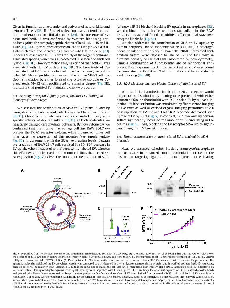
We used a HEK293 cell clone that expresses the humanmembrane-associated heterodimeric interleukin-15 complex, IL-15:IL-15Ra (hetIL-15) [22,23], to examine whether optimally puri-fied EV maintain functional hetIL-15 on their surface (Fig. 3A).
D.C. Watson et al. / Biomaterials 105 (2016) 195e205200
Given its function as an expander and activator of natural killer andcytotoxic T-cells [23], IL-15 is being developed as a potential cancerimmunotherapeutic in clinical studies [29]. The presence of EV-associated hetIL-15 was confirmed by Western blot using anti-bodies against the two polypeptide chains of hetIL-15, IL-15 and IL-15Ra (Fig. 3B). Upon surface expression, the full length ~59 kDa IL-15Ra is cleaved and secreted as a soluble ~42 kDa molecule [23].Indeed, EV-associated IL-15Rawas mostly of the larger membrane-associated species, which was also detected in association with celllysates (Fig. 3C). Flow cytometric analysis verified that hetIL-15 wasassociated with the EV surface (Fig. 3D). The bioactivity of EV-associated hetIL-15 was confirmed in vitro by using an estab-lished MTT-based proliferation assay on the human NK-92 cell line.Upon stimulation by either form of the cytokine (soluble or EV-associated), NK-92 cells proliferated to a similar degree (Fig. 3E),indicating that purified EV maintain bioactive properties.
3.4. Scavenger receptor A family (SR-A) mediates EV binding tomonocytes/macrophages
We assessed the contribution of SR-A to EV uptake in vitro byusing dextran sulfate, a molecule known to block this receptor[30,31]. Chondroitin sulfate was used as a control for any non-specific activity of dextran sulfate [30,31], as both molecules arenegatively charged carbohydrate polymers. By flow cytometry, weconfirmed that the murine macrophage cell line RAW 264.7 ex-presses the SR-A1 receptor isoform, while a panel of tumor celllines lacks the expression of this receptor (see SupplementaryFig. S5). In agreement with the SR-A1 expression levels, dextranpre-treatment of RAW 264.7 cells resulted in a 30e50% decrease inEV uptake when incubated with fluorescently-labeled EV, whereasthis effect was not observed in the tumor cell lines that lacked SR-A1 expression (Fig. 4A). Given the contemporaneous report of BLT-1
Fig. 3. EV purified from hollow fiber bioreactor and containing surface hetIL-15 retain IL-15 bthe presence of IL-15 cytokine in cell lysate and in bioreactor-derived EV from a HEK293 cellcell lysate is from parental HEK293 cell line. (C) EV-associated IL-15Ra is primarily membrapparent molecular weight of the EV-associated protein was compared to that detected insecreted protein). The majority of EV-associated IL-15Ra is the same size as that of the cell-vesicular surface. Flow cytometry histograms show signal intensity from EV probed with PEand probed with fluorophore-conjugated antibody to detect presence of surface cytokineHEK293 cell clone stably overexpressing the cytokine. (E) EV-associated IL-15 is bioactive in vas quantified by mean MTT assay O.D in 6 wells per sample (mean ± SEM). Magenta line repHEK293 cell clone overexpressing hetIL-15. Black line represents triplicate bioactivity asseHEK293 cell EV resulted in MTT O.D. <0.25.
(a known SR-B1 blocker) blocking EV uptake in macrophages [32],we combined this molecule with dextran sulfate in the RAW264.7 cell assay, and found an additive effect of dual scavengerreceptor blockade (Fig. S6).
We also addressed the contribution of SR-A on EV uptake byhuman peripheral blood mononuclear cells (PBMC), a heteroge-neous population of primary human cells. PBMC, pretreated withdextran sulfate, were exposed to labeled EV, and EV uptake indifferent primary cell subsets was monitored by flow cytometry,using a combination of fluorescently labeled monoclonal anti-bodies. These experiments demonstrated that most EV were boundtomonocytes and that 30e60% of this uptake could be abrogated bySR-A blocking (Fig. 4B).
3.5. SR-A blockade changes biodistribution of administered EV
We tested the hypothesis that blocking SR-A receptors wouldimpact EV biodistribution by treating mice pretreated with eitherdextran sulfate or chondroitin with DiR-labeled EV by tail vein in-jection. EV biodistribution was monitored by fluorescence imagingof live mice as well as excised organs. Imaging performed at 2 hpost-injection of EV showed that SR-A blockade decreased liveruptake of EV by ~50% (Fig. 5). In contrast, SR-A blockade by dextransulfate significantly increased the amount of EV circulating in theplasma (Fig. 5). Thus, blocking the EV receptor SR-A led to signifi-cant changes in EV biodistribution.
3.6. Tumor accumulation of administered EV is enabled by SR-Ablockade
Next, we assessed whether blocking monocyte/macrophageuptake results in enhanced tumor accumulation of EV, in theabsence of targeting ligands. Immunocompetent mice bearing
ioactivity. (A) Schematic representation of EV bearing hetIL-15. (B) Western blot showsclone that stably overexpresses the IL-15 heterodimer complex (IL-15:IL-15Ra). Controlane anchored. Western blot of IL-15Ra associated with bioreactor EV preparation. Thethe cell lysate (transmembrane protein) and in purified secreted hetIL-15 (truncated,associated (membrane-anchored) cytokine. (D) EV-associated hetIL-15 is displayed on-conjugated aIL-15 antibody. EV were first captured on aCD63 antibody-coated beads. Control EV were derived from parental HEK293 cells and hetIL-15 EV came from aitro. Bioactivity assessed as proliferation of the NK92 cell line following 72 h incubation,resents bioactivity of 3 independent EV preparations from bioreactor supernatants of assment of protein standard. Incubation of cells with equal protein amount of control

Fig. 4. EV uptake in monocytes/macrophages is largely mediated by the Scavengerreceptor A family (SR-A). Cells were pretreated with dextran sulfate (SR-A blocker) orchondroitin sulfate (dextran control) for 30 min. After washing away inhibitor, stainedEV were added for 2 h. Flow cytometry was used to quantify EV uptake by fluorescentintensity of cells. Boxes represent range of values (min to max), and line in box denotesgroup mean. Uptake compared between groups by multiple t-tests, with Holm-Sidakcorrection for multiple comparisons (Cumulative alpha-error <0.05). One (*), two(**), and three (***) asterisks denote p < 0.05, p < 0.01, and p < 0.001, respectively. (A)Blocking SR-A with dextran sulfate decreased EV uptake by 30e50% only in RAW cells,and not in cell lines that lack SR-A expression (in three independent experiments).Viability of cells assessed by Live/Dead fluorescent stain (Invitrogen) showed no effectof treatment conditions in the tested cell lines (data not shown). (B) Monocytes uptakethe majority of EV among primary human PBMC cultured in vitro, through SR-A (n ¼ 4).EV uptake was quantified by mean fluorescent intensity above background, andnormalized to EV uptake by untreated monocytes of each donor sample.
D.C. Watson et al. / Biomaterials 105 (2016) 195e205 201
subcutaneous 4T1 breast cancer cell tumors were pretreated withthe SR-A blocker, followed by systemic delivery of labeled EV. Im-aging of live mice over a period of 24 h showed that significantintratumoral EV accumulation could only be observed in animalspretreated with dextran sulfate (Figs. 6 and 7), even though SR-Ablockade of the liver diminished over 24 h (Fig. 6C, right panel).Ex vivomeasurements also showed that EV tumor uptake increasedby ~3-fold over the 24-h period (Fig. 7).
4. Discussion
In this study, we developed methods for the purification ofbioactive EV, carrying membrane-embedded heterodimeric IL-15.These results indicate the presence of a third form of hetero-dimeric IL-15 in addition to the soluble and the cell membrane-embedded forms [22,23,33,34]. Presence of functional hetIL-15 onEV may have significant physiologic roles and implications. It waspreviously shown that IL-15 Receptor alpha (the stabilizingcomponent of hetIL-15) is expressed on EV from cultured humandendritic cells, which can directly activate NK cells only in the
presence of exogenous recombinant IL-15 (active cytokinecomponent of hetIL-15) [35]. Contrarily, EV purified fromHEK293 cells expressing hetIL-15 maintain IL-15 bioactivitywithout addition of exogenous cytokine, which could be a signifi-cant benefit when using EV as therapeutics. Furthermore, it re-mains to be examined whether primary human dendritic cells alsosecrete functionally significant EV bearing complete hetIL-15 underconditions not tested previously (e.g. stimulation by certain toll-like receptor ligands) [36].
We developed a methodology for the mass production of highlypurified, bioactive EV using a lab-scale hollow fiber bioreactor,which may facilitate further in vivo studies. This method presentsmany advantages over conventional culture that likely contribute tothe higher EV production yield and purity that we observed. First,lab-scale hollow fiber bioreactors enable the sustained mainte-nance of large numbers of cells (estimated by the manufacturer tobe in the order of 109 cells) within a standard incubator, withminimal maintenance. Specifically, a 2 L bottle of medium is simplyreplaced every 2 days. More importantly, all of the cultured cellssecrete EV into the relatively small volume of the culture cartridgeextracapillary space (~60 mL) resulting in EV-rich conditionedmedium that can be used directly for EV purification. Our findingssuggest that this method yields 40-fold more EV particles pervolume of conditioned medium vs. conventional cell culture. Thus,to match a single daily harvest of the hollow-fiber bioreactor(20 mL), an estimated 53 large (175 cm2) flasks producing 800 mLconditioned medium would be required. Moreover, cells grown inhollow-fiber bioreactors readily adapt to sustained growth inprotein-free medium, which also facilitates EV purificationwithoutserum contaminants, as highlighted by our comparative proteomicsstudy. On the other hand, bioreactor EV preparations may have amore diverse population of EV, as suggested by the increased sizerange. Thus, further purification of preparations (e.g. by densitygradient) may be required for some applications.
Our findings showing the greatly enhanced yield of EV purifiedfrom hollow-fiber cultures may also have significant implicationsfor the development of clinical grade EV therapeutics, given thathollow-fiber methods have already been used to expand primaryhuman cells under cGMP conditions [25,26].
Moreover, we show for the first time that blocking SR-A, a novelreceptor for EV, enables accumulation of even untargeted EV inseveral tissues, including tumors (Fig. 8). Our in vitro findingsshowed that monocytes/macrophages are uniquely affected by SR-A blockade with respect to EV uptake. Interestingly, in the mixedPBMC culture, the blockade of monocyte EV uptake was accom-panied by an increase in uptake by T-cells, perhaps a result of EVbinding to a different receptor on the latter. In vivo, increased cir-culation and accumulationwithin tumors coincidedwith decreasedliver uptake, confirming previous findings that liver macrophagesrapidly clear administered EV in the absence of scavenger receptorblockade [17]. When chondroitin sulfate or dextran sulfate wereadministered to mice prior to labeled EV injection, we observed anincrease in splenic uptake of EV compared to mice receiving EValone (Fig. 5). A possible explanation for this could be the traffickingof circulating monocytes to the spleen after uptake of eithernegatively charged polymer, as has been described previously fornegatively charged poly(lactic-co-glycolic acid) (PLGA) formula-tions [37]. At the highest dose of dextran, even though monocytesare still possibly sequestered in the spleen, their EV uptake capacitywould be largely inhibited, resulting in the lower splenic EV uptakeof this group. In any case, SR-A blockade by dextran sulfateconsistently decreased liver uptake of EV, and significantlyincreased their ability to accumulate within tumors.
Considering the massive EV clearance by the RES under physi-ological conditions, our results are important for EV therapeutic

Fig. 5. SR-A blockade decreases liver clearance of EV. FVB mice were pre-treated with 0.6 mg dextran sulfate, chondroitin sulfate, or PBS. 15 mg DiR-stained EV were then injectedintravenously and mice were imaged 2 h later. (A) Live animal imaging showing dramatically decreased liver uptake of EV with SR-A blocker pre-treatment. (B) Organ bio-distribution of administered EV changes after SR-A blockade. A representative image of excised organs is shown from each group. Plasma is shown from three different mice of eachgroup. (C) Quantitation of EV accumulation in excised organs showed decrease of liver uptake, and increase in circulating EV at 2 h in the mice receiving dextran sulfate. Barsrepresent group mean þ SEM (n ¼ 3). Uptake was compared between groups using ANOVA, with Turkey correction for multiple comparisons. One (*), two (**), and three (***)asterisks denote p < 0.05, p < 0.01, and p < 0.001, respectively.

Fig. 6. Kinetics of EV uptake by liver and tumor following single injection of SR-A blocker in subcutaneous 4T1 tumor-bearing Balb/c mice (tumor location indicated by white circle).Mice were pretreated with either PBS, 0.6 mg dextran or chondroitin, injected IV with 15 mg stained EV, and imaged live at the indicated time points. (A) Serial live animal imagingshowing early inhibition of EV uptake by the liver, and gradual accumulation of EV in the tumor only in mice pre-treated with the SR-A blocker dextran sulfate. (B) Time course of EVuptake (mean ± SEM) in tumor and liver of 4T1 bearing Balb/c mice (n ¼ 4 per group), analyzed by 2-way ANOVA. Tumor accumulation increases significantly with time in SR-Ablocked mice (left panel), even though liver uptake blockade was transient, following a single injection of dextran sulfate (right panel).
D.C. Watson et al. / Biomaterials 105 (2016) 195e205 203
applications. Our findings make the case for developing potent SR-A blockers with favorable toxicity profiles. Combining scavengerreceptor blockade with EV targeting using relevant ligands mayfurther improve the targeted delivery of therapeutic RNA, viralvectors or any other molecules. Given that small molecules andpeptides with the potential to block SR-A and other scavenger re-ceptors are under development and some are in clinical trials[38e40], blocking RES uptake in therapeutic settings comprises apromising approach to enable a broad spectrum of effective EV-based therapeutics.
Author contributions
D.C.W., D.B., A.S., C.B., A.V., J.B., M.M., M.S., A.M.K., and J.C.J.conducted experiments. G.N., B.K.F., X.C., I.G., and G.N.P. supervisedresearch. D.C.W., D.B., A.S., and G.N.P. wrote and edited themanuscript.
Conflict of interest
The authors declare no competing financial interest.

Fig. 7. Significant EV uptake detected at 24 h in excised 4T1 tumors only from mice pre-treated with the SR-A blocker dextran sulfate. Mice were pretreated with either PBS, 0.6 mgdextran or chondroitin, and injected IV with 15 mg stained EV. (A) Fluorescence imaging of excised tumors showing increased EV uptake in dextran-pretreated mice. (B) Quantitationof EV accumulation in excised tumors of SR-A blocked mice, after 24 h. Bars display group mean ± SEM. Uptake compared between groups by ANOVA, with Sidak correction formultiple comparisons. Three asterisks (***) denote p < 0.001.
Fig. 8. Schematic depiction of the application of SR-A blockade for tumor delivery of EV carrying therapeutic molecules.
D.C. Watson et al. / Biomaterials 105 (2016) 195e205204
Acknowledgments
We thank William C. Kopp and Yanyu Wang for the IL-15bioactivity testing; Ming Zhou for mass spectrometry; John Cad-well for discussions and for providing Fibercell hollow-fiber ma-terials; Joseph Meyer for artwork; and Terry Jones foradministrative support. Research supported by the IntramuralResearch Program of the National Institutes of Health, NationalCancer Institute, Center for Cancer Research. Research also sup-ported by Admune/Novartis through a collaborative agreementwith the National Cancer Institute/NIH, USA. Opinions,
interpretations, conclusions, and recommendations are those of theauthors and are not necessarily endorsed by the U.S. Army.
Appendix A. Supplementary data
Supplementary data related to this article can be found at http://dx.doi.org/10.1016/j.biomaterials.2016.07.003.
References
[1] L. Zitvogel, A. Regnault, A. Lozier, J. Wolfers, C. Flament, D. Tenza, P. Ricciardi-
D.C. Watson et al. / Biomaterials 105 (2016) 195e205 205
Castagnoli, G. Raposo, S. Amigorena, Eradication of established murine tumorsusing a novel cell-free vaccine: dendritic cell-derived exosomes, Nat. Med. 4(5) (1998) 594e600.
[2] S.H. Kim, N.R. Bianco, W.J. Shufesky, A.E. Morelli, P.D. Robbins, MHC class IIþexosomes in plasma suppress inflammation in an antigen-specific and Fasligand/Fas-dependent manner, J. Immunol. 179 (4) (2007) 2235e2241.
[3] L. Alvarez-Erviti, Y. Seow, H. Yin, C. Betts, S. Lakhal, M.J. Wood, Delivery ofsiRNA to the mouse brain by systemic injection of targeted exosomes, Nat.Biotechnol. 29 (4) (2011) 341e345.
[4] O.P. Wiklander, J.Z. Nordin, A. O'Loughlin, Y. Gustafsson, G. Corso, I. Mager,P. Vader, Y. Lee, H. Sork, Y. Seow, N. Heldring, L. Alvarez-Erviti, C.E. Smith, K. LeBlanc, P. Macchiarini, P. Jungebluth, M.J. Wood, S.E. Andaloussi, Extracellularvesicle in vivo biodistribution is determined by cell source, route of admin-istration and targeting, J. Extracell. Vesicles 4 (2015) 26316.
[5] M.J. Haney, N.L. Klyachko, Y. Zhao, R. Gupta, E.G. Plotnikova, Z. He, T. Patel,A. Piroyan, M. Sokolsky, A.V. Kabanov, E.V. Batrakova, Exosomes as drug de-livery vehicles for Parkinson's disease therapy, J. Control Release 207 (2015)18e30.
[6] R.B. Rountree, S.J. Mandl, J.M. Nachtwey, K. Dalpozzo, L. Do, J.R. Lombardo,P.L. Schoonmaker, K. Brinkmann, U. Dirmeier, R. Laus, A. Delcayre, Exosometargeting of tumor antigens expressed by cancer vaccines can improve antigenimmunogenicity and therapeutic efficacy, Cancer Res. 71 (15) (2011)5235e5244.
[7] S. El-Andaloussi, Y. Lee, S. Lakhal-Littleton, J. Li, Y. Seow, C. Gardiner,L. Alvarez-Erviti, I.L. Sargent, M.J. Wood, Exosome-mediated delivery of siRNAin vitro and in vivo, Nat. Protoc. 7 (12) (2012) 2112e2126.
[8] B. Gyorgy, Z. Fitzpatrick, M.H. Crommentuijn, D. Mu, C.A. Maguire, Naturallyenveloped AAV vectors for shielding neutralizing antibodies and robust genedelivery in vivo, Biomaterials 35 (26) (2014) 7598e7609.
[9] G. Toffoli, M. Hadla, G. Corona, I. Caligiuri, S. Palazzolo, S. Semeraro, A. Gamini,V. Canzonieri, F. Rizzolio, Exosomal doxorubicin reduces the cardiac toxicity ofdoxorubicin, Nanomed. (Lond.) 10 (19) (2015) 2963e2971.
[10] C. Lasser, M. Eldh, J. Lotvall, Isolation and characterization of RNA-containingexosomes, J. Vis. Exp. 59 (2012) e3037.
[11] H. Shin, C. Han, J.M. Labuz, J. Kim, J. Kim, S. Cho, Y.S. Gho, S. Takayama, J. Park,High-yield isolation of extracellular vesicles using aqueous two-phase system,Sci. Rep. 5 (2015) 13103.
[12] D.W. Greening, R. Xu, H. Ji, B.J. Tauro, R.J. Simpson, A protocol for exosomeisolation and characterization: evaluation of ultracentrifugation, density-gradient separation, and immunoaffinity capture methods, Methods Mol.Biol. 1295 (2015) 179e209.
[13] J.P. Mitchell, J. Court, M.D. Mason, Z. Tabi, A. Clayton, Increased exosomeproduction from tumour cell cultures using the Integra CELLine Culture Sys-tem, J. Immunol. Methods 335 (1e2) (2008) 98e105.
[14] H.G. Lamparski, A. Metha-Damani, J.-Y. Yao, S. Patel, D.-H. Hsu, C. Ruegg, J.-B. Le Pecq, Production and characterization of clinical grade exosomes derivedfrom dendritic cells, J. Immunol. Methods 270 (2002) 211e226.
[15] S. Ohno, M. Takanashi, K. Sudo, S. Ueda, A. Ishikawa, N. Matsuyama, K. Fujita,T. Mizutani, T. Ohgi, T. Ochiya, N. Gotoh, M. Kuroda, Systemically injectedexosomes targeted to EGFR deliver antitumor microRNA to breast cancer cells,Mol. Ther. 21 (1) (2013) 185e191.
[16] T. Smyth, M. Kullberg, N. Malik, P. Smith-Jones, M.W. Graner,T.J. Anchordoquy, Biodistribution and delivery efficiency of unmodifiedtumor-derived exosomes, J. Control Release 199 (2015) 145e155.
[17] T. Imai, Y. Takahashi, M. Nishikawa, K. Kato, M. Morishita, T. Yamashita,A. Matsumoto, C. Charoenviriyakul, Y. Takakura, Macrophage-dependentclearance of systemically administered B16BL6-derived exosomes from theblood circulation in mice, J. Extracell. Vesicles 4 (2015) 26238.
[18] S. Bala, T. Csak, F. Momen-Heravi, D. Lippai, K. Kodys, D. Catalano,A. Satishchandran, V. Ambros, G. Szabo, Biodistribution and function ofextracellular miRNA-155 in mice, Sci. Rep. 5 (2015) 10721.
[19] M. Morishita, Y. Takahashi, M. Nishikawa, K. Sano, K. Kato, T. Yamashita,T. Imai, H. Saji, Y. Takakura, Quantitative analysis of tissue distribution of theB16BL6-derived exosomes using a streptavidin-lactadherin fusion protein andiodine-125-labeled biotin derivative after intravenous injection in mice,J. Pharm. Sci. 104 (2) (2015) 705e713.
[20] D.G. You, G. Saravanakumar, S. Son, H.S. Han, R. Heo, K. Kim, I.C. Kwon, J.Y. Lee,J.H. Park, Dextran sulfate-coated superparamagnetic iron oxide nanoparticlesas a contrast agent for atherosclerosis imaging, Carbohydr. Polym. 101 (2014)1225e1233.
[21] K. Yuyama, H. Sun, S. Mitsutake, Y. Igarashi, Sphingolipid-modulated exosomesecretion promotes clearance of amyloid-beta by microglia, J. Biol. Chem. 287(14) (2012) 10977e10989.
[22] E. Chertova, C. Bergamaschi, O. Chertov, R. Sowder, J. Bear, J.D. Roser,R.K. Beach, J.D. Lifson, B.K. Felber, G.N. Pavlakis, Characterization and favorablein vivo properties of heterodimeric soluble IL-15.IL-15Ralpha cytokinecompared to IL-15 monomer, J. Biol. Chem. 288 (25) (2013) 18093e18103.
[23] C. Bergamaschi, M. Rosati, R. Jalah, A. Valentin, V. Kulkarni, C. Alicea,
G.M. Zhang, V. Patel, B.K. Felber, G.N. Pavlakis, Intracellular interaction ofinterleukin-15 with its receptor alpha during production leads to mutualstabilization and increased bioactivity, J. Biol. Chem. 283 (7) (2008)4189e4199.
[24] M.L. Heinemann, M. Ilmer, L.P. Silva, D.H. Hawke, A. Recio, M.A. Vorontsova,E. Alt, J. Vykoukal, Benchtop isolation and characterization of functionalexosomes by sequential filtration, J. Chromatogr. A 1371C (2014) 125e135.
[25] K. T.N. Startz, R. Peters, B. Nankervis, M. Jones, R. Kilian, N. Frank, B. Vang,D. Hill, Maturation of dendritic cells from CD14þ monocytes in an automatedfunctionally closed hollow fiber bioreactor system, Cytotherapy 16 (4) (2014)S29.
[26] V. Boah, N. Frank, D. Hill, M. Domicina, M. Jones, B. Nankervis, R. Peters,T.P. Starz, K. Nguyen, Expansion of Adult Bone Marrow-derived MesenchymalStem Cells in the Quantum® Cell Expansion System Produces TherapeuticDoses under Hypoxic Conditions, International Society for Stem Cell ResearchMeeting, Boston, MA, 2013. Available at: https://www.terumobct.com/location/emea/products-and-services/Pages/Quantum-Materials.aspx.
[27] J. Webber, A. Clayton, How pure are your vesicles? J. Extracell. Vesicles 2(2013).
[28] J. Li, Y. Lee, H.J. Johansson, I. Mager, P. Vader, J.Z. Nordin, O.P. Wiklander,J. Lehtio, M.J. Wood, S.E. Andaloussi, Serum-free culture alters the quantityand protein composition of neuroblastoma-derived extracellular vesicles,J. Extracell. Vesicles 4 (2015) 26883.
[29] K.C. Conlon, E. Lugli, H.C. Welles, S.A. Rosenberg, A.T. Fojo, J.C. Morris,T.A. Fleisher, S.P. Dubois, L.P. Perera, D.M. Stewart, C.K. Goldman, B.R. Bryant,J.M. Decker, J. Chen, T.A. Worthy, W.D. Figg Sr., C.J. Peer, M.C. Sneller,H.C. Lane, J.L. Yovandich, S.P. Creekmore, M. Roederer, T.A. Waldmann,Redistribution, hyperproliferation, activation of natural killer cells and CD8 Tcells, and cytokine production during first-in-human clinical trial of recom-binant human interleukin-15 in patients with cancer, J. Clin. Oncol. 33 (1)(2015) 74e82.
[30] T. Thelen, Y. Hao, A.I. Medeiros, J.L. Curtis, C.H. Serezani, L. Kobzik, L.H. Harris,D.M. Aronoff, The class A scavenger receptor, macrophage receptor withcollagenous structure, is the major phagocytic receptor for Clostridium sor-dellii expressed by human decidual macrophages, J. Immunol. 185 (7) (2010)4328e4335.
[31] G.V. Limmon, M. Arredouani, K.L. McCann, R.A. Corn Minor, L. Kobzik, F. Imani,Scavenger receptor class-A is a novel cell surface receptor for double-strandedRNA, FASEB J. 22 (1) (2008) 159e167.
[32] M.P. Plebanek, R.K. Mutharasan, O. Volpert, A. Matov, J.C. Gatlin, C.S. Thaxton,Nanoparticle targeting and cholesterol flux through scavenger receptor typeB-1 inhibits cellular exosome uptake, Sci. Rep. 5 (2015) 15724.
[33] C. Bergamaschi, J. Bear, M. Rosati, R.K. Beach, C. Alicea, R. Sowder, E. Chertova,S.A. Rosenberg, B.K. Felber, G.N. Pavlakis, Circulating IL-15 exists as hetero-dimeric complex with soluble IL-15Ralpha in human and mouse serum, Blood120 (1) (2012) e1e8.
[34] M. Thaysen-Andersen, E. Chertova, C. Bergamaschi, E.S. Moh, O. Chertov,J. Roser, R. Sowder, J. Bear, J. Lifson, N.H. Packer, B.K. Felber, G.N. Pavlakis,Recombinant human heterodimeric IL-15 complex displays extensive andreproducible N- and O-linked glycosylation, Glycoconj. J. 33 (3) (2016)417e433.
[35] S. Viaud, M. Terme, C. Flament, J. Taieb, F. Andre, S. Novault, B. Escudier,C. Robert, S. Caillat-Zucman, T. Tursz, L. Zitvogel, N. Chaput, Dendritic cell-derived exosomes promote natural killer cell activation and proliferation: arole for NKG2D ligands and IL-15Ralpha, PLoS One 4 (3) (2009) e4942.
[36] E. Mortier, T. Woo, R. Advincula, S. Gozalo, A. Ma, IL-15Ralpha chaperones IL-15 to stable dendritic cell membrane complexes that activate NK cells viatrans presentation, J. Exp. Med. 205 (5) (2008) 1213e1225.
[37] D.R. Getts, R.L. Terry, M.T. Getts, C. Deffrasnes, M. Muller, C. van Vreden,T.M. Ashhurst, B. Chami, D. McCarthy, H. Wu, J. Ma, A. Martin, L.D. Shae,P. Witting, G.S. Kansas, J. Kuhn, W. Hafezi, I.L. Campbell, D. Reilly, J. Say,L. Brown, M.Y. White, S.J. Cordwell, S.J. Chadban, E.B. Thorp, S. Bao, S.D. Miller,N.J. King, Therapeutic inflammatory monocyte modulation using immune-modifying microparticles, Sci. Transl. Med. 6 (219) (2014), 219ra217.
[38] C. Neyen, A. Pluddemann, P. Roversi, B. Thomas, L. Cai, D.R. van der West-huyzen, R.B. Sim, S. Gordon, Macrophage scavenger receptor A mediatesadhesion to apolipoproteins A-I and E, Biochemistry 48 (50) (2009)11858e11871.
[39] M.S. Sulkowski, M. Kang, R. Matining, D. Wyles, V.A. Johnson, G.D. Morse,V. Amorosa, D. Bhattacharya, K. Coughlin, F. Wong-Staal, M.J. Glesby,A.C.T.G.A.P. Team, Safety and antiviral activity of the HCV entry inhibitorITX5061 in treatment-naive HCV-infected adults: a randomized, double-blind,phase 1b study, J. Infect. Dis. 209 (5) (2014) 658e667.
[40] S. Marleau, D. Harb, K. Bujold, R. Avallone, K. Iken, Y. Wang, A. Demers,M.G. Sirois, M. Febbraio, R.L. Silverstein, A. Tremblay, H. Ong, EP 80317, aligand of the CD36 scavenger receptor, protects apolipoprotein E-deficientmice from developing atherosclerotic lesions, FASEB J. 19 (13) (2005)1869e1871.