review article androgen receptor, egfr, and...
TRANSCRIPT

Review ArticleAndrogen Receptor, EGFR, and BRCA1 as Biomarkers inTriple-Negative Breast Cancer: A Meta-Analysis
Li Zhang,1 Cheng Fang,1 Xianqun Xu,1 Anling Li,1 Qing Cai,2 and Xinghua Long1
1 Zhongnan Hospital of Wuhan University, Wuhan 430071, China2Hubei University of Chinese Medicine, Wuhan 430061, China
Correspondence should be addressed to Xinghua Long; [email protected]
Received 18 July 2014; Revised 5 November 2014; Accepted 7 November 2014
Academic Editor: Gagan Deep
Copyright © 2015 Li Zhang et al.This is an open access article distributed under the Creative Commons Attribution License, whichpermits unrestricted use, distribution, and reproduction in any medium, provided the original work is properly cited.
Objective. More and more evidences demonstrate that androgen receptor (AR), epidermal growth factor receptor (EGFR), andbreast cancer susceptibility gene 1 (BRCA1) have unique clinical implications for targeted therapy or prognosis in triple-negativebreast cancer (TNBC). Therefore, we conducted a meta-analysis to summarize the possible associations. Methods. We retrievedpublished articles about AR, EGFR, and BRCA1 in TNBC from PubMed and EMBASE.The analysis was performed with Rev-Man5.2 software. Results. A total of 38 articles were eligible for the meta-analysis. Our study showed that the expression level of EGFR(OR = 6.88, 𝑃 < 0.00001) and the prevalence of BRCA1mutation (RR = 5.26, 𝑃 < 0.00001) were higher in TNBC than non-TNBC.In contrast, the expression level of AR was lower in TNBC than non-TNBC (OR = 0.07, 𝑃 < 0.00001). In the subgroup relatedto EGFR expression, the level of EGFR expression was significantly increased in Asians (OR = 9.60) compared with Caucasians(OR = 5.53) for TNBC patients. Additionally, the prevalence of BRCA1 mutation in Asians (RR = 5.43, 𝑃 < 0.00001) was higherthan that in Caucasians (RR = 5.16, 𝑃 < 0.00001). Conclusions. The distinct expression of AR and EGFR and the prevalence ofBRCA1 mutation indicated that AR, EGFR, and BRCA1 might be unique biomarkers for targeted therapy and prognosis in TNBC.
1. Introduction
As one of the most common malignant tumors in femalepatients worldwide, breast cancer is recognized as a hetero-geneous cancer, which shows substantial diversities relatedto biological behavior, therapeutic response, and clinical out-come [1]. Gene expression analysis can make breast cancersfall into at least 5 subtypes that are luminal A, luminal B,normal-like, basal-like, and human epidermal growth factorreceptor 2 overexpression by using DNA microarrays [1–3].Besides having different molecular pathology and clinicalmanifestation, different subtypes have different response totreatments. Triple-negative breast cancer (TNBC) character-ized by the absence of estrogen receptor alpha (ER) and pro-gesterone receptor (PR) expression andHER2 overexpressionis mostly basal-like subgroup of breast cancers [1, 3] andaccounts for 10%–20% of all breast cancers [4]. Conventionalhormonal or anti-HER2 targeted therapies have no favorablevalue for TNBC which lacks known common therapeutictargets. Moreover, TNBC patients have recurrence rate of
6.7%–10.5% which is higher compared with an overall rate of2.1%–6.4% among all breast cancer patients and have shortertimes to recurrence ranging from 19 to 40 months versus 35to 67months in non-TNBCpatients [5].Therefore, aggressiveclinical behavior and poor prognosis make it urgent to searchfor appropriate biomarkers for effective treatment optionsand judgment of prognosis.
As a subtype of breast cancer, TNBC is also a highlydiverse group of cancers with unique molecular subtypes,which have distinct clinicopathologic characteristics andreact dissimilarly to targeted agents and chemotherapy. Byanalyzing gene expression profiles, Lehmann et al. [4] iden-tified 6 TNBC molecular subtypes with distinct characteris-tics which included two basal-like (BL1 and BL2), an immun-omodulatory (IM), amesenchymal-like (M), amesenchymal-stem-like (MSL), and a luminal androgen receptor (LAR)subtype. The BL1 and BL2 were enriched in DNA damageresponse (ATR/BRCA) pathways and cell cycle genes (e.g.,EGFR), and the LAR subtype was featured by androgenreceptor (AR) signaling. Park et al. [6] demonstrated that
Hindawi Publishing CorporationBioMed Research InternationalVolume 2015, Article ID 357485, 12 pageshttp://dx.doi.org/10.1155/2015/357485

2 BioMed Research International
BRCA1 was a coactivator of AR and might directly modulateAR signaling. Furthermore, a recent study showed thatAR expression correlated with activated membrane receptorkinase EGFR in TNBC patients, and concomitant adminis-tration of anti-androgen bicalutamide with EGFR inhibitordecreased the amount of AR, along with an antiproliferativeeffect [7]. Moreover, BRCA1-related breast cancer is associ-ated with a basal-like phenotype in which EGFR is a basalmarker [2] and approximately one in five BRCA1 mutatedbreast cancers which were negative for ER and PR expressedAR [8]. Therefore, there are certain interaction relationsbetweenAR, EGFR, and BRCA1 in the initiation and progres-sion of TNBC, which have not been fully investigated. How-ever, it may be worthwhile carrying out more studies regard-ing the unique functions of these biomarkers in TNBC.
Comparedwith ERor PR in breast cancers, few researchesare conducted for the roles of androgen and androgen recep-tor (AR) in breast cancers. AR, a member of the steroidhormone receptor family, is reported to be expressed in morethan 70% of breast cancer [9, 10] and detected only in 25–35%of TNBCs [10, 11]. Low expression of AR is associated withdistant metastasis in the AR-positive TNBCs [12]. Previousstudies about AR expression indicated that AR-negativeTNBC showed significantly poorer outcomes with regard tothe disease-free survival and overall survival than the AR-positive TNBC [1, 13], which suggested that AR expressioncould be a valuable prognostic marker in TNBC.
In general, not all TNBCs have a basal-like phenotype andnot all basal-like breast cancers are TNBCs [2], but TNBCtakes the place of the basal-like breast cancer in the applica-tion of clinical diagnosis and treatment as a result of realisticfeasibility that immunohistochemical method is more feasi-ble for large-scale clinical application or retrospective studiesthan gene expression signature. Therefore, EGFR, as a basalmarker and a transmembrane receptor on the cell surface,can have distinctive expression level and functions in TNBCs.More and more emerging data demonstrated that the expres-sion level of EGFR was increased especially in TNBC [1, 10,14]. In addition, its expression has been displayed to be con-cerned in neoplasms proliferation, invasion, and angiogenesis[15]. Thus, EGFR may play a distinct role in targeted therapyfor specific inhibitors and prognosis in TNBC.
The cancer suppressor gene BRCA1 is involved in theprocess of DNAdamage repair, recombination, cell cycle, andtranscription [16]. Intriguingly, BRCA1-mutated tumors arecorrelated with the basal-like phenotype [2, 17]. Moreover, itis believed that BRCA1 mutation accounts for the progress ofhereditary breast cancers and is in connection with uniqueclinicopathological characteristics compared with sporadicbreast cancers [18]. Emerging data demonstrate that BRCA1mutation is more likely to be identified in TNBC comparedwith non-TNBC, with BRCA1 mutation rate of 50%–87% inTNBC patients [3, 18–21]. Therefore, the hypothesis shouldbe taken into account that there is an association betweenBRCA1 mutation carriers, basal-like phenotype, and TNBC,revealing a new angle for the research into the treatment andprognosis in TNBCs.
It is the interest of studies for AR expression, EGFRexpression, and BRCA1 mutation in TNBC patients, which
offer help for treatment options or prognosis for TNBCs.Although the three biomarkers have been intensively studied,most of the research studied them, respectively, and theresearch results were not fully consistent.Thus, we performeda meta-analysis to systematically evaluate AR expression,EGFR expression, and the risk of BRCA1 mutation in TNBC.
2. Material and Methods
2.1. Publication Search. We collected literature by searchingPubMed and EMBASE, with a combination of the followingkeywords: “AR and triple-negative breast cancer,” “EGFRand triple-negative breast cancer,” and “BRCA1 mutation andtriple-negative breast cancer,” respectively, up to May 2014.We evaluated potentially relevant literature by scanning theirtitles and abstracts, or even full texts in the condition of hav-ing no idea about eligibility for publications. In addition, wepaid an attention to the references of the qualified articles tosee if there were more eligible ones.
2.2. Inclusion Criteria. Candidate studies included in themeta-analysis had to satisfy all the following criteria: relatedtoARor EGFRor BRCA1 inTNBC; use a case-control design;sufficient published data for estimating an odds ratio (OR)or risk ratio (RR) with 95% confidence interval (CI); beinglimited to human subjects.
2.3. Excluding Criteria. The following criteria were used toexclude published studies: reviews and letters; lack of keyinformation (the total number in TNBC group and non-TNBC group; the event number about the AR expression orEGFR expression or BRCA1 mutation in TNBC and non-TNBC) for calculatingOR for the expression ofAR andEGFRor RR for the risk of BRCA1 mutation and 95% CI; non-English articles.
2.4. Data Extraction. For each of the eligible articles, the fol-lowing data was extracted independently by two researchers(Zhang and Fang): first author’s surname, year of publication,study origin, study objects, measuring method, positivejudgement standards, and so on (Tables 1, 2, and 3).
2.5. Statistical Analysis. Crude ORs with their 95% CI wereused to assess the strength of association between the expres-sion of AR and TNBC and EGFR expression and TNBC,respectively. The same was RR and 95% CI for the risk ofBRCA1mutation in TNBC.The significance of the pooledORorRRwas determined by the𝑍-test, and𝑃 < 0.05was consid-ered as statistically significant. Pooled OR or RR was carriedout with Review Manager 5.2 software recommended byCochrane Collaboration. In this meta-analysis, we define theexposed group as triple-negative breast cancer (TNBC) andnonexposed group as non-triple-negative breast cancer (non-TNBC). There are three parts in our analysis: one is forAR expression and TNBC; one is for EGFR expression andTNBC; and the last is for having the risk of BRCA1 mutationin TNBC.
Heterogeneity in meta-analysis is concerned with thevariation in research outcomes among different literature.

BioMed Research International 3
Table 1: Main characteristics of all studies included in the meta-analysis for AR.
References Year Origin Case-controls Antibody source Dilution Cut-off valueRakha et al. [1] 2007 UK 277/1370 BioGenex 1 : 30 >0%Pristauz et al. [8] 2010 Austria 44/91 Dako 1 : 100 Not availableGasparini et al. [10] 2014 USA 371/256 Dako 1 : 100 >5%Chae et al. [22] 2011 Korea 12/157 Dako Not available >10Hu et al. [23] 2011 USA 211/1256 Dako 1 : 200 >1%Koo and Jung [24] 2011 Korea 8/109 Lab Vision Corp 1 : 100 Not availableLuo et al. [25] 2010 China 137/132 Zymed 1 : 100 >1%Loibl et al. [26] 2011 Germany 111/562 Dako 1 : 150 >1%Micello et al. [27] 2010 Italy 138/88 Novocastra 1 : 20 >10%Niemeier et al. [28] 2010 USA 30/159 Dako 1 : 50 >10%Ogawa et al. [29] 2008 Japan 42/185 Dako 1 : 100 >10%Park et al. [30] 2009 Korea 63/350 Dako Not available >10%Park et al. [31] 2011 Korea 156/775 Thermo Scientific Not available >10%Peters et al. [32] 2012 Australia 18/36 Not available Not available Not available
Table 2: Main characteristics of all studies included in the meta-analysis for EGFR.
References Year Origin Case-controls Antibody source Dilution Cut-off valueRakha et al. [1] 2007 UK 249/1293 Novocastra 1 : 10 >10%Gasparini et al. [10] 2014 USA 381/262 Dako 1 : 100 >10%Tang et al. [14] 2012 China 40/158 Dako Not available >0%Koo and Jung [24] 2011 Korea 8/109 Novocastra 1 : 50 >10%Nogi et al. [33] 2009 Japan 26/85 Novocastra Not available >0%Nozoe et al. [34] 2011 Japan 7/30 Dako 1 : 10 >10%Pillai et al. [35] 2012 Malaysia 18/18 Dako 1 : 50 >1%Ryden et al. [36] 2010 Sweden 87/299 Dako Prediluted >1%Tan et al. [37] 2008 UK 31/209 Zymed 1 : 50 Not availableTawfik et al. [38] 2010 USA 151/379 Zymed 1 : 20 Not available
Table 3: Main characteristics of all studies included in the meta-analysis for BRCA1.
References Year Origin Case-controls Method of BRCA1 testingHaffty et al. [3] 2006 USA 34/65 Not mentionedPristauz et al. [8] 2010 Austria 44/91 MLPAAtchley et al. [18] 2008 USA 93/384 Not mentionedMusolino et al. [19] 2007 Italy 20/26 DHPLCLi et al. [20] 2008 China 17/61 SSCP, DHPLCByrski et al. [21] 2008 Poland 28/37 Multiplex allele-specific PCRBayraktar et al. [39] 2013 USA 44/108 Not mentionedChen et al. [40] 2009 China 25/107 DHPLC, germline DNAComen et al. [41] 2011 USA 64/387 pyrosequencingKwong et al. [42] 2009 China 59/146 MLPALee et al. [43] 2011 Norway 156/1011 PCRNoh et al. [44] 2013 Korea 52/178 PCR, Sequencher softwareOu et al. [45] 2013 China 24/55 PCR-DHPLCPhuah et al. [46] 2012 Japan 110/321 MLPAXu et al. [47] 2012 China 76/276 HRM-PCRYip et al. [48] 2009 Malaysia 23/58 MLPAZhang et al. [49] 2012 China 96/271 PCR, BigDyeRakha et al. [50] 2009 UK 27/216 Not mentioned
4 BioMed Research International
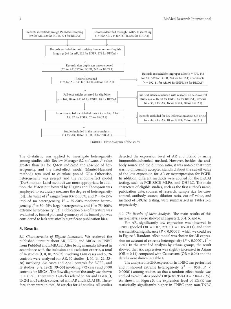
Records identified through PubMed searching (69 for AR, 320 for EGFR, 274 for BRCA1)
Records identified through EMBASE searching(184 for AR, 744 for EGFR, 666 for BRCA1)
Records excluded for not studying human or non-English language (46 for AR, 232 for EGFR, 278 for BRCA1)
Records after duplicates were removed
Records screened for AR, 389 for EGFR, 244 for BRCA1) or abstracts
Full-text articles assessed for eligibility Full-text articles excluded with reasons: no case-control
AR, 17 for EGFR, 52 for BRCA1)
Studies included in the meta-analysis
Records excluded for key information about OR or RR
(32 for AR, 287 for EGFR, 242 for BRCA1)
(175 for AR, 545 for EGFR, 420 for BRCA1)
(14 for AR, 10 for EGFR, 18 for BRCA1)
Records excluded for improper titles (n = 779, 146
(n = 192, 11 for AR, 93 for EGFR, 88 for BRCA1)
(n = 169, 18 for AR, 63 for EGFR, 88 for BRCA1) studies (n = 46, 30 for EGFR, 16 for BRCA1); reviews(n = 38, 2 for AR, 16 for EGFR, 20 for BRCA1)
Records selected for detailed review ( n = 85, 16 for
(n = 47, 2 for AR, 10 for EGFR, 35 for BRCA1)
Figure 1: Flow diagram of the study.
The 𝑄-statistic was applied to investigate heterogeneityamong studies with Review Manager 5.2 software. 𝑃 valuegreater than 0.1 for 𝑄-test indicated the absence of het-erogeneity, and the fixed-effect model (Mantel-Haenszelmethod) was used to calculate pooled ORs. Otherwise,heterogeneity was present and the random-effect model(DerSimonian-Lairdmethod)wasmore appropriate. In addi-tion, the 𝐼2-test put forward by Higgins and Thompson wasemployed to accurately measure the degree of heterogeneity[51].The value of 𝐼2 ranges from 0% to 100%, and 𝐼2 = 0∼25%implied no heterogeneity, 𝐼2 = 25∼50% moderate hetero-geneity, 𝐼2 = 50∼75% large heterogeneity, and 𝐼2 = 75∼100%extreme heterogeneity [52]. Publication bias of literature wasevaluated by funnel plot, and symmetry of the funnel plot wasconsidered to lack statistically significant publication bias.
3. Results
3.1. Characteristics of Eligible Literature. We retrieved thepublished literature about AR, EGFR, and BRCA1 in TNBCfrom PubMed and EMBASE. After being manually filtered inaccordance with the inclusion and exclusion criteria, a totalof 14 studies [1, 8, 10, 22–32] involving 1,618 cases and 5,526controls were analyzed for AR, 10 studies [1, 10, 14, 24, 33–38] involving 998 cases and 2,842 controls for EGFR, and18 studies [3, 8, 18–21, 39–50] involving 992 cases and 3,798controls for BRCA1.The flow diagram of the studywas shownin Figure 1. There were 3 articles related to AR and EGFR [1,10, 24] and 1 article concernedwithAR andBRCA1 [8].There-fore, there were in total 38 articles for 42 studies. All studies
detected the expression level of AR and EGFR by usingimmunohistochemical method. However, besides the anti-body source and the dilution ratio, it was notable that therewas no universally accepted standard about the cut-off valueof the low expression for AR or overexpression for EGFR.In addition, different methods were applied for the BRCA1testing, such as PCR-SSCP, MLPA, and DHPLC. The maincharacters of eligible studies, such as the first author’s name,publication date, sources of research, sample size for case-control, antibody source, dilution ratio, cut-off value, andmethod of BRCA1 testing, were summarized in Tables 1–3,respectively.
3.2. The Results of Meta-Analysis. The main results of thismeta-analysis were showed in Figures 2, 3, 4, 5, and 6.
For AR, significantly low expression was observed inTNBC (pooled OR = 0.07, 95% CI = 0.05–0.11), and therewas statistical significance (𝑃 < 0.00001), which we could seein Figure 2. Random-effect model was chosen for AR expres-sion on account of extreme heterogeneity (𝑃 < 0.00001, 𝐼2 =79%). In the stratified analysis by ethnic groups, the resultshowed that AR expression was slightly increased in Asians(OR = 0.11) compared with Caucasians (OR = 0.06) and thedetails were shown in Table 4.
The analysis of EGFR expression in TNBCwas performedand it showed extreme heterogeneity (𝐼2 = 85%, 𝑃 <0.00001) among studies, so that a random-effect model wasapplied to calculate a pooledOR (6.88, 95%CI = 3.84–12.35).As shown in Figure 3, the expression level of EGFR wasstatistically significantly higher in TNBC than non-TNBC

BioMed Research International 5
Study or subgroup
Chae et al., 2011Gasparini et al., 2014Hu et al., 2011Koo and Jung, 2011Loibl et al., 2011Luo et al., 2010Micello et al., 2010Niemeier et al., 2010Ogawa et al., 2008Park et al., 2009Park et al., 2011Peters et al., 2012Pristauz et al., 2010Rakha et al., 2007
Total (95% CI)Total events
Events
192781
2438423
18222137
36
386
Total
12371211
8111137138304263
1561844
277
1618
Events
126209
107657
33411086
1481242795203076
1000
4175
Total
157256
125610956213288
1591853507753691
1370
5526
Weight
2.6%9.9%
10.3%2.5%9.5%8.8%4.3%4.8%8.2%8.9%9.5%4.1%6.5%
10.1%
100.0%
M-H, random, 95% CI
0.02 [0.00, 0.18]0.07 [0.05, 0.11]0.10 [0.07, 0.14]0.13 [0.02, 1.10]0.19 [0.12, 0.31]0.08 [0.04, 0.14]0.01 [0.00, 0.04]0.01 [0.00, 0.03]0.37 [0.19, 0.73]0.14 [0.08, 0.24]0.08 [0.05, 0.12]0.04 [0.01, 0.18]0.04 [0.01, 0.10]0.06 [0.04, 0.08]
0.07 [0.05, 0.11]
TNBC Non-TNBC Odds ratio Odds ratioM-H, random, 95% CI
0.002 0.1 1 10 500Favours [TNBC] Favours [non-TNBC]
Heterogeneity: 𝜏2 = 0.35; 𝜒2 = 63.05, df = 13 (P < 0.00001); I2 = 79%Test for overall effect: Z = 13.18 (P < 0.00001)
Figure 2: Forest plot of studies evaluating OR of AR expression in TNBC compared with non-TNBC. The events of TNBC and the eventsof non-TNBC refer to the number of TNBC patients with positive expression of AR and the number of non-TNBC patients with positiveexpression of AR, respectively. The squares and horizontal lines correspond to the specific OR and 95% CI for every study. The area of thesquares reflects the study specific weight. The diamond stands for the pooled OR and 95% CI.
Study or subgroup
Gasparini et al., 2014
Koo and Jung, 2011
Nogi et al., 2009
Nozoe et al., 2011
Pillai et al., 2012
Rakha et al., 2007
Ryden et al., 2010
Tan et al., 2008
Tang et al., 2012
Tawfik et al., 2010
Total (95% CI)
Total events
Events
210
1
13
3
11
92
36
16
10
97
489
Total
381
8
26
7
18
249
87
31
40
151
998
Events
37
0
21
3
2
194
34
6
28
34
359
Total
262
109
85
30
18
1293
299
209
158
379
2842
Weight
13.6%
2.6%
10.7%
5.7%
6.3%
14.0%
12.8%
9.7%
11.3%
13.2%
100.0%
M-H, random, 95% CI
7.47 [5.00, 11.16]
43.80 [1.64, 1169.84]
3.05 [1.22, 7.60]
6.75 [1.00, 45.77]
12.57 [2.19, 72.27]
3.32 [2.46, 4.48]
5.50 [3.15, 9.60]
36.09 [12.32, 105.73]
1.55 [0.68, 3.53]
18.23 [11.23, 29.59]
6.88 [3.84, 12.35]
TNBC Non-TNBC Odds ratio Odds ratioM-H, random, 95% CIs
0.01 0.1 1 10 100Favours [TNBC] Favours [non-TNBC]
Heterogeneity: 𝜏2 = 0.61; 𝜒2 = 61.48, df = 9 (P < 0.00001); I2 = 85%Test for overall effect: Z = 6.47 (P < 0.00001)
Figure 3: Forest plot of studies evaluating OR of EGFR expression in TNBC compared with non-TNBC.The events of TNBC and the eventsof non-TNBC refer to the number of TNBC patients with positive expression of EGFR and the number of non-TNBC patients with positiveexpression of EGFR, respectively. The squares and horizontal lines correspond to the specific OR and 95% CI for every study. The area of thesquares reflects the study specific weight. The diamond stands for the pooled OR and 95% CI.

6 BioMed Research International
Study or subgroup
2.2.1 Dako antibodyGasparini et al., 2014Nozoe et al., 2011Pillai et al., 2012Ryden et al., 2010Tang et al., 2012
Subtotal (95% CI)Total events
2.2.2 Novocastra antibodyKoo and Jung, 2011Nogi et al., 2009Rakha et al., 2007
Subtotal (95% CI)Total events
2.2.3 Zymed antibodyTan et al., 2008Tawfik et al., 2010
Subtotal (95% CI)Total events
Total (95% CI)Total events
Events
2103
113610
270
11392
106
1697
113
489
Total
3817
188740
533
826
249283
31151182
998
Events
3732
3428
104
021
194
215
634
40
359
Total
2623018
299158767
10985
12931487
209379588
2842
Weight
13.6%5.7%6.3%
12.8%11.3%49.7%
2.6%10.7%14.0%27.3%
9.7%13.2%23.0%
100.0%
M-H, random, 95% CI
7.47 [5.00, 11.16]6.75 [1.00, 45.77]
12.57 [2.19, 72.27]5.50 [3.15, 9.60]1.55 [0.68, 3.53]5.02 [2.64, 9.56]
43.80 [1.64, 1169.84]3.05 [1.22, 7.60]3.32 [2.46, 4.48]3.42 [2.18, 5.37]
36.09 [12.32, 105.73]18.23 [11.23, 29.59]21.55 [12.08, 38.45]
6.88 [3.84, 12.35]
TNBC Non-TNBC Odds ratio Odds ratioM-H, random, 95% CI
0.01 0.1 1 10 100Favours [TNBC] Favours [non-TNBC]
Heterogeneity: 𝜏2 = 0.04; 𝜒2 = 2.40, df = 2 (P = 0.30); I2 = 17%Test for overall effect: Z = 5.36 (P < 0.00001)
Heterogeneity: 𝜏2 = 0.30; 𝜒2 = 12.20, df = 4 (P = 0.02); I2 = 67%Test for overall effect: Z = 4.92 (P < 0.00001)
Heterogeneity: 𝜏2 = 0.05; 𝜒2 = 1.30, df = 1 (P = 0.25); I2 = 23%Test for overall effect: Z = 10.40 (P < 0.00001)
Heterogeneity: 𝜏2 = 0.61; 𝜒2 = 61.48, df = 9 (P < 0.00001); I2 = 85%
(P < 0.00001); I2 = 92.0%
Test for overall effect: Z = 6.47 (P < 0.00001)Test for subgroup differences: 𝜒2 = 24.89, df = 2
Figure 4: Forest plot of subgroup related to antibody source evaluating EGFR expression in TNBC compared with non-TNBC.
Table 4: Subgroup analyses based on ethnic for AR expression, EGFR expression, and BRCA1mutation in TNBC compared with non-TNBC.
Variables AR EGFR BRCA1𝑁 OR (95% CI) 𝑃
𝐻𝐼
2𝑁 OR (95% CI) 𝑃
𝐻𝐼
2𝑁 RR (95% CI) 𝑃
𝐻𝐼
2
Total 14 0.07 (0.05–0.11) <0.001 79% 10 6.88 (3.84–12.35) <0.001 85% 18 5.26 (4.42–6.26) 0.14 27%Ethnic
Asian 6 0.11 (0.06–0.21) 0.002 73% 5 9.60 (3.50–26.30) 0.01 68% 9 5.43 (4.06–7.25) 0.71 0%Caucasian 8 0.06 (0.03–0.09) <0.001 82% 5 5.53 (2.84–10.77) <0.001 87% 9 5.16 (4.16–6.40) 0.02 55%𝑁: numbers of data sets;𝑃𝐻:𝑃 value of𝑄-test for heterogeneity test;𝑃𝐻 < 0.1 indicates that there is heterogeneity and random-effect model is used to calculatepooled OR or RR and 95% CI. Otherwise, fixed-effect model is used.
(𝑃 < 0.00001). Because of the extreme heterogeneity, we dida subgroup analysis according to the antibody source and theresult was shown in Figure 4.Theheterogeneitywasmarkedlyreduced in the subgroup and it indicated that the antibodysource was one of the factors for the extreme heterogeneity.Moreover, we also did stratified analysis related to ethnicity,and it was worth noting that EGFR expression was signifi-cantly increased in Asians (OR = 9.60) compared with Cau-casians (OR = 5.53) for TNBC patients (Table 4).
In Figure 5, the outcome of heterogeneity for the riskof BRCA1 mutation in TNBC was that 𝐼2 = 27% and𝑃heterogeneity = 0.14, so a fix effect model was used to calculatethe pooled RR (5.26, 95% CI = 4.42–6.26). It indicated thatwomenwith TNBCwere approximately five timesmore likely
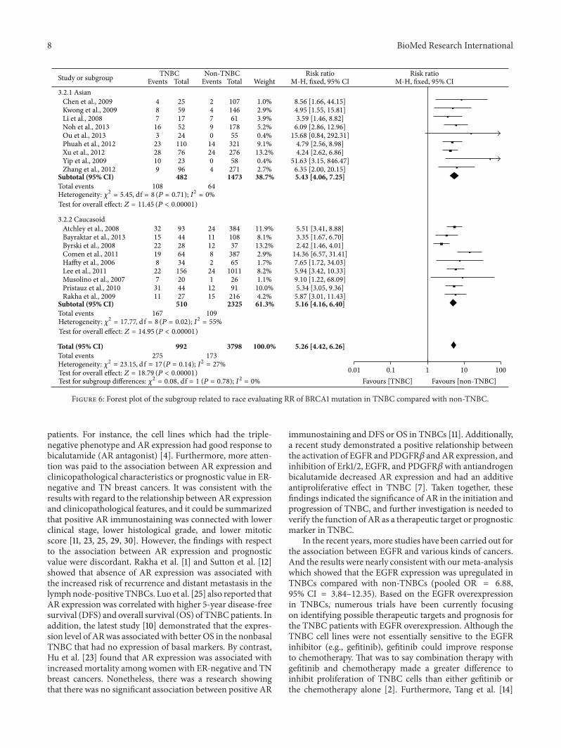
to have BRCA1 mutation compared with non-TNBC and itwas statistically significant (𝑃 < 0.00001). In the subgroupwith regard to ethnicity, the pooled RR (5.43, 95% CI =4.06–7.25) for theAsianswas higher than the pooledRR (5.16,95%CI = 4.16–6.40) for the Caucasians, which indicated thatthe prevalence of BRCA1 mutation was higher in the Asianscompared with the Caucasians (Figure 6).
Finally, publication bias of the eligible studies was evalu-ated by Funnel plots, respectively. As shown in Figure 7, thefunnel plots about AR and EGFR were almost symmetric,which meant that there was absent indication for significantpublication bias. However, there was slight asymmetry in thefunnel plot about BRCA1,which indicated that therewasmildpublication bias between the eligible articles about BRCA1.

BioMed Research International 7
Study or subgroup
Atchley et al., 2008Bayraktar et al., 2013Byrski et al., 2008Chen et al., 2009Comen et al., 2011Haffty et al., 2006Kwong et al., 2009Lee et al., 2011Li et al., 2008Musolino et al., 2007Noh et al., 2013Ou et al., 2013Phuah et al., 2012Pristauz et al., 2010Rakha et al., 2009Xu et al., 2012Yip et al., 2009Zhang et al., 2012
Total (95% CI)Total events
Events
3215224
1988
2277
163
23311128109
275
Total
93442825643459
15617205224
1104427762396
992
Events
2411122824
247190
1412152404
173
Total
38410837
10738765
1461011
6126
17855
32191
21627658
271
3798
Weight
11.9%8.1%
13.2%1.0%2.9%1.7%2.9%8.2%3.9%1.1%5.2%0.4%9.1%
10.0%4.2%
13.2%0.4%2.7%
100.0%
M-H, fixed, 95% CI
5.51 [3.41, 8.88]3.35 [1.67, 6.70]2.42 [1.46, 4.01]
8.56 [1.66, 44.15]14.36 [6.57, 31.41]7.65 [1.72, 34.03]4.95 [1.55, 15.81]5.94 [3.42, 10.33]3.59 [1.46, 8.82]
9.10 [1.22, 68.09]6.09 [2.86, 12.96]
15.68 [0.84, 292.31]4.79 [2.56, 8.98]5.34 [3.05, 9.36]
5.87 [3.01, 11.43]4.24 [2.62, 6.86]
51.63 [3.15, 846.47]6.35 [2.00, 20.15]
5.26 [4.42, 6.26]
TNBC Non-TNBC Risk ratio Risk ratioM-H, fixed, 95% CI
0.001 0.1 1 10 1000Favours [TNBC] Favours [non-TNBC]
Test for overall effect: Z = 18.79 (P < 0.00001)Heterogeneity: 𝜒2 = 23.15, df = 17 (P = 0.14); I2 = 27%
Figure 5: Forest plot of studies evaluating RR of BRCA1 mutation in TNBC compared with non-TNBC.The events of TNBC and the eventsof non-TNBC refer to the number of TNBC patients with BRCA1 mutation and the number of non-TNBC patients with BRCA1 mutation,respectively. The squares and horizontal lines correspond to the specific RR and 95% CI for every study. The area of the squares reflects thestudy specific weight. The diamond stands for the pooled RR and 95% CI.
4. Discussion
In thismeta-analysis, the expression of AR and EGFR and therisk of BRCA1 mutation in TNBC are explored. We find thatEGFR is overexpressed and the risk of having BRCA1 muta-tion is higher in TNBC than non-TNBC. Nevertheless, ARexpression is downregulated in TNBC compared with non-TNBC.The distinct characteristics of AR, EGFR, and BRCA1indicate that they can play crucial roles in targeted therapy orprognosis in TNBC.
There are some advantages in ourmeta-analysis. First, themost obvious superiority is that there is substantial samplesize to improve the credibility for statistical analysis. Second,the conclusions are more generalizable on account of eligibleliterature frommany different geographic distributions, suchas USA, UK, Australia, and China. Finally, we extracted datainformation required from individual literature and compos-ited the outcome instead of summary consequence.
However, several limitations in our meta-analysis shouldbe discussed. First, there is extreme heterogeneity for theoutcomes of AR expression (𝑃 < 0.00001, 𝐼2 = 79%) andEGFR expression (𝑃 < 0.00001, 𝐼2 = 85%). The detectionmethods of AR and EGFR are both immunohistochemistry(IHC), but the antibody source, dilution rate, cut-off value,and ethnicity are different for AR and EGFR in differentstudies (Tables 1 and 2). On the basis of the four factors, weproceedwith subgroup analyses; only the heterogeneity aboutOR related to EGFR expression is markedly reduced in thesubgroup connected with antibody source (Figure 4). Hence,
the heterogeneity is probably attributed to the variances withregard to features of population, the subtypes of TNBC, thedisease stages, the antibody source, the dilution rate, the cut-off value of AR and EGFR expression, and so forth. Second,although there is no evidence of significant publication biasfor the articles about AR and EGFR in our meta-analysis,there was slight asymmetry in the funnel plot about BRCA1.Hence, cautions should be taken on the account of mildpublication bias between the eligible articles about BRCA1.Besides, only the English articles were selected, which cancertainly give rise to language bias. In addition, positiveresults are prone to be published, which may make certainbias.
As we know, TNBC is a heterogeneous disease that hashigh diversity associatedwith biology, etiology, and treatmentstrategies. In addition, TNBCs have amore aggressive clinicalbehavior in part due to poor differentiation. Furthermore,due to the lack of the conventional hormonal or anti-HER2therapeutic targets, the standardized treatment strategieshave not been formulated and the chemotherapy is the onlymodality of systemic therapy for these cancers. Based on theabove, it is urgently needed to find new prognostic indicatorsand therapeutic method for TNBCs.
The meta-analysis indicates that the expression level ofAR is lower in TNBCs than non-TNBCs (pooled OR = 0.07,95% CI = 0.05–0.11), but more and more studies demon-strated that about one-third of TNBCs were AR-positive [11,23, 25, 27, 29, 30]. This represents a potential opportunity fornovel targeted therapy in positive AR expression of TNBC
8 BioMed Research International
Study or subgroup
3.2.1 AsianChen et al., 2009Kwong et al., 2009Li et al., 2008Noh et al., 2013Ou et al., 2013Phuah et al., 2012Xu et al., 2012Yip et al., 2009Zhang et al., 2012
Subtotal (95% CI)Total events
3.2.2 CaucasoidAtchley et al., 2008Bayraktar et al., 2013Byrski et al., 2008Comen et al., 2011Haffty et al., 2006Lee et al., 2011Musolino et al., 2007Pristauz et al., 2010Rakha et al., 2009
Subtotal (95% CI)Total events
Total (95% CI)Total events
Events
487
163
2328109
108
321522198
227
3111
167
275
Total
2559175224
110762396
482
9344286434
156204427
510
992
Events
24790
142404
64
24111282
241
1215
109
173
Total
10714661
17855
32127658
2711473
38410837
38765
10112691
2162325
3798
Weight
1.0%2.9%3.9%5.2%0.4%9.1%
13.2%0.4%2.7%
38.7%
11.9%8.1%
13.2%2.9%1.7%8.2%1.1%
10.0%4.2%
61.3%
100.0%
M-H, fixed, 95% CI
8.56 [1.66, 44.15]4.95 [1.55, 15.81]3.59 [1.46, 8.82]
6.09 [2.86, 12.96]15.68 [0.84, 292.31]
4.79 [2.56, 8.98]4.24 [2.62, 6.86]
51.63 [3.15, 846.47]6.35 [2.00, 20.15]5.43 [4.06, 7.25]
5.51 [3.41, 8.88]3.35 [1.67, 6.70]2.42 [1.46, 4.01]
14.36 [6.57, 31.41]7.65 [1.72, 34.03]5.94 [3.42, 10.33]9.10 [1.22, 68.09]5.34 [3.05, 9.36]
5.87 [3.01, 11.43]5.16 [4.16, 6.40]
5.26 [4.42, 6.26]
TNBC Non-TNBC Risk ratio Risk ratioM-H, fixed, 95% CI
0.01 0.1 1 10 100Favours [TNBC] Favours [non-TNBC]
Test for overall effect: Z = 11.45 (P < 0.00001)Heterogeneity: 𝜒2 = 5.45, df = 8 (P = 0.71); I2 = 0%
Test for overall effect: Z = 14.95 (P < 0.00001)Heterogeneity: 𝜒2 = 17.77, d f = 8 (P = 0.02); I2 = 55%
Test for overall effect: Z = 18.79 (P < 0.00001)Heterogeneity: 𝜒2 = 23.15, d f = 17 (P = 0.14); I2 = 27%
Test for subgroup differences: 𝜒2 = 0.08, df = 1 (P = 0.78); I2 = 0%
Figure 6: Forest plot of the subgroup related to race evaluating RR of BRCA1 mutation in TNBC compared with non-TNBC.
patients. For instance, the cell lines which had the triple-negative phenotype and AR expression had good response tobicalutamide (AR antagonist) [4]. Furthermore, more atten-tion was paid to the association between AR expression andclinicopathological characteristics or prognostic value in ER-negative and TN breast cancers. It was consistent with theresults with regard to the relationship between AR expressionand clinicopathological features, and it could be summarizedthat positive AR immunostaining was connected with lowerclinical stage, lower histological grade, and lower mitoticscore [11, 23, 25, 29, 30]. However, the findings with respectto the association between AR expression and prognosticvalue were discordant. Rakha et al. [1] and Sutton et al. [12]showed that absence of AR expression was associated withthe increased risk of recurrence and distant metastasis in thelymphnode-positive TNBCs. Luo et al. [25] also reported thatAR expression was correlated with higher 5-year disease-freesurvival (DFS) and overall survival (OS) of TNBCpatients. Inaddition, the latest study [10] demonstrated that the expres-sion level of ARwas associated with better OS in the nonbasalTNBC that had no expression of basal markers. By contrast,Hu et al. [23] found that AR expression was associated withincreased mortality among women with ER-negative and TNbreast cancers. Nonetheless, there was a research showingthat there was no significant association between positive AR
immunostaining and DFS or OS in TNBCs [11]. Additionally,a recent study demonstrated a positive relationship betweenthe activation of EGFR and PDGFR𝛽 and AR expression, andinhibition of Erk1/2, EGFR, and PDGFR𝛽 with antiandrogenbicalutamide decreased AR expression and had an additiveantiproliferative effect in TNBC [7]. Taken together, thesefindings indicated the significance of AR in the initiation andprogression of TNBC, and further investigation is needed toverify the function of AR as a therapeutic target or prognosticmarker in TNBC.
In the recent years, more studies have been carried out forthe association between EGFR and various kinds of cancers.And the results were nearly consistent with ourmeta-analysiswhich showed that the EGFR expression was upregulated inTNBCs compared with non-TNBCs (pooled OR = 6.88,95% CI = 3.84–12.35). Based on the EGFR overexpressionin TNBCs, numerous trials have been currently focusingon identifying possible therapeutic targets and prognosis forthe TNBC patients with EGFR overexpression. Although theTNBC cell lines were not essentially sensitive to the EGFRinhibitor (e.g., gefitinib), gefitinib could improve responseto chemotherapy. That was to say combination therapy withgefitinib and chemotherapy made a greater difference toinhibit proliferation of TNBC cells than either gefitinib orthe chemotherapy alone [2]. Furthermore, Tang et al. [14]

BioMed Research International 9
SE (l
og[O
R])
0
0.5
1
1.5
OR
20.001 0.1 1 10 1000
(a)
SE (l
og[O
R])
OR
0
0.5
1
1.5
20.001 0.1 1 10 1000
(b)
SE (l
og[R
R])
0
0.5
1
1.5
2
RR0.001 0.1 1 10 1000
(c)
Figure 7: Funnel plots for evaluating publication bias for the eligible articles about AR (a), EGFR (b), and BRCA1 (c). As shown in the figures,the funnel plots were almost symmetric and no evidence of publication bias was observed in this analysis.
indicated that the EGFR overexpression predicted betterresponse to neoadjuvant chemotherapy and increased patho-logic complete response rates in TNBCs compared with non-TNBCs. Nonetheless, the previous study outcome demon-strated that patients with EGFR-positive TNBC had aless favorable response to neoadjuvant chemotherapy thanpatients with EGFR-negative TNBC [33]. In a word, theexpression level of EGFR is higher in TNBCs than non-TNBCs [1, 10, 14, 24, 33–38], but further studies evaluating theassociation between EGFR and neoadjuvant chemotherapyare required to promote molecular targeting therapy forTNBC. Furthermore, there was evidence that 94% ofearly-stage high-grade TNBC with a basal-like phenotypeexpressedMUC1, andMUC1 and EGFR interacted in nucleusof BC cell to facilitate the association of EGFRwith transcrip-tionally active promoter regions, which provided a rationalefor MUC1-based immunotherapy in TNBC patients withEGFR expression [53].
The prevalence of BRCA1 mutation in familial or early-onset breast cancer led more andmore studies to concentrateon the role of BRCA1 in TNBC [19, 40, 41, 43, 46, 49]. Thepooled RR (5.26, 95% CI = 4.42–6.26) of our meta-analysisshowed that the risk of BRCA1 mutation was about five times
in TNBC compared with non-TNBC. Intriguingly, mountingevidence indicated that breast cancers with BRCA1 mutationwere more likely to exhibit triple-negative phenotype com-pared with the BRCA1 noncarriers [18, 20, 40–44, 49], whichshowed that BRCA1 could play a unique role in the progres-sion of TNBCs. In the subgroup regarding race, the pooledRR (5.43, 95% CI = 4.06–7.25) for the Asian was higher thanthe pooled RR (5.16, 95% CI = 4.16–6.40) for the Caucasoid,which indicated that the prevalence of BRCA1 mutation washigher in the Asian compared with the Caucasoid. Moreover,there was no heterogeneity (𝐼2 = 0%, 𝑃 = 0.71) in the Asiansubgroup. Thus, the race was in a certain contribution to theheterogeneity and a factor causing the different prevalenceof BRCA1 mutation between Caucasoid and Asian. Severalstudies had been conducted for the relationship related tothe BRCA1-associated breast cancers and therapeutic effects.Byrski et al. [21] reported that early-onset breast cancerpatients with BRCA1 mutation had poorer response to theneoadjuvant chemotherapy of the spindle poison docetaxel.Nevertheless, PARP inhibitors damaging DNA single-strandbreak repair could benefit patients with BRCA1 mutation[54]. However, few attentions were paid to the therapeuticeffects of neoadjuvant chemotherapy in TNBC patients with

10 BioMed Research International
BRCA1 mutation. Based on the above, it is necessary tofurther evaluate the relationship between BRCA1 mutationand TNBCs and implement the effective strategies for TNBCpatients with BRCA1 mutation in the future.
In addition, some research studies have been carried outfor other therapeutic target receptors (e.g., VEGFR and folatereceptor) and novel inhibitors (e.g., inhibitor of mTOR andhistone deacetylase) for the preclinical and clinical treatmentof TNBC. Similar to EGFR, the vascular endothelial growthfactor receptor (VEGFR) has been explored as a therapeutictarget receptor in breast cancer. An open-label, randomisedphase 3 trial demonstrated that bevacizumab (VEGFRinhibitor) was ineffective in adjuvant treatment in unselectedTNBC patients but may have some efficacy in metastaticTNBC [55]. A recent study demonstrated that 80% of TNBCpatients expressed folate receptor a (FRA) and FRA expres-sion was significantly associated with a worse disease-freesurvival [56]. Additionally, the PIK3CA gene is commonlymutated in TNBC. The inhibition of the PI3K pathway anddownstream mammalian target of rapamycin (mTOR) hasbeen identified as a promising therapeutic strategy for treat-ing TNBC, and a phase 2 trial demonstrated that everolimus(mTOR inhibitor)-carboplatin combination was efficaciousinmetastatic TNBC [57]. Furthermore, epigenetic alterationsare known for promoter initiation and progression of cancers.Targeting such epigenetic events via histone deacetylaseinhibitor (HDI) has been explored in the treatment ofTNBC. A recent study showed that HDI treatment induced“BRCAness” and synergistic lethality with PARP inhibitorsand cisplatin against human TNBC cells [58].
Based on the gene expression profiling, triple-negativebreast cancer is a heterogeneous disease with uniquemolecu-lar subtypes which have different clinicopathological featuresand clinical outcomes. Taken collectively, AR, EGFR, andBRCA1 play distinct roles as biomarkers in the progressionof TNBCs. In view of the EGFR overexpression and theimmunohistochemical feasibility of clinical practice, EGFRmight be superior to AR and BRCA1 as biomarker forTNBCs. However, the unique role of AR and BRCA1 cannotbe ignored for targeted treatment strategies and prognosisjudgement. Furthermore, the previous study reported that20% of the hereditary BRCA-related breast cancers hadtriple-negative phenotype and expressed AR [8]. To date,the relationship among the three biomarkers is not clearand further investigation should be warranted before thecombination of three biomarkers is applied to the clinicalmanagement of TNBC patients.
5. Conclusion
Overall, by quantifying synthesis of all published studiesof AR, EGFR, and BRCA1, the meta-analysis demonstratedthat the expression level of EGFR and the risk of BRCA1mutation were higher in TNBC compared with non-TNBCand AR expression was downregulated. Our study can give avaluable clue for the targeted therapy or judging prognosis ofTNBC patients. More clinical research should be performedbefore AR, EGFR, or BRCA1 can be proved to be commonbiomarkers for routine clinical practices.
Conflict of Interests
The authors declare that there is no conflict of interestsregarding the publication of this paper.
Authors’ Contribution
Li Zhang and Cheng Fang contribute equally to this work.
Acknowledgments
The Project-sponsored by SRF for ROCS, SEM. This workwas supported by grants from the National Natural ScienceFoundation of China (30873044 and 81272372).
References
[1] E. A. Rakha, M. E. El-Sayed, A. R. Green, A. H. S. Lee, J. F.Robertson, and I. O. Ellis, “Prognostic markers in triple-nega-tive breast cancer,” Cancer, vol. 109, no. 1, pp. 25–32, 2007.
[2] B. Corkery, J. Crown, M. Clynes, and N. O’Donovan, “Epider-mal growth factor receptor as a potential therapeutic target intriple-negative breast cancer,” Annals of Oncology, vol. 20, no. 5,pp. 862–867, 2009.
[3] B. G. Haffty, Q. Yang, M. Reiss et al., “Locoregional relapseanddistantmetastasis in conservativelymanaged triple negativeearly-stage breast cancer,” Journal of Clinical Oncology, vol. 24,no. 36, pp. 5652–5657, 2006.
[4] B. D. Lehmann, J. A. Bauer, X. Chen et al., “Identification ofhuman triple-negative breast cancer subtypes and preclinicalmodels for selection of targeted therapies,” The Journal ofClinical Investigation, vol. 121, no. 7, pp. 2750–2767, 2011.
[5] L. Steward, L. Conant, F. Gao, and J. A. Margenthaler, “Predic-tive factors and patterns of recurrence in patients with triplenegative breast cancer,” Annals of Surgical Oncology, vol. 21, no.7, pp. 2165–2171, 2014.
[6] J. J. Park, R. A. Irvine, G. Buchanan et al., “Breast cancersusceptibility gene 1 (BRCA1) is a coactivator of the androgenreceptor,” Cancer Research, vol. 60, no. 21, pp. 5946–5949, 2000.
[7] M. D. Cuenca-Lopez, J. C. Montero, J. C. Morales, A. Prat, A.Pandiella, andA.Ocana, “Phospho-kinase profile of triple nega-tive breast cancer and androgen receptor signaling,” BMCCancer, vol. 14, no. 1, pp. 302–311, 2014.
[8] G. Pristauz, E. Petru, E. Stacher et al., “Androgen receptorexpression in breast cancer patients tested for BRCA1 andBRCA2 mutations,” Histopathology, vol. 57, no. 6, pp. 877–884,2010.
[9] A. Gucalp and T. A. Traina, “Triple-negative breast cancer: roleof the androgen receptor,” Cancer Journal, vol. 16, no. 1, pp. 62–65, 2010.
[10] P. Gasparini, M. Fassan, L. Gascione et al., “Androgen receptorstatus is a prognostic marker in non-basal triple negative breastcancers and determines novel therapeutic options,” PLOS ONE,vol. 9, no. 2, Article ID e88525, 2014.
[11] I. Mrklic, Z. Pogorelic, V. Capkun, and S. Tomic, “Expression ofandrogen receptors in triple negative breast carcinomas,” ActaHistochemica, vol. 115, no. 4, pp. 344–348, 2013.
[12] L. M. Sutton, D. Cao, V. Sarode et al., “Decreased andro-gen receptor expression is associated with distant metastasesin patients with androgen receptor-expressing triple-negativebreast carcinoma,” The American Journal of Clinical Pathology,vol. 138, no. 4, pp. 511–516, 2012.

BioMed Research International 11
[13] D. Tang, S. Xu, Q. Zhang, and W. Zhao, “The expression andclinical significance of the androgen receptor and E-cadherinin triple-negative breast cancer,” Medical Oncology, vol. 29, no.2, pp. 526–533, 2012.
[14] Y. Tang, L. Zhu, Y. Li et al., “Overexpression of epithelial growthfactor receptor (EGFR) predicts better response to neo-adjuvantchemotherapy in patients with triple-negative breast cancer,”Journal of Translational Medicine, vol. 10, supplement 1, articleS4, 2012.
[15] J.-S. Guillamo, S. de Bouard, S. Valable et al., “Molecular mech-anisms underlying effects of epidermal growth factor recep-tor inhibition on invasion, proliferation, and angiogenesis inexperimental glioma,” Clinical Cancer Research, vol. 15, no. 11,pp. 3697–3704, 2009.
[16] A. R. Venkitaraman, “Cancer susceptibility and the functions ofBRCA1 and BRCA2,” Cell, vol. 108, no. 2, pp. 171–182, 2002.
[17] J. S. Reis-Filho and A. N. Tutt, “Triple negative tumours: acritical review,” Histopathology, vol. 52, no. 1, pp. 108–118, 2008.
[18] D. P. Atchley, C. T. Albarracin, A. Lopez et al., “Clinical andpathologic characteristics of patients with BRCA-positive andBRCA-negative breast cancer,” Journal of Clinical Oncology, vol.26, no. 26, pp. 4282–4288, 2008.
[19] A. Musolino, M. A. Bella, B. Bortesi et al., “BRCA mutations,molecular markers, and clinical variables in early-onset breastcancer: a population-based study,” Breast, vol. 16, no. 3, pp. 280–292, 2007.
[20] W.-F. Li, Z. Hu, N.-Y. Rao et al., “The prevalence of BRCA1 andBRCA2 germline mutations in high-risk breast cancer patientsof Chinese Han nationality: two recurrent mutations wereidentified,” Breast Cancer Research and Treatment, vol. 110, no.1, pp. 99–109, 2008.
[21] T. Byrski, J. Gronwald, T. Huzarski et al., “Response to neo-adjuvant chemotherapy in women with BRCA1-positive breastcancers,” Breast Cancer Research and Treatment, vol. 108, no. 2,pp. 289–296, 2008.
[22] B. J. Chae, A. Lee, J. S. Bae, B. J. Song, and S. S. Jung, “Expressionof nuclear receptor DAX-1 and androgen receptor in humanbreast cancer,” Journal of Surgical Oncology, vol. 103, no. 8, pp.768–772, 2011.
[23] R. Hu, S. Dawood, M. D. Holmes et al., “Androgen recep-tor expression and breast cancer survival in postmenopausalwomen,” Clinical Cancer Research, vol. 17, no. 7, pp. 1867–1874,2011.
[24] J. S. Koo and W. Jung, “Clinicopathlogic and immunohis-tochemical characteristics of triple negative invasive lobularcarcinoma,”YonseiMedical Journal, vol. 52, no. 1, pp. 89–97, 2011.
[25] X. Luo, Y.-X. Shi, Z.-M. Li, and W.-Q. Jiang, “Expression andclinical significance of androgen receptor in triple negativebreast cancer,” Chinese Journal of Cancer, vol. 29, no. 6, pp. 585–590, 2010.
[26] S. Loibl, B. M. Muller, G. von Minckwitz et al., “Androgenreceptor expression in primary breast cancer and its predictiveand prognostic value in patients treated with neoadjuvantchemotherapy,” Breast Cancer Research and Treatment, vol. 130,no. 2, pp. 477–487, 2011.
[27] D. Micello, A. Marando, N. Sahnane, C. Riva, C. Capella, andF. Sessa, “Androgen receptor is frequently expressed in HER2-positive, ER/PR-negative breast cancers,” Virchows Archiv, vol.457, no. 4, pp. 467–476, 2010.
[28] L. A. Niemeier, D. J. Dabbs, S. Beriwal, J. M. Striebel, and R.Bhargava, “Androgen receptor in breast cancer: expression in
estrogen receptor-positive tumors and in estrogen receptor-negative tumors with apocrine differentiation,”Modern Pathol-ogy, vol. 23, no. 2, pp. 205–212, 2010.
[29] Y. Ogawa, E. Hai, K. Matsumoto et al., “Androgen receptorexpression in breast cancer: relationship with clinicopatholog-ical factors and biomarkers,” International Journal of ClinicalOncology, vol. 13, no. 5, pp. 431–435, 2008.
[30] S. Park, J. Koo, H. S. Park et al., “Expression of androgenreceptors in primary breast cancer,” Annals of Oncology, vol. 21,no. 3, pp. 488–492, 2009.
[31] S. Park, J. S. Koo,M. S. Kimet al., “Androgen receptor expressionis significantly associated with better outcomes in estrogenreceptor-positive breast cancers,” Annals of Oncology, vol. 22,no. 8, pp. 1755–1762, 2011.
[32] K. M. Peters, S. L. Edwards, S. S. Nair et al., “Androgen receptorexpression predicts breast cancer survival: the role of geneticand epigenetic events,” BMC Cancer, vol. 12, pp. 132–141, 2012.
[33] H. Nogi, T. Kobayashi, M. Suzuki et al., “EGFR as paradoxi-cal predictor of chemosensitivity and outcome among triple-negative breast cancer,”Oncology Reports, vol. 21, no. 2, pp. 413–417, 2009.
[34] T. Nozoe, E. Mori, T. Iguchi et al., “Immunohistochemicalexpression of epidermal growth factor receptor in breast can-cer,” Breast Cancer, vol. 18, no. 1, pp. 37–41, 2011.
[35] S. K. K. Pillai, A. Tay, S. Nair, and C.-O. Leong, “Triple-negativebreast cancer is associatedwith EGFR,CK5/6 and c-KIT expres-sion in Malaysian women,” BMC Clinical Pathology, vol. 12,article 18, 2012.
[36] L. Ryden, K. Jirstrom, M. Haglund, O. Stal, and M. Ferno, “Epi-dermal growth factor receptor and vascular endothelial growthfactor receptor 2 are specific biomarkers in triple-negativebreast cancer. Results from a controlled randomized trial withlong-term follow-up,” Breast Cancer Research and Treatment,vol. 120, no. 2, pp. 491–498, 2010.
[37] D. S. P. Tan, C.Marchio, R. L. Jones et al., “Triple negative breastcancer: molecular profiling and prognostic impact in adjuvantanthracycline-treated patients,” Breast Cancer Research andTreatment, vol. 111, no. 1, pp. 27–44, 2008.
[38] O. Tawfik, K. Davis, B. F. Kimler et al., “Clinicopathologicalcharacteristics of triple-negative invasivemammary carcinomasin African-American versus Caucasian women,” Annals ofClinical and Laboratory Science, vol. 40, no. 4, pp. 315–323, 2010.
[39] S. Bayraktar, A. M. Gutierrez-Barrera, H. Lin et al., “Outcomeof metastatic breast cancer in selected women with or withoutdeleterious BRCAmutations,”Clinical and ExperimentalMetas-tasis, vol. 30, no. 5, pp. 631–642, 2013.
[40] W. Chen, K. Pan, T. Ouyang et al., “BRCA1 germline mutationsand tumor characteristics in Chinese women with familial orearly-onset breast cancer,” Breast Cancer Research and Treat-ment, vol. 117, no. 1, pp. 55–60, 2009.
[41] E. Comen, M. Davids, T. Kirchhoff, C. Hudis, K. Offit, and M.Robson, “Relative contributions of BRCA1 and BRCA2 muta-tions to “triple-negative” breast cancer in Ashkenazi Women,”Breast Cancer Research and Treatment, vol. 129, no. 1, pp. 185–190, 2011.
[42] A. Kwong, L. P. Wong, H. N. Wong et al., “Clinical and patho-logical characteristics of Chinese patients with BRCA relatedbreast cancer,”TheHUGO Journal, vol. 3, no. 1, pp. 63–76, 2009.
[43] E. Lee, R. McKean-Cowdin, H. Ma et al., “Characteristics oftriple-negative breast cancer in patients with aBRCA1mutation:results from a population-based study of young women,” Jour-nal of Clinical Oncology, vol. 29, no. 33, pp. 4373–4380, 2011.

12 BioMed Research International
[44] J. M. Noh, B.-K. Han, D. H. Choi et al., “Association betweenBRCA mutation status, pathological findings, and magneticresonance imaging features in patients with breast cancer at riskfor the mutation,” Journal of Breast Cancer, vol. 16, no. 3, pp.308–314, 2013.
[45] J. Ou, T.Wu, R. Sijmons, D. Ni,W. Xu, andH.Upur, “Prevalenceof BRCA1 and BRCA2 germline mutations in breast cancerwomen of multiple ethnic region in Northwest China,” Journalof Breast Cancer, vol. 16, no. 1, pp. 50–54, 2013.
[46] S.-Y. Phuah, L.-M. Looi, N.Hassan et al., “Triple-negative breastcancer and PTEN (phosphatase and tensin homologue)loss arepredictors of BRCA1 germline mutations in women with early-onset and familial breast cancer, but not in womenwith isolatedlate-onset breast cancer,” Breast Cancer Research, vol. 14, no. 6,article R142, 2012.
[47] J. Xu, B.Wang, Y. Zhang, R. Li, Y.Wang, and S. Zhang, “Clinicalimplications for BRCA gene mutation in breast cancer,”Molec-ular Biology Reports, vol. 39, no. 3, pp. 3097–3102, 2012.
[48] C.-H. Yip, N. A. Taib, W. Y. Choo, S. Rampal, M. K.Thong, andS. H. Teo, “Clinical and pathologic differences between BRCA1-,BRCA2-, and non-BRCA-associated breast cancers in amultira-cial developing country,”World Journal of Surgery, vol. 33, no. 10,pp. 2077–2081, 2009.
[49] J. Zhang, R. Pei, Z. Pang et al., “Prevalence and characterizationof BRCA1 and BRCA2 germline mutations in Chinese womenwith familial breast cancer,” Breast Cancer Research and Treat-ment, vol. 132, no. 2, pp. 421–428, 2012.
[50] E. A. Rakha, S. E. Elsheikh, M. A. Aleskandarany et al.,“Triple-negative breast cancer: distinguishing between basaland nonbasal subtypes,” Clinical Cancer Research, vol. 15, no. 7,pp. 2302–2310, 2009.
[51] J. P. T. Higgins and S. G.Thompson, “Quantifying heterogeneityin ameta-analysis,” Statistics inMedicine, vol. 21, no. 11, pp. 1539–1558, 2002.
[52] J. P. T. Higgins, S. G. Thompson, J. J. Deeks, and D. G. Altman,“Measuring inconsistency in meta-analyses,” British MedicalJournal, vol. 327, no. 7414, pp. 557–560, 2003.
[53] A. Siroy, F. W. Abdul-Karim, J. Miedler et al., “MUC1 isexpressed at high frequency in early-stage basal-like triplenegative breast cancer,” Human Pathology, vol. 44, no. 10, pp.2159–2166, 2013.
[54] O. Gluz, C. Liedtke, N. Gottschalk, L. Pusztai, U. Nitz, andN. Harbeck, “Triple-negative breast cancer: current status andfuture directions,” Annals of Oncology, vol. 20, no. 12, pp. 1913–1927, 2009.
[55] D. Cameron, J. Brown, R. Dent et al., “Adjuvant bevacizumab-containing therapy in triple-negative breast cancer (BEAT-RICE): primary results of a randomised, phase 3 trial,” TheLancet Oncology, vol. 14, no. 10, pp. 933–942, 2013.
[56] Z. Zhang, J.Wang, D. E. Tacha et al., “Folate receptor 𝛼 associatewith triple-negative breast cancer and poor prognosis,”Archivesof Pathology & LaboratoryMedicine, vol. 138, no. 7, pp. 890–895,2014.
[57] J. C. Singh, Y. Novik, S. Stein et al., “Phase 2 trial of everolimusand carboplatin combination in patients with triple negativemetastatic breast cancer,” Breast Cancer Research, vol. 16, no. 2,article R32, 2014.
[58] K.Ha,W. Fiskus, D. S. Choi et al., “Histone deacetylase inhibitortreatment induces “BRCAness” and synergistic lethality withPARP inhibitor and cisplatin against human triple negativebreast cancer cells,” Oncotarget, vol. 5, no. 14, pp. 5637–5650,2014.

Submit your manuscripts athttp://www.hindawi.com
Stem CellsInternational
Hindawi Publishing Corporationhttp://www.hindawi.com Volume 2014
Hindawi Publishing Corporationhttp://www.hindawi.com Volume 2014
MEDIATORSINFLAMMATION
of
Hindawi Publishing Corporationhttp://www.hindawi.com Volume 2014
Behavioural Neurology
EndocrinologyInternational Journal of
Hindawi Publishing Corporationhttp://www.hindawi.com Volume 2014
Hindawi Publishing Corporationhttp://www.hindawi.com Volume 2014
Disease Markers
Hindawi Publishing Corporationhttp://www.hindawi.com Volume 2014
BioMed Research International
OncologyJournal of
Hindawi Publishing Corporationhttp://www.hindawi.com Volume 2014
Hindawi Publishing Corporationhttp://www.hindawi.com Volume 2014
Oxidative Medicine and Cellular Longevity
Hindawi Publishing Corporationhttp://www.hindawi.com Volume 2014
PPAR Research
The Scientific World JournalHindawi Publishing Corporation http://www.hindawi.com Volume 2014
Immunology ResearchHindawi Publishing Corporationhttp://www.hindawi.com Volume 2014
Journal of
ObesityJournal of
Hindawi Publishing Corporationhttp://www.hindawi.com Volume 2014
Hindawi Publishing Corporationhttp://www.hindawi.com Volume 2014
Computational and Mathematical Methods in Medicine
OphthalmologyJournal of
Hindawi Publishing Corporationhttp://www.hindawi.com Volume 2014
Diabetes ResearchJournal of
Hindawi Publishing Corporationhttp://www.hindawi.com Volume 2014
Hindawi Publishing Corporationhttp://www.hindawi.com Volume 2014
Research and TreatmentAIDS
Hindawi Publishing Corporationhttp://www.hindawi.com Volume 2014
Gastroenterology Research and Practice
Hindawi Publishing Corporationhttp://www.hindawi.com Volume 2014
Parkinson’s Disease
Evidence-Based Complementary and Alternative Medicine
Volume 2014Hindawi Publishing Corporationhttp://www.hindawi.com