review article - amazon web services · 255 jornal brasileiro de pneumologia 31(3) - mai/jun de...
TRANSCRIPT

254
Nóbrega, BB et al.
Pulmonary sarcoidosis: high-resolution computed tomography findings
Pulmonary sarcoidosis: high-resolution computed
tomography findings*
BRUNO BARCELOS DA NÓBREGA, GUSTAVO DE SOUZA PORTES MEIRELLES, GILBERO SZARF,DANY JASINOWODOLINSKI, JORGE ISSAMU KAVAKAMA
J Bras Pneumol 2005; 31(3): 254-60.
*Study conducted at the Universidade Federal de São Paulo - Escola Paulista de Medicina (UNIFESP- EPM), Centro de Medicina Diagnóstica FleuryInstitute of Radiology at the Hospital das Clínicas of the Faculdade de Medicina da Universidade de São Paulo (INRAD-HCFMUSP)Correspondence to: Bruno Barcelos da Nóbrega. Alameda Ribeirão Preto, 551 apto.14, Bela Vista. CEP 01331-001, São Paulo, SP.Phone: 55 11 288-4801. E-mail: [email protected]: 4 August 2004. Accepted, after review: 29 October 2004.
Key words: Sarcoidosis. Lung. High-resolution computed tomography.
Sarcoidosis is a systemic disease of unknown etiology, characterized by noncaseating granulomas. Althoughit may affect any organ, morbidity and mortality are most commonly related to pulmonary involvement,which is found in 80-90% of patients. This study illustrates the principal manifestations of sarcoidosis seenin high-resolution computed tomography scans, including typical as well as atypical forms.
INTRODUCTION
Sarcoidosis is a multisystemic disease ofunknown et iology that is var iable in i tspresentat ion, progress ion and prognosis .Pulmonary involvement is seen in up to 90% ofpatients, and 20-25% of those present permanentfunctional impairment(1-4).
Pulmonary involvement tends to be bilateraland asymmetric, predominantly found in the upperlobes. Radiologic findings are atypical in 25% ofthe cases, and chest X-rays reveal normal resultsin 5-10% of patients(5,6).
The objective of this study was to present, in asuccinct and illustrative way, the main aspects of
pulmonary sarcoidosis revealed in high-resolutioncomputed tomography scans.
TYPICAL FINDINGS
Nodules. The nodular pattern is the pattern mostfrequently seen in pulmonary sarcoidosis. The nodulesare generally small and present perilymphatic distribution,involving the peribronchovascular cuffs, interlobularsepta, subpleural region, centrilobular areas and theentire length of the fissures (Figures 1 to 3). Nodulesmay be large or cavitary, sometimes mimickingneoplasms, and are found in 15-25% of the cases(Figure 4)(1,4,7-10).
Review Article

255
Jornal Brasileiro de Pneumologia 31(3) - Mai/Jun de 2005
Figure 1. Micronodular form. Micronodules in thesubpleural regions (arrows)
Figure 2 A and B. Micronodular pattern. Micronodules inthe upper lobes, some of which are confluent (arrows)
Figure 3. Micronodular form. Confluent peripheralmicronodules (arrows)
Figure 4. Nodular form. Axial images of the middle third(A) and base (B) of the lungs, revealing multiple peripheralnodules of various dimensions (arrows)
256
Nóbrega, BB et al.
Pulmonary sarcoidosis: high-resolution computed tomography findings
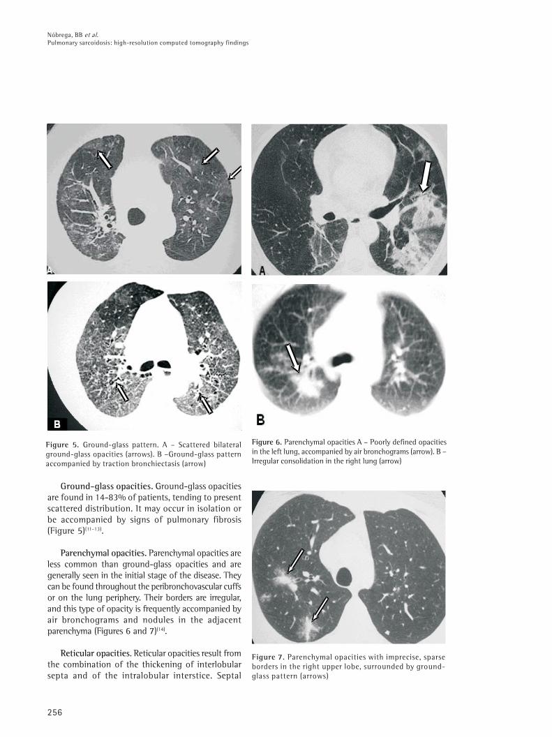
Figure 5. Ground-glass pattern. A – Scattered bilateralground-glass opacities (arrows). B –Ground-glass patternaccompanied by traction bronchiectasis (arrow)
Figure 6. Parenchymal opacities A – Poorly defined opacitiesin the left lung, accompanied by air bronchograms (arrow). B –Irregular consolidation in the right lung (arrow)
Figure 7. Parenchymal opacities with imprecise, sparseborders in the right upper lobe, surrounded by ground-glass pattern (arrows)
Ground-glass opacities. Ground-glass opacitiesare found in 14-83% of patients, tending to presentscattered distribution. It may occur in isolation orbe accompanied by signs of pulmonary fibrosis(Figure 5)(11-13).
Parenchymal opacities. Parenchymal opacities areless common than ground-glass opacities and aregenerally seen in the initial stage of the disease. Theycan be found throughout the peribronchovascular cuffsor on the lung periphery. Their borders are irregular,and this type of opacity is frequently accompanied byair bronchograms and nodules in the adjacentparenchyma (Figures 6 and 7)(14).
Reticular opacities. Reticular opacities result fromthe combination of the thickening of interlobularsepta and of the intralobular interstice. Septal

257
Jornal Brasileiro de Pneumologia 31(3) - Mai/Jun de 2005
Figure 8. Reticular opacities. Pulmonary sarcoidosis withseptal thickening of the upper lobes, forming polygonalarches (arrows), simulating lymphangitic carcinomatosis
Figure 9. Reticular opacities. A – Irregular thickening ofperibronchovascular cuffs (arrows). B – thickening of posteriorbasal interlobular septa (arrow) accompanied by distortion ofthe lung architecture
Figure 10. Areas of air trapping in the lower lobes (arrows)
thickening is typically found along theperibronchovascular cuffs but can also be seen inother topographies (Figure 8). Irregular linearopacities adjacent to the bronchovascular cuffs havealso been described and attributed to early
manifestations of pulmonary fibrosis (Figure 9)(7,9).
Air trapping. Air trapping during expiration isrelatively frequent, due to the presence ofperibronchial or submucosal granulomas or toperibronchiolar fibrosis, leading to small airwayobliteration (Figure 10) (15).
Fibrosis. Parenchymal abnormalities may evolve tofibrosis, which is accompanied by architecturaldistortion, volumetric loss, linear opacities,honeycombing, fibrotic masses, bronchiectasis andtraction bronchiolectasis. Fibrosis is usually found inthe upper and middle lung fields (Figures 11 and 12)(7).
Bronchiectas is . Bronchiectas is i s anuncommon finding in sarcoidosis and is frequentlyassociated with fibrosis (traction bronchiectasis).Other less common et iologies are ai rwayobstruction caused by granulomas and extrinsiccompression caused by lymph nodes (Figure 13)(6).
ATYPICAL FINDINGS
Pseudotumoral form. In some cases, coalescence
of interstitial granulomas results in large conglomerates
with the formation of masses (pseudotumors not
accompanied by air bronchograms) that compress
adjacent air spaces (Figures 14 and 15)(16).
Cysts. The etiology of cysts is uncertain. Of all the
possible causes, peripheral air imprisonment, alveolar
distention caused by endobronchial component, the
destruction of the alveolar parenchyma, and retractions
and collapses of the surrounding parenchyma have been
reported (Figure 16)(7).
True cavities. True cavities are extremely rare.Infectious causes and massive fibrosis with cysts must

258
Nóbrega, BB et al.
Pulmonary sarcoidosis: high-resolution computed tomography findings
Figure 11. Fibrosis. A – Traction bronchiectasis (arrows),architectural distortion and ground-glass pattern, showingthe classical aspect of fibrosis. B – Irregular parenchymalopacities accompanied by architectural distortion(arrows). C – Fibrotic conglomerate forming masses withretractile aspect (arrows)
Figure 12. Fibrosis. Posterior subpleural honeycombing,predominantly in the lower lobes (arrows)
Figure 13A. Bronchiectasis (arrows) associated with peripheral micronodules.B – Irregular fibrotic mass with traction bronchiectasis (arrows)
first be ruled out. True cavities, as well as apical cysts,may be the focus of saprophytic colonizations(17,18).
Bronchial changes (thickening/stenosis).Lenique et al. reported thickening of the bronchialwalls, caused by the deposition of granulomas alongthe peribronchovascular interstice, in 65% of patients(Figure 17)(19).
Lobar atelectasis. The occlusion of a lobarbronchus and consequent atelectasis may be causedby intraluminal disease or extrinsic compression.There are no radiological signs that help distinguishbetween lobar atelectasis caused by sarcoidosis andthat resulting from other causes(17).
Unilateral pulmonary parenchymal lesions. It hasbeen reported in the literature that unilateralparenchymal changes are caused mainly by localizedinterstitial disease, accompanied by an alveolar pattern,or by localized interstitial disease, accompanied by areticulonodular pattern or presenting as a pulmonarycoin lesion(7,17).
AB

259
Jornal Brasileiro de Pneumologia 31(3) - Mai/Jun de 2005
Figure 14. Pseudotumoral form. Mass with irregular borders inthe right upper lobe (arrow). Ground-glass areas characterized inthe left lower lobe and adjacent to the mass (arrows)
Figure 15. Pseudotumoral form. A – High-resolutioncomputed tomography wi th lung window showingspiculated mass in the posterior basal segment of thele f t lower lobe (a r row) . B – Dens i ty measurementduring the pre-contrast phase (40 UH). C – Scan threeminutes after intravenous administration of iodinatedcontrast material , highlighting the lesion (maximumdensity of 76 UH). Swensen’s protocol posit ive(16).Anatomopatho log ica l examinat ion o f the su rg i ca lsample confirmed sarcoidosis
Fairy-ring sign. The "fairy-ring sign" iscaused by multiple peripheral granulomatousnodules bordering areas of preserved lungparenchyma (Figure 18)(20)..
Figura 16. Sarcoidose com cistos bilaterais associados afístula broncopleural com formação de hidropneumotóraxà direita (setas)
Figure 17. Bronchial involvement. Unilateral presentation,characterized by concentric thickening of the bronchialwalls (arrows), with nodules with lymphatic distributionin the right lower lobe
Figure 18. Fairy-ring sign. Micronodules with ring-likedistribution (arrows) surrounding relatively preserved lungparenchyma

260
Nóbrega, BB et al.
Pulmonary sarcoidosis: high-resolution computed tomography findings
CONCLUSIONS
Because of the broad spectrum of presentations,pulmonary sarcoidosis may mimic other interstitialpulmonary diseases. High-resolution computedtomography is the most sensitive and specificimaging method for the evaluation of this disease.
In those cases in which atypical manifestationspredominate, the knowledge of the varioustomographic patterns helps us limit the differentialdiagnosis and select the best location for an eventualbiopsy.
REFERENCES
1. Traill ZC, Maskell GF, Gleesson FV. High-resolution CTf indings of pulmonary sarco idos i s . AJR 1997;168:1557-60.
2. Remy-Jardin M, Giroud F, Remy J, Wattine L, Wallaert B,Duhamel A. Pulmonary sarcoidosis: role of CT in theevaluation of disease activity and functional impairmentin prognosis assessement. Radiology 1994;191:675-80.
3. Miller BH, Rosado-de-Christenson ML, Mcadans HP,Jishback NF. Thoracic sarcoidosis: radiologic-pathologiccorrelation. Radiographics 1995;15:421-37.
4. Müller NL, Kulling P, Miller RR. The CT findings ofpulmonary sarcoidosis: analysis of 25 patients. AJR1989;152:1179-82.
5. Webb WR, Müller NL, Naidich DP. Doenças caracterizadasprincipalmente por opacidades nodulares ereticulonodulares. In: Webb WR, Müller NL, Naidich DP. TCde alta resolução do pulmão. 3a ed. Rio de Janeiro:Guanabara Koogan; 2002:245-336.
6. Hamper VM, Fishman EK, Khouri NF, Jonhs CT, WongKP, S iege lman SS. Typica l and atypica l CTmanifestations of pulmonary sarcoidosis. J ComputAssist Tomogr 1986, 10:929-36
7. Chiles C. Imaging features of thoracic sarcoidosis.Semin Roentgenol. 2002;37:82-93.
8. Nishimura K, Itoh H, Kitaichi M, Nagai S, Izumi T.Pulmonary sarcoidosis: correlat ion of CT andhistopathologic findings. Radiology 1993;189:105-9.
9. Brauner MW, Lenoir S, Grenier P, Cluzel P, Batteste JP,Valleyre D. Pulmonary sarcoidosis: CT assessment oflesion reversibility. Radiology 1992;182:349-54.
10. Nakatsu M, Hatabu H, Morikawa K , Vematsu H, Ohio Y,Nishimura K, et al. Large coalescent parenchymal nodulesin pulmonary sarcoidosis: “Sarcoid galaxy” sign. AJR2002;178:1389-93.
11 . Brauner MW, Grenier P, Monpoint D, Lenoir S, GremouxH. Pulmonary sarcoidosis: evaluation with high-resolution CT. Radiology 1989;174:467-71.
12 . Neto ALF, Marchiori E, Capone D, Mogami R. Aspectosde tomografia computadorizada de alta resolução nasarcoidose. Radiol Bras 1996: 29:325-30.
13. Lynch DA, Webb WR, Gainsu G, Itulbarg M, Golden J.Computed tomography in pulmonary sarcoidosis. JComput Assist Tomogr 1989; 13:405-10.
14 . Jokoh T, Ikezoe J, Takeuchi N, Kohuo N, Tomiyama N,Akira M, et al. CT findings in pseudoalveolar sarcoidosis.J Comput assist Tomogr 1992;16:904-7
15. Hansell DM, Milne DG, Wisher ML, Wells AV. Pulmonarysarcoidosis: morphologic associat ions of airf lowobstruct ion at th in sect ion CT. Radio logy1998;209:697-704
16. Swensen SJ, Viggiano RW, Midthun DE, Müller NL,Sher r ick A, Yamashi ta K, et a l . Lung NoduleEnhancement at CT: Multicenter Study1 Radiology2000; 214: 73-80.
17 . Rockoff SD, Rohatgi PK. Unusual manifestation ofthoracic sarcoidosis. AJR 1985;144:513-28.
18 . Ichikawa Y, Fujimoto K, Shiraishi T, Oizumi K. Primarycavitary sarcoidosis: high resolution CT findings. AJR1994;163:745
19. Lenique F, Brauner MW, Grenier P, Bettesti JP, LoixauA, Valeyre D. CT assessment of bronchi in sarcoidosis:endoscopic and pathologic correlations. Radiology1995;194:419-23.
20 . Marlow TJ, Krapiva PI, Schabel SI, Judson MA. The“fairy“ ring :a new radiographic finding in sarcoidosis.Chest 1999 ; 115 : 275-6.