reversible clustering of magnetic nanobiocatalysts for … of oleic acid-coated iron oxide magnetic...
TRANSCRIPT

Electronic Supplementary Material (ESI) for Chemical Communications This journal is © The Royal Society of Chemistry 2011
S1
Electronic Supplementary Information (ESI) for Chemical Communications
This journal is © The Royal Society of Chemistry of Chemistry 2011
Reversible Clustering of Magnetic Nanobiocatalysts for High-
Performance Biocatalysis and Easy Catalyst Recycling
Thao P. N. NGO,a Wei ZHANG, a Wen WANG a and Zhi LI* a
a Department of Chemical and Biomolecular Engineering, National University of Singapore,
4 Engineering Drive 4, Singapore 117576
E-mail: [email protected]; Fax: 65-6779 1936; Tel: 65-6516 8416
List of content:
1. Materials
2. Characterization methods
3. Synthesis of oleic acid-coated iron oxide magnetic nanoparticles (OA-MNPs)
4. Synthesis of magnetic nanoparticles containing iron oxide core and poly(glycidyl methacrylate) shell
(GMA-MNPs)
5. Synthesis of aldehyde functionalized magnetic nanoparticles (CHO-MNPs)
6. Measurements of zeta-potential and hydrodynamic size of CHO-MNPs at different pH
7. Production and purification of ketone reductase RDR
8. Immobilization of enzymes on magnetic nanoparticles and formation of fine nanobiocatalyst
9. Immobilization of enzymes on magnetic nanoparticles and formation of reversibly clustered RDR-
MNPs (RC RDR-MNPs)
10. Controlled synthesis of reversibly clustered nanobiocatalysts
11. Separation of the reversible clusters of RC RDR-MNPs
12. Catalysis of RC RDR-MNPs for reduction of 7-methoxy-2-tetralone with cofactor recycling
13. Synthesis of racemic 7-methoxy-2-tetralol by ketone reduction of 7-methoxy-2-tetralone with NaBH4
14. Preparation of (R)-7-methoxy-2-tetralol by ketone reduction of 7-methoxy-2-tetralone with RC
nanobiocatalysts
15. Stability of RC nanobiocatalysts at different pH and temperature
16. Recycling of RC nanobiocatalysts in bioreduction of 7-methoxy-2-tetralone
17. Reduction of 7-methoxy-2-tetralone with cofactor recycling catalyzed by free RDR at higher substrate
concentration
Electronic Supplementary Material (ESI) for Chemical CommunicationsThis journal is © The Royal Society of Chemistry 2012

Electronic Supplementary Material (ESI) for Chemical Communications This journal is © The Royal Society of Chemistry 2011
S2
18. Immobilization of enzymes on magnetic nanoparticles and formation of reversibly clustered TLL-
MNPs (RC TLL-MNPs)
19. Separation of the reversible clusters of RC TLL-MNPs
20. Catalysis of RC TLL-MNPs for hydrolysis of p-nitrophenyl butyrate
21. Recycling of RC TLL-MNPs in hydrolysis of p-nitrophenyl butyrate
22. References
Electronic Supplementary Material (ESI) for Chemical CommunicationsThis journal is © The Royal Society of Chemistry 2012
Electronic Supplementary Material (ESI) for Chemical Communications This journal is © The Royal Society of Chemistry 2011
S3
1. Materials
Ferric chloride hexahydrate (FeCl3.6H2O, 97%), ferrous chloride tetrahydrate (FeCl2.4H2O, 99%),
potassium oleate (40 wt. % in H2O), ammonium hydroxide (28% NH3 in H2O), ammonium persulfate
((NH4)2S2O8, ≥98.0%), glycidyl methacrylate (97%), ethylenediamine (NH2CH2CH2NH2, 99%), 4,7,10-trioxa-
1,13-tridecanediamine (C10H24N2O3, 95%), glutaraldehyde solution (CH2(CH2CHO)2, 25% in H2O), 7-methoxy-
2-tetralone (C11H12O2, 97%), n-hexadecane (CH3(CH2)14CH3, 99%), ampicillin (>99%) were purchased from
Sigma-Aldrich. β-Nicotinamide adenine dinucleotide phosphate disodium salt (C21H26N7Na2O17P3, 99%) was
purchased from Merck. Isopropyl--D-thiogalactopyranoside (IPTG) (>99%) was obtained from 1st BASE.
Isopropanol and ethyl acetate was purchased from Tedia. The medium components tryptone and yeast extract
were purchased from Biomed Diagnostics.
2. Characterization methods
Transmission electron microscope (TEM) was performed on JEOL: JEM-2010 model. Field emission
scanning electron microscope (FE-SEM) was performed on JEOL: JSM-6700F model. Vibrating sample
magnetometer (VSM) data were collected on VSM7407. Zetasizer nano-ZS from Malvern was used to measure
the hydrodynamic size distribution and zeta-potential of particles in solution. Optical microscopy were taken on
Leica TCS SP5 (10x).
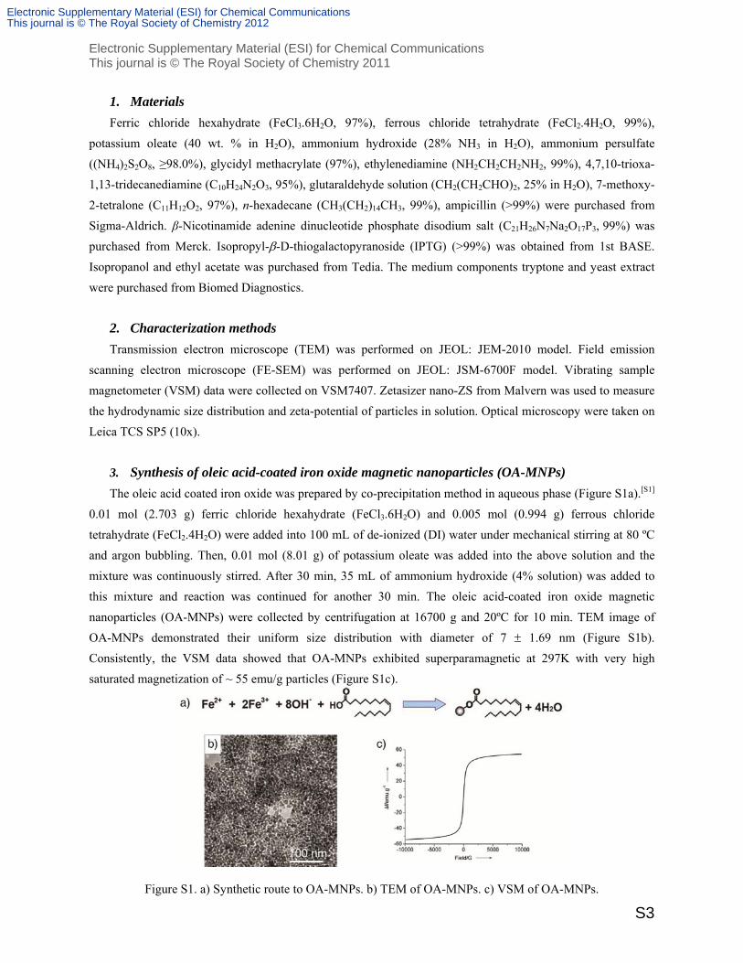
3. Synthesis of oleic acid-coated iron oxide magnetic nanoparticles (OA-MNPs)
The oleic acid coated iron oxide was prepared by co-precipitation method in aqueous phase (Figure S1a).[S1]
0.01 mol (2.703 g) ferric chloride hexahydrate (FeCl3.6H2O) and 0.005 mol (0.994 g) ferrous chloride
tetrahydrate (FeCl2.4H2O) were added into 100 mL of de-ionized (DI) water under mechanical stirring at 80 ºC
and argon bubbling. Then, 0.01 mol (8.01 g) of potassium oleate was added into the above solution and the
mixture was continuously stirred. After 30 min, 35 mL of ammonium hydroxide (4% solution) was added to
this mixture and reaction was continued for another 30 min. The oleic acid-coated iron oxide magnetic
nanoparticles (OA-MNPs) were collected by centrifugation at 16700 g and 20ºC for 10 min. TEM image of
OA-MNPs demonstrated their uniform size distribution with diameter of 7 1.69 nm (Figure S1b).
Consistently, the VSM data showed that OA-MNPs exhibited superparamagnetic at 297K with very high
saturated magnetization of ~ 55 emu/g particles (Figure S1c).
Figure S1. a) Synthetic route to OA-MNPs. b) TEM of OA-MNPs. c) VSM of OA-MNPs.
Electronic Supplementary Material (ESI) for Chemical CommunicationsThis journal is © The Royal Society of Chemistry 2012

Electronic Supplementary Material (ESI) for Chemical Communications This journal is © The Royal Society of Chemistry 2011
S4
4. Synthesis of magnetic nanoparticles containing iron oxide core and poly(glycidyl
methacrylate) shell (GMA-MNPs)
The OA-MNPs synthesized above was purified by high gradient magnetic separation (HGMS) to wash
away the un-reacted oleic acid and ammonium hydroxide. After that, these water based ferrofluid was used to
fabricate the magnetic iron oxide core-poly(glycidyl methacrylate) shell nanoparticles (GMA-MNPs). Different
sizes of the GMA-MNPs could be controlled in nanoscale by tuning the density of OA-MNPs. Reaction of 1
mg/mL, 0.48 mg/mL and 0.267 mg/mL of OA-MNPs with 9mg ammonium persulfate (APS) initiator and
0.126mL glycidyl methacrylate (GMA) in total 25mL mixture volume gave mean diameter of GMA-MNPs
42.5 ± 4.15 nm, 62.2 ± 4.99 nm and 107 ± 7.5 nm, respectively (as shown in Figure S2).
The amount of epoxy groups attached on GMA-MNPs was determined by method developed by Sundberg
et al.[S2] 100L of GMA-MNPs (8.3mg/mL solution) was added into 1.5 mL of sodium thiosulphate solution
(1.3 M). Reaction between epoxy groups on GMA-MNPs and sodium thiosulphate formed OH-. pH of this
reaction mixture was neutralized by addition of HCl until the reaction was completed. The amount of oxirane
presented on GMA-MNPs was then calculated from the amount of HCl used. The result showed that 3 mmol of
epoxy groups were attached per gram GMA-MNPs.
Figure S2. a) Synthetic route to GMA-MNPs. b)-d) TEM of GMA-MNPs: b) with a diameter of 42.5 ± 4.1 nm,
c) with a diameter of 62.2 ± 5.0 nm, and d) with a diameter of 107 ± 7.5 nm.
5. Synthesis of aldehyde functionalized magnetic nanoparticles (CHO-MNPs)
To synthesize aldehyde funtionalized magnetic nanoparticles, GMA-MNPs synthesized above were firstly
functionalized with amine before functionalized with aldehyde groups. To introduce the amine functional
groups, 0.011 mol (2.4 mL) of 4,7,10-trioxa-1,13-tridecanediamine was added into 48mL aqueous solution
containing 0.112g GMA-MNPs and incubated under magnetic stirring at 80oC for 24 h. Afterwards, the GMA-
MNPs with functional amine groups of 4,7,10-trioxa-1,13-tridecanediamine (NH2-MNPs) were collected by
centrifugation at 21000 g for 20 min and washed by DI water several times. To introduce the aldehyde
functional groups on the surface of nanoparticles, 135 mL glutaraldehyde (10% solution) containing 0.11 g
NH2-MNPs was incubated at room temperature under mild shaking for 18 h. The aldehyde-containing
Electronic Supplementary Material (ESI) for Chemical CommunicationsThis journal is © The Royal Society of Chemistry 2012

Electronic Supplementary Material (ESI) for Chemical Communications This journal is © The Royal Society of Chemistry 2011
S5
nanoparticles (CHO-MNPs) were washed several times in DI water. After washed, 0.1g CHO-MNPs were
obtained. TEM image and DLS measurement showed no dramatic difference in shape and size of GMA-MNPs,
NH2-MNPs and CHO-MNPs with diameter of 63nm from TEM image and 157nm from DLS (Figure S3).
Figure S3. a) Synthetic route to NH2-MNPs and CHO-MNPs. b) TEM of NH2-MNPs. c) TEM of CHO-MNPs.
d) DLS of NH2-MNPs. e) DLS of CHO-MNPs. f) DLS of GMA-MNPs.
Electronic Supplementary Material (ESI) for Chemical CommunicationsThis journal is © The Royal Society of Chemistry 2012
Electronic Supplementary Material (ESI) for Chemical Communications This journal is © The Royal Society of Chemistry 2011
S6
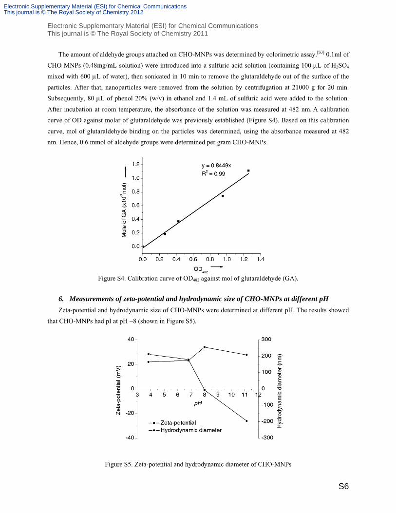
The amount of aldehyde groups attached on CHO-MNPs was determined by colorimetric assay.[S3] 0.1ml of
CHO-MNPs (0.48mg/mL solution) were introduced into a sulfuric acid solution (containing 100 L of H2SO4
mixed with 600 L of water), then sonicated in 10 min to remove the glutaraldehyde out of the surface of the
particles. After that, nanoparticles were removed from the solution by centrifugation at 21000 g for 20 min.
Subsequently, 80 L of phenol 20% (w/v) in ethanol and 1.4 mL of sulfuric acid were added to the solution.
After incubation at room temperature, the absorbance of the solution was measured at 482 nm. A calibration
curve of OD against molar of glutaraldehyde was previously established (Figure S4). Based on this calibration
curve, mol of glutaraldehyde binding on the particles was determined, using the absorbance measured at 482
nm. Hence, 0.6 mmol of aldehyde groups were determined per gram CHO-MNPs.
Figure S4. Calibration curve of OD482 against mol of glutaraldehyde (GA).
6. Measurements of zeta-potential and hydrodynamic size of CHO-MNPs at different pH
Zeta-potential and hydrodynamic size of CHO-MNPs were determined at different pH. The results showed
that CHO-MNPs had pI at pH ~8 (shown in Figure S5).
Figure S5. Zeta-potential and hydrodynamic diameter of CHO-MNPs
Electronic Supplementary Material (ESI) for Chemical CommunicationsThis journal is © The Royal Society of Chemistry 2012

Electronic Supplementary Material (ESI) for Chemical Communications This journal is © The Royal Society of Chemistry 2011
S7
7. Production and purification of ketone reductase RDR
E.coli Pet28a RDR (N-histag) containing histag-RDR were inoculated onto LB agar plates (10 g tryptone, 5
g yeast extract and 5 g NaCl in 1 liter DI water with 1.5% agar) containing kanamycin (50 g/mL), and grown
overnight at 37°C.[S4] A single colony of this strain grown on the LB agar plate was inoculated into 100 mL of
LB medium with kanamycin (50 g/mL), and then the cells were grown at 250 rpm and 37°C for 12 h to OD600
of 1.46. 10 mL of this pre-culture of E. coli Pet28a RDR (N-histag) was added to 1 L of TB medium (1 L DI
water, 12 g Bacto tryptone, 24 g Bacto yeast extract, 4 mL glycerol, KH2PO4 2.3 g, K2HPO4 12.54 g) containing
kanamycin 50 g/mL. The mixture was shaken at 250 rpm and 37°C. Samples were taken at different time
points for OD600 measurement. When the OD600 reached around 1, IPTG was added to 0.25 mM and then the
mixture was continuously shaken at 250 rpm, 30°C. The cells were harvested at the late exponential phase
(OD600 ~ 13) after 21 h by centrifugation at 5400 g and 4oC for 10 min, then washed with potassium phosphate
(KP) buffer (5 mM; pH 7.5), and stored at -80°C.
2.4g wet cells of E. coli Pet28a RDR (N-histag) were suspended in 120 mL Immidazole buffer (10 mM; pH
8), then passed through a homogenizer (Constant cell disruption system) twice at 20,000 lb/in2. The resulting
mixture was centrifuged at 21,000 g and 4°C for 30 min to remove the cell debris. 120 mL of cell free extract
(8.92 mg protein/mL) was filtrated through membrane filter (pore diameter 0.2 µm), mixed with 8 mL Ni-NTA
resin and shaken at 4oC and 30 rpm on a rocking chair for 1 h for effective binding of histag-RDR onto the Ni-
NTA resin. Then, the mixture of lysate and Ni-NTA in Immidazole buffer was loaded into an empty column
and successively washed with 60 mL of Immidazole 20 mM, 80 mL of Immidazole 30 mM, and 24 mL of
Immidazole 50 mM. The active and purified fractions of histag-RDR were eluted when adding 8 mL of
Immidazole 250 mM (5 times). The purified histag-RDR (7 mg enzymes/mL) was desalted via Amicon ultra-15
10K centrifugal filter devices and stored in Tris buffer (20 mM, pH 8) containing glycerol 20%. The protein
concentration was determined by using the Bradford protein content assay with bovine serum albumin as a
standard. To make the Sodium dodecyl sulfate-polyacrylamide gel electrophoresis (SDS-PAGE), 15 g proteins
was loaded on a gel containing 0.1% SDS and 10% acrylamide, the gel was stained with a 0.1% solution of
Coomassie brilliant blue and destained by soaking in DI water in 1 h.
262.6 mg histag-RDR enzymes were purified by Ni-NTA column from cell free extract containing 1.07 g
protein. SDS-PAGE (shown in Figure S6) showed the proteins in cell free extract of E.coli pET28a histag-RDR
(lane 2), washing solution of immodazole (10 mM, 20 mM, 50 mM) (lane 3, 4 and 5 respectively) and purified
histag-RDR (lane 6). Only one clear and dark band of histag-RDR was visible in the purified histag-RDR
solution (compared to many bands shown from cell free extract sample), thus histag-RDR was successfully
purified by Ni-NTA with high purity.
Electronic Supplementary Material (ESI) for Chemical CommunicationsThis journal is © The Royal Society of Chemistry 2012

Electronic Supplementary Material (ESI) for Chemical Communications This journal is © The Royal Society of Chemistry 2011
S8
Figure S6. SDS-PAGE: L1–marker, L2–cell free extract of E.coli pET28a histag-RDR, L3–washing solution of
immidazole 10mM, L4–washing solution of immidazole 20mM, L5–washing solution of immidazole 50mM,
L6–purified histag-RDR.
The purified histag-RDR and cell free extract were used for reduction of 7-methoxy-2-tetralone. 1 mL of
Tris buffer (6 mM, pH 8) containing MgCl2 (1 mM), 7-methoxy-2-tetralone (0.65 mM), NADH (1 mM) and
0.02 mg histag-RDR immobilized on RC nanobiocatalysts were mixed and shaken at 1000 rpm and 30oC for
5min. In order to prepare samples for GC measurement, samples were extracted in same amount of ethyl acetate
(EtOAc) containing 2 mM of n-hexadecane as internal standard. Concentration of 7-methoxy-2-tetralone and 7-
methoxy-2-tetralol were analyzed by using an Agilent GC HP-5 column (described in part 12). As a result,
activity of the purified histag-RDR was 1.75 times higher than activity of cell free extract.
8. Immobilization of enzymes on magnetic nanoparticles and formation of fine nanobiocatalyst
2 mL of 18.2m ultrapure water (salt free, pH 5.5) containing 2.57 mg CHO-MNPs and 0.18 mg purified
histag-RDR was mildly shaken in aqueous solution at 4oC and 30 rpm on a rocking chair. After 4 h, fine
nanobiocatalysts (RDR-MNPs) were obtained without clustering. TEM image of RDR-MNPs demonstrated no
dramatic change in shape or size of particles before and after immobilization (Figure S7).
Figure S7. a) TEM and b) FESEM of the fine nanobiocatalyst (RDR-MNPs)
Electronic Supplementary Material (ESI) for Chemical CommunicationsThis journal is © The Royal Society of Chemistry 2012

Electronic Supplementary Material (ESI) for Chemical Communications This journal is © The Royal Society of Chemistry 2011
S9
9. Immobilization of enzymes on magnetic nanoparticles and formation of reversible clusters
In another setting of experiment, enzymes were immobilized in the same procedure that 2.57 mg CHO-
MNPs and 0.18 mg purified histag-RDR was mildly shaken at 4oC and 30 rpm on a rocking chair for 4 h but in
2 mL of phosphate buffer (7 mM, pH 8). As a result, reversible cluster of magnetic nanobicatalysts was
obtained. Afterwards, the RC RDR-MNPs was treated with Tris buffer (0.1 M, pH 8) to block the un-reacted
aldehyde groups on the particles, and then washed several times to remove free RDR by using external
magnetic field. The results showed that around 2.57mg RC RDR-MNPs were obtained. 76% of histag-RDR
added were immobilized on CHO-MNPs, resulting in loading capacity of 53 mg enzymes/g particles. They
relatively polydispersed with regular shape and mean size of about 8.5 m but individual fine nanobiocatalysts
with diameter of 68 7.4 nm were observed as a discrete entity in the microsphere of RC RDR-MNPs (in
Figure 2b-c of the main text). However, hydrodynamic size of RC nanobiocatalysts after sonication was
measured and showed not significantly different from that of the fine RDR-MNPs, indicating that RC
nanobiocatalysts can dissociate into fine RDR-MNPs (Figure S8-9). The overall synthesis of the biocatalysts is
highly reproducible with 89% yield from GMA-MNPs (i.e. 0.1g RC RDR-MNPs were produced from 0.112g
GMA-MNPs).
Figure S8. a) DLS of RC RDR-MNPs after sonication. b) DLS of fine RDR-MNPs.
Figure S9. FESEM of RC RDR-MNPs after sonication.
Electronic Supplementary Material (ESI) for Chemical CommunicationsThis journal is © The Royal Society of Chemistry 2012

Electronic Supplementary Material (ESI) for Chemical Communications This journal is © The Royal Society of Chemistry 2011
S10
10. Controlled synthesis of reversibly clustered nanobiocatalysts
The formation of RC nanobiocatalysts was examined at different pH value and phosphate buffer
concentration in the same procedure described in part 9. The results were listed in Table S1. RC
nanobiocatalysts were not formed at pH 5.5, even at high salt concentration. At pH 8, RC nanobiocatalysts were
not formed in the absence of salt or salt concentration below 2mM. In different experiment, CHO-MNPs
without immobilized enzymes were observed to be well dispersed at pH 8 in different phosphate buffer (from
6.5 to 90 mM). It means that particles did not clustere before the addition of enzymes.
Table S1: Catalyst formed by immobilization under different pH and phosphate buffer concentration.[a]
Phosphate buffer (mM) Catalyst immobilized at pH 5.5 Catalyst immobilized at pH 8
0
1
2
4
7
20
50
90
RDR-MNPs
RDR-MNPs
RDR-MNPs
RDR-MNPs
RDR-MNPs
RDR-MNPs
RDR-MNPs
RDR-MNPs
RDR-MNPs
RDR-MNPs
RC RDR-MNPs
RC RDR-MNPs
RC RDR-MNPs
RC RDR-MNPs
RC RDR-MNPs
-
[a]: Immobilization condition: 2 mL of phosphate buffer at different concentration, 2.57 mg CHO-MNPs and
0.18 mg purified histag-RDR was mildly shaken at 4oC and 30 rpm on a rocking chair for 4 h.
In another experiment, enzymes were firstly immobilized in ultra pure water (pH 8) for 4 h to form fine
nanobiocatalysts with a loading capacity of 37 mg enzymes/g particles. After that, phosphate buffer was added
to 7 mM. Consequently, RC nanobiocatalysts were quickly formed within 10 min and a higher enzyme loading
was also achieved (48 mg enzymes/g particles). After 1 h, immobilization finished with loading capacity at 53
mg enzymes/g particles (Figure S10). The RC nanobiocatalysts were sonicated in untra pure water, centrifuged
and then enzyme amount in this supernatant was determined. A negligible amount of enzymes was found in this
supernatant, indicating that enzymes were mostly covalently immobilized on particles before clustered.
Figure S10. Enzyme concentration in supernatant during immobilization in ultra pure water (pH 8). Phosphate
buffer was added to 7mM at 4h.
Electronic Supplementary Material (ESI) for Chemical CommunicationsThis journal is © The Royal Society of Chemistry 2012

Electronic Supplementary Material (ESI) for Chemical Communications This journal is © The Royal Society of Chemistry 2011
S11
11. Separation of the reversible clusters of RC RDR-MNPs
The separation of the reversible clusters of RC RDR-MNPs was done under external magnetic field. It was
observed that RC RDR-MNPs were completed separated within 4s (as illustrated in Figure 3 of the main text).
12. Catalysis of RC nanobiocatalysts for reduction of 7-methoxy-2-tetralone with cofactor
recycling
1 mL of Tris buffer (6 mM, pH 8) containing MgCl2 (1 mM), 7-methoxy-2-tetralone (10.5 mM),
isopropanol (48 mM), NADH (0.0012 mM) and 0.1 mg histag-RDR immobilized on RC nanobiocatalysts were
shaken at 1000 rpm and 30oC. Samples were taken at 5 min, 20 min, 40 min and 60 min and they were extracted
in higher amount of ethyl acetate (EtOAc) (12 times higher) containing 2 mM of n-hexadecane as internal
standard. To quantify concentrations of 7-methoxy-2-tetralone and 7-methoxy-2-tetralol, sample were analyzed
by using an Agilent GC HP-5 column (25 m by 0.32 mm) with an inlet temperature of 290°C and a detector
temperature of 310°C. The temperature program was as follows: temperature increased from 60°C to 195°C at a
rate of 15°C/min, from 195°C to to 200°C at a rate of 5°C/min, then from 200°C to 280°C at a rate of 30°C/min
and kept at 280°C for 1 min. The retention times were 9.4 min for 7-methoxy-2-tetralone, 9.6 min for 7-
methoxy-2-tetralol and 9 min for n-hexadecane. It was observed that catalytic performance of RC RDR-MNPs
was as same as that of free enzyme. After 60 min, 10.2 mM of 7-methoxy-2-tetralol was produced by RC RDR-
MNPs while 9.7 mM of product was produced by free histag-RDR. The final turn over number (TTN) for
NADH recycling was calculated by dividing the number of mol product formed by the number of mol NADH
added. For production of 10.2 mM 7-methoxy-2-tetralol, NADH was recycled for 8,500 times.
For up-scale, shaking flask of 10 mL of Tris buffer (6 mM, pH 8) containing MgCl2 (1 mM), 7-methoxy-2-
tetralone (10.5 mM), isopropanol (48 mM), NADH (0.0012 mM) and 1 mg free histag-RDR or histag-RDR
immobilized on RC nanobiocatalysts were shaken at 300 rpm and 30oC for 60 min. After biotransformation,
crude product was extracted with EtOAc and analyzed by GC. Again, catalytic performance of RC RDR-MNPs
was as same as that of free enzyme in this large scale (Figure S11).
Figure S11. Time course of biotransformation on a 10mL-scale with shaking at 300 rpm.
Electronic Supplementary Material (ESI) for Chemical CommunicationsThis journal is © The Royal Society of Chemistry 2012

Electronic Supplementary Material (ESI) for Chemical Communications This journal is © The Royal Society of Chemistry 2011
S12
13. Synthesis of racemic 7-methoxy-2-tetralol by ketone reduction of 7-methoxy-2-tetralone with
NaBH4
To 20 mL of anhydrous methanol (MeOH) containing 1.66 mmol (0.2925 g) of 7-methoxy-2-tetralone
under Argon atmosphere at -20oC were gradually added 3.18 mmol (0.12 g) of NaBH4. The reaction mixture
was allowed to warm to room temperature and stirred for 4 h (TLC control, 1hexane : 5 EtOAc, Rf = 0.25).
Afterwards, this mixture was quenched by the addition of 0.6 mL acetone, 20 mL water and few drops of
concentrated HCl before removing MeOH under reduced pressure. The remaining aqueous phase was extracted
with 20 mL of EtOAc (3 times). After that, the organic extracts were dried by Na2SO4, filtered and then
evaporated under reduced pressure to give racemic 7-methoxy-2-tetralol as solid. The product was analyzed by
GC with >99% purity (as shown in Figure S12a). 93% of yield of product (0.274 g) was obtained.
The product ee was analyzed by HPLC on Daicel Chiralcel OJ column (4.6 mm x 250 mm) with detection
of 210 nm and column temperature at 30oC. n-hexane (90% v/v) and isopropanol (10% v/v) was used as mobile
phase with a flow rate of 1 mL/min. The retention time were 10.45 min for (R) 7-methoxy-2-tetralol and
12.8min for (S) 7-methoxy-2-tetralol, which is similar to the reported data (Figure S12b).[S5]
Figure S12. a) GC chromatogram and b) chiral HPLC chromatogram of of racemic 7-methoxy-2-tetralol.
Electronic Supplementary Material (ESI) for Chemical CommunicationsThis journal is © The Royal Society of Chemistry 2012

Electronic Supplementary Material (ESI) for Chemical Communications This journal is © The Royal Society of Chemistry 2011
S13
14. Preparation of (R)-7-methoxy-2-tetralol by ketone reduction of 7-methoxy-2-tetralone with
RC nanobiocatalysts
7-methoxy-2-tetralol was produced from biotransformation by catalysis of free enzymes and the RC
nanobiocatalysts. 6 shaking flask of 10 mL of Tris buffer (6 mM, pH 8) containing MgCl2 (1 mM), 7-methoxy-
2-tetralone (10.5 mM), isopropanol (48 mM), NADH (0.0012 mM) and 1 mg free histag-RDR or histag-RDR
immobilized on RC nanobiocatalysts were shaken at 300 rpm and 30oC for 60 min. After biotransformation,
crude product was extracted with 120 mL of EtOAc (3 times). After that, the organic extracts were evaporated
under reduced pressure to give 7-methoxy-2-tetralol as liquid. The product was analyzed by GC with 96%
purity (as shown in Figure S13a,c).
The product was further purified by flash chromatography on silica column (1hexane : 5 EtOAc, Rf = 0.25) to
give pure 7-methoxy-2-tetralol as solid. The purity of 7-methoxy-2-tetralol was >99% by GC analysis (Figure
S13b,d). Yield of product produced from biotransformation by catalysis of free enzymes and the RC
nanobiocatalysts after purified was 84% and 85%, respectively (0.093 g and 0.0944 g product).
Figure S13. a)-b). GC chromatograms of product mixture from biotransformation with free enzyme: a) before
purification and b) after purification. c)-d). GC chromatograms of product mixture from biotransformation with
RC nanobiocatalysts: c) before purification and d) after purification.
The product ee was analyzed by HPLC as describe in part 14. As shown in Figure S14, the purified
products from biotransformation with both free enzymes and RC nanobiocatalysts have >99% ee.
Electronic Supplementary Material (ESI) for Chemical CommunicationsThis journal is © The Royal Society of Chemistry 2012
Electronic Supplementary Material (ESI) for Chemical Communications This journal is © The Royal Society of Chemistry 2011
S14
Figure S14. Chiral HPLC chromatograms of purified 7-methoxy-2-tetralol produced with a) free enzyme and b)
RC RDR-MNPs.
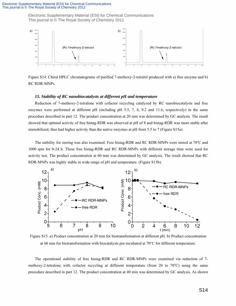
15. Stability of RC nanobiocatalysts at different pH and temperature
Reduction of 7-methoxy-2-tetralone with cofactor recycling catalyzed by RC nanobiocatalysts and free
enzymes were performed at different pH (including pH 5.5, 7, 8, 9.2 and 11.6, respectively) in the same
procedure described in part 12. The product concentration at 20 min was determined by GC analysis. The result
showed that optimal activity of free histag-RDR was observed at pH of 8 and histag-RDR was more stable after
immobilized, thus had higher activity than the native enzymes at pH from 5.5 to 7 (Figure S15a).
The stability for storing was also examined. Free histag-RDR and RC RDR-MNPs were stored at 70oC and
1000 rpm for 0-24 h. These free histag-RDR and RC RDR-MNPs with different storage time were used for
activity test. The product concentration at 60 min was determined by GC analysis. The result showed that RC
RDR-MNPs was highly stable in wide range of pH and temperature. (Figure S15b).
Figure S15. a) Product concentration at 20 min for biotransformation at different pH. b) Product concentration
at 60 min for biotransformation with biocatalysts pre-incubated at 70C for different temperature.
The operational stability of free histag-RDR and RC RDR-MNPs were examined via reduction of 7-
methoxy-2-tetralone with cofactor recycling at different temperature (from 20 to 70oC) using the same
procedure described in part 12. The product concentration at 40 min was determined by GC analysis. As shown
Electronic Supplementary Material (ESI) for Chemical CommunicationsThis journal is © The Royal Society of Chemistry 2012

Electronic Supplementary Material (ESI) for Chemical Communications This journal is © The Royal Society of Chemistry 2011
S15
in Figure S16, activity of free histag-RDR was optimal from 30 to 50oC and dramatically decreased at 70oC,
while activity of RC RDR-MNPs was stay the same from 20 to 70oC.
Figure S16. Product concentration from biotransformation of reduction of 7-methoxy-2-tetralone with free
enzyme and RC RDR-MNPs at different reaction temperature.
16. Recycling of RC nanobiocatalysts in bioreduction of 7-methoxy-2-tetralone
Recycling of RC RDR-MNPs was conducted for reduction of 7-methoxy-2-tetralone with cofactor
recycling in the same procedure described in part 12. 1 mL of Tris buffer (6 mM, pH 8) containing MgCl2 (1
mM), 7-methoxy-2-tetralone (10.5 mM), isopropanol (48 mM), NADH (0.0012 mM) and 0.1 mg histag-RDR
immobilized on RC nanobiocatalysts were shaken at 3000 rpm and 30oC. After 20 min, RC RDR-MNPs were
induced and separated by external magnetic field. The product mixture in supernatant was extracted in higher
amount of ethyl acetate (EtOAc) (12 times higher) containing 2 mM of n-hexadecane and used for GC analysis.
The RC RDR-MNPs were washed several time before being reused in new cycles. RC nanobiocatalysts were
added to 1 mL of Tris buffer (6 mM, pH 8) containing MgCl2 (1 mM), 7-methoxy-2-tetralone (10.5 mM),
isopropanol (48 mM), NADH (0.0012 mM), and the reaction mixture were shaken at 1000 rpm and 30oC to
start the new cycle of reaction for another 20 min. The result showed that after 14 recycles, the catalyst still
remained 80% of its original activity (described in Figure 4c in the main text). 125 mM of 7-methoxy-2-tetralol
intermediate was totally produced in 14 cycles (80% yield). The NADH was recycled for 6000 to 7700 times in
each cycle.
17. Reduction of 7-methoxy-2-tetralone with co-factor recycling catalyzed by free histag-RDR at
higher substrate concentration
1 mL of Tris buffer (6 mM, pH 8) containing MgCl2 (1 mM), 7-methoxy-2-tetralone (31.5 mM),
isopropanol (144 mM), NADH (0.0036 mM) and 0.2 mg histag-RDR were shaken at 1000 rpm and 30oC. The
substrate and co-factor were added either in one-pot from the beginning or stepwise at 0 min, 10 min and 20
min. After 30min, the substrate and product were extracted and quantified in the same procedure described in
Electronic Supplementary Material (ESI) for Chemical CommunicationsThis journal is © The Royal Society of Chemistry 2012

Electronic Supplementary Material (ESI) for Chemical Communications This journal is © The Royal Society of Chemistry 2011
S16
part 12. After 30 min, 24.5mM and 29.2mM of 7-methoxy-2-tetralol was produced by free histag-RDR via
stepwise and one-pot addition of 31.5 mM of substrate and 0.0036 mM of co-factor, respectively.
Figure S17. Product () and substrate () concentration from the biotransformation of 31.5mM 7-methoxy-2-
tetralone, isopropanol (144 mM), NADH (0.0036 mM), 0.2mg/ml enzymes with a) stepwise addition of
substrate and co-factor after each 10min; and b) one-pot addition of substrate and co-factor from the beginning.
18. Immobilization of enzymes on magnetic nanoparticles and formation of reversibly clustered
TLL-MNPs (RC TLL-MNPs)
The Thermomyces Lanuginosus lipase (TLL) (molecular weight of 30KD)[S6] was also immobilized on the
CHO-MNPs in the same procedure of RC RDR-MNPs synthesis (in part 9) as another example to prove the
concept. 1.5 mg CHO-MNPs and 0.15 mg TLL was mildly shaken in 1 mL of phosphate buffer (7 mM, pH 8) at
4oC and 30 rpm on a rocking chair for 4 h. As a result, reversible cluster of magnetic nanobiocatalysts was
obtained. Afterwards, the RC TLL-MNPs was washed several times to remove free enzymes by using external
magnetic field. 42% of TLL added were immobilized on CHO-MNPs, resulting in loading capacity of 42 mg
enzymes/g particles. The results showed that RC TLL-MNPs has regular shape and mean size of about 27 m.
But during shaking, they were dissociated into smaller nano-particles, and only a few particles with size of
200nm-2m were observed in the microscopy shown in Figure S18 (particles with size less than 200nm are
invisible in the microscopy due to detection limit). The overall synthesis of TLL-MNPs is highly reproducible
with 89% yield from GMA-MNPs.
Figure S18. Optical microscopy of RC Lipase-MNPs a) before and b) after shaking at 300rpm and 30C
for 2 min.
Electronic Supplementary Material (ESI) for Chemical CommunicationsThis journal is © The Royal Society of Chemistry 2012

Electronic Supplementary Material (ESI) for Chemical Communications This journal is © The Royal Society of Chemistry 2011
S17
19. Separation of the reversible clusters of RC TLL-MNPs
The separation of the reversible clusters was done under external magnetic field. It was observed that RC
TLL-MNPs were completed separated within 4s (as illustrated in Figure S19)
Figure S19. Magnetic separation of RC TLL-MNPs: a) t=0, b) t=4sec.
20. Catalysis of RC TLL-MNPs for hydrolysis of p-nitrophenyl butyrate
8 microgram of free lipase or TLL immobilized on RC was used for hydrolysis of p-nitrophenyl buturate
(p-NPB) (5mM) in the presence of K-buffer (7mM, pH 7.5) at 30ºC in 5ml scale in the shaking flask at 300
rpm. Samples were taken at 1 min, 2 min, 3 min, 5 min and 10 min. Ethanol 95% was added to stop the
reaction. The amount of p-nitrophenol produced in the reaction mixture was determined by UV-Vis at the
wavelength of 400nm. It was observed that after 10 min, 4.0 and 3.9 mM p-nitrophenol were produced under
catalysis of free enzyme and RC TLL-MNPs, respectively, in 78% yields (illustrated in Figure S20).
Figure S20. Time course of biotransformation catalyzed by free TLL and RC TLL-MNPs.
21. Recycling of RC TLL-MNPs in hydrolysis of p-nitrophenyl butyrate
Recycling of RC TLL-MNPs was conducted for hydrolysis of p-nitrophenyl butyrate in the same procedure
described in part 19. 8 microgram of free lipase or TLL immobilized on RC was used for hydrolysis of p-
nitrophenyl buturate (p-NPB) (5mM) in the presence of K-buffer (7mM, pH 7.5) at 30ºC in 5ml scale in the
rocking chair at 300 rpm in 10 min. The RC TLL-MNPs were separated and collected by external magnetic
field. The amount of p-nitrophenol produced in the reaction mixture was determined by UV-Vis at the
wavelength of 400nm. The RC RDR-MNPs were washed and then reused in new cycles. RC TLL-MNPs were
added to 5 mL of K-buffer (7 mM, pH 7.5) containing p-NPB (5 mM), and the reaction mixture were shaken at
Electronic Supplementary Material (ESI) for Chemical CommunicationsThis journal is © The Royal Society of Chemistry 2012

Electronic Supplementary Material (ESI) for Chemical Communications This journal is © The Royal Society of Chemistry 2011
S18
300 rpm and 25oC to start the new cycle of reaction for another 10 min. The result showed that after 9 recycles,
the catalyst still remained 92% of its original activity (described in Figure S21).
Figure S21. Recycling of RC TLL-MNPs in the hydrolysis of p-nitrophenyl butyrate.
22. References
[S1] W. Wang, Y. Xu, D. I. C. Wang and Z. Li, J. Am. Chem. Soc. 2009, 131, 12892.
[S2] L. Sundberg and J. Porath, J. Chromatography A, 1974, 90, 87.
[S3] M. Ghasemi, M. Minier, M. Tatoulian and F. Arefi-Khonsari, Langmuir, 2007, 23, 11554.
[S4] W. L. Tang, Z. Li and H. Zhao,Chem. Commun., 2010, 46, 5461.
[S5] T. Honda, A. Fujii, K. Inoue, Y. Yasohara, Y. Itagaki, K. Maehara, T. Takeda and Y. Ueda,
European patent 20020788671, 2002.
[S6] N. Li, M.-H. Zong and D. Ma, J. Biotechnol., 2009, 140, 250.
Electronic Supplementary Material (ESI) for Chemical CommunicationsThis journal is © The Royal Society of Chemistry 2012