resident orientation to nephrology & renal transplant...
TRANSCRIPT
RESIDENT ORIENTATION TO NEPHROLOGY & RENAL TRANSPLANT
@ St. Michael’s Hospital Third Edition January 2010
Introduction & Objectives Page 2
Table of Contents I. INTRODUCTION .......................................................................................................................................................... 3
Rotation Objectives: Core Medical Trainees.......................................................................................................... 3 Nephrology & Renal Transplantation Administrative Structure............................ Error! Bookmark not defined. Nephrology & Renal Transplantation Housestaff Responsibilities ...................................................................... 5
II. IN-PATIENT WARD EXPERIENCE ............................................................................................................................ 6 III. CONSULTATION EXPERIENCE............................................................................................................................... 6 IV. OUT-PATIENT EXPERIENCE................................................................................................................................... 7 V. EDUCATION AND RESEARCH EXPERIENCE......................................................................................................... 8
A. Teaching................................................................................................................................................................. 8 B. Rounds ................................................................................................................................................................... 9 C. Research .............................................................................................................................................................. 10
VI. HAEMODIALYSIS ................................................................................................................................................... 11 Resident responsibilities for chronic HD patients................................................................................................ 11 Dialysis Orders......................................................................................................................................................... 12 Vascular Access for Haemodialysis ...................................................................................................................... 14 Problems with Central Venous Dialysis Catheters............................................................................................... 15 Renal Replacement Therapy for Acute Kidney Injury .......................................................................................... 19 Continuous Renal Replacement Therapy (CRRT) ................................................................................................ 20 Management of Intoxications ................................................................................................................................. 22 Home Hemodialysis................................................................................................................................................. 27
VII. PERITONEAL DIALYSIS ....................................................................................................................................... 28 CAPD (Continuous Ambulatory Peritoneal Dialysis) ........................................................................................... 29 PD PERITONITIS ...................................................................................................................................................... 31 PD CATHETER INSERTION..................................................................................................................................... 36 Short Term Management......................................................................................................................................... 36 Long Term Management ......................................................................................................................................... 36 Catheter Dysfunction............................................................................................................................................... 37 INTRAPERITONEAL (IP) MEDICATIONS................................................................................................................ 37
VIII. RENAL TRANSPLANTATION .............................................................................................................................. 39 A. PRE-OP PROCEDURES ...................................................................................................................................... 39 DECEASED DONOR TRANSPLANT ....................................................................................................................... 39 B. POST-OP MANAGEMENT ................................................................................................................................... 42 C. POST-OP MEDICATIONS .................................................................................................................................... 43 D. DELAYED GRAFT FUNCTION ............................................................................................................................ 49 E. COMPLICATIONS IN TRANSPLANT PATIENTS................................................................................................ 50
IX. GENERAL NEPHROLOGY ..................................................................................................................................... 54 A. Renal Function .................................................................................................................................................... 54 B. Acute Renal Failure............................................................................................................................................. 55 1. Pre-Renal .............................................................................................................................................................. 55 2. Post Renal ............................................................................................................................................................ 55 3. Renal ..................................................................................................................................................................... 55 C. Glomerular Diseases- A Classification ............................................................................................................. 57
Acknowledgements..................................................................................................................................................... 60
Introduction & Objectives Page 3
I. INTRODUCTION Welcome to Nephrology. In addition to goals and objectives, this manual details some of the practical issues relating to your time on the nephrology ward (8CS), the consult service and the outpatient clinics. There are more detailed (and often more up to date) protocols on 8CS for many of the issues related to specific subjects in transplantation and dialysis. Rotation Objectives: Core Medical Trainees A) Data Gathering/Knowledge General Nephrology
1) To understand the approach to renal failure acute vs chronic if acute: pre-renal, renal, post-renal primary vs secondary glomerular, tubular, vascular, interstitial
2) To understand the conservative management of acute renal failure
3) To understand the management of chronic renal failure specific therapies (e.g. immunotherapy) non-specific therapies (e.g. HTN control, RASS blockade,
dietary protein intake, etc.) complication therapies ( e.g. diuretic therapy )
4) To understand drug therapy of hypertension and an approach to renal artery disease 5) To understand the workup of hematuria and proteinuria 6) To understand and develop an approach to common fluid electrolyte
and acid base problems (hypo & hypernatremia, hypo & hyperkalemia, metabolic acidosis & alkalosis)
7) To understand the indications for dialysis, the basic concepts
regarding selection of dialysis modality and the basic components of renal replacement therapies (i.e. diffusive vs. convection clearance or dialysis vs. ultra-filtration, respectively)
Introduction & Objectives Page 4
Renal Transplantation
1) To understand the basics of renal transplantation, including indications, management of immunotherapy, mechanisms and management of rejection, and infection in an immunocompromised host, transplant ethics
B) Choice and Use of Ancillary Tests
1) To be able to perform a competent microscopic urine examination, and to understand the assessment of renal function with serum creatinine and creatinine clearance
2) To understand the indications for and complications of kidney biopsy C) Performance under Emergency Conditions
1) Management of acute hyperkalemia 2) Management of acute poisoning e.g. Methanol, ASA 3) Management of hypertensive emergencies 4) Competence in securing temporary vascular access for haemodialysis
D) Supplementary
1) Ability to perform competent general nephrology consultation 2) Knowledge of immunology of transplantation and glomerulonephritis 3) Knowledge of renal osteodystrophy, indications for parathyroidectomy 4) Optimizing dialysis prescription for acute kidney injury in critically ill
patients
Introduction & Objectives Page 5
Nephrology & Renal Transplantation Housestaff Responsibilities (Education: Dr. Jordan Weinstein) The Nephrology rotation is a combined inpatient ward, consult and ambulatory clinic experience. There is a strong teaching component. 8 am teaching sessions (Mon-Thurs), 12:00 (FRI) During a two-month rotation, the 5 nephrology house staff will be divided into two teams of 3 & 2 residents. Each team will spend 2-3 weeks on the nephrology ward (8CS) and one 5-6 weeks on the consult rotation. If a house staff member is doing only one month, the choice of service may not be guaranteed as team assignments will be based on needs and vacation times. Each team will have an attending nephrologist and a nephrology trainee as the day to day leader/teacher. ( attendings for 2 week period) Formal rounds with the attending nephrologist will take place twice weekly, usually Monday and Friday mornings, although there can be some flexibility to accommodate the schedules of the attending, fellow and housestaff. IT IS EXTREMELY IMPORTANT THAT MORNING SIGN-OVER and EVENING SIGN-OUT TAKE PLACE WITH BOTH TEAMS PRESENT, AS THE ON-CALL RESIDENT MUST BE FAMILIAR WITH ALL WARD AND CONSULT PATIENTS. The charge nurse in dialysis (X5228) must be contacted first thing in the AM and last thing in the PM by the consult and the ward fellow to update the needs of the acute dialysis patients. Given vacations, post-call, CRISP, and half-days back, it is foreseeable that the numbers on one of the teams could at times be as low as ZERO. During these times, it is expected that the teams will ‘cross-cover’ for each other. REMEMBER THAT PATIENT CARE COMES FIRST! During the consult month, house staff will be expected to attend ~6 ambulatory nephrology clinics. These constitute part of your evaluation. All formal teaching rounds are organized for the entire group of housestaff.
Inpatient/Consultation Page 6
II. IN-PATIENT WARD EXPERIENCE
Admissions to ward include patients from the hemo unit, home dialysis unit, acute transplants and transplant-related issues, nephrologists’ offices and ER. Most renal patients will be not be admitted to 8CS. Admissions should be reserved for those patients with primarily nephrologic issues. For example, a hemodialysis patient with an acute CVA should be admitted to team medicine and followed by the nephro consult team. All ward admissions are under the attending nephrologist for that period or under the weekend attending nephrologist. The patient will be transferred to the care of the attending at the end of the weekend or holiday. Discharges are planned the night prior to discharge with notification of patient and nursing staff. It is of utmost importance that discharge summaries be dictated immediately upon discharge, with copies sent to family physicians, referring internists and staff. Notes regarding Transplant, Hemodialysis, Peritoneal Dialysis patients are to be dictated STAT, with copies to be sent to the respective units. There is a daily meeting at 8:00 AM (or earlier on teaching mornings) on 8 CS of all the Nephrology Housestaff and the attending nephrologists to ensure adequate transfer of tasks and responsibilities. Bullet rounds with team and 8CS staff occur at ~09:30 every morning to highlight the plans of the day for each in-patient. Brief team/staff meetings are held as the need arises to review clinical problems or new consults. Ward rounds will be arranged at least twice weekly by attending staff. Progress notes are required q72 hours, more frequently if patient's condition warrants should always conclude with the statement "Discussed with staff". III. CONSULTATION EXPERIENCE The Consult Service offers the opportunity to deal with renal or fluid and electrolyte disorders in patients who are on other services. Many consults come from the critical care units where acute kidney injury is the most likely reason for consult request. The ‘Consult Opinion’ is expected to be an expert opinion. Although Consults are part of the responsibility of renal trainees, they are an integral component of Internal Medicine Trainee experience on the nephrology service. Patients with acute kidney injury should be re-assessed early each day to ensure adequate planning for any dialysis needs.
Introduction & Objectives Page 7
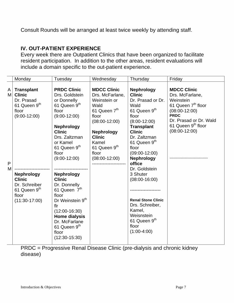
Consult Rounds will be arranged at least twice weekly by attending staff. IV. OUT-PATIENT EXPERIENCE Every week there are Outpatient Clinics that have been organized to facilitate resident participation. In addition to the other areas, resident evaluations will include a domain specific to the out-patient experience.
Monday Tuesday Wednesday Thursday Friday AM PM
Transplant Clinic Dr. Prasad 61 Queen 9th floor (9:00-12:00) ----------------------- Nephrology Clinic Dr. Schreiber 61 Queen 9th floor (11:30-17:00)
PRDC Clinic Drs. Goldstein or Donnelly 61 Queen 9th floor (9:00-12:00) Nephrology Clinic Drs. Zaltzman or Kamel 61 Queen 9th floor (9:00-12:00) ----------------------Nephrology Clinic Dr. Donnelly 61 Queen 7th floor Dr Weinstein 9th flr (12:00-16:30) Home dialysis Dr. McFarlane 61 Queen 9th floor (12:30-15:30)
MDCC Clinic Drs. McFarlane, Weinstein or Wald 61 Queen 7th floor (08:00-12:00) Nephrology Clinic Kamel 61 Queen 9th floor (08:00-12:00) ----------------------
Nephrology Clinic Dr. Prasad or Dr. Wald 61 Queen 9th floor (8:00-12:00) Transplant Clinic Dr. Zaltzman 61 Queen 9th floor (09:00-12:00) Nephrology office Dr. Goldstein 3 Shuter (08:00-16:00) -------------------- Renal Stone Clinic Drs. Schreiber, Kamel, Weisnstein 61 Queen 9th floor (1:00-4:00)
MDCC Clinic Drs. McFarlane, Weinstein 61 Queen 7th floor (08:00-12:00) PRDC Dr. Prasad or Dr. Wald 61 Queen 9th floor (08:00-12:00) ------------------------------
PRDC = Progressive Renal Disease Clinic (pre-dialysis and chronic kidney disease)
Introduction & Objectives Page 8
MDCC = Multidisciplinary Diabetes Complications Clinic (diabetes, nephropathy, hypertension) Each resident will be sent an e-mail from Michelle Gottwald (Dr. Schreiber’s assistant) asking them when they will not be available in terms of (a) holidays, (b) other days off, (c) ambulatory medicine clinics. Once this is available, a clinic schedule per rotation will be distributed on the first day of the rotation. Clinics are assigned during “Ward” month These clinics are expected to start on time, and it is expected that Residents/Fellows will be present on time. The staff person covering the clinic will communicate clinic cancellation ahead of time. V. EDUCATION AND RESEARCH EXPERIENCE A. Teaching All residents will present a formal PowerPoint presentation on a topic of their choice at the Friday noon rounds during the two month rotation. The outline of these presentations should be reviewed the one of the nephrologists several days before the rounds. The presentations should be ~35 min with ample time for interactive discussion. The goals of this exercise are: 1) to develop an expertise in 1 area of nephrology/transplant 2) to gain expertise in formal presentation skills 3) to teach your peers
Introduction & Objectives Page 9
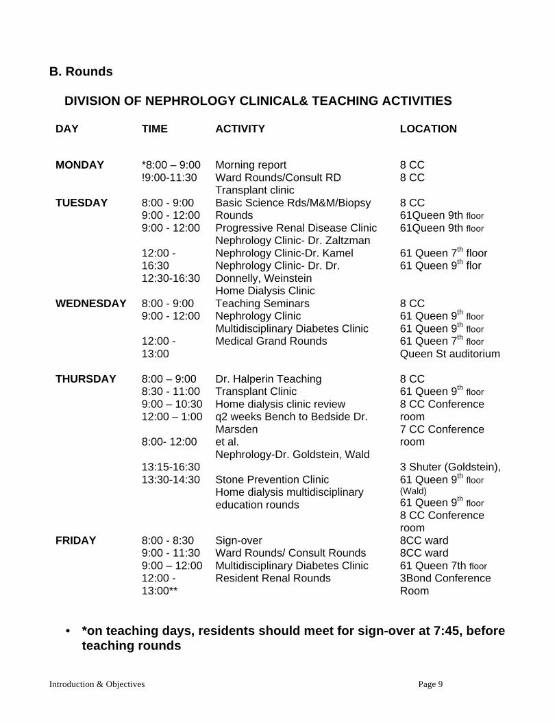
B. Rounds
DIVISION OF NEPHROLOGY CLINICAL& TEACHING ACTIVITIES
DAY TIME ACTIVITY LOCATION
MONDAY *8:00 – 9:00 !9:00-11:30
Morning report Ward Rounds/Consult RD Transplant clinic
8 CC 8 CC
TUESDAY 8:00 - 9:00 9:00 - 12:00 9:00 - 12:00 12:00 -16:30 12:30-16:30
Basic Science Rds/M&M/Biopsy Rounds Progressive Renal Disease Clinic Nephrology Clinic- Dr. Zaltzman Nephrology Clinic-Dr. Kamel Nephrology Clinic- Dr. Dr. Donnelly, Weinstein Home Dialysis Clinic
8 CC 61Queen 9th floor 61Queen 9th floor 61 Queen 7th floor 61 Queen 9th flor
WEDNESDAY 8:00 - 9:00 9:00 - 12:00 12:00 - 13:00
Teaching Seminars Nephrology Clinic Multidisciplinary Diabetes Clinic Medical Grand Rounds
8 CC 61 Queen 9th floor 61 Queen 9th floor 61 Queen 7th floor Queen St auditorium
THURSDAY 8:00 – 9:00 8:30 - 11:00 9:00 – 10:30 12:00 – 1:00 8:00- 12:00 13:15-16:30 13:30-14:30
Dr. Halperin Teaching Transplant Clinic Home dialysis clinic review q2 weeks Bench to Bedside Dr. Marsden et al. Nephrology-Dr. Goldstein, Wald Stone Prevention Clinic Home dialysis multidisciplinary education rounds
8 CC 61 Queen 9th floor 8 CC Conference room 7 CC Conference room 3 Shuter (Goldstein), 61 Queen 9th floor (Wald) 61 Queen 9th floor 8 CC Conference room
FRIDAY 8:00 - 8:30 9:00 - 11:30 9:00 – 12:00 12:00 - 13:00**
Sign-over Ward Rounds/ Consult Rounds Multidisciplinary Diabetes Clinic Resident Renal Rounds
8CC ward 8CC ward 61 Queen 7th floor 3Bond Conference Room
• *on teaching days, residents should meet for sign-over at 7:45, before
teaching rounds
Introduction & Objectives Page 10
• ** every 2 months on the third Friday of the month, rounds are Nephro-ICU joint rounds 13:00-14:00
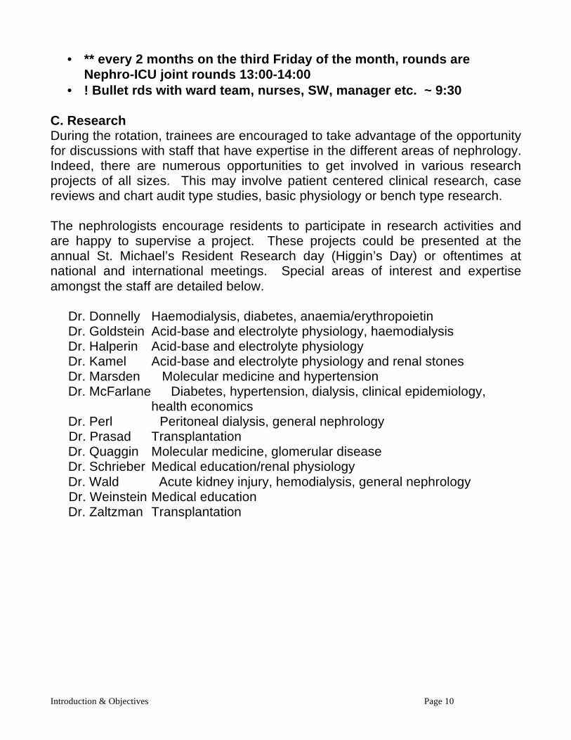
• ! Bullet rds with ward team, nurses, SW, manager etc. ~ 9:30 C. Research During the rotation, trainees are encouraged to take advantage of the opportunity for discussions with staff that have expertise in the different areas of nephrology. Indeed, there are numerous opportunities to get involved in various research projects of all sizes. This may involve patient centered clinical research, case reviews and chart audit type studies, basic physiology or bench type research. The nephrologists encourage residents to participate in research activities and are happy to supervise a project. These projects could be presented at the annual St. Michael’s Resident Research day (Higgin’s Day) or oftentimes at national and international meetings. Special areas of interest and expertise amongst the staff are detailed below. Dr. Donnelly Haemodialysis, diabetes, anaemia/erythropoietin Dr. Goldstein Acid-base and electrolyte physiology, haemodialysis Dr. Halperin Acid-base and electrolyte physiology Dr. Kamel Acid-base and electrolyte physiology and renal stones Dr. Marsden Molecular medicine and hypertension Dr. McFarlane Diabetes, hypertension, dialysis, clinical epidemiology,
health economics Dr. Perl Peritoneal dialysis, general nephrology Dr. Prasad Transplantation Dr. Quaggin Molecular medicine, glomerular disease Dr. Schrieber Medical education/renal physiology Dr. Wald Acute kidney injury, hemodialysis, general nephrology Dr. Weinstein Medical education Dr. Zaltzman Transplantation
Hemodialysis Page 11
VI. HAEMODIALYSIS The haemodialysis units are located on 8CC. The main unit on 8CC is open from Sunday 22:00 until Saturday 23:00. An on-call dialysis nurse is available after hours and Sundays for emergency dialysis needs. If a nurse is required to come in, this decision is made with renal resident and the attending nephrologist. The nurse has been instructed to confirm with the housestaff that the attending nephrologist has been informed. He/she will verify this with you. Most patients are dialyzed 3X/week, but some receive short daily dialysis and some receive nocturnal dialysis. The thrice weekly shifts are on Monday, Wednesday, Friday (MWF) or Tuesday, Thursday, Saturday (TTS). All Nephrology Patients: It is of utmost importance that renal patients do not have intravenous or heparin locks inserted into the cephalic veins from the wrist to the shoulder. MD to make notation on initial blood work orders to avoid cephalic vein cannulation. Resident responsibilities for chronic HD patients Residents do not provide first line care for the out patient dialysis patients as there are Renal staff and Renal trainees involved in the care of these patients (see schedule on ward and in the hemo unit). Residents are responsible for in-patients and patients on the consultation service who are on dialysis (acute or chronic patients). Any acute dialysis needs should be brought to the attention of the charge nurse the night before or immediately to their attention at the beginning of the morning schedule (07:30) This includes writing the orders, monitoring therapies and signing over issues to the MD responsible for the care of the patients upon the discharge of the patient to the outpatient setting. In addition, admissions from the dialysis unit will be directed to the residents. Residents are needed to respond to acute issues for dialysis patients if the assigned nephrologist has already left after seeing the patients. These visits are to be completed in consultation with the nephrologist assigned to that patient (see schedule on the ward and in the hemo unit). Ideally the hemo nurse will first contact the MD assigned to the patient who may then contact the housestaff to assist in the care of the patient if an onsite visit of the MD is required. However, if the nurse feels the issue is of such an urgent nature, the hemo nurse will
Hemodialysis Page 12
contact the housestaff directly. In all such cases, the MD responsible for the patient should be informed either immediately or first thing in the morning. Dialysis Orders A written consent must be obtained by the physician for line insertions and for haemodialysis treatments. HBsAg should be done and ideally the results known prior to the first treatment as HBsAg positive patients are isolated for their dialysis treatment. If a patient may need dialysis in the foreseeable future, order the HBsAg early. Dialysis orders need to be written in advance (a day ahead if possible) for all acute patients and for all chronic haemodialysis patients admitted to hospital. For stable chronic in-patients, orders can be written once per week unless changes are required. Note: for historical reasons, the dialysate is often referred to as the “bath”. Orders are written in the usual order section of the chart and include:
1) Dialyser: usually Xenium 210
2) Time: for chronic patients 4 hours is typical (very uremic patients may need more frequent but short dialysis runs performed daily to avoid the dialysis disequilibrium syndrome)
3) Blood pump speed (BPS); maximum as tolerated. Conventionally
dialyzed patients typically use a blood pump speed between 300-450 ml/ min. Patients receiving nocturnal hemodialysis (with treatment times over 6 hours) may have a slower blood pump speed (200-350 mL/min). Acute and/or very uremic patients may need slower speeds (ex. ~200 ml/min) initially to avoid the dialysis disequilibrium syndrome)
3) [Na+] in dialysate: usually 140 mmol/L, but can use ‘ramped sodium”
([Na+] starting at 145- to 160 mmol/L, reducing to 140 over first 3 hours of treatment). Ramped sodium can be used when the patient is having difficulties with hypotension on dialysis or when it is difficult to reach the patients ultra-filtration target.
4) [K+]in dialysate: 1.5, 2.5 or 3.5 mmol/l. The aim is for a pre-dialysis K+ of
4.0 – 5.5. Suggest starting at a dialysate K+ of 2.5mmol/L if the pre-dialysis K+ is at target.
Hemodialysis Page 13
5) Ultra-filtration : refers to amount of net fluid removal required over the course of the dialysis treatment. Usual target is to achieve the patient’s “target” or “dry” weight. This is the weight that the patient would be if they had no peripheral edema or excess ECF volume. The dry weight is determined by clinical assessment of the ECF volume of the patient. As ultra-filtration removes fluid directly from the intra-vascular space, achieving enough ultra-filtration such that the patient reaches their target weight can be difficult if the patient is hypotensive or if they are significantly above their target weight (typically 3 or more kg above target). In such situations, it may be preferable to have the patient dialyzed more frequently (e.g. short daily haemodialysis) with the ultra-filtration demand spread over a greater number of treatments. Ultra-filtration orders can be written in terms of a) target weight or b) litres to be removed. The first option (target weight) is preferable to ordering number of litres to be removed, however, if the patient cannot be weighed, the second option becomes necessary. During any given dialysis session, ultrafiltration may be “ramped” such that a greater proportion of the total uf is removed earlier in the dialysis session (eg, remove 40% over 1st hour, 30% over 2nd, 20% over 3rd and 10% over 4th).
6) BP support usually important in ICU setting. Can involve ramping Na,
ramping ultrafiltration, saline infusions, blood, albumin, pentaspan or inotropic support. Wrapping of legs with Tensor bandages to enhance interstitial to intravascular fluid shifts within the ECF compartment may be helpful.
7) Heparin standard anticoagulation.
a) normal : 1000 unit bolus followed by 1000 units/hour; discontinue heparin 60 minutes prior to the end of dialysis if patient has an AV fistula or graft; in patients with central venous catheters, continue heparin until the end of the treatment
b) tight: as above except, 500 unit bolus followed by 500 units/hour c) none (the nurse will flush the dialysis membrane periodically with normal saline to prevent clotting).
Heparin orders will depend on risk of bleeding. For patients with active bleeding or at high risk for bleed, then the heparin choice should be “none”.
8) Other Can give meds with dialysis. Some antibiotics are prescribed at end of dialysis. RBC transfusions, if required, are usually given while on dialysis.
Hemodialysis Page 14
Vascular Access for Haemodialysis Three types of vascular access are available for hemodialysis. The access coordinator (6353) can facilitate the investigations and management of vascular access issues. Note:
• IVs and BP measurements should be avoided in the limb that has an AV graft or fistula or one in which a graft or fistula is being planned
1. AV Fistula A surgical anastamosis of patient’s artery to vein ideally placed in the non-dominant forearm, with upper arm or leg sites also possible. Arterialization of the vein is usually adequate by 6-8 weeks. Patency can be confirmed by feeling a thrill and/or hearing a bruit over the fistulae site. This is the preferred type of vascular access for chronic hemodialysis.
2. AV Graft Connection of an artery to vein using a “Gortex” vascular graft, usually in the upper arm or thigh. It can be used earlier than a fistula after creation (within 1 week) but is more problematic in terms of thrombosis and infections. A graft is the preferred access for chronic hemodialysis when a fistula cannot be created or fails to mature.
3. Central venous catheters
a) temporary catheters are placed by housestaff and fellows under staff supervision as needed. These are double lumen catheters placed in femoral or internal jugular vein. They are used when dialysis is urgently required.
The line insertion/removal cart is located in the room 8009 cc (technical lab-back of dialysis unit). The cart must be signed out in the book attached to the cart. The cart is check and stocked at least once per week. Use the “Site Rite” (a portable ultrasound) to localize vessel. NO MORE THAN TWO ATTEMPTS SHOULD BE MADE BEFORE CONSULTING THE RENAL FELLOW OR THE STAFF NEPHROLOGIST. A formal sterile procedure with gown and mask and skin scrub with betadine is used. All lines are sutured securely in place. The exit site is covered with a 2X2 gauze with betadine ointment. This is covered with a 4X4 gauze and taped with a Tegaderm that has had a slit cut in it to accommodate the exit of the catheter.

Hemodialysis Page 15
Citrate (4%) is instilled in each lumen (check volume of each lumen, it is printed on the side of the catheter) as a locking solution. If heparin is using as the locking solution, dose, the dose is 5000 units/lumen (i.e. .5cc of 1:10,000 diluted with saline to the volume of the internal lumen). A CXR is needed to rule out complications of insertion, such as pneumothorax, and to check that the tip of the tip of the IJ line is in the SVC or the right atrium. b) permanent Commonly referred to as an “Uldall Cook” or “UC line” or other long term catheter. The catheter is placed in the internal jugular (IJ), then runs through a subcutaneous tunnel and exits the skin about 5 cm below the clavicle. Tunnelled catheters are inserted by interventional radiology under fluoroscopy. This is the preferred method for central venous catheter insertion. Can usually get it on day of request, by calling radiology at X5886 and faxing the requisition to X5380. Note: Central venous lines are the least preferred long-term access for chronic haemodialysis. They are associated with thrombosis, infection, and inadequate dialysis.The advantage is that they can be used immediately.
Problems with Central Venous Dialysis Catheters 1. Line migration: Do NOT push the catheter back in. If there is sufficient
catheter length remaining in the central vein, the dialysis can be done and plans made to replace the catheter before the next dialysis. If it is a temporary catheter and there is good flow through the venous port, the line can be changed over a guidewire. If there is no flow through the venous port, a new line insertion is required.
2. Cannot aspirate from the arterial port: If you can infuse without resistance,
the arterial port is likely against the wall of the vein or the patient is ECF volume depleted- play the patient in slight Trendelenburg or reverse the lines.
• If you cannot infuse without resistance the arterial lumen is blocked. You can infuse TPA and wait 1 hour in an attempt to lyse the thrombus.
3. Cannot aspirate from the venous port: Check to see if line kinked - if so
should be changed and sutured to minimize kinking. • If you can infuse without resistance, either the patient is volume contracted or
there is a "ball valve" thrombus. Try positioning the patient in a supine
Hemodialysis Page 16
position or in the lateral decubitus position (line side up), or TPA respectively. • If it is a left sided line perhaps the tip is against the superior vena cava and a
longer line could be necessary. Ensure there is no pain on infusion as in a very rare instances, the tip could have migrated through the wall of the vein.
• Otherwise, if the patient is not volume contracted, TPA fails and the line is not against a wall, the line should be changed.
4. Line started out fine but now BPS above 200 generate arterial insufficiency: The initial good function rules out kinks and thrombi and suggests mechanical problem (line sucking on vein wall) due to circulating volume contraction. Patients with low serum albumin can be circulating volume contracted yet still have +++ edema. Use tilt stretcher with patient head down and you will have to remove volume slowly. Support stockings will minimize problem. Be patient, the line sucking the vein wall produces spasm and aggravates the problem
5. Both ports aspirate well but BPS >200 are problematic: Is patient
circulating volume contracted? If you cannot be sure, place head down on tilt stretcher and give 200-500 cc normal saline and evaluate impact. If JVP clearly elevated, do not give the saline, just place head down.
• If all efforts fail, check X-ray position of line to see if long line will be beneficial and have line changed.
6. Cannot aspirate from or infuse into either port: Ensure the clamps have
not left the silastic compressed; check for kinking, clotting, locking solution protocol, coumadin protocol. Check CXR. Trial of TPA or new line as necessary
7. Suspected line sepsis (see policy and procedure algorithm)
• If a patient with a catheter develops signs & symptoms of infection, always consider CVC-associated bacteremia. However, CVC-associated bacteremia should be a diagnosis of exclusion after all other potential infectious foci have been considered.
• Look for redness/discharge at the exit site for evidence of exit site infection and for fullness or tenderness along the catheter tunnel for evidence of tunnel infection.
• Most common source of fever and sepsis in HD patients at SMH is due to staph species.
• Need blood culture (consider doing two or three blood cultures) and a C&S swab from the exit site
Hemodialysis Page 17
• Start empiric therapy with Ancef 2 gm IV last hour of dialysis and gentamycin (2 mg/kg load; 1mg/kg post dialysis for 3 weeks).
• Treatment is usually for 3 weeks and is adjusted at the next dialysis pending culture results and antibiotic sensitivities.
• Antibiotics are adjusted at the next dialysis based on the culture results and C&S. If the cultures are negative, the patient is reviewed before simply terminating the intended 3 weeks of therapy.
• Consider line removal at the onset of the infection only if the patient is very toxic. Otherwise, the decision for line removal is deferred until the next visit. Line change is considered if symptoms of fever persist after 48 hours of appropriate antibiotic therapy, tunnel infections and recurrent infections. Otherwise, at St. Michael’s Hospital lines are considered salvageable and routine replacement for a single episode of line sepsis is not advocated. This is a guideline and does not replace good clinical judgement.
• All lines get antibacterial ointment (polysporin or betadine if patient is polysporin sensitive) applied to the exit site at each dressing change.
Notes Regarding Patients Receiving Intensive Haemodialysis Many St. Michael's Hospital dialysis patients are receiving higher than conventional doses of haemodialysis. Examples of intensive haemodialysis include: • short daily in-centre (4 or 5 times per week for 2.5 to 3.5 hours) • nocturnal in-centre (3 times per week for 7- 8 hours overnight) • short daily home (5 or 6 times per week for 2.5 to 3.5 hours) • nocturnal home (5 to 7 times per week for 6 - 8 hours overnight) Patients receiving intensive haemodialysis may have non-standard dialysis bath compositions. For example, they may be on a higher than normal potassium concentration or have phosphate (as fleet phospha soda) added to the dialysate. These patients often can have a far more liberal diet and fluid intake. If an intensively dialyzed patient is admitted to hospital and converted to a conventional dialysis dose (4 hours 3 times per week), their entire dialysis prescription should be reassessed, and dietary restrictions should be started as appropriate. In general, it is best to keep a patient who normally performs an intensive form of haemodialysis on an intensive form (e.g. in-centre intermittent nocturnal haemodialysis) while they are admitted.
Hemodialysis Page 18
Some Hints for the Consult Team When chronic dialysis patients are admitted to other services, the nephrology consult team will be consulted in order to manage dialysis related issues. In addition to ordering dialysis, the consult team should monitor for co-interventions ordered by other teams that may require major modifications in a dialysis patient.
1) Medications It is crucial to review the pre-admission medication list for all dialysis patients and ensure that these are continued or held (as appropriate) during the hospitalization. The patient’s medication list may be found in his/her dialysis chart and should be reviewed at the outset of each hospitalization. At the time of discharge, it is important to identify dose adjustments to pre-existing medications, medications that have been discontinued permanently and medications that need to be restarted after being temporarily held. This information should be conveyed to the patient’s primary nephrologist. For all patients with acute or chronic (including chronic dialysis patients) kidney disease, the medication list should be frequently reviewed o ensure that all medications that have been prescribed by the admitting service are not contraindicated in the setting of kidney disease. Furthermore, the consult service should verify if the medication dose and frequency is appropriate for the degree of kidney function. Note that some medications need to be given POST-dialysis and in some cases, post- dialysis supplemental doses are needed. 2) Bowel preparation Magnesium, citrate, aluminium and phosphate containing bowel medications should be avoided. Fleet enemas (primarily PO4), in particular, should always be avoided. If bowel preparation is required, the preferred solution is CoLyte or PegLyte. Although large volumes are typically required, these solutions (when used appropriately) are not absorbed and are not contraindicated in dialysis patients. 3) Maintenance IV solutions The volume of delivered IV solutions should be monitored in dialysis patients. Standard maintenance orders (e.g. normal saline at 100cc/hour) could rapidly lead to volume overload in a patient with CKD. 4) Dialysis dose and prescription
Hemodialysis Page 19
Patients who are acutely ill may require modification of their dialysis prescription. If oral intake is reduced, patients may require a higher dialysate potassium concentration, and may require supplementation of calcium or phosphate. Ultra-filtration requirements can be significantly altered during a hospital admission. Patients may become uremic on what was previously an adequate dialysis dose, and either more frequent treatment, longer treatments or both may be required. Ultra-filtration, solute removal, and biochemical parameters should be monitored longitudinally, with adjustments of the dialysis prescription made as required.
Renal Replacement Therapy for Acute Kidney Injury Renal replacement therapy for acute kidney injury (AKI) is frequently required when conservative measures fail to prevent or control life-threatening complications of AKI (eg, congestive heart failure, hyperkalemia). In addition, severely ill patients with established AKI and no evidence of impending renal recovery are often started on renal replacement therapy. This criterion for renal replacement therapy initiation is more subjective and there is no consensus on the optimal time for commencement of renal replacement therapy in AKI. Patients with AKI who require renal replacement therapy may be managed with intermittent hemodialysis (IHD), continuous renal replacement therapy (CRRT) or sustained low efficiency dialysis (SLED). The same patient may receive one or more of these modalities at different times of their course in hospital depending on the evolving clinical circumstances. PLEASE REVIEW ALL ACUTE RENAL REPLACEMENT THERAPY PRESCRIPTIONS WITH THE RENAL FELLOW AND/OR STAFF ON A DAILY BASIS. Intermittent Hemodialysis (IHD) In intermittent hemodialysis (IHD), conventional dialysis machines and prescriptions (akin to those used in the chronic setting as above) are applied to the setting of AKI. IHD may be administered anywhere in the hospital. IHD is typically reserved for patients with AKI who are hemodynamically stable, including patients who received SLED or CRRT who have become hemodynamically stable. IHD is the most efficient form of dialysis and is the most effective way to manage life-threatening hyperkalemia (even if the patient is hemodynamically unstable) and intoxications (see below). The typical session duration is 3-4 hours at blood pump speeds of 300-400 mL/min. Anticoagulation is preferred but sessions may be feasibly administered with no heparin.
Hemodialysis Page 20
Hemodialysis nurses administer IHD and sessions need to be arranged in coordination with the Hemodialysis Case Manager or the dialysis nurse on call. Continuous Renal Replacement Therapy (CRRT)
Continuous renal replacement therapy (CRRT) provides 24-hour renal replacement therapy using relatively slow blood flow and ultrafiltration rates. Hemodynamically unstable patients are those who are most likely to benefit. CRRT may be administered as continuous veno-venous hemodialysis (CVVHD), continuous veno-venous hemofiltration (CVVH) and continuous veno-venous hemodiafiltration (CVVHDF). CRRT is only available in the MSICU and CVICU and is administered using dedicated PrismaflexTM machines. Critical care nurses are responsible for setup of the machines and administration of therapies. The Nephrology consult service is responsible for ordering, monitoring and adjusting the CRRT prescription. Pre-printed orders are available in the MSICU and CVICU to help guide CRRT prescription. One set of orders is employed for patients receiving heparin or no anticoagulation; another set is used for patients receiving regional citrate anticoagulation. Completed orders must be reviewed with the Nephrology fellow and/or staff prior to submission to the ICU nurse. Clearance modes in CRRT CVVHD employs dialysis exclusively as the mode of clearance; all solute removal is by diffusion. CVVH utilizes hemofiltration as the only mode of clearance. All solute removal is by convection. Pure CVVHD cannot be delivered on the Prismaflex as there is an obligate 200 mL/hr of hemofiltration that must be given for technical reasons. Practically speaking, most patients will receive a combination of dialysis (diffusion) and hemofiltration (convection) in the form of continuous veno-venous hemodiafiltration (CVVHDF). Indications for CRRT • patients who require acute hemodialysis and are hemodynamically unstable to
the point where conventional haemodialysis is very high risk (this is a judgement call; note that in very unstable patients, even CRRT may not be tolerated)
• patients with very large ultra-filtration needs, coupled with large IV infusion rates
Intensity of CRRT The total effluent dose per hour should 20-25 mL/kg/hr.
Hemodialysis Page 21
Anticoagulation for CRRT Anticoagulation is almost always required in CRRT although under some rare circumstances, CRRT may be attempted with no anticoagulation. Two basic options for anticoagulation exist:
REGIONAL CITRATE ANTICOAGULATION Regional citrate anticoagulation provides isolated anticoagulation in the extracorporeal circuit through the chelation of calcium by exogenous citrate which “paralyzes” the coagulation cascade. Systemic anticoagulation and hypocalcemia are avoided by the concurrent administration of exogenous calcium intravenously. All patients are eligible for regional citrate anticoagulation although this strategy is relatively contraindicated in patients with hepatic dysfunction due to the risk of citrate accumulation as a result of impaired liver metabolism. Regional citrate anticoagulation is especially indicated for patients who cannot have heparin (e.g. HIT) or systemic anticoagulation in general (e.g. patients at high risk of bleeding such as those who have had recent surgery, active/recent bleeding or thrombocytopenia). PRACTICALLY SPEAKING: • Citrate chelates Ca+2 in the circuit; at ionized calcium of < 0.40 mmol/L, the
coagulation pathway is inactivated. • CaCl2 is re-infused to the patient to maintain normal systemic ionized calcium • Ionized calcium in both the circuit and the patient are monitored • Circuit ionized calcium is adjusted by a sliding scale of citrate infusion rates • Patient ionized calcium is adjusted by a sliding scale of CaCl2 infusion rates • To maximize citrate effectiveness and to minimize the amount of citrate used,
a Ca+2 free dialysate is used (PrismocalTM) • Anticipate potential problems:
o metabolic alkalosis may develop after about 2 day as citrate is a source of bicarbonate…manage with ultra-filtration (i.e. remove high bicarbonate) and replace this fluid with saline. (see pre-printed orders)
o citrate accumulation may develop in patients with liver problems who are not able to metabolize the citrate load. This is recognized by an expansion of the citrate gap (i.e. total calcium-ionized calcium).
HEPARIN
Systemic unfractionated heparin may be administered to prevent clotting of the extracorporeal circuit. aPTT is targeted to 60-85 seconds using a protocol that is
Hemodialysis Page 22
found in the pre-printed orders. This is identical to the “high PTT nomogram” that is used across the hospital for patients receiving unfractionated heparin. Sustained low efficiency dialysis (SLED) Sustained low efficiency dialysis (SLED) utilizes conventional dialysis equipment (as in IHD) applied over a more prolonged treatment time and with a lower blood flow (as in CRRT). As such, SLED has been described as a hybrid therapy that captures the benefits of CRRT (ie, greater hemodynamic tolerability) and IHD (ie, lower material costs, option for no anticoagulation). SLED may be administered in any critical care unit (MSICU, CVICU, TNICU or CCU) by a hemodialysis nurse. Patients with hemodynamic instability (similar to those being considered for CRRT) should receive primary consideration for SLED. Treatment duration is 8 hours at a blood flow of 200 mL/min and a dialysate flow of 350 mL/min. SLED may be administered with no anticoagulation. Given the intensive nature of the dialysis provided by SLED, patients may become hypophosphatemic after 1-2 SLED sessions; phosphate may be added to the dialysate in such individuals for subsequent sessions. All aspects of the SLED prescription are found in the pre-printed SLED orders which should be completed for each session.
Management of Intoxications All poisonings should be managed with the supervision of renal fellow and staff Nephrologist. The management of intoxication requires a high dose of high efficiency dialysis. Slow low-efficiency techniques such as PD or CVVHD should not be used unless there is no other option (ex. intoxicated PD patient being managed at a peripheral hospital that does not offer dialysis awaiting transfer to SMH). Poison Control Telephone Number: (416) 813-5900 Hemodialysis • For solutes that have low MW, are not protein bound, and are water soluble • Concurrent: renal failure, acid-base disturbance, electrolyte or volume
abnormality correctable by dialysis • Requires vascular access and anticoagulation Hemoperfusion
Hemodialysis Page 23
• Blood passes through a cartridge with activated charcoal or other sorbents • For toxins that are more lipid-soluble, higher MW • May cause thrombocytopenia • May be less destabilizing than HD if hypotensive • Requires vascular access and anticoagulation
Examples of intoxications
Methanol • Industry solvent (e.g. windshield washer fluid, antifreeze) • T1/2 variable: 12-20 hrs, minimum lethal dose 50-100 ml • Metabolism – oxidation to 1) formaldehyde and 2) formic acid • Clinical manifestations
Early Stage (< 6 hrs): non-specific, mild or transient: inebriation, drowsiness
Delayed Stage (6-30 hrs): Vertigo/N/V abdo pain • Kussmaul breathing • Blurred vision (papilledema, disc hyperemia)→ blindness • Seizures, opisthotonus, coma → death • Lab findings: AGMA, osmolar gap, ↑ formate level, ↑ lactate level, ↑
amylase (pancreatitis) • Toxic levels: >10mmol/L (50 mg% or 500 mg/L)
ANY level with anion gap metabolic acidosis • 4 ml methanol has caused blindness - 15 ml of methanol can be lethal!!!!
• Metabolized by alcohol dehydrogenase - has lower affinity for methanol than ethanol.
• Metabolized into formic acid - causes the large anion gap metabolic acidosis. • Prognosis dependant on amount of methanol metabolized and determined by the
time between ingestion and treatment, the amount of ethanol on board, the degree of acidosis and the extent of the visual disturbance.
• Diagnosis is usually made by history and biochemical “footprints”. An anion gap metabolic acidosis with an osmolar gap between measured and calculated osmolality is classic (calculated osmolality = Na x 2 + urea + glucose). The difference represents the mosmoles of methanol and can be used to guess the level until levels are available.
Management: Alcohol dehydrogenase inhibition and Hemodialysis Alcohol dehydrogenase inhibition
Hemodialysis Page 24
• Ethanol is given as an antidote - orally or by IV. Aim for a blood level of 100 mg% (20-25 mmol/L). The alcohols are distributed across total body water.
Oral Ethanol • Loading dose of 40 gm ethanol. (Absolute or 95% ethanol has SG of
0.8 gm/mL.) This works out to 50 mL of absolute ethanol or 120 mL of 40% ethanol like scotch. The maintenance dose is 12 mL of absolute or 30 mL (1 oz) of whisky per hour with frequent measurements to ensure levels as above.
IV Ethanol • Begin with IV bolus of 0.5 gm ethanol/ kg • NOTE: Must be diluted to a 15% solution or less to be non toxic.
Mix 72 mL absolute ethanol in 500 mL D5W or NS to give a solution of 10 gm/100 mL i.e. 100 gm/L. A 70 kg man gets 350 mL of this solution or 35 gm. This is followed by maintenance of 10 gm (100 ml) per hour. Continue infusion even if dialysis is in progress to make up for metabolized ethanol.
• Fomepizole For acute management of methanol or ethylene glycol intoxication at peripheral hospital until patient is stable for transport. This drug is very costly and is routinely available at SMH.
Hemodialysis • Hemodialysis indicated for serum methanol levels > 10 mmol/L, or
even at lower levels if anion gap metabolic acidosis is present. • Dialyze at Qb of 300 or more • Ethanol is added to the dialysate (500cc of 100% ethanol to 8L of
the bicarbonate concentrate) to avoid blood ethanol from being dialyzed from the patient. Change dialyser q 6 hr
• Continue to dialyze to methanol level < 5 mmol/L. By the time this result is back, actual level will be much lower. D/C dialysis and send final methanol level. Dialysis often needed for > 10 hours.
• PD is less effective but may be of some use in those who cannot be hemodialyzed. Add ethanol to the PD fluid.
Follow the blood levels on a flow sheet
Ethylene Glycol • Component of antifreeze and solvents. Dialysis indicated for level > 6
mmol/L or lower levels with anion gap acidosis • T 1/2 is 3 hours
Hemodialysis Page 25
• Lethal dose ~ 100 mL. • S/S - neurological– drunkenness to coma, tachypnea, pulmonary
edema, flank pain and RF • Classically, but not always, crystalluria (needle shaped or envelope
shaped crystals) • Management is same as methanol intoxication, i.e. ethanol and
hemodialysis.
Theophylline • Chronic intoxication – more severe clinical manifestations than acute
and may have liver or renal involvement contributing to intoxication • Acute - usually intentional overdose • Toxic levels 450 umol/L in acute overdose or 220 umol/L in chronic
overdose • Small vol of distribution + low rate of clearance - effectively cleared by
HD and charcoal hemoperfusion (HP) (hemoperfusion approx 2x as effective due to removal of protein-bound drug)
• Use two sites for venous catheters • HD – use max blood flow, minimum 4 hours • HP – use charcoal cartridge, saturates in about 2 hours and hence the
cartridge must be changed q2h • Serial HD-HP delays saturation of HP cartridge • No guidelines re level to dialyze to, advisable to continue to < 100umol/L
Lithium
• Therapeutic range: 0.4-1.3 mEq/L • Toxic manifestations may appear >1.5 mEq/L • Clinical manifestations:
Acute intoxication: N/V, neuromuscular irritability, coarse tremor, ataxia, slurred speech, confusion, fever, stupor, coma, CV collapse Chronic intoxication: polyuria & NDI, renal acidification defects, CIN, thyromegaly
• Lab manifestations: leukocytosis; ECG: flattened T's, AV blocks, QT prolongation
Management
• Well hemodialyzable • Hemodialysis for 8-12 hours and monitor post plasma Li levels q4h
for 36 hours
Hemodialysis Page 26
Indications: Li level > 4.0 meq/L Li level >2.5 meq/L if symptomatic or renal insufficiency Goal: sustained level of 1 mmol/L 8 hrs post HD
• Dialyze 8-12 hours • Monitor for post HD rebound as slow equilibration between extra and
intracellular lithium. May require repeated HD treatments
Salicylates • Aspirin, oil of wintergreen (topically) • Minimum lethal dose 10 g ASA; levels useful 6 hrs post ingestion • Acute ingestion: 1 tab/kg = severe (1 tab = 325 mg) • Metabolism – ASA hydrolyzed to salicylic acid → glycinated to salicyluric
acid in liver→ excreted via kidneys; urine pH > 7.0 enhances excretion • Clinical manifestations • Chronic ingesters : HA, tinnitus, ↓hearing, dizziness, weakness, N/V,
↑RR, confusion • Acute/severe intoxications: above + fever, seizures, coma, ARDS • Acid base disturbances: • Respiratory alkalosis → resp alk + AG metabolic acidosis → metabolic
acidosis
Management • Systemic and urine alkalinzation urine: goal urine pH >7.5 • Hemodialysis Indications: Salicylate level > 7 mmol/L
Seizures/coma Severe metabolic acidosis, especially with renal failure Non cardiogenic pulmonary edema
Especially if elderly, smoker, acute on chronic ingestion
Peritoneal Dialysis Page 27
Home Hemodialysis While the majority of HD patients are on in-centre HD three times weekly, there is a subset of patients who are trained to do HD at home. For these patients, there is no universal HD prescription, as patients have more flexibility to individualize their treatment. Some common examples of home dialysis prescriptions include short daily HD (3 hrs, 5-6 days per week) and nocturnal HD (7-8 hours, 3-6 nights per week). Patients are generally seen in clinic at 2 month intervals, or sooner if necessary. For any home HD issue, it is important to consult with the multidisciplinary team in the Home Dialysis office on the 8th floor of the Cardinal Carter wing (416-864-5794).
Peritoneal Dialysis Page 28
VII. PERITONEAL DIALYSIS The peritoneal membrane can be used to perform dialysis (PD). Dialysate is infused into the peritoneal cavity, and allowed to dwell for a period of time, during which toxins diffuse out of the blood into the dialysate. Ultrafiltration (UF) also occurs during this time, as fluid is drawn out of the circulation by the osmotic force of compounds such as dextrose in the dialysate. Once the dwell is over, the dialysate is drained from the peritoneum, and fresh dialysate is instilled. The process of draining spent dialysate and reinstilling fresh solution is known as an “exchange”. PD comes in a variety of forms, which are discussed below. For any peritoneal dialysis issue, it is important to consult with the multidisciplinary team in the Home Dialysis office on the 8th floor of the Cardinal Carter wing (416-864-5794). Peritoneal Dialysis Solutions Standard dialysate (Dianeal™) comes in four dextrose concentrations, 0.5%, 1.5%, 2.5% and 4.25%. The higher the percentage of dextrose, the more likely UF will be achieved. As a rough guide, a 2 L 4-hour dwell with 1.5% solution should result in 0-100 cc of UF, while a 2.5% solution should result in 150-250 cc UF and a 4.25% solution should produce > 400 cc UF. A 0.5% bag is usually only used when the patient is hypotensive and volume contracted, as it typically results in a net negative UF (i.e. less fluid is drained out than instilled, with a net infusion of solution into the intravascular space). A more precise estimate of expected UF for a given dextrose concentration can be achieved by consulting the patient’s log book from home. On occasion, it is necessary to specify the concentration of dextrose in the dialysate, in order to give more precise instructions to the nursing staff. When a target weight is ordered and no specification is given regarding dialysate dextrose concentration, the nursing staff will consult a pre-made table that will provide them with guidance on how to select the dextrose concentration. There are three specialized PD solutions available: (1) Extraneal™ (icodextrin) – uses a non-dextrose molecule to provide the osmotic force for UF. In contrast to dextrose-based solutions, Icodextrin is not readily absorbed into the bloodstream, so there is not dissipation of the osmotic gradient for UF. This solution is therefore ideal for a long dwell in order to prevent net absorption of fluid and to promote UF. Icodextrin should typically only be used for one long (6 to 16 hour) exchange daily.
Peritoneal Dialysis Page 29
(2) Nutrineal™ - an amino acid-based solution. This solution can be used to supplement amino acids in malnourished patients. The amount of supplementation is relatively small, and use of this solution does not preclude the need to identify and treat causes of malnutrition. Nutrineal should not be used with short dwell times (eg. night cycler) as the nutritional benefit will be minimized. Since both Extraneal and Nutrineal are not dextrose-based, they have the advantage of not leading to absorption large quantities of dextrose as does standard dialysate. Nutrineal should only be used for one exchange daily as the amino acid load can cause metabolic acidosis. Nutrineal also does not produce a large volume of UF, and should not be used in patients requiring aggressive ultrafiltration. (3) Physioneal™ – a neutral pH, bicarbonate-buffered solution (all other bags use lactate as a buffer). Physioneal may be tried in patients with persistent abdominal pain during/after infusion as this pain may be due to the low pH of standard solutions. Others have suggested using Physioneal for long term preservation of the peritoneal membrane, although supportive data is lacking. When used, Physioneal should replace all Dianeal exchanges. It should be noted that all three specialty PD solutions are significantly more expensive than standard dialysate, and should therefore only be used when clinically indicated. CAPD (Continuous Ambulatory Peritoneal Dialysis) This simple form of dialysis is the most common method of doing peritoneal dialysis around the world (although not at St. Michael's Hospital). Patients performing CAPD typically perform four 2 L exchanges per day, usually upon awakening, at lunch, at dinner and prior to bed. Exchanges are done using a “twin-bag” system, consisting of an empty drain bag and a full dialysate bag connected by a Y connector. A CAPD exchange first involves connecting the Y connector of the twin-bag to the patient’s PD catheter. Spent dialysate is then drained into the drain bag. Once the peritoneum is empty, fresh dialysate is instilled into the peritoneum from the dialysate bag. This process typically takes from 20 to 40 minutes. When prescribing CAPD, order volume of exchange (usually 2L), frequency of exchanges (usually 4x/day), additives (usually none), target weight specifying whether or not this includes the exchange volume (eg. dry weight 75 kg empty). Example: CAPD 2 L fill volumes qid, target weight 68 kg (full with 2 L)
Peritoneal Dialysis Page 30
APD (Automated Peritoneal Dialysis) An automated machine called a “cycler” can be used to instill and drain dialysate from the peritoneal cavity. Typically such a machine is used as night, with the cycler performing the dialysis exchanges while the patient sleeps. Normally, a larger number of exchanges can be ordered than would be practical during the day (eg. 4-5 exchanges over 9 hours). In some cases, the patient can tolerate a larger volume of dialysate at night as well (eg. 2.5L) because of lower intra-abdominal pressure when supine. While CAPD bags are usually 2 or 2.5L, APD bags are usually 5L. APD can be performed in one of three ways depending on the desired dialysis dose, discussed below: (1) NIPD (Nightly Intermittent Peritoneal Dialysis) In this form of dialysis the cycler is used to perform exchanges during the night. In the morning, as the dialysis program is ending, the cycler drains the patient, who then disconnects from the machine and remains empty through the day. The cycle begins again the next evening, when the patient hooks back up to the cycler. To order NIPD, specify the number and volume of exchanges, the total number of hours of the cycler program, and the dextrose concentration of the dialysate or the patient’s target weight. Example 1: NIPD, 5 exchanges over 9 hrs, 2L fill volume, no last fill, target weight 65 kg Example 2: NIPD, 5 exchanges over 9 hrs, 2L fill volume, no last fill, 2.5% Dianeal for all exchanges (2) CCPD (Continuous Cyclic Peritoneal Dialysis) Continuous cyclic peritoneal dialysis is similar to NIPD, but rather than have the patient empty during the day, the patient carries dialysate for part or all of the day. This is performed using the “last fill” option of the cycler. In NIPD, at the end of the cycler program the cycler drains the patient until they are empty, at which point the patient disconnects from the machine. In CCPD, the patient is drained, and then is refilled from a separate dialysate bag, and the patient completes the cycle program with fluid in their peritoneal cavity. This fluid is either kept in for the whole day and drained at the start of the next night cycle, or is drained at some point during the day. The last fill usually comes from a standard CAPD-type dialysate bag. Icodextrin solution is a good choice if the last fill is to dwell until the late afternoon or evening in order to prevent fluid absorption and promote UF. Order CCPD as you would NIPD, but you must also specify the composition, volume and duration of the last fill.
Peritoneal Dialysis Page 31
Example: CCPD, 5 exchanges over 9 hrs, 2 L fill volume, 1.5%/2.5% Dianeal. Last fill 2L Icodextrin. (In this example, since the total night volume is 10L, the 1.5%/2.5% means that the patient/nurse will use one 5L bag of 1.5% solution and one 5L bag of 2.5% solution). (3) Enhanced CCPD In patients who are not able to achieve adequate dialysis with CCPD, additional twin-bag exchange(s) can be added to the CCPD (night cycler + last fill) prescription. A CAPD twin bag is used for the daytime exchange. Order enhanced CCPD as you would standard CCPD, but you must also specify the composition, volume and timing of the additional exchanges, as well as any additives as required. Example: Enhanced CCPD, 5 exchanges over 9 hrs, 2 L fill volume, 1.5%. Last fill 2L of Icodextrin. Twin bag exchange 2L of 2.5% at 4 PM For patients admitted to hospital, enhanced CCPD can also be delivered by continuous use of the cycler. This may meet the patient’s medical requirements, as well as being more convenient for the nursing staff. PD PERITONITIS PD peritonitis requires two of the following three criteria to be fulfilled:
1. Signs/Symptoms of peritoneal inflammation 2. Cloudy bags (or WBC count > 100 with >50% neutrophils) 3. Positive dialysate culture or Gram stain
Initial Assessment (1) Clinical examination with particular attention to assessment of:
- abdomen for symptoms and signs of peritoneal inflammation (eg. rebound)
- peritoneal catheter exit site; send swab for C&S if drainage present - rule out presence of incarcerated hernia
(2) Order first dialysate bag to be sent for Gram stain, C&S and cell count with differential. If patient is dry or on cycler, try to allow a dwell of 2 – 4 hours in order to get a meaningful sample. If the patient is too ill, a one hour dwell will suffice (cell count may not be as high as with longer dwell, but if true infection,
Peritoneal Dialysis Page 32
percentage of neutrophils should be high). Bloodwork on admission - CBC and differential, electrolytes, creatinine, urea, calcium, phosphate, protein, albumin (3) Order antibiotics (see below for details) (4) Order dialysis prescription, including target weight. Antibiotic usually given IP once daily in 6 hour dwell (4 hour dwell minimum)
-CAPD prescription usually does not need modification -Cycler patients should have antibiotics added to the longest dwell. For a cycler patient who normally does only NIPD, you can add a 6-hour day dwell (eg. last fill or daytime exchange) on top of the usual nocturnal prescription. -Patients may require higher % dialysate bags as peritoneal inflammation may lead to more rapid glucose absorption and therefore less UF
(5) Order additional intraperitoneal additives: Heparin 1000 u/L until effluent clears, then 500 U/L prn if fibrin still present Xylocaine 2%, 5cc/L prn for abdominal pain KCl, insulin as required (6) Order frequency for effluent sampling for inpatients (cell count daily or q2d until <100, and culture q2days until first “no growth”; then q4days until total of 3 “no growths”) (7) For patients with residual renal function who are receiving vancomycin, check vancomycin levels at 72 hours post-initial dose. (8) Hold phosphate binders or calcium supplements if peritonitis is severe (due to constipation). Order appropriate diet and all other medications. (9) Patients on peritoneal dialysis who present with peritonitis are managed as outpatients unless severity indicates hospital admission. Admission is also required for patients unable to manage at home. ***If no decrease in cell counts in 3-4 days or if count fell initially and then increased, repeat culture and consider (1) inappropriate antibiotics for organism, (2) associated exit site/tunnel infection, (3) secondary peritonitis (eg. ischemic bowel, cholecystitis, diverticulitis, appendicitis, pancreatitis). Management of non-resolving peritonitis after 5 days of appropriate antibiotics is catheter removal.
Peritoneal Dialysis Page 33
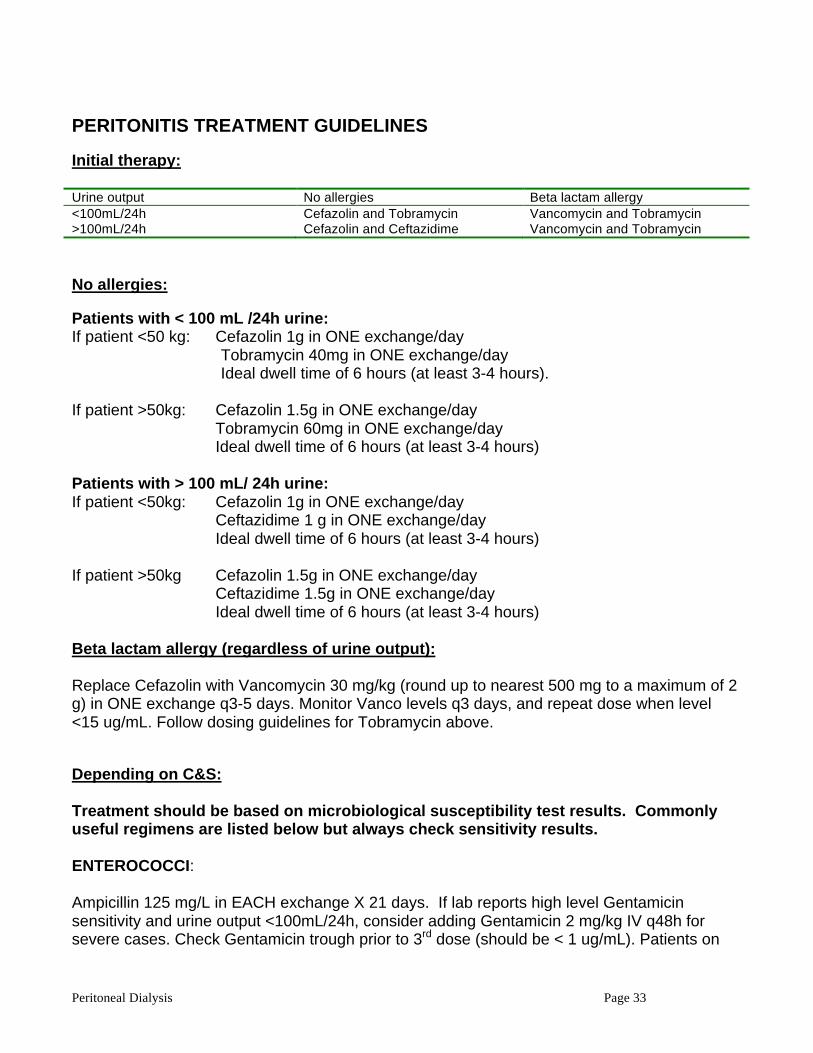
PERITONITIS TREATMENT GUIDELINES Initial therapy: Urine output No allergies Beta lactam allergy <100mL/24h Cefazolin and Tobramycin Vancomycin and Tobramycin >100mL/24h Cefazolin and Ceftazidime Vancomycin and Tobramycin No allergies: Patients with < 100 mL /24h urine: If patient <50 kg: Cefazolin 1g in ONE exchange/day
Tobramycin 40mg in ONE exchange/day Ideal dwell time of 6 hours (at least 3-4 hours).
If patient >50kg: Cefazolin 1.5g in ONE exchange/day
Tobramycin 60mg in ONE exchange/day Ideal dwell time of 6 hours (at least 3-4 hours) Patients with > 100 mL/ 24h urine: If patient <50kg: Cefazolin 1g in ONE exchange/day Ceftazidime 1 g in ONE exchange/day Ideal dwell time of 6 hours (at least 3-4 hours) If patient >50kg Cefazolin 1.5g in ONE exchange/day Ceftazidime 1.5g in ONE exchange/day Ideal dwell time of 6 hours (at least 3-4 hours) Beta lactam allergy (regardless of urine output): Replace Cefazolin with Vancomycin 30 mg/kg (round up to nearest 500 mg to a maximum of 2 g) in ONE exchange q3-5 days. Monitor Vanco levels q3 days, and repeat dose when level <15 ug/mL. Follow dosing guidelines for Tobramycin above. Depending on C&S: Treatment should be based on microbiological susceptibility test results. Commonly useful regimens are listed below but always check sensitivity results. ENTEROCOCCI: Ampicillin 125 mg/L in EACH exchange X 21 days. If lab reports high level Gentamicin sensitivity and urine output <100mL/24h, consider adding Gentamicin 2 mg/kg IV q48h for severe cases. Check Gentamicin trough prior to 3rd dose (should be < 1 ug/mL). Patients on
Peritoneal Dialysis Page 34
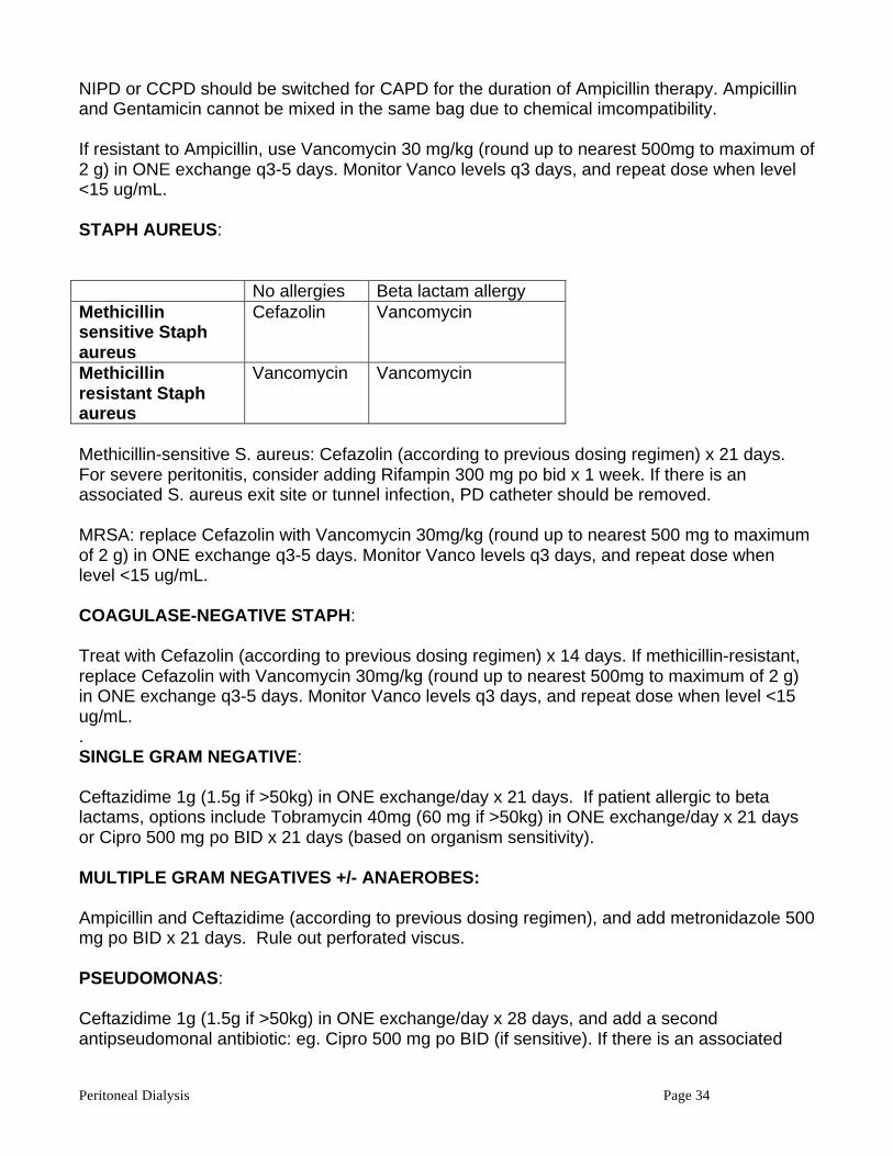
NIPD or CCPD should be switched for CAPD for the duration of Ampicillin therapy. Ampicillin and Gentamicin cannot be mixed in the same bag due to chemical imcompatibility. If resistant to Ampicillin, use Vancomycin 30 mg/kg (round up to nearest 500mg to maximum of 2 g) in ONE exchange q3-5 days. Monitor Vanco levels q3 days, and repeat dose when level <15 ug/mL. STAPH AUREUS: No allergies Beta lactam allergy Methicillin sensitive Staph aureus
Cefazolin Vancomycin
Methicillin resistant Staph aureus
Vancomycin Vancomycin
Methicillin-sensitive S. aureus: Cefazolin (according to previous dosing regimen) x 21 days. For severe peritonitis, consider adding Rifampin 300 mg po bid x 1 week. If there is an associated S. aureus exit site or tunnel infection, PD catheter should be removed. MRSA: replace Cefazolin with Vancomycin 30mg/kg (round up to nearest 500 mg to maximum of 2 g) in ONE exchange q3-5 days. Monitor Vanco levels q3 days, and repeat dose when level <15 ug/mL. COAGULASE-NEGATIVE STAPH: Treat with Cefazolin (according to previous dosing regimen) x 14 days. If methicillin-resistant, replace Cefazolin with Vancomycin 30mg/kg (round up to nearest 500mg to maximum of 2 g) in ONE exchange q3-5 days. Monitor Vanco levels q3 days, and repeat dose when level <15 ug/mL. . SINGLE GRAM NEGATIVE: Ceftazidime 1g (1.5g if >50kg) in ONE exchange/day x 21 days. If patient allergic to beta lactams, options include Tobramycin 40mg (60 mg if >50kg) in ONE exchange/day x 21 days or Cipro 500 mg po BID x 21 days (based on organism sensitivity). MULTIPLE GRAM NEGATIVES +/- ANAEROBES: Ampicillin and Ceftazidime (according to previous dosing regimen), and add metronidazole 500 mg po BID x 21 days. Rule out perforated viscus. PSEUDOMONAS: Ceftazidime 1g (1.5g if >50kg) in ONE exchange/day x 28 days, and add a second antipseudomonal antibiotic: eg. Cipro 500 mg po BID (if sensitive). If there is an associated
Peritoneal Dialysis Page 35
Pseudomonas exit site or tunnel infection, PD catheter should be removed, and patient should be treated with IV antibiotics for 2 weeks after catheter removal. Consider Nystatin therapy for all patients receiving antibiotics Definitions Refractory peritonitis: failure of the effluent to clear after 5 days of appropriate antibiotics Relapsing peritonitis: an episode that occurs within 4 weeks of completion of therapy of a prior episode with the same organism or a sterile episode Recurrent peritonitis: an episode that occurs within 4 weeks of completion of therapy of a prior episode but with a different organism Repeat peritonitis: an episode that occurs more than 4 weeks after completion of therapy of a prior episode with the same organism Catheter-related peritonitis: Peritonitis in conjunction with an exit-site or tunnel infection with the same organism Indications for PD catheter removal FUNGAL-remove immediately Refractory peritonitis Relapsing peritonitis Refractory exit site/tunnel infection Fungal peritonitis Pseudomonas or S. aureus catheter-related peritonitis Consider catheter removal if not responding to therapy Mycobacterial peritonitis Multiple enteric organisms Reference: ISPD: PD-related Infections Recommendations: 2005 Update (www.ispd.org) ANTIBIOTIC PROPHYLAXIS FOR PD PATIENTS PD catheter insertion: Ancef 1 g IV 1 hour pre-procedure (or Vanco 1 g IV if beta-lactam allergy) Dental procedures: Amoxil 2 g po 1 hour pre-procedure (or Clindamycin 600 mg po if beta-lactam allergy) Colonoscopy: If no allergies:
Ampicillin 2 g IV 1 hour pre-procedure Ceftazidime 1.5 g IV 1 hour pre-procedure (1 g if < 50 kg)
Peritoneal Dialysis Page 36
Flagyl 500 mg po 1 hour pre-procedure and 500 mg 12 hours post If beta-lactam allergy: Vancomycin 1 g IV 1 hour pre-procedure Tobramycin 1 mg/kg IV 1 hour pre-procedure Flagyl 500 mg po 1 hour pre-procedure and 500 mg 12 hours post PD catheter manipulation: same protocol as for colonoscopy PD CATHETER INSERTION Prior to PD catheter insertion, please inform home dialysis unit. At St. Michael’s Hospital, PD catheters may be inserted by interventional radiology (IR), or by urology in the OR (usually open surgical approach, with laparoscopy reserved for complex cases). Pre-Insertion (1) All patients going for PD catheter insertion should be given prophylactic antibiotics prior to insertion: Ancef 1 gr IV 1 hour pre-insertion (or Vancomycin 1 gr IV if beta-lactam allergy). (2) Hold anticoagulants and antiplatelet therapies 1 week prior to catheter insertion (can individualize decision re. ASA/Plavix based on cardiac risk). (3) Hold calcium and iron for 1 week pre-insertion as they may predispose to constipation (4) Administer Klean Prep 250 cc po OD x 3 days prior to catheter insertion (can increase dose if necessary) Short Term Management (1) Sterile PD dressing to cover exit site and catheter until site heals (about 2 weeks) (2) Flushes should be done for any patient with a new catheter. This is done to assess the catheter function and to remove fibrin and blood from the peritoneal cavity. Order 500cc volume “in and out” until the effluent clears. Long Term Management Standard nursing protocols for catheter exit care are used once the initial dressing is removed. Catheter care is every second day routinely with antibacterial soap and water followed by 2% chlorhexidine and a dry dressing. Twice weekly is the minimum frequency; exit site care should be increased for drainage or infected sites. Gentamicin ointment should be applied routinely by all
Peritoneal Dialysis Page 37
PD patients with each dressing change (with the exception of patients colonized with MRSA, who should use mupirocin ointment instead). Catheter Dysfunction If there is poor catheter flow, determine whether the difficulty is with outflow alone or both inflow and outflow. The most common cause of slow outflow is constipation.
• If there are problems with both inflow and outflow, consider mechanical obstruction of the catheter by fibrin/clot. Have the nurse irrigate with heparin and saline.
• Order abdominal X-ray to assess catheter position and presence of constipation. The PD catheter tip should be seen in the pelvis.
• If the catheter is in good position and there is evidence of constipation, increase bowel regimen and reassess in 2-3 days
• If the catheter tip has migrated out of the pelvis, refer patient for radiologic catheter manipulation
• If the catheter is in good position, there is no evidence of constipation and there is no improvement after catheter irrigation with heparin, the patient may have catheter dysfunction due to omentum wrapping around the catheter or adhesions (neither of which are visible on standard imaging techniques). Refer patient for radiologic manipulation. If catheter dysfunction is recurrent, consider referral to Dr. Pace for laparoscopic assessment +/- omentopexy/adhesiolysis.
INTRAPERITONEAL (IP) MEDICATIONS Heparin Indicated if fibrin is present in bags or for slow drainage. For CAPD, may be used in all bags or overnight bag only in relation to presence of fibrin.
Dose (Non-peritonitis): 500 units/litre Dose (Peritonitis): 1000 units/litre until effluent clears
Maxeran Used for diabetic gastropathy as an alternative if oral route not beneficial. Dose: 5 mg/litre for control of nausea Potassium Chloride Intraperitoneal KCl is not usually added, but may be considered if alternatives such as increased dietary intake or oral potassium supplementation are not possible. It may be used for inpatients but is generally avoided in the outpatient setting.
Peritoneal Dialysis Page 38
Usual dose = 2 - 4 mEq/L. This dose will limit diffusive removal of K but will not supplement K to the patient. Max dose 10 mEq/L
Xylocaine without Epinephrine For abdominal cramps or pain only after investigations support that the pain is related to dialysate solution (ie. avoid risk of masking pain related to other causes) Dose: 1.25 - 5.0 mL/ of 1% or 2% xylocaine without epinephrine Insulin Therapy in CAPD Although insulin can be placed in the PD bags, typically patients with diabetes on PD are managed in a standard manner with subcutaneous insulin. As with most people with type 2 diabetes, the preferred initial insulin therapy would be a long-acting insulin given at bedtime. A short-acting insulin can be given before meals. Twice daily mixed insulin (ex. 30/70) is also possible, but less likely to control the blood sugar well. When starting PD, the carbohydrate load associated with dextrose containing dialysate can significantly raise the blood sugar. All patients (regardless of diabetes status) who are starting peritoneal dialysis should have their blood sugars monitored during the initiation phase, and hypoglycaemic therapies should be adjusted appropriately. Conversely, a patient who is changing from peritoneal dialysis to hemodialysis may require a significant decrease in hypoglycaemic therapy at the time of the switch.
Renal Transplantation Page 39
VIII. RENAL TRANSPLANTATION A. PRE-OP PROCEDURES LIVING DONOR TRANSPLANT There are many variations on the living donor theme:
1. Biologically related donors ( siblings, parents , children) 2. Emotionally-related donors ( spouse, friend) 3. Non-emotional directed donation 4. Matched-Paired donation 5. Living anonymous donors 6. Domino donation
Donor admitted to Urology service day of surgery; you are not responsible for the donor. Recipient admitted to Nephrology service day prior to surgery; you are responsible pre-op and post-op.
1) Brief history of any recent hospitalization, surgery, illness or blood transfusions that would preclude an elective procedure (eg. recent MI)
2) Focused physical exam 3) If signs or symptoms of infection (fever, leukocytosis, etc) are present,
consider delaying the surgery 4) If patient is on dialysis, they should be dialyzed the day prior to surgery.
Assess the need for an additional run of dialysis: - volume overload (clinical exam, CXR) - hyperkalemia (K > 5.0)
If patient is on CCPD, ensure they get their usual dialysis overnight 5) Ensure that you have the patient’s chart for review from the transplant clinic
(61 Queen office x3665). Ensure that you have viral serology for the recipient AND the donor. There will be a recent note from the transplant physician outlining the plan for this recipient, including potential for participation in clinical research
6) ORDERS: Use the ready-made transplant orders for pre-op, post-op, and medications (more on these later). ALL patients require a cross-match. Ensure CXR and ECG are reviewed the evening of admission.
7) Consider beta-blockers for patients with medium and high cardiac risk 8) TYPE 1 Diabetics (Some Type 2) will need an insulin drip started in the
morning 9) Order all regular medications except anti-coagulants
DECEASED DONOR TRANSPLANT There are variations on the deceased donor theme:
1) DEATH BY NEUROLOGIC CRITERIA (NDD or Brain death) 2) DEATH BY CARDIAC CRITERIA (Donation after cardiac death(DCD))
Renal Transplantation Page 40
For each of these 2, deceased donors can be subclassified as:
A) STANDARD CRITERIA (SCD-Donors less than age 60, or healthy donors , <age 50
B) EXTENDED CRITERIA (ECD-Donors 60 yrs and older, or age 50-59 with 2/3 of the following: hypertension, spontaneous intercranial bleed, Scr>132 umol/l)
You will get a call from Trillium Gift Of Life (TGOL) that a kidney has become available for transplant. They will give you the name of the 1st (and usually 2nd) matched recipient. The recipient(s) will be notified by nephrology resident for urgent admission to hospital and urgent surgery.
1) When you get the call, obtain as much information regarding the kidney and donor as possible DONOR INFO IS CONFIDENTIAL AND SHOULD NOT BE RECORDED IN RECIPIENT CHART (only document serology in chart for future reference):
♦ age of donor ♦ nature of injury to donor ♦ amount of time on pressors ♦ baseline and most recent creatinine and GFR of donor ♦ underlying co morbidities (diabetes, hypertension, etc) ♦ anatomy of kidney (# of arteries and veins) ♦ time at which kidney was or expected to be procured ♦ blood type of donor ♦ serology of donor (CMV, EBV, HepB, HepC, HIV, HTLV)
2) Call urology resident and inform them of a potential transplant 3) Notify 8CC charge nurse of a potential transplant 4) Obtain the SMH transplant list on 8CC and find the recipient
This list is available by hard copy on 8cc or alternatively can be accessed via transplant data base thru the web (YOU WILL GET ACCESS TO THIS SITE AT THE TIME OF YOUR ORIENTATION, AS IT CAN ALSO BE USE TO OBTAIN LAB, CLNICAL AND MEDICATION DATA ON ALL POST-TRANSPLANT RECIPIENTS WHO MAY GET ADMITED TO THE HOSPITAL)
♦ ensure that donor and recipient blood types match ♦ ensure that recipient is not “ON HOLD” for transplant (HD on list) ♦ check to see recipient’s peak PRA and most recent PRA ♦ get recipient’s contact information
5) all recipient ♦ home phone # or cell#
Renal Transplantation Page 41
♦ dialysis unit (phone # at back of transplant list) When speaking to recipient:
♦ introduce yourself and reason for calling ♦ ensure they are ready for transplant (not “on hold”) ♦ ask brief Hx for recent hospitalization, surgery, recent or ongoing
illness, or recent blood products, and time of most recent dialysis ♦ instruct to hold all anticoagulants, stay NPO, and come directly to
8CC nursing station 6) Consider calling in a second recipient if first is highly sensitized (PRA >
75%) 7) Obtain chart from Transplant Clinic. After hours this involves calling
security to let you into 61 Queen 9th floor and unlock transplant office for you.Please sign out the chart and feel free to bring it(them) back to the main hospital with you. Please don’t lose the charts as it is our only record of these patients. These recipients are from dialysis centers from across the province so the data is only available in hard copy and is not available on the SMH electronic system.
8) When recipient arrives perform focused history and physical exam, obtain blood work (including a STAT cross-match), CXR, ECG.
9) If signs or symptoms of infection (fever, leukocytosis, etc) are present, consider cancelling surgery and calling in another recipient
10)assess the need for urgent hemodialysis: ♦ volume overload (clinical exam, CXR) ♦ hyperkalemia (K > 5.0)
11) Cross-match: (HLA lab : 416-340-4995) ♦ All recipients will have a cross-match against potentil donor ♦ In the following cases the results of a STAT cross-match (await
results and must be negative BEFORE beginning surgery): - recent blood products
- current PRA > 20% - peak PRA > 50% - recipient has had prior transplant - most recent cross-match > 3 months old
- If patient has a 0 % PRA and has not had a cross match with the current donor, it is ok to proceed as long as there have been no sensitizing events ie no recent blood transfusion. ALL CROSS MATCH RESULTS WILL BE CALLED IN TO THE ATTENDING STAFF PHYSICIAN as THE TECHNOLOGY IS CHANGING AND THERE ARE MANY NUANCES AND SUBLITIES. (AHG-CDC vs FLOW)If cross-match is definitively positive, you must call in another recipient. THESE RESULTS in ~5 hours
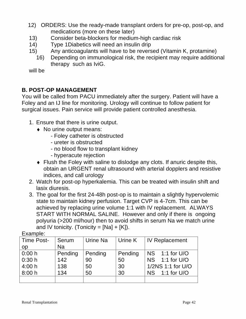
Renal Transplantation Page 42
12) ORDERS: Use the ready-made transplant orders for pre-op, post-op, and medications (more on these later)
13) Consider beta-blockers for medium-high cardiac risk 14) Type 1Diabetics will need an insulin drip 15) Any anticoagulants will have to be reversed (Vitamin K, protamine)
16) Depending on immunological risk, the recipient may require additional therapy such as IviG.
will be
B. POST-OP MANAGEMENT You will be called from PACU immediately after the surgery. Patient will have a Foley and an IJ line for monitoring. Urology will continue to follow patient for surgical issues. Pain service will provide patient controlled anesthesia.
1. Ensure that there is urine output. ♦ No urine output means:
- Foley catheter is obstructed - ureter is obstructed - no blood flow to transplant kidney - hyperacute rejection
♦ Flush the Foley with saline to dislodge any clots. If anuric despite this, obtain an URGENT renal ultrasound with arterial dopplers and resistive indices, and call urology
2. Watch for post-op hyperkalemia. This can be treated with insulin shift and lasix diuresis.
3. The goal for the first 24-48h post-op is to maintain a slightly hypervolemic state to maintain kidney perfusion. Target CVP is 4-7cm. This can be achieved by replacing urine volume 1:1 with IV replacement. ALWAYS START WITH NORMAL SALINE. However and only if there is ongoing polyuria (>200 ml/hour) then to avoid shifts in serum Na we match urine and IV tonicity. (Tonicity = [Na] + [K]).
Example: Time Post-op
Serum Na
Urine Na Urine K IV Replacement
0:00 h 0:30 h 4:00 h 8:00 h
Pending 142 138 134
Pending 90 50 50
Pending 50 30 30
NS 1:1 for U/O NS 1:1 for U/O 1/2NS 1:1 for U/O NS 1:1 for U/O
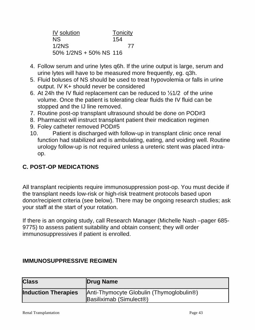
Renal Transplantation Page 43
IV solution Tonicity
NS 154 1/2NS 77 50% 1/2NS + 50% NS 116
4. Follow serum and urine lytes q6h. If the urine output is large, serum and urine lytes will have to be measured more frequently, eg. q3h.
5. Fluid boluses of NS should be used to treat hypovolemia or falls in urine output. IV K+ should never be considered
6. At 24h the IV fluid replacement can be reduced to ½1/2 of the urine volume. Once the patient is tolerating clear fluids the IV fluid can be stopped and the IJ line removed.
7. Routine post-op transplant ultrasound should be done on POD#3 8. Pharmacist will instruct transplant patient their medication regimen 9. Foley catheter removed POD#5 10. Patient is discharged with follow-up in transplant clinic once renal
function had stabilized and is ambulating, eating, and voiding well. Routine urology follow-up is not required unless a ureteric stent was placed intra-op.
C. POST-OP MEDICATIONS All transplant recipients require immunosuppression post-op. You must decide if the transplant needs low-risk or high-risk treatment protocols based upon donor/recipient criteria (see below). There may be ongoing research studies; ask your staff at the start of your rotation. If there is an ongoing study, call Research Manager (Michelle Nash –pager 685-9775) to assess patient suitability and obtain consent; they will order immunosuppressives if patient is enrolled. IMMUNOSUPPRESSIVE REGIMEN
Class Drug Name
Induction Therapies Anti-Thymocyte Globulin (Thymoglobulin®) Basiliximab (Simulect®)
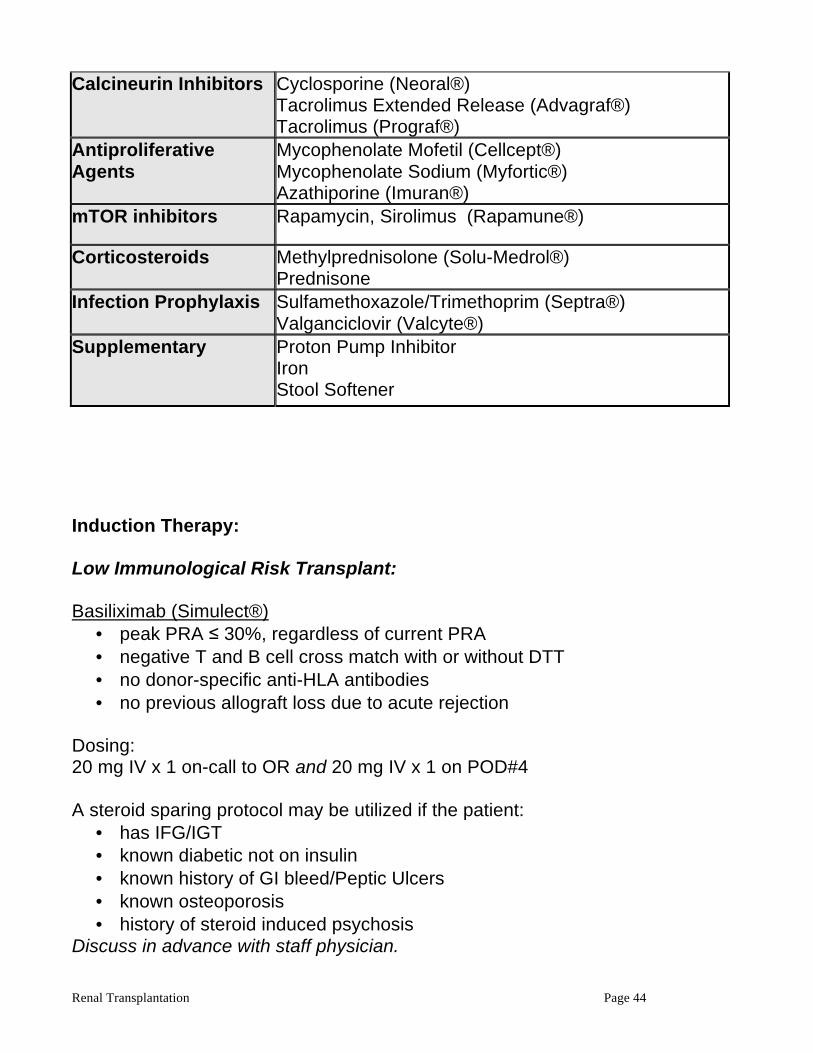
Renal Transplantation Page 44
Calcineurin Inhibitors Cyclosporine (Neoral®) Tacrolimus Extended Release (Advagraf®) Tacrolimus (Prograf®)
Antiproliferative Agents
Mycophenolate Mofetil (Cellcept®) Mycophenolate Sodium (Myfortic®) Azathiporine (Imuran®)
mTOR inhibitors Rapamycin, Sirolimus (Rapamune®)
Corticosteroids Methylprednisolone (Solu-Medrol®) Prednisone
Infection Prophylaxis Sulfamethoxazole/Trimethoprim (Septra®) Valganciclovir (Valcyte®)
Supplementary Proton Pump Inhibitor Iron Stool Softener
Induction Therapy: Low Immunological Risk Transplant: Basiliximab (Simulect®)
• peak PRA ≤ 30%, regardless of current PRA • negative T and B cell cross match with or without DTT • no donor-specific anti-HLA antibodies • no previous allograft loss due to acute rejection
Dosing: 20 mg IV x 1 on-call to OR and 20 mg IV x 1 on POD#4 A steroid sparing protocol may be utilized if the patient:
• has IFG/IGT • known diabetic not on insulin • known history of GI bleed/Peptic Ulcers • known osteoporosis • history of steroid induced psychosis
Discuss in advance with staff physician.
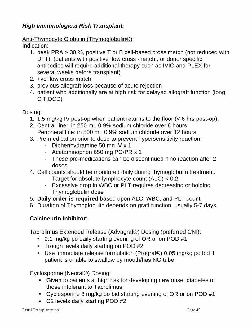
Renal Transplantation Page 45
High Immunological Risk Transplant: Anti-Thymocyte Globulin (Thymoglobulin®) Indication:
1. peak PRA > 30 %, positive T or B cell-based cross match (not reduced with DTT), (patients with positive flow cross -match , or donor specific antibodies will require additional therapy such as IVIG and PLEX for several weeks before transplant)
2. +ve flow cross match 3. previous allograft loss because of acute rejection 4. patient who additionally are at high risk for delayed allograft function (long
CIT,DCD) Dosing:
1. 1.5 mg/kg IV post-op when patient returns to the floor (< 6 hrs post-op). 2. Central line: in 250 mL 0.9% sodium chloride over 8 hours
Peripheral line: in 500 mL 0.9% sodium chloride over 12 hours 3. Pre-medication prior to dose to prevent hypersensitivity reaction:
- Diphenhydramine 50 mg IV x 1 - Acetaminophen 650 mg PO/PR x 1 - These pre-medications can be discontinued if no reaction after 2
doses 4. Cell counts should be monitored daily during thymoglobulin treatment.
- Target for absolute lymphocyte count (ALC) < 0.2 - Excessive drop in WBC or PLT requires decreasing or holding
Thymoglobulin dose 5. Daily order is required based upon ALC, WBC, and PLT count 6. Duration of Thymoglobulin depends on graft function, usually 5-7 days. Calcineurin Inhibitor:
Tacrolimus Extended Release (Advagraf®) Dosing (preferred CNI):
• 0.1 mg/kg po daily starting evening of OR or on POD #1 • Trough levels daily starting on POD #2 • Use immediate release formulation (Prograf®) 0.05 mg/kg po bid if
patient is unable to swallow by mouth/has NG tube
Cyclosporine (Neoral®) Dosing: • Given to patients at high risk for developing new onset diabetes or
those intolerant to Tacrolimus • Cyclosporine 3 mg/kg po bid starting evening of OR or on POD #1 • C2 levels daily starting POD #2
Renal Transplantation Page 46
Cyclosporine (Sandimmune®) IV:
• IV Cyclosporine is used if the patient is unable to take oral cyclosporine or other oral immunosuppressant agents (i.e: tacrolimus)
• Dose: o < 100 mg: dilute in 100 mL D5W or 0.9% sodium chloride o 100-500 mg: dilute in 250 mL D5W or 0.9% sodium chloride
• Administered by slow IV infusion over a period of no less than 2 hours, usual administration time is 4-6 hours.
• Can be given via peripheral or central vein • IV Cyclosporine is associated with anaphylactoid reactions due to
polyoxyethylated castor oil vehicle in the solution. Physician to remain on the nursing unit for the first 30 minutes following the start of infusion. Keep anaphylaxis kit at bedside.
Mycophenolate (Cellcept®) Dosing:
• 500 mg po qid starting evening of OR or on POD#1 • If well tolerated, you may change dose to 1 gram po bid • Alternative to those intolerant to Mycophenolate Mofetil (GI symptoms)
is the enteric-coated active drug Mycophenolate Sodium (Myfortic®). • 500 mg Cellcept = 360 mg of Myfortic
Mycophenolate Mofetil (Cellcept®) IV:
• Given to those patients unable to take oral capsule, tablet, or suspension
• Can be given for up to 14 days, patients should be switched to oral Mycophenolate once they can tolerate oral medication
• Dose: 1 gram IV bid in 140 mL of D5W administered by slow IV infusion over a period of no less than 2 hours. Final concentration is 6 mg/mL.
• Can be given via peripheral or central vein
Steroid Dosing: • Methylprednisolone (Solumedrol®): 2mg/kg IV on-call to OR and then IV
q12h x 48 hours • Prednisone:
• POD # 3-7: 1 mg/kg po daily • POD # 8-14: 0.5 mg/kg po daily • POD # 15 onward: 20 mg po daily until transplant clinic follow
up Others:
Renal Transplantation Page 47
• Iron: Ferrous Fumarate (Palafer®) 300 mg po daily • Stool Softener: Docusate Sodium (Colace®) 100 mg po bid • GI Symptoms: Lansoprazole (Prevacid®) 30 mg po daily • DVT Prophylaxis: Heparin 5000 units sc bid
Surgical Antimicrobial Prophylaxis
1. All transplant recipients receive surgical prophylaxis: • Patients less than or equal to 80 kg: Cefazolin 1 gm IV on call to OR
OR • Patients greater than 80 kg: Cefazolin 2 gm IV on call to OR
OR • Penicillin Allergic: Clindamycin 600 mg IV on call to OR
Infection Prophylaxis:
CMV:
Recipient CMV Donor CMV
Low-Risk (no
Thymoglobulin) High-Risk
(Thymoglobulin)
+ + None Yes (Valganciclovir)
+ - None Yes (Valganciclovir)
- + Yes (Valganciclovir)
Yes (Valganciclovir)
- - None None
Start Valgancyclovir (Valcyte®) 450 mg Tablet POD #3. Dose is based on renal function (see below). Creatinine Clearance (mL/min) CMV Prophylaxis Dosage 60 or greater 900 mg once daily 40-59 450 mg once daily 25-39 450 mg every 2nd day 10-24 450 mg twice weekly
Renal Transplantation Page 48
< 10 (oral suspension only) 100 mg three times weekly
PCP:
All recipients receive Sulfamethoxazole/Trimethoprim 400/80 mg po daily (Septra® Single Strength) for PCP prophylaxis for 1 year. If patient is sulfa allergic, use Dapsone 100 mg po daily.
Other Post Transplant Medications
1. Pre-op meds should be reassessed after transplant (1) most medications should be resumed on POD #1 (2) NSAIDs, ACE inhibitors, and ARBs should be held until transplant
has normal GFR (3) renal medications (phosphate binders, calcitriol, erythropoietin
stimulating agents) can be discontinued 2. Iron supplements are usually added for approximately 3-6 months post
transplant Therapeutic Drug Monitoring Tacrolimus (Advagraf® & Prograf®): • Draw Tacrolimus trough level immediately prior to the morning dose at 10 A.M • Time to steady state after initiation of therapy or after change of dose = 2.5 – 3
days Target Troughs
Time Out From Transplant Target Level (ug/L) 0 – 14 days (1st 2 weeks) 5 - 10 14 – 90 days (2 weeks – 3 month)
5 - 8
> 90 days (> 3 months) 3 - 7 Cyclosporine Oral: Target C2 Level • Draw cyclosporine C2 level exactly 2 hours after the morning dose at 8 A.M.
Renal Transplantation Page 49
Time Out From Transplant Target C2 Level (ug/L)
0 – 30 days 1200 – 1500 1 – 2 months 1000 – 1300 2 – 3 months 900 – 1100 3 – 6 months 800 – 1000 6 – 12 months 500 – 900 > 12 months 300 – 600 Cyclosporine IV: Target Trough • Draw cyclosporine trough level immediately prior to morning dose at 8 A.M
Time From Transplant Target Trough Level (ug/L)
0-1 month 350 – 450 1 – 3 months 300 – 350 3 – 6 months 250 – 300 6 – 12 months 200 – 250 > 12 months 100 – 200 NOTE:
• Cyclosporine Oral to IV dose conversion = 3:1 • Tacrolimus Oral to Cyclosporine Oral dose conversion =1:75
D. DELAYED GRAFT FUNCTION Common causes: Pre-renal:
- hypovolemia - post-op complications (pulm embolus, MI, etc) - tacrolimus/cyclosporine toxicity - other meds: NSAIDs, ACE inhibitors - renal artery embolus or thrombosis
Renal: Venous/ arterial thrombus
- post-ischemic ATN - hyperacute or accelerated rejection
Post-renal:
Renal Transplantation Page 50
- ureteric stricture - lmyphocele or urinoma - hematoma
Investigations:
- assess volume status - check tacrolimus or cyclosporine levels - check pre-op cross-match
Obtain transplant ultrasound with dopplers and resistive (ALWAYS DO ULTRASOUND and DOPPLER URGENTLY IF ANURIC)
- indices - consider transplant biopsy
Management:
- ensure adequate effective circulating volume - hold any nephrotoxic drugs if possible - reduce tacrolimus/cyclosporine levels and substitute basiliximab 20mg
IV days 1 and 4 - address any specific post-renal complication or rejection
Risk Factors for Post-Ischemic ATN
- prolonged cold-ischemia time ( >24h ) - prior sensitization in a prior transplant - increased donor age - nephrotoxic agents (pressors, contrast dye) in donor - subarachnoid hemorrhage in donor - vasculopathy in donor or recipient
E. COMPLICATIONS IN TRANSPLANT PATIENTS General Care of Hospitalized Transplant Patients
1) Most transplant patients are on low doses of prednisone. Consideration must be made for relative adrenal insufficiency:
- severe stress (sepsis, ACS, major surgery)
o hydrocortisone 100mg IV q8h o once stable change to prednisone taper (for example):
Renal Transplantation Page 51
day 1 50mg day 2 30mg day 3 20mg day 4 15mg day 5 10mg day 6 5mg
- mild to moderate stress (uncomplicated infection, day surgery) o triple steroid dose for 3 days
2) Patients who cannot take PO medications can receive some medications
IV - 1mg prednisone PO -> 4mg hydrocortisone IV (divide dose q8-12h) - 3mg cyclosporine PO -> 1mg cyclosporine IV (q12h dosing) - Tacrolimus 0.05mg/kg/day continuous IV infusion - 1mg MMF PO -> 1mg MMF IV (q12h dosing, no adjustment needed)
3) Nephrotoxic agents should be avoided as much as possible
- avoid radiocontrast dye when possible; premedicate with NAC 600mg PO q12h and IV NS at 75cc/h
- avoid aminoglycoside antibiotics; use fluoroquinolones or cephalosporins instead
- judicious use of NSAID or ACE inhibitors when needed REMEMBER: Immunosuppressed patients have a reduced inflammatory response to infection and tissue damage. Have a high degree of suspicion for occult infection and order appropriate imaging. Acute Renal Failure in Renal transplant Patients Common causes Weeks 1-12:
- Acute rejection - Tacrolimus/cyclosporine toxicity - Hypovolemia - Urinary obstruction (ureteric stricture or fluid collection) - CMV infection - Recurrence of primary disease (esp. FSGS and HUS/TTP)
Common causes at > 3 months:
- Hypovolemia - Tacrolimus/cyclosporine toxicity - Acute rejection - Recurrence of primary disease
Renal Transplantation Page 52
- BK virus nephritis - Post-transplant lymphoproliferative disorder - De novo renal disease
Investigations:
- history focusing on medication changes and adherence, history of CMV or EBV mismatch, and recent illness or volume loss
- physical exam focusing on volume status - labs including urine electrolytes, urine dip and microscopy for sediment,
serum tacrolimus or cyclosporine levels, and routine blood work - transplant ultrasound with dopplers
Treatment Guidelines for Acute Allograft Rejection
1. Pulse steroids may be given without affecting the diagnostic yield of the biopsy
2. Thymoglobulin is given in the same way as high-risk transplants, with a dose of 2mg/kg IV q24h
a. Premedicate with diphenhydramine and Tylenol for first 2 days b. Ensure gancyclovir IV prophylaxis c. Septra for PCP prophylaxis d. Nystatin mouthwash
3. Reason for rejection should be addressed a. Inadequate immunosuppressive regimen b. Decreased tacrolimus/cyclosporine levels c. Non-adherence to medications d. Drug interactions with immunosuppressants
4- Antibody-mediated acute rejections ( C4D+) in addition to thymoglobulin may require additional therapies such as plasmapheresis and IVIG.
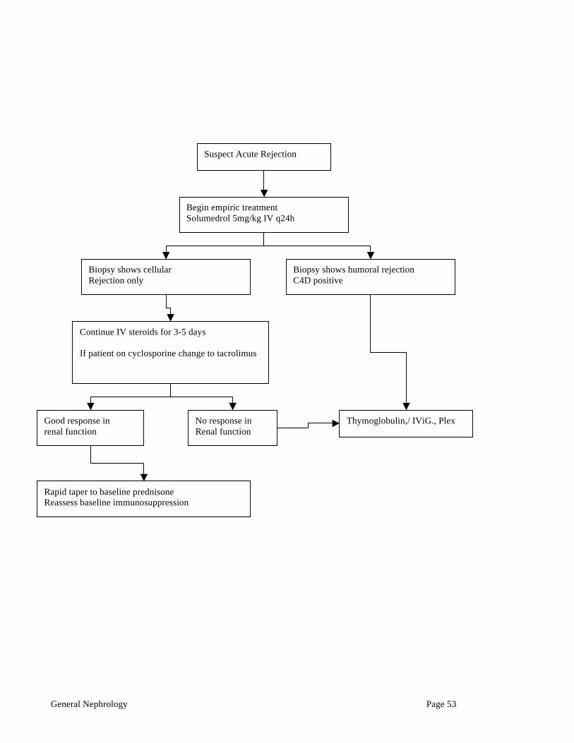
General Nephrology Page 53
Suspect Acute Rejection
Begin empiric treatment Solumedrol 5mg/kg IV q24h
Biopsy shows cellular Rejection only
Biopsy shows humoral rejection C4D positive
Continue IV steroids for 3-5 days If patient on cyclosporine change to tacrolimus
Good response in renal function
No response in Renal function
Thymoglobulin,/ IViG., Plex
Rapid taper to baseline prednisone Reassess baseline immunosuppression
General Nephrology Page 54
IX. GENERAL NEPHROLOGY A. Renal Function
Kidneys:
1) filter, remove wastes 2) maintain salt & water balance 3) phoshate & calcium balance 4) maintain N Hgb ( EPO) 5) control blood pressure 6) maintain Acid/ base balance 7) regulate all major cations/ anions
• simplest estimate of GFR is Plasma creatinine (Pcr) • PCr is directly proportional to muscle mass and indirectly related to GFR • Pcr is filtered but also secreted by renal tubules, particularly when GFR
falls. Therefore at low levels of GFR, Pcr and Clearance of Cr over-estimate true GFR.
• REMEMBER look at your patient when using Pcr to estimate GFR • Normal GFR 100-140 ml/ min
An 80 year old women who weighs 40 KG has an estimated GFR of only 30 ml/ min with a “normal” Pcr of 80 µmol/l Whereas, a 20 year old 100 kg man has a GFR of 120 ml/min with a PCr of 120 µmol /l. CrCl in ml/min= (140-age) x LBW/ PCr (multiply x 1.2 for male) Divide by 60 for CrCl in ml/sec CrCl : creatinine clearance LBW: lean body weight in Kg PCr : plasma creatinine
General Nephrology Page 55
B. Acute Renal Failure
A decrease in baseline GFR occurring over days to weeks with a potential for reversibility
1. Pre-Renal
Decrease Effective Circulating Volume ( ECFV) ie “what the kidneys see”
Volume loss: renal, GI, blood, skin, third space sequestration Edematous states : CHF, cirrhosis, nephrotic syndrome Renal ischemia: hepato-renal, NSAIDS, bilateral RAS, ACEi , ARB
Urine lytes : UNa < 20 meq/l, UCl < 20 ( unless recent diuretic)
FeNa = 100 x[UNa] [Pcr ]/[PNa] [Ucr] FeNa; pre renal < 1, ATN >1
U/A: bland
2. Post Renal “The presence of urine does not exclude obstruction”
Anatomical Functional Prostatic hypertrophy DM Cervical Ca anticholinergic meds Strictures neurogenic bladder stones External compression; colon ca, lymphoma Hematoma, papilary necrosis, Retroperitoneal fibrosis
Most readily diagnosed by ultrasound demonstrating hydronephrosis U/A: bland
3. Renal
Pre-Glomerular
• Malignant hypertension • Scleroderma renal crisis • Cholesterol emboli • thrombotic micro-angiopathy
General Nephrology Page 56
U/A: most often bland, sometimes proteinuria, occasionally, RBCs, eosinophilss Tubular
• ATN (long list: MOST COMMON): shock, sepsis, drugs ( AG antibiotics), contrast, rhabdomylysis, NSAIDs
U/A: heme granular casts, tubular cells, RBCs, crystals Interstitial
• Idiopathic • Drugs; antibiotics, NSAIDs, diuretics, CNI • Infections • Infiltration, leukemia, lymphoma • Radiation • renal transplant : rejection CNI, polyoma, pyelonephritis
U/A: casts, WBC, WBC casts, eosinophiluria Glomerular (see GN) ARF usually seen in context of RPGN: U/A : RBCs & RBC casts
General Nephrology Page 57
C. Glomerular Diseases- A Classification
1. Proliferative (a.k.a., “Nephritic”): = ACTIVE URINARY SEDIMENT, RBCs, OR RBC CASTS Characterized by hypertension, edema, hematuria, RBC casts, and mild-moderate proteinuria.
1. Mesangial Proliferative: Most common GN, microscopic hematuria, gross hematuria with URI, rarely nephrotic, rarely cresentic (RPGN)
• IgA Nephropathy ( seen in IBD, celiac, arthritis, genetic causes) • Henoch-Schonlein Purpura (systemic)
2. Endothelial Proliferative (Post-Infectious Glomerulonephritis):
• Post streptococcal GN (PSGN) • Subacute bacterial endocarditis (SBE) • Ventriculo-peritoneal shunts • Visceral abscesses (pulmonary, intra-abdominal)
3. Crescentic,Epithelial Proliferative (Rapidly Progressive Glomerulonephritis): NEED TO ESTABLISH DIAGNOSIS, SINCE THESE CAN BE LIFE THREATENING
• Type 1: Anti-GBM GN I ) Renal (Anti-GBM Disease) II ) Pulmonary-Renal (Goodpasture’s Syndrome) • Type 2: Immune Complex GN
I ) Primary (MPGN) II ) Secondary (SLE, PSGN, IgA,cryoglobulins)
• Type 3: Pauci-immune Crescentic GN (ANCA +ve) I ) Idiopathic or Pauci-immune Crescentic GN
General Nephrology Page 58
II ) Wegener’s Granulomatosis, Microscopic Polyangiitis, Lll)Churg-Strauss Syndrome 2. Non-Proliferative (a.k.a., “Nephrotic”): BENIGN URINE, FEW or NO RBCs Characterized by proteinuria > 3.5 g/1.73 m2/24 hours, hypoalbuminemia, edema, hyperlipidemia, lipiduria, and hypercoagulability. 1. Primary (a.k.a., Idiopathic):
a) Minimal Change Disease (lipoid nephrosis, nil disease, epithelial cell disease) Most common in children.
• Adult form • Child form • Associations include viral URTI, recent immunizations,
hypersensitivity to drugs (e.g., NSAIDS) or bee stings, paraneoplastic (e.g., Hodgkin’s Disease)
b) Focal Segmental Glomerulosclerosis (FSGS)
• Idiopathic • Associations include heroin abuse, HIV, reflux nephropathy,
chronic pyelonephritis, massive obesity, solitary kidney (renal ablation or renal agenesis), idiosyncratic reaction to drugs (e.g., NSAIDS), circulating factor, genetic causes
c) Membranous Glomerulonephritis (MGN)
• Idiopathic • Associations include hepatitis B antigenemia, autoimmune
diseases (SLE, DM, MCTD, thyroiditis), cancer, drugs (gold, penicillamine, captopril), malaria, syphilis
d) Membranoproliferative Glomerulonephritis (MPGN)(SOMETIMES
CLASSIFIED AS PROLIFERATIVE)
• Type 1 (subendothelial deposits) • Type 2 (intramembrane deposits, dense deposit disease) • Associations include SLE, cryoglobulinemia, hepatitis C, other
chronic viral and bacterial infections, partial lipodystrophy, sickle cell anemia, complement deficiencies
2. Secondary (associated with systemic diseases):
General Nephrology Page 59
• DM, SLE ( antiphospholpid), Multiple myeloma, Amyloidosis
Acknowledgements Page 60
Acknowledgements This 3rd edition has been generously sponsored by Novartis Canada I would like to thank the following who have contributed to the preparation of this manual
1) Division of Nephrology staff at SMH 2) Helen Fanous
THE BEST NEPHROLOGY WEBSITE IN THE WORLD! : UKidney.com (Developed and administered by Dr. Jordan Weinstein) Dr Jeff Zaltzman Educational Director of Nephrology Trainees and Fellows and @ SMH JANUARY 2010