antibody-mediated rejection: prevention, pathophysiology,treatment, brenda muth, rn, ms, acnp...
TRANSCRIPT

Antibody-Mediated Rejection:Prevention, Pathophysiology,Treatment,
Brenda Muth, RN, MS, ACNP
Transplant Nephrology
and Desensitization
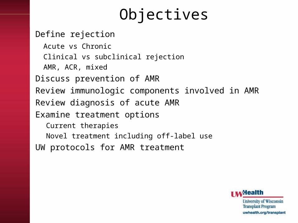
ObjectivesDefine rejection
Acute vs Chronic
Clinical vs subclinical rejection
AMR, ACR, mixed
Discuss prevention of AMR
Review immunologic components involved in AMR
Review diagnosis of acute AMR
Examine treatment optionsCurrent therapies
Novel treatment including off-label use
UW protocols for AMR treatment

Definitions
AMRGraft rejection caused by Ab directed against HLA molecules, ABO antigens or endothelial cell antigens
Clinical RejectionBiopsy confirmed with associated graft dysfunction
Subclinical RejectionHistological changes specific for acute rejection on protocol biopsy w/o graft dysfunction

Definitions• Hyperacute Rejection
– Due to preformed Ab
• Early AMR– Due to amnestic Ab response
• Late AMR – Due to de novo DSA production
• Chronic AMR– Recall secondary DSA response
• Mixed rejection• Accomodation
– Resistance to injury in presence of Ab

Lymphocytes
SCSCMyeloidStem cell
Lymphoid Stem Cell
NKNKTTBB
Lymphoblast
Thymus
Bon
e M
arro
w
PCPCY
Y
Y
Y
Y
Y
CD 20CD 20

The sequence of letters & numbers identify which HLA gene it is & it’s
location
HLA-DQB3*0101 (DR52)
Gene Allele(alternate form of the same gene)
Specific HLA protein

DonorOrgan
Capillary Endothelial
cell
Formation of
Antigen-Ab complex
DonorOrgan
Capillary Endothelial
cell
Formation of
Antigen-Ab complex
C1 complex
Pathophysiology of AMR
Damaged Cell
Releases platelet
aggregationfactors,
cytokines
Damaged Cell
Releases platelet
aggregationfactors,
cytokines
Endothelial cell
necrosis
Endothelial cell
necrosis
Schwartz, NEJM 2010
C4
C4b C4d
C4d is by-product and marker of complement activation
4

Prognosis of AMR
• Vasculopathy
• Fibrosis
• Loss of graft function/graft loss

Diagnostic Criteria for Kidney AMR
Must have at least 2 of the following• Presence of anti-donor antibodies• C4d staining
– C4d1: Minimal C4d stain/detection: 1<10%– C4d2: Focal C4d stain/positive: 10–50%– C4d3: Diffuse C4d stain/positive: >50%
• Morphologic evidence of tissue injury– Capillary and or glomerular inflammation (ptc/g >0)
and/or thromboses
• Graft dysfunction

Diagnostic criteria for Pancreas AMR
• Acute AMR: (all 3 criteria)– Circulating DSA– morphologic evidence of microvascular
tissue injury– C4d staining of interacinar capillaries
• Suspicious for AMR – 2 of 3 criteria
Drachenberg et all, AJT 2011(9)

Consensus Statement for Heart AMR
• Based on pathology:– Histology
• Endothelial activation• Intravascular macrophages• Neutrophilic infiltrates• Capillary destruction• Interstitial edema, hemorrhage
– Immunopathology
• May include DSA, graft dysfunction
Kobashigawa et al, Journal of Heart and Lung Tx, 2011 (3).

Diagnostic Criteria for Liver AMR
• Lack of clear criteria for diagnosis.
• May include:– Positive C4d staining of sinusoidal
endothelium– Proliferation of small bile ducts– Sinusoidal accumulation of neutrophils– cholestasis
Kozlowski, et al, Liver Transplantation, 2011

Diagnostic Criteria for Lung AMR
• Difficult to diagnose
• May be determined by prominence of B cells and plasma cells in inflammatory infiltrate, endothelialitis and small airway inflammation
Takemoto et al, AJT, 2004

Antibody mediated rejection
Nickeleit, Neph Dial and Transplant 18: 2232-2239, 2003
Peritubular capillaritis and focal interstitial hemorrhage

Nickeleit, Neph Dial and Transplant 18: 2232-2239, 2003
Peritubular capillaryImmunofluorescent Stainingfor C4d
Peritubular capillary Immunohistochemistry staining for C4d.

AMR Treatment
CD20CD20
PCPCY
YY
YY
TT

AMR Treatment
• Suppression of T cell response
• Elimination of circulating Ab
• Inhibition of Ab
• Suppression/Depletion of B cells

Suppression of T-cell ResponseDepletional Antilymphocyte Ab (rATG) • Has multiple anti-T cell Ab specificities, costimulatory pathways,
cell adhesion molecules, cell surface molecules expressed on B cells and plasma cells.
• Usually used as adjuvant therapy in AMR• Used for severe or steroid resistant ACR• FDA approved for Kidney transplant rejection
Steroids• inhibits IL-1,IL-2, IL-6 production, T-cell proliferation, cytokine
gene transcription & antigen presentation
MPA• Prevents proliferation of T & B-cells • FDA approved for kidney
CNI • Both CsA & Tac inhibit T & B-cell activation and proliferation• FDA approved for kidney, liver, heart
Singh et al, Transplantation Review, 2009Samaniego et al, Nature Clinical Practice, 2006micromedex

Elimination of Circulating AbPlasmapheresis• Fast, effective method of eliminating DSA• Used in combination with other therapies • Adverse effects:
– Nonselective removal of proteins, bleeding diatheses, volume contraction, requires replacement fluid (albumin), allergic reactions, bld borne pathogens, need HD access
• Dose: – 1-1.5 total plasma volume QD or QOD (3 to 6 treatments), followed by
maintenance PP – Decision to stop PP should be based on:
• elimination of donor-directed HLA antibody• establishment of good graft function • graft failure
• Cost: ~ $2000.00 per treatmentApheresis Guidelinesconsidered a therapeutic option when AMR has been confirmed by Bx
and/or + DSA & immunosuppressive treatment has not been effective.
Singh et al, Transplantation Review, 2009Apheresis Guidelines, 2009

Inhibition of Antibody Immune Globulin Highly purified IgG from large pools of human plasma diluted in
sterile water +/- glucose, sodium. Non-FDA labeled use
• Action:– immunomodulatory effects on T cells, macrophages, cytokine
synthesis, B-cell function & regulatory action on complement system – Down regulates antibody/blocks HLA Ab from binding to targets– T & B cell suppression
• Adverse effects: – Arthralgias, mylagias, HA, HTN, hypotension, MI, Hypercoagulability, allergic reactions, volume overload, AKI
• Dose:– T ½ = 3 weeks– Range 100 mg/kg to 2 gm/kg– ~ $500.00 for 100mg/kg dose
Singh et al, Transplantation Review, 2009Micromedex

Suppression/Depletion of B-cells
RituximabGenetically engineered chimeric MoAb w/ mouse fused with
human IgG. • Indication:
– FDA approved for Non-hodgkin’s Lymphoma, rheumatoid arthritis. Use for AMR is off label.
• Action: – binds to the CD20 antigen located on pre-B & mature B
lymphocytes: mediates B cell lysis – Depletes CD19 & CD20 (Chemical splenectomy)– No effect on plasma cells
• Adverse effects: – Infusion and hypersensitivity reactions, cytopenias, fever,
infection risk including association with BK• Dose
– 375 mg/m2 BSA IV– Duration of treatment ?– ~ $650.00
Singh et al, Transplantation Review, 2009Micromedex

Depletion of Plasma cell
BortezomibReversible proteasome inhibitor • Indication: FDA approved for multiple
myeloma. Use for AMR is off label. • Dose: 1.3 to 1.5 mg/m2 IV day 1, 4, 8, 11.• Adverse effects:
– Neuropathy, plt, WBC, GI symptoms
• Cost ~$2000 per injectionUW experience:
used in kidney, liver and pancreas AMRSteroids + PP + IVIG + Bortezomib +/- ATG
Everly et al, Transplantation, 2008Djamali et al, Clinical Transplants, 2009Sollinger et al, WTC Abstract 2010

Complement Inhibition
Eculizumb• Recombinant humanized monoclonal IgG antibody
produced from murine myeloma cells that inhibits the cleavage of C5
• Indication: PNH, atypical HUS• Blocks graft injury in presence of DSA, may suppress
plasma cells.• Adverse effects:
– Risk of neisseiria meningitis, need immunization• Mayo monitored DSA, B & T flow CM, protocol Bx.• Dose:
– 600 mg IV injection qw to q2w– Duration of therapy unknown– Cost $5000 for 300 mg vial

Monitoring during treatment
• Graft function• Infection
– Viral, bacterial, fungal
• Bone marrow suppression– Leukopenia, thrombocytopenia, anemia
• DSA• Immunosuppression• Repeat biopsy
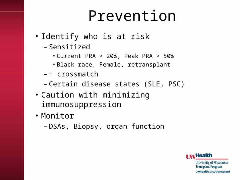
Prevention• Identify who is at risk
– Sensitized• Current PRA > 20%, Peak PRA > 50%• Black race, Female, retransplant
– + crossmatch– Certain disease states (SLE, PSC)
• Caution with minimizing immunosuppression
• Monitor– DSAs, Biopsy, organ function

UW Kidney Transplant Rejection Protocols
Suspicious Dex 50 mg IV + taper
IA, IB Dex 100 mg IV + taper
IIA, IIB, III Dex 100 + taper + ATG
C4d< 50% +/-
Banff I
Early: (<3m) Dex 50mg + taper + TPE 3 to 5 treatments + IVIG 100 mg/kg after each TPE
Late: (> 3m)Dex 50 mg + taper + IVIG 100 mg/kg weekly x 4wks
C4d> 50%+/-
Banff I
Early: Dex 100 mg + taper + TPE 3 to 5 treatments + IVIG 100mg/kg
Late: Dex 100 mg + taper + IVIG 100mg/kg x 4wks
C4d < 50%+
Banff II or III
Dex 100mg/kg + taper + TPE 3 to 5 treatments + IVIG 100mg/kg + either ATG or Bortezomib

Conclusion
• AMR can occur at any time
• Biopsy confirmed diagosis
• Due to preformed Ab, memory response or de novo Ab production
• Causes vascular injury & fibrosis to long term graft survival
• Can be recalcitrant to treatment risk for infectious complications after
treatment

Thank you!