research system microscope...
TRANSCRIPT

RESEARCH SYSTEM MICROSCOPE
BX51/BX61BX2 SERIES
Everything for digital imaging
Everything for digital imaging
Photos courtesy of:Junko Kyozuka, Associate Professor, Graduate School of Agricultural and Life Sciences, The Tokyo University (P.11 below)Prof. Nakatani, Biochemistry course, School of medicine, Showa University (P.13 above)Prof. Tadokoro, Pathology class, School of medicine, St. Marianna University (P.15)
Printed in Japan M1540E-0806B
This catalog is printed by enviromentally-friendly waterless printing system with soy ink.
Specifications are subject to change without any obligation on the part of the manufacturer.
OLYMPUS CORPORATION obtains ISO9001/ISO14001
ISO9001 CertificationOLYMPUS CORPORATION Micro-Imaging System Division
IISO14001 CertificationOLYMPUS CORPORATION Ina-Plant
008
U K A SENVIRONMENTAL
MANAGEMENT
Certified ISO 14001 by

BX51System Microscope
1
The new BX2 series addresses the research demands of the future with Olympus' mostadvanced optical system to date. UIS2 optics deliver the world's highest standard offluorescence performance, along with the image quality and clarity needed for progressof fast-developing life science research programs. With increased S/N ratio, high opticaltransmission, and diverse illumination capabilities, the UIS2 optical system providesexcellent performance over a newly extended wavelength range between UV and IR.This improvement meets all current demands in fluorescence digital imaging andprovides a firm foundation for future developments. As modern research advances toever higher levels of complexity and sophistication, the need for quality anddependability makes the BX2 series today's most convincing solution.
The UIS2 optical system: a new evolutionary advance in fluorescence digital imaging.
BX61Motorized System Microscope

2

400 450 500 550 600 6500
10203040
506070
8090
100
Transmittance (%
)
Wavelength (nm)
BP470-495DM505
BA510-550
High-qualityfluorescence mirror unitsfor fluorescence proteinsThe HQ type mirror units are ideal for thewavelength characteristics ofECFP/EGFP/EYFP/DsRed. With sharpupstroke and high transmission, the mirrorunit efficiently transmits the fluorescenceemitted from fluorescence proteins. Thisallows bright observation images even withweak excitation light, while preventingfluorescence fading and minimizing thechances of cell damage.
Higher S/N ratio enables clear capture ofweak fluorescence emissions.
High S/N objectives detect evenslight fluorescence emission
Mirror unit with straylight reducing functionto eliminate noise
High-performance filters with hightransmission, optimized to individualfluorochrome characteristics
Transmittance (%
)
400 450 500 550 600 6500
10203040
506070
8090
100
Wavelength (nm)
DM485BP460-480
ExBA495-540
Em
World leading fluorescenceperformance — a vital key to modernlife science researchThe ideal in fluorescence observation is tocapture high-contrast images with thelowest exposure to excitation light, thusminimizing the chances of cell damage andfluorescence fading. With increased S/Nratio, high transmission of the objectives andthe high performance mirror unit, Olympus'UIS2 optics provide excellent performancein fluorescence by obtaining bright imagesfrom weak fluorescence signals.
The best S/N ratio...andthe best fluorescenceperformanceOlympus UIS2 objectives provide the bestS/N ratio by employment of totally newdesign to curtail autofluorescence from allpossible sources — glass material, coatingand cementing material. UIS2 objectives
U-MGFPHQ
U-MNIBA3
Stray light reducing functionOlympus mirror units are equippedwith an unique function to eliminatethe stray light that could increase thebackground noise in fluorescence image.
Improved performanceof interference typefluorescence mirror unitsThe new fluorescence mirror units achievehigh S/N ratio by application of new coatingtechnology to the filters and optimal designof excitation and emission filters'characteristic.The hard coating, which prolongs thelifetime of filter, is applied to all Olympusfluorescence mirror units.
Light source
Stray light
Excitation filter
Emission filter
Stray light reducing function
Fluorescence light for observation
Objective
Specimen
Excitation light: Illumination light
Dichromatic mirror
Image captured by the UIS2 objective
S N
S N
S NN
3

4
Trinocular tube compatible fornear infrared region
Camera adapter compatiblefor near infrared region
High sensitivity cooled CCD camera
Lamp housing with asphericalcollector lens providingexcellent excitation efficiency
Image captured by a conventional objective
High sensitivity cooled CCD cameraThe cooled CCD camera DP30BW isdesigned for fluorescence imaging. Itfeatures high sensitivity, low noise andvibration-free operation.
Excellent trinocular tube performanceeven in the near infrared region The trinocular tube U-TR30NIR improves the transmission and compensates foraberrations over a wider wavelength range.A new multi-coatings is applied to thetrinocular optical surfaces to widen the IRspectral characteristics and allow forobservation of newly developedfluorochromes in the near-infrared region.
Camera adapter suitable for near infrared regionUsers can choose from a variety of lowmagnification camera adapters with C-mount, all IR compatible.
High transmission across a widewavelength spectrumThe latest UIS2 objectives achieve a flat,high transmission over a wide wavelengthspectrum, from visible to near infrared —thanks to the incorporation of a newly-developed ultra-wide wavelength reflectionprevention coating (UW multi-coatings). Theimprovement in transmission in the nearinfrared region is especially notable, andtypifies the high performance which makesUIS2 objectives the natural choice in manyleading-edge research fields.
High N.A. objectives forfluorescence imagingThe BX2 series features thenewly-developed PLAPON60XO objective,offering the world's highest N.A. (1.42) forfluorescence imaging, and theUPLSAPO100XO with high 1.4 N.A. andadvanced universal features. In addition totheir outstanding fluorescence S/N ratio,they enjoy UV transmission. TheUPLSAPO100XO objective is especiallynotable for maintaining its transmissiondown to the 340nm wavelength.
S N
S Signal Up
N Noise Down
Fluorescence
achieve high N.A. while reducingautofluorescence, two benefits previouslyconsidered incompatible. With these improvements, UIS2 objectivesprovide the best fluorescence image. Up to near infrared compensation for
chromatic aberrationThe Super Apochromat performance ofUPLSAPO series objectives compensatesfor all chromatic aberrations, from visible toup to 1000nm wavelength light. Clearimages without color shift are provided evenin multi-color observations. Imaging all theway from UV to IR can be performed with asingle objective.
q U-TV0.35xC-2 w U-TV0.5xC-3 e U-TV0.63xCr U-TV1x-2+U-CMAD3
q w e r
5
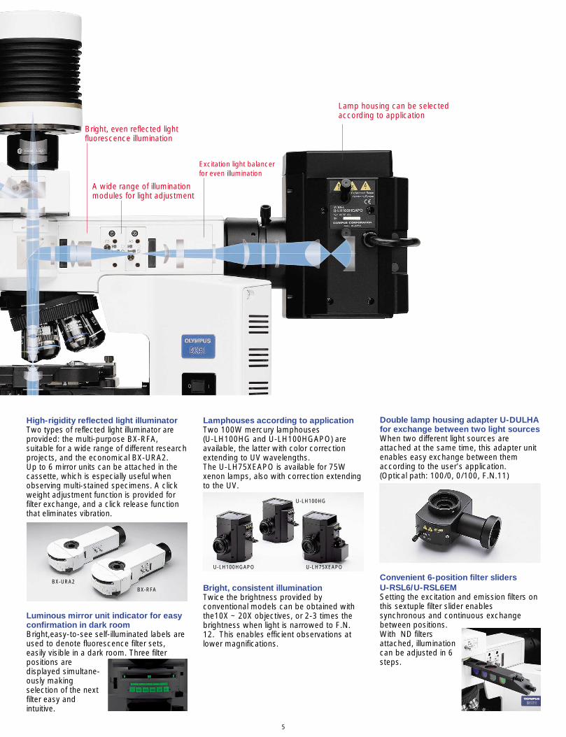
High-rigidity reflected light illuminatorTwo types of reflected light illuminator areprovided: the multi-purpose BX-RFA,suitable for a wide range of different researchprojects, and the economical BX-URA2. Up to 6 mirror units can be attached in thecassette, which is especially useful whenobserving multi-stained specimens. A clickweight adjustment function is provided forfilter exchange, and a click release functionthat eliminates vibration.
Convenient 6-position filter sliders U-RSL6/U-RSL6EMSetting the excitation and emission filters onthis sextuple filter slider enablessynchronous and continuous exchangebetween positions.With ND filtersattached, illuminationcan be adjusted in 6steps.
Lamphouses according to applicationTwo 100W mercury lamphouses (U-LH100HG and U-LH100HGAPO) areavailable, the latter with color correctionextending to UV wavelengths. The U-LH75XEAPO is available for 75Wxenon lamps, also with correction extendingto the UV.
BX-URA2BX-RFA
U-LH100HGAPO U-LH75XEAPO
U-LH100HG
Bright, even reflected lightfluorescence illumination
A wide range of illuminationmodules for light adjustment
Excitation light balancerfor even illumination
Lamp housing can be selectedaccording to application
Luminous mirror unit indicator for easyconfirmation in dark roomBright,easy-to-see self-illuminated labels areused to denote fluorescence filter sets,easily visible in a dark room. Three filterpositions aredisplayed simultane-ously makingselection of the nextfilter easy andintuitive.
Bright, consistent illuminationTwice the brightness provided byconventional models can be obtained withthe10X ~ 20X objectives, or 2-3 times thebrightness when light is narrowed to F.N.12. This enables efficient observations atlower magnifications.
Double lamp housing adapter U-DULHAfor exchange between two light sourcesWhen two different light sources areattached at the same time, this adapter unitenables easy exchange between themaccording to the user's application. (Optical path: 100/0, 0/100, F.N.11)

6
Rectangular field stop for digital imaging/U-RFSSThe rectangular field stop can be set to theexact size of the imaging sensor to avoidfading outside of the imaging area anddamaging sensitivetissue.
Pinhole field stop module/BX-RFSPOTThis slider makes it possible to use the lightsource as a spotlight, illuminating tinyindividual areas on the fluorescencespecimen — an especially valuable featurein experimental work.The slider is attachedto the BX-RFAfluorescenceilluminator in the fieldstop position.
Unnecessary exposure areacaused by a round field stop
Blue enhanced
Blue and red enhanced
Green and red enhanced
Neuro glear cells
Fluorescence excitation balancers/U-EXBABG, U-EXBAUB, U-EXBAUGWhen observing double and triple stainedspecimens, both observation andphotography can be conducted byarranging or altering the fluorescencebrightness while freely changing theexcitation light for each stained color. Anexcitation balancer is attached in the parallellight path, so there is no unevenness in thevisual field.
Confocal laser scanning biological microscope/FV1000The FluoView/FV1000 is a next-generation imaging system designed forhigh-resolution, confocal observation of both fixed and live cells.The FV1000 offers advances in confocal system performancewhile providing the speed and sensitivity required forlive cell imaging with minimal risk of damage tospecimens. In addition, the FV1000 offers a revolutionarysynchronized laser scanning system called the SIMScanner. While one laser stimulates, the secondlaser simultaneously provides high-resolutionimaging. This coordination of laser stimulation andimaging makes the FV1000 an ideal choice forFRAP, FLIP and photoactivation.
FV1000
Fluorescence accessories

Microscope digital camera/ DP71High-resolution digital images equivalent to 12.5 million pixels captured inapprox. 3 seconds — from brightfield to fluorescence.
Thanks to its high-speed hardware, theDP71 can capture high-resolution imagesequivalent to 12.5 million pixels in around 3seconds. The camera's high sensitivity andlow noise (equivalent to the level of ISO1600) ensure clear fluorescence imaging,while the resolution quality allows preciserepresentation of particular specimen areas.
By shifting the pixels of the 1.45 million pixel 2/3 inch CCD (onepixel = 6.45µm), it is possible to record still images equivalent tothe maximum image recording size (4080 X 3072) or effectiveimage size of 12.5 million pixels.
High sensitivity cooled CCD camera/ DP30BWHigh sensitivity, vibration-free cooled CCD camera for fast 15 frames/spreview window.
Using its Peltier-cooled system, theDP30BW offers quiet, vibration-freeoperation. Combined with the built-in shutterand highly effective synchronizedbackground subtract function (dark currentis electronically reduced on the PCI board),these features enable high-quality recordingof even weak fluorescence images.
7
Varied illumination and advanced optics deliver top quality digital images.
Excellent color reproduction fromdaylight illuminationSince Olympus microscopes can apply idealcolor temperature at natural daylight (5500K)throughout the light source, the objectivesand the CCD camera, the camera capturescolor information accurately and providesfaithful reproduction on the display.
0350 400 450 500 550 600 650 700 750 800 850 900
10
20
30
40
50
60
70
80
90
100
Wavelength (nm)
High transmittance(UPLSAPO10x+U-TR30NIR)
Transmittance (%
)
UIS2 optics provide high transmissionfor clear, flat imagesIn the UIS2 optical system, improvedtransmission and compensation forchromatic aberration over a widewavelength spectrum are not onlycharacteristics of the objectives, but also ofimage forming components such as thetrinocular tube and video camera adapter.As a result, images at all magnification levelsare flat, sharp, clear and free from colorshift.
Optimal trinocular tube for digitalimagingIn digital imaging, the best light intensitybalance between the observation side andthe digital camera side should be equal.Olympus' new trinocular tube U-TR30NIRprovides a choice of three light pathexchange: 100% for binocular, 100% forcamera, or 50% each for binocular andcamera.

• In simple time-lapse image mode, theshutter can be opened/closed before/afterthe exposure by synchronous use ofOlympus shutters for the BX61.
• Intuitive, easy-to-use GUI. Tool bar itemscan be user-customized and menu iconsrestricted to frequently used functions.• Comments with arrow, time and scale canbe stored with individual images.
• Focus indicator function shows overallimage focusing status; line profile functionenables detailed focusing on selected areas.• Three different metering areas (30%, 1%and 0.1%) can be selected, and moved overthe specimen as required, allowing highlyprecise exposures. A spot centering buttonis provided to locate and highlight the centerof the image.
Configuration example
• Recorded images can be stored in AVI orMPEG-1 formats, for intermittent shooting ofstill images or "movie" recording of liveimages.
• Separate color/grayscale images capturedfor each excitation wavelength in a multistained specimen can be combined in layersto create a single composite image.
8
Configuration example
Imaging software (for DP71, DP30BW)/DP-BSWDP-BSW is a user-friendly image capturingsoftware package with a simple, easy-to-use GUI (Graphical User Interface) and highspeed live display. It can be used to controldifferent types of motorized units, and toperform both still time-lapse images and liveimage movie recording.
Digital Imaging

9
UIS2 optics inherit high expandability As heir to Olympus' infinity-corrected opticalsystem, in which the tube lens is built intothe observation tube, UIS2 optics display noimage deterioration even when manydifferent optical components or equipmentare inserted in the parallel light path. Thisinherent expandability gives users amplefreedom to construct the system in a waythat meets their specific requirements.
UW (Ultra wideband) multi-coatingsreduces autofluorescence and improvesS/N ratio By using carefully selected raw materials forglass, and applying advanced UW multi-coatings technology, Olympus has reducedobjective autofluorescence and significantlyimproved the S/N ratio.
Flat, high transmission over widewavelength range from UV to IRUW multi-coatings also yields a flat, hightransmission over a wide wavelength range,ensuring high performance in research tasksusing different types of fluorochromes.
UPLSAPO100XO
UPLAPO100XO
Focus (µm)
Wavelength (nm)
Comparing chromatic aberration compensation levels:(The smaller the figure the better)
UPLSAPO series chromatic aberration compensation
-0.5
0
400 450 500 550 600 650 700 750 800 850 900
0.5
1
1.5
2
(new) UPLSAPO100XO
(conventional) UPLAPO100XO
High transmittanceUPLSAPO100XO
Transmittance(%
)
0300 400 500 600 700 800
10
20
30
40
50
60
70
80
90
100 (new) UPLSAPO100XO
(conventional) PLAPO100XO
Complete chromatic aberrationcompensation up to near infraredregionUPLSAPO objectives completely eliminatechromatic aberration up to the near infraredregion, matching the ability of SuperApochromat objectives to provide clearimages without overlapping colors or colorshift. As a result, a single objective canperform imaging from UV to IR wavelengths.
The advanced UIS2 system delivers high performance over a wider wavelength spectrum.
■ UPLSAPO seriesThanks to the application of Olympus' original UW multi-coatings, these Super Apochromat objectives fullycompensate for both spherical and chromatic aberrationsfrom the UV to the near infrared region. Their sensitivity tofluorescence emissions ensures the acquisition of sharp,clear images, without color shift, even in brightfield andNomarski DIC observations. For quality and performance,they offer an unbeatable solution to every kind of digitalimaging need.
■ PLAPON seriesDesigned for unsurpassed resolution and contrast, thesePlan Apochromat objectives keep chromatic aberrationdown to an absolute minimum. The PLAPON60XO, to which the UW multi-coatings isapplied, is the first in the world to achieve N.A. 1.42 forfluorescence imaging.

10
■ UPLFLN (UPLFLN-PH) seriesThese plan objectives also provide flat images with hightransmission up to the near infrared region of thespectrum through the employment of UW multi-coatings.With their high S/N ratio, excellent resolution and highcontrast imaging, they are especially effective inbrightfield and Nomarski DIC observations. The UPLFLN-PH series is optimized for phase contrastobservation.
■ PLN(PLN-PH) seriesIdeal for a range of clinical and research applications,these high quality objectives feature excellent flatness upto F.N. 22 in transmitted brightfield (phase contrast)observation. The PLN-PH series is specifically designedfor phase contrast work.
Excellent Optics
Cover Cor- Iris Water proofObjective N.A. W.D. F.N. glass Immer- Spring rec- dia- and
(mm) thickness sion tion phragm oil proof(mm) ring function
UPLSAPO 4X 0.16 13 26.5 —
UPLSAPO 10X 0.40 3.1 26.5 0.17
UPLSAPO 20X 0.75 0.6 26.5 0.17 _
UPLSAPO 20XO 0.85 0.2 26.5 — Oil _
UPLSAPO 40X 0.90 0.18 26.5 0.11-0.23 _ _
UPLSAPO 60XW 1.20 0.28 26.5 0.15-0.21 Water _ _ _
UPLSAPO 60XO 1.35 0.15 26.5 0.17 Oil _ _
UPLSAPO 100XO 1.40 0.13 26.5 0.17 Oil _ _
PLAPON 1.25X 0.04 5 26.5 —
PLAPON 2X 0.08 6.2 26.5 —
PLAPON 60XO 1.42 0.15 26.5 0.17 Oil _ _
PLAPON 60XOTIRFM 1.45 0.1 26.5 0.13-0.19 Oil _ _ _
UPLFLN 4X 0.13 17 26.5 —
UPLFLN 10X 0.30 10 26.5 —
UPLFLN 20X 0.50 2.1 26.5 0.17 _
UPLFLN 40X 0.75 0.51 26.5 0.17 _
UPLFLN 40XO 1.30 0.2 26.5 0.17 Oil _ _
UPLFLN 60X 0.90 0.2 26.5 0.11-0.23 _ _
UPLFLN 60XOI 1.25-0.65 0.12 26.5 0.17 Oil _ _ _
UPLFLN 100XO2 1.30 0.2 26.5 0.17 Oil _ _
UPLFLN 100XOI2 1.3-0.6 0.2 26.5 0.17 Oil _ _ _
UPLFLN 10XPH 0.30 10 26.5 —
UPLFLN 20XPH 0.50 2.1 26.5 0.17 _
UPLFLN 40XPH 0.75 0.51 26.5 0.17 _
UPLFLN 60XOIPH 1.25-0.65 0.12 26.5 0.17 Oil _ _ _
UPLFLN 100XO2PH 1.30 0.2 26.5 0.17 Oil _ _
PLN 2X 0.06 5.8 22 —
PLN 4X 0.10 18.5 22 —
PLN 10X 0.25 10.6 22 —
PLN 20X 0.40 1.2 22 0.17 _
PLN 40X 0.65 0.6 22 0.17 _
PLN 50XOI 0.9-0.5 0.2 22 — Oil _ _ _
PLN 100XO 1.25 0.15 22 — Oil _
Cover Cor- Iris Water proofObjective N.A. W.D. F.N. glass Immer- Spring rec- dia- and
(mm) thickness sion tion phragm oil proof(mm) ring function
PLN 10XPH 0.25 10.6 22 —
PLN 20XPH 0.40 1.2 22 0.17 _
PLN 40XPH 0.65 0.6 22 0.17 _
PLN 100XOPH 1.25 0.15 22 — Oil _
PLN 4XP 0.10 18.5 22 —
ACHN 10XP 0.25 6 22 —
ACHN 20XP 0.40 3 22 0.17
ACHN 40XP 0.65 0.45 22 0.17 _
ACHN 100XOP 1.25 0.13 22 — Oil _
* All UIS2 objectives and WHN eyepieces: lead-free eco-glass
Cover Cor- Iris OilObjective N.A. W.D. F.N. glass Immer- Spring rec- dia- proof
(mm) thickness sion tion phragm cap(mm) ring
UPLAPO 10XO3 0.40 0.24 26.5 0.17 Oil _ _
UPLAPO 40XOI3 1.00-0.50 0.12 26.5 — Oil _ _ (_)
PLAPO 40X 0.95 0.13 26.5 0.11-0.23 _ _
UPLFL 4XP 0.13 13 26.5 —
UPLFL 10XP 0.30 3.1 26.5 —
UPLFL 20XP 0.50 1.6 26.5 0.17 _
UPLFL 40XP 0.75 0.51 26.5 0.17 _
UPLFL 100XO3P 1.30 0.1 26.5 0.17 Oil _
PLFL 100X 0.95 0.2 26.5 0.14-0.2 _ _
(_): Oil proof cap applicable
UIS2 objectives *
UIS objectives

11 12
Clear, high-contrast imaging from lowto high magnifications.
Clear, high-contrast observation ofstained specimensImage contrast is significantly enhanced bycombining UIS2 objectives with the UIS2eyepiece WHN, which features multi-
Task-specific brightfield condenseroptionsAccording to their purpose, users canchoose from the U-SC3, a swing-outcondenser suitable for observations from1.25X-100X; the U-AC2, a highly cost-efficient Abbe-type model; the U-AAC,whose Aplanat-Achromat designcomprehensively eliminates chromaticaberration; and the U-ULC-2, a specialcondenser for ultra low magnifications.* Select the U-ULC2 condenser for optimal digital imaging with the
1.25X objective.
Bone marrow
Kidney
U-ULC-2U-AAC
U-AC2
U-SC3
*1 Choose upon objective magnification
For high contrast For high resolution General
Specimen Thin Thick —
DIC slider (objective side) U-DICTHC U-DICTHR U-DICT*1 U-DICTS*1
• High resolution with less glareU-DICTHR
This unit enables observations with highresolution but less glare even for thickspecimens used in developmental andgenetic research, such as finely-structureddiatoms, embryos, zebrafish and C. elegans.
• High all-round performanceU-DICT, U-DICTS
Suitable for observing a wide range ofgeneral specimens, such as tissue.
• High contrast for thin specimensU-DICTHC
High contrast can be obtained even in highmagnification observations of thinspecimens, such as culture cells.
PtK2 cell
C.elegance
A shoot apical meristem of rice
New DIC observation system optimizes the specimen image at wider magnifications.
Optimum shearing value according tothe specimenThree types of prisms with different shearingvalue are provided to define contrast andresolution.
New DIC system allows wider selectionMore DIC-compatible objectives areavailable in UIS2, and users can select themost suitable shearing value for a givenspecimen from among 10X to 100Xobjectives. In addition, combination withother observation methods and componentsis simpler and more convenient.
Objective
Specimen
Analyzer
Polarizer
DIC slider(Objective side)
DIC prism(Condenser side)
Universal condenser/U-UCD8This condenser, with built-in polarizer, allowssimultaneous attachment of up to 8 opticalcomponents, freely combined or easilyswitched.
Septuple revolving nosepiece forDIC/simple POL/U-D7REEquipped with a DIC slider slot, the U-D7REseptuple revolving nosepiece allowssimultaneous attachment of 7 objectivesfrom low to high magnifications. It isespecially suitable for combined DIC andfluorescence observations.
Nomarski DIC Brightfield
Conventional imageUIS2 image
coatings on all its surfaces. This makes theimage background look whiter, so that thestained area of the specimen stands outmore clearly.

13
Ideal phase contrast observation with excellent image clarity.
High-contrast observation of internalstructure of live cells/fungus• UPLFLN-PH series objectives have hightransmission, producing well-balancedimages with high contrast even at lowmagnifications. They are suitable forsimultaneous fluorescence, brightfield anddarkfield observations.
Phase contrast accessories
High-quality darkfield effect at all magnifications.
Observing algae in water, or muscletissueTwo darkfield condensers are provided:dry darkfield condenser U-DCD, formagnifications from 10X to 100X (up to N.A.0.80); and oil immersion darkfield condenserU-DCW, for magnifications from 20X to 100X(up to N.A. 1.2).* Please consult your nearest Olympus dealer for applicable objectives.
Ovarian cancer
Spirogyra
U-DCD U-DCW
Phase Contrast
Darkfield

• Mounting an attachable cross-movementmechanical stage (U-FMP) onto the circularrotatable stage makes for improvedobservation efficiency. Interference betweenthe mechanical stage and the objectives iseliminated, so that images of superb qualitycan be effortlesslyobserved at all objectivemagnification.
14
Polarizing observation for wide-arearetardation measurement.
q U-TP530 w U-TP137 e U-TAD r U-CBRI t U-CBR2
y U-CWE2 u U-CSE i U-CBE o U-CTB
Vitamin C Amyloid
U-POC-2U-P4RE
U-CPA
U-OPAU-AN360P-2
t
yui
o
q we
r
• With the U-CPA conoscopic observationattachment, the changeover betweenorthoscopic and conoscopic observationmethods is simple and quick — just slide theBertrand lens control knob in or out.
• UPLFL-P series objectives, designed forobservation under polarizing light, can beused with the revolving nosepiece U-P4RE,which provides a centering function, and thespecial polarizing light condenser U-POC-2.Also available as an option is the sextuplerevolving nosepiece U-P6RE, which allowsperfect alignment of the light path among 3objectives.
• The circular rotatable graduated stage hastwo centering knobs and allows smoothsample rotation. By setting a click stop every45 degrees, it enables accurate observationand measurement.
U-FMP
Polarizing
Compensator Measurement range ApplicationsThick Berek (U-CTB) 0-11,000nm (20λ) Measurement of high retardation level (R*>3λ),
(crystals, macromolecules, fiber, etc.)Berek (U-CBE) 0-1,640nm (3λ) Measurement of retardation level
(crystals, macromolecules, living organisms, etc.)Senarmont compensator 0-546nm (1λ) Measurement of retardation level (crystals, living organisms, etc.)(U-CSE) Enhancement of image contrast (living organisms, etc.)Brace-Koehler compensator 1/10λ 0-55nm (1/10λ) Measurement of low retardation level (living organisms, etc.)(U-CBR1)Brace-Koehler compensator 1/30λ 0-20nm (1/30λ) Enhancement of image contrast (living organisms, etc.)(U-CBE2)Quartz wedge (U-CWE2) 500-2,200nm (4λ) Approximate measurement of retardation level
(crystal, macromolecules, etc.)*R= retardation levelFor more accurate measurement, it is recommended that compensators (expect U-CWE2) be used together with the interference filter45-IF546.
Measuring range of compensators

15
DC power source with no flickerThe microscope body's power source isdirect current, which delivers brightobservation images without flicker.
Metal construction for maximum rigidityThe microscope bodies are made fromaluminum alloy to ensure the high rigidityneeded for consistent performance andlong-term durability.
New advances in ergonomics secure improved observation efficiency.
Rackless stage designBX2 series microscopes feature a wire-driven stage from which the X-axis guidedoes not protrude. This design provides arigid and precise X-Y translation. The X-Ymovement weight is freely adjustable. Thestage surface has a ceramic coating whichprovides excellent wear resistance andensures consistently smooth specimenmovement.
Smooth, light rubber knob movementA rubber cap allowing light and accurateone-finger operation is available as option.
Grooved oil stageFor operators who frequently use highmagnification oil immersion objectives,Olympus offers a special stage with agroove for oil run-off, to prevent glass slidesfrom sticking to the surface.
Swing-out U-SC3 condenser allowsobservation over wide areaThe swing-out U-SC3 condenser is suitablefor all observations from 1.25X to 100X. Nospecial condenser is required for work atultra lowmagnifications.
Up to 4 filters can be mountedSpace is provided for an optional fourthfilter. This allows any filter to be insertedfreely, and the built-in frosted filter to bechanged. Changing to direct lightobservation is aone-touchoperation.
1.25X
4X
10X
100X
40X
Pulmonaryadenocarcinoma
Ergonomics

Motorized revolving nosepiece/U-D6REMMotorized sextuple revolving nosepiecewith slider slot for Nomarski DIC.
Motorized universal condenser/U-UCD8A8 position universal condenser. Differentcombinations of designated opticalcomponents allow for various kinds oftransmitted light observation. Automaticcontrol of optical component exchange, toplens swing out and aperture iris diaphragm.
16
High-efficiency motorized systemmeets more sophisticated researchdemands.
■ Lamp preset and lamp on/off buttonMounted on the front left side of themicroscope frame.■ Fine/coarse and stage escape buttonMounted on the left side of the microscopeframe.■ Hand switch/U-HSTR2Hand set used to control the microscopewhile conducting visual observations. ■ Control box/BX-UCBMotorized modules attached to themicroscope are controlled via this controlbox, which is linked to the computer via anRS232C connector.
U-HSTR2
BX-UCB
Filter wheels /U-FWR, U-FWO and U-FWTMotorized exchange of 6 filters. 3 kindsof filters can be attached simultaneously:U-FWR (ø32, 25) for excitation, U-FWO(ø32, 25) for emission and U-FWT(ø32)for transmitted light.
Reflected light illuminator/BX-RFAAThis motorized turret can load up to 6fluorescence mirror units. Also, equippedwith motorized shutter.
Light adjustment buttons
Stage adjustment buttons
Auto focus unit / U-AFP1Maintains steady auto focusing with1.25X to 100X objectives. Suitable for allobservation methods except phasecontrast. A host personal computer andBX-UCB are required.
Motorized Image Capture Microscope
U-AFP1
U-AFDPT
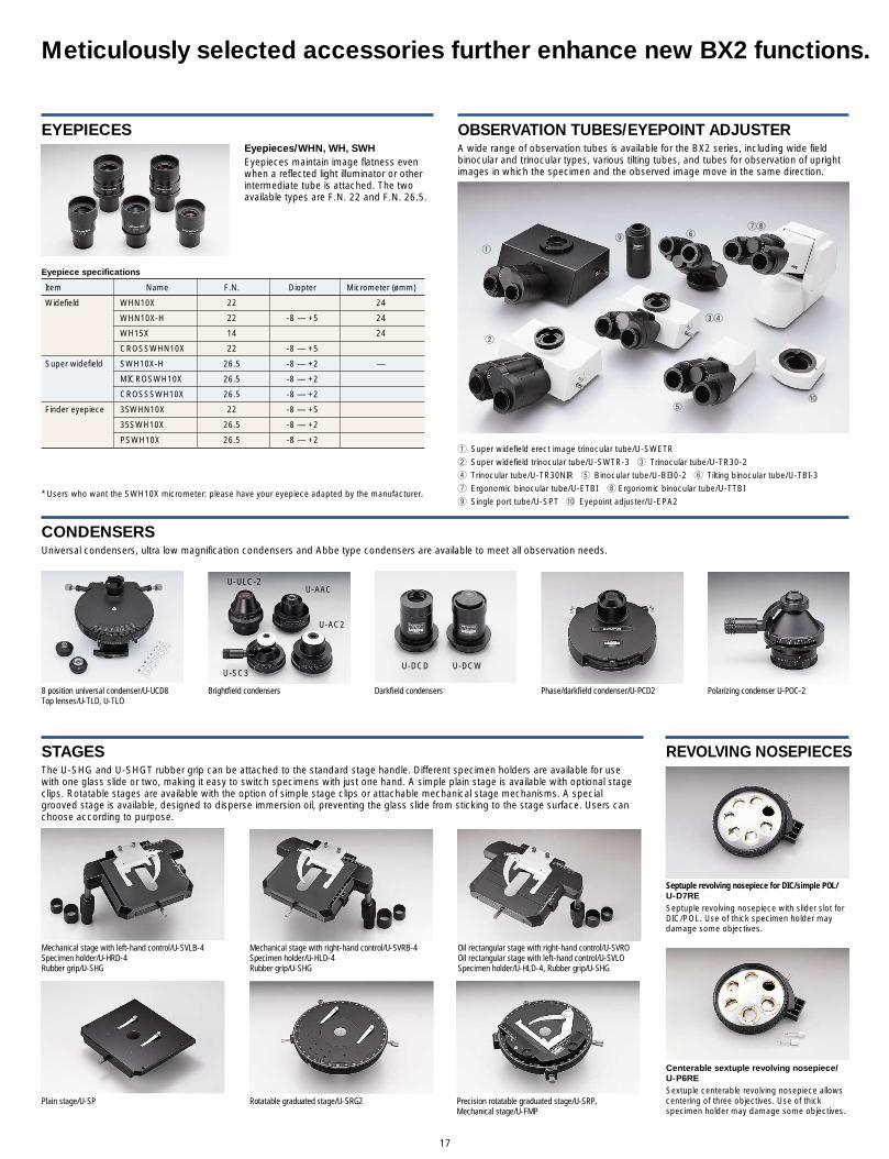
The U-SHG and U-SHGT rubber grip can be attached to the standard stage handle. Different specimen holders are available for usewith one glass slide or two, making it easy to switch specimens with just one hand. A simple plain stage is available with optional stageclips. Rotatable stages are available with the option of simple stage clips or attachable mechanical stage mechanisms. A specialgrooved stage is available, designed to disperse immersion oil, preventing the glass slide from sticking to the stage surface. Users canchoose according to purpose.
17
Centerable sextuple revolving nosepiece/U-P6RESextuple centerable revolving nosepiece allowscentering of three objectives. Use of thickspecimen holder may damage some objectives.
Septuple revolving nosepiece for DIC/simple POL/U-D7RESeptuple revolving nosepiece with slider slot forDIC/POL. Use of thick specimen holder maydamage some objectives.
STAGES
Mechanical stage with left-hand control/U-SVLB-4Specimen holder/U-HRD-4Rubber grip/U-SHG
Plain stage/U-SP
Mechanical stage with right-hand control/U-SVRB-4Specimen holder/U-HLD-4Rubber grip/U-SHG
Rotatable graduated stage/U-SRG2 Precision rotatable graduated stage/U-SRP, Mechanical stage/U-FMP
Oil rectangular stage with right-hand control/U-SVROOil rectangular stage with left-hand control/U-SVLOSpecimen holder/U-HLD-4, Rubber grip/U-SHG
OBSERVATION TUBES/EYEPOINT ADJUSTERA wide range of observation tubes is available for the BX2 series, including wide fieldbinocular and trinocular types, various tilting tubes, and tubes for observation of uprightimages in which the specimen and the observed image move in the same direction.
REVOLVING NOSEPIECES
CONDENSERS
8 position universal condenser/U-UCD8Top lenses/U-TLD, U-TLO
Brightfield condensers Darkfield condensers Phase/darkfield condenser/U-PCD2 Polarizing condenser U-POC-2
U-DCD U-DCW
Universal condensers, ultra low magnification condensers and Abbe type condensers are available to meet all observation needs.
Meticulously selected accessories further enhance new BX2 functions.
q
w
er
t
yui
o
!0
q Super widefield erect image trinocular tube/U-SWETR
w Super widefield trinocular tube/U-SWTR-3 e Trinocular tube/U-TR30-2
r Trinocular tube/U-TR30NIR t Binocular tube/U-BI30-2 y Tilting binocular tube/U-TBI-3
u Ergonomic binocular tube/U-ETBI i Ergonomic binocular tube/U-TTBI
o Single port tube/U-SPT !0 Eyepoint adjuster/U-EPA2
U-AAC
U-AC2
U-ULC-2
U-SC3
Eyepieces/WHN, WH, SWHEyepieces maintain image flatness evenwhen a reflected light illuminator or otherintermediate tube is attached. The twoavailable types are F.N. 22 and F.N. 26.5.
EYEPIECES
Eyepiece specifications
Item Name F.N. Diopter Micrometer (ømm)
Widefield WHN10X 22 24
WHN10X-H 22 -8 — +5 24
WH15X 14 24
CROSSWHN10X 22 -8 — +5
Super widefield SWH10X-H 26.5 -8 — +2 —
MICROSWH10X 26.5 -8 — +2
CROSSSWH10X 26.5 -8 — +2
Finder eyepiece 35WHN10X 22 -8 — +5
35SWH10X 26.5 -8 — +2
PSWH10X 26.5 -8 — +2
* Users who want the SWH10X micrometer: please have your eyepiece adapted by the manufacturer.

18
Drawing attachment/U-DAThe drawing attachment projects an image ofthe pencil and drawing surface into the visualfield. Tracing of microscopic structures is madeeasier and more accurate.
INTERMEDIATE UNITS
U-DA
U-DAL10X
Simple polarizing attachmentSimple polarizing observation can beaccomplished with the combination of U-KPAintermediate attachment for simple polarizingobservation, U-ANT analyzer for transmitted lightand U-POT polarizer.
GROUP OBSERVATION SYSTEMS
CAMERA ADAPTERS
Dual port/U-DPThe dual port may be used for a variety ofpurposes: separating the image by spectralcomposition (e.g. directing fluorescence to oneport, infrared to the other), as an illuminationport for adding a new incident light source or asa C-mount compatible trinocular port for imageoutput. A 1X image formation lens is alsoprovided.
Trinocular intermediate attachments/U-TRUThis intermediate trinocular attachment can beused simultaneously with the inclinable binocularobservation tube (U-TBI-3). Two light paths areselectable: 100% light for binocular observationor 20% for binocular observation and 80% forimaging through the trinocular port.
Multi observation bodies/BX2-DO, BX2-SDO, BX2-MDO-5, BX2-MDO-10Olympus discussion systems are invaluable for research studies, lab training, andeducation. Multi-view configurations are available to accommodate between 2 and 10participants. The pointer is powered by LED, so there is no need for concern aboutsudden lamp failure.
BX2-DO
BX2-SDO
BX2-MDO-5BX2-MDO-10
Camera adapters
w
eq
TR-AdaptersThe single port tube of the trinocular tube is detachable, and can be used with variouscameras via a range of adapters. Using the U-TV1X-2, video can be shot directly with noneed for a shooting lens. The potential of your microscope is greatly increased by itsmultiple image utilization capabilities.
U-POT
U-KPA
U-ANT
U-DP1×C
U-DP
q U-PMTVCw U-PMTVe U-PMTV1Xr U-SMADt U-CMAD3y U-TMADu U-BMADi U-FMTo U-TV0.25XC!0 U-TV0.5X!1 U-TVZ!2 U-TV0.35XC-2!3 U-TV0.5XC-3!4 U-TV0.63XC!5 U-TV1X-2
r t y
u i
Magnification changer/U-CAThis intermediate magnification changerexpands the capability of UIS2 objectives,optimizing the imaged field without theinterruption of rotating the objective lens; 1X / 1.25X / 1.6X / 2X
Filter cassette/U-FCUse of this cassette enables fast exchangeamong three filters (with ø45mm and below2.8mm thickness).
Arrow pointer/U-APTEnables insertion of a red or green LED arrowfor display on a monitor.
Magnification changer/U-ECA, U-ECA1.6XThis intermediate magnification changerexpands the capability of UIS2 objectives,optimizing the imaged field without theinterruption of rotating the objective lens; U-ECA: 1X / 2X, U-ECA1.6X: 1X / 1.6X
Accessories
!0
!4 !5
!1
!2 !3
o

19
*1 Slight vignetting may occur in the periphery of the field due to the top lens. This occurs in observation only. *2 U-FWCO 1.25× should be mounted on U-FWT *3 Optional
BX51/61 specifications
BX51 BX61
Microscope frame Optical system UIS2 optical system
Focus Vertical stage movement: 25mm Motorized focus/vertical stage movement: 25mm, Stage stroke with coarse adjustment limit stopper 0.01µm increments, maximum speed: 3mm/s, Torque adjustment for coarse adjustment knobs coarse/fine changeover button, stage shunting button and Stage mounting position variable stage up/down buttonHigh sensitivity fine focusing knob (minimum adjustment gradations: 1µm)
Illuminator Built-in Koehler illumination for transmitted light 12V100W halogen bulb Light preset switchLight intensity LED indicator Built-in filters (LBD-IF, ND6, ND25, option)
Revolving nosepiece Interchangeable reversed quintuple/sextuple/septuple nosepieceMotorized sextuple revolving nosepiece with slider slot for DICSeptuple revolving nosepiece for DIC/simple POL
Observation Widefield •Widefield binocular, inclined 30° •Widefield tilting binocular, inclined 5°-35°tube (F.N. 22) •Widefield trinocular, inclined 30° •Widefield tilting/telescoping binocular, inclined 0°-25°, telescoping 0-45mm
Super widefield Super widefield trinocular, inclined 24°(F.N. 26.5)
Stage Ceramic-coated coaxial stage with left or right hand low drive control: with rotating mechanism and torque adjustment mechanism, optional rubber grips available(Non stick grooved coaxial, plain, rotatable stages are also available)
Condenser •Abbe (N.A. 1.1), for 4×—100× •Swing out Achromatic (N.A. 0.9), for 1.25×—100× (swing-out: 1.25×—4×)•Achromatic Aplanatic (N.A. 1.4), for 10×—100וUniversal (N.A. 1.4/0.9), for 2×—100× (swing-out: 2×—4×, with oil top lens: 20×—100×)
Motorized fluorescence illuminator *3 Motorized reflected fluorescence, 6-position mirror turret unit, motorized shutter changeover speed: shutter speed: 0.1 s
Motorized universal condenser *3 8-position with motorized AS, turret and top lens swing out mechanism (N.A. 1.4—0.9), for 1.25×*1*2—100×Motorized transmitted filter wheel *3 To be mounted on light exit, 6 positions, ø32, filter thickness: up to 6mm
Motorized reflected filter wheel *3 To be mounted between the lamphouse and the frame, 6 positions, ø25/ø32, filter thickness: up to 6mm
Motorized observation filter wheel *3 To be mounted between the frame and the observation tube, 6 positions, ø25/ø32, filter thickness: up to 6mm
Hand switch *3 Control of septuple revolving nosepiece, 6-position mirror turret illumination unit and 8-position condenser
Control box *3 Serial interface RS232C, built-in transmitted/reflected halogen power supply

20
BX51 dimensions (unit: mm) BX61 dimensions (unit: mm)
BX-UCB dimensions (unit: mm)BX51+BX-RFA dimensions (unit: mm)
U-HSTR2 dimensions (unit: mm)BX51+U-DO3 dimensions (unit: mm)
BX51+U-MDO10 dimensions (unit: mm)
415
209
*412
*187175
6545
84341
90317.5
70
SHUTTER
*470
.8
*187
124
45
8420
944
0.5
70341
415500
175
450
317.5
FOCUS
175
317.5
*187
*496
.826
124
4520
984
341415
530.5
70
See manual
151.
4
212
216
12515
310332 (depth)
OFFON
SW2
SW1
HS
RS232C
ERR
NP
Z/AFCDTTLFW3FW2FW1ASRSHTMU
RMT
BX-UBC
OB
146
7°
180
105
8070
6050
40
3020
10900
*191.5
*451
.5
312.8
4520
9
84
6545
70 341415433.6
4554
1640.6 (standard interpupillary distance)
319
415.
4
467.
1
1086.91341.7
P
12
4
6
8
10
11
9
(740
.6)
(740
.6)
600.
8
965.
6
594.5 637.1
(594.5)(637.1)
Weight: 45kg Power consumption: 160W
Weight: 20.5kg Power consumption: 160WThe length marked with an asterisk (*) may vary according to interpupillary distance. Distance for figure shown is 62mm.
Weight: 27kg Power consumption: 390WThe length marked with an asterisk (*) may vary according to interpupillary distance. Distance for figure shown is 62mm.
Weight: 18kg Power consumption: 140WThe length marked with an asterisk (*) may vary according to interpupillary distance. Distance for figure shown is 62mm.
Weight: 0.4kg
Weight: 5kg Power consumption: 250W
Weight: 37kg Power consumption: 500WThe length marked with an asterisk (*) may vary according to interpupillary distance. Distance for figure shown is 62mm.

SYSTEM DIAGRAM
21
U-TVZZoomcamera port
U-TV1X-2Direct imagecamera port
U-TV0.5XCamera port with 0.5X lens
U-TV0.35XC-2C mountcamera port with0.35X lens
U-TV0.25XCC mountcamera port with0.25X lens
U-SPTSingleport tube
U-DPT-2Double port tube
PEPhoto eyepiece
U-PMTVCamera port with 0.3X lens
U-AFP1*5
Auto focus unit
U-AFDPT*5
AF double port tube
U-PMTV1XCamera port
U-CMAD3C mountadapter
U-BMADBayonetmountadapter
U-TADPlate adapter
COMPENSATORS
U-P4RECenterable revolvingnosepiece
OBJECTIVESfor polarising observation
U-ANTAnalyser fortransmitted light
U-ANTAnalyser fortransmitted light
U-DICTDIC slider fortransmitted light
OBJECTIVES
U-D6RESextuple revolvingnosepiece forDIC/simple POL
U-P6RECenterable sextuple revolving nosepiece
U-D7RESeptuple revolvingnosepiece forDIC/simple POL
U-5RE-2Quintuplerevolvingnosepiece
U-SRG2Rotatable stage
U-SRPPrecision rotatable stage
U-SPPlain stage
U-HLS-4,U-HLST-4Specimen holder
U-HLD-4, U-HLDT-4Specimen holder
U-HRD-4, U-HRDT-4Specimen holder
U-POTPolarizer
U-FCFilter cassette
BX51TFBX51 transmitted frame
BX51TRFBX51 transmitted & reflected frame
U-SWTR-3Super widefieldtrinocular tube
U-SWETRSuper widefielderect image trinocular tube
U-BI30-2Binocular tube
U-TBI-3*1
Tilting binocular tube
U-ETBIErgonomic erect image binocular tube
U-TTBIErgonomic binocular tube
U-FMTF mount adapter
TR-Adapter
U-TMADT mount adapter
U-SMADSonymountadapter
U-FMPMechanical stage
U-CSTCenteringtarget
U-TLOOil top lens
U-TLDDry top lens
Optical devices
U-DICTSShift DIC slider fortransmitted lightU-DICTHRHigh resolution DICslider for transmitted lightU-DICTHCHigh contrast DIC sliderfor transmitted light
U-SVLOOil rectangular stage with left-hand control
U-SVROOil rectangularstage with right-hand control
U-SVLB-4Mechanical stages withleft-handcontrol
U-SVRB-4Mechanical stages withright-handcontrol
U-ETR-4*2
Erect imagetrinocular tube
U-GANGout analyser
*WHN10X, WHN10X-H,CROSS WHN10XEyepiecesU-CT30Centering eyepiece
*35WHN10XEyepiece
▲SWH10X, SWH10X-H,CROSS SWH10X,MICRO SWH10XEyepiecesU-CT30Centering eyepiece
▲ 35SWH10X, PSWH10XEyepieces
● U-TV1X-2, U-TV0.5X, U-TV0.25XC, U-TV0.35XC-2, U-TV0.5XC-3, U-TV0.63XC, U-TVZ
U-SHGRubber grip U-SHGTRubber grip
Filter (ø45)
*4
*1Slight vignetting may occur in the periphery of the field of view in combination with an additional intermediate attachment.
*2 Sight vignetting may occur in the periphery of the field of view in combination with fluorescence illuminator.
*3 Stand is a standard equipment of the U-MDOSV and U-MDO10R3.
*4 Dedicated port for cameras.
*5 Dedicated for BX61.
*6 Standard accessory of BX51TF and BX61TRF frames.
Illustrations colored cyan shows motorized units.
U-DPTSMulti double port tube
U-CMDPTSC-mount adapter for U-DPTS
U-PMDPTSPM-mount adapterfor U-DPS
*6

22
U-TV0.5XC-3C mountcamera port with0.5X lens
U-TV0.63XCC mountcamera port with0.63X lens
U-ECAMagnification changer 2XU-ECA1.6XMagnification changer 1.6X
U-LH100-3100W halogen lamp housing
U-RFSSRectangular field stop
U-RFSSRectangular field stop
U-ANAnalyser for reflected light
U-RSL66 position filter slider
U-RSL6EM6 position filter slider
Slider adaptor
BX-URA2BX reflected light illuminator
U-CPAIntermediate attachment for conoscopic and orthoscopic observation U-AN360P-2
Rotatable analyserU-OPA
Intermediate attachment for orthoscopic observation
U-PCD2Phase/darkfield condenser
U-POC-2Polarisingcondenser
U-AACAchromatic/Aplanaticcondenser
U-AC2Abbe condenser
U-SC3Swing-out condenser
U-ULC-2Ultra low condenser
U-DCDDarkfieldcondenser,dry
U-DCWDarkfieldcondenser,oil
U-HSTR2Hand switch
BX-UCBBX control box
BX61TRFBX61 transmitted & reflected frame
U-TR30-2Trinocular tube
U-TR30NIRTrinocular tube
U-TRU*2
Trinocular intermediate attachment
U-DO3Dual observation attachment
U-MDO10B3Multi observation body for 10 persons
U-MDOB3Multi observation body
U-MDOSV*3
Multi observation side viewer
Stand*3
U-MDO10R3*3
Multi observation body for 10 persons
U-SDO3Side by side observation attachment
U-CAMagnification changer
U-KPAIntermediate attachment forsimple polarising observation
U-ANTAnalyser for transmitted light
U-EPA2Eyepoint adjuster
U-APTArrow pointer
U-DP*3
Dual port U-DP1xCDual port 1X
U-DADrawing attachment
U-DAL10XDrawing attachment 10X
U-25ND6-2, U-25ND25-2, U-25ND50-2 ND filterMIRROR
UNITS
U-TLOOil top lens
U-TLDDry top lens
U-UCD88 positionuniversal condenser
Optical devices
U-LH100HG100W mercury lamp housing
U-LH75XEAPO75W xenon apo lamp housing
U-LH100HGAPO100W mercury apo lamp housing
BX-RFABX fluorescence illuminator
U-EXBABGExcitation balancer BGU-EXBAUBExcitation balancer UBU-EXBAUGExcitation balancer UG
U-UCDTP530Tint plate for sensitive color observation
MICROSCOPE DIGITAL CAMERAS, ANALOG CAMERAS
Power source for 75W xenon lamp
Power source for 100W mercury lamp
U-D6REMMotorized sextuple revolving nosepiece for DIC/simplePOL
BX-RFAAMotorized fluorescence illuminator
U-FWOObservation filter wheel U-FWR
Reflected filter wheel
U-UCD8AMotorizeduniversal condenser
U-FWTTransmitted filter wheel
PCBX2BSWControl software
U-ZPCB2Z control board
U-AFP1*5
Control board
U-ANAnalyser for reflected light
U-RSL66 position filter slider
U-RSL6EM6 position filter slider
Slider adapter
U-25ND6-2, U-25ND25-2, U-25ND50-2ND filter
U-EXBABGExcitation balancer BGU-EXBAUBExcitation balancer UBU-EXBAUGExcitation balancer UG
BX-RFSPOTRectangular field stop
BX-RFSPOTRectangular field stop
MIRROR UNITS
U-DULHADouble lamp house adapter
U-RMTExtension cord

RESEARCH SYSTEM MICROSCOPE
BX51/BX61BX2 SERIES
Everything for digital imaging
Everything for digital imaging
Photos courtesy of:Junko Kyozuka, Associate Professor, Graduate School of Agricultural and Life Sciences, The Tokyo University (P.11 below)Prof. Nakatani, Biochemistry course, School of medicine, Showa University (P.13 above)Prof. Tadokoro, Pathology class, School of medicine, St. Marianna University (P.15)
Printed in Japan M1540E-0806B
This catalog is printed by enviromentally-friendly waterless printing system with soy ink.
Specifications are subject to change without any obligation on the part of the manufacturer.
OLYMPUS CORPORATION obtains ISO9001/ISO14001
ISO9001 CertificationOLYMPUS CORPORATION Micro-Imaging System Division
IISO14001 CertificationOLYMPUS CORPORATION Ina-Plant
008
U K A SENVIRONMENTAL
MANAGEMENT
Certified ISO 14001 by