research paper polymer microneedles for …drugdelivery.chbe.gatech.edu/papers/2006/park pharm res...
TRANSCRIPT

Research Paper
Polymer Microneedles for Controlled-Release Drug Delivery
Jung-Hwan Park,1 Mark G. Allen,2 and Mark R. Prausnitz1,3,4
Received December 2, 2005; accepted January 11, 2006
Purpose. As an alternative to hypodermic injection or implantation of controlled-release systems, this
study designed and evaluated biodegradable polymer microneedles that encapsulate drug for controlled
release in skin and are suitable for self-administration by patients.
Methods. Arrays of microneedles were fabricated out of poly-lactide-co-glycolide using a mold-based
technique to encapsulate model drugsVcalcein and bovine serum albumin (BSA)Veither as a single
encapsulation within the needle matrix or as a double encapsulation, by first encapsulating the drug
within carboxymethylcellulose or poly-L-lactide microparticles and then encapsulating drug-loaded
microparticles within needles.
Results. By measuring failure force over a range of conditions, poly-lactide-co-glycolide microneedles
were shown to exhibit sufficient mechanical strength to insert into human skin. Microneedles were also
shown to encapsulate drug at mass fractions up to 10% and to release encapsulated compounds within
human cadaver skin. In vitro release of calcein and BSA from three different encapsulation formulations
was measured over time and was shown to be controlled by the encapsulation method to achieve release
kinetics ranging from hours to months. Release was modeled using the Higuchi equation with good
agreement (r 2Q 0.90). After microneedle fabrication at elevated temperature, up to 90% of
encapsulated BSA remained in its native state, as determined by measuring effects on primary,
secondary, and tertiary protein structure.
Conclusions. Biodegradable polymer microneedles can encapsulate drug to provide controlled-release
delivery in skin for hours to months.
KEY WORDS: controlled-release drug delivery; microneedles; protein stability; transdermal drugdelivery.
INTRODUCTION
Conventional drug delivery using pills or injection isoften not suitable for new protein, DNA, and other therapies(1,2). Devices for controlled release of such compounds havebeen developed, which enable slow delivery over hours toyears. Controlled release is often achieved by encapsulatingdrugs within biodegradable polymer matrices, from whichrelease is governed by drug diffusion and polymer erosion.Decades of research on this topic have yielded clinicalproducts, such as the Lupron Depot, which delivers leupro-lide acetate systemically for months (3), and the Gliadelwafer, which administers carmustine locally to the brain fordays to weeks (4).
A limitation, however, of controlled-release systems isthat they typically require hypodermic needle injection ofpolymeric microparticles or possibly surgical implantation ofmacroscopic devices within the body. These painful andinvasive procedures are generally not suitable for self-administration by patients and therefore are limited to use inhospitals or clinics.
The goal of this study was to develop a minimallyinvasive polymeric controlled-release system suitable for self-administration without the pain or complexity of currentcontrolled-release devices. Rather than using a hypodermicneedle to introduce polymeric microparticles into the body,we propose redesigning the microparticles to have the shapeof microneedles and thereby give these polymeric particlesthe functionality of both needles and drug matrices forcontrolled release (Fig. 1). By integrally forming thesemicroscopic needles onto a patch substrate, arrays of drug-loaded microneedles could be inserted into the skin and wornlike a transdermal patch for slow release over time. An al-ternative approach would involve intentionally separating thepatch base from the needles after insertion into the skin,thereby leaving the drug-filled needles invisibly buried in theskin for slow release. Because these microneedles are madeof FDA-approved, biodegradable polymer, they should safelydisappear after drug delivery is complete. Previous studieshave shown that microneedles are painless (5,6).
0724-8741/06/0500-1008/0 # 2006 Springer Science + Business Media, Inc. 1008
Pharmaceutical Research, Vol. 23, No. 5, May 2006 (# 2006)DOI: 10.1007/s11095-006-0028-9
1 Wallace H. Coulter Department of Biomedical Engineering at
Georgia Tech and Emory University, Georgia Institute of Technol-
ogy, Atlanta, Georgia 30332, USA.2 School of Electrical and Computer Engineering, Georgia Institute
of Technology, Atlanta, Georgia 30332, USA.3 School of Chemical and Biomolecular Engineering, Georgia In-
stitute of Technology, Atlanta, Georgia 30332, USA.4 To whom correspondence should be addressed. (e-mail: prausnitz@
gatech.edu)

Microneedles have previously been proposed and devel-oped for related applications. Solid microneedles have beenused to pierce the skin for increased permeability (7) as wellas to provide a substrate on which drug can be coated (8) orencapsulated (9) for rapid release. Using this approach, arange of compounds has been delivered to the skin, includingproteins, such as insulin and human growth hormone; geneticmaterial, including plasmid DNA and oligonucleotides; andvaccines directed against hepatitis B and anthrax (10,11).Hollow microneedles have also been developed for infusionof drug solutions into the skin (12Y14). However, we believethat this is the first study to address the use of microneedlesto encapsulate drug for controlled-release delivery (15).
Guided by previous microneedle studies, controlled-release microneedles should measure hundreds of microns inlength and have a radius of curvature less than 10 mm at the tipto ensure easy penetration into skin by manual insertion (16).Microneedles of this size can penetrate past the skin’s outerbarrier of stratum corneum and deliver drug to the epidermisand superficial dermis, where drug can diffuse rapidly forlocal delivery to skin or systemic distribution via uptake bydermal capillaries. Through the use of biodegradable poly-mers such as poly-lactide-co-glycolide (PLGA), well-estab-lished controlled-release mechanisms can be exploited tocontrol release from microneedles (17,18).
Microneedles of the proposed dimensions can be madeby adapting microfabrication technology (19). Althoughmicrofabrication often involves lithography and etching ofsilicon, the field is being expanded to include laser cutting,molding, and other fabrication techniques to produce micro-devices made of other materials, including metals andpolymers. By leveraging these technologies of the microelec-tronics industry, methods to make microneedles shouldprovide reproducible mass production at disposable cost.
MATERIALS AND METHODS
Fabrication of Biodegradable Microneedles
Fabrication of Microneedle Master Structures and Molds
Microneedles were fabricated by first making masterstructures using lithography-based methods, then creatinginverse molds of these master structures, and finally prepar-ing replicate microneedles by melting biodegradable polymerformulations into the molds. In this way, one master structurecould be used to make multiple molds, which could each beused to make multiple replicates. Microneedles were fabri-cated using two different geometries: beveled tip and taperedcone. Methods to fabricate these master structures and moldshave been described in detail previously (20) and are sum-marized below.
Beveled-tip microneedle master structures were fabri-cated out of SU-8 epoxy using standard UV-lithographictechniques (20). SU-8 epoxy (SU-8 100; MicroChem, New-ton, MA, USA) was coated onto a silicon wafer andlithographically patterned into cylinders in the shape of thedesired needles. The space between the cylinders was filledwith a sacrificial polymer (PLGA 85/15, Sigma-Aldrich, St.Louis, MO, USA) and a copper mask was patterned toasymmetrically cover the tops of the epoxy cylinders andsome of the sacrificial polymer on one side of each cylinder.Reactive ion etching (RIE; Plasma Therm, St. Petersburg,FL, USA) partially removed the uncovered sacrificial layerand asymmetrically etched the tips of the adjacent epoxycylinders. All remaining sacrificial polymer was removed byethyl acetate, leaving an array of epoxy cylinders withasymmetrically beveled tips. This array of needles was coatedwith poly(dimethylsiloxane) (PDMS; Sylgard 184, DowCorning, Midland, MI, USA), which was subsequently peeledoff to make an inverse mold.
Tapered-cone microneedles were fabricated using anovel microlens technique (Y.-K. Yoon, J.-H. Park, and M.G. Allen. Multidirectional UV lithography for complex 3-DMEMS structures. J MEMS, in press). A chromium layer wasfirst deposited and patterned on a glass substrate to form anarray of circular dots of exposed glass. Isotropic wet etchingof the exposed glass was then performed to create concavewells, which were filled with SU-8 epoxy cast on the surface.The refractive index mismatch between glass and SU-8 epoxycreated an array of integrated microlenses. After soft-baking,the SU-8 film was exposed from the bottom (i.e., through theglass) to UV light, which passed through the microlenses toform latent images in the SU-8 epoxy as ray traces from thelenses. After development of the SU-8 epoxy, the resultingtapered-cone microneedle master structure was used to makean inverse PDMS mold.
Fabrication of Microneedles Encapsulating Drug
To prepare microneedles encapsulating drug for con-trolled release, PDMS microneedle molds were first filledwith a model drug formulation and then filled with a PLGAmelt, which was allowed to cool and solidify. Three for-mulations were used to achieve different timescales ofcontrolled release. For rapid release, the model drug was
Fig. 1. Controlled-release drug delivery using polymer microneedles.
Polymeric controlled release is often achieved by encapsulating drug
within microparticles, which are then injected into the body using a
hypodermic needle (shown on left). Polymer microneedles can
similarly be designed to encapsulate drug for controlled release, but
can be directly inserted into the skin without the need for
hypodermic injection (shown on right).
1009Polymer Microneedles for Controlled Release

directly encapsulated within the microneedles. For slowerrelease, drug was first encapsulated either within carboxy-methylcellulose (CMC) or poly-L-lactide (PLA), which wasthen encapsulated within microneedles. The process issummarized in Fig. 2.
For the first formulation, calcein or Texas-Red-labeledbovine serum albumin (BSA) powder (used as received fromSigma-Aldrich or Molecular Probes, Eugene, OR, USA,respectively) was suspended in acetonitrile (Sigma-Aldrich)at a solids content of 10% (w/v) and then homogenized for5 min at 10,000 rpm (PowerGen 700 homogenizer, FisherScientific, Pittsburgh, PA, USA) to make drug microparticles.The homogenized particles, with a broad size distribution overthe approximate range of 1Y100 mm, were filtered first througha 30-mm filter, and then the filtrate was passed through a 1-mmfilter (nylon net filter, Millipore, Billerica, MA, USA). Thefinal solids cake containing particles 1Y30 mm in size wasredispersed in acetonitrile at a solids content >20% (w/v). Theresulting suspension was poured onto a PDMS microneedlemold and placed in a vacuum chamber at j20 kPa for õ5 min.This filled the mold with drug particles by first allowing thevacuum to force the drug suspension into the mold cavities andthen evaporate off the organic solvent. Residual particlesremaining on the surface of the mold were removed usingadhesive tape (Blenderm, 3M, St. Paul, MN, USA). Asdescribed previously (20), the mold was then filled withmelted PLGA (PLGA 50/50, 1.2 dL/g, Birmingham Polymer,Birmingham, AL, USA) in a vacuum oven at 135-C andj70 kPa for 10Y20 min. After cooling, the resulting micro-needles with encapsulated drug were manually removed fromthe mold.
To retard release from microneedles using a double-encapsulation formulation approach (21), calcein was firstencapsulated within CMC microparticles, which were thenencapsulated within microneedles. A CMC solution wasprepared by dissolving 0.25 g of CMC sodium salt (referenceviscosity of 400Y800 cP @ 2% aqueous solution; Sigma-Aldrich) in 9.6 mL of deionized (DI) water for 12 h on a 50-Chot plate with stirring at 300 rpm. Then, 25 mg calcein(Sigma-Aldrich) was dissolved in the CMC solution at acalcein: CMC ratio of 1:10 (w/w). The resulting clearsolution was poured onto aluminum foil and dried to removewater for 6 h under j50 kPa of vacuum. The resulting filmcontaining calcein dispersed in a solid CMC matrix waspulverized by an agate mortar and pestle to form particlesmeasuring a few hundred microns to a few millimeters in size.These large particles were dispersed in acetonitrile, homoge-nized, and filtered to yield particles of 1Y30 mm in size, asdescribed above. The average diameter of the particles was 9.6mm, with a standard deviation of 6.2 mm, determined byanalyzing scanning electron microscope (SEM) images. Thesmall CMC particles encapsulating calcein were finally loadedinto a PDMS mold, which was subsequently filled with PLGA,as described above, to form PLGA microneedles, whichencapsulated CMC particles that further encapsulated calcein.
To slow release still more, a similar approach was used,where calcein was first encapsulated within PLA micropar-ticles, which were then encapsulated within microneedles.Using the well-known double-emulsion technique to makePLA microparticles (22), 50 mg of calcein was dissolved in 15mL DI water and 0.2 g of PLA (L-PLA, 1.0 dL/g; Birming-
ham Polymer) was separately dissolved in 2 mL methylenechloride (Sigma-Aldrich). Then, 200 ml of the calcein solutionwas homogenized in 2 mL of PLA solution for 2 min at15,000 rpm. The resulting water-in-oil emulsion was homoge-nized in 50 mL of an aqueous solution of 0.1% polyvinylalcohol (Sigma-Aldrich) for 2 min at 10,000 rpm, whichproduced a water-in-oil-in-water emulsion. After mixing for3 h at 300 rpm, the methylene chloride was extracted into the
Fig. 2. Method to fabricate polymer microneedles that encapsulate
drug for controlled release. First, a suspension of drug particles is
filled into a microneedle mold. Evaporation of the solvent leaves
solid drug particles partially filling the mold. Pellets of biodegradable
polymer are then melted into the mold under vacuum. Cooling and
solidification of the polymer yields biodegradable polymer micro-
needles with encapsulated drug particles.
1010 Park, Allen, and Prausnitz
continuous phase, which solidified the discontinuous phaseinto PLA microparticles encapsulating calcein. Micropar-ticles of 1Y30 mm in size were isolated by filtration, loadedinto a PDMS mold, and encapsulated in PLGA micro-needles, as described above.
Characterization of Microneedles Containing Drug
In Vitro Release Test in Saline
To measure release rates from microneedles, an arraycontaining 100Y200 needles encapsulating one of the formula-tions of calcein or BSA was attached to the bottom or side of a30-mL glass vial (All-Pak, Bridgeville, PA, USA) containing 5or 10 mL of phosphate-buffered saline (PBS, pH 7.4, Sigma-Aldrich) filtered using a 0.2-mm filter (Millipore). Glass vials,PBS, and magnetic stir bars were autoclaved prior to use. Thevials were magnetically stirred at 300 rpm and incubated in a37-C water bath. Periodically, a 100-ml aliquot of PBS wassampled from each vial, replaced with fresh PBS, and analyzedto determine the concentration of calcein or Texas Red-labeled BSA by calibrated spectrofluorometry (QM-1, PhotonTechnology International, South Brunswick, NJ, USA). Mea-sured concentrations were converted into cumulative drugreleased (Mt) by accounting for PBS volume. Total drugcontent (M0) was determined by placing microneedles in 1 NNaOH overnight at the end of each experiment to fullydegrade remaining PLGA and PLA and thereby release allencapsulated drug (23). The resulting solution was returnedto pH 7.4 using HCl before analysis. M0 was typically õ1 mgfor calcein only, BSA only, and calcein in CMC microparticlesand õ0.1 mg for calcein in PLA microparticles.
Drug release from microneedle formulations was mod-eled using the Higuchi equation (24), which indicates that dif-fusion-mediated release should be proportional to the squareroot of time.
Mt
M0¼ 6
ffiffiffiffiffiffiffiffi
D�
r2�
r
ð1Þ
In this expression, D is the apparent diffusion coefficientof the drug in the polymer matrix, t is time, and r is the radiusof the microneedle (50 mm) (25). This equation was fitted toexperimental data to yield D, which is the only unknown(26). The fitting procedure used a least-squares method thatminimized the differences between experimental and theo-retical values.
In Vitro Release Test in Skin
To study the dissolution and release of drug in skin,microneedles encapsulating calcein were inserted into full-thickness human cadaver skin and placed in a sealed chamber at4-C. Refrigeration was used to avoid dehydration and degra-dation of the skin. Recognizing that skin properties and drugdelivery kinetics are different at the experimental temperatureof 4-C and the body temperature of 37-C, we conducted thisexperiment to qualitatively verify that our microneedles insertand that encapsulated drug is released in the skin.
After 8 h, the needles were removed and any residualcalcein on the skin surface was cleaned off with wet tissue
paper. The spatial profile of calcein released in the skin wasthen imaged by confocal microscopy (LSM 510; Zeiss,Thornwood, NY, USA). Human cadaver skin was obtainedfrom the Emory University Body Donor Program withapproval from the Georgia Tech and Emory UniversityInstitutional Review Boards.
Microneedle Failure Force Measurement
To determine the effect of calcein encapsulation onmicroneedle mechanical properties, the microneedle failureforce was measured as described previously (16,20). Briefly,stressYstrain curves were generated using a displacement-force test station (Model 921A, Tricor Systems, Elgin, IL,USA) while pressing an array of 35 microneedles against astainless steel surface at a rate of 1.1 mm/s until a presetmaximum load (19.6 N) was reached. Microneedles had abase radius of 100 mm, tip radius of 12 mm, and height of 1mm. Microneedle failure was indicated by a sudden drop inapplied force. After each test, microneedles were visuallyinspected by microscopy to confirm that all microneedles haddeformed and failed uniformly. Failure force was determinedat calcein contents of 0, 2, and 10% prepared using a single-encapsulation method.
Protein Stability
Because encapsulation of drugs within microneedlesinvolves a brief exposure to a high-temperature polymermelt, the encapsulation process could be damaging to drugs,especially proteins. To assess possible damage, proteinstability was tested by measuring protein solubility, dynamiclight scattering, and circular dichroism (CD), using BSA as amodel protein (27). Because protein content in microneedlesis small, larger samples were generated by dispersing 500 mgof homogenized BSA particles in a 27.5 g solution of 10%(w/w) PLGA in acetonitrile and then pouring the suspen-sion onto aluminum foil. A thin polymer film encapsulatingBSA particles was formed by evaporating off the acetoni-trile for 5 h under j67 kPa vacuum. The film was cut into3 � 3-cm squares and placed on a 1 cm-thick PDMS filmto simulate the molding process. Samples prepared in thisway were placed in the oven at 135-C for predeterminedtimes. After cooling, PLGA samples were dissolved again inacetonitrile, and the BSA particles were recovered by filtra-tion, washed with methylene chloride, and dried for 6Y8 hunder j30 kPa vacuum. Each condition was tested intriplicate.
As a first measure of stability, BSA samples weredissolved in PBS to determine the fraction of BSA remainingsoluble after thermal exposure, which includes both nativeBSA and reversibly denatured BSA. Insoluble aggregateswere removed by centrifugation at 23,000 � g for 20 min (28).Protein concentration in the supernatant was determined bythe Lowry protein assay (29).
The presence of soluble aggregates of BSA was detectedby dynamic light scattering (30). BSA particles were dis-solved in PBS at a concentration of 800 mg/mL and filtered toremoved insoluble aggregates, and 45 ml of solution wasplaced in a quartz cuvette (Proterion, now Wyatt Technolo-gy, Santa Barbara, CA, USA) at room temperature for
1011Polymer Microneedles for Controlled Release
dynamic light scattering measurements (DynaPro-MS/X,Proterion) using CONTIN analysis.
To identify possible changes in the ratio of a-helix/b-sheet components of BSA structure, CD polarimetry mea-surements were performed using BSA particles dissolved inPBS at a concentration of 70 mg/mL (J700, Jasco, Easton,MD, USA) (31). CD spectra were obtained over a wave-length range of 400Y190 nm with a sensitivity of 20 mdeg anda response time of 2 s.
RESULTS
Fabrication of Microneedles for Transdermal Drug Delivery
Fabrication of Microneedle Master Structures
The first step to make polymer microneedles for con-trolled-release drug delivery involved fabricating master
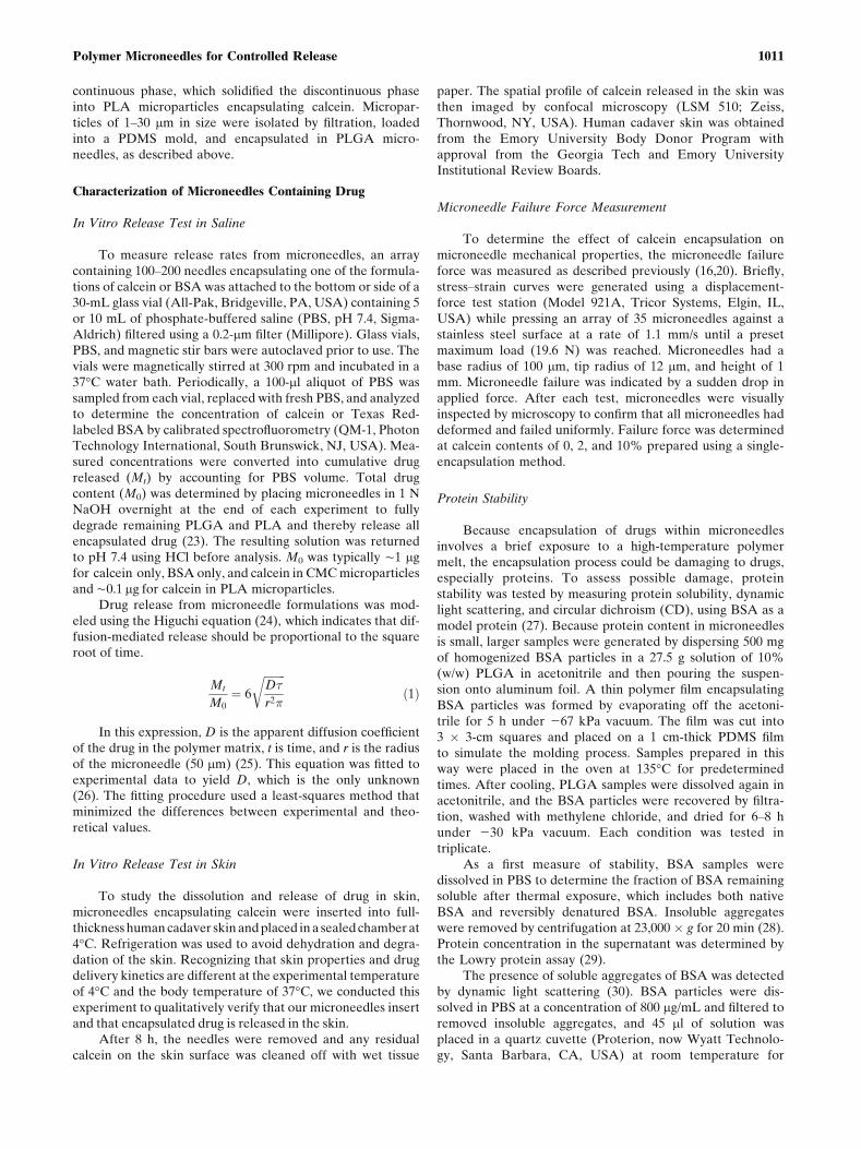
structures using microelectromechanical systems (MEMS)techniques. These master structures were then used to makemolds, which were in turn used to make replicate micro-needles out of biodegradable polymers. Two different geom-etries of microneedle master structures were fabricated outof SU-8 epoxy using lithography-based methods. Representa-tive beveled-tip microneedles are shown in Fig. 3A and have abase radius of 50 mm, a tip radius of 5 mm, and a height of600 mm. The needles are positioned in a 20 � 6 array with acenter-to-center spacing between needles of 400 and 1400 mm.The entire array occupies an area of 9 � 9 mm. Geometricparameters of beveled-tip microneedle arrays, such as needle-to-needle spacing, needle base radius, and base shape, werecontrolled by adjusting the size, shape, and spacing of thelithography mask. The needle height was controlled by thethickness of SU-8 photoresist casting and etching parame-ters. The tip sharpness was controlled by the etchingparameters.
Fig. 3. Microscopy images of microneedles. A section of an array of (A) bevel-tip microneedles
and (B) tapered-cone microneedles used as master structures (imaged by SEM). Making a mold
and using it to prepare polymer microneedles as described in Fig. 2 yielded (C) bevel-tip and
(D) tapered-cone microneedles made of PLGA and encapsulating calcein within their tips
(imaged by fluorescence and bright-field microscopy, respectively). Using a double-encapsula-
tion method produced microneedles that encapsulate microparticles that, in turn, encapsulate
calcein. (E) Cutting off the tip of a PLGA microneedle reveals the PLA microparticles within
(imaged by SEM). (F) A complete 20 � 10 array of PLGA microneedles is shown (imaged by
flash photography).
1012 Park, Allen, and Prausnitz

Representative tapered-cone microneedles are shown inFig. 3B and have a base radius of 100 mm, a tip radius of 2.5 mm,and a height of 750 mm. The needles are positioned in a 10� 20array with center-to-center spacing between needles of 400and 800 mm. The entire array occupies an area of 9 � 9 mm.Needle-to-needle spacing, needle base radius, and base shapewere controlled by adjusting the size, shape, and spacing of thelithography mask. Needle height, taper, and tip sharpnesswere controlled by the optical properties and geometry of theintegrated lenses (see BMaterials and Methods^).
Polymer Microneedles Encapsulating Drug
Using PDMS molds created using the microneedlemaster structures described above, PLGA microneedles werefabricated with encapsulated calcein or BSA, which served asmodel drugs. Figure 3C and D shows beveled-tip andtapered-cone microneedles, respectively, with encapsulatedcalcein. In this case, most calcein was entrapped near the tipsof microneedles to be sure that all of the model drug wasdelivered, even if the microneedles only inserted partiallyinto the skin. Calcein was loaded as solid particles into themicroneedles at compositions up to 10% of needle mass.Similar results were seen for encapsulation of BSA inmicroneedles.
To facilitate slower release, the model drug was firstencapsulated either within CMC or PLA microparticles, whichwere then encapsulated within microneedles. Figure 3E showsa PLGA microneedle with encapsulated PLA microparticlescontaining 5Y10% calcein. The tip of the microneedle hasbeen cut off, thereby exposing the PLA microneedles within.Although these microneedles are made of a solid polymermatrix, a continuous PLGA domain surrounds discrete CMCor PLA microdomains having the size and shape of themicroparticles that formed them. To supplement the highlymagnified views of microneedles, Fig. 3F shows the size andshape of a complete PLGA microneedle array.
Controlled Release of Drug from Microneedles
Release from Microneedles Using a Single-Encapsulation
Formulation
Polymer microneedles encapsulating drugs were devel-oped to serve as a minimally invasive method of con-trolled-release drug delivery that is similar to injectablemicroparticulate systems already in clinical use, but does notinvolve the pain and inconvenience of hypodermic needleinjection. To test this idea, controlled release from micro-needles loaded with calcein was measured in vitro. Using asingle-encapsulation formulation (i.e., calcein was directlyentrapped within the PLGA matrix of the microneedles),calcein release showed zero-order kinetics over a period of4 h, after which 93% of encapsulated calcein was released(Fig. 4A). Controlled release of BSA from a similar formula-tion showed slower kinetics, where 80% of BSA was releasedafter 5 days (Fig. 4A).
These kinetics are consistent with release controlled bydrug diffusion through the microneedle polymer matrix, asopposed to release due to polymer degradation and dissolu-tion. First, significant degradation of polymer should not have
occurred over the timescale of hours to days. PLGAdegradation is known to occur over a timescale of months(32). This is confirmed by microscopy analysis of micro-needles discussed below, which shows that calcein wasreleased before significant needle degradation was observed.Second, degradation-controlled release should be a weakfunction of drug molecular size (17,18). In contrast, diffusion-controlled release is a strong function of drug molecular size,which is consistent with the order-of-magnitude difference inrelease rates between calcein and BSA.
As a companion to quantitative release studies, micro-needles were imaged by light microscopy during release anddegradation. Before release was initiated, calcein was encap-sulated within microneedles, especially at the tips, asindicated by the dark regions in the otherwise transparentPLGA microneedles (Fig. 5A). After 9 h of release in PBS,microneedles showed little change in geometry, indicatingthat significant polymer degradation and dissolution did notyet occur, but were largely devoid of encapsulated calcein, asindicated by the white voids from which calcein was released(Fig. 5B). This image is consistent with release measurementsin Fig. 4, which show release kinetics of hours. Subsequentincubation in strong base to rapidly degrade microneedlesand fully release any residual entrapped calcein yieldedalmost fully degraded microneedles (Fig. 5C).
Fig. 4. Cumulative release of model compounds from PLGA micro-
needles prepared using different formulations to control release
kinetics in vitro. (A) Release of calcein (P) and BSA (Í)
encapsulated within microneedles. The inset has an expanded time
axis to better show release over the initial 4 h. (B) Release of calcein
from microneedles that encapsulated (P) calcein only, (r) CMC
microparticles containing calcein, or (Í) PLA microparticles con-
taining calcein. Data are presented as average T SEM (n = 3Y5).
1013Polymer Microneedles for Controlled Release

Release from Microneedles Using Double-EncapsulationFormulations
To achieve slower release, calcein was first encapsulatedwithin CMC microparticles, which were then encapsulatedwithin PLGA microneedles. Using this approach, calceinrelease from microneedles showed steady release over 4 days(Fig. 4B). This demonstrates that double encapsulation usingCMC can slow release kinetics by more than an order ofmagnitude. Some calcein may not have been encapsulatedwithin CMC, which could explain the burst effect seen duringthe first hours of release.
To achieve slow release over a still longer time, calceinwas first encapsulated within PLA microparticles, which werethen encapsulated within PLGA microneedles. Using thisapproach, calcein release from microneedles was muchslower, exhibiting an initial burst (Fig. 4B), followed by slowrelease over a 2-month period (discussed below). Althoughnot examined in this study, the double-encapsulation ap-proach lends itself to achieving release over other timescales,which can be controlled by formulation of drugs in micro-particles with different polymer compositions using well-known methods (33).
Modeling Controlled Release from Microneedles
Controlled release from polymer matrix systems can bemodeled using the Higuchi equation describing Fickiandiffusion, which predicts that drug release increases linearlywith the square root of time [see Eq. (1)]. This relationshipshould be valid at times following the initial burst-effectrelease of nonencapsulated drug and at times before polymerdegradation can play a role and the apparent diffusioncoefficient is no longer constant. During this release period,the apparent diffusion coefficient of drug inside the micro-needles is the only unknown in the Higuchi equation and wasdetermined by nonlinear regression using experimental datafor controlled release of calcein and BSA from microneedleswith single- and double-encapsulation formulations shown inFig. 4. Fits of Eq. (1) to these experimental data are shownin Fig. 6. The corresponding correlation coefficients werer 2 = 0.92, 0.90, 0.99, and 0.96 for calcein, calcein entrappedin CMC microparticles, calcein entrapped in PLA micro-particles, and BSA, respectively.
Based on these fitted equations, the apparent diffusioncoefficient of calcein in PLGA microneedles was 1.2 �10j10 cm2/s (Fig. 6A). The diffusion coefficient of free calceinin water has previously been calculated to be 5.0 � 10j6 cm2/s(13), which is four orders of magnitude greater. Thus, calceinencapsulation within PLGA microneedles significantlyslowed calcein diffusion for controlled release. The apparentdiffusion coefficient of BSA in PLGA microneedles was 3.0 �10j12 cm2/s (Fig. 6D), which is five orders of magnitudesmaller than free BSA diffusivity in water, 5.9 � 10j7 cm2/s(13). This is again consistent with release controlled byreduced BSA diffusivity in the PLGA matrix.
Encapsulation of calcein in CMC and PLA micro-particles reduced the apparent diffusivity still further. CMCencapsulation reduced the diffusivity to 4.7 � 10j12 cm2/s(Fig. 6B), which is more than an order of magnitude lowerthan without CMC. Encapsulation in PLA microparticlesfurther reduced the diffusion coefficient by another twoorders of magnitude to a value of 6.2 � 10j14 cm2/s.
Microneedle Insertion and Controlled Release in Skin
Controlled Release from Microneedles into Skin
Because encapsulated microneedles are envisioned foruse in skin, we assessed the ability of microneedles to insertinto skin and release drug. In the first experiment, an array ofmicroneedles with encapsulated calcein was inserted intohuman cadaver skin and then removed after 9 h. Imaging the
Fig. 5. Microscopic images of microneedles during controlled
release and degradation in vitro (imaged by bright-field microscopy).
(A) Initially, microneedles encapsulated calcein, as indicated by dark
regions in the otherwise transparent PLGA microneedles. (B) After
incubation in PBS for 9 h, almost all calcein was released from the
microneedles, as indicated by the white voids. (C) Incubation in
concentrated NaOH rapidly degraded the microneedles and fully
released any residual calcein.
1014 Park, Allen, and Prausnitz

skin surface by fluorescence microscopy revealed fluores-cence at each site of microneedle insertion, indicating thatcalcein was released into the skin (Fig. 7A). In a companionexperiment, skin was treated in the same way and thenimaged by confocal microscopy (Fig. 7B). This revealed littlefluorescence at the skin surface and intense fluorescencedeeper in the skin, with a peak fluorescence at 125Y160 mmbelow the surface, which is just below the dermalYepidermaljunction and near the dermal capillary bed (34). Thisobservation further demonstrates that polymer microneedlescan release encapsulated compounds within the skin. Becausecalcein was loaded into the tips of these microneedles, theseimages also indicate that the microneedles did not insert totheir full 750-mm length during this in vitro hand insertion. Ifdesired, insertion at higher velocity, with vibration, or withskin under tension (as found on the body in vivo) canincrease insertion depth by reducing skin deflection duringinsertion (16,35,36).
Mechanics of Microneedle Insertion into Skin
The ability of microneedles to insert into skin alsodepends on needle mechanical properties. Although metalmicroneedles used in other studies are extremely strong,
polymer microneedles require special attention to mechani-cal strength. Our previous study of polymer microneedles(without encapsulated drug) showed that microneedles madeof PLGA and other polymers could be designed withsufficient strength for reliable insertion without breaking(20). Because the encapsulation of drug could weakenmicroneedles, the force required to cause failure of PLGAmicroneedles was measured at 0, 2, and 10% loading withcalcein. As shown in Fig. 8, microneedles without encapsu-lated drug failed at a force of 163 T 10 mN per needle.Previous measurements and calculations have shown thatneedles of the same geometry insert into human skin with aforce of 45 mN per needle (16). The safety factorVdefined asthe ratio of failure force to insertion forceVis therefore 3.6,which means that these microneedles insert into the skin witha force much less than the failure force.
Encapsulation of 2% calcein in needles of the samegeometry lowered the failure force to 91 T 30 mN per needle,which indicates that encapsulation weakened the needles, butstill maintained a safety factor of 2.0. Encapsulation probablyweakened the microneedles because calcein particles aremechanically weaker than PLGA and poor adhesion betweencalcein particles and the PLGA matrix provided sites formechanical failure.
Fig. 6. Modeling of controlled release from PLGA microneedles prepared using different
formulations: (A) calcein only, (B) calcein encapsulated in CMC microparticles, (C) calcein
encapsulated in PLA microparticles, and (D) BSA only. Data were obtained from Fig. 4. The
Higuchi equation (Eq. 1) was fitted to the data, as shown by the solid line. For these fits, the t =
0 data points were excluded to eliminate release due to the initial burst effect. In (C), only data
up to 100 h were fitted because the effects of polymer degradation became significant at later
times. Values of effective diffusivity and correlation coefficients resulting from these fits are
presented in the text. The x-axes are presented as the square root of time because the Higuchi
equation predicts a linear dependence of release on the square root of time for diffusion-
controlled transport.
1015Polymer Microneedles for Controlled Release

Encapsulation of 10% calcein lowered the failure forcefurther to 40 T 2 mN per needle, which reduced the safetyfactor to 0.89. Because this value is less than 1.0, it meansthat these microneedles mechanically fail before insertinginto skin. However, the microneedles used for this study wererelatively blunt (12-mm tip radius) and long (1-mm length).Sharpening tip radius is known to decrease insertion forceand shortening needle length is known to increase failure
force (16,20). Thus, it is possible that redesigned PLGAmicroneedles with 10% drug capsulation might be madestrong enough for insertion into skin without failure.
Protein Stability During Microneedle Fabrication
Encapsulation in microneedles exposed drugs to the hightemperature of melted PLGA, which could be damaging,especially to temperature-sensitive proteins. The possibilityof damage was reduced by selecting a polymer that can bemelted at a relatively low temperature (135-C), minimizingthe exposure time to elevated temperature to 10Y20 min, andkeeping encapsulated drug in the solid state, which reducesconformational mobility and thereby increases protein sta-bility (37). To assess possible protein damage, the effect ofexposing BSA to melted PLGA under encapsulation con-ditions was measured after different exposure times usingthree different protein stability assays. BSA was chosen as amodel protein, although additional studies will be needed toassess stability of other proteins.
First, irreversible denaturation that led to insolubleprotein aggregates was assessed by measuring aqueous BSAsolubility after various treatments. Exposure to the solvent-based processes used during encapsulation, but without anythermal exposure, did not affect BSA solubility (0 minthermal exposure in Fig. 9A). Exposure to elevated temper-ature for 10, 20, or 30 min lowered protein solubility by 10,18, and 32%, respectively, presumably due to irreversibleaggregation. After 1 h at elevated temperature, essentially allprotein was denatured.
To determine if additional protein aggregates existedamong the soluble fraction of BSA, we further tested samplesby dynamic light scattering. As shown in Fig. 9B, a 10-minexposure to elevated temperature had no significant effect,whereas 20- and 30-min exposures caused increasing levels ofaggregation that formed particles measuring tens of nano-meters in size.
Fig. 7. Fluorescence microscopy images of calcein delivered into
human cadaver skin using polymer microneedles. (A) A 100-needle
array of PLGA microneedles containing õ1 2g of calcein was inserted
into full-thickness cadaver skin for 9 h. After removing the needles,
the skin surface was imaged en face by fluorescence microscopy, which
shows calcein delivery into the skin at the site of each microneedle
insertion. (B) Confocal microscopy of the site of one needle insertion
shows calcein release within the skin below the skin surface, which is
expected for release of calcein encapsulated primarily at the needle
tip. Optical section depths are (B1) 20 mm, (B2) 55 mm, (B3) 90 mm,
(B4) 125 mm, (B5) 160 mm, (B6) 195 mm below the skin surface.
Fig. 8. Mechanical strength of polymer microneedles as a function of
calcein encapsulation. The force-per-needle required to fracture an
array of 35 microneedles decreased with increasing calcein content.
Microneedles had a geometry of 12-mm tip radius, 100-2m base
radius, and 1-mm length. The dashed line indicates the expected
force required for insertion of a microneedle of these dimensions into
skin (16). Data are presented as average T SEM (n = 7).
1016 Park, Allen, and Prausnitz

Finally, circular dichroism measurements were made todetect the presence of intermediate species by measuring theratio of a-helix/b-sheet (27). As shown in Fig. 9C, there waslittle difference in the spectra of BSA exposed to thermaltreatments for up to 30 min, indicating little or no formationof intermediate species. In contrast, a positive control of BSA
incubated in aqueous solution for 25 min at 67-C exhibited alarge spectral shift corresponding to significant conforma-tional changes. This observation is consistent with theexpectation that keeping protein in the solid state increasesstability during thermal processing.
DISCUSSION
Advantages of Controlled Releasefrom Polymer Microneedles
A limitation of conventional controlled-release polymerformulations is that they often require surgical implantationor hypodermic needle injection (1,2). This limits the ability ofsuch systems to be self-administered and generally requiresthe time and expense of trained clinical personnel. Conven-tional transdermal patches provide up to 1 week of controlleddelivery, but only for the small subset of drugs that can crossskin at useful rates (38). Thus, the possibility of a controlled-release, microneedle-based delivery method could provide asignificant advance that captures the ease of use afforded by apatch and the versatile controlled-release properties ofbiodegradable polymer systems.
Microneedles can be painlessly inserted into the skin in aminimally invasive manner that lends itself to self-adminis-tration by patients (5). As designed in this study, the intactmicroneedle patch could be left in place on the skin for anextended period and later removed. Alternatively, the patchcould be designed to intentionally break off the needles,leaving them invisibly embedded in the skin, so that the patchbacking could be discarded. In this scenario, microneedleswould need to be designed to remain anchored within theskin to prevent unintentional expulsion. By using FDA-approved materials, such as PLGA and CMC, controlled-release microneedles are likely to be safe. By leveragingadvanced microelectronics industry fabrication technology,microneedles are likely to be mass-produced at disposablecosts (e.g., US$0.10 per patch) (11).
Controlled-release microneedles require no power supplyor sophisticated controllers, which also reduces cost andcomplexity. This contrasts with most other minimally invasivetransdermal delivery methods under development (38). More-over, intersubject variations in skin diffusional barrierproperties are less important because delivery is largelycontrolled by polymer properties of diffusivity and degrada-tion rate. In this study, in vitro release kinetics ranging fromhours to months were demonstrated using single- and double-encapsulation methods, which should be broadly applicableto many drugs.
Limitations of Controlled Releasefrom Polymer Microneedles
Despite many advantages, controlled-release micronee-dles have limitations that constrain their possible applica-tions. One disadvantage of the microneedle fabricationmethod used in this study is that it involves melted polymerthat exposes encapsulated drugs to elevated temperature.Although this may not pose difficulties for some small-molecule drugs, this study showed that 10% of encapsulated
Fig. 9. Stability of BSA after encapsulation within PLGA using
thermal exposures similar to those used to fabricate microneedles.
(A) BSA solubility in water decreased with increasing length of
exposure to the PLGA melt at 135-C. (B) Dynamic light scattering
shows that the size distribution of BSA molecules did not change
after a 10-min exposure to the PLGA melt, but BSA aggregation was
evident after longer exposures. (C) Circular dichroism shows that
BSA spectra did not change significantly after exposure of solid-state
BSA to the PLGA melt, but BSA in aqueous solution was damaged.
1017Polymer Microneedles for Controlled Release
BSA was irreversibly aggregated after a 10-min exposure tothe polymer melt and that longer exposures led to moreextensive denaturation of primary and secondary structure.Thus, applications involving delivery of heat-sensitive drugsare possible, but may lead to partial denaturation. Efforts inour laboratory are underway to develop different methods tofabricate polymer microneedles that do not involve elevatedtemperatures that damage proteins.
Polymer microneedles for controlled-release delivery arealso constrained by needle mechanical properties. Althoughpolymer microneedles can be designed to be strong enoughto reliably insert into skin, the addition of encapsulated drugcan weaken them. This study found that microneedles with a2% drug loading retained sufficient mechanical strength, butneedles with a 10% loading did not. Although redesign ofneedle geometry should increase the maximum drug loadingthat retains needle strength, there is clearly an upper limit,which constrains the maximum possible dose that can bedelivered.
Perhaps the greatest shortcoming of controlled-releasemicroneedles is the limited dose that can be administered.Because drug is encapsulated within microneedles and micro-needles are, of course, very small, the maximum total dose thatcan be administered is likely to be less than 1 mg. This estimateis based on the calculation that one microneedle has a mass onthe order of 10 mg. Thus, a 100-needle patch with 2% drugloading contains 20 mg of drug. Better needle design maypermit a patch with 100Y1000 needles loaded with 10% drug,which corresponds to 100Y1000 mg. Coating the surface ofmicroneedles could further increase dose.
Although the maximum dose constrains applications,controlled-release delivery of up to 1 mg has a number ofcandidate drugs on the market, with more likely to beapproved in the future. For example, interferon a-2A forhepatitis C, interferon b-1A for multiple sclerosis, anderythropoietin for anemia have doses of 33 mg/week, 132mg/week and 100 mg/day, respectively (39). Thus, controlledrelease of 1 mg of drug would last for 30 weeks, 7 weeks, and10 days, respectively. Vaccine delivery presents anothercompelling opportunity. For example, hepatitis B vaccineand influenza vaccine require antigen doses of just 10 and 45mg, respectively. Moreover, the rich dendritic cell populationin the skin has been shown to increase immune response tovaccines (40), which provides a further motivation formicroneedle-based vaccine delivery to the skin.
CONCLUSION
Polymeric controlled-release drug delivery is a powerfultechnology with demonstrated clinical utility. To remove theneed for administration by clinical personnel, this studydeveloped a microneedle-based controlled-release devicedesigned for self-administration at home. Using this ap-proach, drug can be encapsulated within biodegradablepolymer microneedles for controlled release in the skin usingfabrication methods designed for inexpensive mass produc-tion. Drug-release kinetics ranging from hours to monthswere controlled by encapsulating drugs directly within thePLGA microneedle matrix or encapsulating drug withinCMC or PLA microparticles, which were then encapsulatedwithin the needle. Effective microneedle design required
drug loadings of less than 10% to maintain needle mechan-ical strength, limited exposure to elevated temperatureduring needle processing to maintain protein stability, andselection of drugs with total doses less than 1 mg, due to theinherently small size of microneedles. Overall, this studydemonstrates the feasibility of using polymer microneedlesfor controlled-release drug delivery in skin.
ACKNOWLEDGMENTS
We thank Jin-Woo Park, Hak-Jun Sung, and Ping MingWang for helpful discussions and Gary Meek for photograph-ing Fig. 3F. This work was supported in part by the NationalInstitutes of Health. J.-H. P., M. G. A., and M. R. P. aremembers of the Microelectronics Research Center, andJ.-H. P. and M. R. P. are members of the Institute forBioengineering and Bioscience and the Center for DrugDesign, Development and Delivery at Georgia Tech.
REFERENCES
1. R. Langer. Drug delivery and targeting. Nature 392:5Y10 (1998).2. H. Rosen and T. Abribat. The rise and rise of drug delivery. Nat.
Rev. Drug Discov. 4:381Y385 (2005).3. P. Periti, T. Mazzei, and E. Mini. Clinical pharmacokinetics of
depot leuprorelin. Clin. Pharmacokinet. 41:485Y504 (2002).4. H. Brem, S. Piantadosi, P. C. Burger, M. Walker, R. Selker,
N. A. Vick, K. Black, M. Sisti, S. Brem, and G. Mohr, et al.Placebo- controlled trial of safety and efficacy of intraoperativecontrolled delivery by biodegradable polymers of chemotherapyfor recurrent gliomas. The Polymer-Brain Tumor TreatmentGroup. Lancet 345:1008Y1012 (1995).
5. S. Kaushik, A. H. Hord, D. D. Denson, D. V. McAllister, S.Smitra, M. G. Allen, and M. R. Prausnitz. Lack of painassociated with microfabricated microneedles. Anesth. Analg.92:502Y504 (2001).
6. J. A. Mikszta, J. B. Alarcon, J. M. Brittingham, D. E. Sutter, R.J. Pettis, and N. G. Harvey. Improved genetic immunization viamicromechanical disruption of skin-barrier function and tar-geted epidermal delivery. Nat. Med. 8:415Y419 (2002).
7. S. Henry, D. McAllister, M. G. Allen, and M. R. Prausnitz.Microfabricated microneedles: a novel method to increasetransdermal drug delivery. J. Pharm. Sci. 87:922Y925 (1998).
8. M. Cormier, B. Johnson, M. Ameri, K. Nyam, L. Libiran, D. D.Zhang, and P. Daddona. Transdermal delivery of desmopressinusing a coated microneedle array patch system. J. Control.Release 97:503Y511 (2004).
9. T. Miyano, Y. Tobinaga, T. Kanno, Y. Matsuzaki, H. Takeda,M. Wakui, and K. Hanada. Sugar micro needles as transdermicdrug delivery system. Biomed. Microdevices 7:185Y188 (2005).
10. M. R. Prausnitz. Microneedles for transdermal drug delivery.Adv. Drug Deliv. Rev. 56:581Y587 (2004).
11. M. Prausnitz, J. Mikszta, and J. Raeder-Devens. Microneedles.In E. Smith and H. Maibach (ed.), Percutaneous PenetrationEnhancers, CRC Press, Boca Raton, FL, 2005, pp. 239Y255.
12. J. G. E. Gardeniers, R. Luttge, J. W. Berenschot, M. J. de Boer,Y. Yeshurun, M. Hefetz, R. van’t Oever, and A. van den Berg.Silicon micromachined hollow microneedles for transdermalliquid transport. J. MEMS 6:855Y862 (2003).
13. D. V. McAllister, P. M. Wang, S. P. Davis, J.-H. Park, P. J.Canatella, M. G. Allen, and M. R. Prausnitz. Microfabricatedneedles for transdermal delivery of macromolecules and nano-particles: fabrication methods and transport studies. Proc. Natl.Acad. Sci. USA 100:13755Y13760 (2003).
14. R. K. Sivamani, B. Stoeber, G. C. Wu, H. Zhai, D. Liepmann,and H. Maibach. Clinical microneedle injection of methylnicotinate: stratum corneum penetration. Skin Res. Technol.11:152Y156 (2005).
1018 Park, Allen, and Prausnitz
15. J. H. Park, S. P. Davis, Y. K. Yoon, M. R. Prausnitz, andM. G. Allen. Micromachined biodegradable microstructures.In The 16th Annual International Conference on Micro ElectroMechanical Systems, IEEE, Piscataway, NJ, 2003, pp. 371Y374.
16. S. P. Davis, B. J. Landis, Z. H. Adams, M. G. Allen, and M. R.Prausnitz. Insertion of microneedles into skin: measurement andprediction of insertion force and needle fracture force. J.Biomech. 37:1155Y1163 (2004).
17. W. M. Saltzman. Drug Delivery: Engineering Principles for DrugTherapy, Oxford University Press, New York, 2001.
18. D. G. Kanjickal and S. T. Lopina. Modeling of drug release frompolymeric delivery systemsVa review. Crit. Rev. Ther. DrugCarr. Syst. 21:345Y386 (2004).
19. M. J. Madou. Fundamentals of Microfabrication: The Science ofMiniaturization, CRC Press, Boca Raton, FL, 2002.
20. J.-H. Park, M. G. Allen, and M. R. Prausnitz. Biodegradablepolymer microneedles: fabrication, mechanics and transdermaldrug delivery. J. Control. Release 104:51Y66 (2005).
21. H. K. Lee, J. H. Park, and K. C. Kwon. Double-walledmicroparticles for single shot vaccine. J. Control. Release 44:283Y293 (1997).
22. J.-P. Benoit, H. Marchais, H. Rolland, and V. V. Velde. Bio-degradable microspheres: advances in production technology. InS. Benita (ed.), Microencapsulation, Marcel Dekker, New York,1996, pp. 35Y72.
23. R. K. Gupta, A. C. Chang, P. Griffin, R. Rivera, Y. Y. Guo, andG. R. Siber. Determination of protein loading in biodegradablepolymer microspheres containing tetanus toxoid. Vaccine15:672Y678 (1997).
24. Y. W. Chien (ed.), Novel Drug Delivery Systems: Fundamentals,Developmental Concepts, Biomedical Assessments, Marcel Dek-ker, New York, 1991.
25. R. W. Baker and H. K. Lonsdale. Controlled release: mecha-nisms and rates. In A. C. Tanquary and R. E. Lacey (ed.),Controlled Release of Biologically Active Agents. Advances inExperimental Medicine and Biology, Vol. 47, Plenum Press, NewYork, 1974, pp. 15Y72.
26. N. Faisant, J. Siepmann, and J. P. Benoit. PLGA-based micro-particles: elucidation of mechanisms and a new, simple mathe-
matical model quantifying drug release. Eur. J. Pharm. Sci.15:355Y366 (2002).
27. D. Bulone, V. Martorana, and P. L. San Biagio. Effects ofintermediates on aggregation of native bovine serum albumin.Biophys. Chem. 91:61Y69 (2001).
28. W. R. Liu, R. Langer, and A. M. Klibanov. Moisture-inducedaggregation of lyophilized proteins in the solid state. Biotechnol.Bioeng. 37:177Y184 (1991).
29. O. H. Lowry, N. J. Rosebrough, A. L. Farr, and R. J. Randall.Protein measurement with the Folin phenol reagent. J. Biol.Chem. 193:265Y275 (1951).
30. B. J. Berne and R. Pecora. Dynamic Light Scattering: WithApplications to Chemistry, Biology, and Physics, Dover Pub-lications, Mineola, NY, 2000.
31. G. D. Fasman (ed.). Circular Dichroism and the ConformationalAnalysis of Biomolecules. Plenum, New York, 1996.
32. L. Lu, C. A. Garcia, and A. G. Mikos. In vitro degradation ofthin poly(DL-lactic-co-glycolic acid) films. J. Biomed. Mater.Res. 46:236Y244 (1999).
33. K. Park (ed.), Controlled Drug Delivery: Challenges and Strate-gies. American Chemical Society, Washington, DC, 1997.
34. N. A. Monteiro-Riviere. Comparative anatomy, physiology, andbiochemistry of mammalian skin. In D. W. Hobson (ed.),Dermal and Ocular Toxicology, CRC Press, Boca Raton, FL,1991, pp. 3Y71.
35. P. J. Rousche and R. A. Normann. A method for pneumaticallyinserting an array of penetrating electrodes into cortical tissue.Ann. Biomed. Eng. 20:413Y422 (1992).
36. M. Yang and J. D. Zahn. Microneedle insertion force reductionusing vibratory actuation. Biomed. Microdevices 6:177Y182(2004).
37. T. Arakawa, S. J. Prestrelski, W. C. Kenney, and J. F. Carpenter.Factors affecting short-term and long-term stabilities of proteins.Adv. Drug Deliv. Rev. 46:307Y326 (2001).
38. M. R. Prausnitz, S. Mitragotri, and R. Langer. Current statusand future potential of transdermal drug delivery. Nat. Rev.Drug Discov. 3:115Y124 (2004).
39. Physicians’ Desk Reference. Thomson PDR, Montvale, NJ, 2005.40. S. Mitragotri. Immunization without needles. Nat. Rev. Immunol.
5:905Y916 (2005).
1019Polymer Microneedles for Controlled Release