research open access the characteristics of oviposition ... · research open access the...
TRANSCRIPT
Qin et al. Reproductive Biology and Endocrinology 2013, 11:65http://www.rbej.com/content/11/1/65
RESEARCH Open Access
The characteristics of oviposition and hormonaland gene regulation of ovarian follicledevelopment in Magang geeseQingming Qin1, Aidong Sun3, Rihong Guo1, Mingming Lei2, Shijia Ying2 and Zhendan Shi2*
Abstract
Background: Egg laying in Magang geese is characterized by extended interruption between clutches and lowinglaying rate. Both the ovarian follicular development and ovulation characteristics, and the associated endocrine andmolecular regulatory mechanisms involved are poorly understood, but could be important for guidingdevelopment of molecule aided selection of egg laying performances in geese. This study, therefore, recordedegg-laying characteristics of Magang geese, and the endocrine and molecular regulatory mechanisms of ovarianfollicular development, maturation, and ovulation in Magang geese.
Methods: Oviposition, ovarian follicle development, and reproductive hormone and gene expression profiles wereobserved in a small flock of Magang geese.
Results: Greater than 73% of eggs were laid during the day. The average oviposition interval was 46.8 h (36–55 h). Ittook approximately 18 days for large white follicles to develop into mature F1 follicles; follicular growth was exponential.LHR expression levels increased from the small to the large mature follicles, but FSHR expression decreased in thegranulosa and thecal layers. As the follicles matured, inhibin alpha and inhibin betaA expression increased in thegranulosa layer. Activin IR, activin IIRA, activin IIRB, and beta-glycan expressions also increased as the follicles increased insize, but were more abundantly expressed in the thecal than in the granulosa layers. During the oviposition cycle, plasmaconcentrations of gonadal hormones decreased rapidly, whereas the level of PGFM peaked around ovulation. The profilesof activin, inhibin, follistatin, estradiol, and progesterone leading to ovulation were characterized.
Conclusions: The molecular and endocrine mechanisms that regulate follicular development in Magang geese aresimilar to those in chickens. Moreover, gonadotropin regulation and interaction between activin, inhibin, and follistatinsecretion may govern 3-stage maturation in the final preovulatory follicles in Magang geese. The rapid rebound of post-ovulatory secretions of inhibin and follistatin may inhibit recruitment of new SYF recruitment once a sequence of eggs isstarted, and may limit the egg clutch size to no more than the number of LYFs present before the first sequence egg.
Keywords: Oviposition, Ovarian follice development, Gene expression, Hormone profile, Magang geese
BackgroundDomestic geese are economically important for their abil-ity to efficiently digest and utilize grass to produce meat,eggs, and feathers. The Magang goose is indigenous toGuangdong Province in southern China, with an egg pro-duction rate of 30 to 50 eggs per goose per annum. Totalgoose production is about 53 million annually, within a
* Correspondence: [email protected] of Animal Science, Jiangsu Academy of Agricultural Sciences,Nanjing 210014, ChinaFull list of author information is available at the end of the article
© 2013 Qin et al.; licensee BioMed Central LtdCommons Attribution License (http://creativecreproduction in any medium, provided the or
national total of 650 million or about 93% of the total gooseproduction of the world [1]. The reproductive behaviorsof Magang geese are characterized by strong seasonal egg-laying, strong incubation tendency, and a low egg-layingrate of no more than 40% [2,3]. These factors limit theannual laying capacity to approximately 35 eggs [4], withless than 30 goslings hatched. In contrast, prolific goosebreeds have laying rates exceeding 60%, with at least 60eggs laid in a single season [3,5]. Improving reproductiveefficiency is an important goal for the Magang goose breed-ing program. Aside from eliminating incubation-prone
. This is an Open Access article distributed under the terms of the Creativeommons.org/licenses/by/2.0), which permits unrestricted use, distribution, andiginal work is properly cited.
Qin et al. Reproductive Biology and Endocrinology 2013, 11:65 Page 2 of 10http://www.rbej.com/content/11/1/65
individuals, one of the selection methods involves iden-tifying frequent layers or geese with short ovipositionintervals. This requires accurate recording of each ovi-position by individual geese. Avian egg-laying is an eventcoordinated by hormones secreted by the pituitary glandand ovarian follicles [6,7] and by receptors for these fac-tors on the surface of follicular cells [8]. Studying hormo-nal profiles during egg-laying cycles and gene expressionpatterns during follicular development will provide insightsinto the endocrine regulatory mechanisms of follicularmaturation and ovulation and reveal the key roles of genesthat determine oviposition cycle and egg-laying rate. Dis-covery of these genes is also important for molecule-aidedselection of egg-laying performance [9]. The endocrinemechanisms that regulate the egg-laying cycle has beendocumented for the chickens [6,10,11], in which egg lay-ing and ovarian follice recruitment are almost continu-ous, and so have the secretion patterns of progesteroneand pgFM for graylag goose (Anser anser) [12]. However,for the Magang geese (Anser cygnoid) in which not onlyegg laying rate is low, but also the clutch or sequence eggsize is limited to no more than the number of large yellowfollicles (LYF) available before the first egg in a sequence,resulting extended interruption of over 20 days betweenthe egg clutches [2]. The mechanisms regulating the fol-licle maturation, ovulation and oviposition have not beenstudied and are poorly understood. Therefore, this studyaimed to characterize the egg-laying characteristics ofMagang geese and to understand how particular endo-crine and molecular factors influence ovarian folliculardevelopment and maturation that culminate in ovulationand egg-laying.
MethodsAnimal experimentsExperiment 1: Egg-laying characteristics of free-range geeseSeventy laying geese were each fitted with an RFID (radiofrequency identification) ring on the shank and were di-vided into 7 small flocks of 10 fowls each. Each flockwas confined in a light-proof room (15 m2) and exposedto a short lighting program (11 L:13D). Each room wasequipped with 2 nest boxes, each fitted with an RFIDsignal detector connected to a computer reader for indi-vidual identification. Video monitoring of laying behaviorand the presence of eggs in the nest boxes helped deter-mine the timing of oviposition; 527 ovipositions wererecorded.
Experiment 2: Ovarian follicular developmentOn day 18 after termination of incubation, 4 non-incubating geese were selected; each was given an orallyadministered gelatin capsule containing 200 mg SudanBlack dye. A fat-soluble substance, Sudan Black is absorbedinto the blood stream and readily deposited with yolk lipids
onto the surface of each developing ovarian follicle,which includes large yellow follicles (LYF, with diameters> 9 mm), small yellow follicles (SYF, light yellow withdiameters < 8 mm), and large white follicles (LWF, whitefollicles with diameters < 6 mm). The eggs were collectedduring the next 18 days and hard-boiled. The yolks wereseparated and transversely cut open to reveal the ring ofSudan Black. The ring diameter was measured with a pairof sliding calipers and used to calculate follicle volumeand construct the follicle development curve.
Experiment 3: Blood sampling during the oviposition cycleBlood samples were collected from 65 laying geese. Toavoid causing excessive stress, which would influencenormal oviposition, each bird was sampled only 3 times,each 72 h apart. Ten birds were sampled at each timepoint: 2, 4, 6, 12, 18, 24, 30, 34, 36, 38, 40, 42, 44, 45, 46,and 47 h after recorded oviposition, within the theoret-ical 48 h ± 2 h oviposition cycle. A denser sampling wasarranged surrounding the oviposition and ovulation pe-riods to allow accurate measurement of the variations inhormone concentration. All samples were collected fromthe wing vein using heparinized syringes. Plasma was sep-arated within 3 h of collection, centrifuged at 2000 × g for20 min at room temperature, and stored at −20°C.
Experiment 4: Collection of ovarian tissuesUsing the RFID detection system and video camera moni-toring, 10 Magang geese were selected and slaughtered45 min after laying their first egg. Ovarian follicles (LYF,SYF, and LWF) were collected. The granulosa and thecallayers were separated from the 5 largest LYFs (F1 to F5);whole SYFs and LWFs were snap-frozen in liquid nitrogenand stored at −70°C. All experimental procedures wereapproved by South China Agricultural University ResearchCommittee under the Animal Experimentation Code 2003.
Hormone concentration measurementsConcentrations of estradiol (E2) and P4 were measuredwith medical diagnosis RIA kits (Beijing Northern Biotech-nology Institute, Beijing, China). To minimize interferencefrom steroid-binding proteins, 100 μL sample aliquots weremixed with an equal volume of 0.01 M PBS (pH7.4) andpreheated in a 70°C water bath for 30 min to denaturebinding proteins and release bound hormone. The sampleswere analyzed according to the protocols supplied by thekit provider. The E2 kit had a sensitivity of 1 pg · mL-1,detection range of 1 pg · mL-1 to 160 pg · mL-1, and anintra-assay coefficient of variation (CV) of less than 10%;and those for P4 were 0.2 ng · mL-1, 0.5 ng · mL-1 to10 ng · mL-1, and less than 10%, respectively.Plasma PGFM (the metabolite of PGF2α), activin, in-
hibin A, and follistatin were measured using ELISA kits(R&D Systems China, Shanghai, China). The PGFM assay
Qin et al. Reproductive Biology and Endocrinology 2013, 11:65 Page 3 of 10http://www.rbej.com/content/11/1/65
had a sensitivity of 1 pg ·mL-1, detection range of 1 pg ·mL-1
to 160 pg · mL-1, and intra-assay CV of less than 15%;those for activin were 0.2 ng · L-1, 0 ng ·mL-1 to 40 ng ·mL-1,and less than 10%; those for inhibin A were 1.0 pgmL-1,0 pg · mL-1 to 2000 pg · mL-1, and less than 10%; and thosefor follistatin were 0.2 ng · mL-1, 0 ng · mL-1 to 8.0 ng · mL-1,and less than 10%. Serial dilutions of goose samples withhigh readings were used to generate binding patterns par-allel to the standard curves, thus to validate the assays formeasuring goose samples.
Gene expression analysisReal-time quantitative PCR was performed to quantifyβ-actin, LHR, FSHR, β-glycan, activin R, inhibin α, andinhibin βA mRNA expression in various tissues. Total RNAwas extracted from the tissues using Trizol (Invitrogen),then reverse-transcribed to synthesize the first strandcDNA using a ReverTra Ace qPCR RT Kit (Toyobo, Osaka,Japan). PCR was performed in a 50 μL reaction volumeusing SYBR Green I Master Mix (Toyobo, Osaka, Japan)and 2.5 pmol primers (Table 1). An ABI PRISM_7500 se-quence detection system (Applied Biosystems, Foster City,CA, USA) was used to determine the sequences of thereaction products. Upon completion of real-time Q-PCR,the threshold cycle (Ct, defined as the cycle at which astatistically significant increase in the magnitude of thesignal generated by the PCR was first detected) valueswere calculated by the sequence detection software (SDSVersion 1.2.2, Applied Biosystems). Expression levels wereexpressed as 2-ΔCt (ΔCt = Ctgene – Ctβ-actin). All assays
Table 1 Primers used in real-time quantitative PCR of genes i
Gene name Accession number Prime sequences (5′ to 3′)
LHR U31987.1 upstream: CTCTGTGATAACTTGC
downstream: AAGGCATGACTGTG
FSHR NM_205079.1 upstream: CAACCTTCCCAAACTA
downstream: ATTTCCTGAATCCC
ß-Glycan M77809.1 upstream: GTGAACTTCCCAATAG
downstream: AGTGTCCAGACCG
Activin IIRα D31899.1 upstream: ACTGACTTCCTCAAGG
downstream: GAGATGGCGGGTT
Inhibin α NM_001031257.1 upstream: GGGCTGGAAGAGGTA
downstream: GGAGGACACGTCG
Inhibin ßA NM_205206.1 upstream: CAGTGGACACCCGAA
downstream: GCACAGGTCACAG
Activin IR NM_204560.1 upstream: TGCCTGGATGGTTTCG
downstream: GCCATTTCTCATCG
Activin IIRβ NM_204317.1 upstream: ATTTACTACAACGCCA
downstream: CAGCCTTTCTTCAC
β-Actin L08165.1 upstream: GAGAGAAATTGTGCGT
downstream: TCCATACCCAAGAA
were performed in triplicate, and the amplified frag-ments were sequenced and verified through Blast searchesagainst known chicken sequences in GenBank.
Statistical analysisThe egg laying pattern during a day was calculated as thepercentage of ovipositions detected at each hour of theday over the total number of ovipositions in Experiment 1.Laying interval was calculated as the mean time between2 adjacent ovipositions in a lay-incubation cycle inExperiment 2. Differences in PGFM, E2, P4, activin,inhibin A, and follistatin concentrations at each layingtime were analyzed by one-way analysis of variance, andthe means were compared by the Tukey-Kramer methodwith each time point as a treatment level. Gene expressionwas analyzed with each follicle class as a treatment level.All values are expressed as mean ± SEM. All statistical ana-lyses were performed with SAS software Version 8.01 (SASInstitute Inc. Cary, NC. USA).
ResultsEgg-laying characteristicsFigure 1A depicts the time distribution of eggs laid duringa day, calculated from 527 eggs laid by 69 geese duringone laying–incubation cycle. The majority of eggs werelaid during the day, comprising 73.1% of all eggs laid.Only 26.9% of ovipositions occurred at night (18:00 pmto 6:00 am). The brief drop in oviposition around9:00 am was associated with the lights coming on at
n goose samples
Annealing temperature (°C) PCR product (bp)
GTAT 52.8 119
GAT 52.8 279
C 52.8 327
ATT
CA
TAGAAC
CTAA 52.8 131
TATGT 55.8 114
GGTGAAGAT 55.8 113
CAGGTGAT
AGA
GCAAT
TC 55.8 189
GGACT
ACT 55.8 143
CAG
GA 55.8/52.5 195
AGAT
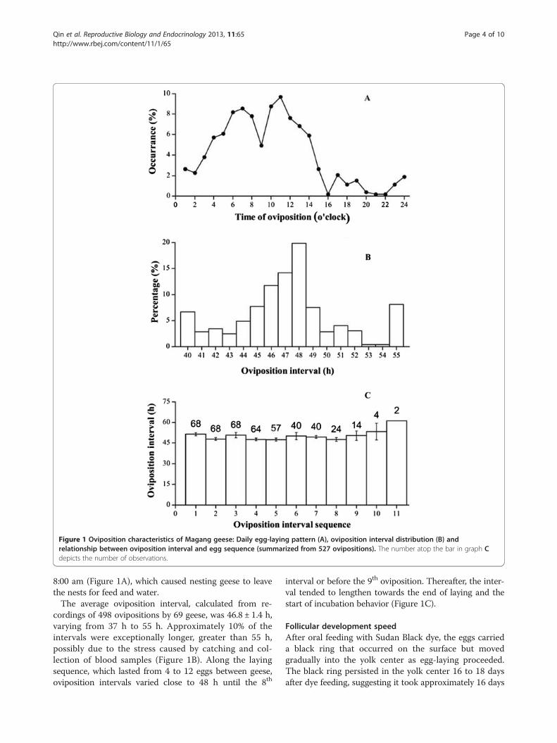
Figure 1 Oviposition characteristics of Magang geese: Daily egg-laying pattern (A), oviposition interval distribution (B) andrelationship between oviposition interval and egg sequence (summarized from 527 ovipositions). The number atop the bar in graph Cdepicts the number of observations.
Qin et al. Reproductive Biology and Endocrinology 2013, 11:65 Page 4 of 10http://www.rbej.com/content/11/1/65
8:00 am (Figure 1A), which caused nesting geese to leavethe nests for feed and water.The average oviposition interval, calculated from re-
cordings of 498 ovipositions by 69 geese, was 46.8 ± 1.4 h,varying from 37 h to 55 h. Approximately 10% of theintervals were exceptionally longer, greater than 55 h,possibly due to the stress caused by catching and col-lection of blood samples (Figure 1B). Along the layingsequence, which lasted from 4 to 12 eggs between geese,oviposition intervals varied close to 48 h until the 8th
interval or before the 9th oviposition. Thereafter, the inter-val tended to lengthen towards the end of laying and thestart of incubation behavior (Figure 1C).
Follicular development speedAfter oral feeding with Sudan Black dye, the eggs carrieda black ring that occurred on the surface but movedgradually into the yolk center as egg-laying proceeded.The black ring persisted in the yolk center 16 to 18 daysafter dye feeding, suggesting it took approximately 16 days
Qin et al. Reproductive Biology and Endocrinology 2013, 11:65 Page 5 of 10http://www.rbej.com/content/11/1/65
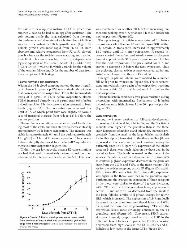
for LWFs to develop into mature F1 LYFs, which tookanother 2 days to be laid as an egg after ovulation. Theyolk volume inside the ring, calculated from the ringcircumference and diameter of each size class of follicle,was used to construct a follicle growth curve (Figure 2).Follicle growth was most rapid from F6 to F2. Bothabsolute and relative expansions from F2 to F1 slowed,possibly because the follicles were maturing and reachedtheir limit. This curve was best fitted by a 4-parameterlogistic equation of V = −0.663 + 58.231/(1 + 72.130 * exp(−0.371*D)) (R2 = 99.9%), in whichV stands for the volumeof the ovarian follicle and D for the number of days fromthe small yellow follicle stage.
Plasma hormone concentrationsWithin the 48-h blood sampling period, the most signifi-cant change in plasma pgFM was a single abrupt peakthat corresponded to oviposition. From the intermediatelevels of 2 pg/mL at 1.5 h before oviposition, plasmaPGFM increased abruptly to a 5 pg/mL peak 0.5 h beforeoviposition. After 2 h, the concentration returned to basallevels (Figure 3A). The concentrations remained lowuntil 28 h, at which point they rose slightly to form asecond marginal increase from 4 h to 1.5 h before thenext oviposition.Plasma P4 concentrations remained at basal levels dur-
ing most of the cycle, and then started to increase steadilyapproximately 10 h before oviposition. The increase wasstable for approximately 6 h until the peak (approximately1.2 ng/mL) at 3 h to 4 h before oviposition. The concen-trations abruptly decreased to a nadir (~0.2 ng/mL) im-mediately after oviposition (Figure 3B).Within the egg-laying cycle, plasma E2 concentrations
reached their nadir immediately before oviposition, thenrebounded to intermediate levels within 5 h. This level
Figure 2 Ovarian follicular development curve constructedfrom diameter of Sudan Black dye circumference yolk of laideggs from 4 Magang geese. Vertical bars represent the standarderror of the mean.
was maintained for another 30 h before increasing fur-ther and peaking over 4 h, or about 6 h to 5 h before thenext oviposition (Figure 3C).The cycle trough of activin A was detected 3 h before
oviposition, earlier than for E2 and P4 (Figure 3D). Within3 h, activin A transiently increased to approximately1.80 ng/mL until 10 h after oviposition. A second in-crease started thereafter, and steadily rose to the peaklevel at approximately 34 h post-oviposition, or 14 h be-fore the next oviposition. The peak lasted for 8 h andstarted to decrease 6 h before the next oviposition. Thus,the prelaying plasma activin A peak occurred earlier andlasted much longer than those of E2 and P4.Changes in plasma inhibin were marked by a sudden
fall 1.5 h prior to oviposition (Figure 3E). The concentra-tions immediately rose again after oviposition, reachinga plateau within 16 h that lasted until 3 h before thenext oviposition.Plasma follistatin exhibited a two-phase variation during
oviposition, with intermediate fluctuations 10 h beforeoviposition and a high plateau 5 h to 30 h post-oviposition(Figure 3F).
Gene expressionAmong the 8 genes pertinent to follicular development,expression of inhibin alpha, inhibin βA, and the 3 activin Rsubunits were higher in the granulosa than in the thecallayer. Expression of inhibin α and inhibin βA increased pro-gressively from the small to the large follicles, particularlyfor inhibin alpha (Figure 4A). In the theca, inhibin βA wasexpressed at low levels and without variation among thedifferently sized LYF (Figure 4B). Expression of the inhibinreceptor β-glycan was much higher in the theca than in thegranulosa layer. The levels increased in the theca of thesmallest F5 until F2, and then decreased in F1 (Figure 4C).In contrast, β-glycan expression decreased in the granulosalayer from the LWFs and SYFs, to the more mature LYFs.As for the activin receptors, activin IR (Figure 4D), activinIIRα (Figure 4E), and activin IIRβ (Figure 4F), expressionwas higher in the thecal layer than in the granulosa layer.Furthermore, the changes in expression of these receptorsin the theca were similar to those of β-glycan: increasingwith LYF maturity. In the granulosa layer, expression ofactivin IR and activin IIRα decreased from the small tothe large follicles similar to β-glycan, except for activinIIRβ, which increased. The expression of LHR graduallyincreased in the granulosa and thecal layers of LWFs,SYFs, and the more mature preovulatory LYFs, with theexpression levels most strikingly enhanced in the F1granulosa layer (Figure 4G). Conversely, FSHR expres-sion was inversely proportional to that of LHR in thedifferent class of follicles. In particular, FSHR expressiondecreased from high levels in the LYFs, SWFs, and F5follicles to low levels in the larger LYFs (Figure 4H).
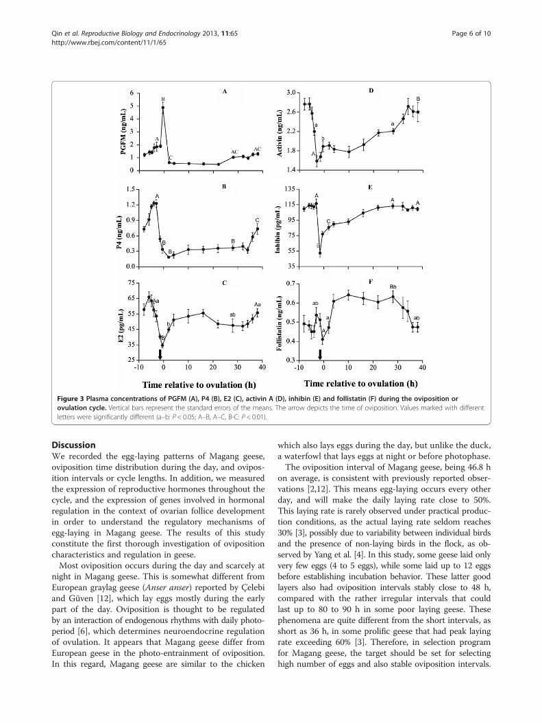
Figure 3 Plasma concentrations of PGFM (A), P4 (B), E2 (C), activin A (D), inhibin (E) and follistatin (F) during the oviposition orovulation cycle. Vertical bars represent the standard errors of the means. The arrow depicts the time of oviposition. Values marked with differentletters were significantly different (a–b: P < 0.05; A–B, A–C, B-C: P < 0.01).
Qin et al. Reproductive Biology and Endocrinology 2013, 11:65 Page 6 of 10http://www.rbej.com/content/11/1/65
DiscussionWe recorded the egg-laying patterns of Magang geese,oviposition time distribution during the day, and ovipos-ition intervals or cycle lengths. In addition, we measuredthe expression of reproductive hormones throughout thecycle, and the expression of genes involved in hormonalregulation in the context of ovarian follice developmentin order to understand the regulatory mechanisms ofegg-laying in Magang geese. The results of this studyconstitute the first thorough investigation of ovipositioncharacteristics and regulation in geese.Most oviposition occurs during the day and scarcely at
night in Magang geese. This is somewhat different fromEuropean graylag geese (Anser anser) reported by Çelebiand Güven [12], which lay eggs mostly during the earlypart of the day. Oviposition is thought to be regulatedby an interaction of endogenous rhythms with daily photo-period [6], which determines neuroendocrine regulationof ovulation. It appears that Magang geese differ fromEuropean geese in the photo-entrainment of oviposition.In this regard, Magang geese are similar to the chicken
which also lays eggs during the day, but unlike the duck,a waterfowl that lays eggs at night or before photophase.The oviposition interval of Magang geese, being 46.8 h
on average, is consistent with previously reported obser-vations [2,12]. This means egg-laying occurs every otherday, and will make the daily laying rate close to 50%.This laying rate is rarely observed under practical produc-tion conditions, as the actual laying rate seldom reaches30% [3], possibly due to variability between individual birdsand the presence of non-laying birds in the flock, as ob-served by Yang et al. [4]. In this study, some geese laid onlyvery few eggs (4 to 5 eggs), while some laid up to 12 eggsbefore establishing incubation behavior. These latter goodlayers also had oviposition intervals stably close to 48 h,compared with the rather irregular intervals that couldlast up to 80 to 90 h in some poor laying geese. Thesephenomena are quite different from the short intervals, asshort as 36 h, in some prolific geese that had peak layingrate exceeding 60% [3]. Therefore, in selection programfor Magang geese, the target should be set for selectinghigh number of eggs and also stable oviposition intervals.
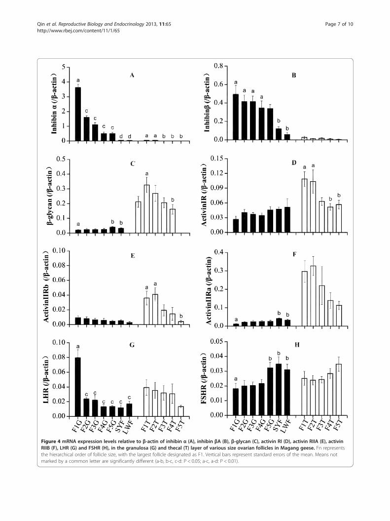
Figure 4 mRNA expression levels relative to β-actin of inhibin α (A), inhibin βA (B), β-glycan (C), activin RI (D), activin RIIA (E), activinRIIB (F), LHR (G) and FSHR (H), in the granulosa (G) and thecal (T) layer of various size ovarian follicles in Magang geese. Fn representsthe hierarchical order of follicle size, with the largest follicle designated as F1. Vertical bars represent standard errors of the mean. Means notmarked by a common letter are significantly different (a-b, b-c, c-d: P < 0.05; a-c, a-d: P < 0.01).
Qin et al. Reproductive Biology and Endocrinology 2013, 11:65 Page 7 of 10http://www.rbej.com/content/11/1/65
Qin et al. Reproductive Biology and Endocrinology 2013, 11:65 Page 8 of 10http://www.rbej.com/content/11/1/65
In addition, the oviposition interval became increasinglylonger towards the end of the clutch. Lengthening of theoviposition interval at the end of the clutch might resultfrom the lowering of LH secretion caused by rising secre-tion of PRL [2], which would slow follicle maturation.Follice developmental speed was recorded by marking
follicle diameter with Sudan Black dye. This not onlyunraveled the time course required for SYFs to developto ovulation, 18 days, but also helped to construct amathematical model of follicle growth that was hithertounknown. The 18 days for SYFs to develop to ovulationcould be established by laying of the previous 8 to 10 eggsin a clutch, but it is an accurate reflection of naturalconditions. It took between 20 to 25 days from the endof incubation to laying a new clutch of eggs in Maganggeese [2]. In other words, the time required for LWFsto develop to ovulatory maturity is 20 or so days onaverage. In addition, during practical production, the laying-incubation cycle in Magang geese is approximately 50 days[2]. This consists of the 7 to 10 days for terminating incu-bation behavior, the 20 to 25 days required for initiationof laying, and the 18 to 20 days required to lay thewhole clutch of eggs.During the stages of egg-laying, ovarian follicles of differ-
ent sizes are exposed to the same endocrine milieu, whichaffects each individual follicle through the expression ofreceptors on the cell surface. The factors involved inovarian follicular development and ovulation include pi-tuitary gonadotropins [11] and the autocrine/paracrinefactors secreted by the follicles themselves [8,13]. Wecharacterized the expression profiles of these factors inthis study. The dramatic upregulation of LHR and inhibinalpha subunit, in the largest F1 follicle, and also changingpatterns of other genes in granulosa and theca layers, wereall similar in Magang goose to those reported for chicken[14-17]. These results indicate that the progression offollicular development and the molecular mechanismsinvolved, i.e., the endocrine regulation by gonadotropinand autocrine/paracrine regulation by inhibin/activinR,are much similar for geese and chickens even thoughthe former have lower laying rates than the latter [2].Apart from the aforementioned differences in chickens,
geese have much longer oviposition cycles. This could bedue to the maturation process and hormonal profile,which affects the oviposition cycle. In this study, theconcentrations of 6 hormones or metabolite were mea-sured within a single ovulation cycle. We found that thechanging hormonal patterns were consistent with thosepreviously reported for chickens [6,8,10] and Graylag goose[12]. Among the hormones, PGF2α plays a pivotal role inoviposition cycle regulation in avian species, such thatthe enhanced synthesis and release of uterotonic PGF2αstimulates uterine muscle contraction, which culminatesin expulsion of the egg [12,18]. The blood concentrations
of PGFM, the metabolic molecular form of PGF2α, arenormally measured to reflect changes in PGF2α secretions[12,18]. In this study, the single peak in plasma PGFMwas detected during the narrow oviposition time window,similar to previous results in chickens and geese [12,18].PGF2α is synthesized in uterine and follicle tissues andits concentration peaks can be detected in the blood ofchickens independent of oviposition [16]. Therefore,analyzing the expression and structure of the genesassociated with PGF2α synthesis and their associationwith oviposition cycle length and egg-laying rate in geeseare of particular interest.For progesterone, another hormone instrumental to
ovulation, the plasma concentrations during the ovipos-ition cycle were comparable to those of Magang geese(Anser cygnoids) under laying state [2], but much lowerthan those reported for Graylag geese (Anser anser) [12].These differences may result from interspecies differencesin progesterone secretion or from differences in assaymethods. This issue needs to be clarified in future studies.Nevertheless, the interoviposition progesterone variationpattern in Magang geese strikingly resembles that reportedfor Graylag geese [12], with the preovulatory P4 increasingat 14 h and peaking 3 h before oviposition. Considering theinterval between the preovulatory P4 peak and ovulation isabout 3.5 hours in both geese and chickens [6,10,11], eventhough the goose ovulation cycle is about 22 h longer thanin chickens, goose F1 follicles clearly take longer to mature.In other words, follicular development and maturationimmediately after ovulation occurs at a slower pace in geese.This may be a factor in the prolonged oviposition cycle ofgeese and may reduce egg-laying frequency and rate.Immediate to ovulation, both plasma concentrations
of activin and inhibin decreased dramatically. This indi-cated the mature F1 follicle also secrets large amount ofactivin in Magang geese. This phenomen is contrary tosituation in the chicken that activin was considered to bemainly secreted by less developed F4 to F3 follicles [8].Also, the residual inhibin concentration nearing 50 pg/mlwas about half the preovulatory peak of 110 pg/ml, indi-cating the F2 follicle also secreted copious amount of in-hibin, rather than the small amount in the chicken model[8]. Ready secretion of inhibin by F2 was also seen by therapid rebound of plasma concentration in less than 5 hrafter the ovulation, also followed by follistatin concentra-tion rebounds. Compared with inhibin secretion reboundat 10 hr post-ovulation in chicken hens whose ovipositininterval was only 24 hr or so and new SYF recruitmentoccurs daily [11], post-ovulatory inhibin secretion inMagang geese was recovered highly rapid. Moreover, theseinfer that, during laying a sequence or clutch of eggs, copi-ous amount of these hormones are always present inblood circulation. Since inhibin and follistatin counteractthe activin’s role of promoting small follicle development
Qin et al. Reproductive Biology and Endocrinology 2013, 11:65 Page 9 of 10http://www.rbej.com/content/11/1/65
[8], the continued presence of inhibin and follistatin inblood may inhibit new SYF recruitment into preovultoryLYF development after Magang geese entering into lay [2],causing clutch egg size limited to no more than thenumber of LYFs present before laying of the first egg in asequence. This also explains taking place of the near 20-dayinterruption between two clutches of eggs as discussedabove, and also in some non-incubating geese [3].Within 10 h to 15 h after ovulation, the hormonal pro-
file is characterized by the static secretion of progesterone,transient secretions of activin and inhibin, and robustsecretions of estradiol and follistatin. Activin A enhancesgranulosa cell proliferation and mediates expression ofFSHR and LHR [8,13]. The increase in follistatin secretionduring the first 12 after ovulation should counteract theeffects of low transient activin A secretions to delay follicu-lar maturation during this stage and to inhibit progesteronesecretion. Nevertheless, increasing gonadotropin secretionsfollowing ovulation of the F1 follicle and subsequentwithdrawal of negative feedback [6,19] may stimulate theremaining F2 follicle and enhance estradiol secretion.From mid-cycle onwards, i.e., 15 h after ovulation, the
plasma activin A concentrations continued to increase withinhibin concentrations (though at a lower magnitude),whereas follistatin concentrations remained stable. Thesechanges in hormonal balance or increase in “activin tone”may regulate the second stage of follicular developmentor maturation, i.e., functional upgrading of the F2 fol-licle to F1.As the F1 follicle continues to mature, plasma activin
A concentrations continue to increase, peaking at 34 hafter ovulation, or 14 h before the next ovulation. Thisactivin A peak occurred concomitantly with the fall offollistatin concentrations, but with preovulatory increasesin P4 and estradiol. Considering the plasma inhibin con-centrations remain stable during this stage, the “activintone” is undoubtedly strengthened, facilitating final matur-ation of the F1 follicle that ultimately triggers the preovu-latory LH surge and ovulation. The earlier decrease inplasma E2 concentrations compared with P4 before ovula-tion is also consistent with the phenomenon observed inchickens, wherein the more mature F1 follicle secretes lessE2 [20]. This indicates that as the preovulatory F1 folliclematures, gonadal steroidogenesis shifts from E2 to P4.
Competing interestsThe authors declare that they have no competing interests.
Authors’ contributionsQMQ, ADS, RHG, MML, SJY and ZDS devised the study and participated in itsdesign. QMQ did the practical analysis, advised by RHG. QMQ and RHGsampled the material. ZDS and MML wrote the manuscript. MML, SJY and ZDScorrected the manuscript. All authors read and approved the final manuscript.
AcknowledgementsThis work was supported by the National Natural Science Foundation ofChina grant (30871795), and the China Agricultural Research System grant
(CARS-43-16). The authors want to thank the generous help of providingresearch facility and animals by Qingxing Golden Geese Company, Ltd.
Author details1College of Animal Sciences, South China Agricultural University, Guangzhou510642, China. 2Institute of Animal Science, Jiangsu Academy of AgriculturalSciences, Nanjing 210014, China. 3Institute of Food Safety and MonitoringTechnology, Jiangsu Academy of Agricultural Sciences, Nanjing 210014,China.
Received: 19 March 2013 Accepted: 8 July 2013Published: 16 July 2013
References1. Shen G, Gong GF, Lv SY: Current waterfowl production and future
tendency in China. Waterfowl world 2011, 5:7–12.2. Huang YM, Shi ZD, Liu Z, Liu Y, Li XW: Endocrine regulations of
reproductive seasonality, follicular development and incubation inMagang geese. Anim Reprod Sci 2008, 104:344–358.
3. Shi ZD, Tian YB, Wu W: Controlling reproductive seasonality in the geese:a review. Worlds Poult Sci J 2008, 64:343–355.
4. Yang CZ, Cai LC, Rong ZK: Observation results of the nucleus flock ofselection program of Magang geese. Journal of Fushan University (NaturalScience Edition) 2001, 19:50–62.
5. Bogenfüst F: Importance of technological developments on improvement ofreproduction parameters of geese. Proceedings of the 3rd World WaterfowlConference; 2005:119–122.
6. Etches RJ: The ovary. Wallingford, Oxon, UK: CAB International; 1996.7. Bahr JM, Johnson AL: Regulation of the follicular hierarchy and ovulation.
J Exp Zool 1984, 232:495–500.8. Lovell TM, Gladwell RT, Groome NP, Knight PG: Ovarian follicle
development in the laying hen is accompanied by divergent changes ininhibin A, inhibin B, activin A and follistatin production in granulosa andtheca layers. J Endocrinol 2003, 177:45–55.
9. Chang MT, Cheng YS, Huang MC: Novel genetic markers of the carbonicanhydrase II gene associated with egg production and reproductiontraits in Tsaiya ducks. Reprod Domest Anim 2013, 48:98–104.
10. Chen CC, Johnson PA: Expression and regulation of mRNA for inhibin/activin alpha- and betaA-subunits in the granulosa layer of the twolargest preovulatory follicles during the hen ovulatory cycle. Gen CompEndocrinol 1997, 107:386–393.
11. Lovell TM, Vanmontfort D, Bruggeman V, Decuypere E, Groome NP,Knight PG, Gladwell RT: Circulating concentrations of inhibin-relatedproteins during the ovulatory cycle of the domestic fowl (Gallusdomesticus) and after induced cessation of egg laying. J Reprod Fertil2000, 119:323–328.
12. Celebi F, Guven B: Plasma concentrations of 13,14-dihydro-15-ketopgF2alpha and progesterone during the oviposition cycle of thedomestic goose (Anser anser domesticus). Poult Sci 2001, 80:225–227.
13. Davis AJ, Brooks CF, Johnson PA: Activin A and gonadotropinregulation of follicle-stimulating hormone and luteinizing hormonereceptor messenger RNA in avian granulosa cells. Biol Reprod 2001,65:1352–1358.
14. Lovell TM, Gladwell RT, Cunningham FJ, Groome NP, Knight PG: Differentialchanges in inhibin A, activin A, and total alpha-subunit levels ingranulosa and thecal layers of developing preovulatory follicles in thechicken. Endocrinology 1998, 139:1164–1171.
15. Lovell TM, Knight PG, Gladwell RT: Differential expression of mRNAsencoding the putative inhibin co-receptor (betaglycan) and activintype-I and type-II receptors in preovulatory and prehierarchical folliclesof the laying hen ovary. J Endocrinol 2006, 188:241–249.
16. Takahashi I, Nakamura Y, Hamada Y, Nakazawa K: Immunohistochemicalanalysis of proteoglycan biosynthesis during early development of thechicken cornea. J Biochem 1999, 126:804–814.
17. Sweeney SA, Johnson PA: Messenger RNA and protein expressionanalysis of betaglycan in the pituitary and ovary of the domestic hen.Biol Reprod 2005, 72:172–178.
18. Hammond RW, Burke WH, Hertelendy F: Influence of follicular maturationon progesterone release in chicken granulosa cells in response to turkeyand ovine gonadotropins. Biol Reprod 1981, 24:1048–1055.
Qin et al. Reproductive Biology and Endocrinology 2013, 11:65 Page 10 of 10http://www.rbej.com/content/11/1/65
19. Johnson PA, Brooks C, Wang SY, Chen CC: Plasma concentrations ofimmunoreactive inhibin and gonadotropins following removal ofovarian follicles in the domestic hen. Biol Reprod 1993, 49:1026–1031.
20. Johnson PA, Stoklosowa S, Bahr JM: Interaction of granulosa and thecalayers in the control of progesterone secretion in the domestic hen. BiolReprod 1987, 37:1149–1155.
doi:10.1186/1477-7827-11-65Cite this article as: Qin et al.: The characteristics of oviposition andhormonal and gene regulation of ovarian follicle development inMagang geese. Reproductive Biology and Endocrinology 2013 11:65.
Submit your next manuscript to BioMed Centraland take full advantage of:
• Convenient online submission
• Thorough peer review
• No space constraints or color figure charges
• Immediate publication on acceptance
• Inclusion in PubMed, CAS, Scopus and Google Scholar
• Research which is freely available for redistribution
Submit your manuscript at www.biomedcentral.com/submit