research open access clinicopathological characteristics ... · research open access...
TRANSCRIPT

Ito et al. Journal of Experimental & Clinical Cancer Research 2013, 32:2http://www.jeccr.com/content/32/1/2
RESEARCH Open Access
Clinicopathological characteristics and optimalmanagement for esophagogastric junctionalcancer; a single center retrospective cohort studyHiroaki Ito*, Haruhiro Inoue, Noriko Odaka, Hitoshi Satodate, Michitaka Suzuki, Shumpei Mukai, Yusuke Takehara,Hiroyuki Kida and Shin-ei Kudo
Abstract
Background: Esophagogastric junctional (EGJ) cancer occurs in the mucosa near the esophagogastric junction, andhas characteristics of both esophageal and gastric malignancies; its optimal treatment strategy is controversial.
Methods: We conducted a single-center retrospective cohort study of the patients who underwent curativesurgery with lymphadenectomy for EGJ cancer. Tumor specimens were categorized by histology and location intofour types—centered in the esophagus < 5 cm from EGJ (type E), which were subtyped as (i) squamous-cellcarcinoma (SQ) or (ii) adenocarcinoma (AD); (iii) any histological tumor centered in the stomach < 5 cm from EGJ,with EGJ invasion (type Ge); (iv) any histological tumor centered in the stomach < 5 cm from EGJ, without EGJinvasion (type G)—and classified by TNM system; these were compared to patients’ clinicopathologicalcharacteristics and survival outcomes.
Results: A total of 92 EGJ cancer patients were studied. Median follow-up of surviving patients was 35.5 months.Tumors were categorized as 12 type E (SQ), 6 type E (AD), 27 type Ge and 47 type G; of these 7 (58.3%), 3 (50%), 19(70.4%) and 14 (29.8%) and 23 patients, respectively, had lymph node metastases. No patients with type E (AD) andGe tumors had cervical lymph node metastasis; those with type G tumors had no nodal metastasis at cervical andmediastinal lymph nodes. Multivariate analysis showed that type E (AD) tumor was an independent prognosticfactor.
Conclusions: We should distinguish type Ge tumor from type E (AD) tumor because of the clinicopathological andprognostic differentiation. Extended gastrectomy with or without lower esophagectomy according to tumorlocation and lower mediastinal and abdominal lymphadenectomy are recommended for EGJ cancer.
Trial registration: University Hospital Medical Information Network in Japan, UMIN000008596.
Keywords: Esophagogastric junctional cancer, Esophageal cancer, Gastric cancer, Lymph node metastasis
BackgroundGastric and esophageal cancers are, respectively, thefourth and eighth most common cancers in the world,and the second and sixth most common causes ofcancer-related death, affecting approximately 736,000and 406,000 people in 2008 [1]. Esophagogastric junc-tional cancer (EGJC), which is increasing in Westerncountries, is a tumor occurring at the mucosa between thelower esophagus and cardia, and has clinicopathological
* Correspondence: [email protected] Disease Center, Showa University Northern Yokohama Hospital,35-1 Chigasakichuo Tsuzuki-ku, Yokohama 224-8503, Japan
© 2013 Ito et al.; licensee BioMed Central Ltd.Commons Attribution License (http://creativecreproduction in any medium, provided the or
characteristics of both esophageal and gastric malignan-cies [2,3].Siewert classification is widely used to categorize EGJ
adenocarcinoma [4,5]. Siewert defines adenocarcinomaof the distal esophagus, such as that from specializedesophageal metaplasia (e.g., Barrett’s esophagus) as typeI; cardiac carcinoma, from the cardia epithelium orwithin 1 cm (along the esophagus) or 2 cm (in the stom-ach) from the EGJ as type II; and subcardial gastric car-cinoma with epicenter in the proximal 5 cm of thestomach, which infiltrates the EGJ and distal esophagus,as type III. Because the Siewert type I tumor is located
This is an Open Access article distributed under the terms of the Creativeommons.org/licenses/by/2.0), which permits unrestricted use, distribution, andiginal work is properly cited.

Figure 1 Tumor classification. We categorized tumors near the EGJ into four types according to its location and main histological type.Categorization criteria were: (i) squamous-cell carcinoma with epicenter in the esophagus within 5 cm from EGJ (type E (SQ)); (ii)adenocarcinoma with epicenter in the esophagus within 5 cm from EGJ (type E (AD)); (iii) any histological tumor with epicenter in the stomachwithin 5 cm from EGJ, with EGJ invasion (type Ge); (iv) any histological tumor with epicenter in the stomach within 5 cm from EGJ, without EGJinvasion (type G). Type E (SQ), E (AD) and Ge tumors were categorized as esophageal cancer; type G tumor was categorized as gastric cancer bythe American Joint Committee on Cancer/International Union Against Cancer (AJCC/UICC) Cancer Staging Manual. Siewert type I and III tumorswere categorized as type E (AD) and Ge tumors, and Siewert type II tumor was categorized as type E (AD) or Ge tumor in this study.
Ito et al. Journal of Experimental & Clinical Cancer Research 2013, 32:2 Page 2 of 12http://www.jeccr.com/content/32/1/2
in the lower esophagus, it can be treated as loweresophageal cancer; whereas type III tumor has similarclinicopathological characteristics to cardiac cancer be-cause of its location. However, Siewert type II tumor is ametastatic threat to both thoracic and abdominal areas,as it crosses the EGJ. Subtotal esophagectomy offers onlya limited benefit and should not be performed for typeII cancer. The TNM staging system according to the sev-enth edition of the American Joint Committee on Can-cer/International Union Against Cancer (AJCC/UICC)Cancer Staging Manual defined EGJC, including of
Figure 2 Flow diagram of the patients in this study. Total 92 patients wjunctional cancer at the Digestive Disease Center, Showa University Northewere retrospectively studied.
squamous-cell carcinoma and adenocarcinoma centeredin the esophagus within 5 cm, and in the proximal 5 cmof the stomach with crossing the EGJ [6,7]. AJCC/UICCalso categorizes any cardiac cancer without EGJ invasionas gastric cancer regardless of its location. Different sta-ging systems are applied to esophageal squamous-cellcarcinoma and esophageal adenocarcinoma.Surgery is effective treatment for resectable esophageal
[8,9] and gastric cancer [10-12]. However, as esophagect-omy is generally more invasive than gastrectomy [13], weshould be careful in treating EGJC with esophagectomy.
ho underwent curative surgical resection for esophagogastricrn Yokohama Hospital between October 2001 and December 2010

Table 1 Patient characteristics (n = 92)
Variables
Age (year, mean ± SD) 65.9 ± 9.4
Sex Male 72 (78.3%)
Female 20 (21.7%)
Siewert classification Type I adenocarcinoma 2 (2.2%)
Type II adenocarcinoma 16 (17.4%)
Type III adenocarcinoma 11 (12.0%)
Not applicable 63 (68.5%)
Macro type Type 0 36 (39.1%)
Type 1 4 (4.3%)
Type 2 26 (28.3%)
Type 3 21 (22.8%)
Type 4 1 (1.1%)
Type 5 4 (4.3%)
Preoperative chemotherapy No 79 (85.9%)
Yes 13 (14.1%)
Extent of surgical resection Subtotal esophagectomy with partial gastrectomy 14 (15.2%)
Proximal gastrectomy with partial esophagectomy 30 (32.6%)
Total gastrectomy with partial esophagectomy 48 (52.2%)
Extent of lymph node dissection Abdominal, mediastinal and cervical 11 (12.0%)
Abdominal and mediastinal 9 (9.8%)
Abdominal and lower mediastinal† 27 (29.3%)
Abdominal 45 (48.9%)
Pathological tumor size (mm, mean ± SD) 46.1 ± 23.7
Main histologic type Adenocarcinoma 79 (85.9%)
Squamous-cell carcinoma 13 (14.1%)
Lymphatic invasion L0 32 (34.8%)
L1 60 (65.2%)
Venous invasion V0 32 (34.8%)
V1–2 60 (65.2%)
Pathological depth of tumor invasion pT1 33 (35.9%)
pT2 11 (12.0%)
pT3 35 (38.0%)
pT4 13 (14.1%)
Lymph node metastasis pN0 47 (51.1%)
pN1 19 (20.7%)
pN2 14 (15.2%)
pN3 12 (13.0%)
Distant metastasis pM0 72 (78.3%)
pM1 20 (21.7%)
TNM stage pStage I 36 (39.1%)
pStage II 19 (20.7%)
pStage III 17 (18.5%)
pStage IV 20 (21.7%)
Ito et al. Journal of Experimental & Clinical Cancer Research 2013, 32:2 Page 3 of 12http://www.jeccr.com/content/32/1/2

Table 1 Patient characteristics (n = 92) (Continued)
Adjuvant chemotherapy No 43 (46.7%)
Yes 49 (53.3%)† Including lower thoracic paraesophageal, diaphragmatic and posterior mediastinal lymph node.
Ito et al. Journal of Experimental & Clinical Cancer Research 2013, 32:2 Page 4 of 12http://www.jeccr.com/content/32/1/2
We studied clinicopathological characteristics of patientswith EGJC to investigate its optimal management.
MethodsStudy designWe performed a single center, retrospective cohort study.We studied patients who underwent curative surgery forEGJC, including lymph node dissection, at the DigestiveDisease Center, Showa University Northern YokohamaHospital, between October 2001 and December 2010.Clinicopathological data and prognosis were taken frommedical records.
PatientsWe studied patients with cancer in the lower esophagusand cardia. Inclusion criteria were: (i) presence of histolo-gically proven carcinoma centered within the lower 5 cmof the esophagus and the upper 5 cm of the stomach; (ii)clinically solitary tumors; (iii) no prior endoscopic resec-tion or surgical treatment; and (iv) patient aged 20–80 years. The exclusion criteria were: (i) presence of severeorgan dysfunction; (ii) presence of metachronous and syn-chronous malignancy; and (iii) presence of pathologicalnon-curative findings.
All patient data were approved for use by the institu-tional review board of Showa University NorthernYokohama Hospital. This study was registered with theUniversity Hospital Medical Information Network inJapan (No. UMIN000008596).
ClassificationAlthough Siewert classification is one of the most widelyused criteria for EGJC, it is generally used for onlyadenocarcinoma. EGJC, including squamous cell carcin-oma, has been defined by the seventh edition of AJCC/UICC TNM Cancer Staging Manual. However, it doesnot cover all of the cancer near the EGJ—for example alocalized gastric adenocarcinoma with centered in thestomach within 5 cm from EGJ. Thus, we categorizedtumors near the EGJ into four types, according to loca-tion and main histological type (Figure 1). Categorizationcriteria were: (i) squamous-cell carcinoma centered inthe esophagus within 5 cm from EGJ (type E (SQ)); (ii)adenocarcinoma centered in the esophagus within 5 cmfrom EGJ (type E (AD)); (iii) any histological tumor cen-tered in the stomach within 5 cm from EGJ, with EGJ in-vasion (type Ge); (iv) any histological tumor centered inthe stomach within 5 cm from EGJ, without EGJ invasion
(type G). All disease was pathologically staged using theseventh edition of AJCC/UICC TNM Cancer StagingManual [6,7]. Thus, types E and Ge tumors were stagedas esophageal cancer, and type G tumor was staged asgastric cancer.
Statistical analysisStatistical analysis was performed using JMP 9.0.3 (SASInstitute, Cary, USA). We used Fisher’s exact test andPearson’s chi-squared test to compare the characteristicsof the patients and pathological findings. The nonpara-metric Kruskal–Wallis test was used to assess differencesamong patients’ age groups, number of dissected lymphnodes and pathological tumor size. Kaplan–Meier curvesof estimated overall survival were generated and com-pared, using a 2-sided log-rank test. To investigate prog-nostic factors, Cox proportional hazard analysis wasused. Multivariate analysis included tumor types andvariables with P < 0.10 in univariate analysis. P < 0.05was considered statistically significant.
ResultsPatient characteristicsA total of 92 patients were included in this study(Figure 2). Median follow-up of surviving patients was35.5 months. Patients’ characteristics are summarized inTable 1. Approximately 80% of them were men; theiraverage age was 65.9 years (range: 35–80 years). Four-teen (15.2%), 30 (32.6%) and 48 (52.2%) patients under-went subtotal esophagectomy with partial gastrectomy,proximal gastrectomy with partial esophagectomy andtotal gastrectomy with partial esophagectomy, respect-ively. Twenty-four patients underwent splenectomy toremove involved lymph nodes at the splenic hilum. Thir-teen patients (14.1%) received preoperative chemother-apy. Histologically, 79 (85.9%) and 13 (14.1%) of 92patients had tumors mainly composed with adenocarcin-oma and squamous cell carcinoma. Mean pathologicaltumor size was 46.1 mm. Two, 16 and 11 tumors werecategorized as Siewert types I, II and III, respectively;Siewert classification was not applicable to theremaining 63 tumors. In 63 tumors which did not applyto Siewert classification, 50 and 13 tumors were mainlycomposed with adenocarcinoma and squamous cell car-cinoma. However 15 and 48 tumors centered in theesophagus and the stomach, only one tumor had esopha-gogastric junctional invasion. Eighteen (19.6%), 27(29.3%) and 47 (51.1%) tumors were categorized type E,G and Ge, respectively. The mean number of dissected
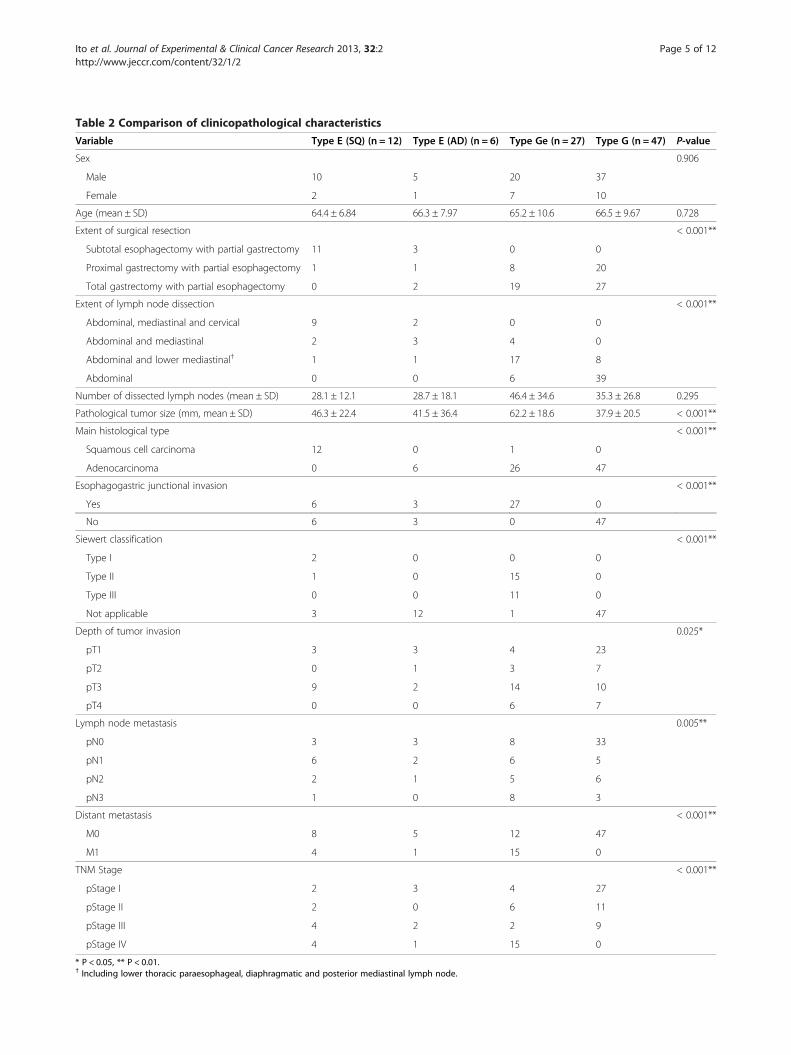
Table 2 Comparison of clinicopathological characteristics
Variable Type E (SQ) (n = 12) Type E (AD) (n = 6) Type Ge (n = 27) Type G (n = 47) P-value
Sex 0.906
Male 10 5 20 37
Female 2 1 7 10
Age (mean ± SD) 64.4 ± 6.84 66.3 ± 7.97 65.2 ± 10.6 66.5 ± 9.67 0.728
Extent of surgical resection < 0.001**
Subtotal esophagectomy with partial gastrectomy 11 3 0 0
Proximal gastrectomy with partial esophagectomy 1 1 8 20
Total gastrectomy with partial esophagectomy 0 2 19 27
Extent of lymph node dissection < 0.001**
Abdominal, mediastinal and cervical 9 2 0 0
Abdominal and mediastinal 2 3 4 0
Abdominal and lower mediastinal† 1 1 17 8
Abdominal 0 0 6 39
Number of dissected lymph nodes (mean ± SD) 28.1 ± 12.1 28.7 ± 18.1 46.4 ± 34.6 35.3 ± 26.8 0.295
Pathological tumor size (mm, mean ± SD) 46.3 ± 22.4 41.5 ± 36.4 62.2 ± 18.6 37.9 ± 20.5 < 0.001**
Main histological type < 0.001**
Squamous cell carcinoma 12 0 1 0
Adenocarcinoma 0 6 26 47
Esophagogastric junctional invasion < 0.001**
Yes 6 3 27 0
No 6 3 0 47
Siewert classification < 0.001**
Type I 2 0 0 0
Type II 1 0 15 0
Type III 0 0 11 0
Not applicable 3 12 1 47
Depth of tumor invasion 0.025*
pT1 3 3 4 23
pT2 0 1 3 7
pT3 9 2 14 10
pT4 0 0 6 7
Lymph node metastasis 0.005**
pN0 3 3 8 33
pN1 6 2 6 5
pN2 2 1 5 6
pN3 1 0 8 3
Distant metastasis < 0.001**
M0 8 5 12 47
M1 4 1 15 0
TNM Stage < 0.001**
pStage I 2 3 4 27
pStage II 2 0 6 11
pStage III 4 2 2 9
pStage IV 4 1 15 0
* P < 0.05, ** P < 0.01.† Including lower thoracic paraesophageal, diaphragmatic and posterior mediastinal lymph node.
Ito et al. Journal of Experimental & Clinical Cancer Research 2013, 32:2 Page 5 of 12http://www.jeccr.com/content/32/1/2

Table 3 Number of patients with positive nodes
Variable Type E (SQ) (n = 12) Type E (AD) (n = 6) Type Ge (n = 27) Type G (n = 47) P-value
Overall 7/12 (58.3%) 3/6 (50.0%) 19/27 (70.4%) 14/47 (29.8%) 0.003**
Depth of tumor invasion
pT1 2/3 (66.7%) 0/3 2/4 (50.0%) 0/23 0.001**
pT2 – 1/1 (100%) 2/3 (66.7%) 3/7 (42.9%) 0.497
pT3 5/9 (55.6%) 2/2 (100.0%) 9/14 (64.3%) 6/10 (60.0%) 0.697
pT4 – – 6/6 (100%) 5/7 (71.4%) 0.269
Main histological type
Squamous-cell carcinoma 7/12 (66.7%) – 0/1 – 0.462
Adenocarcinoma – 3/6 (50.0%) 19/26 (73.1%) 14/47 (29.8%) 0.002**
Location of lymph node†
Cervical LN 2/9 (22.2%) 0/2 – – 0.655
Upper–middle mediastinal 0/11 0/5 0/4 – –
Lower mediastinal‡ 2/12 (16.7%) 2/6 (33.3%) 2/20 (10.0%) 0/8 0.298
Perigastric LN 6/12 (50.0%) 3/6 (50.0%) 17/27 (63.0%) 13/47 (27.7%) 0.026*
Left paracardial 1 2 8 2
Right paracardial 3 3 10 5
Lesser curvature 4 1 13 10
Greater curvature 0 1 4 1
Suprapyloric 0 0 0 0
Infrapyloric 0 0 1 0
LN along left gastric artery 2/12 (16.7%) 1/6 (16.7%) 5/27 (18.5%) 7/47 (14.9%) 0.983
LN at Celiac trunk 0/6 0/3 1/19 (5.3%) 2/24 (8.3%) 0.837
LN along hepatic artery 0/3 0/1 3/19 (15.8%) 1/27 (3.7%) 0.459
LN along splenic artery 0/2 1/3 (33.3%) 2/22 (9.1%) 1/23 (4.3%) 0.356
LN at splenic hilum – – 3/17 (17.6%) 0/9 0.262
* P < 0.05; ** P < 0.01.† Number of the patients with nodal metastasis/number of the patients underwet lymph node dissection (%).‡ Lower thoracic paraesophageal, diaphragmatic and posterior mediastinal lymph node.LN Lymph node.
Ito et al. Journal of Experimental & Clinical Cancer Research 2013, 32:2 Page 6 of 12http://www.jeccr.com/content/32/1/2
lymph nodes was 37.2 ± 28.0 (SD) in each patient. Forty-five (48.9%) of 92 patients had lymph node metastases(pN1–3). Thirty-six (39.1%), 19 (20.7%), 17 (18.5%) and20 (21.7%) patients were pathologically staged I, II, IIIand IV, respectively. Forty-nine patients (53.3%) had pre-operative chemotherapy.Comparison of clinicopathological characteristics among
type E (SQ), E (AD), Ge and G tumor group are summar-ized in Table 2. There were significant differences in extentof surgical resection, pathological tumor size, main histo-logical type, depth of tumor invasion (pT category), lymphnode metastasis (pN category), distant metastasis (pM cat-egory) and TNM tumor stage. Histologically, 26 (96.3%) of27 type Ge tumor and all 47 type G tumors were adenocar-cinoma. Patients with Type G tumors tended to have earl-ier stage diseases than the other tumor groups.Incidence of lymph node metastases were summarized
in Table 3. Seven (58.3%) of 12 type E (SQ) tumors, 3(50.0%) of 6 type E (AD) tumors, 19 (70.4%) of 27 type
Ge tumors and 14 (29.8%) of 47 type G tumors hadlymph nodes metastases (P = 0.003). Although incidenceof nodal metastasis in pT1 tumor was significantly lowerin the type G tumor group than the other type tumorgroups, there was no significant difference in pT2, pT3and pT4 tumors among 4 tumor groups. With regard tolymph node location, no nodal metastasis in the cervicaland mediastinal lymph nodes was seen in the type Gtumor group. Although nodal metastases in perigastriclymph nodes were seen in all tumor types, only one nodalmetastasis in intra-abdominal lymph nodes, except forperigastric lymph nodes, was recognized in type E tumorgroup. Nodal metastasis at the splenic hilum was seen inonly in the Ge tumor group. As a result, incidence ratesfor nodal metastasis in cervical, mediastinal, and perigas-tric lymph nodes differed among 4 patients groups.Clinicopathological characteristics and clinical courses
of seven patients with cervical or mediastinal lymphnode metastasis were summarized in Table 4. The

Table 4 Clinicopathological findings of patients with cervical and mediastinal lymph node metastasis
Case Tumortype
CervicalLN
MediastinalLN
Age Sex Tumor size(mm)
Distance†
Macroscopictype
Histologicaltype
pT pN pM Stage Initial recurrencesite
Status
1 E (SQ) SC – 64 M 50 65 Type 0 SQ (por) T3 N3 M0 IIIC LN, lt. adrenal grand Deceased
2 E (SQ) SC LTP 57 M 87 69 Type 0 SQ (por) T1 N2 M1 IV LN Deceased
3 E (SQ) – EH 72 M 25 40 Type 2 SQ (mod) T3 N1 M0 IIIA LN Deceased
4 E (AD) – EH 73 F 110 100 Type 0 AD (por) T2 N1 M0 IIB Peritoneum Deceased
5 E (AD) – LTP, ID 62 M 45 55 Type 2 AD (mod) T3 N1 M0 IIIA LN Deceased
6 Ge – LTP 68 M 80 30 Type 1 AD (mod) T3 N3 M0 IIIC Deceased (othercause)
7 Ge – EH 41 M 65 25 Type 3 AD (por) T3 N3 M1 IV LN Alive with relapse† Distance between proximal edge of tumor and EGJ in mm.AD adenocarcinoma, EH Esophageal hiatus, ID Infradiaphragmatic, LTP Lower thoracic paraesophageal, LN Lymph node, mod moderately differentiated. por: poorly differentiated, SC, Supraclavicular, SQ Squamous-cellcarcinoma.
Itoet
al.JournalofExperim
ental&ClinicalCancer
Research2013,32:2
Page7of
12http://w
ww.jeccr.com
/content/32/1/2

Figure 3 Overall survival of patients. (A) Patients with pT1–4 tumors (n = 92). Type G tumor group demonstrated higher overall survival ratecompared with type E adenocarcinoma (AD) (P = 0.013) tumor group. Although not significantly, the type G tumor group had a higher survivalrate than the type E squamous-cell carcinoma (SQ) (P = 0.366) and Ge (P = 0.850) tumor group. (B) Patients with pT2–4 Tumors (n = 59). The typeE (AD) tumor group demonstrated significantly lower overall survival rate compared with the type Ge (P = 0.001) and type G (P = 0.003) tumorgroup. The type E (AD) tumor group had a lower survival rate than the type E (SQ) tumor group (P = 0.076) although not significantly.
Ito et al. Journal of Experimental & Clinical Cancer Research 2013, 32:2 Page 8 of 12http://www.jeccr.com/content/32/1/2
location of mediastinal positive nodes was localized inthe lower mediastinal area. Six of 7 patients had diseaserecurrence and 5 patients were deceased. One patientdied of another cause without disease recurrence.
Surgical outcomesThe 5-year overall survival rate was 56.6%. Thirty-threepatients had disease recurrence. Thirty-four patientsdeceased. Twenty-five, 1 and 8 patients died of cancer,surgical complication and other causes. Overall survivalrates were compared among the patients with type E(SQ), E (AD), G and Ge tumors. In patients with pT1–4tumors, the type G tumor group (overall 5-year survivalrate was 64.4%) demonstrated higher overall survivalrate compared with type E (AD) (overall 5-year survivalrate was 33.3%) (P = 0.013) tumor group. Although notsignificantly, the type G tumor group had a higher sur-vival rate than the type E (SQ) (overall 5-year survivalrate was 50.0%) (P = 0.366) and Ge (overall 5-year sur-vival rate was 51.9%) (P = 0.850) tumor group
(Figure 3A). Because the type G tumor group had rela-tively early-stage disease, survival rates were calculatedin patients with pT2–4 tumor. In the pT2–4 group, thetype E (AD) tumor group demonstrated significantlylower overall survival rate compared with the type Ge(overall 5-year survival rate was 49.4%) (P = 0.001) andtype G (overall 5-year survival rate was 42.8%)(P = 0.003) tumor group. The type E (AD) tumor grouphad a lower survival rate than the type E (SQ) tumorgroup (overall 5-year survival rate was 44.4%) (P = 0.076)although not significantly (Figure 3B).
Prognostic factorA univariate Cox proportional hazard analysis showedthat lymphatic invasion (P < 0.001) and venous invasion(P < 0.001), depth of tumor invasion (pT category;P < 0.001), lymph node metastasis (pN category;P < 0.001), distant metastasis (M category; P = 0.028)were statistically significant for survival. Sex, age and
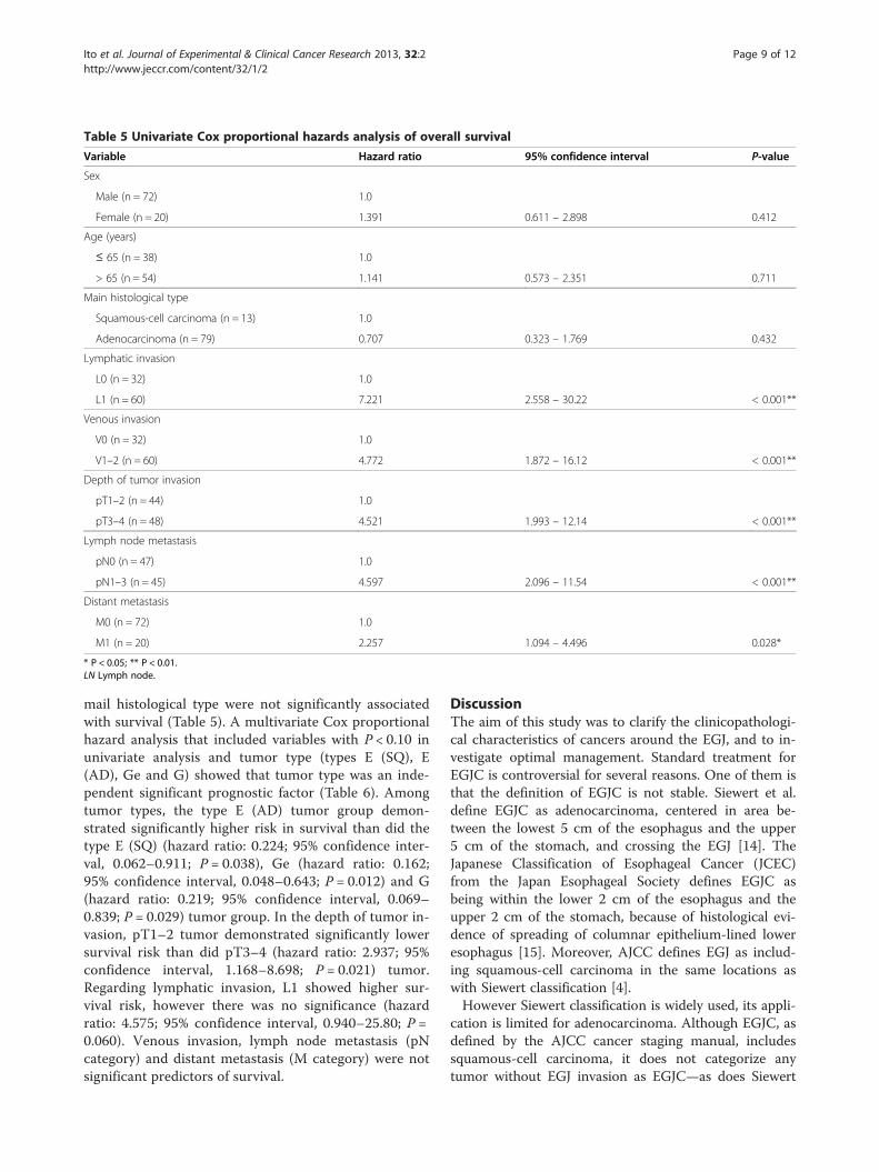
Table 5 Univariate Cox proportional hazards analysis of overall survival
Variable Hazard ratio 95% confidence interval P-value
Sex
Male (n = 72) 1.0
Female (n = 20) 1.391 0.611 – 2.898 0.412
Age (years)
≤ 65 (n = 38) 1.0
> 65 (n = 54) 1.141 0.573 – 2.351 0.711
Main histological type
Squamous-cell carcinoma (n = 13) 1.0
Adenocarcinoma (n = 79) 0.707 0.323 – 1.769 0.432
Lymphatic invasion
L0 (n = 32) 1.0
L1 (n = 60) 7.221 2.558 – 30.22 < 0.001**
Venous invasion
V0 (n = 32) 1.0
V1–2 (n = 60) 4.772 1.872 – 16.12 < 0.001**
Depth of tumor invasion
pT1–2 (n = 44) 1.0
pT3–4 (n = 48) 4.521 1.993 – 12.14 < 0.001**
Lymph node metastasis
pN0 (n = 47) 1.0
pN1–3 (n = 45) 4.597 2.096 – 11.54 < 0.001**
Distant metastasis
M0 (n = 72) 1.0
M1 (n = 20) 2.257 1.094 – 4.496 0.028*
* P < 0.05; ** P < 0.01.LN Lymph node.
Ito et al. Journal of Experimental & Clinical Cancer Research 2013, 32:2 Page 9 of 12http://www.jeccr.com/content/32/1/2
mail histological type were not significantly associatedwith survival (Table 5). A multivariate Cox proportionalhazard analysis that included variables with P < 0.10 inunivariate analysis and tumor type (types E (SQ), E(AD), Ge and G) showed that tumor type was an inde-pendent significant prognostic factor (Table 6). Amongtumor types, the type E (AD) tumor group demon-strated significantly higher risk in survival than did thetype E (SQ) (hazard ratio: 0.224; 95% confidence inter-val, 0.062–0.911; P = 0.038), Ge (hazard ratio: 0.162;95% confidence interval, 0.048–0.643; P = 0.012) and G(hazard ratio: 0.219; 95% confidence interval, 0.069–0.839; P = 0.029) tumor group. In the depth of tumor in-vasion, pT1–2 tumor demonstrated significantly lowersurvival risk than did pT3–4 (hazard ratio: 2.937; 95%confidence interval, 1.168–8.698; P = 0.021) tumor.Regarding lymphatic invasion, L1 showed higher sur-vival risk, however there was no significance (hazardratio: 4.575; 95% confidence interval, 0.940–25.80; P =0.060). Venous invasion, lymph node metastasis (pNcategory) and distant metastasis (M category) were notsignificant predictors of survival.
DiscussionThe aim of this study was to clarify the clinicopathologi-cal characteristics of cancers around the EGJ, and to in-vestigate optimal management. Standard treatment forEGJC is controversial for several reasons. One of them isthat the definition of EGJC is not stable. Siewert et al.define EGJC as adenocarcinoma, centered in area be-tween the lowest 5 cm of the esophagus and the upper5 cm of the stomach, and crossing the EGJ [14]. TheJapanese Classification of Esophageal Cancer (JCEC)from the Japan Esophageal Society defines EGJC asbeing within the lower 2 cm of the esophagus and theupper 2 cm of the stomach, because of histological evi-dence of spreading of columnar epithelium-lined loweresophagus [15]. Moreover, AJCC defines EGJ as includ-ing squamous-cell carcinoma in the same locations aswith Siewert classification [4].However Siewert classification is widely used, its appli-
cation is limited for adenocarcinoma. Although EGJC, asdefined by the AJCC cancer staging manual, includessquamous-cell carcinoma, it does not categorize anytumor without EGJ invasion as EGJC—as does Siewert

Table 6 Multivariate Cox proportional hazards analysis of overall survival
Variable Hazard ratio 95% confidence interval P-value
Tumor type
Type E (AD) (n = 6) 1.0
Type E (SQ) (n = 12) 0.224 0.062 – 0.911 0.038*
Type Ge (n = 27) 0.162 0.048 – 0.643 0.012*
Type G (n = 47) 0.219 0.069 – 0.839 0.029*
Lymphatic invasion
L0 (n = 32) 1.0
L1 (n = 60) 4.575 0.940 – 25.80 0.060
Venous invasion
V0 (n = 32) 1.0
V1–2 (n = 60) 0.966 0.196 – 5.170 0.967
Depth of tumor invasion
pT1–2 (n = 44) 1.0
pT3–4 (n = 48) 2.937 1.168 – 8.698 0.021*
Lymph node metastasis
pN0 (n = 47) 1.0
pN1–3 (n = 45) 1.460 0.463 – 5.607 0.537
Distant metastasis
M0 (n = 72) 1.0
M1 (n = 20) 1.097 0.428 – 2.794 0.846
* P < 0.05.
Ito et al. Journal of Experimental & Clinical Cancer Research 2013, 32:2 Page 10 of 12http://www.jeccr.com/content/32/1/2
classification. Although it estimates prognosis well usingdifferent staging systems for squamous-cell carcinomaand adenocarcinoma, this method may be too complexfor clinicians; whereas the JCEC system, which treatsmost limited tumors as EGJC, is more precise.Because of the unstable definition of EGJCs, clinico-
pathological characters and treatment strategies have notbeen unified. Siewert et al. argued that complete surgicalresection and lymph node metastasis were independentprognostic factors in type II adenocarcinoma, and subtotalesophagectomy had less survival effectiveness for thepatients with type II adenocarcinoma [5]. Hasegawa et al.reported that about 40%, 60% and 90% of patients withtype I, II and III tumors, respectively, had lymph node me-tastases, and recommended complete resection for im-proving survival [16]. Schiesser et al. reported thatsubtotal esophagectomy and extended total gastrectomyshould be performed for type I and type II–III tumor [17].With regard to surgical approach, Sasako et al. showedthat the left thoracoabdominal approach did not improvesurvival after the abdominal-transhiatal approach andleads to increased morbidity in patients with cancer of thecardia or subcardia [18]. Kakeji et al. reported that esopha-gectomy with mediastinal and abdominal lymphadenect-omy was adequate for squamous-cell carcinoma, andthat extended total gastrectomy with lower mediastinaland abdominal lymphadenectomy was suitable for
adenocarcinoma [19]. Carboni et al. maintained effects ofextended gastrectomy by an abdominal–trans-hiatal ap-proach for EGJC [20]. Conversely, Chau et al. reportedthat performance status, liver metastasis, peritoneal me-tastasis and alkaline phosphatase were independent prog-nostic factors in patients with locally advanced andmetastatic EGJC, and that prognoses of patients with re-current disease were no better than those without surgery[21].We studied any tumor centered in area between the
lowest 5 cm of the esophagus and the upper 5 cm of thestomach, regardless of histological type and EGJ inva-sion, and simply categorized them in 4 groups includingtype E (SQ), E (AD), Ge and G.Whereas type E (SQ), E (AD) and Ge tumors in this
study are categorized as esophageal cancer by AJCC/UICC criteria, these tumor groups show differences inclinicopathological characteristics. In lymph node metas-tasis, approximately 60%, 50%, 70% and 30% of thepatients with type E (SQ), E (AD), Ge and G tumors re-spectively had lymph node metastases in this study. Cer-vical lymph node metastases were recognized in onlytype E (SQ) tumor group. Because type E (AD) tumorwas based on columnar epithelium, its histological be-havior was thought to be similar to cardiac adenocarcin-oma; however, type E (AD) tumor showed a nodalmetastatic spreading pattern similar to that of type Ge

Ito et al. Journal of Experimental & Clinical Cancer Research 2013, 32:2 Page 11 of 12http://www.jeccr.com/content/32/1/2
tumor in this study. Although it seems reasonable tounite type E (AD) and Ge tumors as a group on thebasis of lymphadenectomy extent, the patients with typeE (AD) tumor showed significantly lower survival ratesthan other type tumor groups. Although not signifi-cantly, patients with type E (AD) tumor had higher inci-dence of nodal metastasis at mediastinal lymph nodethan did patients in tumor groups, and all mediastinalpositive nodes existed in lower mediastinal area. Thus,subtotal esophagectomy is not necessary for type E (AD)and Ge tumor, if complete tumor resection can beachieved. Because no cervical or mediastinal lymph nodemetastasis was recognized in the type G tumor group,we should not perform subtotal esophagectomy for typeG tumor. In multivariate analysys, tumor type (type E(AD)) was an independent risk factor for survival of thepatients with EGJC in this study. The prognosis of cer-vical or mediastinal node positive patients was poor. Be-cause survival benefit by cervical and mediastinallymphadenectomy for the node positive patients withEGJC is limited, we should carefully perform subtotalesophagectomy, and cervical and mediastinal lymphade-nectomy for EGJC patients. Therefore, extended gastrec-tomy with or without lower esophagectomy, accordingto tumor location, and lower mediastinal and abdominallymphadenectomy is thought to be adequate for patientswith EGJC, including type E (SQ) tumor.Although lymphatic invasion, venous invasion, depth
of tumor invasion (T category), lymph node metastasis(N category) and distant metastasis (M category) weresignificantly prognostic factors in the univariate analysis,tumor type (types E (SQ), E (AD), Ge and G) and depthof tumor invasion (pT3–4 tumor) were significant in themultivariate analysis in this study. It was reported thatcomplete surgical resection and lymph node metastasiswere independent prognostic factors in type II adenocar-cinoma [5]. We believe that the lack of a significant dif-ference between the prognosis and lymph nodemetastasis can be explained by limitations of this studysuch as the small sample size. Distant metastasis (M cat-egory) was not significantly prognostic factor in themultivariate analysis in study. AJCC/UICC TNM stagingsystem for esophageal cancer defines nodal metastasisalong lesser curvature as distant metastasis, althoughlymph node along lesser curvature is one of the main re-gional lymph nodes of gastric cancer. Because majorityof the patient with M1 disease had no hematogenousmetastasis in this study, there was a possibility that dis-tant metastasis was not significant for prognosis in thisstudy.Reim et al. reported that chemotherapy to be more ef-
ficacious for EGJC than for distal gastric cancer [22].The treatment efficacy of chemotherapy before or aftersurgery is unclear in this small scale retrospective cohort
study. To clarify optimal treatment strategy for EGJC,we should confirm the results in this study using a largescale prospective study.
ConclusionsPatients with type E (AD) and Ge tumor had no cervicallymph node metastasis, and those with type G tumorhad no nodal metastasis at cervical and mediastinallymph node. The incidence of mediastinal lymph nodemetastasis of type E (AD) tumor group was higher thantype Ge tumor group, and survival rate of the patientswith type Ge tumor is significantly higher than thosewith type E (AD) tumor. Therefore we should distin-guish type Ge tumor from type E (AD) tumor. Based onour findings from a retrospective analysis in this cohortstudy, we suggest performing extended gastrectomy withor without lower esophagectomy, according to tumor lo-cation, and lower mediastinal and abdominal lymphade-nectomy for EGJC.
Competing interestsThe authors declare that they have no competing interests.
Authors’ contributionsHI (Hiroaki Ito)* conceived and designed the study, collected clinical data,and performed the statistical analysis and interpretation of data. HI (HaruhiroInoue) participated in the study design and performed interpretation of data.NO, HS, MS, SM, YT and HK collected clinical data. SK participated in thestudy design and coordination. All authors read and approved the finalmanuscript.
AcknowledgementsWe are extremely grateful to all the patients and to the clinical staff whocared for these patients. We also are thankful to Dr. Shigeharu Hamatani forhis reliable pathological diagnoses.
Received: 9 December 2012 Accepted: 4 January 2013Published: 7 January 2013
References1. World Health Organization. International Agency for Research on Cancer:
GLOBOCAN 2008. Cancer Incidence and Mortality World Wide. 2008[http://globocan.iarc.fr/]
2. Pohl H, Welch HG: The role of overdiagnosis and reclassification in themarked increase of esophageal adenocarcinoma incidence. J Nat CancerInst 2005, 97:142–146.
3. Lu YK, Li YM, Gu YZ: Cancer of esophagus and esophagogastric junction:analysis of results of 1,025 resections after 5 to 20 years. Ann ThoracicSurg 1987, 43:176–181.
4. Siewert JR, Feith M, Stein HJ: Biologic and clinical variations ofadenocarcinoma at the esophago-gastric junction: relevance of atopographic-anatomic subclassification. J Surg Oncol 2005, 90:139–146.
5. Siewert JR, Stein HJ, Feith M: Adenocarcinoma of the esophago-gastricjunction. Scand J Surg 2006, 95:260–269.
6. Edge SB, Byrd DR, Compton CC, et al (Eds): AJCC Cancer Staging Manual. 7thedition. New York: Springer; 2009.
7. Sobin LH, Gospodarowicz MK, Wittekind C: TNM Classification of MalignantTumors. 7th edition. Oxford: Wiley-Blackwell; 2010.
8. Berger B, Stahlberg K, Lemminger A, Bleif M, Belka C, Bamberg M: Impact ofradiotherapy, chemotherapy and surgery in multimodal treatment oflocally advanced esophageal cancer. Oncol 2011, 81:387–394.
9. Stahl M: Is there any role for surgery in the multidisciplinary treatment ofesophageal cancer? Ann Oncol 2010, 21:283–285.
10. Nakajima T, Nishi M, Kajitani T: Improvement in treatment results ofgastric cancer with surgery and chemotherapy: experience of 9,700
Ito et al. Journal of Experimental & Clinical Cancer Research 2013, 32:2 Page 12 of 12http://www.jeccr.com/content/32/1/2
cases in the Cancer Institute Hospital. Tokyo. Sem Surg Oncol 1991,7:365–372.
11. Peeters KC, van de Velde CJ: Improving treatment outcome for gastriccancer: the role of surgery and adjuvant therapy. J Clinical Oncol 2003,21:272–273.
12. Diazde Liano A, Yarnoz C, Artieda C, Aguilar R, Viana S, Artajona A, Ortiz H:Results of R0 surgery with D2 lymphadenectomy for the treatment oflocalised gastric cancer. Clin Translat Oncol 2009, 11:178–182.
13. Siewert JR, Stein HJ, Sendler A, Fink U: Surgical resection for cancer of thecardia. Sem Surg Oncol 1999, 17:125–131.
14. Siewert JR, Stein HJ: Classification of adenocarcinoma of theoesophagogastric junction. British J Surg 1998, 85:1457–1459.
15. Japan Esophageal Society: Japanese Classification of Esophageal Cancer.10th edition: part I. Esophagus 2009, 6:1–25.
16. Hasegawa S, Yoshikawa T, Cho H, Tsuburaya A, Kobayashi O: Isadenocarcinoma of the esophagogastric junction different betweenJapan and western countries? The incidence and clinicopathologicalfeatures at a Japanese high-volume cancer center. World J Surg 2009,33:95–103.
17. Schiesser M, Schneider PM: Surgical strategies for adenocarcinoma of theesophagogastric junction. Recent Results Cancer Res 2010, 182:93–106.
18. Sasako M, Sano T, Yamamoto S, Sairenji M, Arai K, Kinoshita T, Nashimoto A,Hiratsuka M: Left thoracoabdominal approach versus abdominal-transhiatal approach for gastric cancer of the cardia or subcardia: arandomised controlled trial. Lancet Oncol 2006, 7(8):644–651.
19. Kakeji Y, Yamamoto M, Ito S, Sugiyama M, Egashira A, Saeki H, Morita M,Sakaguchi Y, Toh Y, Maehara Y: Lymph node metastasis from cancer ofthe esophagogastric junction, and determination of the appropriatenodal dissection. Surg Today 2012, 42:351–358.
20. Carboni F, Lorusso R, Santoro R, Lepiane P, Mancini P, Sperduti I, Santoro E:Adenocarcinoma of the esophagogastric junction: the role ofabdominal-transhiatal resection. Ann Surg Oncol 2009, 16:304–310.
21. Chau I, Norman AR, Cunningham D, Waters JS, Oates J, Ross PJ: Multivariateprognostic factor analysis in locally advanced and metastatic esophago-gastric cancer–pooled analysis from three multicenter, randomized,controlled trials using individual patient data. J Clin Oncol 2004,22:2395–2403.
22. Reim D, Gertler R, Novotny A, Becker K, Ebert M, Dobritz M, Langer R,Hoefler H, Friess H, et al: Adenocarcinomas of the esophagogastricjunction are more likely to respond to preoperative chemotherapy thandistal gastric cancer. Ann Surg Oncol 2012, 19:2108–2118.
doi:10.1186/1756-9966-32-2Cite this article as: Ito et al.: Clinicopathological characteristics andoptimal management for esophagogastric junctional cancer; a singlecenter retrospective cohort study. Journal of Experimental & Clinical CancerResearch 2013 32:2.
Submit your next manuscript to BioMed Centraland take full advantage of:
• Convenient online submission
• Thorough peer review
• No space constraints or color figure charges
• Immediate publication on acceptance
• Inclusion in PubMed, CAS, Scopus and Google Scholar
• Research which is freely available for redistribution
Submit your manuscript at www.biomedcentral.com/submit