research open access decreased gabab receptor function in
TRANSCRIPT

RESEARCH Open Access
Decreased GABAB receptor function in thecerebellum and brain stem of hypoxic neonatalrats: Role of glucose, oxygen and epinephrineresuscitationThoppil R Anju, Sadanandan Jayanarayanan and Cheramadatikudiyil S Paulose*
Abstract
Background-: Hypoxia during the first week of life can induce neuronal death in vulnerable brain regions usuallyassociated with an impairment of cognitive function that can be detected later in life. The neurobiological changesmediated through neurotransmitters and other signaling molecules associated with neonatal hypoxia are animportant aspect in establishing a proper neonatal care.
Methods-: The present study evaluated total GABA, GABAB receptor alterations, gene expression changes in GABABreceptor and glutamate decarboxylase in the cerebellum and brain stem of hypoxic neonatal rats and theresuscitation groups with glucose, oxygen and epinephrine. Radiolabelled GABA and baclofen were used forreceptor studies of GABA and GABAB receptors respectively and Real Time PCR analysis using specific probes forGABAB receptor and GAD mRNA was done for gene expression studies.
Results-: The adaptive response of the body to hypoxic stress resulted in a reduction in total GABA and GABABreceptors along with decreased GABAB receptor and GAD gene expression in the cerebellum and brain stem.Hypoxic rats supplemented with glucose alone and with oxygen showed a reversal of the receptor alterations andchanges in GAD. Resuscitation with oxygen alone and epinephrine was less effective in reversing the receptoralterations.
Conclusions-: Being a source of immediate energy, glucose can reduce the ATP-depletion-induced changes inGABA and oxygenation, which helps in encountering hypoxia. The present study suggests that reduction in theGABAB receptors functional regulation during hypoxia plays an important role in central nervous system damage.Resuscitation with glucose alone and glucose and oxygen to hypoxic neonatal rats helps in protecting the brainfrom severe hypoxic damage.
Keywords: GABAB neonatal hypoxia, cerebellum and brain stem
BackgroundHypoxia is one of the most common reasons for neona-tal morbidity and mortality, causing reduced oxygensupply to the vital organs [1] and injury to the develop-ing brain [2-5]. The response of central nervous systemto hypoxia is vital in revealing mechanisms that
participate in coordinated behavior of respiratory andvasomotor activities [6,7].The ventilatory response to acute hypoxia (hypoxic
ventilatory response; HVR) in humans and some othermammalian species is biphasic [8,9]. The initial rise inventilation (early phase of the HVR) is followed by amarked decline after several minutes to values above theprehypoxic level. This decline in ventilation has beentermed “ventilatory roll-off” or “hypoxic ventilatorydecline” (HVD). Several neurotransmitters and neuro-modulators, such as g-aminobutyric acid (GABA),
* Correspondence: [email protected] Neurobiology and Cell Biology Unit, Centre for Neuroscience,Department of Biotechnology, Cochin University of Science and Technology,Cochin-682022 Kerala, India
Anju et al. Journal of Biomedical Science 2011, 18:31http://www.jbiomedsci.com/content/18/1/31
© 2011 Anju et al; licensee BioMed Central Ltd. This is an Open Access article distributed under the terms of the Creative CommonsAttribution License (http://creativecommons.org/licenses/by/2.0), which permits unrestricted use, distribution, and reproduction inany medium, provided the original work is properly cited.

[10-13] serotonin [14], adenosine, [15,16] and platelet-derived growth factor [17,18] play important roles inHVD. The alterations in neurotransmitter signaling inthe respiratory control centers in brain stem andstressed breathing facilitating regions in cerebellar deepnuclei highly influence the ventilatory response of thebody.At synaptic transmission level, experimental hypoxia
or hypoxia/ischemia increases the release of aminoacidneurotransmitters [19-23], causing an imbalance in nor-mal activity of glutamatergic and GABAergic neurones,resulting in acute cell excitotoxicity. Endogenous GABAacting on GABAA or GABAB receptors modulates venti-lation during room air breathing as well that the ventila-tory response to acute and sustained hypoxia [24].Rhythm generation in mature respiratory networks isinfluenced strongly by synaptic inhibition. Zhang et al,2002 [24] reported that GABAB-receptor-mediated post-synaptic modulation plays an important role in therespiratory network from P0 on. GABAB-receptor-mediated presynaptic modulation develops with a longerpostnatal latency, and becomes predominant within thefirst postnatal week [25].GABAB receptors may contribute essentially to the
modulation of respiratory rhythm in adult mammalsand may be involved in the control of respiratory neuro-nal discharge [26]. GABA, which is metabolized inGABA shunts, is produced through a-decarboxylationof glutamic acid catalyzed by glutamate decarboxylase(GAD; EC 4.1.1.15) under the presence of cofactor pyri-doxal 5’-phoshate. GAD, the rate limiting enzyme ofGABA synthesis and a key protein in the GABA path-way, is used as a marker for GABAergic activity.Thus, understanding the diagnosis, pathogenesis,
resuscitation and treatment of those infants sufferinghypoxic brain injury is paramount to reducing disability,improving survival and enhancing quality of life. Upondelivery, 5–10% of all newborns require some degree ofresuscitation and assistance to begin breathing [27-29].The aim of resuscitation is to prevent neonatal deathand adverse long-term neurodevelopment sequelae asso-ciated with neonatal hypoxic event [30] and rapidlyreverse fetal hypoxemia, and acidosis [31]. Debateregarding the optimal concentration of oxygen at initia-tion of resuscitation continues in the international com-munity. The present study focused on understandingthe alterations in GABA content, total GABA andGABAB receptors and GAD expression in the cerebel-lum and brain stem of hypoxic neonatal rats and theeffects of various resuscitations on these alterations. Theeffectiveness of various resuscitation methods likeadministration of 100% oxygen and intravenous fluidslike 10% glucose and 0.10 g/Kg body wt epinephrinealone and in combinations in the management of
hypoxia was analyzed to understand the neuroprotectiverole of glucose supplementation. Understanding themolecular mechanisms involved in the regulation ofneurotransmitter receptors will lead to better therapiesfor neonatal hypoxia-ischemia.
Materials and methodsAnimalsNeonatal Wistar rats were purchased from Amrita Insti-tute of Medical Sciences, Kochi. Neonatal rats of fourdays old were weighed and used for experiments. Allgroups of neonatal rat were maintained with theirmothers under optimal conditions - 12 hour light and12 hour dark periods and were fed standard food andwater ad libitum. All animal care and procedures weretaken in accordance with the institutional, NationalInstitute of Health guidelines and CPCSEA guidelines.
Induction of Acute Hypoxia in Neonatal RatsWistar neonatal rats of 4-days old (body weight, 6.06 ±0.45 g) were used for the experiments and were groupedinto seven as follows: (i) Control neonatal rats weregiven atmospheric air (20.9% oxygen) for 30 minutes(C); (ii) Hypoxia was induced by placing the neonatalrats in a hypoxic chamber provided with 2.6% oxygenfor 30 minutes (Hx); (iii) Hypoxic neonatal rats wereinjected 10% dextrose (500 mg/Kg body wt) intra-perito-neally (i.p.) (Hx+G). (iv) Hypoxic neonatal rats weresupplied with 100% oxygen for 30 minutes (Hx+O); (v)Hypoxic neonatal rats were injected 10% dextrose (500mg/Kg body wt. i.p.) and treated with 100% oxygen for30 minutes (Hx+G+O). (vi) Hypoxic neonatal rats wereinjected 10% dextrose (500 mg/Kg body wt), epinephrine(0.1 μg/Kg body wt. i.p.) and treated with 100% oxygenfor 30 minutes (Hx+G+E+O) (vii) Hypoxic neonatal ratswere injected with epinephrine (0.10 g/Kg body wt) i.p.(Hx + E). The experimental animals were maintained inthe room temperature for one week.
Tissue preparationControl and experimental neonatal rats were sacrificedby decapitation. The cerebellum and brain stem weredissected out quickly over ice according to the proce-dure of Glowinski and Iversen, 1966 [32] and was storedat -80°C for various experiments.
Quantification of GABA content Using [3H]RadioligandsGABA content in the cerebellum and brain stem of con-trol and experimental rat groups was quantified by dis-placement method of Kurioka et al, 1981 [33] where theincubation mixture contained 30 nM [3H]GABA withand without GABA at a concentration range of 10-8 Mto 10-4 M. The unknown concentrations were deter-mined from the standard displacement curve using
Anju et al. Journal of Biomedical Science 2011, 18:31http://www.jbiomedsci.com/content/18/1/31
Page 2 of 11

appropriate dilutions and calculated for μ moles/gm wt.of the tissue
GABA Receptor Binding Assay[3H] GABA binding to the GABA receptor was assayedin Triton X-100 treated synaptic membranes [33].Crude synaptic membranes were prepared usingsodium-free 10 mM tris buffer, pH 7.4. Each assay tubecontained a protein concentration of 0.1 - 0.2 mg. Insaturation binding experiments, 5 nM to 40 nM concen-trations of [3H]GABA was incubated with and withoutexcess of unlabelled GABA (100 μM) and in competi-tion binding experiments the incubation mixture con-tained 30 nM of [3H] GABA with and without GABA ata concentration range of 10-8M to 10-4M were used.
GABAB Receptor Binding Assay[3H] baclofen binding to the GABAB receptor wasassayed in Triton X-100 treated synaptic membranes[33]. Crude synaptic membranes were prepared usingsodium-free 10 mM tris buffer, pH 7.4. Each assay tubecontained a protein concentration of 0.1 - 0.2 mg. Insaturation binding experiments, 5 nM to 40 nM concen-trations of [3H]baclofen was incubated with and withoutexcess of unlabelled baclofen (100 μM) were used.Protein was measured by the method of Lowry et al,
1951 [34] using bovine serum albumin as standard.
Linear regression analysis of the receptor binding datafor Scatchard plotsThe data was analysed according to Scatchard, 1949[35]. The specific binding was determined by subtractingnon-specific binding from the total. The binding para-meters, maximal binding (Bmax) and equilibrium disso-ciation constant (Kd), were derived by linear regressionanalysis by plotting the specific binding of the radioli-gand on X-axis and bound/free on Y-axis. The maximalbinding is a measure of the total number of receptorspresent in the tissue and the equilibrium dissociationconstant is the measure of the affinity of the receptorsfor the radioligand. The Kd is inversely related to recep-tor affinity.
Nonlinear regression analysis for displacement curveCompetitive binding data was analyzed using non-linearregression curve-fitting procedure (GraphPad PRISM™,San Diego, USA). The data of the competitive bindingassays were represented graphically with the log of con-centration of the competing drug on x-axis and percen-tage of the radioligand bound on the y-axis. Thesteepness of the binding curve can be quantified with aslope factor, often called a Hill slope. A one-site compe-titive binding curve that follows the law of mass actionhas a slope of 1.0 and a two site competitive binding
curve has a slope less than 1.0. The concentration ofcompetitor that competes for half the specific bindingwas defined as EC50, which is same as IC50. The affinityof the receptor for the competing drug is designated asKi and is defined as the concentration of the competingligand that binds to half the binding sites at equilibriumin the absence of radioligand or other competitors.
Gene expression studies in cerebellum and brain stemRNA was isolated from the cerebellum and brain stemusing Tri reagent. Total cDNA synthesis was performedusing ABI PRISM cDNA Archive kit. Real-Time PCRassays were performed in 96-well plates in an ABI 7300Real-Time PCR instrument (Applied Biosystems, FosterCity, CA, USA). PCR analyses were conducted withgene-specific primers and fluorescently labeled Taqprobe for GABA B (Rn 00578911) and GAD1 (Rn00690304_g1) designed by Applied Biosystems. Endo-genous control (b-actin) labeled with a reporter dye wasused as internal control. All reagents were purchasedfrom Applied Biosystems. The real-time data were ana-lyzed with Sequence Detection Systems software version1.7. All reactions were performed in duplicate.The ΔΔCT method of relative quantification was used
to determine the fold change in expression. This wasdone by first normalizing the resulting threshold cycle(CT) values of the target mRNAs to the CT values ofthe internal control b-actin in the same samples (ΔCT =CT Target - CT b-actin). It was further normalized withthe control (ΔΔCT = ΔCT - CT Control). The foldchange in expression was then obtained (2-ΔΔCT).
Statistical analysisThe equality of all the groups was tested by the analysisof variance (ANOVA) technique for different values ofp. Further the pair wise comparisons of all the experi-mental groups were studied using Students-Newman-Keuls test at different significance levels. The testingwas performed using GraphPad Instat (Ver. 2.04a, SanDiego, USA) computer program.
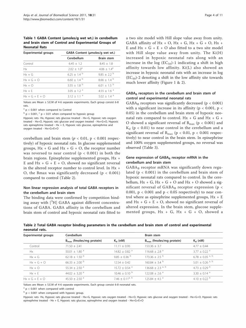
ResultsGABA Content in the cerebellum and brain stem ofcontrol and experimental neonatal ratsThe GABA content was decreased significantly (p <0.001) in the cerebellum and brain stem of hypoxic neo-natal rats compared to control. The decreased contentwas reversed to near normal in glucose supplementedgroups - Hx + G and Hx + G + O (Table 1).
Total GABA receptors in the cerebellum and brain stemof control and experimental neonatal ratsReceptor studies for total GABA showed a significantdecrease in receptor number compared to control in the
Anju et al. Journal of Biomedical Science 2011, 18:31http://www.jbiomedsci.com/content/18/1/31
Page 3 of 11
cerebellum and brain stem (p < 0.01, p < 0.001 respec-tively) of hypoxic neonatal rats. In glucose supplementedgroups, Hx + G and Hx + G + O, the receptor numberwas reversed to near control (p < 0.001) in both thebrain regions. Epinephrine supplemented groups, Hx +E and Hx + G + E + O, showed no significant reversalin the altered receptor number to control level. In Hx +O, the Bmax was significantly decreased (p < 0.001)compared to control (Table 2).
Non linear regression analysis of total GABA receptors inthe cerebellum and brain stemThe binding data were confirmed by competition bind-ing assay with [3H] GABA against different concentra-tions of GABA. GABA affinity in the cerebellum andbrain stem of control and hypoxic neonatal rats fitted to
a two site model with Hill slope value away from unity.GABA affinity of Hx + O, Hx + G, Hx + G + O, Hx +E and Hx + G + E + O also fitted to a two site modelwith Hill slope value away from unity. The Ki(H)increased in hypoxic neonatal rats along with anincrease in the log (EC50)-1 indicating a shift in highaffinity towards low affinity. Ki(L) also showed anincrease in hypoxic neonatal rats with an increase in log(EC50)-2 denoting a shift in the low affinity site towardsmuch lower affinity (Figure 1 & 2).
GABAB receptors in the cerebellum and brain stem ofcontrol and experimental neonatal ratsGABAB receptors was significantly decreased (p < 0.001)with a significant increase in its affinity (p < 0.001, p <0.05) in the cerebellum and brain stem of hypoxic neo-natal rats compared to control. Hx + G and Hx + G +O showed a significant reversal of Bmax (p < 0.001) andKd (p < 0.01) to near control in the cerebellum and asignificant reversal of Bmax (p < 0.01, p < 0.001 respec-tively) to near control in the brain stem. In epinephrineand 100% oxygen supplemented groups, no reversal wasobserved (Table 3).
Gene expression of GABAB receptor mRNA in thecerebellum and brain stemGABAB receptor mRNA was significantly down regu-lated (p < 0.001) in the cerebellum and brain stem ofhypoxic neonatal rats compared to control. In the cere-bellum, Hx + G, Hx + G + O and Hx + O showed a sig-nificant reversal of GABAB receptor expression (p <0.001, p < 0.001 and p < 0.05 respectively) to near con-trol where as epinephrine supplemented groups, Hx + Eand Hx + G + E + O, showed no significant reversal ofaltered expression. In the brain stem, glucose supple-mented groups, Hx + G, Hx + G + O, showed a
Table 1 GABA Content (μmoles/g wet wt.) in cerebellumand brain stem of Control and Experimental Groups ofNeonatal Rats
Experimental groups GABA Content (μmoles/g wet wt.)
Cerebellum Brain stem
Control 6.45 ± 1.2 8.45 ± 1.8
Hx 2.02 ± 1.0a 4.06 ± 1.4a
Hx + G 6.25 ± 1.4 b 9.85 ± 2.2 b
Hx + G + O 6.60 ± 1.4 b 8.66 ± 1.4 b
Hx + O 3.55 ± 1.8 b 6.01 ± 1.5 b
Hx + E 3.05 ± 1.2 a 4.55 ± 1.6 a
Hx + G + E + O 3.12 ± 1.1 a 5.02 ± 1.4 a
Values are Mean ± S.E.M of 4-6 separate experiments. Each group consist 6-8rats.a p < 0.001 when compared to Controlb p < 0.001, c p < 0.01 when compared to hypoxic group
Hypoxic rats- Hx, Hypoxic rats glucose treated - Hx+G, Hypoxic rats oxygentreated - Hx+O, Hypoxic rats glucose and oxygen treated - Hx+G+O, Hypoxicrats epinephrine treated - Hx + E, Hypoxic rats glucose, epinephrine andoxygen treated - Hx+G+E+O
Table 2 Total GABA receptor binding parameters in the cerebellum and brain stem of control and experimentalneonatal rats.
Experimental groups Cerebellum Brain stem
Bmax (fmoles/mg protein) Kd (nM) Bmax (fmoles/mg protein) Kd (nM)
Control 71.50 ± 2.41 11.11 ± 0.95 153.36 ± 3.7 4.77 ± 0.44
Hx 50.01 ± 1.80 a 14.82 ± 0.82 a 116.68 ± 2.8 a 3.77 ± 0.22 a
Hx + G 62.18 ± 1.50 b 9.85 ± 0.36 b 173.36 ± 2.5 b 6.78 ± 0.35 a, b
Hx + G + O 66.33 ± 2.00 b 12.54 ± 0.42 160.84 ± 3.4 b 5.01 ± 0.26 a, b
Hx + O 55.34 ± 2.50 a 15.72 ± 0.54 a 136.68 ± 2.3 a, b 4.73 ± 0.29 b
Hx + E 44.02 ± 3.20 a 10.46 ± 0.10 b 122.08 ± 2.6 a 3.30 ± 0.14 a
Hx + G + E + O 45.50 ± 2.50 a 7.46 ± 0.11a, b 125.84 ± 4.5 a 4.10 ± 0.22 b
Values are Mean ± S.E.M of 4-6 separate experiments. Each group consist 6-8 neonatal rats.a p < 0.001 when compared with controlb p < 0.001 when compared with hypoxic group.
Hypoxic rats- Hx, Hypoxic rats glucose treated - Hx+G, Hypoxic rats oxygen treated - Hx+O, Hypoxic rats glucose and oxygen treated - Hx+G+O, Hypoxic ratsepinephrine treated - Hx + E, Hypoxic rats glucose, epinephrine and oxygen treated - Hx+G+E+O
Anju et al. Journal of Biomedical Science 2011, 18:31http://www.jbiomedsci.com/content/18/1/31
Page 4 of 11

significant reversal of the gene expression (p < 0.001) tonear control, whereas Hx + O, Hx + E and Hx + G + E+ O showed a down regulated GABAB receptor expres-sion (p < 0.01, p < 0.001, p < 0.001 respectively) without a significant reversal to near control (Figure 3).
Gene expression of GAD mRNA in the cerebellum andbrain stemThe expression of glutamate decarboxylase in cerebel-lum and brain stem also showed a significant down reg-ulation (p < 0.001) in the hypoxic group compared tocontrol. The cerebellar and brain stem GAD expressionwas significantly reversed to near control in Hx + G, Hx+ G + O and Hx + O whereas in Hx + E and Hx + G +E + O, there was no significant reversal to near control(Figure 4).
DiscussionHypoxia–ischemia (HI) occurring before or shortly afterbirth is a major cause of life-threatening injury and life-long disability [36]. HI results in multi-organ failure andstructural/functional damage especially devastating tothe cardiovascular, renal, gastrointestinal and central
nervous systems [37,38]. HI brain injury is very complexand has different neuropathological manifestationsdepending on the maturity of the newborn. Many of thestructural changes that occur during the initial postnatalperiod in rodents are consistent with those seen duringthe late prenatal period in human brain development.Thus, exposure of rat to hypoxia on postnatal day 4includes many of the neurodevelopmental events thatmay be affected by hypoxia in preterm human infants.In the present study, we investigated the functional reg-ulation of GABAB receptors and GAD in hypoxic neo-natal rats and the role of glucose, oxygen andepinephrine in altering the receptor status.Numerous studies by different groups have confirmed
that both inhibitory and excitatory amino acids areinvolved in the acute hypoxic ventilatory response[39-42]. Increases in GABA as a consequence of brainhypoxia can lead to depression of ventilation, whichbecomes more apparent in the absence of peripheralchemoreceptors. Blockade of GABA by biccuculine cansignificantly reduce this depressive effect of GABA onventilation during hypoxia in chemodenervated animalor the newborn [43-45].
Figure 1 Displacement of [3H] GABA against GABA in cerebellum of control and experimental neonatal rats. Competition studies werecarried out with 30 nM [3H] GABA in each tube with the unlabelled GABA concentrations varying from 10-8 to10-4 M. Values are representationof 4-6 separate experiments. Data from the curves as determined from nonlinear regression analysis using computer program PRISM fitted to atwo-site model. The affinity for the first and second site for the competing drug is designated as Ki-1 (for high affinity) and Ki-2 (for low affinity).EC50 is the concentration of competitor that competes for half the specific binding. The equation built-in to the program is defined in terms ofthe log (EC50). If the concentrations of unlabelled compound are equally spaced on a log scale, the uncertainty of the log (EC50) will besymmetrical, but uncertainty of the EC50 will not be symmetrical
Anju et al. Journal of Biomedical Science 2011, 18:31http://www.jbiomedsci.com/content/18/1/31
Page 5 of 11

The present study reports a significant decrease in totalGABA and GABAB receptor number with a down regu-lated receptor expression and glutamate decarboxylaseexpression in the cerebellum and brain stem regions ofhypoxic neonatal rats. The decreased expression of GAD
in turn results in the inhibition of GABA synthesizingpathway, which can be correlated to the decreased GABAreceptors. The decreased GABA receptor is a response ofthe body to encounter hypoxic ventilatory decline. Thereduction in GABAB receptor may help in overcoming
Figure 2 Displacement of [3H] GABA against GABA in brain stem of control and experimental neonatal rats. Competition studies werecarried out with 30 nM [3H] baclofen in each tube with the unlabelled baclofen concentrations varying from 10-12 to10-4 M. Values arerepresentation of 4-6 separate experiments. Data from the curves as determined from nonlinear regression analysis using computer programPRISM fitted to a two-site model. The affinity for the first and second site for the competing drug is designated as Ki-1 (for high affinity) and Ki-2(for low affinity). EC50 is the concentration of competitor that competes for half the specific binding. The equation built-in to the program isdefined in terms of the log (EC50). If the concentrations of unlabelled compound are equally spaced on a log scale, the uncertainty of the log(EC50) will be symmetrical, but uncertainty of the EC50 will not be symmetrical.
Table 3 GABAB receptor binding parameters in the cerebellum and brain stem of control and experimental neonatalrats.
Experimental groups Cerebellum Brain stem
Bmax (fmoles/mg protein) Kd (nM) Bmax (fmoles/mg protein) Kd (nM)
Control 71.50 ± 2.41 11.11 ± 0.95 74.27 ± 1.20 13.31 ± 1.00
Hx 50.01 ± 1.80 a 14.82 ± 0.82 a 51.84 ± 1.50 a 14.44 ± 0.99 b
Hx + G 62.18 ± 1.50 b 9.85 ± 0.36 b 69.41 ± 1.40 b 20.47 ± 0.99 a
Hx + G + O 66.33 ± 2.00 b 12.54 ± 0.42 70.47 ± 1.10 c 26.10 ± 1.20 a
Hx + O 55.34 ± 2.50 a 15.72 ± 0.54 a 49.10 ± 1.10 a 16.36 ± 1.50 a
Hx + E 44.02 ± 3.20 a 10.46 ± 0.10 b 43.59 ± 1.5 a 14.53 ± 0.99 b
Hx + G + E + O 45.50 ± 2.50 a 7.46 ± 0.11a, b 53.95 ± 1.5 a 13.90 ± 0.99 b
Values are Mean ± S.E.M of 4-6 separate experiments. Each group consist 6-8 neonatal rats.a p < 0.001, b p < 0.05 when compared with controlc p < 0.001 when compared with hypoxic group.
Hypoxic rats- Hx, Hypoxic rats glucose treated - Hx+G, Hypoxic rats oxygen treated - Hx+O, Hypoxic rats glucose and oxygen treated - Hx+G+O, Hypoxic ratsepinephrine treated - Hx + E, Hypoxic rats glucose, epinephrine and oxygen treated - Hx+G+E+O
Anju et al. Journal of Biomedical Science 2011, 18:31http://www.jbiomedsci.com/content/18/1/31
Page 6 of 11

the ventilatory decline during hypoxia but at the cost ofsevere central nervous system dysfunction. Louzoun-Kaplan et al, 2008 [46] reported that prenatal hypoxia atgestation day 17 in mice caused an immediate decreasein fetal cerebral cortex levels of glutamate decarboxylase.Decreased levels of key proteins in the GABA pathway inthe cerebral cortex may lead to high susceptibility to sei-zures and epilepsy in newborns after prenatal or perinatalhypoxia. In the elevated plus maze, the agonist of GABA-B receptor was reported to improve consolidation of pas-sive avoidance in rats undergoing hypoxia [47]. GABAB
receptor-mediated activation of TASK-1 or a relatedchannel provides a presynaptic autoregulatory feedbackmechanism that modulates fast synaptic transmission inthe rat carotid body [48]. The signaling cascade that trig-gers the altered transcription of GABA-B receptor andGAD under hypoxic stress can be related to the activa-tion of apoptotic pathways by triggering Bax expression
and deactivating CREB expression coupled with the acti-vation of HIF. The accumulation of HIF-1a in ischemicor hypoxic tissues promote adaptive mechanisms for cellsurvival [49] and was found to be an important mediatorof hypoxia-induced tolerance to ischemia [50]. AlthoughHIF-1a is essential for adaptation to low oxygen levels, ithas also been shown in vitro to mediate hypoxia-inducedgrowth arrest and apoptosis [51]. The increased Hif 1mRNA expression under hypoxia facilitates angiogenesis,vasodialation and erythropoiesis. But in severe hypoxiccases, HIF-1a is accumulated and leads to cell death byactivating different target genes [52]. The role of HIF-1ain mediating pro death and pro survival responses, isdependent on the duration [53] and types of pathologicalstimuli [54] as well as the cell type that it induces [55].We observed that glucose supplementation to hypoxic
neonates alone and along with 100% oxygen showed areversal in the altered GABAB receptor parameters and
Figure 3 Real time PCR amplification of GABAB receptor subunit in mRNA form the cerebellum (A) and brain stem (B) of control andexperimental neonatal rats. The ΔΔCT method of relative quantification was used to determine the fold change in expression. The relativeratios of mRNA levels were calculated using the ΔΔCT method normalized with b-actin. CT value as the internal control and Control CT value asthe caliberator. PCR analyses were conducted in the cerebellum (A) and brain stem (B) with gene-specific primers and fluorescently labeled Taqprobe GABAB (Rn 00578911)
Anju et al. Journal of Biomedical Science 2011, 18:31http://www.jbiomedsci.com/content/18/1/31
Page 7 of 11

GAD expression in the cerebellum and brain stem. Glu-cose supplementation provides an instant source ofenergy to the brain cells thereby preventing ATP deple-tion mediated cell death. Hattori and Wasterlain, 2004[56] observed a reduction in the blood glucose levelsand substantially increased cerebral glucose utilization[57] as a result of hypoxic stress in experimental rats.Mónica Lemus et al, 2008 [58] reported that GABAB
receptor agonist (baclofen) or antagonists (phaclofenand CGP55845A) locally injected into nucleus tractussolitarius modified arterial glucose levels and brain glu-cose retention.The standard approach to resuscitation neonatal
hypoxia is to use 100% O2. Further, resuscitation with100% is recommended as a beneficial short-term therapythat is generally thought to be non-toxic [31,59].Although the use of 100% O2 appears intuitive to maxi-mize the gradient required to drive O2 into hypoxiccells [30], a building body of evidence derived from
animal models, has demonstrated that although resusci-tation with 100% O2 improves restoration of cerebraland cortical perfusion, it may occur at the price ofgreater biochemical oxidative stress [31]. Resuscitationwith 100% O2 significantly increased glutamate and gly-cine in the dorsal cortex contralateral to the ligatedcommon carotid artery, compared to piglets resuscitatedwith 21% O2. These data suggest that persistent changesin neurochemistry occur 4 days after hypoxic ischemiaand further studies are warranted to elucidate the conse-quences of this on neonatal brain development [60]. Weobserved that 100% oxygen resuscitation for neonatalhypoxia is not as effective as the combination of glucoseand oxygen or administration of glucose alone. In cere-bellum and brain stem of 100% oxygen resuscitatedgroups, GABAB receptors showed a significant decreasecompared to control. One hundred percentage of oxy-gen generated abnormally high levels of reactive oxygenspecies (ROS) which causes dysfunction of defensive
Figure 4 Real time PCR amplification of GAD mRNA form the cerebellum (A) and brain stem (B) of control and experimental neonatalrats. The ΔΔCT method of relative quantification was used to determine the fold change in expression. The relative ratios of mRNA levels werecalculated using the ΔΔCT method normalized with b-actin. CT value as the internal control and Control CT value as the caliberator. PCRanalyses were conducted in the cerebellum (A) and brain stem (B) with gene-specific primers and fluorescently labeled Taq probe GAD1 (Rn00690304_g1).
Anju et al. Journal of Biomedical Science 2011, 18:31http://www.jbiomedsci.com/content/18/1/31
Page 8 of 11

antioxidant system of cells by altering enzyme activity[61,62] and act as a factor for neurodegeneration [63].Hypoxemic piglets resuscitated with 100% O2 alsoshowed increased cerebral injury, cortical damage andearly neurologic disorders [64-66]. Previous studies onacetylcholinesterase [67], GABAA and serotonin recep-tors [68] reported the neuroprotective role of glucoseand combination of glucose and oxygen resuscitationand the damaging effects of oxygen supplementationalone. The reduction in GABAB receptor number in thecerebellar and brain stem regions during oxygen supple-mentation is suggested to be due to tissue damagecaused by the formation of free radicals or reactive oxy-gen species and the changes in amino acids resulting inneuronal cell death. During oxygen resuscitation, theaccumulation of ROS activates the over stimulation ofHIF 1 alpha which can in turn results in the activationof apoptotic pathways by altering the expression of tran-scription factors like CREB and NF-Kappa-B.Epinephrine is routinely used in the resuscitation for
persistent severe neonatal hypoxia. The present studypoints out the adverse effects of epinephrine supplemen-tation, alone and even in combination with glucose andoxygen, by studying the changes in GABAB receptor,expression of GABAB receptor and GAD in the brainstem and cerebellum. The GABAB receptor was signifi-cantly decreased in epinephrine treated groups. A reflexaction of epinephrine firing occurs during hypoxia. Sup-plementation of epinephrine to already excited systemresults in its hyper activity and it affects the balance ofvarious neurotransmitters like dopamine [69] and gluta-mate. Epinephrine induces a hypoxia-neovascularizationcascade and plays a primary role in vascular prolifera-tion within soft tissues [70]. It is reported that repetitivehypoxic stress induced by labour is a powerful stimulusfor catecholamine release in fetus and is accompaniedby typical alterations of fetal heart rate. The high influxof this excitatory neurotransmitter affects the balance ofother neurotransmitters thereby disrupting the cascadeof signal transduction.There has been much interest in the acute neurologi-
cal changes associated with neonatal hypoxia, along withthe mechanisms of subsequent central nervous systemdysfunction in the adult [71-74]. Hypoxia during thefirst week of life can induce neuronal death in vulner-able brain regions usually associated with an impairmentof cognitive function that can be detected later in life[75]. Postnatal hypoxia resulting from lung immaturityand respiratory disturbances in infants is an importantpathophysiological mechanism underlying the devastat-ing neurological complications. This points the impor-tance of a proper resuscitation program to overcomeneonatal hypoxia for a better intellect in the later stagesof life.
ConclusionsOur studies point out the neuroprotective role of glu-cose in the management of neonatal hypoxic stress. Thedown regulated GABAB receptor in cerebellum andbrain stem led to hypoxia induced ventilatory declineand activation of apoptotic pathways. These receptoralterations are reversed back to near control by thetimely resuscitation with glucose, alone and in combina-tion with oxygen. The deleterious effect of oxygen aloneand epinephrine resuscitation in neuronal responsethrough alterations in neurotransmitters was alsoobserved. Thus it is suggested that glucose administra-tion immediately after hypoxia with oxygenated air as aresuscitation programme will be of tremendous advan-tage especially in neonatal care. Deeper understandingof mechanisms, through which hypoxia regulates theneurotransmitters, could point towards the developmentof new therapeutic approaches to reduce or suppressthe pathological effects of hypoxia.
AcknowledgementsThis work was supported by the research grants from DBT, DST, ICMR, Govt.of India and KSCSTE, Govt. of Kerala to Dr. C. S. Paulose. Anju T R thanksCouncil of Scientific and Industrial Research for Senior Research Fellowship.
Authors’ contributionsTRA carried out the receptor assays, gene expression and drafted themanuscript. SJ participated participated in the design of the study andperformed the statistical analysis. CSP conceived of the study andparticipated in its design and coordination. All authors read and approvedthe final manuscript.
Competing interestsThe authors declare that they have no competing interests.
Received: 5 January 2011 Accepted: 12 May 2011Published: 12 May 2011
References1. Low JA, Froese AB, Galbraith RS, Smith JT, Sauerbrei EE, Derrick EJ: The
association between preterm newborn hypotension and hypoxemia andoutcome during the first year. Acta Paediatrica 1993, 82:433-437.
2. Delivoria-Papadopoulos M, Mishra POM: Mechanisms of perinatal cerebralinjury in fetus and newborn. Annals of the New York Academy of Sciences2000, 900:159-168.
3. Li C, Jackson RM: Reactive species mechanisms of cellularhypoxicreoxygenation injury. American Journal of Physiology 2002,282:227-241.
4. Rodrigo J, Fernandez AP, Serrano J, Peinado MA, Martinez A: The role offree radicals in cerebral hypoxia and ischemia. Free Radical Biology andMedicine 2005, 39:26-50.
5. Xu W, Chi L, Row BW, et al: Increased oxidative stress is associated withchronic intermittent hypoxia-mediated brain cortical neuronal cellapoptosis in a mouse model of sleep apnea. Neuroscience 2004,126:313-323.
6. Acker T, Acker H: oxygen sensing need in CNS function: Physiologicaland pathological implications. Journal of Experimental Biology 2004,207:3171-3188.
7. Solomon IC: Excitation of phrenic and sympathetic output during acutehypoxia: Contribution of medullary oxygen detectors. RespirationPhysiology 2000, 121:101-117.
8. Neubauer JA, Melton JE, Edelman NH: Modulation of respiration duringbrain hypoxia. J Appl Physiol 1990, 68:441-451.
Anju et al. Journal of Biomedical Science 2011, 18:31http://www.jbiomedsci.com/content/18/1/31
Page 9 of 11

9. Weil JV, Zwillich CW: Assessment of ventilatory response to hypoxia.Chest 1976, 70(1):124-128.
10. Kneussl MP, Pappagianopoulos P, Hoop B, Kazemi H: Reversibledepressiozn of ventilation and cardiovascular function byventriculocisternal perfusion with γ-aminobutyric acid in dogs. Am RevRespir Dis 1986, 133:1024-1028.
11. Taveira da Silva AM, Hartley B, Hamosh P, Quest JA, Gillis RA: Respiratorydepressant effects of GABA α- and β-receptor agonists in the cat. J ApplPhysiol 1987, 62:2264-2272.
12. Kazemi H, Hoop B: Glutamic acid and gamma-aminobutyric acidneurotransmitters in central control of breathing. J Appl Physiol 1991,70:1-7.
13. Richter DW, Schmidt-Garcon P, Pierrefiche O, Bischoff AM, Lalley PM:Neurotransmitters and neuromodulators controlling the hypoxicrespiratory response in anaesthetized cats. J Physiol 1999, 514:567-578.
14. Di Pasquale E, Morin D, Monteau R, Hilaire G: Serotonergic modulation ofthe respiratory rhythm generator at birth: an in vitro study in the rat.Neurosci Lett 1992, 143:91-95.
15. Neylon M, Marshall JM: The role of adenosine in the respiratory andcardiovascular response to systemic hypoxia in the rat. J Physiol 1991,440:529-545.
16. Elnazir B, Marshall JM, Kumar P: Postnatal development of the pattern ofrespiratory and cardiovascular response to systemic hypoxia in thepiglet: the roles of adenosine. J Physiol 1996, 492:573-585.
17. Gozal D, Simakajornboon N, Czapla MA, Xue YD, Gozal E, Vlasic V, Lasky JA,Liu JY: Brainstem activation of platelet-derived growth factor-β receptormodulates the late phase of the hypoxic ventilatory response.J Neurochem 2000, 74:310-319.
18. Simakajornboon N, Kuptanon T: Maturational changes inneuromodulation of central pathways underlying hypoxic ventilatoryresponse. Respir Physiol Neurobiol 2005, 149:273-286.
19. Cataltepe O, Towfighi J, Vannucci RC: Cerebrospinal fluid concentrationsof glutamate and GABA during perinatal cerebral hypoxia-ischemia andseizures. Brain Res 1996, 709:326-330.
20. Hagberg H, Andersson P, Kjellmer I, Thiringer K, Thordstein M: Extracellularoverflow of glutamate, aspartate, GABA and taurine in the cortex andbasal ganglia of fetal lambs during hypoxia-ischemia. Neurosci Lett 1987,78:311-317.
21. Rego AC, Santos MS, Oliveira CR: Oxidative stress, hypoxia, and ischemia-like conditions increase the release of endogenous amino acids bydistinct mechanisms in cultured retinal cells. J Neurochem 1996,66:2506-2516.
22. Saransaari P, Oja SS: Enhanced GABA release in cell-damaging conditionsin the adult and developing mouse hippocampus. Int J Devl Neurosci1997, 15(2):163-174.
23. Saransaari P, Oja SS: Release of endogenous glutamate, aspartate, GABA,and taurine from hippocampal slices from adult and developing miceunder cell-damaging conditions. Neuochem Res 1998, 23:563-570.
24. Zhang W, Barnbrock A, Gajic S, Pfeiffer A, Ritter B: Differential ontogeny ofGABAB-receptor-mediated pre- and postsynaptic modulation of GABAand glycine transmission in respiratory rhythm-generating network inmouse. The Journal of Physiology 2002, 540:435-446.
25. Suzuki M, Tetsuka M, Endo M: GABA(B) receptors in the nucleus tractussolitarii modulate the carotid chemoreceptor reflex in rats. Neurosci Lett1999, 260:21-24.
26. Yang AL, Lo MJ, Ting H, Chen JS, Huang CY, Lee SD: GABAA and GABABreceptors differentially modulate volume and frequency in ventilatorycompensation in obese Zucker rats. J Appl Physiol 2007, 102:350-357.
27. Schubert S, Brandl U, Brodhun M, Ulrich C, Spaltmann J, Fiedler N, Bauer R:Neuroprotective effects hypoxia–ischemia in newborn piglets. Brain Res2005, 1058:129-136.
28. Davis PG, Tan A, O’Donnell CPF, Schulze A: Resuscitation of newborninfants with 100% oxygen or air: a systematic review and meta-analysis.Lancet 2004, 364:1329-33.
29. Tan A, Schulze A, O’Donnell CPF, Davis PG: Air versus oxygen forresuscitation of infants at birth. The cochrane database of systemic reviews2006 2005, 2.
30. Corff KE, McCann DL: Room air resuscitation versus oxygen resuscitationin the delivery room. J Perinat Neonat Nurs 2005, 19:379-90.
31. Martin RJ, Walsh MC, Carlo WA: Reevaluating neonatal resuscitation with100% oxygen. Am J Respir Crit Care Med 2005, 172:1360.
32. Glowinski J, Iversen LL: Regional studies of catecholamines in the ratbrain: The disposition of [3H] Norepinephrine, [3H] DOPA in variousregions of the brain. J Neurochem 1966, 13:655-669.
33. Kurioka S, Toshiaki K, Makoto M: Effects of sodium and bicarbonate ionson gamma amino butyric acid receptor binding in synaptic membranesof rat brain. J Neurochem 1981, 37:418-421.
34. Lowry OH, Rosebrough NJ, Farr AL, Randall J: Protein measurement withfolin phenol reagent. J Biol Chem 1951, 193:265-275.
35. Scatchard G: The attractions of proteins for small molecules and ions.Ann NY Acad Sci 1949, 51:660-672.
36. du Plessis AJ, Volpe JJ: Perinatal brain injury in the preterm and termnewborn. Curr Opin Neurol 2002, 15:151-7.
37. Shah P, Riphagen S, Beyene J, Perlman JM: Multiorgan dysfunction ininfants with post-asphyxial hypoxic–ischemic encephalopathy. Arch DisChild Fetal Neonatal Ed 2004, 89:F152-5.
38. Vento M, Sastre J, Asensi MA, Vina J: Room-air resusciatation causes lessdamage to heart and kidney than 100% oxygen. Am J Respir Crit CareMed 2005, 172:1393-8.
39. Mitra J, Prabhakar NR, Overholt JL, Cherniack NS: Respiratory effects of N-methyl-D-aspartate on the ventrolateral medullary surface. J Appl Physiol1989, 67:1814-1819.
40. Lin J, Suguihara C, Huang J, Hehre D, Devia C, Bancalari E: Effect of N-methyl-D-aspartate-receptor blockage on hypoxic ventilatory responsein unanesthetized piglets. J Appl Physiol 1996, 80:1759-1763.
41. Gozal D, Gozal E, Torres JE, Gozal YM, Nuckton TJ, Hornby PJ: Nitric oxidemodulates ventilatory responses to hypoxia in the developing rat. Am JRespir Crit Care Med 1997, 155:1755-1762.
42. Gozal D, Graff GR, Torres JE, Khicha SG, Nayak GS, Simakajornboon N, Gozal E:Cardiorespiratory responses to systemic administration of a proteinkinase C inhibitor in conscious rats. J Appl Physiol 1998, 84:641-648.
43. Hayashi F, Lipski J: The role of inhibitory amino acids in control ofrespiratory motor output in an arterially perfused rat. Respir Physiol 1992,89:47-63.
44. Huang J, Suguihara C, Hehre D, Lin J, Bancalari E: Effects of GABA receptorblockage on the respiratory response to hypoxia in sedated newbornpiglets. J Appl Physiol 1994, 77:1006-1010.
45. Soto-Arape I, Burton MD, Kazemi H: Central amino acid neurotransmittersand the hypoxic ventilatory response. Am J Respir Crit Care Med 1995,151:1113-1120.
46. Louzoun-Kaplan V, Zuckerman M, Regino Perez-Polo J, Golan HM: Prenatalhypoxia down regulates the GABA pathway in newborn mice cerebralcortex; partial protection by MgSO4.. International Journal ofDevelopmental Neuroscience 2008, 26:77-85.
47. Car H, Oksztel R, Nadlewska A, Wi K: Baclofen prevents hypoxia-inducedconsolidation impairment for passive avoidance in rats. PharmacologicalResearch 2001, 44:329-335.
48. Fearon IM, Zhang M, Vollmer C, Nurse CA: GABA mediates autoreceptorfeedback inhibition in the rat carotid body via presynaptic GABAB
receptors and TASK-1. The Journal of Physiology 2003, 553:83-94.49. Bergeron M, Gidday JM, Yu AY, Semenza GL, Ferriero DM, Sharp FR: Role of
hypoxia-inducible factor-1 in hypoxia-induced ischemic tolerance inneonatal rat brain. Ann Neurol 2000, 48:285-296.
50. Bernaudin M, Nedelec AS, Divoux D, MacKenzie ET, Petit E, Schumann-Bard P: Normobaric hypoxia induces tolerance to focal permanentcerebral ischemia in association with an increased expression ofhypoxia-inducible factor-1 and its target genes, erythropoietin andVEGF, in the adult mouse brain. J Cereb Blood Flow Metab 2002,22:393-403.
51. Goda N, Ryan HE, Khadivi B, McNulty W, Rickert RC, Johnson RS: Hypoxia-inducible factor 1alpha is essential for cell cycle arrest during hypoxia.Mol Cell Biol 2003, 23:359-369.
52. Semenza GL, Agani F, Feldser D, Iyer N, Kotch L, Laughner E, Yu A: Hypoxia,HIF-1, and the pathophysiology of common human diseases. Adv ExpMed Biol 2000, 475:123-130.
53. Halterman MW, Federoff HJ: HIF-1alpha and p53 promote hypoxia-induced delayed neuronal death in models of CNS ischemia. Exp Neurol1999, 159:65-72.
54. Aminova LR, Chavez JC, Lee J, Ryu H, Kung A, Lamanna JC, Ratan RR:Prosurvival and prodeath effects of hypoxia-inducible factor-1alphastabilization in a murine hippocampal cell line. J Biol Chem 2005,280:3996-4003.
Anju et al. Journal of Biomedical Science 2011, 18:31http://www.jbiomedsci.com/content/18/1/31
Page 10 of 11

55. Vangeison G, Carr D, Federoff HJ, Rempe DA: The good, the bad, and thecell type-specific roles of hypoxia inducible factor-1 alpha in neuronsand astrocytes. J Neurosci 2008, 28:1988-1993.
56. Hattori H, Wasterlain CG: Posthypoxic glucose supplement reduceshypoxicischemic brain damage in the neonatal rat. Ann Neurol 2004,28:122-128.
57. Vannucci SJ, Hagberg H: Hypoxia-ischemia in the immature brain. J ExpBiol 2004, 207:3749-3154.
58. Lemus Mónica, Montero S, Cadenas JL, Lara JJ, Tejeda-Chávez HR, Álvarez-Buylla R, de Álvarez-Buylla Elena Roces: GabaB receptors activation in theNTS blocks the glycemic responses induced by carotid body receptorstimulation. Autonomic Neuroscience 2008, 141:73-82.
59. Kuisma M, Boyd J, Voipio V, Alaspaa A, Roine RO, Rosenberg P: Comparisonof 30 and the 100% inspired oxygen concentrations during early post-resuscitation period: a randomised controlled pilot study. Resuscitation2006, 69:199-206.
60. Lauren LJ, Po-Yin C, Obaid L, Emara M, Johnson ST, Bigam DL, Todd KG:Persistent neurochemical changes in neonatal piglets after hypoxia-ischemia and resuscitation with 100%, 21% or 18% oxygen. Resuscitation2008, 77:111-120.
61. Bandyopadhyay U, Das D, Ranajit K, Banerjee V: Reactive oxygen species:Oxidative damage and pathogenesis. Current Science 1999, 77:658-666.
62. Anju TR, Athira B, Paulose CS: Superoxide dismutase functional regulationin neonatal hypoxia: Effect of glucose, oxygen and epinephrine. Indian JBiochem Biophys 2009, 46:166-171.
63. Matharan TS, Laemmel E, Duranteau J, Vicaut E: After hypoxia and glucosedepletion causes reactive oxygen species production by mitochondria inHUVEC. American Journal of Physiology: Regulatory Integrative andComparative Physiology 2004, 287:R1037-R1043.
64. Temesvari P, Karg E, Bódi I, Németh I, Pintér S, Lazics K: Impaired earlyneurologic outcome in newborn piglets reoxygenated with 100%oxygen compared with room air after pneumothorax-induced asphyxia.Pediatric Research 2001, 49:812-819.
65. Munkeby BH, Borke WB, Bjornland K, Sikkeland LL, Borge GL, Halvorsen B,Saugstad OD: Resuscitation with 100% O2 increases cerebral injury inhypoxemic piglets. Pediatric Research 2004, 56(5):783-790.
66. Shimabuku R, Ota A, Pereyra S, Veliz B, Paz E, Nakachi G: Hyperoxia with100% oxygen following hypoxia-ischemia increases brain damage innewborn rats. Biology of Neonate 2005, 88:168-171.
67. Chathu F, Krishnakumar A, Paulose CS: Acetylcholine esterase activity andbehavioral response in hypoxia induced neonatal rats: Effect of glucose,oxygen and epinephrine supplementation. Brain and cognition 2008,68:59-66.
68. Anju TR, Jobin M, Jayanarayanan S, Paulose CS: Cerebellar 5-HT2A receptorfunction under hypoxia in neonatal rats: Role of glucose, oxygen, andepinephrine resuscitation. Respir Physiol Neurobiol 2010, 172(3):147-153.
69. Binoy J, Nandhu MS, Paulose CS: Dopamine D(1) and D(2) receptorfunctional down regulation in the cerebellum of hypoxic neonatal rats:Neuroprotective role of glucose and oxygen, epinephrine resuscitation.Pharmacological Research 2009.
70. Karacaoglu E, Bayram I, Celiköz B, Zienowicz RJ: Does sustainedepinephrine release trigger a hypoxia-neovascularization cascade? PlastReconstr Surg 2007, 119:858-64.
71. Soulier V, Peyronnet J, Pequignot JM, Cottet-Emard JM, Lagercrantz H,Dalmaz Y: Long-term impairment in the neurochemical activity of thesympathoadrenal system after neonatal hypoxia in the rat. Pediatr Res1997, 42:30-38.
72. Peterson BS: Brain imaging studies of the anatomical and functionalconsequences of preterm birth for human brain development. Ann NYAcad Sci 2003, 1008:219-237.
73. Lindahl E, Michelsson K, Helenius M, Parre M: Neonatal risk factors andlater neurodevelopmental disturbances. Dev Med Child Neurol 1988,30:571-589.
74. Berg AT: Childhood neurological morbidity and its association withgestational age, intrauterine growth retardation and perinatal stress.Paediatr Perinat Epidemiol 1988, 2:229-238.
75. Casolini P, Zuena AR, Cinque C: Sub-neurotoxic neonatal anoxia inducessubtle behavioural changes and specific abnormalities in brain group-Imetabotropic glutamate receptors in rats. J Neurochem 2005, 95:137-145.
doi:10.1186/1423-0127-18-31Cite this article as: Anju et al.: Decreased GABAB receptor function inthe cerebellum and brain stem of hypoxic neonatal rats: Role ofglucose, oxygen and epinephrine resuscitation. Journal of BiomedicalScience 2011 18:31.
Submit your next manuscript to BioMed Centraland take full advantage of:
• Convenient online submission
• Thorough peer review
• No space constraints or color figure charges
• Immediate publication on acceptance
• Inclusion in PubMed, CAS, Scopus and Google Scholar
• Research which is freely available for redistribution
Submit your manuscript at www.biomedcentral.com/submit
Anju et al. Journal of Biomedical Science 2011, 18:31http://www.jbiomedsci.com/content/18/1/31
Page 11 of 11