research in veterinary science -...
TRANSCRIPT
Research in Veterinary Science 102 (2015) 27–33
Contents lists available at ScienceDirect
Research in Veterinary Science
j ourna l homepage: www.e lsev ie r .com/ locate /y rvsc
Functional and regenerative effects of local administration of autologousmononuclear bone marrow cells combined with silicone conduit ontransected femoral nerve of rabbits
Anelise Bonilla Trindade a,⁎, Pedro Schestatsky b, Vítor Félix Torres b, Cristiano Gomes a,Giordano Cabral Gianotti a, AnaHelena da Rosa Paz c, Paula Barros Terraciano c, JaneteMaria VolpatoMarques a,Karina Magano Guimarães a, Dominguita Lühers Graça d,Elizabeth Obino Cirne-Lima c, Emerson Antonio Contesini e
a Programa de Pós-graduação em Ciências Veterinárias, Universidade Federal do Rio Grande do Sul (UFRGS), Porto Alegre, RS, Brazilb Neurology Department, Eletroneuromyography, Hospital de Clínicas de Porto Alegre, Porto Alegre, RS, Brazilc Laboratory of Embryology and Cell Differentiation, CPE, Hospital de Clínicas de Porto Alegre, Universidade Federal do Rio Grande do Sul (UFRGS), Porto Alegre, RS, Brazild Laboratory Animal Pathology, Universidade Federal de Santa Maria (UFSM), Santa Maria, RS, Brazile Department of Animal Medicine, Universidade Federal do Rio Grande do Sul (UFRGS), Porto Alegre, RS, Brazil
⁎ Corresponding author at: Veterinary Hospital, UniversSul, Rua Bento Gonçalves, 9090, CEP 91540-000 Porto Ale
E-mail address: [email protected] (A.B. Trindad
http://dx.doi.org/10.1016/j.rvsc.2015.07.0080034-5288/© 2015 Elsevier Ltd. All rights reserved.
a b s t r a c t
a r t i c l e i n f oArticle history:Received 6 June 2014Received in revised form 3 June 2015Accepted 9 July 2015Available online xxxx
Keywords:Femoral nerve regenerationAutograftAutologous mononuclear cellCell therapyMicrosurgery
The inoculation of cells into injury sites can accelerate and improve the quality of nerve regeneration. This studyaimed to evaluate the functional and regenerative effects of mononuclear autologous bone marrow cells(MABMC) combined with silicon conduit grafting in rabbit femoral nerves. Twenty-eight animals were allocatedto one of two groups: treatment group (TG) or control group (CG), divided according to the time of evaluation, ateither 50 or 75 days. After neurotmesis of the femoral nerve, surgical repair was performedwith nerve autograftsin silicon conduits, leaving a 5 mm gap in both groups. The TG received MABMC in silicon conduits, and CGreceived a sham saline inoculum. Histological, clinical and electrophysiological analyses detected no differencesbetween groups, but analysis of leg diameter showed that TG diameters were larger. This cell therapy did notimprove regeneration of the femoral nerve, but there was a tendency for better functional recovery.
© 2015 Elsevier Ltd. All rights reserved.
1. Introduction
The peripheral nerves are susceptible to all of the same types oftraumas that other tissues can suffer, but when the continuity of anerve structure is interrupted, transmission of nervous impulses isstopped and functional activities are disrupted (Mattar and Azze,2008) causing considerable disability and/or permanent physical inca-pacity (Ignatiadis et al., 2007). This is why there are scientists all overthe world conducting research designed to increase the understandingof the nerve regeneration process and employing a variety of treatmentapproaches in attempts to find the ideal method with which injurednerves can be restored to full functionality (Santos and André, 2007).
There are reports of successful surgical repair of peripheral nerves inliterature going back to the nineteenth century (Ignatiadis et al., 2007),when suggested that direct repair of nerves could be achieved by
idade Federal do Rio Grande dogre, RS, Brazil.e).
drawing together the epineurium. However, not all nerve injuries pro-vide the conditions for direct anastomosis and significant nerve tissuelosses demand grafts or conduits allowing communication betweenthe two extremities (Mimura et al., 2004; Ignatiadis et al., 2007).These conduits or regeneration guides are made from biological or syn-theticmaterials and are considered viable alternatives, for grafting tech-niques, performing the function of helping to guide axonal growth froma sectioned nerve, containing the diffusion of neurotrophic and neuro-protective factors with regenerative functions produced by the nervestumps (Murakami et al., 2003; Yin et al., 2007; Zhang et al., 2008;Salomone et al., 2013). Even using these techniques, regeneration isstill a slow process and one that is not always completed, since the sili-cone remainswithin the body and can cause foreign body reactions andmay compress the nerve, leading to loss of function and formation of aneuroma (Yin et al., 2007). In recent attempts to achieve recuperationof both nerve continuity and function, a treatment combining cell ther-apy and trophic factors with thewell-established technique of nerve re-pair using tubes has been tested (Chen et al., 2007; Colomé et al., 2008;Wang et al., 2011; Costa et al., 2013; Mohammadi et al., 2013) with en-couraging results in terms of nerve regeneration.

28 A.B. Trindade et al. / Research in Veterinary Science 102 (2015) 27–33
The objective of this study was to evaluate the functional and histo-logical effects of a combination of silicone tube conduit and inoculationof autologousmononuclear bonemarrow cells on rabbit femoral nervesafter neurotmesis.
Fig. 1. Nerve femoral of rabbit. Location of the femoral nerve and nerve section.
2. Materials and methods
2.1. Animals
A total of 28 New Zealand rabbits (Oryctolagus cuniculus) of bothsexes (17 females and 11 males), aged 4 ± 1 months and with bodymass of 3.52 kg ± 0.64 were obtained from the Universidade Federalde Santa Maria (UFSM) central animal house.
The animals were given a period of at least 5 days to acclimatize attheHospital de Clínicas de Porto Alegre (HCPA) animal experimentationunit where they were fed on a commercial rabbit food in pellets andwater ad libitum and housed in individual cages at a controlled temper-ature of 18.9 °C with mean air humidity of 68.2% and a 12 + 12 light–dark cycle.
The animals were allocated to one of two equal sized groups atrandom: Control (CG, n = 14) or Treatment (TG, n = 14), which werethen further subdivided into two equal subsets for evaluation at either50 or 75 days (CG50, n = 7; TG50, n = 7; CG75, n = 7; TG75, n = 7).
This study was reviewed by the HCPA Animal Research Ethics Com-mittee for compliance with the principles and standards regulating theuse of experimental animals and was approved under protocol number07672.
2.2. Harvesting and processing autologous mononuclear bone marrow cells
Bone marrow was harvested in advance of the surgical procedure,under sterile conditions and all samples were handled separately.
Autologous mononuclear bone marrow cells were extracted fromthe greater tubercle of the humerus of both limbs or until a minimumbone marrow aspirate volume of 5 mL had been collected. Animalswere anesthetized with ketamine hydrochloride (20 mg·kg−1),midazolam (0.5 mg·kg−1) and pethidine hydrochloride (5 mg·kg−1)via intramuscular (i.m) injection and then isoflurane vaporization in100% oxygen was initiated. All of the animals were given intravenous(i.v) enrofloxacin (5 mg·kg−1) at the point of anesthetic induction.
Bone marrow aspirate was homogenized and washed twice withD-MEM culture medium containing 10% fetal bovine serum and 1%penicillin and then added to conical tubes containing Ficoll-hypaque. The resulting cell suspension was centrifuged for 5 min at160 ×g (Eppendorf®, USA).
The cell pelletwas resuspended in 3mLof completeD-MEMmedium.Another conical tube was prepared by adding 3 mL of Ficoll-hypaque(proportion 1:1) and then the cell suspension was pipetted onto thegradient, down the side of the tube, before the tube was centrifuged for20 min at 110 ×g and 18 °C. After centrifugation, the mononuclear cellsat the interface were removed once more, placed in another conicaltube and centrifuged for a further 5 min at 200 ×g. The cell pellet wasthen resuspended in 1 mL of PBS. Cells were quantified and tested forviability using trypan blue vital stain.1
2 Medicone, Cachoeirinha, RS, Brazil.3 Ketofen, Rhodia, Mérieux, Paulínia, SP, Brazil.
2.3. Cell transplantation
A total of 1 × 10⁶ viable cells in a volume of 0.2mLwere transplantedthe same day, into the silicone prosthesis, which was itself attached tothe right femoral nerve that had been transected. For cell transplanta-tion, it was used a needle 12.7mm×33mm(29G) attached in a syringeand administered through the distal end of the silicone tube.
1 Trypan blue, Acros Organic, Geel, Belgium.
2.4. Surgical procedure and transplantation of autologous mononuclearbone marrow cells
With the animal positioned in decubitus dorsal and under general an-esthetic, a transverse incision was made at the right groin. The femoralnervewas located and completely sectioned, without removing any por-tion of the nerve (Fig. 1). A 7.5 mm long section of hollow cylindricalsilicone tubing,2 with internal diameter of 1.5mmand external diameterof 2.42 mmwas fitted over the extremities of the sectioned nerve. Thiswas achieved by placing the tube between the nerve stumps and thensliding the proximal stump into the tube and suturing it to the epineuraltissues with a single simple stitch using 6–0 nylonmonofilament thread.The procedure was repeated for the distal stump, leaving a gap ofapproximately 5 mm between the nerve extremities. A surgicalmicroscope4 was used to aid suturing with 40× image magnification.
In the TG, a volume of 0.2mL containing 1 × 10⁶mononuclear autol-ogous cells, previously harvested from bone marrow aspirate wasinoculated into the space within the silicone tube. In the CG, 0.2 mL of0.9% NaCl solution was used instead (Fig. 2).
At the end of surgery and for the 2 following days, all rabbits weregiven ketoprofen3 (1.0 mg·kg−1 IM, SID). They were also given trama-dol hydrochloride4 (2.5 mg·kg−1 IM, SID) for 5 days and enrofloxacin5
was used as systemic antibiotic therapy at a dosage of 5 mg·kg−1 IM,SID, for the first 5 postoperative days. The surgical wound was cleanedusing 0.9% NaCl solution every 24 h until healed.
2.5. Clinical assessment
After the date of the surgical procedure (day zero) animals wereassessed clinically every 10 days. The area innervated by the femoralnerve was tested for sensitivity using a needle and animals were alsotested for conscious proprioception. The thickness of both pelvic limbswas measured using tape measure. Finally, the animals' ambulatoryability was assessed before and after the nerve section procedure,
4 Tramadol, Cristália Produtos Químicos Farmacêuticos LTDA, Itapira, SP, Brazil.5 Flotril, Indústria Química e Farmacêutica Schering-Plough S/A, Rio de Janeiro, RJ, Brazil.

Fig. 2. Placing the silicone tube between the nerve stumps and suturing it to the epineuraltissues with a single simple stitch. The procedure was repeated for the distal stump, leav-ing a gap of approximately 5mmbetween the nerve extremities. In the therapy group, themononuclear cell of the bone marrow was inoculated into the space within the siliconetube. In the CG, 0.9% NaCl solution was used instead.
29A.B. Trindade et al. / Research in Veterinary Science 102 (2015) 27–33
both on a smooth and a rubberized surface. Ambulation was gradedeither as zero –with no apparent claudication – or as 1— if claudicationwas evident.
For the last assessment of each animals' gait, the plantar surface ofthe pelvic limbs was stained with nontoxic tempera paint, making itpossible to assess its gait by analyzing the tracks it left (Fig. 3).
Fig. 3. Clinical assessment of ambulation of the rabbits by dyeing the plantar region of the hinddisproportionate support of the right pelvic limb operated (black arrow) with respect to the co
All clinical assessments were conducted on all animals in bothgroups sequentially, before the surgical procedure and every 10 daysduring the entire postoperative evaluation period.
2.6. Electrophysiological assessment of the femoral nerve
The animals were tested for femoral nerve conductivity after anes-thesia following the same anesthetic protocol described for the surgicalprocedure. The velocity of nerve conduction was assessed in both limbsof 16 rabbits, comparing per individual the operated limb and thehealthy contralateral limb. Nine of these animals were from TG andseven were from CG.
The nerve conductivity tests were conducted by trained neurophysi-ologists who were blind to the animals' data. Animals underwent thistest on the day they were to be euthanized, either 50 or 75 days aftersurgery.
With the animal positioned in decubitus dorsal, the femoral nervewas stimulated electrically by inserting intradermal needles into themedial section of the iliac spine, lateral to the femoral artery, whichwas located by digital palpation. The mean intensity of the electricalcurrent was 10 ± 5.2 mA. Compound motor action potential (CMAP)was recorded using 2 cm superficial electrodes arranged longitudinallyalong the vastus medialis muscle, on the medial face of the pelvic limb(active electrode), and patella (reference electrode). Stimuli lasted0.2 ms and reached 1 Hz using constant voltage from a power supply.
Compoundmotor action potentials were recorded using pairs of discelectrodeswith a 9mmconducting surface. Responseswere recorded at10 second intervals,with gain of 0.2 to 1.0mVand frequency of 0.1 Hz to0.5 kHz. Motor latency, amplitude, the distance between electrodes andspontaneous muscle activity were all assessed bilaterally.
2.7. Euthanasia
At the end of the evaluation periods (either 50 or 75 days), all animalswere euthanized. They were first sedated with ketamine hydrochloride
limbs. (A) Animals with no claudication. (B) Animals showing lameness, seen through thentralateral limb.

Fig. 4.Graph showing average values of circumference (cm) of the operated limb of theCGand TG animals in seven week assessments.
30 A.B. Trindade et al. / Research in Veterinary Science 102 (2015) 27–33
(20mg·kg−1) combinedwithmidazolam (0.5 mg·kg−1) andmorphine(0.5mg·kg−1), via IM injection, and then a sodium thiopental6 overdosewas administered via IV injection until cardiorespiratory arrest.
2.8. Macroscopic assessment
After euthanasia, the operated femoral nerve was assessed directlyfor nerve recovery at the site of tube placement, on the basis of whetherthere was a nerve bridge between the stumps and on the basis of pres-ence or absence of signs of infection. After direct observation, the nervesegment was removed surgically and immersed in a 10% buffered for-malin solution % (17 samples) or 2.5% glutaraldehyde (10 samples) forhistological assessment, depending onwhether theywould be observedwith optical or electronic microscopy.
2.9. Microscopic assessment
The 17 samples that had been fixed in 10% buffered formalin wereset in paraffin blocks and sectioned into 3 μm slices. They were laterstained for microscopic assessment using hematoxylin–eosin (HE), orperiodic acid-Schiff (PAS), or Masson's trichrome and picrosirius.
Ten samples were fixed in 2.5% glutaraldehyde with 0.1 M sodiumcacodylate buffer at a pH of 7.4. Two of these samples (one each fromthe CG and TG) were washed once more in a sodium cacodylate buffersolution and then dehydrated using ascending concentrations of etha-nol (30, 50, 70, 85, 90, 95 and 100%) and acetone P.A. (100%), maintain-ing the samples in each concentration for 15min, with the exception ofthe 100% concentration, in which samples were immersed three timesfor 15 min each time.
The samples were then dried in a critical point dryer, using liquidcarbon dioxide, fixed to specimen holders with conductive carbon-glue tape and metallized with a 35 nm gold and palladium coating inan ion sputter coater for 2 min. The samples were then analyzed in ascanning electron microscope, operating at 15 kV and magnificationsbetween 35 and 55 times.
When analyzing the samples stainedwithHE, the followingwere re-corded if present in the regeneration site or the nerve segments cranialand caudal to it: inflammatory cells (eosinophils), digestion chambers(Wallerian degeneration), hemosiderin granules, and granulomas orgiant cells.
When analyzing the samples stained with PAS, the following obser-vations were recorded: orientation of fibers (longitudinal, disordered orpresence of neuroma) and presence of digestion chambers orWalleriandegeneration (graded as zero if absent, 1 if present). The presence ofcollagen (blue fibers) was investigated usingMasson trichrome stainedsamples, observing the entire slice, in the center and the periphery, andgrading 0 for absent, 1 for weak, 2 for average, and 3 for accentuated.Picrosirius was used to test for the presence of collagen type I (red-or-ange tinged fibers), graded as zero when absent and 1 when present.
The histological slides were graded for the variables listed above, insequence, by the same pathologist who was blind to which group eachsample came from.
2.10. Statistical analysis
The chi-square test (SPSS version 18.0 for Windows) was used forstatistical analysis of the results of the clinical assessments of consciousproprioception, patellar reflex and sensitivity to needle prick. The sametest was used to compare gait at 3 weeks with gait at 7 weeks. General-ized estimating equations (GEE) were used to evaluate differences indegree of claudication between assessmentweeks. Histological analysisresults for HE, PAS, Picrosirius and Masson's trichrome were analyzedusing the chi-square test. Electroneuromyography results and pelvic
6 Sodium thiopental, Cristália ProdutosQuímicos Farmacêuticos LTDA, Itapira, SP, Brazil.
limb circumferencemeasurementswere analyzedusingGEE. The signif-icance level adopted was 5% (p b 0.05) for all tests.
3. Results
3.1. Clinical assessment
There was no statistical difference between groups in the results forconscious proprioception, patellar reflex or local sensitivity to needleprick. However, the circumferencemeasurements of the operated pelviclimbwere statistically smaller for the CG than the TG (Fig. 4) up until thefourth week (first week: p b 0.001; secondweek: p b 0.001; third week:p b 0.001; fourth week: p = 0.038).
After three weeks, 92.9% of CG animals and 78.6% of TG animals ex-hibited some degree of claudication during the gait assessments. Aftersevenweeks, 57.7% of the CG and 28.6% of the TG exhibited somedegreeof gait impairment. Notwithstanding, the differences in gait assessmentresults were not statistically significant forweek 3 orweek 7 (p=0.596and p = 0.592 respectively).
3.2. Electrophysiological assessment
There were no statistically significant differences between the TGand CG groups' electrophysiological assessment results, according toGEE (Fig. 5). Mean amplitudes and latencies for healthy and operatedlimbs for both TG and CG are listed in Table 1.
3.3. Macroscopic assessment
Direct observation of the site where the femoral nerve had beenbridged by the silicon tubing demonstrated the viability of the surgicaltechnique, since all of the animals in both groups and after both 50and 75 days exhibited nerve regrowth bridging the gap between thesectioned nerve stumps. There were no occurrences of dehiscence ofthe sutures holding the silicon prosthesis to the nerve nor was thereany evidence of infection in any of the animals.
3.4. Microscopic assessment
Therewere no statistical differences between the CGand TG in termsof the results for HE, PAS, Picrosirius, Masson trichrome or Toluidineblue at either of the assessment times (Fig. 6). The samples stainedwith HE showed that, in general, both CG and TG animals had organizednerve fibers.Wallerian degeneration at the distal end of the regeneratedsegment, inflammatory reactions and/or hemosiderin deposits andgranuloma were all observed rarely.
The CG50 and TG50 animals exhibited greater Wallerian degenera-tion than the 75-day groups; but the difference did not attain statisticalsignificance. Two animals in TG50 and one in CG50 exhibited signs of

Fig. 5. Graphical analysis of the electrophysiology of both hind limbs of rabbits and therapy control group at 75 days postoperatively.
Table 1Comparison of mean values and standard deviations of the latency and amplitude of thehealthy femoral nerve compared with the femoral nerve of the operated animals CG andTG.
Latency (ms) Amplitude (mV)
Control group Operated nerve 1.70 ± 0.45 4.38 ± 4.20Healthy nerve 1.37 ± 0.13 10.16 ± 3.63
Therapy group Operated nerve 2.51 ± 1.69 3.57 ± 2.80Healthy nerve 1.45 ± 0.49 10.82 ± 3.01
31A.B. Trindade et al. / Research in Veterinary Science 102 (2015) 27–33
recent hemorrhage in tissue sections, in the form of hemosideringranules revealed by HE staining (Fig. 6A).
The PAS results revealed no statistically significant differencesbetween groups for any of the variables studied (Fig. 6B). Four CG ani-mals (n = 9, 44.4%) and two TG animals (n = 6, 33.3%) exhibitedWallerian degeneration (p = 0.67). In the CG, analysis of fiber
Fig. 6. Neuronal tissues of rabbits underwent neurotomy with immediate neurorrhaphy of the50days. Hematoxylin–eosin. Bar 100 μm. B)Disclosure of aligned nervefibers (*), digestion chamControl group 50 days. Bar 300 μm. C) Thin collagenfibers in bright blue (arrows) between nerveare indicated by gray coloring, themyelin sheaths highlighted indarkblue color. Are views of dig300 μm. (For interpretation of the references to color in this figure legend, the reader is referre
orientation (n = 9) detected just one animal (7.1%) with disorderedfibers and three (21.4%) with neuroma. In the TG (n = 6), just twoanimals (14.3%) exhibited neuroma (p = 0.57).
There was no statistically significant difference in presence orabsence of collagen type I at the nerve regeneration site, tested usingPicrosirius staining. All CG animals (n = 9) exhibited a weak presenceof collagen type I and just one TG animal (n = 6) did not (p = 0.30).
Collagen was assessed in Masson trichrome-stained samples(Fig. 6C). Four CG animals (n = 6) did not exhibit collagen throughoutthe nerve (66.7%), while in the TG group (n = 8), seven animals(87.5%) did not (p=0.30). Five CG animals (83.3%), and six TG animals(75%) did not exhibit collagen at the center of the nerve (p=0.71) andthree CG animals (50%) and four TG animals (57.1%) did not exhibitcollagen at the nerve periphery (p = 0.69).
The toluidine blue results revealed digestion chambers and raremetachromatic mast cells in some sections in all animals (Fig. 6D).
right femoral nerve. A) Hemosiderin granules indicated by the black arrow. Control groupbers (Wallerian degeneration) (•), continuity solution allowed by the suture (arrow). PAS.fiber ashes.Masson's trichrome. Treatment group 75days. Bar 300 μm.D)The nervefibersestion chambers (dc) and blood vessels (arrow). Control group50days. Toluidine blue. Bard to the web version of this article.)
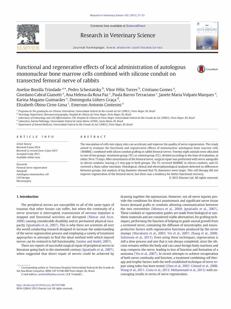
Fig. 7. Scanning electronmicroscopyof the femoral nerve. A) Surface of the femoral nerve of treatment group 75. B) Longitudinal image showing continuity offibers in the central region ofthe nerve of animals of treatment group 75.
32 A.B. Trindade et al. / Research in Veterinary Science 102 (2015) 27–33
Scanning microscopy revealed no differences between the CGanimal and the TG animal in terms of continuity or orientation ofnerve fibers (Fig. 7).
4. Discussion
Regeneration techniques employing tubes are considered viablealternatives to grafting since the latter suffers from certain limitations,including achieving a good fit in terms of both length and diameter ofthe nerve; the need for subsequent surgery to the same patient, withloss of donor nerve function; formation of neuromas and greater post-operative pain (Yin et al., 2007;Mohammadi et al., 2013). In the presentstudy, use of a silicone tube to guide regeneration of rabbit femoralnerves subjected to neurotmesis facilitated union of the sectionednerve stumps, guided nerve fiber growth and acted as a scaffold tomaintain the mononuclear fraction of bone marrow at the injury site.The silicone tube interposed between the stumps provided adequatesupport for regeneration of the femoral nerve of rabbits, as confirmedby analysis of specimens prepared with several different types ofhistological stains.
The marrow aspirate was harvested from the greater tubercle of thehumerus, as described by Grindem et al. (2002), Zago (2006) andColomé et al. (2008), who suggest that this is one possible location forobtaining bone marrow samples. Choice of this site was important toavoid harvesting traumas interfering with animals' claudication assess-ment results.
Although an injured femoral nerve exhibits distinctive clinicalcharacteristics, the neurological clinical assessment of animals involveda certain degree of difficulty because some animals sometimes took along time to respond to the stimuli. This might be explicable by thefact that in the wild these animals have many predators, meaningthey become stressed easily andwill often become agitated or immobileduring clinical examinations, as described by Vernau et al. (2007).
The gait analysis was important to describe evaluations of formsof locomotion such as walking and running. The rabbits' gait wasexamined neurologically on the basis of strength and coordination(Chrisman, 1985). Although this was a subjective analysis, it was ofreal importance because it provided the opportunity to observe andcompare the clinical evolution of the group given cell therapy againstthat of the group treated with tubing only. Additionally, all analyseswere conducted by the same evaluator in order to reduce variations.Since rabbits do not have fused digits, they increase the length of theirstride in order to increase the size of the limb and duration of move-ments, increasing the power with which they can push against theground and the degree to which they extend and flexion the spinalcolumn (Fostowicz-Frelik, 2007). These characteristics justify impreg-nating the plantar region of the pelvic limbswith paint in order to inves-tigate claudication, since, when they push harder against the ground inorder to jump, both of the stained limbs should leave well-defined
marks, which is what was observed with the majority of animals inboth groups, suggesting functional recovery of the nerve. Therefore,the animals who were only treated with silicone guides and thosewho had both the tube procedure and cell therapy exhibited the samequality of gait at all analysis points, according to GEE.
In contrast, although the animals had normal gait, the electrophysi-ological test showed that there had been a reduction in amplitude andan increase in latency, which indicate axonal injury and myelin sheathinjury, respectively, in all of the animals assessed. The explanationthat was found for this results is that neither the velocity of nerve con-ductivity nor the peak motor action potential asses the whole nervefunction, but a fraction of the population of nerve fibers (Kanaya et al.,1996). This explains the lack of a significant correlation between post-operative nerve conductivity velocity and the number of axons regener-ated, since histological, clinical neurological assessment and gaitassessment results all demonstrated nerve regeneration and functionalrecovery. Similar results have been reported by Chen et al. (2007) andSandrini et al. (2007).
A number of different staining techniques were used for the histo-logical analysis, including hematoxylin–eosin (HE), picrosirius, Massontrichrome and periodic acid-Schiff (PAS) and also scanning electronmi-croscopy. No statistically significant differences were observed in any ofthe variables studied histologically.
The hemosiderin observed in HE histological analysis of someanimals signified the presence of recent hemorrhage, caused by the iat-rogenic injury to vessels close to the nerve when the specimens werecollected. Hemosiderin is a pigment that is formed by degradation ofhemoglobin and, like ferritin, it contains iron and can be deposited inexcessive quantities in tissues in a localized or systemic manner. Local-ized deposits are found after hemorrhages, when hemosiderin can beobserved in adjacent macrophages some hours after bleeding begins(Costa-Val et al., 2006).
The finding that control group animals had eosinophils andplasmacytes but treatment group animals did not is the result of thefact that cell therapy promotes faster resolution of the immunoresponse,which facilitates removal of myelin and axonal fragments, favoringproduction of neurotrophic factors,which in turn benefit axonal regener-ation (Colomé et al., 2008). Immunoresponse would therefore have de-clined or ceased in the treated group by the time of assessment.
Animals in the CG and animals in TG exhibited aligned nerve fibersat both assessment times, using PAS staining. This can be explained bythe fact that the silicone tube technique reduces manipulation of thenerve and the quantity of suture material required at the anastomosissite, in addition to providing sufficient support for new regeneration,which makes it more likely that axons will be aligned in the directionof the distal stump (Gibson and Daniloff, 1989; Rodrigues et al., 2012).
The weak presence of collagen type I and type IV observed in CGanimals and TG animals, seen on picrosirius and Masson trichrome-stained specimens, could be considered a beneficial factor, since

33A.B. Trindade et al. / Research in Veterinary Science 102 (2015) 27–33
collagen is needed for formation of the normal extracellular matrix andplays an important role regulating the function of Schwann cells; butafter an injury, collagen production often exceeds the ideal and impedesgrowth of the axon, which delays nerve regeneration (Koopmans et al.,2009).
Macroscopically, all animals exhibited regeneration tissueinterconnecting the nerve stumps and there were no statistical differ-ences between the groups. This can be explained by the fact that surgerywas conducted respecting the principles of microsurgery and neurosur-gery and with the aid of magnification. These results are similar to re-sults reported by Chen et al. (2007), Colomé et al. (2008), Costa et al.(2013) and Mohammadi et al. (2013). In general, histological findingsindicated good nerve regeneration, in agreement with the gait assess-ments of all animals, since at the last assessment all of them had lowclaudication indices.
Several different studies have demonstrated that cell therapy offersadvantages for neuronal regeneration,when comparedwith individualswho did not receive it (Chen et al., 2007; Hu et al., 2007; Costa et al.,2013), suggesting that the mononuclear fraction of bone marrow se-crete biologically active molecules and attracts neurotrophic factors,providing an appropriate environment early on and contributing to anacceleration of nerve regeneration (Chen et al., 2007). This mechanismof action of MSC it is known as paracrine effect (Chen et al., 2008;Linero and Chaparro, 2014). Since the survival and differentiation ofMSC at the site of the lesion is limited, this mechanism starts in thefirst hours after transplantation of cells and, it is proposed that paracrinesignaling is the primary mechanism of their therapeutic effects (Horieet al., 2012; Linero and Chaparro, 2014).
In this study, therewas a tendency for animalswhich underwent celltherapy to exhibit better functional recovery andmaintenance of a largeproportion of the original diameter of the treated limb, which was astatistically significant difference. The advantages of combining celltherapy with the silicon guide tube technique would have been moreevident if a larger number of cases had been evaluated.
5. Conclusions
In this study, local administration of the mononuclear fraction ofautologous bone marrow together with fitting of a silicone conduit didnot improve regeneration of transected femoral nerves of rabbits, butthe limbdiameter of animals given cell therapywas significantly greaterthan in the control group, demonstrating a tendency towards betterfunctional recovery.
Declaration of conflicting interests
The authors declared nopotential conflicts of interestwith respect tothe research, authorship and/or publication of this article.
Acknowledgments
We are grateful to the Hospital de Clínicas de Porto Alegre-Fundo deIncentivo a Pesquisa (www.hcpa.ufrg.br) for providingfinancial supportfor the study and for paying for the translation of this article. The articlewas translated by Robert Coulthard for Scientific Linguagem Ltda.
References
Chen, C.J., Ou, Y.C., Liao, S.L., Chen,W.Y., Chen, S.Y.,Wu, C.W.,Wang, C.C.,Wang,W.Y., Huang,Y.S., Hu, S.H., 2007. Transplantation of bone marrow stromal cells for peripheral nerverepair. Exp. Neurol. 204, 443–453.
Chen, Y., Shao, J.Z., Xiang, L.X., Dong, X.J., Zhang, G.R., 2008. Mesenchymal stem cells: apromising candidate in regenerative medicine. Int. J. Biochem. Cell Biol. 40, 815–820.
Chrisman, C. (Ed.), 1985. Neurologia dos Pequenos Animais. Roca, São Paulo (55-61 pp.).Colomé, L.M., Gomes, C., Crosignani, N., Paz, A.H., Lugo, A.A., Guimarães, K.M., Foerstrow,
L.P., Tessari, J.P., Colomé, L.M., Graça, D.L., Meurer, L., Passos, E.P., Pippi, N.L.,Contesisni, E.A., Cirne-Lima, E.O., 2008. Utilização de células-tronco autólogas demedula óssea na regeneração do nervo tibial de coelhos mediante técnica detubulização com prótese de silicone. Ciên. Rural 38, 2529–2534.
Costa, H.J.Z.R., Costa, Z.R., Bento, R.F., Salomone, R., Azzi-Nogueira, D., Zanatta, D.B., Costa,M.P., Silva, C.F., Strauss, B.E., Haddad, L.A., 2013. Mesenchymal bonemarrow stemcellswithin polyglycolic acid tube in vivo after six weeks enhance facial nerve regeneration.Brain Res. 1510, 10–21.
Costa-Val, R., Nunes, T.A., Oliveira e Silva, R.C., Souza, T.K.P., 2006. Efeito da oxigenoterapiahiperbárica em ratos submetidos à ligadura das veias hepáticas: avaliação damortalidade e da histologia do fígado e baço. Acta Cir. Bras. 21, 51–56.
Fostowicz−Frelik, L., 2007. The hind limb skeleton and cursorial adaptations of the Plio-Pleistocene rabbit Hypolagus beremendensis. Acta Palaeontol. Pol. 52, 447–476.
Gibson, K.L., Daniloff, J.K., 1989. Peripheral nerve repair. Comp. Cont. Educ. Pract. Vet. 11,938–944.
Grindem, C.B., Neel, J.A., Juopperi, T.A., 2002. Cytology of bone marrow. Vet. Clin. N. Am.Small Anim. Pract. 32, 1313–1374.
Horie, M., Choi, H., Lee, R.H., Reger, R.L., Ylostalo, J., et al., 2012. Intra-articular injection ofhuman mesenchymal stem cells (MSCs) promote rat meniscal regeneration by beingactivated to express Indian hedgehog that enhances expression of type II collagen.Osteoarthr. Cartil. 20, 1197–1207.
Hu, J., Zhu, Q.T., Liu, X.L., Xu, Y.B., Zhu, J.K., 2007. Repair of extended peripheral nervelesions in rhesus monkeys using acellular allogenic nerve grafts implanted withautologous mesenchymal stem cells. Exp. Neurol. 204, 658–666.
Ignatiadis, I.A., Yiannakopoulos, C.K., Barbitsioti, A.D., Avram, A.M., Patralexis, H.G.,Tsolakis, C.K., Papalois, A.E., Xenakis, T.H., Beris, A.E., Soucacos, P.N., 2007. Diversetypes of epineural conduits for bridging short nerve defects. An experimental studyin the rabbit. Microsurgery 27, 98–104.
Kanaya, F., Firrell, J.C., Breidenbach, W.C., 1996. Sciatic function index, nerve conductiontests, muscle contraction and axon morphometry as indicators of regeneration.Plast. Reconstr. Surg. 98 (1264), 1996.
Koopmans, G., Hasse, B., Sinis, N., 2009. The role of collagen in peripheral nerve repair. Int.Rev. Neurobiol. 87, 363–379.
Linero, I., Chaparro, O., 2014. Paracrine effect of mesenchymal stem cells derived fromhuman adipose tissue in bone regeneration. PLoS ONE 9, e107001. http://dx.doi.org/10.1371/journal.pone.0107001.
Mattar Jr., R., Azze, R.J. (Eds.), 2008. Lesões dos nervos periféricos- Atualização emtraumatologia do aparelho locomotor. Faculdade de Medicina da Universidade deSão Paulo, São Paulo, pp. 03–27.
Mimura, T., Dezawa, M., Kanno, H., Sawada, H., Yamamoto, I., 2004. Peripheral nerveregeneration by transplantation of bone marrow stromal cell-derived Schwanncells in adult rats. J. Neurosurg. 101, 806–812.
Mohammadi, R., Amini, K., Yousefi, A., Abdollabi-Pirbazari, M., Belbasi, A., Abedi, F., 2013.Functional effects of local administration of thyroid hormone combinedwith chitosanconduit after sciatic nerve transection in rats. J. Oral Maxillofac. Surg. 71, 1763–1776.
Murakami, T., Fujimoto, Y., Yasunaga, Y., Ishida, O., Tanaka, N., Ikuta, Y., Oshi, M., 2003.Transplanted neuronal progenitor cells in a peripheral nerve gap promote nerverepair. Brain Res. 974, 17–24.
Rodrigues, M.C.O., Rodrigues, A.A., Glover, L.E., Voltareli, J., Borlongan, C.V., 2012. Periph-eral nerve repair with cultured Schwann cells: getting closer to the clinics. Sci. WorldJ. 2012, 1–11.
Salomone, R., Bento, R.F., Costa, H.J.Z.R., Azzi-Nogueira, D., Ovando, P.C., Da-Silva, C.F.,Zanatta, D.B., Strauss, B.E., Haddad, L.A., 2013. Bone marrow stem cells in the facialnerve regeneration from isolated stumps. Muscle Nerve 48, 423–429.
Sandrini, F.A.L., Pereira-Júnior, E.D., Gay-Escoda, C., 2007. Anastomose do nervo facial decoelhos com cola de fibrina: estudo da velocidade de condução nervosa. Rev. Bras.Otorrinolaringol. 73, 196–201.
Santos, T.S., André, E.S., 2007. Avaliação funcional da marcha do rato após estimulaçãoelétrica do músculo gastrocnêmio desnervado. Rev. Neuroci. 15, 120–124.
Vernau, K., Osofsky, A., LeCouteur, R.A., 2007. The neurological examination and lesionlocalization in the companion rabbit (Oryctolagus cuniculus). Vet. Clin. N. Am. Exot.Anim. Pract. 10, 731–758.
Wang, X., Luo, E., Li, Y., Hu, J., 2011. Schwann-like mesenchymal stem cells within veingraft facilitate facial nerve regeneration and remyelination. Brain Res. 1383, 71–80.
Yin, D., Wang, X., Yan, Y., Zhang, J., 2007. Preliminary studies on peripheral nerve regen-eration using a new polyurethane conduit. J. Bioact. Compat. Polym. 22, 143–159.
Zago, M.A., 2006. Células-tronco: origens e propriedades. In: Zago, M.A., Covas, D.T. (Eds.),Células-tronco: a nova fronteira da medicina. Atheneu, São Paulo, pp. 3–20.
Zhang, H., Wei, Y.T., Tsang, K.S., Sun, C.R., Huang, J.L.H., Cui, F.Z., An, Y.H., 2008. Implanta-tion of neural stem cells embedded in hyaluronic acid and collagen compositeconduit promotes regeneration in a rabbit facial nerve injury model. J. Transl. Med.6, 1–11.