research articleglobalresearchonline.net/journalcontents/v52-1/23.pdf · size, extent of spread and...
TRANSCRIPT

Int. J. Pharm. Sci. Rev. Res., 52(1), September - October 2018; Article No. 23, Pages: 125-132 ISSN 0976 – 044X
International Journal of Pharmaceutical Sciences Review and Research . International Journal of Pharmaceutical Sciences Review and Research Available online at www.globalresearchonline.net
© Copyright protected. Unauthorised republication, reproduction, distribution, dissemination and copying of this document in whole or in part is strictly prohibited.
.
. Available online at www.globalresearchonline.net
125
Zeinab M. Klaab1, Faizah A. AlMalki1, Aziza M. Hassan**1&2
1Biotechnology Department, Faculty of Science, Taif University, Taif, K.S.A. 2 Cell Biology Department, National Research Centre, 33 Bohouth St., Dokki, Giza, Egypt.
*Corresponding author’s E-mail: [email protected].
Received: 10-08-2018; Revised: 27-08-2018; Accepted: 08-09-2018.
ABSTRACT
Ziziphus jujuba (Z. jujuba) represent a conventional plant that human use from long time ago for feeding and treating a large variety of diseases including anti-inflammation, anticancer and antioxidant, due to its bioactive gradients. Hormonal chemotherapy, such as Tamoxifine (Tam) and its derivatives, is widely used in cancer treatment. It is an effective treatment for human breast cancers; however, the drug resistance problem considers being a major obstacle to responding the treatment successfully. The excellent understanding of the mechanisms and strategies for overcoming drug resistance can highly progress therapeutic outcomes for patients. The present study focused on studying the anticancer and apoptotic activity of Z. jujuba in breast cancer cells as well as its impact on chemoresistance in vitro through MTT assay, DNA fragmentation and mRNA expression level of apoptotic genes. The results demonstrated that Z. jujuba extract exert a time-dependent cytotoxic action against breast cancer cells represented in significantly reduced viability and high potent inhibitory activity toward the proliferation of MCF-7 cells. Also, the DNA fragmentation indicated that apoptosis induced. It is noteworthy that cytotoxicity of Tamoxifine combined with Z. jujuba, at the tested doses, is remarkably higher compared to treatment with Tamoxifen alone. We expect that these studies can lead up to a hopeful future for cancer patients.
Keywords: Ziziphus Extract, Breast Cancer Cells, Tamoxifen, in vitro.
INTRODUCTION
ancer continues to be a serious health problem and one of the leading causes of death in the world.1 Breast cancer (BC) is both the most common
diseases malignancies of cancer-related deaths and the most frequently diagnosed type of cancer (~25% of all cancers) worldwide among women2. Its occurrence around the world is predicted to increase to 2.3 million by 2030 3. In 2009, about 1.308 new BC cases were reported in the Kingdom of Saudi Arabia (KSA) and it was the ninth leading cause of death for females in 2010
4-5. According
to Saudi Cancer Registry, BC patient was 25% from all cancer cases registered with expectation for increasing incidence in the KSA which lined with population`s growth and aging
6-7.
Breast cancer treatment strategies are largely dependent on patient preference and tumor characteristics including size, extent of spread and cancer cell classification.8 Treatment usually involves surgery, radiation therapy (before or after surgery), hormonal therapy, chemotherapy, or targeted therapy. Targeted therapeutic approaches are the induction of apoptosis or inhibition of several processes in the cell such as anti- apoptosis, cell cycle progression, signal transduction and angiogenesis 9-
10.
BC can be controlled by using hormonal treatments like Tamoxifen (Tam). Tam has the FDA approval for BC prevention and treatment. It is a selective estrogen receptor (ER) modulator and considered as the first targeted cancer therapy. Additionally, Tam is a usual used
drug in routine clinical practice and it represents an excellent level of treatment for ER+ and breast tumors 11. On the bases of this result, Tam bound ER complex prevents the genes from being switched on by estrogen, leading to inhibition of estrogenic effects which are responsible for cancer cell growth or proliferation so, it is known as antiestrogen 12. Although it is an effective treatment it has back draw effects such as development of recurrent of tumors, 40% of patients eventually relapse and resistance to the drug over time 13.
Different clinical methods, like as complementary and alternative medicines, have been evaluated in addition to chemotherapy, radiation, and surgery in treating cancer. In recent years, researchers draw their attention to the natural compounds included in plants beneficial effects against a variety of diseases for their projected low costs, fewer side effects, and low toxicity in respect to the standard chemotherapies treatment. The development of new agents such as medicinal herbs with anticancer effects can herald a promising future in cancer treatment14–15. The practice of application of medicinal plants for relief from many illnesses dates back to ancient times
16.
Plants have many phenolic compounds, flavonoids, lignans, and phytosterols that have beneficial effects. Studies show that these compounds can inhibit local estrogen synthesis and promote epigenetic changes.
17
According to the most recent studies, phytoestrogens have demonstrated positive effects on BC prognosis, through receptor binding antagonism; it potentially
Anticancer Activities of Ziziphus Jujuba Extract on Breast Cancer Cells in vitro
C
Research Article

Int. J. Pharm. Sci. Rev. Res., 52(1), September - October 2018; Article No. 23, Pages: 125-132 ISSN 0976 – 044X
International Journal of Pharmaceutical Sciences Review and Research . International Journal of Pharmaceutical Sciences Review and Research Available online at www.globalresearchonline.net
© Copyright protected. Unauthorised republication, reproduction, distribution, dissemination and copying of this document in whole or in part is strictly prohibited.
.
. Available online at www.globalresearchonline.net
126
inhibits BC and work synergistically with the chemotherapeutic drugs.18
Many foods and plants in Arabic region have described as phytoestrogens sources including: nuts, oilseeds, soy contents and jujuba plant. The last one (jujuba) has been known and used from long past decades for nutrition and the treatment of a broad spectrum of diseases such as human cancers. Ziziphus jujuba (family: Rhamnaceae) is a herbal plant used in traditional medicine. It can be used for the curing of several health problems concerning with skin, liver, urinary disorders, obesity, anemia, cancer, and also for blood purification 19. The mature jujuba fruit has great beneficial bioactive compounds with anticancer activity 20. The Z. jujuba fruit contain different compounds including: triterpenic acids, flavonoids, cerebrosides, phenolic acids, α-tocopherol, β-carotene, and unsaturated fatty acids
21. In addition, it has more total
phenolic compounds that exhibit antioxidant activities 22.
The triterpenic acids, present in the extract, are in the form of free acids or glycones such as saponins, with multiple medical and biological effects including anti-inflammatory, anti-microbial, antioxidant effects 23, hepatoprotective 20, and inhibit of P-glycoprotein (Pgp) in cancer cells 24. In recent years, triterpenic acids became attractive products for scientists and health care researchers due to their anticancer and antitumor activities. 25-26
This study aims to investigate the antiprolifiration and apoptosis-inducing effects of Z. jujuba extract and Tam, each individually and in combination, on human breast cancer cells MCF7 and explore of Z. jujuba extract impact on chemoresistance in vitro.
MATERIALS AND METHODS
Materials and reagents
Dulbecco‘s Modified Eagle Medium (DMEM media), Penicillin-Streptomycin Solution (10.000 Units/ml Penicillin, 10.000 µ/ml Streptomycin) and Fetal Bovine serum (FBS) were obtained from Gibco by life technologies, (USA), Trizol reagent (Ambion RNA by life technology, USA). Gene expression Master Mix, (FIREPol®) and MTT were obtained from Bio Basic INC, (Canada). All other chemicals used were of a high grade.
Cell Line
The breast cancer cells with a positive estrogen receptor (MCF-7) cell line were provided by Dr. Thamer Ahmed F. Bouback from Cell Culture Laboratory, Department of Biology, Faculty of Science, King Abdul-Aziz University, Saudi Arabia which was originally obtained from American Type Culture Collection (ATCC).
Ziziphus jujuba Extract
Ziziphus jujuba extract capsules *500 mg) of pure jujuba fruit extract without any additives (GMP, FDA, REGSTERED FACITY. USA) and was prepared freshly just before treatment.
Tamoxifen Drug
Pure Tamoxifen (Tam) tablets (EBEWE Pharma®- UK) provided from King Abdul-Aziz Specialist hospital, Taif, KSA. The tablet was dissolved in absolute ethanol to make a stock solution of 10 mg/ml.
Experimental Design and Groups
To investigate the effects of Z. jujuba extract and Tam, each individually and in combination, on MCF-7 cells in vitro, the MCF-7 cells were cultured and plated at 1× 10
6
cells/well in 6-well plates in DMEM medium containing 10% fetal bovine serum (FBS), 1% penicillin-streptomycin and were allowed to attach overnight at 37 ºC in a humidified atmosphere containing 5% CO2
27. After 24 h in basal medium, these cells were treated on day 0 with medium containing different treatments and then further incubated for 24, 48 and 72h. The experimental groups were: (1) NC: Untreated MCF-7 cells, (2) Tam-10: MCF-7 treated with Tam at a dose of 10 µg/ml (effective dose)
28,
(3) Tam-1: MCF-7 treated with Tam at a dose of 1 µg/ml , (4) Z-Group: MCF-7 treated with Z. jujuba extract at a concentration of 1 mg/ml which represent the IC50
29, (5)
Tam-1& Z-Group: MCF-7 treated as groups 3 and 4, (6) Tam-10& Z-Group: MCF-7 treated as groups 2 and 4.
Methods
Cell Proliferation Assay
The cells viability and the cytotoxic effects of Tam and Z. jujuba extract on the MCF-7 cells were determined using the 3-(4,5-dimethylthiazol- 2-yl)2,5- diphenyl tetrazolium (MTT) assay 30-31. The cells were seeded onto the 96-well plates and different treatments of Z. jujuba extract and Tam each individually and in combination were added to the cells. After incubation at different time courses (24, 48 and 72h), the medium was removed, and 40 μl MTT solution (5mg/ml) was added to each well for 2-4 h. The supernatant was removed and 140 μl of dimethyl sulfoxide (DMSO) was added to the wells to dissolve any precipitate. The absorbance was measured at wavelength of 540 nm using a plate reader (MR-96A).
DNA Fragmentation for Apoptosis
The detection of DNA fragmentation is used as a biochemical marker for the measurement of apoptosis. The internucleosomal DNA fragmentation was assessed by electrophoresis of extracted genomic DNA from MCF-7 cells as described by the protocol of BioVision‘s Quick Apoptotic DNA Ladder Detection Kit (Bio-Vision, Life Science SOURCE ™, USA). The DNA pellet was dissolved in 30 µl DNA suspension buffer and electrophoresed in 1.2% agarose gel containing 0.5 µg/ml ethidium bromide. The gel was run for 1-2 h DNA samples visualized by UV light and photographed. DNA Fragmentation percentages were evaluated colorimetrically using Diphenylamine (DPA), as described by Burton, 32 and modified by Perandones et al., 33
. The colorimetric reaction was then measured spectrophotometrically at 575 nm.
Int. J. Pharm. Sci. Rev. Res., 52(1), September - October 2018; Article No. 23, Pages: 125-132 ISSN 0976 – 044X
International Journal of Pharmaceutical Sciences Review and Research . International Journal of Pharmaceutical Sciences Review and Research Available online at www.globalresearchonline.net
© Copyright protected. Unauthorised republication, reproduction, distribution, dissemination and copying of this document in whole or in part is strictly prohibited.
.
. Available online at www.globalresearchonline.net
127
Gene Expression Analysis
The total RNA isolation was carried out with the TRIzole reagent. The extracted RNA was immediately used in RT-PCR to generate first-strand cDNA. The first-strand cDNA synthesis was performed using the reverse transcription kit (Maxime RT PreMix) obtained from iNtRON Biotechnology, (Korea). RT reactions were carried out using 2 μg RNA, subjected previously to DNase digestion, and a reverse transcription reagents, followed by PCR. The PCR amplification reactions were carried out in a thermal cycler (PXE 0.5 Thermo). The PCR product was run on a 1.5 % agarose gel in Tris-Borate-EDTA buffer and visualized on a UV Transillumenator. The ethidium bromide-stained gel bands were scanned and the signal intensities were quantified by Gel-Pro software (version 3.1 for Windows 3). The ratio between the levels of the target genes-amplification product and GAPDH was calculated to normalize for initial variation in sample concentration as a control for reaction efficiency
34. Gene
expression was analyzed using the following pairs of primers,
35 listed in Table 1.
Table 1: The sequences of oligonucleotide primers used to amplify the studied genes
Gene Forward primer
sequences
Reverse primer
sequences
GAPDH
5-
GGTGCCGGTTCAG
GTACTCAGTCA-3
5-
TTGTGGCCTTCTTTG
AGTTCGGTG-3
Bcl-2
5-
TTGTGGCCTTCTTT
GAGTTCGGTG-3
5-
GGTGCCGGTTCAGG
TACTCAGTCA-3
Bax
5-
AGGACAGGCACA
AACACGCACC-3
5-
TAACAGTTCCTGCAT
GGGCGGC-3
Statistical Analysis
All the results were statistically analyzed using the SPSS.11 program. The significance of the differences
among treatment groups was determined by (One Way – ANOVA) followed by Duncan's multiple test 36-37. The values were expressed as Mean ± SE. The data were considered to be statistically significant if the probability had a value of P < 0.05.
RESULTS
Effects of Z. jujuba extract and Tamoxifen on Proliferation Percentage of MCF-7Cells
The effect of Z. jujuba extract on cell survival was determined in order to investigate the antiproliferative effect of the extract and Tam, each individually or in combination on MCF-7 cell line. Cells were cultured and treated with the extract at selected dose, which represent the IC50, (1 mg/ml) while Tam drug was used at either the effective dose (10 µg/ml) or at 1/10th that dose (1µg/ml) for 24, 48 and 72 h then cell assessed for count, morphology and viability by Trypan blue staining and MTT assay. The results summarized in Tables 2 & 3 and Figure 1. As shown, the jujuba extract exhibited significant anti-proliferative effect on cell viability of MCF-7 (P <0.05) in a time-dependent manner when compared with control MCF-7 untreated cells. However the growth inhibitory effect was more prominent against MCF-7 cells when the jujuba extract combined with the Tam 10 (10 µg/ml) at about 31.2% vs. 29.2%, 48.0 % vs. 42.74 % and 67 % vs. 52.7 after treatment for 24, 48 and 72 h, respectively. Interestingly, Tam alone, at the effective dose (10 µg/ml), inhibited the proliferation of MCF-7 cells by only 25.3 %, 40.4 % and 58.1 % after 24, 48 and 72 h, respectively as represented in Figure (1). However, the maximum inhibition in the proliferation percent was shown after 72h (Figure 1).
Table 2: The Effects of Tamoxifen and Z. jujuba extract Treatment on The proliferative Percentage of MCF-7 Cells at different times
Treatments Groups Proliferative Percentage of Treated MCF-7 cells
After 24 h After 48 h After 72 h
Untreated Cells 95.3 ± 0.75a 97.54 ± 1.38
a 98.2 ± 0.69
a
Z .jujuba 66.10 ± 1.15d 54.80 ± 0.46 cd 45.50 ± 1.44 d
Tam 1 81.60 ± 0.92b 62.90 ± 1.67 b 57.80 ± 1.04 b
Tam 10 70.00 ± 0.87c 57.10 ± 0.64 c 40.10 ± 0.57 e
Z. jujuba +Tam 1 79.80 ± 1.04b 55.30 ± 1.90 cd 50.90 ± 1.09 c
Z. jujuba + Tam 10 64.10 ± 0.63d 49.50 ± 0.87 e 31.20 ± 0.69 fg
Means with different superscripts (a, b, c, d, e, f and g) between groups in the same column are significantly different at P< 0.05. Cell numbers were counted and data are expressed as the percentage of untreated control. Tam 1 or Tam 10: Tamoxifen at 1 or 10 µg/ml.

Int. J. Pharm. Sci. Rev. Res., 52(1), September - October 2018; Article No. 23, Pages: 125-132 ISSN 0976 – 044X
International Journal of Pharmaceutical Sciences Review and Research . International Journal of Pharmaceutical Sciences Review and Research Available online at www.globalresearchonline.net
© Copyright protected. Unauthorised republication, reproduction, distribution, dissemination and copying of this document in whole or in part is strictly prohibited.
.
. Available online at www.globalresearchonline.net
128
Figure 1: Effects of Tamoxifen and Z. jujuba extract Treatment on Growth Inhibition Percentage of MCF-7 Cells for 24, 48 and 72 hours. Results were obtained from three independent experiments and expressed as Mean ± SEM; Tam: Tamoxifen
Table 3: Effects of Tamoxifen and Z. Jujuba Treatment on the Viability Percentage of MCF-7 Cells at different times.
Treatments Groups Viability Percentage of Treated MCF-7cells
After 24 h After 48 h After 72h
Untreated Cells 91.8 ± 1.04a 95.23 ± 1.73a 96.6 ± 0.92 a
Z .jujuba 80.8 ± 1.17c 57.9 ± 1.09 c 48.90 ± 0.63 c
Tam 1 88.12 ± 0.95 ab 80.42 ± 0.81 b 70.32 ± 1.32 b
Tam 10 76.20 ± 0.69 cd 51.83 ± 1.03 d 40.95 ± 0.63 d
Z. jujuba +Tam 1 78.10 ± 1.73cd 56.30 ± 1.32 c 40.58 ± 0.80 d
Z. jujuba + Tam 10 70.02 ± 1.44e 45.76 ± 1.00 f 36.56 ± 0.86 e
Means with different superscripts (a, b, c, d, e, and f) between groups in the same column are significantly different at P< 0.05. Cell numbers were counted and data are expressed as the percentage of untreated control. Tam 1 or Tam 10: Tamoxifen at 1 or 10 µg/ml.
Effects of Z. jujuba extract and Tamoxifen on morphological changes in MCF-7 Cells
To characterize the cell death induced by jujuba extract, the morphology of the cells was examined under the microscope. The data depicted in Figure (2) indicated that treatment of MCF-7 cells with jujuba extract and/or Tam resulted in obvious morphological changes presented by cell shrinkage, blebbing and cellular
detachment compared to the normal attached MCF-7 cells as shown in Figures (2). When MCF-7 cells were treated with Tam at a dose of 10 µg/ml plus jujuba extract for 72 h, a significant cellular detachment and decreased in the cellular crowding was observed in the MCF-7 cells and they were shrunken and fewer in number indicating a sever cytotoxic effects.
Figure (2): Inverted microscopic photographs of MCF-7 Cells untreated (a) and treated with Tam10 (b); Z. jujuba; (c) and with Tam 10 & Z. jujuba after 48 h Arrows indicate morphologically apoptotic changes, including condensed and fragmented nuclei, and apoptotic bodies.
Effects of Z. jujuba extract and Tamoxifen on the DNA fragmentation percentage induced in MCF-7 cells
The apoptotic effect of jujuba extract by DNA fragmentation assay was further evaluated by measuring
the level of fragmented DNA colorimetrically using Diphenylamine (DPA), the data presented in Figure (3) and by comparing DNA profiles on agarose gel electrophoresis as the migration of fragmented DNA was observed Figure (4).
Int. J. Pharm. Sci. Rev. Res., 52(1), September - October 2018; Article No. 23, Pages: 125-132 ISSN 0976 – 044X
International Journal of Pharmaceutical Sciences Review and Research . International Journal of Pharmaceutical Sciences Review and Research Available online at www.globalresearchonline.net
© Copyright protected. Unauthorised republication, reproduction, distribution, dissemination and copying of this document in whole or in part is strictly prohibited.
.
. Available online at www.globalresearchonline.net
129
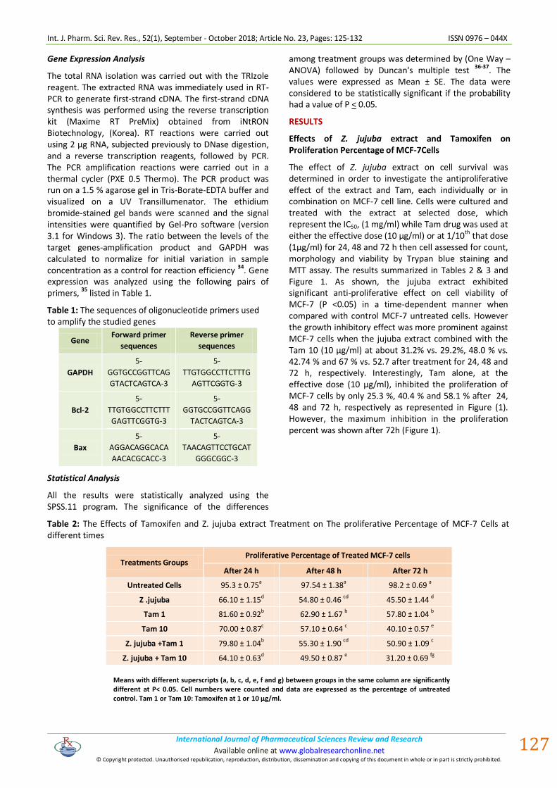
Figure 3: The effects of Z. jujuba and Tamoxifen 10 treatments on DNA fragmentation % in MCF-7 cells after 48h. Results were obtained from three independent experiments and expressed as Mean ± SEM
The results illustrated that DNA fragmentation was clearly detected in MCF-7 cells at all treated groups. However, the level of DNA fragmentation became more prominent with the combination of jujuba extract and Tam (10 µg/ml) as shown in Figures (3 & 4).
Figure (4): Agarose gel electrophoresis showing DNA fragmentation induced inMCF-7 cells treated with different treatment at the indicated doses after 48 h: Lane 1 (Cont) control, Lane 2 (Z) effect of Z. jujuba; Lane 3 (Tam1) effect of Tamoxifen 1, Lane 4 (Tam10) effect of Tamoxifen 10, Lane 5 (Z+T1) effect of jujuba and Tam1; Lane 6 (Jujuba+Tam10) effect of Ziziphus and Tamoxifen10.
These observations further supported the apoptotic changes appearing in the microscopic results as mentioned above. The low molecular DNA fragments could be easily detected on the lanes of the combined
treatments and this appearance is a typical characteristic of apoptosis.
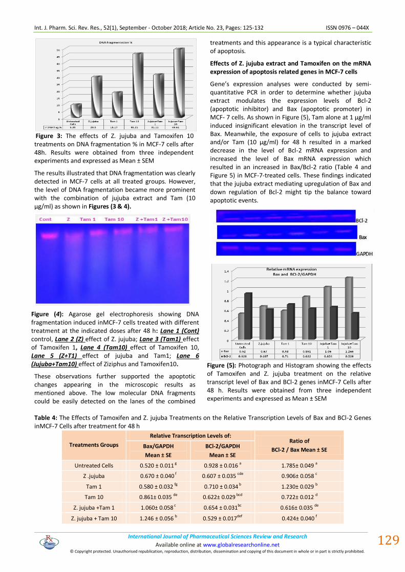
Effects of Z. jujuba extract and Tamoxifen on the mRNA expression of apoptosis related genes in MCF-7 cells
Gene’s expression analyses were conducted by semi-quantitative PCR in order to determine whether jujuba extract modulates the expression levels of Bcl-2 (apoptotic inhibitor) and Bax (apoptotic promoter) in MCF- 7 cells. As shown in Figure (5), Tam alone at 1 µg/ml induced insignificant elevation in the transcript level of Bax. Meanwhile, the exposure of cells to jujuba extract and/or Tam (10 µg/ml) for 48 h resulted in a marked decrease in the level of Bcl-2 mRNA expression and increased the level of Bax mRNA expression which resulted in an increased in Bax/Bcl-2 ratio (Table 4 and Figure 5) in MCF-7-treated cells. These findings indicated that the jujuba extract mediating upregulation of Bax and down regulation of Bcl-2 might tip the balance toward apoptotic events.
Figure (5): Photograph and Histogram showing the effects of Tamoxifen and Z. jujuba treatment on the relative transcript level of Bax and BCl-2 genes inMCF-7 Cells after 48 h. Results were obtained from three independent experiments and expressed as Mean ± SEM
Table 4: The Effects of Tamoxifen and Z. jujuba Treatments on the Relative Transcription Levels of Bax and BCl-2 Genes inMCF-7 Cells after treatment for 48 h
Treatments Groups
Relative Transcription Levels of: Ratio of
BCl-2 / Bax Mean ± SE Bax/GAPDH
Mean ± SE
BCl-2/GAPDH
Mean ± SE
Untreated Cells 0.520 ± 0.011 g 0.928 ± 0.016 a 1.785± 0.049 a
Z .jujuba 0.670 ± 0.040 f 0.607 ± 0.035 cde 0.906± 0.058 c
Tam 1 0.580 ± 0.032 fg 0.710 ± 0.034 b 1.230± 0.029 b
Tam 10 0.861± 0.035 de 0.622± 0.029 bcd 0.722± 0.012 d
Z. jujuba +Tam 1 1.060± 0.058 c 0.654 ± 0.031bc 0.616± 0.035 de
Z. jujuba + Tam 10 1.246 ± 0.056 b 0.529 ± 0.017def 0.424± 0.040 f

Int. J. Pharm. Sci. Rev. Res., 52(1), September - October 2018; Article No. 23, Pages: 125-132 ISSN 0976 – 044X
International Journal of Pharmaceutical Sciences Review and Research . International Journal of Pharmaceutical Sciences Review and Research Available online at www.globalresearchonline.net
© Copyright protected. Unauthorised republication, reproduction, distribution, dissemination and copying of this document in whole or in part is strictly prohibited.
.
. Available online at www.globalresearchonline.net
130
DISCUSSION
Chemotherapy considers the standard medical treatment for cancer. However, the resistance of cancer cells to nearly all kinds of anticancer and chemotherapeutic drugs has become predominant, and approximately equal 80-90% of deaths in cancer patients are indirectly or directly attributed to drug resistance
38. On the other
hand, the serious side effects of chemotherapy and radiation therapy have made many cancer patients seek alternative complementary methods of treatment 39. The combination treatment is one of the developmental trends for increasing the efficacy and/or minimizing these side effects 40. Fortunately, natural products with diverse chemical structures and pharmacological anticancer effects serve as effective substances against drug resistance. Therefore, a new natural source with anticancer activities would aid in finding new strategy for cancer therapy. In the present research, the anticancer effect of Z. jujuba extract on breast cancer cell was investigated as well as its effect against drug resistance was evaluated in vitro.
In the present work, the results illustrated that the conventional drug Tamoxifen at a dose of 10 µg / ml inhibited the proliferation of MCF-7 treated cells, reduced the percentage of viable cells and significantly induced DNA fragmentation followed by up-regulation in the expression levels of apoptotic genes. However, treatment MCF-7 cells with Tam at 1µg / ml resulted insignificant changes were observed. In these concerns, Ichikawa et al. 28 reported that treatment with Tam inhibited the proliferation of MCF-7 cells in a time- and dose-dependent manner which confirmed the present results. Also, our data were similar to those reported in the earlier experimental reports 41-42. Consistently with these results, Mandlekar et al. 43 and Kim et al. 44
demonstrated that Tam induces cytotoxicity and apoptosis in MCF-7 cells. Moreover, the combination of Tam and Z. jujuba significantly decreased cellular proliferation and reduced the viability percentage of MCF-7 cells at about 50 % and 60% after treatment for 48 h and 72 h, respectively; indicating severe cytotoxic effects in MCF-7 treated cells. Also, the combined treatment was significantly induced apoptosis and increased the percentage of DNA fragmentation in the treated cells to about 31.1 % vs 29.5 and 18.7 in the MCF-7 cells treated with Z. jujuba or Tam 1, respectively. In addition, a significant up-regulating in the expression levels of apoptotic genes followed Z. jujuba along with Tam 1 and Tam 10 treatments.
Z. jujuba is one of the most valuable herbs with terrific medicinal ingredients and demonstrated a significant inhibitory effect on various cancer cells’ proliferation 45-
47. The present study showed that Z. jujuba extract at 1 mg/ml, significantly reduced the proliferation and viability of MCF-7 treated cells, either alone or in combination treatment in a time dependant manner. In these concerns, Plastina et al. 47 reported the cytotoxic
effects of Ziziphus fruit extracts on breast cancer cells based on the MTT assay. Also, Huang et al. 48 reported that this fruit has shown anticancer activity on human liver cancer cells that are highly resistant to chemotherapy drugs. Vahedi et al. showed that Z. jujuba extract induced morphological changes, including cell shrinkage and detachment in the HeLa and Hep-2 cells.
46
According to inverted microscopic studies, we demonstrated that aqueous extract of Z. jujuba fruit caused some cell morphological variation, such as changes in cell adhesion to the surface of the plate, decreased relative size with round shape and increased internal complexity in the treated cells after 48 h. A typical apoptotic appearance on MCF-7 cells was observed in the treated cells after 72 h.
Numerous studies have suggested that herbs exert potent anticarcinogenic effects due to their ability to induce apoptosis and the combination chemotherapy is a more efficient method in the treatment of cancer, compared to single agent treatment 49-50. The current study indicated a considerable dose reduction when both Z. jujuba and Tam were used along with each other. While Tam alone decreased MCF7 cell growth, adding Ziziphus extract, at a dose of 1 mg/ml to Tam at a dose of 1 µg/ml resulted in a remarkable inhibition reach to the inhibitory effects of Tam treatment alone at 10 µg/ml. This is a significant observation since drug resistance is the main problem for cancer chemotherapy and worldwide search for new drug with minimal toxicity is on the way. This combined effect is probably because of the combination of the drug and Ziziphus extract with different mechanisms of action, proposing that this combination could act in a multifactorial pathway
51. In
addition, the different metabolic responses between the extracts and commercial drugs (Tam) might in part, contribute to the synergistic effect of these two agents and minimize Tam side effects by reducing the dosage.
Bcl-2 family plays central roles in apoptotic events; this family includes anti-apoptotic members (such as Bcl-2) and proapoptotic members (such as Bax). In the present study we used DNA fragmentation and expression analysis for the apoptotic regulating genes to ascertain the molecular mechanism of apoptosis resulted in MCF-7 treated cells. When MCF-7 cells were exposed to Z. jujuba extracts, at a dose of 1 mg/ml, for 48 h, resulted in a significant increased in Bax mRNA expression level, a response associated with a significant down regulation in the BCl2 expression level, lead to significant reduction in the BCl2/Bax ration. Our finding was consistent with Hoshyar et al. 29, who reported that Z. jujuba extract had a cytotoxic effect and produced apoptosis on breast cancer cells in rats. Due to the fact that tumor progression is closely related to inflammation and oxidative stress, compounds that have antioxidant activity, such as Z. jujuba, can be anti-carcinogens and these confirmed the present results.

Int. J. Pharm. Sci. Rev. Res., 52(1), September - October 2018; Article No. 23, Pages: 125-132 ISSN 0976 – 044X
International Journal of Pharmaceutical Sciences Review and Research . International Journal of Pharmaceutical Sciences Review and Research Available online at www.globalresearchonline.net
© Copyright protected. Unauthorised republication, reproduction, distribution, dissemination and copying of this document in whole or in part is strictly prohibited.
.
. Available online at www.globalresearchonline.net
131
CONCLUSION
The present results mean that the Z. jujuba could have a potential anticancer activity against breast cancer, in vitro and the Tamoxifen drug, at 1 µg/ml, when combined with Z. jujuba extract, at indicated dose, was more sensitive and its effects was comparable to the therapeutic dose (10 µg/ml) . These results open a new avenue for the treatment of breast cancer, but also providing a basis for further research.
REFERENCES
1. Rebecca LS, Kimberly DM and Ahmedin J. Cancer Statistics, CA CANCER J CLIN. 2018, 68:7–30. doi: 10.3322/caac.2144
2. Mafu TS, September AV and Shamley D. The potential role of angiogenesis in the development of shoulder pain, shoulder dysfunction, and lymphedema after breast cancer treatment. Cancer management and research, 10, 2018, 81–90. Doi: 10.2147/CMAR.S151714; PMID: 29391829
3. Alanazi IO and Khan Z. Understanding EGFR Signaling in Breast Cancer and Breast Cancer Stem Cells: Overexpression and Therapeutic Implications. Asian Pacific journal of cancer prevention, 17, 2016, 445-453. Doi: http://dx.doi.org/10.7314/APJCP.2016.17.2.445; PMID: 26925626.
4. Lozano R, Naghavi M, Foreman K, Lim S, Shibuya K and Aboyans K. Global and regional mortality from 235 causes of death for 20 age groups in 1990 and 2010 . The Lancet, 380, 2012, 2095–2128. Doi: 10.1016/S0140-6736(12)61728-0; PMID: 23245604.
5. Bcheraoui CE, Basulaiman M, Wilson S, Daoud F, Tuffaha M, AlMazroa M, Memish Z, Al Saeedi M and Mokdad AH. Breast Cancer Screening in Saudi Arabia: Free but Almost No Takers. PLoS ONE 10(3), 2015 e0119051. doi:10.1371/journal.pone.0119051; PMID: 25774520
6. Saudi Cancer Registry. (SCR). Kingdom of Saudi Arabia, Saudi Health Council. Cancer Incidence Report in 2013. ISSN: 2013, 1658-0559.
7. Bazarbashi S, Al Eid H and Minguet J. Cancer Incidence in Saudi Arabia: 2012 Data from the Saudi Cancer Registry. Asian Pac J Cancer Prev. 18 (9), 2017, 2437-2444. doi:10.22034/APJCP.2017.18.9.2437; PMID: 28952273
8. Tang Y, Wang Y, Kiani MF and Wang B. (Classification, Treatment Strategy, and Associated Drug Resistance in Breast Cancer. Clin Breast Cancer. 16(5); 2016, 335-343. doi: 10.1016/j.clbc.2016.05.012; PMID: 27268750
9. Schlotter CM, Vogt U, Allgayer H and Brandt B. Molecular targeted therapies for breast cancer treatments. Breast Cancer Res.10: 2008, 211. doi:https://doi.org/10.1186/bcr2112.
10. Ahmed, K.; Kren, B.T.; Abedin, M.J.; Vogel, R.I.; Shaughnessy, D.P.; Nacusi, L.; Korman, V.L.; Li, Y.; Dehm, S.M.; Zimmerman, C.L. CK2 targeted RNAi therapeutic delivered via malignant cell-directed tenfibgen nanocapsule: Dose and molecular mechanisms of response in xenograft prostate tumors. Oncotarget. 7, 2016, 61789–61805.doi: 10.18632, PMID: 27557516.
11. Cronin-Fenton DP, Damkier P and Lash TL. Metabolism and transport of tamoxifen in relation to its effectiveness: New perspectives on an ongoing controversy. Future Oncol. 10, 2014, 107–122. doi: 10.2217/fon.13.168; PMID: 24328412.
12. Marczell I, Nyiro G, Kiss A, Kovacs B, Bekesi G, Racz K and Patocs A. Membrane-bound estrogen receptor alpha initiated signaling is dynamin dependent in breast cancer cells. European Journal of Medical Research. 2018, 23-31. https://doi.org/10.1186/s40001. 2018, 0328-7; PMID: 29880033
13. Rondón-Lagos M, Villegas VE, Rangel N and Sánchez MC. Zaphiropoulos, P.G. Tamoxifen Resistance: Emerging Molecular
Targets. Int. J. Mol. Sci.17, 2016, 1357. doi: 10.3390/ijms, 2016; 17081357; PMID: 27548161.
14. Thakur A and Pathak SR. Chapter 14 - Introduction to medicinally important constituent from chinese medicinal plants. Synthesis of Medicinal Agents from Plants. 2018, 333-349. doi: https://doi.org/10.1016/B978-0-08-102071-5.00014-3
15. Dabaghian FH, Hassani A, Nayeri N, Shojaii A and Entezari M. Anti-Proliferative and Apoptotic Effects of Aqueous Extract of Ziziphus Jujube in Human Thyroid Carcinoma Cell Lines (C643). Int J Cancer Manag. 11, 2018, e65820. doi: 10.5812/ijcm.65820.
16. Hosseinzadeh S, Jafarikukhdan A, Hosseini A and Armand R. The Application of Medicinal Plants in Traditional and Modern Medicine: A Review of Thymus vulgaris. International Journal of Clinical Medicine. 6, 2015, 635-642. doi: http://dx.doi.org/10.4236/ijcm.2015.69084.
17. Bilal I, Chowdhury A, Davidson J and Whitehead S. Phytoestrogens and prevention of breast cancer: The contentious debate, World J Clin Oncol. 5(4), 2014, 705-712. doi: 10.5306/wjco.v5.i4.705; PMID: 2530217.
18. Hsieh CJ, Hsu YL, Huang YF and Tsai EM. Molecular Mechanisms of Anticancer Effects of Phytoestrogens in Breast Cancer. Curr Protein Pept Sci. 19(3), 2018, 323-332. doi: 10.2174/1389203718666170111121255; PMID: 28079011.
19. Li Y, Guo S, Ren Q , Wei D, Zhao M, Su S, Tang Z and Duan J. Pharmacokinetic Comparisons of Multiple Triterpenic Acids from Jujubae Fructus Extract Following Oral Delivery in Normal and Acute Liver Injury Rats. Int. J. Mol. Sci. 19(7), 2018, 2047.doi: https://doi.org/10.3390/ijms19072047; PMID: 30011885.
20. Tahergorabi Z, Abedini M, Mitra M, Fard MH and Beydokhti H. "Ziziphus jujuba": A red fruit with promising anticancer activities. Pharmacognosy Reviews 9. 18, 2015, 99-106. doi: 10.4103/0973-7847.162108; PMID: 26392706.
21. Gao QH, Wu CS and Wang M. The jujube (Ziziphus jujube Mill.) fruit: Review of knowledge of fruit composition and health benefits. J Agric Food Chem.61, 2013, 3351-63. doi: 10.1021/jf4007032; PMID: 2348059.
22. Xie P, You F, HuangL and Zhang C. Comprehensive assessment of phenolic compound and antioxidant performance in the developmental process of jujube (Ziziphus jujuba Mill.). Journal of Functional Foods. V:36, 2017, 233-242. doi: https://doi.org/10.1016/j.jff.2017.07.012.
23. Mesaik AM, Poh HW , Bin OY, Elawad I and Alsayed B. In Vivo Anti-Inflammatory, Anti-Bacterial and Anti-Diarrhoeal Activity of Ziziphus Jujuba Fruit Extract. Macedonian Journal of Medical Sciences. 6(5), 2018, 757-766. doi: https://doi.org/10.3889/oamjms.2018.168; PMID: 29875842.
24. Abdallah HM, Al-Abd AM, El-Dine RS and El-Halawany AM. P-glycoprotein inhibitors of natural origin as potential tumor chemo-sensitizers: A-review. Journal of Advanced Research, 2015, 45–62. doi: http://dx.doi.org/10.1016/j.jare.2014.11.008; PMID: 25685543.
25. Taechakulwanijya N, Weerapreeyakul N, Barusrux S and Siriamornpun S. Apoptosis-inducing effects of jujube (Zǎo) seed extracts on human Jurkat leukemia T cells. Chin Med, 11, 2016, 15. doi: 10.1186/s13020-016-0085-x; PMID: 27042202.
26. Seca AML and Pinto DCG. (2018). Plant Secondary Metabolites as Anticancer Agents: Successes in Clinical Trials and Therapeutic Application. Int J Mol Sci. 19(1), 2018, 263. doi: 10.3390/ijms19010263; PMID: 29337925.
27. Thomas W, Nga T, Toshihiko K, Xia W, Petra AT and Kristina UW. Cancer chemo preventive properties of orally bioavailable flavonoids--Methylated versus unmethylated flavones. Biochem Pharmacol.73, 2017, 1288. doi: 10.1016/j.bcp.2006.12.028.

Int. J. Pharm. Sci. Rev. Res., 52(1), September - October 2018; Article No. 23, Pages: 125-132 ISSN 0976 – 044X
International Journal of Pharmaceutical Sciences Review and Research . International Journal of Pharmaceutical Sciences Review and Research Available online at www.globalresearchonline.net
© Copyright protected. Unauthorised republication, reproduction, distribution, dissemination and copying of this document in whole or in part is strictly prohibited.
.
. Available online at www.globalresearchonline.net
132
28. Ichikawa A, Ando J and Suda K. G1 arrest and expression of cyclin-dependent kinase inhibitors in tamoxifen-treated MCF-7 human breast cancer cells. Hum Cell. 21, 2008, 28–37. doi: 10.1111/j.1749-0774.2008.00048.x; PMID: 18397472.
29. Hoshyar R, Mohaghegh Z, Torabi N and Abolghasem A. Antitumor activity of aqueous extract of Ziziphus jujube fruit in breast cancer: An in vitro and in vivo study. Asian Pacific Journal of Reproduction.4 (2), 2015, 116-122. doi: https://doi.org/10.1016/S2305-0500(15)30007-5.
30. Rasul A, Khan M, Yu B, Ma T and Yang H. Xanthoxyletin, a coumarin induces S phase arrest and apoptosis in human gastric adenocarcinoma SGC-7901 cells. Asian Pac J Cancer Prev.12, 2011, 1219-23. PMID: 21875271
31. Rasul A, Yu B, Yang LF, Ali M, Khan M, Ma T and Yang H. Induction of Mitochondria-mediated apoptosis in human gastric adenocarcinoma SGC-7901 cells by kuraridin and Nor-kurarinone isolated from Sophora flavescens. Asian Pac J Cancer Prev.12, 2011, 2499-2504. PMID: 22320946.
32. Burton, K. A study of the conditions and mechanism of the diphenylamine reaction for the colorimetric estimation of deoxyribonucleic acid. The Biochemical Journal. 62(2), 1956, 315-323. Doi: 10.1042/bj0620315; PMID: 13293190.
33. Perandones CE, Illera VA, Peckham D, Stunz LL and Ashman RF. Regulation of apoptosis in vitro in mature murine spleen T cells. J Immunol.151(7), 1993, 3521. PMID: 8376790.
34. Raben N, Nichols RC, Martiniuk F and Plotz PH. A model of mRNA splicing in adult lysosomal storage disease (glycogenosis type II). Hum. Molec.Gen. 5, 1996, 995-1001. doi: https://doi.org/10.1093/hmg/5.7.995.
35. Sun Y-F, Song C-K, Viernstein H, Unger F, Liang Z-S. Apoptosis of human breast cancer cells induced by microencapsulated betulinic acid from sour jujube fruits through the mitochondria transduction pathway. Food Chemistry 138, 2013, 1998–2007, doi: 10.1016/j.foodchem.2012.10.079. Epub 2012; PMID: 23411336
Walter RA and Duncan DB. A Bayes rule for the symmetric multiple comparison problems. J. of the American Statistical Association.64, 1969, 1484-1503 doi: http://dx.doi.org/10.2307/2286085.
36. Senedecor GW and Cochran WG. Statistical methods. 7th ed. Ames, Iowa, Iowa State University Press. 1980, 334-364.
37. Yuan R1, Hou Y1, Sun W1, Yu J1, Liu X1, Niu Y1, Lu JJ1, Chen X1. Natural products to prevent drug resistance in cancer chemotherapy: a review. Ann N Y Acad Sci. ; 1401(1), 2017, 19-27. doi: 10.1111/nyas.13387
38. Watkins, CL, Fernandez-Robles, C, Miller, KM, Pine, A, Stern TA. Use of Complementary and Alternative Medicine by Patients With Cancer. Prim Care Companion CNS Disord ; 13(2),2011, 1000-1011. doi: 10.4088/PCC.10f01011
39. Qi F1, Li A, Inagaki Y, Gao J, Li J, Kokudo N, Li XK, Tang W. Chinese herbal medicines as adjuvant treatment during chemo- or radio-therapy for cancer. Biosci Trends. 4(6), 2010, 297-307. PMID : 21248427
40. Sutherland RL, Green MD, Hall RE, Reddel RR and Taylor IW. Tamoxifen indues accumulation of MCF 7 human mammary carcinoma cells in the G0/G1 phase of the cell cycle. Eur J Cancer Clin Oncol. 19, 1983, 615–21. Doi: https://doi.org/10.1016/0277-5379(83)90177-3; PMID: 6683633
41. Budtz PE. Role of proliferation and apoptosis in net growth rates of human breast cancer cells (MCF-7) treated with oestradiol and/or tamoxifen. Cell Prolif. 32, 1999, 289-302. Doi: https://doi.org/10.1046/j.1365-2184.1999.3250289.x; PMID: 10619490.
42. Mandlekar S, Yu R, Tan TH and Kong AN. Activation of caspase-3 and c-Jun NH2-terminal kinase-1 signaling pathways in tamoxifen-induced apoptosis of human breast cancer cells. Cancer Res.1;60, 2000, 5995–6000. Doi: Published November 2000; PMID: 11085519.
43. Kim Y., Young S., Moon A. PHTHALATES INHIBIT TAMOXIFENINDUCED APOPTOSIS IN MCF-7 HUMAN BREAST CANCER CELLS. Journal of Toxicology and Environmental Health, Part A, 67, 2010, 2025–2035, DOI: 10.1080/15287390490514750; PMID: 15513900
44. Huang YL, Yen GC, Sheu F and Chau CF. Effects of water-soluble carbohydrate concentrate from Chinese jujube on different intestinal and fecal indices. J Agric Food Chem.56, 2008, 1734-9. doi: 10.1021/jf072664z, PMID: 18251499
45. Vahedi F, Fathi Najafi M and Bozari K. Evaluation of inhibitory effect and apoptosis induction of Zyzyphus jujube on tumor cell lines, an in vitro preliminary study. Cytotechnology. 56, 2008, 105-111. doi: 10.1007/s10616-008-9131-6; PMID: 19002848.
46. Plastina P, Bonofiglio D, Vizza D, Fazio A, Rovito D and Giordano C. Identification of bioactive constituents of Ziziphus jujube fruit extracts exerting antiproliferative and apoptotic effects in human breast cancer cells. J Ethnopharmacol.140, 2012, 325-32. doi:10.1016/j.jep.2012.01.022; PMID: 22301448.
47. Huang X, Kojima-Yuasa A, Norikura T, Kennedy DO, Hasuma T and Matsui-Yuasa I. Mechanism of the anti-cancer activity of Zizyphus jujuba in HepG2 cells. Am J Chin Med. 35, 2007, 517–532 .doi: 10.1142/S0192415X0700503X ; PMID : 18251499.
48. Shahneh FZ, ValiyarI S, Azadmehr A, Hajiaghaee R, Bandehagh A and Baradaran B. In vitro cytotoxic and apoptotic activity of four Persian medicine plants on human leukemia and lymphoma cells. Asian Pac J Trop Dis. 4, 2014, s415-s420. https://doi.org/10.1016/S2222-1808(14)60480-1
49. Parvathaneni M, Rao Batt G, Gray A and Gummalla P. Investigation of anticancer potential of hypophyllanthin and phyllanthin against breast cancer by in vitro and in vivo méthode. Asian Pac J Trop Dis. 4, 2014, s71- s76. doi: https://doi.org/10.1016/S2222-1808(14)60417-5
50. Yoshimaru T, Komatsu M, Matsuo T, Chen YA, Murakami Y and Mizuguchi K. Targeting BIG3–PHB2 interaction to overcome tamoxifen resistance in breast cancer cells. Nat. Commun. 4, 2013, 2443–2456. doi: 10.1038/ncomms3443; PMID: 25685543.
Source of Support: Nil, Conflict of Interest: None.