research articleglobalresearchonline.net/journalcontents/v51-2/23.pdf1departement of chemistry,...
TRANSCRIPT
Int. J. Pharm. Sci. Rev. Res., 51(2), July - August 2018; Article No. 23, Pages: 129-136 ISSN 0976 – 044X
International Journal of Pharmaceutical Sciences Review and Research . International Journal of Pharmaceutical Sciences Review and Research Available online at www.globalresearchonline.net
© Copyright protected. Unauthorised republication, reproduction, distribution, dissemination and copying of this document in whole or in part is strictly prohibited.
.
. Available online at www.globalresearchonline.net
129
Toto Subroto1,2*, Tubagus H. Noerraya1, Muhammad Yusuf 1,2, Saadah Diana Rachman1 , Khomaini Hasan3 1Departement of Chemistry, Faculty of Mathematics and Natural Sciences, Universitas Padjadjaran, Indonesia.
2Research Centre for Molecular Biotechnology and Bioinformatics Universitas Padjadjaran, Indonesia. 3Faculty of Medicine, Universitas Jenderal Achmad Yani, Cimahi - Jawa Barat, Indonesia.
*Corresponding author’s E-mail: [email protected]
Received: 10-07-2018; Revised: 30-07-2018; Accepted: 10-08-2018.
ABSTRACT
Currently, the activation of recombinant thrombin as a component of fibrin sealant is performed by ecarin, a metalloprotease which is extracted from snake venom. However, the utilization of ecarin is costly, due to its purification process for therapeutic use. Autoactivation of thrombin precursor (prethrombin-2) can be a good alternative. It is done by substituting the amino acid residues in the activation domain, such as E14e, D14l, and E18 with alanine, and G41m with proline (EDGE). Through these mutations, the precursor can be converted automatically to the active thrombin. The gene of autoactivated thrombin precursor can be expressed in Arctic Express Escherichia coli (DE3) containing Cpn10 and Cpn60 as the refolding agents to decrease the formation of inclusion bodies. Therefore, this study aims to express the mutant gene of EDGE prethrombin-2pH in Arctic Express E. coli (DE3) and to confirm its expression using SDS-PAGE electrophoresis. As a result, a band of about 63 kDa is shown in the SDS-PAGE electropherogram. It is suggested that the band was thrombin, since there is no other protein in ArcticExpress E. coli (DE3) with similar molecular weight.
Keywords: Thrombin, autoactivation, ecarin, ArcticExpress E. coli (DE3).
INTRODUCTION
ecombinant human thrombin can be produced from cloning and expression technology using a heterologous protein expression system.
Escherichia coli (E. coli) is a host that is easy to use as a protein expression system because of its high expression, fast-breeding, and relatively cheap growth media. E. coli expression systems to produce thrombin by developing pathogen-free recombinant thrombin and can be used as pharmaceutical ingredients in hemostasis or fibrin glue components1.
However, E. coli has a deficiency in posttranslational modification, making recombinant protein expression in this system a huge challenge. The bioactive protein produced is often in the form of an insoluble fraction in a host cell called the inclusion body 2,3.. Another disadvantage of using E. coli as a recombinant protein expression host is the bias phenomenon of the codon. The use of codons derived from organisms which are different from the use by the host can have a significant effect on the protein production process 4. Variation of codon use among species is one of the main factors affecting the expression level of recombinant protein.
This is where synthetic genes serve as an advantage as genes that will be expressed into proteins in heterologous protein expression systems, as in addition to the practicality and flexibility of synthetic genes that can be designed as needed, synthetic genes also allow researchers to perform codon optimization, which can overcome the low frequency of gene expression (codon bias). The codon optimization is performed by altering the
sequence of certain codon nucleotides in humans into codons with high expression frequencies after being compared with the preference codons in host E. coli K12 expression for these amino acids5. In addition, synthetic genes avoid the transmission of disease and allergies. Malaria protein expression has proved that the best and most consistent is obtained by adapting all codons of the malaria gene with the most frequent codon (codon preference) in E. coli.6 In another study, the gene expression containing the optimized codon is faster than the gene containing the unoptimized codon7. The results of protein expression increased at least threefold with codon optimization6.
The thrombin produced from the expression system after purification must be activated for it to function as a protease for fibrinogen. In general, thrombin is activated by Echis carinatus (Ecarin), which is a protease from snake venom. Ecarin will cut the bond between R49 and I50 on protrombin to produce an active thrombin8 . However, activation using ecarin requires considerable cost, besides the need for the thrombin that has been activated by the ecarin to be refined to remove the existing ecarin, as the resulting thrombin will be used as a therapeutic protein. Since the ecarin is obtained from snake venom, there is a real possibility of pathogenic transmission as well as allergic reactions to occur. Thus, a cheaper and safer alternative way to activate thrombin is required.
The autoactivation potential of thrombin precursors (prothrombin-1 and pretrombin-2) has been demonstrated in the study. In his research, it was shown that the E14eA / D14lA / G14mP / E18A (EDGE) mutation
Design, Construction, and Expression of Autoactivated Thrombin Precursor Gene in Escherichia coli Arctic Express (DE3)
R
Research Article
Int. J. Pharm. Sci. Rev. Res., 51(2), July - August 2018; Article No. 23, Pages: 129-136 ISSN 0976 – 044X
International Journal of Pharmaceutical Sciences Review and Research . International Journal of Pharmaceutical Sciences Review and Research Available online at www.globalresearchonline.net
© Copyright protected. Unauthorised republication, reproduction, distribution, dissemination and copying of this document in whole or in part is strictly prohibited.
.
. Available online at www.globalresearchonline.net
130
of pretrombin-2 yielded activate the thrombin after the refolding process. This autoactivation product has a function that is equivalent to the one found in wild animals
9. In this experiment, we aim to express the
prethrombin-2 pH EDGE mutant gene in E.coli ArcticExpress (DE3) and to determine whether the pt-2 EDGE mutant gene is autoactivated to active thrombin by confirming with SDS-PAGE electrophoresis.
MATERIALS AND METHODS
Reagent and chemicals
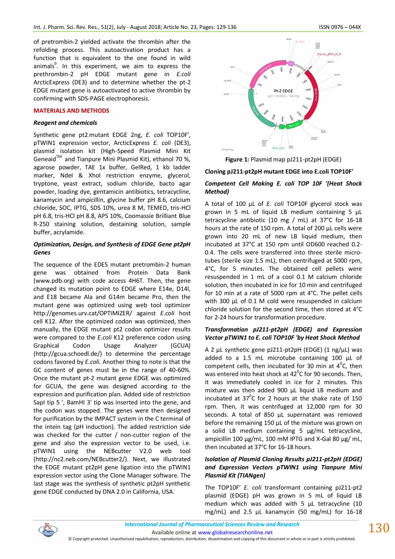
Synthetic gene pt2-mutant EDGE 2ng, E. coli TOP10F', pTWIN1 expression vector, ArcticExpress E. coli (DE3), plasmid isolation kit (High-Speed Plasmid Mini Kit GeneaidTM and Tianpure Mini Plasmid Kit), ethanol 70 %, agarose powder, TAE 1x buffer, GelRed, 1 kb ladder marker, NdeI & XhoI restriction enzyme, glycerol, tryptone, yeast extract, sodium chloride, bacto agar powder, loading dye, gentamicin antibiotics, tetracycline, kanamycin and ampicillin, glycine buffer pH 8.6, calcium chloride, SOC, IPTG, SDS 10%, urea 8 M, TEMED, tris-HCl pH 6.8, tris-HCl pH 8.8, APS 10%, Coomassie Brilliant Blue R-250 staining solution, destaining solution, sample buffer, acrylamide.
Optimization, Design, and Synthesis of EDGE Gene pt2pH Genes
The sequence of the EDES mutant pretrombin-2 human gene was obtained from Protein Data Bank (www.pdb.org) with code access 4H6T. Then, the gene changed its mutation point to EDGE where E14e, D14l, and E18 became Ala and G14m became Pro, then the mutant gene was optimized using web tool optimizer http://genomes.urv.cat/OPTIMIZER/ against E.coli host cell K12. After the optimized codon was optimized, then manually, the EDGE mutant pt2 codon optimizer results were compared to the E.coli K12 preference codon using Graphical Codon Usage Analyzer (GCUA) (http://gcua.schoedl.de/) to determine the percentage codons favored by E.coli. Another thing to note is that the GC content of genes must be in the range of 40-60%. Once the mutant pt-2 mutant gene EDGE was optimized for GCUA, the gene was designed according to the expression and purification plan. Added side of restriction SapI tip 5 ', BamHI 3' tip was inserted into the gene, and the codon was stopped. The genes were then designed for purification by the IMPACT system in the C terminal of the intein tag (pH induction). The added restriction side was checked for the cutter / non-cutter region of the gene and also the expression vector to be used, i.e. pTWIN1 using the NEBcutter V2.0 web tool (http://nc2.neb.com/NEBcutter2/). Next, we illustrated the EDGE mutant pt2pH gene ligation into the pTWIN1 expression vector using the Clone Manager software. The last stage was the synthesis of synthetic pt2pH synthetic gene EDGE conducted by DNA 2.0 in California, USA.
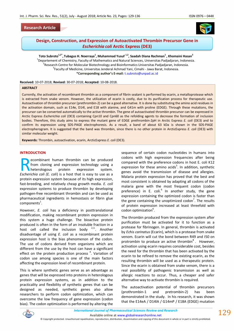
Figure 1: Plasmid map pJ211-pt2pH (EDGE)
Cloning pJ211-pt2pH mutant EDGE into E.coli TOP10F'
Competent Cell Making E. coli TOP 10F '(Heat Shock Method)
A total of 100 μL of E. coli TOP10F glycerol stock was grown in 5 mL of liquid LB medium containing 5 μL tetracycline antibiotic (10 mg / mL) at 37°C for 16-18 hours at the rate of 150 rpm. A total of 200 μL cells were grown into 20 mL of new LB liquid medium, then incubated at 37°C at 150 rpm until OD600 reached 0.2-0.4. The cells were transferred into three sterile micro-tubes (sterile size 1.5 mL), then centrifuged at 5000 rpm, 4°C, for 5 minutes. The obtained cell pellets were resuspended in 1 mL of a cool 0.1 M calcium chloride solution, then incubated in ice for 10 min and centrifuged for 10 min at a rate of 5000 rpm at 4°C. The pellet cells with 300 μL of 0.1 M cold were resuspended in calcium chloride solution for the second time, then stored at 4°C for 2-24 hours for transformation procedure.
Transformation pJ211-pt2pH (EDGE) and Expression Vector pTWIN1 to E. coli TOP10F 'by Heat Shock Method
A 2 μL synthetic gene pJ211-pt2pH (EDGE) (1 ng/μL) was added to a 1.5 mL microtube containing 100 μL of competent cells, then incubated for 30 min at 40C, then was entered into heat shock at 420C for 90 seconds. Then, it was immediately cooled in ice for 2 minutes. This mixture was then added 900 μL liquid LB medium and incubated at 37
0C for 2 hours at the shake rate of 150
rpm. Then, it was centrifuged at 12,000 rpm for 30 seconds. A total of 850 μL supernatant was removed before the remaining 150 μL of the mixture was grown on a solid LB medium containing 5 μg/mL tetracycline, ampicillin 100 μg/mL, 100 mM IPTG and X-Gal 80 μg/ mL, then incubated at 37°C for 16-18 hours.
Isolation of Plasmid Cloning Results pJ211-pt2pH (EDGE) and Expression Vectors pTWIN1 using Tianpure Mini Plasmid Kit (TIANgen)
The TOP10F’ E. coli transformant containing pJ211-pt2 plasmid (EDGE) pH was grown in 5 mL of liquid LB medium which was added with 5 μL tetracycline (10 mg/mL) and 2.5 μL kanamycin (50 mg/mL) for 16-18
Int. J. Pharm. Sci. Rev. Res., 51(2), July - August 2018; Article No. 23, Pages: 129-136 ISSN 0976 – 044X
International Journal of Pharmaceutical Sciences Review and Research . International Journal of Pharmaceutical Sciences Review and Research Available online at www.globalresearchonline.net
© Copyright protected. Unauthorised republication, reproduction, distribution, dissemination and copying of this document in whole or in part is strictly prohibited.
.
. Available online at www.globalresearchonline.net
131
hours, a temperature of 37°C, with a shake rate of 150 rpm, while a TOP10F’ E. coli transformant containing a pTWIN1 expression vector was grown in 5 mL of liquid LB medium which added 5 μL tetracycline (10 mg/mL) and 5 μL ampicillin (100 mg / mL). The TOP10F’ E. coli cell culture was inserted into a 1.5 mL microscentrifuges tube and centrifuged at 12,000 rpm for 1 minute to collect pellets. The cell pellets were added with 250 μL buffer P1 (added RNase) and dissolved with vortex. Furthermore, 250 μL buffer P2 was added, and the microcentrifuges tube was converted 8 times. A 350 μL P3 buffer was added to the microscentrifuges tube and then inverted 8 times. The centrifuges tube was centrifuged at 12,000-13,400 rpm for 10 minutes. The supernatant was transferred into CS column then centrifuged at 12,000-13,400 rpm for 2 minutes. Then the flowthrough was transferred to the CP3 column which had previously been added with 500 μL buffer BL and centrifuged at 12,000-13,400 rpm for 1 min and the flowthrough was removed. The CP3 column containing the supernatant was centrifuged at a rate of 12,000-13,400 rpm for 1 minute and removed its flowthrough. A 500 μL buffer of PD was added to the CP3 column and centrifuged at 12,000-13,400 rpm for 1 minute and then the flowthrough was removed. Then, as much as 600 μL of PW buffer (added ethanol) was added to the CP3 column and centrifuged at 12,000-13,400 rpm for 1 minute and then the flowthrough was discharged. The addition of PW buffer is done 2 times. Thereafter, the CP3 column was centrifuged at a rate of 12,000-13,400 rpm for 2 min and to dry the CP3 column, the column opened and closed, and allowed to stand for several minutes at room temperature, then transferred to a new micros centrifugation tube. The TB buffer was added to 50 μL CP3 column then incubated in 55°C water bath for 3 minutes and centrifuged at 12,000-13,400 rpm for 2 minutes. Then, the TB buffer was added back to 60 μL CP3 column then incubated in water bath temperature 55°C for 3 minutes and centrifuged at 12.000-13,400 rpm for 2 minutes.
Characterization of Plasmid Cloning Results pJ211-pt2pH (EDGE) and Expression Vector pTWIN1 with Restriction Enzyme and Gel Agarose Electrophoresis
The isolation results were characterized by the cutting of one and two restriction enzymes. A total of 5 μL isolates were inserted into the micro tube and added with 0.5 μL Sapl enzyme and 0.5 μL of the BamHI enzyme, and for restriction, by addition of 1 μL buffer cut smart and 3 μL nuclease-free water, then incubated for 2-3 hours at 37°C. As for the cutting of only one restriction enzyme, the enzyme used was BamHI. The results of the restriction were characterized using 0.4% agarose electrophoresis. Markers and mixtures were made containing isolated plasmids to be characterized. The marker contained a mixture of 4 μL nuclease-free water, 1 μL loading dye, 1 μL gel red, and 1 μL ladder DNA 1 kb. Then, 1 μL of loading dye, 1 μL gel red, and 5 μL of plasmid isolation were added without cutting, while for cutting plasmid, all
the volumes of the restriction reaction results were included. Then, the marker and plasmid mixture were introduced into a 0.8% (b / v) agarose well prepared by dissolving 0.32 g agarose in 40 mL TAE 1x buffer. Electrophoresis was then carried out with a voltage of 80 volts for ± 40 minutes and TAE 1x buffer as used as a current delivery medium. The DNA bands were viewed with the help of ultra violet light and were documented with the camera.
Plasmid construction pTWIN1-pJ211-pt2pH (EDGE) and Cloning in E. coli TOP10F'
Cutting Vector Expression pTWIN1 and pJ211-pt2pH (EDGE) by Restriction Enzyme
Isolates pJ211-pt2 (EDGE) pH and pTWIN1 were cut with restriction enzymes BamHI and SAP with a total reaction volume of 20 μL. All remaining isolates were concentrated to a volume of 0 μL and then added with 2 μL buffer cut smart, 1 μL SapI enzyme, 1 μL of BamHI enzyme, and 16 μL of nuclease-free water and then incubated for 2-3 hours at 37°C. The restriction DNA fragment was analyzed with 0.4% agarose electrophoresis. The SapI-pt2-BamHI fragments were cut from the agarose gel and purified using a Tiangel Midi DNA Purification Kit purification kit. In the same way, the pTWIN1 plasmid was also cut from the agarose gel and purified. The ligation of SapI-PT-2-BamHI DNA fragment was put into the pTWIN1 Expression Vector.
Purification of Fragments SapI-pt2-BamHI and pTWIN1
The agarose gel slices (fragment SapI-pt2-BamHI and pTWIN1) were fed into a microscentrifuges tube and then added with 400 μL PN buffer and incubated in 50°C water bath while inverted until dissolved for 10 min. The micros centrifugation tube was cooled to room temperature and allowed to stand for 2 minutes at room temperature. The solution of the DNA fragment was transferred to the CA2 column which had previously been added with 500 μL buffer BL and centrifuged at 12,000-13,400 rpm for 1 minute and then the flowthrough was discharged. The CA2 column containing the DNA fragment solution was centrifuged at a rate of 12,000-13,400 rpm for 1 minute and the flowthrough was removed. Then, as much as 600 μL of PW buffer (added ethanol) was added to the CA2 column and centrifuged at 12,000-13,400 rpm for 1 minute and then the flowthrough was removed. Then, the column was allowed to stand for 2-5 minutes at room temperature. The addition of PW buffer was done 2 times. Thereafter, the CA2 column was centrifuged at a rate of 12,000-13,400 rpm for 2 minutes and for drying the CA2 column, the column was opened and closed and allowed to stand for several minutes at room temperature before being transferred to a new microscentrifuges tube. EB buffer was added to CA2 column of 40 μL then incubated in a water bath at 55°C for 3 minutes and centrifuged at 12,000-13,400 rpm for 2 minutes. The EB buffer was then added back to a 30 μL CA2 column and incubated in a 55°C water bath for 3
Int. J. Pharm. Sci. Rev. Res., 51(2), July - August 2018; Article No. 23, Pages: 129-136 ISSN 0976 – 044X
International Journal of Pharmaceutical Sciences Review and Research . International Journal of Pharmaceutical Sciences Review and Research Available online at www.globalresearchonline.net
© Copyright protected. Unauthorised republication, reproduction, distribution, dissemination and copying of this document in whole or in part is strictly prohibited.
.
. Available online at www.globalresearchonline.net
132
minutes and centrifuged at 12,000-13,400 rpm for 2 minutes.
Ligation of SapI-pt2-BamHI Fragment with Expression Vector pTWIN1
The ligation reaction was carried out with a 1: 8 molar ratio between the pTWIN1 vector and the SapI-pt2-BamHI insert genes. The ligation reaction was carried out using T4 DNA ligase enzyme with total ligation reaction volume of 10 μL at 160C for 16-18 hours.
Transformation pTWIN1-pt2 (EDGE) pH into E.coli TOP10F '
The transformation of pTWIN1-pt2 (EDGE) pH was achieved through the electroporation method. A total of 10 μL recombinant plasmid pTWIN1-pt2 (EDGE) pH were each added to a microscentrifuges tube containing 50 μL competent cells, then pipetted up and down. A micros centrifugation tube containing a mixture of plasmids and competent cells was fed into a cuvette that had been incubated for 10 minutes in an ice bath. Bottom of the cuvette was cleaned and then inserted into a 1500 V voltage regulated electrophorator device. The ms and voltage values used were recorded. As a positive control of the transformation, a transformation of TOP10F E.coli was performed using a quantized plasmid pTYB21. Then, 1 mL of SOC liquid medium was added to the cuvette, resuspended, and fed into a test tube, then incubated for 1 hour at 37°C. at a rate of 250 rpm. A total of 100 μL transformed cultures were grown on an LB solid medium containing antibiotics suitable for selection at 37°C for 16-18 hours. Transformances were taken from a single colony and replicated.
Isolation of cloning Result pTWIN1-pt2 (EDGE) pH
Isolation of the cloning result was done using plasmid insulation kit of GenAid High-Speed Plasmid Mini Kit. The TOP10F E. coli transformant containing pTWIN1-pt2 (EDGE) plasmid pH was grown in 5 mL of liquid LB medium which had been added with 5 μL tetracycline (10 mg / mL) and 5 μL ampicillin (100 mg / mL) for 16-18 hours, temperature 37°C, at the shuffling rate of 150 rpm. The TOP10F E. coli cell culture was inserted into a 1.5 mL micros centrifugation tube and centrifuged at 12,000 rpm for 1 minute to gather the pellets. The cell pellets were added with 200 μL buffer PD1 (added RNase) and dissolved by vortex until dissolved. Then, 200 μL buffer PD2 was added, and the microaspiration tube was inverted 10 times. A 300 μL PD3 buffer was added to the microscentrifuges tube and then inverted 10 times. The centrifugation tube was centrifuged at 13,400 rpm for 5 min. The supernatant was transferred into the PD column then centrifuged at 13,400 rpm for 1 minute and then the flowthrough was discharged. A 400 μL buffer W1 was added to the PD column and centrifuged at 13,400 rpm for 1 minute and then the flowthrough was removed. Then, as much as 600 μL washing buffer (added ethanol) was added to the PD column and centrifuged at 13,400 rpm for 1 minute and then the flowthrough was removed.
After that, the PD column was centrifuged at 13,400 rpm for 5 minutes then the column was transferred to a new micros centrifugation tube. The TE buffer was added to a 30 μL CP3 column then incubated in a 55°C water bath for 3 min before being centrifuged at 13,400 rpm for 2 min. The eluate was then re-inserted in the PD column then incubated in water bath at 55°C for 3 minutes and centrifuged at 13,400 rpm for 2 minutes.
Characterization of pTWIN1-pt2 Isolation Result (EDGE) pH
The isolation results were characterized by the cutting of two restriction enzymes. A total of 5 μL isolates were inserted into the micro tube and added 0.5 μL Sapl enzyme and 0.5 μL of the BamHI enzyme for restriction by addition of 1 μL buffer cut smart and 3 μL nuclease-free water then incubated for 2-3 hours at 37°C. Isolates without the restriction enzymes being cut and the restriction results were characterized using 0.4% agarose electrophoresis.
Expression Plasmid Recombinant pTWIN1-pt2 (EDGE) pH into ArcticExpress E. coli (DE3)
Making Competent Cells
Competent cell production used the same antibiotics previously. But, the concentrations of the antibiotics were instead 1% tetracycline 10 μg / ml and 2% gentamicin 10 μg / ml.
Expression Test
E. coli ArcticExpress (DE3) [pTWIN1-pt2 (EDGE) pH] had been characterized, grown in 5 mL of liquid LB medium which had been added with 5 μL tetracycline (10 mg / mL), 5 μL ampicillin (100 mg / mL), and 10 μL chloramphenicol (100 mg / mL) for 16-18 hours, 37°C, at a rate of 150 rpm. ArcticExpress E. coli cell culture (DE3) was incorporated into an erlenmeyer flask containing 20 mL liquid LB and an appropriate antibiotic for selection, then incubated at 37°C, with a matching rate of 150 rpm to 0.4-0.8 OD600. A total of 1 mL of culture was taken as to (before induction of IPTG) and inserted into the microspiration tube, then the pellets were collected by centrifugation at 8000 g 4oC for 10 min. Then, cultures in the erlenmeyer flasks were added IPTG 0.1 mM / mL culture volume, then incubated at 22°C, with a shuffle rate of 150 rpm for 5-6 hours to OD600 1.0-1.5. A total of 1 mL of culture is taken as ti (after induction of IPTG) and fed into in the microscentrifuges tube, then collected pellet by centrifugation at 8000 g 4oC for 10 minutes. Then the remaining cultures were collected pellet by centrifugation at 8000 g 4°C for 10 minutes. Then, as much as 500 μL of glycine buffer was added. The dialysis was done using a sonicator and centrifuged at 10,000 g at 4°C for 30 minutes. The supernatant was then transferred to a new microscaping tube as a soluble fraction. The pellets were added 100 μl of urea 8 M, then heated to 95°C for 15 min and centrifuged at 8000 g at 4°C for 10 min. The supernatant was fed into a new microscaping
Int. J. Pharm. Sci. Rev. Res., 51(2), July - August 2018; Article No. 23, Pages: 129-136 ISSN 0976 – 044X
International Journal of Pharmaceutical Sciences Review and Research . International Journal of Pharmaceutical Sciences Review and Research Available online at www.globalresearchonline.net
© Copyright protected. Unauthorised republication, reproduction, distribution, dissemination and copying of this document in whole or in part is strictly prohibited.
.
. Available online at www.globalresearchonline.net
133
tube as an insoluble fraction. For to and ti, they were dissolved in 40 μL glycine buffer.
Characterization of pTWIN1-pt2 Gene Expression (EDGE) Results pH with Analysis of SDS-PAGE
For characterization using SDS-PAGE, the process began with gel-making (separating gel and stacking gel). Glass plate sandwich (short plate and spacer plate) was first washed with soap and cleaned with ethanol 70%, then dried. The glass plate sandwich was used to place the mold in gel making. Short plate was placed in front of the spacer plate glass. Both glasses were inserted into the frame casting with the bottom position of both glasses equally then locked. The ready-made glass plate sandwich was then mounted on the casting stand. Solved gel resolving solutions were inserted between the short plate glass gap and the spacer plate, up to two-thirds. 10% acrylamide gel as a resolving gel solution was made by mixing 4.85 mL of sterile distilled water, 2.50 mL 40% acrylamide, 2.50 mL buffer resolving pH 8.8, 100 μL SDS 10%, 50 μL APS 10%, and 5 μL TEMED. Resolving gel solutions were strived to prevent air bubbles from occurring. The distilled water was added to the upper limit of the glass and the solution was allowed for 45 minutes until the resolving gel hardens. The aqueous at the top of the resolving gel was then removed, then dried with a tissue. Stacking gel solution was poured to the upper limit of glass plate sandwich then comb (comb) was inserted. 4% acrylamide gel as a stacking gel solution was made by mixing 3.18 mL of sterile distilled water, 500 μL 40% acrylamide, 1.26 ml buffer stacking pH 6.8, 50 μL SDS 10%, 25 μL APS 10%, and 5 μL TEMED. The gel was left to harden for 1 hour. Gel-glass plate sandwich was then removed from the casting frame and mounted on the electrode assembly with the position of the short plate facing inward and then running 1x buffer was inserted into the electrophoresis tank.
The sample of expression result was taken as much as 40 μL then mixed with 10 μl SDS Sample Buffer 5x in micros centrifugation tube. The mixture was then heated at 95°C for 15 minutes at the water bath, then centrifuged at 8000 g for 1 minute at 4°C to lower the vapor. Each sample of 10 μl and a protein marker of 5 μL were then fed into the well. The electrode cable was paired with the electrophoresis device and the gel was run at 100 volts for ± 120 minutes. The gel was then immersed in staining solution (Coomassie Brilliant Blue R-250 0.25%, 45% methanol, 10% acetic acid) for 1 hour with slow shaking. The gel was then soaked in the destaining solution until the excess gel dye was cleaned off. The gel was then immersed in aquadest which can then be stored in mica plastic to dry to be documented. The positive results of SDS-PAGE were characterized by the emergence of the ± 63 kDa recombinant protein band of intein-pretrombin-2.
RESULTS DISCUSSION
Design, Optimization and Synthesis of EDGE Gene pt2-pH Genes
The EDGE mutant pt2 gene was designed based on the amino acid sequence of the EDES mutant pt2 gene (E14eA / 14lA / E18A / S195A) on the Database Protein database (www.pdb.org) with code access: 4H6T, where A195 was converted back to Ser (corresponding to triad thrombin catalytic) followed by the addition of G14mP mutation, so that the amino acid sequences obtained were as follows (306 amino acids):
TSEYQTFFNPRTFGSGEADCG LRPLFEKKSLEDKTERALLESYIAPRIVAGSDAEIGMSPWQV MLFRK SPQELLCGASLISDRW VLTAAHCLLYPPWDKNFTEND LLVRIGKHSRTRYERNIEKIS MLEKIY IHPR YNWRENLDRDI ALMKLKKPVAFSDYIHPVCLP DRETAASLLQAGYKGRVTGWG NLKETWTANV G KGQPSVLQVV NLPIVERPVCKDSTRIRITDN MFCAGYKPDEGKRGDACEGDS GGPFVMKS PFNN RW YQMGIVS WGEGCDRDGKYGFYTHVFRLK KWIQKVIDQFGE
(Note: Amino acid is thickened and underlined is the amino acid E, D, G, E are mutated into alanine).
Then, this amino acid sequence was converted into its nucleotide sequence using software that can be accessed online, ie EMBOSS Backtranseq (http://www.ebi.ac.uk/Tools/st/emboss_backtranseq/).
After obtaining the nucleotide sequence, the sequence was then analyzed using Graphical Codon Usage Analyzer (GCUA) (http://gcua.schoedl.de/) to see the relative adaptivesness or conformity parameter between the target gene codon (pt2 mutant EDGE) with preferred codon preference in host cell E. coli K12. The codon optimization was performed by replacing the codons of an amino acid in the gene with the preference codon of the amino acid which is preferably E. coli K12 with a relative adaptivesness value of 100%. At the time of doing the optimization codon should also note the basic content of nitrogen Guanin (G) and Cytosine (C) (GC content) that should be in the range of 40-60%. This was so that the double helical DNA strand will not be easily released, because the hydrogen bond between G and C would be very strong if the GC content was less than 40%. It was feared that the double helical DNA strand would be easily removed and interfered with the replication process. Once the gene was optimized, then it was then designed according to the expression vector and the purification system used. The expression vector used was pTWIN1 (NEB) with an Intein Mediated Purification with an Affinity Chitin-binding Tag (IMPACT) system. Since the purification strategy with the selected intein tag, the induction of cuts was with pH (fusion in N-terminal proteins). Once the genes were completed, the next stage was synthesization by DNA 2.0 (http://dna20.com/). The gene was obtained in a circular form with a delivery vector pJ211-pt2pH (EDGE) measuring 3461 bp carrying kanamycin resistance gene (Fig. 1).
Int. J. Pharm. Sci. Rev. Res., 51(2), July - August 2018; Article No. 23, Pages: 129-136 ISSN 0976 – 044X
International Journal of Pharmaceutical Sciences Review and Research . International Journal of Pharmaceutical Sciences Review and Research Available online at www.globalresearchonline.net
© Copyright protected. Unauthorised republication, reproduction, distribution, dissemination and copying of this document in whole or in part is strictly prohibited.
.
. Available online at www.globalresearchonline.net
134
Subcloning pJ211-pt2pH (EDGE) and Expression Vector pTWIN1 to E. coli TOP10F 'with Heat Shock Method
Subcloning was performed for propagation purposes of plasmid pJ211-pt2pH (EDGE) and pTWIN1 expression vector. E. coli TOP10F 'is a highly transformed bacterial bacteria because it could easily receive plasmid and it could grow fast. The competent cells of TOP10F E. coli were prepared using the CaCl2 method
10. Single columns
of transformants E. coli TOP10F 'containing plasmid pJ211-pt2pH (EDGE) were rejuvenated and reprocessed in new solid LB media which had been added tetracycline and kanamycin antibiotics. But, single-transformant colonies containing pTWIN1 vectors were rejuvenated in a solid LB medium containing ampicillin and tetracycline antibiotics. The colonies obtained from the replica were then isolated and characterized to ensure the growing transformer E. coli TOP10F ', containing pJ211-pt2pH (EDGE) plasmids and pTWIN1 expression vectors. To ensure that the isolates obtained contained true plasmids, ie pJ211-pt2 (EDGE) pH and pTWIN1, further analysis was required using two digest enzymes (double digest). With this double digest we can know the exact size of the pH211-pt2 (EDGE) pH gene and also the pTWIN1 vector. Since the DNA marker used was a collection of linear DNA fragments, to determine the size of the still-circular DNA, it must be diluted first by cutting by the appropriate restriction enzyme in accordance with the synthetic gene design pt2 (EDGE) pH that the enzymes used are SapI and BamH.
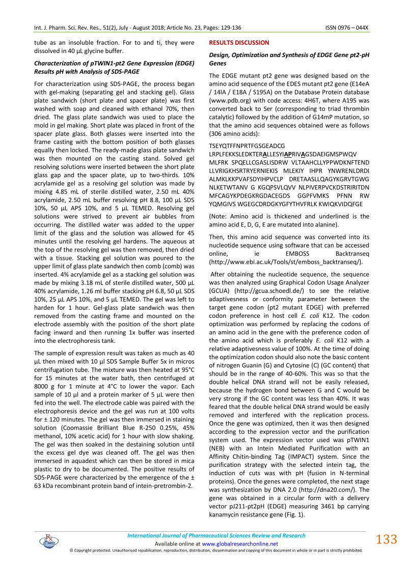
Figure 2: Electropherogram characterization of two restriction enzymes SapI-BamHI plasmid pJ211-pt2 (EDGE) pH and pTWIN1. (M) DNA marker 1 kb; (1) pTWIN1 colony 2; (2) pJ211- pt2 (EDGE) colony pH 2.
The results of restriction with two SapI-BamHI enzymes in lane 1 showed that colonies of 2 transformants TOP10F ' [pTWIN1] positively contained the pTWIN1 vector as successfully cut by the SapI-BamHI restriction enzyme and the presence of a band of molecular weight ± 6540 bp and the band with a molecular weight of ± 835 bb as the backbone of the multi cloning site (MCS), intein 2 ie Mxe GyrA and CBD. Similarly, in the 2 pale colonies of 2 pH2 transformants [pJ211-pt2 (EDGE) positive pH2 contains pJ211-pt2 (EDGE) plasmid pH because in the ± 938 b band there is a band corresponding to the pH2 gene molecule (pH2) gene and at area ± 2524 pb which was the
backbone band of the delivery vector pJ211. Pt2 gene ligation (EDGE) with pT2 expression gene (EDGE) pH in the pT2 expression vector pTWIN1 was performed using T4 DNA ligase (NEB) enzyme catalyzing the phosphodiester bond reaction between the pH 2 end pH 2 (pH 2) gene phosphate group and the hydroxyl group 3 'end of pTWIN1 expression vector. The ligation process was carried out at a temperature of 16°C overnight as it was the optimal temperature and incubation time of T4 DNA ligase enzyme for sticky end ligation (New England Biolabs, 2015). The ligation results could not be visualized by agarose gel electrophoresis because the recombinant plasmid concentrations of the formed DNA were too small.
Recombinant Plasmid Cloning pTWIN1-pt2 (EDGE) pH into E. coli TOP10F '
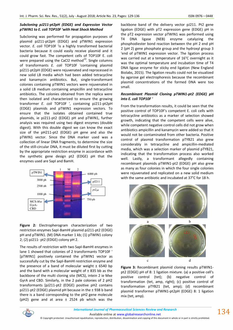
From the transformation results, it could be seen that the positive control of TOP10F's competent E. coli cells with tetracycline antibiotics as a marker of selection showed growth, indicating that the competent cells were alive, while competent negative control cells did not grow when antibiotics ampicillin and kanamycin were added so that it would not be contaminated from other bacteria. Positive control of plasmid transformation pTYB21 also grew considerably in tetracycline and ampicillin-mediated media, which was a selection marker of plasmid pTYB21, indicating that the transformation process also worked well. Lastly, a transformant allegedly containing recombinant plasmids pTWIN1-pt2 (EDGE) pH also grew as many as four colonies in which the four single colonies were rejuvenated and replicated on a new solid medium with the same antibiotic and incubated at 37°C for 18 h.
Figure 3: Recombinant plasmid cloning results pTWIN1-pt2 (EDGE) pH of 8: 1 ligation mixture. (a) a positive cell's positive control (tet); (b) negative control of transformation (tet, amp, right); (c) positive control of transformation pTYB21 (tet, amp); (d) recombinant plasmid transformer pTWIN1-pt2pH (EDGE) 8: 1 ligation mix (tet, amp).
Int. J. Pharm. Sci. Rev. Res., 51(2), July - August 2018; Article No. 23, Pages: 129-136 ISSN 0976 – 044X
International Journal of Pharmaceutical Sciences Review and Research . International Journal of Pharmaceutical Sciences Review and Research Available online at www.globalresearchonline.net
© Copyright protected. Unauthorised republication, reproduction, distribution, dissemination and copying of this document in whole or in part is strictly prohibited.
.
. Available online at www.globalresearchonline.net
135
Recombinant Plasmid Transformation pTWIN1-pt2pH (EDGE) into the ArcticExpress E. coli Expression (DE3)
Transformation to the ArcticExpress E. coli expression host (DE3) was done by heat shock method. After a competent cell was made with CaCl2, then the recombinant plasmid plasmid pTWIN1-pt2 (EDGE) pH colony was added to the competent cell and heat shock was performed at 42°C for 90 seconds. Thereafter, the transformants were collected in a dense LB medium containing tetracycline antibiotics and gentamicin as a selection for ArcticExpress E. coli (DE3) and ampicillin for selection of pH recombinant pTWIN1-pt2 (EDGE) plasmids at 37°C for 18 h and single colonies grown replicated in media New solid LB with the same antibiotic.
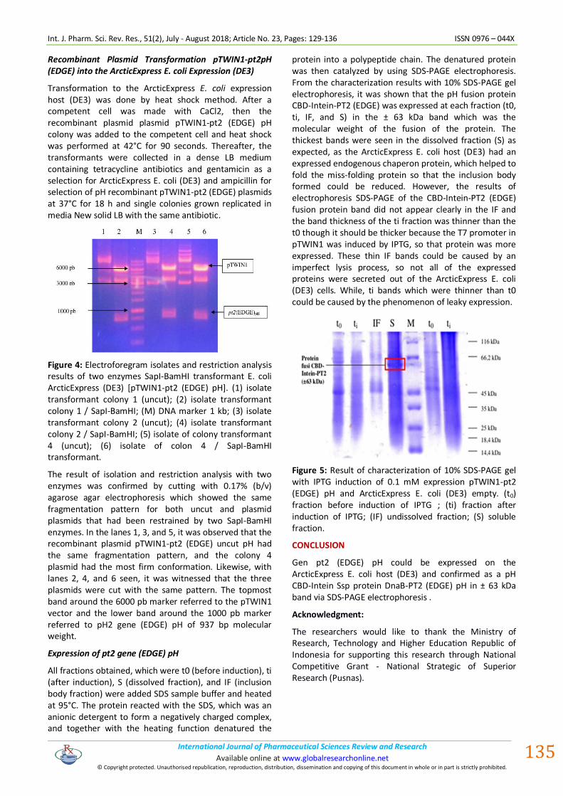
Figure 4: Electroforegram isolates and restriction analysis results of two enzymes SapI-BamHI transformant E. coli ArcticExpress (DE3) [pTWIN1-pt2 (EDGE) pH]. (1) isolate transformant colony 1 (uncut); (2) isolate transformant colony 1 / SapI-BamHI; (M) DNA marker 1 kb; (3) isolate transformant colony 2 (uncut); (4) isolate transformant colony 2 / SapI-BamHI; (5) isolate of colony transformant 4 (uncut); (6) isolate of colon 4 / SapI-BamHI transformant.
The result of isolation and restriction analysis with two enzymes was confirmed by cutting with 0.17% (b/v) agarose agar electrophoresis which showed the same fragmentation pattern for both uncut and plasmid plasmids that had been restrained by two SapI-BamHI enzymes. In the lanes 1, 3, and 5, it was observed that the recombinant plasmid pTWIN1-pt2 (EDGE) uncut pH had the same fragmentation pattern, and the colony 4 plasmid had the most firm conformation. Likewise, with lanes 2, 4, and 6 seen, it was witnessed that the three plasmids were cut with the same pattern. The topmost band around the 6000 pb marker referred to the pTWIN1 vector and the lower band around the 1000 pb marker referred to pH2 gene (EDGE) pH of 937 bp molecular weight.
Expression of pt2 gene (EDGE) pH
All fractions obtained, which were t0 (before induction), ti (after induction), S (dissolved fraction), and IF (inclusion body fraction) were added SDS sample buffer and heated at 95°C. The protein reacted with the SDS, which was an anionic detergent to form a negatively charged complex, and together with the heating function denatured the
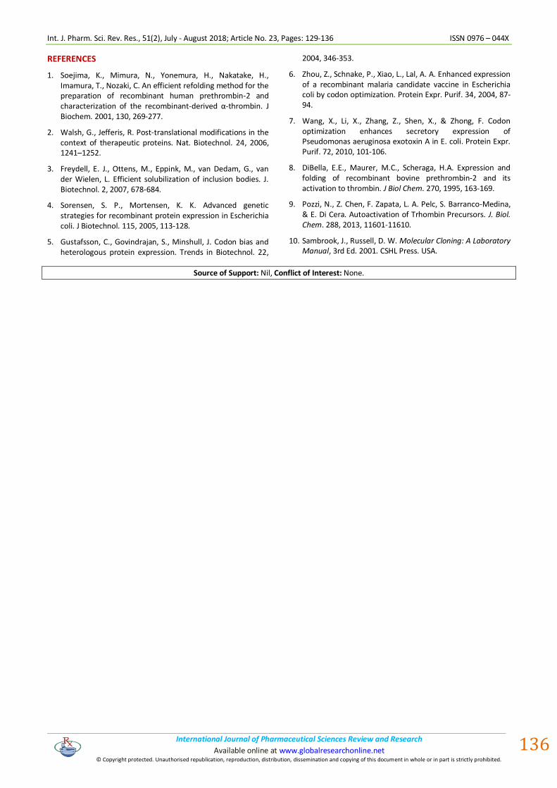
protein into a polypeptide chain. The denatured protein was then catalyzed by using SDS-PAGE electrophoresis. From the characterization results with 10% SDS-PAGE gel electrophoresis, it was shown that the pH fusion protein CBD-Intein-PT2 (EDGE) was expressed at each fraction (t0, ti, IF, and S) in the ± 63 kDa band which was the molecular weight of the fusion of the protein. The thickest bands were seen in the dissolved fraction (S) as expected, as the ArcticExpress E. coli host (DE3) had an expressed endogenous chaperon protein, which helped to fold the miss-folding protein so that the inclusion body formed could be reduced. However, the results of electrophoresis SDS-PAGE of the CBD-Intein-PT2 (EDGE) fusion protein band did not appear clearly in the IF and the band thickness of the ti fraction was thinner than the t0 though it should be thicker because the T7 promoter in pTWIN1 was induced by IPTG, so that protein was more expressed. These thin IF bands could be caused by an imperfect lysis process, so not all of the expressed proteins were secreted out of the ArcticExpress E. coli (DE3) cells. While, ti bands which were thinner than t0 could be caused by the phenomenon of leaky expression.
Figure 5: Result of characterization of 10% SDS-PAGE gel with IPTG induction of 0.1 mM expression pTWIN1-pt2 (EDGE) pH and ArcticExpress E. coli (DE3) empty. (t0) fraction before induction of IPTG ; (ti) fraction after induction of IPTG; (IF) undissolved fraction; (S) soluble fraction.
CONCLUSION
Gen pt2 (EDGE) pH could be expressed on the ArcticExpress E. coli host (DE3) and confirmed as a pH CBD-Intein Ssp protein DnaB-PT2 (EDGE) pH in ± 63 kDa band via SDS-PAGE electrophoresis .
Acknowledgment:
The researchers would like to thank the Ministry of Research, Technology and Higher Education Republic of Indonesia for supporting this research through National Competitive Grant - National Strategic of Superior Research (Pusnas).
Int. J. Pharm. Sci. Rev. Res., 51(2), July - August 2018; Article No. 23, Pages: 129-136 ISSN 0976 – 044X
International Journal of Pharmaceutical Sciences Review and Research . International Journal of Pharmaceutical Sciences Review and Research Available online at www.globalresearchonline.net
© Copyright protected. Unauthorised republication, reproduction, distribution, dissemination and copying of this document in whole or in part is strictly prohibited.
.
. Available online at www.globalresearchonline.net
136
REFERENCES
1. Soejima, K., Mimura, N., Yonemura, H., Nakatake, H., Imamura, T., Nozaki, C. An efficient refolding method for the preparation of recombinant human prethrombin-2 and characterization of the recombinant-derived α-thrombin. J Biochem. 2001, 130, 269-277.
2. Walsh, G., Jefferis, R. Post-translational modifications in the context of therapeutic proteins. Nat. Biotechnol. 24, 2006, 1241–1252.
3. Freydell, E. J., Ottens, M., Eppink, M., van Dedam, G., van der Wielen, L. Efficient solubilization of inclusion bodies. J. Biotechnol. 2, 2007, 678-684.
4. Sorensen, S. P., Mortensen, K. K. Advanced genetic strategies for recombinant protein expression in Escherichia coli. J Biotechnol. 115, 2005, 113-128.
5. Gustafsson, C., Govindrajan, S., Minshull, J. Codon bias and heterologous protein expression. Trends in Biotechnol. 22,
2004, 346-353.
6. Zhou, Z., Schnake, P., Xiao, L., Lal, A. A. Enhanced expression of a recombinant malaria candidate vaccine in Escherichia coli by codon optimization. Protein Expr. Purif. 34, 2004, 87-94.
7. Wang, X., Li, X., Zhang, Z., Shen, X., & Zhong, F. Codon optimization enhances secretory expression of Pseudomonas aeruginosa exotoxin A in E. coli. Protein Expr. Purif. 72, 2010, 101-106.
8. DiBella, E.E., Maurer, M.C., Scheraga, H.A. Expression and folding of recombinant bovine prethrombin-2 and its activation to thrombin. J Biol Chem. 270, 1995, 163-169.
9. Pozzi, N., Z. Chen, F. Zapata, L. A. Pelc, S. Barranco-Medina, & E. Di Cera. Autoactivation of Trhombin Precursors. J. Biol. Chem. 288, 2013, 11601-11610.
10. Sambrook, J., Russell, D. W. Molecular Cloning: A Laboratory Manual, 3rd Ed. 2001. CSHL Press. USA.
Source of Support: Nil, Conflict of Interest: None.