research articleglobalresearchonline.net/journalcontents/v26-1/51.pdf · int. j. pharm. sci. rev....
TRANSCRIPT

Int. J. Pharm. Sci. Rev. Res., 26(1), May – Jun 2014; Article No. 51, Pages: 301-308 ISSN 0976 – 044X
International Journal of Pharmaceutical Sciences Review and Research Available online at www.globalresearchonline.net
© Copyright protected. Unauthorised republication, reproduction, distribution, dissemination and copying of this document in whole or in part is strictly prohibited.
301
Faten K. Abd El-Hady1*, Mohamed S. Abdel-Aziz2, Kamel H. Shaker3, Zeinab A. El-Shahid1, Mohamed A.Ghani4 1Chemistry of Natural Products Department, National Research Center, Egypt.
2Department of Microbial Chemistry, National Research Center, Egypt. 3Chemistry of Natural Compounds Department, National Research Center, Egypt.
4Red Sea Marine parks, Hurghada-Red Sea – Egypt. *Corresponding author’s E-mail: [email protected]
Accepted on: 01-04-2014; Finalized on: 30-04-2014.
ABSTRACT
Acetylcholinesterase (AChE) inhibitors have received considerable attention as alternatives in treatment of Alzheimer's disease (AD). The damage caused by reactive oxygen species is considered a contributing factor to AD. Three fungi (FC1, FC2 and FC3) were isolated from the soft corals; Sinularia sp. and Lobophyton sp. The antioxidant activities (DPPH and Xanthine Oxidase assays), (AChE) inhibitory activity and antimicrobial activity for twelve different fungal extracts were evaluated. Mycelial ethylacetate extract from the static culture of the fungus FC2 (Emericella unguis 8429) had the highest free radical scavenging activity against DPPH and superoxide anion radicals. Mycelial extract from the shake culture of the isolated three fungi have mild scavenging activity, while their supernatant extracts had no free radical scavenging activity. These data are mentioned for the first time. Screening the AChE inhibitory activity: only FC2 had a significant activity for mycelial extract of the static culture, while mild activity was found in the supernatant extract of the same culture. The three fungal extracts in shake conditions had no AChE inhibitory activity. The supernatant and mycelial extracts from the static culture of the fungus FC2 is highly effective against Pseudomonas aeruginosa, Staph. aureus and Candida albicans. The supernatant extract from the static culture of FC3 is highly effective against Pseudomonas aeruginosa, Staph. aureus and Candida albicans. It could be concluded that, the isolated marine fungus Emericella unguis showed different biological activities as AChE inhibitor (reported for the first time), antioxidant, antimicrobial rather than the unidentified other two fungi (FC1, FC3).
Keywords: Marine fungi, Corals, Acetylcholinesterase inhibitors, antioxidant, antimicrobial activities.
INTRODUCTION
ChE inhibitors have received considerable attention as alternatives in treatment of Alzheimer's disease (AD). Alzheimer's disease is
the most common, complex and challenging form of neuro-degenerative disease associated with dementia in the elderly. Acetylcholine is a neurotransmitter inhibited by acetylcholinesterase (AChE), considered to play a role in the pathology of AD.1 Despite the unknown etiology of AD, elevation of acetylcholine amount through AChE enzyme inhibition has been accepted as the most effective treatment strategy against AD. However, the present drugs (tacrine, rivastigmine and donezepil) with AChE inhibitory activity possess some side effects. 2
Therefore, natural AChE inhibitors have become the remarkable alternatives in treatment of AD.
Microbes are vast and largely untapped resources of novel, structurally diverse metabolites. Many of these metabolites possess highly valuable bioactivities to humans. Marine-derived microbes, fungi in particular have long been recognized as potential source of structurally novel and biologically potent metabolites.3,4
Because of their particular living conditions, salinity, nutrition, high pressure, temperature variations, competition with bacteria, viruses and other fungi, they may have developed specific secondary metabolic pathways compared with terrestrial fungi.5 Previous
literature shows that marine-derived fungi have been recognized as one of the tapped sources for new biologically active secondary metabolites including antitumor, antibacterial, antiviral, antifungal, anti- inflammatory and anticancer activities and enzyme inhibitor compounds.6
To this day, there are no reports that marine compounds isolated from microorganisms of the Red Sea area of Egypt have been used for the treatment of Alzheimer's disease. Hence, we tried to study soft coral associated fungi and some of their biological activities. The present investigation is an outcome of such a study on the fungus Emericella unguis associated with the soft coral Sinularia sp. and screen for acetylcholineseterase inhibition activity.
MATERIALS AND METHODS
Soft coral materials
Soft coral samples; Sinularia sp. (from which fungi FC1 and FC2 were isolated) and Lobophyton sp. (from which fungus FC3 was isolated) were collected from Hurghada coast, Red Sea, Egypt. The site is Shaa’b Al areq latitude, N 27° 25ˊ 08.9˝, E 33° 51ˊ 0.5˝ the samples were collected at depth of 5m - 8m in January 2013 and kept frozen until the work-up. The morphological taxonomy of the soft corals was identified by Mohamed A. Ghani –
Coral-Derived Fungi Inhibit Acetylcholinesterase, Superoxide Anion Radical, and Microbial Activities
A
Research Article
Int. J. Pharm. Sci. Rev. Res., 26(1), May – Jun 2014; Article No. 51, Pages: 301-308 ISSN 0976 – 044X
International Journal of Pharmaceutical Sciences Review and Research Available online at www.globalresearchonline.net
© Copyright protected. Unauthorised republication, reproduction, distribution, dissemination and copying of this document in whole or in part is strictly prohibited.
302
environmental researcher -Red Sea Marine parks, Hurghada, Red Sea, Egypt.
Preparation of animal material
Small pieces of inner tissue of fresh Soft coral materials were rinsed three times with sterile sea water (SW); then aseptically cut into small cubes, approx. (0.5 cm3). A total of 50 - 75 cubes of each sample were placed on different isolation media. During the initial investigations, cubes from Soft coral sample were placed in EtOH (70 %) for various times between 5 and 30s and subsequently squeezed three times in sterile sea water (SW) before inoculation.
Isolation of Fungi from Soft coral sample
A measured area of Soft coral tissue (about 1cm3) was excised from the middle internal mesohyl area of the Soft coral using a sterile scalpel. These Soft coral cubes were placed directly on the surface of the agar plates7 or the excised tissue was then homogenized with sterile aged sea water, using a sterile mortar and pestle. The resultant homogenization was serially diluted until 10-6 and preincubated at room temperature for 1hr for the activation of dormant cells. From dilution 10-3 to 10-6
0.1ml of each dilution was used to inoculate suitable solid medium containing antibacterial antibiotics. The plates were then incubated at 30oC for 7-14 days.8 The appeared single fungal colonies were picked up and inoculated on PDA (sea water) slants. The medium used in isolation exhibited the following composition (g/L): yeast extract (1), glucose (1), Ammonium nitrate (1), peptone (0.25), agar (20) and sea water (1000). The pH was adjusted to 7.4.9 The medium10 was supplemented with Streptomycin sulphate (0.1g/L) and Penicillin G (0.1g/L).
Screening medium (Wickerham Medium for Liquid Culture)
For both shake and static cultures this broth medium with the following ingredients (g/L): Yeast extract (3), Malt extract (3), peptone (5), glucose (10) and sea water to make 1000 mL
Small and large scale cultivation for screening
One fungal slant (7-10 old) was used to inoculate two Erlenmeyer flasks; (1L) and each containing 300ml of Wickerham medium for liquid cultures; by making spore suspension using 10ml sterile marine water. The cultures were then incubated at room temperature (shaking and static) for 8 days. Large scale cultivation was carried out using twenty 1L Erlenmeyer flasks for liquid cultures.
Extraction of secondary metabolites
The fungi were harvested at the end of incubation period, centrifuged at 8,000rpm and subjected to extraction. The culture supernatant was extracted with ethyl acetate (3x or till exhaustion) and then evaporated under vacuum. On the other hand the fungal mycelia were first extracted using acetone and evaporated till dryness. The residual part was re-extracted using small volume of ethyl acetate.
Identification of fungal cultures
Fungal culture (FC2) was identified according to a molecular biological protocol by DNA isolation, amplification (PCR) and sequencing of the ITS region. The primers ITS2 (GCTGCGTTCTTCATCGATGC) and ITS3 (GCATCGATGAAGAACGCAGC) were used at PCR while ITS1 (TCCGTAGGTGAACCTGCGG) and ITS4 (TCCTCCGCTTATTGATATGC) were used at sequencing. The purification of the PCR products was carried to remove unincorporated PCR primers and dNTPs from PCR products by using Montage PCR Clean up kit (Millipore). Sequencing was performed by using Big Dye terminator cycle sequencing kit (Applied BioSystems, USA). Sequencing products were resolved on an Applied Biosystems model 3730XL automated DNA sequencing system (Applied BioSystems, USA). Candida sp. was used as control.
Antimicrobial activity Test
Disc agar plate method was done to evaluate the antimicrobial activity of fungal extracts.11 Investigated samples were solubilized in methanol. The antimicrobial activities of 0.5-cm-diameter filter paper disc saturated with about 1mg sample were tested against four different microbial strains, i.e., Staphylococcus aureus (G +ve bacteria), Pseudomonas aeruginosa (G-ve bacteria), Candida albicans (yeast) and Aspergillus niger (fungi). Both bacterial and yeast test microbes were grown on nutrient agar (DSNZ 1) medium (g/L): beef extract (3), peptone (10), and agar (20). Whereas fungal test microbe was grown on Szapek-Dox (DSMZ130) medium (g/L): sucrose (30), NaNO3 (3), MgSO4.7H2O (0.5), KCl (0.5), FeSO4.7H2O (0.001), K2HPO4 (1) and agar (20). The culture of each microorganism was diluted by sterile distilled water to 107 to 108 CFU/ml to be used as inoculum. 0.1ml of the previous inoculum was used to inoculate 1L of agar medium (just before solidification) then poured in Petri-dishes (10cm diameter containing 25ml). Discs (5 mm diameter) were placed on the surface of the agar plates previously inoculated with the test microbe and incubated for 24 hrs. for bacteria and yeast but for 48 hrs. for fungus at 37 and 30oC, respectively.
DPPH radical scavenging activity
DPPH radical scavenging activity of all extracts was analyzed according to a modified procedure of Matsushige and his group.12 1 ml of methanol solution for each extract (100µg/ml) was added to 1 ml of methanol solution of DPPH (60µM). The prepared solutions were mixed and left for 30 min at room temperature. The optical density was measured at 520 nm using a spectrophotometer (UV-1650PC Shimadzu, Japan). Mean of three measurements for each compound was calculated.
Superoxide anion scavenging activity
Superoxide anion scavenging activity was determined according to a modified method of Matsushige and his
Int. J. Pharm. Sci. Rev. Res., 26(1), May – Jun 2014; Article No. 51, Pages: 301-308 ISSN 0976 – 044X
International Journal of Pharmaceutical Sciences Review and Research Available online at www.globalresearchonline.net
© Copyright protected. Unauthorised republication, reproduction, distribution, dissemination and copying of this document in whole or in part is strictly prohibited.
303
group.12 Reaction mixtures containing 1.4 mL of 50 mM Na2CO3 (pH 10.2), 100 µL of 3 mM xanthine, 100 µL of 3 mM EDTA, 100 µL of BSA (1.5 mg/mL), 100 µL of 75 mM Nitro blue tetrazolium, and 50 µL of each compound (100 µg/ml)were preincubated at 30 °C for 10 min, and 50 µL of xanthine oxidase (0.3 unit/mL) was added. After incubation at 30 °C for 20 min, 200 µL of 6 mM CuCl2 was added to stop the reactions and the absorbance was measured at 560 nm.
Acetylcholinesterase (AChE) inhibitory activity
To investigate the AChE-inhibitory activity we followed the method previously described,13,14 with slight modified spectrophotometric procedure. Electric-eel AChE (Sigma) was utilized as source of cholinesterase. Acetylthiocholine iodide (Sigma) was used as substrate for AChE, to perform the reaction. 5,5-Dithiobis-(2-nitrobenzoic acid) (DTNB, Sigma) was utilized for the determination of cholinesterase assay. Investigated samples were solubilized in ethanol. Reaction mixture contained 150 µL of (100 mM) sodium phosphate buffer (pH 8.0), 10 µL of DTNB, 10 µL of test-extract solution and 20 µL of acetylcholinesterase solution were mixed and incubated for 15 min (25°C). 10 mL of acetylthiocholine was added to initiate the reaction. Hydrolysis of acetylthiocholine was monitored by the formation of yellow 5-thio-2-nitrobenzoate anion as the result of the reaction of DTNB with thiocholine, released by the enzymatic hydrolysis of acetylthiocholine, at a wavelength of 412 nm (15 min). All the reactions were performed in triplicate in 96-well micro-plate.
RESULTS
Identification of fungal cultures
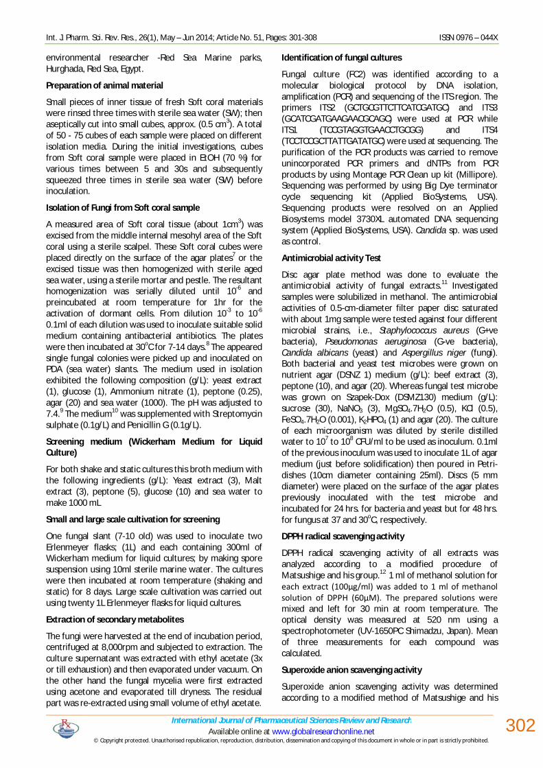
BLAST search for the fungus isolate FC2 revealed 99% similarity to Emericella unguis 8429. The phylogentic tree of this fungal isolate was also constructed (Fig.1).
Antimicrobial activity
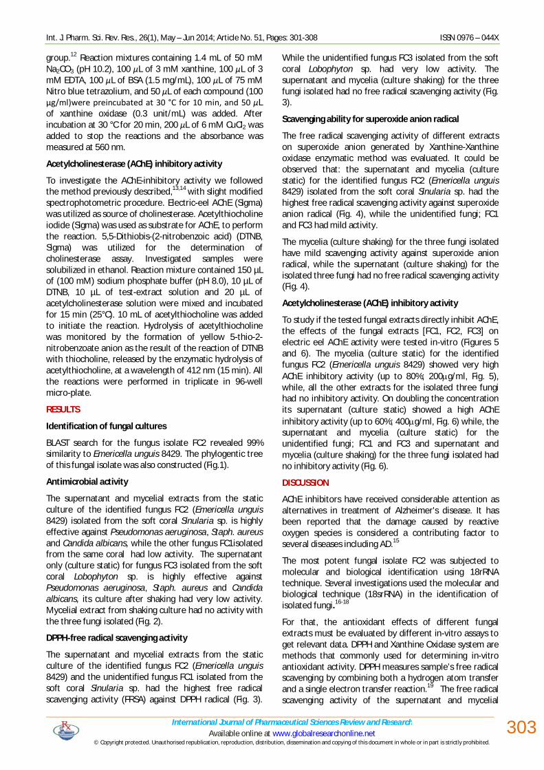
The supernatant and mycelial extracts from the static culture of the identified fungus FC2 (Emericella unguis 8429) isolated from the soft coral Sinularia sp. is highly effective against Pseudomonas aeruginosa, Staph. aureus and Candida albicans, while the other fungus FC1isolated from the same coral had low activity. The supernatant only (culture static) for fungus FC3 isolated from the soft coral Lobophyton sp. is highly effective against Pseudomonas aeruginosa, Staph. aureus and Candida albicans, its culture after shaking had very low activity. Mycelial extract from shaking culture had no activity with the three fungi isolated (Fig. 2).
DPPH-free radical scavenging activity
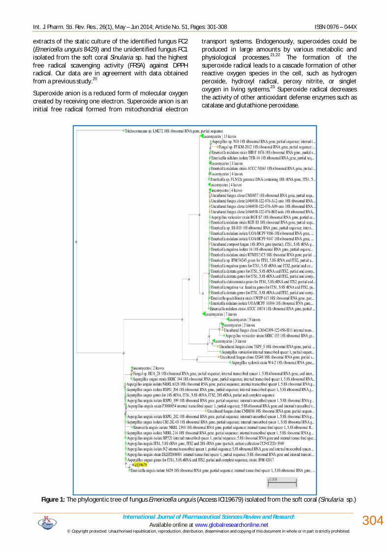
The supernatant and mycelial extracts from the static culture of the identified fungus FC2 (Emericella unguis 8429) and the unidentified fungus FC1 isolated from the soft coral Sinularia sp. had the highest free radical scavenging activity (FRSA) against DPPH radical (Fig. 3).
While the unidentified fungus FC3 isolated from the soft coral Lobophyton sp. had very low activity. The supernatant and mycelia (culture shaking) for the three fungi isolated had no free radical scavenging activity (Fig. 3).
Scavenging ability for superoxide anion radical
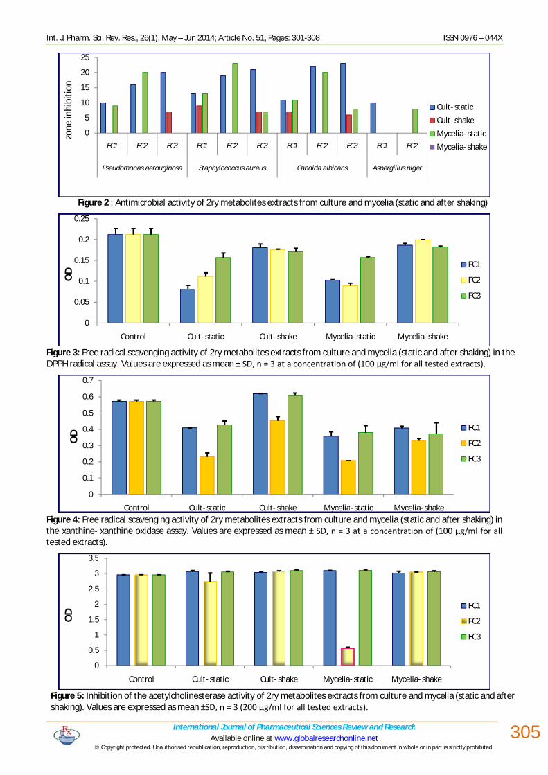
The free radical scavenging activity of different extracts on superoxide anion generated by Xanthine-Xanthine oxidase enzymatic method was evaluated. It could be observed that: the supernatant and mycelia (culture static) for the identified fungus FC2 (Emericella unguis 8429) isolated from the soft coral Sinularia sp. had the highest free radical scavenging activity against superoxide anion radical (Fig. 4), while the unidentified fungi; FC1 and FC3 had mild activity.
The mycelia (culture shaking) for the three fungi isolated have mild scavenging activity against superoxide anion radical, while the supernatant (culture shaking) for the isolated three fungi had no free radical scavenging activity (Fig. 4).
Acetylcholinesterase (AChE) inhibitory activity
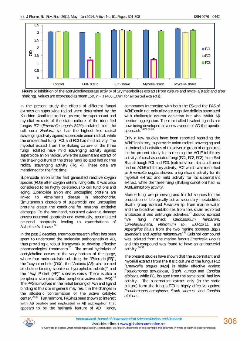
To study if the tested fungal extracts directly inhibit AChE, the effects of the fungal extracts [FC1, FC2, FC3] on electric eel AChE activity were tested in-vitro (Figures 5 and 6). The mycelia (culture static) for the identified fungus FC2 (Emericella unguis 8429) showed very high AChE inhibitory activity (up to 80%; 200g/ml, Fig. 5), while, all the other extracts for the isolated three fungi had no inhibitory activity. On doubling the concentration its supernatant (culture static) showed a high AChE inhibitory activity (up to 60%; 400g/ml, Fig. 6) while, the supernatant and mycelia (culture static) for the unidentified fungi; FC1 and FC3 and supernatant and mycelia (culture shaking) for the three fungi isolated had no inhibitory activity (Fig. 6).
DISCUSSION
AChE inhibitors have received considerable attention as alternatives in treatment of Alzheimer's disease. It has been reported that the damage caused by reactive oxygen species is considered a contributing factor to several diseases including AD.15
The most potent fungal isolate FC2 was subjected to molecular and biological identification using 18rRNA technique. Several investigations used the molecular and biological technique (18srRNA) in the identification of isolated fungi.16-18
For that, the antioxidant effects of different fungal extracts must be evaluated by different in-vitro assays to get relevant data. DPPH and Xanthine Oxidase system are methods that commonly used for determining in-vitro antioxidant activity. DPPH measures sample’s free radical scavenging by combining both a hydrogen atom transfer and a single electron transfer reaction.19 The free radical scavenging activity of the supernatant and mycelial
Int. J. Pharm. Sci. Rev. Res., 26(1), May – Jun 2014; Article No. 51, Pages: 301-308 ISSN 0976 – 044X
International Journal of Pharmaceutical Sciences Review and Research Available online at www.globalresearchonline.net
© Copyright protected. Unauthorised republication, reproduction, distribution, dissemination and copying of this document in whole or in part is strictly prohibited.
304
extracts of the static culture of the identified fungus FC2 (Emericella unguis 8429) and the unidentified fungus FC1 isolated from the soft coral Sinularia sp. had the highest free radical scavenging activity (FRSA) against DPPH radical. Our data are in agreement with data obtained from a previous study.20
Superoxide anion is a reduced form of molecular oxygen created by receiving one electron. Superoxide anion is an initial free radical formed from mitochondrial electron
transport systems. Endogenously, superoxides could be produced in large amounts by various metabolic and physiological processes.21,22 The formation of the superoxide radical leads to a cascade formation of other reactive oxygen species in the cell, such as hydrogen peroxide, hydroxyl radical, peroxy nitrite, or singlet oxygen in living systems.23 Superoxide radical decreases the activity of other antioxidant defense enzymes such as catalase and glutathione peroxidase.
Figure 1: The phylogentic tree of fungus Emericella unguis (Access ICI19679) isolated from the soft coral (Sinularia sp.)
Int. J. Pharm. Sci. Rev. Res., 26(1), May – Jun 2014; Article No. 51, Pages: 301-308 ISSN 0976 – 044X
International Journal of Pharmaceutical Sciences Review and Research Available online at www.globalresearchonline.net
© Copyright protected. Unauthorised republication, reproduction, distribution, dissemination and copying of this document in whole or in part is strictly prohibited.
305
Figure 2 : Antimicrobial activity of 2ry metabolites extracts from culture and mycelia (static and after shaking)
Figure 3: Free radical scavenging activity of 2ry metabolites extracts from culture and mycelia (static and after shaking) in the DPPH radical assay. Values are expressed as mean ± SD, n = 3 at a concentration of (100 µg/ml for all tested extracts).
Figure 4: Free radical scavenging activity of 2ry metabolites extracts from culture and mycelia (static and after shaking) in the xanthine- xanthine oxidase assay. Values are expressed as mean ± SD, n = 3 at a concentration of (100 µg/ml for all tested extracts).
Figure 5: Inhibition of the acetylcholinesterase activity of 2ry metabolites extracts from culture and mycelia (static and after shaking). Values are expressed as mean ±SD, n = 3 (200 µg/ml for all tested extracts).
0
5
10
15
20
25
FC1 FC2 FC3 FC1 FC2 FC3 FC1 FC2 FC3 FC1 FC2
Pseudomonas aerouginosa Staphylococcus aureus Candida albicans Aspergillus niger
Cult- staticCult- shakeMycelia- staticMycelia- shake
zone
inhi
bitio
n
0
0.05
0.1
0.15
0.2
0.25
Control Cult- static Cult- shake Mycelia- static Mycelia- shake
FC1
FC2
FC3
OD
0
0.1
0.2
0.3
0.4
0.5
0.6
0.7
Control Cult- static Cult- shake Mycelia- static Mycelia- shake
FC1
FC2
FC3
OD
0
0.5
1
1.5
2
2.5
3
3.5
Control Cult- static Cult- shake Mycelia- static Mycelia- shake
FC1
FC2
FC3
OD
Int. J. Pharm. Sci. Rev. Res., 26(1), May – Jun 2014; Article No. 51, Pages: 301-308 ISSN 0976 – 044X
International Journal of Pharmaceutical Sciences Review and Research Available online at www.globalresearchonline.net
© Copyright protected. Unauthorised republication, reproduction, distribution, dissemination and copying of this document in whole or in part is strictly prohibited.
306
Figure 6: Inhibition of the acetylcholinesterase activity of 2ry metabolites extracts from culture and mycelia(static and after shaking). Values are expressed as mean ±SD, n = 3 (400 µg/ml for all tested extracts).
In the present study the effects of different fungal extracts on superoxide radical were determined by the Xanthine -Xanthine oxidase system; the supernatant and mycelial extracts of the static culture of the identified fungus FC2 (Emericella unguis 8429) isolated from the soft coral Sinularia sp. had the highest free radical scavenging activity against superoxide anion radical, while the unidentified fungi; FC1 and FC3 had mild activity. The mycelial extract from the shaking culture of the three fungi isolated have mild scavenging activity against superoxide anion radical, while the supernatant extract of the shaking culture of the three fungi isolated had no free radical scavenging activity (Fig. 4). These data are mentioned for the first time.
Superoxide anion is the first generated reactive oxygen species (ROS) after oxygen enters living cells. It was once considered to be highly deleterious to cell functions and aging. Superoxide anion and uncoupling proteins are linked to Alzheimer’s disease in mitochondria. Simultaneous disorders of superoxide and uncoupling proteins create the conditions for neuronal oxidative damages. On the one hand, sustained oxidative damage causes neuronal apoptosis and eventually, accumulated neuronal apoptosis, leading to exacerbations of Alzheimer’s disease.24
In the past 2 decades, enormous research effort has been spent to understand the molecular pathogenesis of AD, thus providing a robust framework to develop effective pharmacological treatments.25 The actual hydrolysis of acetylcholine occurs at the very bottom of the gorge, where four main catalytic sub-sites; the “Esteratic (ES)”, the “oxyanion hole (OX)”, the “Anionic (AS), also termed as choline binding subsite or hydrophobic subsite)” and the “Acyl Pocket (AP)” subsites exists. There is also a peripheral site (also called peripheral active site, PAS).13 The PAS is involved in the initial binding of Ach and ligand binding at this site in general may result in the changes in the allosteric conformation of the active catalytic center.26-29 Furthermore, PAS has been shown to interact with Aβ peptide and implicated in Aβ aggregation that appears to be the hallmark feature of AD. Hence,
compounds interacting with both the ES and the PAS of AChE could not only alleviate cognitive deficits associated with cholinergic neuron depletion but also inhibit Aβ peptide aggregation. These so-called bivalent ligands are now being developed as a new avenue of AD therapeutic approach.14,27,30-33
Only a few studies have been reported regarding the AChE inhibitory, superoxide anion radical scavenging and antimicrobial activities of this diverse group of organisms. In the present study for screening the AChE inhibitory activity of coral associated fungi (FC1, FC2, FC3) from Red Sea, although FC1 and FC3, (extracts from static cultures) had no AChE inhibitory activity, FC2 which was identified as Emericella unguis showed a significant activity for its mycelial extract and mild activity for its supernatant extract, while the three fungi (shaking condition) had no AChE inhibitory activity.
Marine fungi are promising and fruitful sources for the production of biologically active secondary metabolites. Swathi group isolated Fusarium sp. from marine water and the bioactive metabolites from this strain exhibited antibacterial and antifungal activities.34 Jadulco isolated five fungi named: Caldosporium herbarum, Curvularialunata, Penicillium sp., E00-12/11 and Aspergillus flavus from the two marine sponges Jaspis splenderis and Agelas nakamourai.35 Guisinol compound was isolated from the marine fungus Emericella unguis and this compound was found to have an antibacterial activity. 36,37
The present studies have shown that the supernatant and mycelial extracts from the static culture of the fungus FC2 (Emericella unguis 8429] is highly effective against Pseudomonas aeruginosa, Staph. aureus and Candida albicans, while FC1 isolated from the same coral had low activity. The supernatant extract only (in the static culture) form the fungus FC3 is highly effective against Pseudomonas aeruginosa, Staph. aureus and Candida albicans.
0
0.5
1
1.5
2
2.5
3
3.5
Control Cult- static Cult- shake Mycelia- static Mycelia- shake
FC1
FC2
FC3
OD
Int. J. Pharm. Sci. Rev. Res., 26(1), May – Jun 2014; Article No. 51, Pages: 301-308 ISSN 0976 – 044X
International Journal of Pharmaceutical Sciences Review and Research Available online at www.globalresearchonline.net
© Copyright protected. Unauthorised republication, reproduction, distribution, dissemination and copying of this document in whole or in part is strictly prohibited.
307
CONCLUSION
It could be concluded that, the isolated marine fungus Emericella unguis 8429 showed different biological activities as acetylcholinesterase inhibitor (reported for the first time), antioxidant, antibacterial and antifungal rather than the other unidentified two fungi (FC1, FC3).Our study provides (for the first time) primary evidence suggesting that the isolated marine fungus Emericella unguis in further in-vivo studies could play an important role as acetylcholinesterase inhibitor, besides its antioxidant, antibacterial and antifungal activities.
Acknowledgements: This work is financially supported by the bilateral projects within the Executive Programmed of Scientific and Technological Cooperation between Arab Republic of Egypt and Italian Republic for the years 2013 – 2015. Project no. [A2-12-15].
REFERENCES
1. Hebert LE, Scherr PA, Beckeff LA. Age-specific incidence of Alzheimer’s Disease in a community population. AMA 273, 1995, 1354-1359.
2. Schneider LJ. Treatment of Alzheimer’s disease with cholinesterase inhibitors. ClinGeriatr Med 17, 2001, 337-339.
3. Bugni T S, Ireland C M. Marine-derived fungi: a chemically and biologically diverse group of microorganisms. Nat. Prod. Rep. 21, 2003, 143-163.
4. Saleem M, Ali M S, Hussain S, Jabbar A, Ashraf M, Lee Y S. Marine natural products of fungal origin. Nat. Prod. Rep. 24, 2007, 1142-1152.
5. Liberra K and Lindequist U. Marine fungi- a prolific resource of biologically active natural products? Pharmazie 50(H9), 1995, 583-588.
6. Swathi J, Narendra K, Sowjanya KM, Satya AK. Marine fungal metabolites as a rich source of bioactive compounds. African Journal of Biochemistry Research, 7, 2013, 184-196.
7. Subramani R, Kumar R, Prasad P,Aalbersberg W. Cytotoxic and antibacterial substances against multi-drug resistant pathogens from marine sponge symbiont: citrinin, a secondary metabolite of Penicillium sp.Asian Pac J Trop Biomed.3, 2013,291-296.
8. Manilal A,Sabarathnan B,Kiran G S,Sujith S,Shakir C,Selvin J. Antagonisitic potential of marine sponge associated fungi Aspergillusclavatus MFD15. Asian J. Med. Sci. 2(4), 2010, 195-200.
9. Holler U. Isolation, biological activity and secondary metabolite investigations of marine-derived fungi and selected host sponges, PhD thesis, 1999.
10. Rusman Y.Isolation of new secondary metabolites from sponge-associated and plant plant-derived endophytic fungi, PhD thesis, 2006.
11. Srinivasan D, Sangeetha N, Suresh T, Lakshmanaperumalsamy P. Antimicrobial activity of certain Indian medicinal plants used in folkloric medicine. J. Ethnopharmacol., 74, 2001, 217-220.
12. Matsushige K, Basnet P, Kadota S and Namba T. Potent free radical scavenging activity of dicaffeoylquinic acid derivatives from propolis. J. Trad. Med., 13, 1996, 217-228.
13. Khan I, Nisar M, Khan N, Saeed M, Nadeem S, Rehman FU, Karim N, Kaleem WA, Qayum M, Ahmad H, Khan IA. Structural insights to investigate conypododiol as a dual cholinesterase inhibitor from Asparagus adscendens. Fitoterapea. 81, 2010, 1020-25.
14. Khan I, Nisar M, Shah MR, Shah H, Gilani SN, Gul F, Abdullah SM, Ismail M, Khan N, Kaleem WA, Qayum M, Khan HO. Anti-inflammatory activities of Taxusabietane A isolated from Taxuswallichiana Zucc.Fitoterapea, 82, 2011, 1003-07.
15. Giordani RB, Pagliosa LB, Henriques AT, Zuanazzi JAS. Investigacao do Potencial Antioxidante e anticolinesterasico de Hippeastrum (Amaryllidaceae). Quim, Nova, 31, 2008, 2042-2046.
16. Kjer J. New natural products from endophytic fungi from mangrove plants - structure elucidation and biological screening, PhD, Dusseldorf university, Germany, (2010).
17. Ebrahim W,Aly A, Wray V,Proksch P,Debbab A. Unusual octalactones from Corynesporacassiicola, an endophyte of Lagunculariaracemosa. Tetrahedron Letters, 54,2013, 6611-6614.
18. Li Y,Wu CM, Liu D,Proksch P,Guo P, Lin WH. Chartarlactams A-P, phenylspirodrimanes from the sponge-associated fungus Stachybotryschartarum with antihyperlipidemic activities. J. Nat. Prod. 77, 2014, 138-147.
19. Prior RL, Wu X, Schaich. Standerdized methods for the determination of antioxidant capacity and phenolics in foods and dietary supplements. J. Agric. Food Chemistry, 53, 2005, 4290-4302.
20. Abdel-Monem N, Abdel-Azeem AM, El Ashry ESH, Ghareeb DA, Adam AN. Assessment of Secondary Metabolites from Marine-Derived Fungi as Antioxidant. Journal of Medicinal Chemistry, 3, 2013, 60-73 .
21. Blaszczyk J, Kedziora J, Luciak M, Sibinska E, Trznadel K, Pawlicki L. Effect of morphine and naloxone on oxidative metabolism during experimental renal ischemia and reperfusion. Experimental Nephrology ,2, 1994, 364-370.
22. Bedard L, Young MJ, Hall D, Paul T, and Ingold KU. Quantitative study on the peroxidation of human low-density lipoprotein initiated by superoxide and by charged and neutral alkylperoxl radicals. Journal of the American chemistry society, 123, 2001, 12439-12448.
23. Lee J, Koo N, and Min DB. Reactive oxygen species, aging and antioxidative nutraceuticals. Comprehensive Reviews in Food science and food safety, 3, 2004, 21-33.
24. Wu Z, Zhao Y, Zhao B. Superoxide Anion, Uncoupling Proteins and Alzheimer’s Disease. J. Clin. Biochem. Nutr., 46, 2010, 187–194.
25. Dekoski S T. Pathology and pathways of Alzheimer’s disease with an update on new development and treatment. J. Am.Geriatr. Soc., 51, 2003, 314-320.
Int. J. Pharm. Sci. Rev. Res., 26(1), May – Jun 2014; Article No. 51, Pages: 301-308 ISSN 0976 – 044X
International Journal of Pharmaceutical Sciences Review and Research Available online at www.globalresearchonline.net
© Copyright protected. Unauthorised republication, reproduction, distribution, dissemination and copying of this document in whole or in part is strictly prohibited.
308
26. Harel M, Quinn DM, Nair HK, Silman I, Sussman JL. The X-ray structures of transition state analog complex reveal that molecular origin of the catalytic power of the substrate specificity of acetylcholinesterase. J Am ChemSoc. ,118, 1996, 2340–46.
27.
Nisar M, Kaleem WA, Khan I, Adhikari A, Khan N, Shah MR, Khan IA, Qayum M, Samiullah, Ismail M, Aman A. Molecular simulations probing Kushecarpin A as a new lipoxygenase inhibitor. Fitoterapea, 82, 2011, 1008-11.
28. Szegletes T, Mallender WD, Rosenberry TL. Non-equilibrium analysis alters the mechanistic interpretation of inhibition of acetylcholinesterase by peripheral site ligands. Biochemistry, 37, 1998, 4206-16.
29. Zhang Y, Kua J, McCammon JA. Role of the catalytic triad and oxyanion hole in acetylcholinesterase catalysis: An ab initio QM/MM study. J Am ChemSoc., 124,2002, 10572–77.
30. Gaggeri R, Rossi D, Hajikarimian N, Martino E, Bracco F, Grisoli P, Dacarro C, Leoni F, Mascheroni G, Collina S, Azzolina O. Preliminary study on TNFα-blocker activity of AmygdaluslycioidesSpach extracts. Open Nat Prod J. 3, 2010, 20-25.
31. Gaggeri R, Rossi D, Collina S, Mannucci B, Baierl M, Juza M. Quick development of an analytical enantioselective HPLC separation and preparative scale-up for the flavonoid Naringenin. J Chromatogr A., 1218, 2011, 5414-22.
32. Martino E, Ramaiola I, Urbano M, Bracco F, Collina S. Microwave-assisted extraction of coumarin and related compounds from melilotusofficinalis (l.) pallas as an alternative to soxhlet and ultrasound assisted extraction. J. Chromatogr A. 1125, 2006, 147–51.
33. Martino E, Collina S, Rossi D, Bazzoni D, Gaggeri R, Bracco F, Azzolina O. Influence of the extraction mode on the yield of hyperoside, vitexin and vitexin-2”-O-rhamnoside from CrataegusmonogynaJacq. [Hawthorn]. Phytochem Ana., 19, 2008, 534-40.
34. Swathi J,Sowjanya K M,Narendra K, Krishna Satya A. Bioactivity Assay of an Isolated MarineFusariumsps.International Journal of Bio-Science and Bio-Technology, 5(5), 2013, 179-186.
35. Jadulco R C. Isolation and Structure Elucidation of Bioactive Secondary Metabolites from Marine Sponges and Sponge-derived Fungi. PhD, Würzburg University, Germany, 2002.
36. Punyasloke B, Balsam TM, Phillip C. The current status of natural products from marine fungi and their potential as anti-infective agents. J. Ind. Microbiol. Biotechnol., 33, 2006, 325-337.
37. Joan N, Halfdan NP.Fungal depside, guisinol, from a marine derived strain of Emericella unguis.” Phytochemistry, 50, 1999, 263-265.
Source of Support: Nil, Conflict of Interest: None.