research article ethyl acetate fraction of amomum...
TRANSCRIPT

Research ArticleEthyl Acetate Fraction of Amomum xanthioides ExertsAntihepatofibrotic Actions via the Regulation of FibrogenicCytokines in a Dimethylnitrosamine-Induced Rat Model
Sung-Bae Lee Hyeong-Geug Kim Hyo-Seon Kim Jin-Seok Lee Hwi-Jin ImWon-Yong Kim and Chang-Gue Son
Liver and Immunology Research Center Daejeon Oriental Hospital of Oriental Medical College of Daejeon University176-9 Daeheung-ro Jung-gu Daejeon 301-724 Republic of Korea
Correspondence should be addressed to Chang-Gue Son cksondjuackr
Received 25 May 2016 Revised 8 July 2016 Accepted 16 July 2016
Academic Editor Gabino Garrido
Copyright copy 2016 Sung-Bae Lee et al This is an open access article distributed under the Creative Commons Attribution Licensewhich permits unrestricted use distribution and reproduction in any medium provided the original work is properly cited
Amomum xanthioides has been traditionally used to treat diverse digestive system disorders in the Asian countries We investigatedantihepatofibrotic effects of ethyl acetate fraction ofAmomumxanthioides (EFAX) Liver fibrosis is induced by dimethylnitrosamine(DMN) injection (intraperitoneally 10mgkg of DMN for 4 weeks to Sprague-Dawley rats) EFAX (25 or 50mgkg) silymarin(50mgkg) or distilled water was orally administered every dayTheDMN injection drastically altered body and organmass serumbiochemistry and platelet count while EFAX treatment significantly attenuated this alteration Severe liver fibrosis is determinedby trichrome staining and measurement of hydroxyproline contents EFAX treatment significantly attenuated these symptoms aswell as the increase in oxidative by-products of lipid and protein metabolism in liver tissues DMN induced a dramatic activationof hepatic stellate cells and increases in the levels of protein and gene expression of transforming growth factor-beta (TGF-120573) platelet derived growth factor-beta (PDGF-120573) and connective tissue growth factor (CTGF) Immunohistochemical analysesrevealed increases in the levels of protein and gene expression of 120572-smooth muscle actin These alterations were significantlynormalized by EFAX treatment Our findings demonstrate the potent antihepatofibrotic properties of EFAX via modulation offibrogenic cytokines especially TGF-120573 in the liver fibrosis rat model
1 Introduction
Liver fibrosis is a pathological consequence of the woundhealing response to chronic liver injuries leading to theexcessive accumulation of extracellular matrix (ECM) pro-tein in hepatic tissues [1] Liver fibrosis can be induced byvarious causes including chronic alcohol abuse hepatitisviral infections metabolic disorders and autoimmune dis-ease [2 3] Liver fibrosis is reversible in certain conditions butcan commonly progress to liver cirrhosis the final step in liverfibrosis if no proper treatment is given [4] Worldwide 22of total deaths were caused by liver cirrhosis in 2013 [5]
Therefore the development of liver fibrosis is criticalwith respect to the clinical outcome of patients with chronicliver injuries ECMs such as 120572-smoothmuscle actin (120572-SMA)and collagens are generated by activated hepatic stellate cells
(HSCs) [6] ldquoQuiescentrdquo HSCs convert to myofibroblasts viaactivation by fibrogenic cytokines including transforminggrowth factor- (TGF-) 120573 platelet derived growth factor-(PDGF-) 120573 and connective tissue growth factor (CTGF)[7] Accordingly the inhibition of the HSC activation orthe modulation of the above three fibrogenic cytokines istherapeutic target for treatment of liver fibrosis
MeanwhileAmomumxanthioidesWall exBaker (AmomiFructus) is a well-known medicinal herb that has been usedclinically to treat digestive system disorders for more thana thousand years in Asia A xanthioides has been tradi-tionally used to treat indigestion diarrhea and flatulencein China [8] Japan [9] and Thailand [10] which are thecommon complaints in patients with chronic liver diseasesWe previously reported the hepatoprotective effect of Axanthioides in a thioacetamide and a bile duct-ligationmodel
Hindawi Publishing CorporationEvidence-Based Complementary and Alternative MedicineVolume 2016 Article ID 6014380 14 pageshttpdxdoiorg10115520166014380
2 Evidence-Based Complementary and Alternative Medicine
as well as the anti-inflammatory effects in a gastritis model[11ndash13] In additionA xanthioideshas beenwidely prescribedfor the treatment of various liver diseases [14 15] Furtherstudies however have been required especially regarding thepractical dose and a detailed explanation of the pharmaco-logical actions of A xanthioides We therefore compared theantihepatofibrotic capacities of several A xanthioides frac-tions based on in vitro experiments and determined the ethylacetate fraction of Amomum xanthioides (EFAX) with themost potent pharmacological activity at relatively very lowconcentrations
We herein investigated the antihepatofibrotic effects of alow-dose EFAX and explored the underlying mechanisms inrat model of DMN-induced liver fibrosis
2 Materials and Methods
21 Reagents and Chemicals Dimethylnitrosamine (DMN)hydroxyproline p-dimethylaminobenzaldehyde 1133-tet-raethoxypropane (TEP) chloramine-T potassium chloride(KCl) Folin-Ciocalteursquos phenol reagent and hydrochloricacid (HCl) were purchased from Sigma (St Louis MO)Thiobarbituric acid (TBA) was purchased from LancasterCo (Lancashire UK) Histofine was purchased fromNichireiBiosciences (Tokyo Japan) Sodium carbonate was purchasedfrom Kanto perchloric acid and aluminum chloride waspurchased from Junsei Chemical (Tokyo Japan)
22 Preparation of Fractions for A xanthioides Korean Phar-macopoeia standard Amomi Fructus (a dried fruit of Axanthioides) was purchased from Jeong-Seong Pharmacy(Daejeon South Korea) and its identity was confirmed byprofessor Sang-Hoon Oh (Daejeon University) The Amo-mum xanthioides were washed twice using tap water andrinsed with distilled water (DW) The sample was then com-pletely dehydrated by drying in an oven overnight (60∘C)After drying 10 kg samples of A xanthioides were boiledin 100 L of DW for 3 h at 100∘C centrifuged (3000timesg) for20min and then filteredWefirstly obtained thewater extractof Amomum xanthioides (WAX) and the final yield (ww)was 112 (total 112 g voucher specimen numberWAX-2014-W007)
To obtain the methanol and ethyl acetate fractions of Axanthioides we used an organic solvent extraction method(Figure 1) Briefly 10 kg of A xanthioides was ground andextracted in 100 L of absolute methanol for 7 days withshaking On the 7th day 100mL DW was added to 900mLmethanol extract Next the extracts were further fractionatedthree times with petroleum ether (3 times 1 L) to isolate themethanol fraction of Amomum xanthioides (MFAX) Then100mLof the petroleumether extract wasmixedwith 900mLDW (3 times 1 L) and further fractionated two times with ethylacetate (2 times 1 L) to isolate the ethyl acetate fraction ofAmomum xanthioides (EFAX) Finally we obtained a portionof the 100 MFAX and EFAX The final fraction yieldswere 662 (ww) for MFAX (total 662 g voucher specimennumber MFAX-2014-MF001) and 019 (ww) for EFAX
(total 19 g voucher specimen number EFAX-2014-EF002)WAX MFAX and EFAX were stored at minus70∘C and dissolvedin DW for the experiments
23 Fingerprinting Analysis of WAX MFAX and EFAX Todetermine the reproducibility of WAX MFAX and EFAXsamples fingerprinting was performed using ultra-high-performance liquid chromatography-tandem mass spec-trometry (UHPLC-MSMS) Five milligram aliquots of theWAXMFAX and EFAX samples were dissolved in 1mL 90methanol and the solution was filtered Sample solutionsof 10 120583L were subjected to UHPLC-MSMS using an LTQOrbitrap XL linear ion-trapMS Spectrometer (San Jose CA)Separationwas performed on anAccelaUHPLC systemusingan Acquity BEH C18 column (17 120583m 100 times 21mm WatersMilford Connecticut)The columnwas eluted at a flow rate of04mLmin usingwater (in 01 formic acid) and acetonitrile(in 01 formic acid) which were used as mobile phases Aand B respectively The following gradients were applied0-1min 0-1 B in A 1ndash7min 1ndash100 B in A 7ndash10min100ndash1 B in A (linear gradient) The compositional analyseswere conducted using a photodiode array at 200ndash600 nmThe full-scan mass spectra were acquired at 150ndash1500119898119911in positive and negative modes An Orbit rap analyzer wasused for high-resolution mass data acquisition with a massresolving power of 30000 FWHM at 400119898119911 Tandemmass(MSMS) spectra were acquired in data-dependent mode bycollision-induced dissociation The quantitative analysis ofthe major three compounds in EFAX including procyanidinB2 catechin and quercitrin was performed using UHPLC-MSMS (Figure 2)
24 Determination of Total Flavonoid and Phenolic ContentsThe total flavonoid contents of WAX MFAX and EFAXwere measured using a previously developed method [16]Briefly 05mL solutions of each sample (10 wv in absolutemethanol) were separately mixed in a flavonoid assay buffer(15mL methanol 100120583L 10 aluminum chloride 100 120583L1M potassium acetate and 28mL DW) After 30min ofincubation at room temperature 200120583L of the mixture wastransferred to a 96-well plate and absorbance was measuredat 415 nm using a spectrophotometer (Palo Alto CA) Thecalibration curve was obtained by preparing quercetin solu-tions at concentrations from 125 to 100 120583gmL in methanolThe total flavonoid contents values were expressed in terms ofquercetin equivalent (mgg of dry mass) which is a commonreference for flavonoid
The total phenolic contents were measured using theFolin Ciocalteu method [17] The WAX MFAX and EFAXsamples were mixed with 25mL 02N Folin-Ciocalteursquosphenol reagent for 5min after which 2mL 75 gL sodiumcarbonate was added After 2 h of incubation at room tem-perature the optical density of the reaction product wasread at 760 nm using a spectrophotometer The standardcurve was prepared using 50 to 250mgmL solutions ofgallic acid in a methanol-water solution (1 1 vv) The totalphenolic contents valueswere expressed in terms of gallic acid
Evidence-Based Complementary and Alternative Medicine 3
Amomum xanthioides
Methanol extract
Water extract (WAX)
Added distilled water (final 10)
Methanol
fraction (MFAX)
Petroleum ether fraction
Water fraction Ethyl acetate
fraction (EFAX)
Added distilled water (final 90)
(1 kg of powder) (final yield = 112)
(final yield = 662)
Mixed with petroleum ether (1 1)
Added methanol 10 L (RT gt1 week)
Mixed with ethyl acetate (1 1)
(final yield = 019)
Figure 1 Scheme for preparation of EFAX
Rel
ativ
eab
un
dan
ce
WAX
0 2 4 6 8 10 12 14 16 18 20
0
50
100
RT (min)
(a)
Rel
ativ
eab
un
dan
ce
MFAX
0 2 4 6 8 10 12 14 16 18 20
0
50
100
RT (min)
(b)
Rel
ativ
eab
un
dan
ce
RT (min)
EFAX
Procyanidin B2 Catechin Quercitrin
0 2 4 6 8 10 12 14 16 18 20
0
50
100
(c)
Compounds RT (min) MW (g) WAX MFAX EFAX
Procyanidin B2 431 57817
Catechin 475 29008 ND
Quercitrin 724 44811
143 plusmn 005
015 plusmn 003
407 plusmn 005
033 plusmn 007
156 plusmn 008
759 plusmn 006
091 plusmn 005
597 plusmn 009
(120583gmg)Mean plusmn SD
(d)
Figure 2 Fingerprinting analysis WAX MFAX and EFAX were subjected to UHPLC-MSMS Chromatogram of (a) WAX (b) MFAX and(c) EFAX (d) Quantitative analysis of the WAX MFAX and EFAX
equivalent (mgg of dry mass) which is a common referencefor phenolic contents
25 Animals and Experimental Design A total of 30 maleSprague-Dawley rats (6 weeks old 160ndash180 g) were purchased
from Daehanbiolink (Choong-book South Korea) Sevendays of acclimation was allowed at 22 plusmn 2∘C under a 12 hlight12 h dark cycle All of the animals had free access towater and standard chow diet After acclimation all of therats were divided into five groups (119899 = 6 for each group)
4 Evidence-Based Complementary and Alternative Medicine
and orally administered with DW EFAX (25 or 50mgkg)or silymarin (50mgkg) daily for 4 weeks To induce liverfibrosis 10mgkg DMN was intraperitoneally injected on 3consecutive days per week for 4 weeks The groups wereas follows (1) naive group (DW with 09 neutral saline)(2) control group (DW with 10mgkg DMN) (3) EFAX25 (25mgkg EFAX with 10mgkg DMN) (4) EFAX 50(50mgkg EFAX with 10mgkg DMN) and (5) silymarin 50(50mgkg silymarin with 10mgkg DMN) The naive groupwas also intraperitoneally injected with same volume of 09neutral saline for 4 weeks Body weight was measured twicea week and once shortly before sacrifice
On the final day of the experiment the animals weresacrificed under ether anesthesia and whole blood wasisolated from the abdominal aorta The liver and spleentissues were removed and weighed and then collected forbiochemical analyses and other measurements The animalexperiment was conducted in accordance with the Guide forthe Care and Use of Laboratory Animals prepared by theUS National Institutes of Health and was approved by theInstitutional Animal Care and Use Committee of DaejeonUniversity (DJUARB2015-007)
26 Serum Biochemical Analysis Whole blood was isolatedfrom the abdominal aorta and transferred to an EDTA-coated tube (Plymouth UK) Serum samples were obtainedfor subsequent separation after 1 hour of blood clottingusing Vacutainer tubes (Plymouth UK) The platelet countsin each sample were measured using a HEMA VET 850automatic analyzer (Oxford CT) The serum was separatedby centrifugation (3000timesg 15min) following blood clottingThe serum levels of aspartate transaminase (AST) alaninetransaminase (ALT) and total bilirubin were determinedusing an Auto Chemistry Analyzer (Emeryville CA)
27 Histomorphology and Immunohistochemistry for 120572-SmoothMuscle Actin (120572-SMA) On the final day of the exper-iment the liver tissues were removed and weighedThe tissuesamples were fixed with a 10 neutral formalin solutionThe tissues then underwent general processing Paraffin-embedded liver tissues were sectioned (5 120583m) and stainedwith hematoxylin and eosin (HampE) or Massonrsquos trichromedye for histopathological evaluation Immunohistochemistrywas performed with an anti-120572-SMA mouse monoclonalantibody (Cambridge UK) and a Vectastain ABC kit (VectorBurlingame CA) The samples were visualized with Tetram-ethylbenzidine (TMB) substrate and examined under anoptical microscope (times100 magnification)
The liver histological examination was examined andgraded by two independent investigators who were blindto samplesrsquo groups The samples were graded according topublished criteria for magnitude analysis and inflammationThe histomorphological changes for inflammation were alsoscored (times100) using HampE staining (grade 0 naive absenceof pathology to lt5 of maximum pathology grade 1 lt10of maximum pathology grade 2 15 to 20 of maximumpathology grade 3 gt20 of maximum pathology) [18] AMETAVIR fibrosis score from 0 to 4 was used to differentiate
the levels of liver fibrosis Briefly stage 0 indicates no scarringstage 1 indicates minimal scarring stage 2 indicates scarringthat extends outside the vascularized area of the liver stage 3indicates bridging fibrosis that has spread and connected tofibrotic areas and stage 4 indicates advanced scarring of theliver or cirrhosis [19] The number of 120572-SMA positive cells(stained blue-violet color) was also calculated and expressedas a fold change after normalization to the naive group Theinflammation and METAVIR scores and the number of 120572-SMA positive cells were calculated using ImageJ analysissoftware v 167 (NIH Rockville MD)
28 Determination of Hydroxyproline in Liver TissuesHydroxyproline determination was performed with a slightmodification of a previously method described [20] Brieflyliver tissues (200mg) stored at minus70∘C were homogenizedin 2mL 6N HCl and incubated overnight at 110∘C Afterfiltering the acid hydrolysates using a 045 120583m filter (TokyoJapan) 50 120583L samples or hydroxyproline standards in 6NHCl were incubated at 60∘C to dry The dried sampleswere dissolved with methanol (50 120583L) after which 12mL50 isopropanol and 200120583L of chloramine-T solutionwere added to each same sample The samples were thenincubated at room temperature for 10min After incubationEhrlichrsquos solution (13mL) was added and the sampleswere further incubated at 50∘C for 90min The opticaldensity of the reaction product was read at 558 nm using aspectrophotometer A standard curve was constructed usingserial twofold dilutions of a 1mg hydroxyproline solution
29 Determination of Lipid Peroxidation and Protein CarbonylContents in Liver Tissues The levels of malondialdehyde(MDA the final product for lipid peroxidation) in theliver tissues were determined using TBA reactive substancesmethod (TBARS) as described previously [21] The concen-tration of TBARS was expressed as 120583mol per gram of tissueusing TEP as a standardThe protein carbonyl contents in theliver tissues were determined according to themanufacturerrsquosprotocol [22]
210 Determination of the Levels of TGF-1205731 PDGF-BB CTGFandTIMP-1 in Liver Tissues One hundredmilligrams of livertissues was homogenized with RIPA buffer and centrifugedat 10000timesg for 15min at 4∘C The supernatant fraction wasused to determine the levels of fibrosis-related cytokinesTheprotein levels of TGF-1205731 PDGF-BB and tissue inhibitor ofmatrix metalloprotease (TIMP) in the liver tissue were alsodetermined using an ELISA kit (RampD Systems MinneapolisMN) The quantification of CTGF was performed usinga modification of the sandwich ELISA method describedpreviously [23] Briefly a 96-well ELISA plate was coatedwith100mL of goat polyclonal anti-rat antibody at a concentrationof 10mgmL in PBS and 002 sodium azide overnightAfter incubation with blocking buffer (PBS 002 sodiumazide and 1 bovine serum albumin) and washing (fourtimes) 50mL of the sample or recombinant human CTGFstandard was added for 1 hour Then 100mL of the primaryrabbit polyclonal anti-goat antibody (2mgmL) and 50mL of
Evidence-Based Complementary and Alternative Medicine 5
Table 1 Primer sequences used in this experiment
Gene name Primer sequence
Collagen 1a1 51015840-GAT CCT GCC GAT GTC GCT AT-31015840
31015840-TGT AGG CTA CGC TGT TCT TGC A-51015840
120572-SMA 51015840-GAC CCT CTT CCA GCC ATC TTT-31015840
31015840-GTC CTT CCT GAT GTC AAT ATC ACA CT-51015840
TGF-120573 51015840-AGG AGA CGG AAT ACA GGG CTT T-31015840
31015840-AGC AGG AAG GGT CGG TTC AT-51015840
PDGF-BB 51015840-ACC ACT CCA TCC GCT CCT TT-31015840
31015840-TGT GCT CGG GTC ATG TTC AA-51015840
CTGF 51015840-GTG TGT GAT GAG CCC AAG GA-31015840
31015840-CAG TTG GCT CGC ATC ATA GTT G-51015840
TIMP-1 51015840-ATG GAG AGC CTC TGT GGA TAT GTC-31015840
31015840-AGG CAG TGA TGT GCA AAT TTC C-51015840
MMP-2 51015840-TGT GGC AGC CCA TGA GTT C-31015840
31015840-TCG GAA GTT CTT GGT GTA GGT GTA-51015840
Smad7 51015840-TGC AAC CCC CAT CAC CTT AG-31015840
31015840-GAC AGT CTG CAG TTG GTT TGA GA-51015840
BAMBI 51015840-TTA TGT TGG CCT TGC GAA TG-31015840
31015840-TGG TGT CCA TGG AAG CTG TAG T-51015840
120573-actin 51015840-AGG CCA ACC GTG AAA AGA TG-31015840
31015840-CCA GAG GCA TAC AGG GAC AAC-51015840
the secondary donkey anti-rabbit IgG-HRP antibody (both1 2000) were added (Santa Cruz Biotechnology Germany)CTGFwas quantified bymeasuring the absorbance at 405 nmaftermixing the substrate solution and stopping solution (2NH2SO4)
211 Real-Time PCR for Gene Expression in HSC-T6 Cells orLiver Tissues HSC-T6 cells (2 times 106) were seeded in six-well plates with 2mL DMEM with 10 FBS and incubatedovernight at 37∘C and 5 CO2 The cell culture media wasthen changed to serum-free DMEM Next each sampleof WAX MFAX or EFAX (25 or 50 120583gmL dissolved inmethanol initially and then DMEM) was added to the wellsAfter 6 h of incubationwith the samples 1 ngmLTGF-1205731 wastreated for 12 hours The mRNA of HSC-T6 cells or liver tis-sues was isolated using QIAzol reagent (Germantown MD)and used for complementary DNA (cDNA) synthesis usingstandard protocols Real-time PCR was performed using aniQ5 instrumentThe primer sequences were used that is givenin Table 1 120573-actin levels were used for normalization
212 Western Blot Analysis HSC-T6 cells (2 times 106) wereseeded in six-well plates with 2mLDMEMwith 10 FBS andincubated overnight at 37∘C and 5 CO2 The cell culturemedia was then changed to serum-free DMEM Next eachsample of WAX MFAX or EFAX (25 or 50 120583gmL dissolvedin methanol initially and then DMEM) was added to thewells After 6 h of incubation with the samples 1 ngmLTGF-1205731 was treated for 24 hours The protein of HSC-T6
was extracted by RIPA buffer Each sample was separatedby 10 polyacrylamide gel electrophoresis and transferredto polyvinylidene fluoride membranes After blocking in 5skimmilk themembranes were probed overnight at 4∘Cwithprimary antibodies (collagen type 1 120572-SMA and 120573-actin)
213 Statistical Analysis The results are expressed as themeans plusmn SD (standard deviation 119899 = 6) Statisticalsignificance was analyzed by one-way analysis of variance(ANOVA) followed by the Tukey HSD (honest significantdifference) post hoc test In all of the analyses 119901 lt 005119901 lt 001 or 119901 lt 0001 was taken to indicate statisticalsignificance
3 Results
31 Compositional Analysis of WAX MFAX and EFAX Thecompositional analysis for the main chemicals componentsof WAX MFAX and EFAX was performed using UHPLC-MSMSThehistogram indicated that three types of flavonoidfamily chemicals were detected including procyanidin B2catechin and quercitrin at 431 475 and 724min of retentiontime respectively The molecular weights of above chem-icals were confirmed using UHPLC-MSMS analysis andwere as follows procyanidin B2 29008 catechin 57817and quercitrin 44811 From the quantitative analysis EFAXcontained the highest amount of the above three chemicalcomponents than the other extracts (Figure 2)
32 Comparisons of the WAX MFAX and EFAX in TotalFlavonoid and Phenolic Contents EFAX contained the mostflavonoid contents (1517 plusmn 08mgg) compared to WAX(1139 plusmn 04mgg) and MFAX (1301 plusmn 19mgg) In contrastto the total flavonoid contents the total phenolic contentswere the highest inMFAX (18plusmn00mgg) compared toWAX(08 plusmn 00mgg) and EFAX (17 plusmn 01mgg Figures 3(a) and3(b))
33 Comparisons of WAX MFAX and EFAX on AntifibroticGene Expressions and Proteins As measures of antihepatofi-brotic properties of WAX MFAX and EFAX the expressionlevels of two fibrosis-related genes collagen type 1a1 and120572-SMA were examined following treatment in rat derivedHSC-T6 cells Treatment with TGF-1205731 led to a remarkableupregulation of these genes (approximately 29-and 47-foldfor collagen type 1a1 and 120572-SMA resp) Pretreatment withWAX MFAX or EFAX significantly reduced the expressionlevels of collagen type 1a1 (119901 lt 001 for WAX 50 MFAX50 all EFAX treatments) and 120572-SMA (119901 lt 0001 forall pretreatments) compared with the TGF-1205731 treatmentgroup The effects of extracts were strongest in the EFAX50 pretreatment group compared to WAX 50 (119901 lt 0001in all of parameter) or MFAX 50 (119901 lt 001 and 005 incollagen type 1a1 and 120572-SMA) treatment groups (Figures 3(c)and 3(d)) Treatment with TGF-1205731 considerably increased theprotein levels of collagen type 1 and a-SMA in HSC-T6 cellsPretreatment with EFAX (especially EFAX 50) dramaticallynormalized those abnormalities (Figure 3(e))
6 Evidence-Based Complementary and Alternative Medicine
Table 2 Organ weights and serum biochemistries
Naive DMN EFAX 25 EFAX 50 Sily 50Total body mass (g) 33450 plusmn 1861 24330 plusmn 2976 28600 plusmn 1775 26733 plusmn 3129 26883 plusmn 2809Absolute liver mass (g) 1031 plusmn 088 882 plusmn 170 976 plusmn 115 960 plusmn 107 988 plusmn 168Relative liver mass (g100 g) 307 plusmn 016 361 plusmn 037 341 plusmn 028 360 plusmn 022 367 plusmn 046Absolute spleen mass (g) 076 plusmn 006 144 plusmn 027 141 plusmn 021 125 plusmn 026 186 plusmn 035Relative spleen mass (g100 g) 023 plusmn 002 059 plusmn 010 050 plusmn 008 048 plusmn 016 070 plusmn 015AST (IUL) 17333 plusmn 2422 84800 plusmn 42151 32666 plusmn 7633 43200 plusmn 10616 110000 plusmn 71529ALT (IUL) 3333 plusmn 1032 51400 plusmn 16226 22666 plusmn 7089lowast 38800 plusmn 14060 51333 plusmn 25897Total bilirubin (gdL) 010 plusmn 000 120 plusmn 054 036 plusmn 005lowastlowastlowast 040 plusmn 008lowastlowastlowast 120 plusmn 015Platelet (k120583L) 93383 plusmn 4116 22283 plusmn 2968 55066 plusmn 16400lowastlowastlowast 36866 plusmn 2153 27150 plusmn 10687119901 lt 005 119901 lt 0001 compared to Naıve group lowast119901 lt 005 lowastlowastlowast119901 lt 0001 compared to the DMN group (119899 = 6)
34 Effects of EFAX on DMN-Induced Changes in Body andOrgan Weights DMN injection drastically decreased thebody weights (07-fold) compared with the naive groupThe absolute liver weights were slightly reduced (09-fold)but the relative liver weights were considerably increasedby DMN injection (12-fold) compared with naive groupThe DMN injection caused remarkable increases in boththe absolute and relative spleen weights (19- and 26-foldcompared with naive group resp) Compared to DMNgroup the administration of EFAX tended to ameliorate theabove alterations without significances for all of parameters(Table 2) Silymarin (50mgkg) used as a reference drug didnot positively affect all of the parameters
35 Effects of EFAX on Serum Biochemical Parameters andPlatelet Counts Compared with the naive group the DMNinjection dramatically increased the serum levels of ASTALT and total bilirubin approximately 49- 155- and 120-fold respectively In contrast the administration of EFAXsignificantly decreased the above abnormal elevations ofserum ALT (119901 lt 001 for only EFAX 25) and total bilirubinlevels (119901 lt 0001 for EFAX 25 and 50) compared withthe DMN group (but not AST) The DMN also injectiondrastically depleted the platelet counts 43-fold comparedwith naive group while this effect was significantly attenuatedby EFAX treatment (especially EFAX 25) compared to DMNgroup (119901 lt 0001 Table 2) Silymarin did not show positiveeffects on liver enzyme serum levels or platelet counts
36 Effects of EFAX on Histopathological Findings DMNgroup showed marked bridging necrosis inflammation andwide infiltration of inflammatory cells around the centralvein in HampE staining analysis whereas EFAX administrationdrastically ameliorated these alterations (Figures 4(a) and4(d)) Massonrsquos trichrome staining was performed to analyzecollagen synthesis in liver tissues The results showed thathepatofibrotic changes (blue) were considerable in the DMNgroup while EFAX treatment notably inhibited collagen syn-thesis in liver tissues (Figures 4(b) and 4(e)) To investigateHSC activation 120572-SMA levels were analyzed by immuno-histochemistry Strong 120572-SMA signals (blue-violet) wereobserved in the DMN group these signals were considerably
reduced by EFAX administration (Figures 4(c) and 4(f))Thequantitative analyses of the above observations showed thatEFAX treatment had statistically significant effects comparedwith the DMN group (119901 lt 001 and 119901 lt 0001 for EFAX25 and EFAX 50 in inflammation score 119901 lt 0001 forall EFAX treatments in both the Metavirrsquo score and 120572-SMApositive signal) Administration of silymarin also moderatelyattenuated these morphological alterations (119901 lt 0001)
37 Effects of EFAX on Hydroxyproline Lipid Peroxidationand Protein Carbonyl Contents in Liver Tissues DMN injec-tion markedly increased the hydroxyproline contents 21-fold compared with naive group whereas this effect wassignificantly ameliorated by administration with EFAX (119901 lt005 for EFAX 25 and 50 Figure 5(a)) DMN injection alsoinduced considerable increases in MDA (final product oflipid peroxidation) and protein carbonyl contents 21- and 17-fold compared to naive group whereas administration withEFAX significantly decreased abnormal elevations of MDAlevel (119901 lt 001 and 005 for EFAX 25 and 50) and showeda decreasing tendency of protein carbonyl contents (119901 gt005 Figures 5(b) and 5(c)) Silymarin significantly decreasedhepatic protein carbonyl contents (119901 lt 001) but nosignificant effect was observed for MDA or hydroxyprolinecontents
38 Effects of EFAX on Fibrogenic Cytokines andTIMP-1 Levelsin Liver Tissues Comparedwith naive group DMN injectiondrastically elevated the levels of profibrogenic cytokinesincluding TGF-1205731 PDGF-BB and CTGF 77- 35- and 17-fold respectively The administration of EFAX significantlydecreased hepatic protein levels of TGF-1205731 (119901 lt 0001 forEFAX 25 and 50) and PDGF-BB (119901 lt 001 and 005 for EFAX25 and 50) and reduced CTGF levels (119901 gt 005) comparedwith the DMN group (Figures 6(a)ndash6(c)) The protein levelsof TIMP-1 in liver tissues were increased 140-fold comparedwith the naive group whereas this abnormal elevation wassignificantly ameliorated by administration with EFAX (119901 lt0001 for EFAX 25 and 50 Figure 6(d)) Silymarin treatmentresulted in significant decreases in the protein levels of TGF-1205731 (119901 lt 005) and TIMP-1 (119901 lt 001) in liver tissues (but119901 gt 005 for PDGF-BB and CTGF)
Evidence-Based Complementary and Alternative Medicine 7
0
100
200
WAX MFAX EFAX
To
tal
flav
on
oid
co
nte
nts
(m
gg
dry
mas
s)
(a)
0
1
2
WAX MFAX EFAX
To
tal p
hen
ol
com
po
un
ds
(mg
g d
ry m
ass)
(b)
0
1
2
3
WAX MFAX EFAX
Gen
e ex
pre
ssio
n o
f co
llag
en
typ
e 1
a1 (
fold
s)
lowastlowast
lowastlowastlowastlowastlowastlowast
DaggerDagger
daggerdaggerdagger
(120583gmL) (120583gmL) (120583gmL)
Con TGF-1205731 25 50 25 50 25 50
(c)
0
2
4
6
lowastlowastlowastlowastlowastlowast lowastlowastlowast
lowastlowastlowast lowastlowastlowastlowastlowastlowast
Dagger
daggerdaggerdagger
WAX MFAX EFAX (120583gmL) (120583gmL) (120583gmL)
Gen
e ex
pre
ssio
n o
f120572
-SM
A (
fold
s)
Con TGF-1205731 25 50 25 50 25 50
(d)
Naive 25 50 inhibitor
Collagentype 1
Actin
EFAX (120583gmL)
TGF-1205731
TGF-1205731
120572-SMA
(e)
Figure 3 Total flavonoid and phenolic contents and mRNA expression and protein levels in HSC-T6 (a) Total flavonoid contents values areexpressed in terms of quercetin equivalent (mgg of dry mass) and (b) the total phenolic contents values are expressed in terms of gallic acidequivalent (mgg of dry mass) The analysis of mRNA expression levels was performed for (c) Col 1a1 and (d) 120572-SMA using real-time PCR inHSC-T6 cells (e) The protein levels of Col 1 and 120572-SMA are examined using western blot The data are expressed as the mean plusmn SD (119899 = 4)119901 lt 001 119901 lt 0001 compared to the control group lowastlowast119901 lt 001 lowastlowastlowast119901 lt 0001 compared to the TGF-1205731 treatment group daggerdaggerdagger119901 lt 0001comparison of WAX 50 and EFAX 50 dagger119901 lt 005 DaggerDagger119901 lt 001 comparison of MFAX 50 and EFAX 50
39 Effects of EFAX on Gene Expression in Liver TissuesCompared with naive group the gene expression levelsof ECM including collagen type 1a1 and 120572-SMA in livertissues were markedly elevated approximately 18- and 26-fold respectively in DMN group DMN injection also
significantly upregulated the gene expression levels of fibro-genic cytokines including TGF-120573 PDGF-120573 and CTGF18- 22- and 21-fold respectively Compared with naivegroup the DMN injection caused drastic upregulationsof two ECM turnover-related genes TIMP-1 and MMP-2
8 Evidence-Based Complementary and Alternative Medicine
Naive DMN EFAX 25 EFAX 50 Sily 50
100 120583m
(a)
Naive DMN EFAX 25 EFAX 50 Sily 50
100 120583m
(b)
Naive DMN EFAX 25 EFAX 50 Sily 50
100 120583m
(c)
0
1
2
3
4
Naive DMN EFAX 25 EFAX 50 Sily 50
Infl
amm
atio
n s
core
(times10
0)
lowastlowastlowast
lowastlowastlowast
lowastlowast
(d)
0
1
2
3
4
Naive DMN EFAX 25 EFAX 50 Sily 50
lowastlowastlowast
lowastlowastlowast
lowastlowastlowast
M
etav
ir s
core
(times10
0)
(e)
0
10
20
30
40
Naive DMN EXFA 25 EXFA 50 Sily 50
lowastlowastlowast
lowastlowastlowast
lowastlowastlowast
120572-S
MA
po
siti
ve c
ells
(times10
0)
(f)
Figure 4 Histopathological findings and immunohistochemical staining of liver tissues (a) Hematoxylin and eosin staining (HampE)(b) Massonrsquos trichrome staining and (c) immunohistochemistry for 120572-SMA the histological examinations were performed under lightmicroscopy (times100) (d) The inflammation scores (e) METAVIR scores and (f) the number of 120572-SMA positive cells were analyzed The dataare expressed as the mean plusmn SD (119899 = 6) 119901 lt 0001 compared with the naive group lowastlowast119901 lt 001 lowastlowastlowast119901 lt 0001 compared with the DMNgroup
Evidence-Based Complementary and Alternative Medicine 9
0
150
300
450
Naive DMN EFAX 25 EFAX 50 Sily 50
Hyd
roxy
pro
lin
e (120583
gg
tiss
ue)
lowast lowast
(a)
0
30
60
90
120
Naive DMN EFAX 25 EFAX 50 Sily 50
lowast
lowastlowast
MD
A (120583
Mg
tis
sue)
(b)
0
10
20
30
40
Naive DMN EFAX 25 EFAX 50 Sily 50
Pro
tein
car
bo
nyl
(120583
Mg
tis
sue)
lowastlowast
(c)
Figure 5 Contents of hydroxyproline MDA and protein carbonyl in liver tissues (a) Hydroxyproline (b) MDA and (c) protein carbonylcontents were determined in the liver tissues The data are expressed as the mean plusmn SD (119899 = 6) 119901 lt 001 119901 lt 0001 compared with thenaive group lowast119901 lt 005 lowastlowast119901 lt 001 compared with the DMN group
48- and 25-fold In the DMN group the gene expressionlevels of TGF-1205731 antagonists such as Smad7 and BAMAIwere remarkably lowered 03- and 05-fold respectively com-pared with the naive group The administration with EFAXsignificantly normalized gene expression levels of collagentype 1a1 (119901 lt 001 and 005 for EFAX 25 and 50) 120572-SMA(119901 lt 0001 and 005 for EFAX 25 and 50) and fibrogeniccytokines (TGF-120573 119901 lt 001 for EFAX 25 PDGF-120573 119901 lt 005for EFAX 25 and CTGF 119901 lt 0001 for all EFAX treatments)compared with the DMN group (Figure 7(a)) Moreovergene expression levels of TIMP-1 (119901 lt 0001 and 001 forEFAX 25 and 50) MMP-2 (119901 lt 005 for EFAX 50) andBAMBI (119901 lt 001 and 119901 lt 0001 for EFAX 25 and 50)were significantly normalized by EFAX treatment also geneexpression level of Smad7 showed tendency to normalizationby EFAX treatment (Figure 7(b)) Silymarin treatment alsosignificantly normalized the gene expression level of CTGF(119901 lt 0001) but not significant for other parameters
4 Discussion
Many groups have previously attempted to develop antihep-atofibrotic therapeutics Many candidates such as interferon-120574 angiotensin II antagonist and ursodeoxycholic acid haveshown potent antihepatofibrotic effects in animal modelsHowever such treatments have failed to demonstrate anybeneficial effects clinically because they lack antihepatofi-brotic effects [24ndash26] In order to support the clinical rele-vance of traditional use of Amomum xanthioides and eval-uate its potential as an antihepatofibrotic drug the presentstudy investigated the pharmaceutical action and underlyingmechanisms of EFAX the most potent fraction of Amomumxanthioides extract
We adapted a DMN-induced rat hepatofibrosis model forpresent study DMN is a well-known chemotoxin that is usedin experimental model of liver fibrosis [27] As expectedDMN injection considerably elevated the serum levels of
10 Evidence-Based Complementary and Alternative Medicine
0
30
60
90
120
150
Naive DMN EFAX 25 EFAX 50 Sily 50
TG
F-120573
1(n
gm
g p
rote
in)
lowast
lowastlowastlowastlowastlowastlowast
(a)
0
10
20
30
40
50
Naive DMN EFAX 25 EFAX 50 Sily 50
PD
GF
-BB
(n
gm
g p
rote
in)
lowastlowast
lowast
(b)
0
100
200
300
400
500
Naive DMN EFAX 25 EFAX 50 Sily 50
CT
GF
(n
gm
g p
rote
in)
(c)
0
300
600
900
1200
1500
1800
Naive DMN EFAX 25 EFAX 50 Sily 50
TIM
P-1
(n
gm
g p
rote
in)
lowastlowast
lowastlowastlowastlowastlowastlowast
(d)
Figure 6 Determination of fibrogenic cytokines and TIMP-1 levels in liver tissues The quantitative analysis of (a) TGF-1205731 (b) PDGF-BB(c) CTGF and (d) TIMP-1 was performed in liver tissues using ELISA kits The data are expressed as the mean plusmn SD (119899 = 6) 119901 lt 001119901 lt 0001 compared with the naive group lowast119901 lt 005 lowastlowast119901 lt 001 lowastlowastlowast119901 lt 0001 compared with the DMN group
liver enzymes and total bilirubin In addition DMN injectioncaused drastic reduction body weight splenomegaly andthrombocytopenia all of which are typical characteristics ofliver cirrhosis [3 28] These results indicated the successfulinduction of hepatocyte destruction and inflammation aswellas hepatic fibrosis which was evidenced by the infiltrationof inflamed cells in HampE staining and fibrotic changes inMassonrsquos trichrome staining (Table 2 and Figures 4(a) and4(b)) The severity of hepatic injury in present study wasseverer than our previous study (same dose and period ofDMN treatment but using Wistar rat instead of SD rat) Theadministration of EFAX considerably attenuated the aboveabnormalities in the liver enzyme levels and histologicalfinding
The fibrotic changes observed in our model occurredjust prior to cirrhosis as the METAVIR fibrosis score wasgreater than gt 3 The score was decreased to less than 2by administration with EFAX (Figures 4(b) and 4(e)) Theantihepatofibrotic effect of EFAX corresponded well withthe quantitative measurement of hydroxyproline contents
(Figure 5(a)) EFAX treatment also attenuated the DMN-induced oxidative alterations in hepatic tissues as evidencedby measurement of final product for lipid peroxidation(Figure 5(b)) It is well known that oxidative stress contributesto pathological changes that are characterized by hepaticfibrosis via continuous damage to hepatocytes [29 30]
To investigate the underlying mechanisms of EFAX treat-ment we examined its pharmacological activity with respecttoHSCs and the primary fibrogenic cytokines HSCs performa central role in the development of liver fibrosis via the pro-duction of ECM in hepatic tissues [31] Chronic liver damagealters HSCs from a quiescent state to activated state understimulation of three primary fibrogenic cytokines includingTGF-120573 PDGF-120573 and CTGF These cytokines induce theactivation and proliferations of HSCs which consequentlyresult in accumulate excessive ECM in the liver [32] Inthe present study HSC activation by DMN injection wasobserved immunohistochemically by staining for 120572-SMAa potent marker of HSC activation [33] EFAX efficientlyinhibited HSC activation (Figures 4(c) and 4(f)) EFAX
Evidence-Based Complementary and Alternative Medicine 11
0
1
2
3
4
Collagen type 1a1 CTGF
Rel
ativ
e ge
ne
exp
ress
ion
s (f
old
s)
Naive
DMN
EXFA 25
EXFA 50
Sily 50
lowast lowast
lowast
lowastlowastlowastlowast
lowastlowastlowast lowastlowastlowastlowastlowastlowastlowastlowastlowast
120572-SMA
TGF-120573 PDGF-120573
(a)
0
2
4
6
TIMP-1 MMP-2 Smad7 BAMBI
Rel
ativ
e ge
ne
exp
ress
ion
s (f
old
s)
lowastlowastlowast
lowastlowastlowast
lowastlowast
lowastlowast
lowast
Naive
DMN
EXFA 25
EXFA 50
Sily 50
(b)
Figure 7ThemRNA expression levels of liver fibrosis-related genes in liver tissuesThe analyses of mRNA expression levels were performedto determine the mRNA levels of (a) collagen type 1a1 120572-SMA TGF-120573 PDGF-120573 and CTGF and (b) TIMP-1 MMP-2 BAMBI and Smad7using real-time PCR Gene expression is presented with the level in the naive group set as 1 after normalization to 120573-actin The data areexpressed as the mean plusmn SD (119899 = 6) 119901 lt 001 119901 lt 0001 compared to the naive group lowast119901 lt 005 lowastlowast119901 lt 001 lowastlowastlowast119901 lt 0001 compared tothe DMN group
treatment also significantly normalized the dramatic DMN-induced increases in the levels of the two fibrogenic cytokines(TGF-120573 and PDGF-120573) and these effects were observed atboth the protein and mRNA level (Figures 6(a) 6(b) and7(a)) TGF-120573 has the most central role in HSC activationacting both directly and indirectly to induce the expressionof PDGF-120573 as well as CTGF receptors in hepatocytes orHSCs during liver fibrosis [34 35] In the development liverfibrosis PDGF-120573 acts as a potent mitogen or activator ofHSCs and CTGF mediates TGF-120573-induced ECM formationin liver tissues [36 37]These results were in accordance withproteins assays for collagen type 1 and120572-SMA inHSC-T6 cellsunder TGF-120573 stimulation (Figure 3(e))
Our result showed that TGF-120573 was the most stronglyelevated among three fibrogenic cytokines and EFAX treat-ment ameliorated this increase more effectively than theincreases in the other two fibrogenic cytokines BAMBI andSmad7 play important roles in TGF-120573 signal transductionin the context of the pathological development of liverfibrosis BAMBI inhibits the TGF-120573 receptor and Smad7acts as a TGF-120573 inhibitor which can act as a negativefeedback mechanism for TGF-120573 signaling [38] Our datafrom gene expression results well reflected that the EFAXexerted upregulation of the antihepatic fibrotic genes suchas BAMBI and Smad7 (Figure 7(b)) The above results were
supported by the observed gene expression levels of collagentype 1a1 and 120572-SMA which are potent markers of the HSCactivation The gene expression levels of collagen type 1a1and 120572-SMA were markedly upregulated in hepatic tissue byDMN injection as has been observed in previous studies[39 40] EFAX treatment showed strong antihepatofibroticeffects normalizing the altered expression levels of the abovetwo genes (Figure 7(a))
Regarding liver fibrosis collagen generation and degrada-tion are known to be very dynamic processes and are medi-ated by MMPs and TIMPs ECMs are principally degradedby MMPs whereas TIMPs are potent inhibitors of MMPs[41] Therefore the balance between MMPs and TIMPs iscrucial in collagen degradation [42] In the present study wemeasured the protein or mRNA expression levels of MMP-2 and TIMP-1 DMN injection led to dramatic activation ofTIMP-1 at both the protein and gene expression level andMMP-2 gene expression was upregulated The upregulationof MMP-2 gene expression may be a compensatory responseto the excessive accumulation of ECM during liver fibrosiswhich has been observed by other authors [43] Treatmentwith EFAX is thought to activate the degradation of ECMa hypothesis that was supported by both the suppression ofTIMP-1 gene expression and the upregulation ofMMP-2 geneexpression (Figures 6(d) and 7(b))
12 Evidence-Based Complementary and Alternative Medicine
In fact our previous studies demonstrated the anti-hepatofibrotic properties of Amomum xanthioides using awater extract (WAX) and a methanol fraction (MFAX) BothWAX and MFAX attenuated the hepatofibrotic alterationsvia modulation of antioxidant and anti-inflammatory effects[11 44] However WAX and MFAX were potent in theirantihepatofibrotic activity near a dose of 100mgkg whereasa very low dose of EFAX (25mgkg) showed notable activ-ity in the present study In all of the above experimentsthe EFAX 25 group generally showed notable effectivenesson parameters of hepatofibrotic activity These data wouldstrongly provide the clinical relevance of traditional use ofA xanthioides Silymarin is a compound derived from Milkthistle and is the most well-known hepatoprotective agent[45 46] In our current study silymarin treatment showed thepositive effects on especially three fibrogenic cytokines but noeffects on hepatic enzymes and MDA content unexpectedlyThis reason is uncertain which would be associated with thelow dose (50mgkg) in current animal model
Based on UHPLC-MSMS data and in vitro assays wefound that EFAX contained most flavonoid contents (Figures2(a)ndash2(d) and 3(a)) In contrast the total phenolic contents ofEFAX were approximately half those in MFAX (Figure 3(b))The antioxidant capacity of WAX MFAX and EFAX wasvery similar in assays of both DPPH activity and totalantioxidant capacity (data not shown) however under TGF-1205731 stimulation EFAX showed the strongest activity on thegene expression levels of collagen type 1 and 120572-SMA inHSC-T6 cells a rat derived-HSC cell line (Figures 3(c) and3(d)) Concentration of WAX MFAX and EFAX treated inHSC-T6 was decided by cytotoxicity assay (SupplementaryFigure 1 in the Supplementary Material available online athttpdxdoiorg10115520166014380)
Taken together we conclude that the ethyl acetate frac-tion of Amomum xanthioides has potent antihepatofibroticproperties and the underlying mechanisms involve the inac-tivation of HSCs via the regulation of fibrogenic cytokinesespecially TGF-120573
Abbreviations
A xanthioides Amomum xanthioidesANOVA One-way analysis of varianceAST Aspartate transferaseALT Alanine transferase120572-SMA 120572-smooth muscle actinBAMBI Bone morphogenic protein and activin
membrane bound inhibitorCTGF Connective tissue growth factorDMN DimethylnitrosamineDW Distilled waterEFAX Ethyl acetate fraction of Amomum
xanthioidesECM Extracellular matrix proteinHSC Hepatic stellate cellMFAX Methanol fraction of Amomum
xanthioidesMDA MalondialdehydeMMP Matrix metalloprotease
PDGF Platelet derived growth factorSD Sprague-DawleyTGF-120573 Transforming growth factor betaTIMP Tissue inhibitor of matrix
metalloproteaseUHPLC-MSMS Ultra-high-performance liquid
chromatography-tandem massspectrometry
WAX Water extract of Amomum xanthioides
Competing Interests
The authors declare no conflict of interests
Authorsrsquo Contributions
Hyeong-Geug Kim and Chang-Gue Son participated inresearch design Sung-Bae Lee Hyo-Soen Kim and Hyeong-Geug Kim conducted experiments Sung-Bae Lee and Won-Yong Kim conducted fingerprinting analysis and in vitroassay Sung-Bae Lee Jin-Soek Lee andHwi-Jin Im performeddata analysis Sung-Bae Lee Hyeong-Geug Kim and Chang-Gue Son wrote or contributed to the writing of the paper
Acknowledgments
This research was supported by the grant of the TraditionalKoreanMedicine RampDProjectMinistry ofHealthampWelfareRepublic of Korea (HI12C-1920-010014)
References
[1] M Pinzani ldquoPathophysiology of liver fibrosisrdquo Digestive Dis-eases vol 33 no 4 pp 492ndash497 2015
[2] R Bataller andD A Brenner ldquoLiver fibrosisrdquo Journal of ClinicalInvestigation vol 115 no 2 pp 209ndash218 2005
[3] EA Tsochatzis J Bosch andAK Burroughs ldquoLiver cirrhosisrdquoThe Lancet vol 383 no 9930 pp 1749ndash1761 2014
[4] A J van der Meer M J Sonneveld J N L Schouten andH L A Janssen ldquoReversibility of hepatic fibrosisrdquo NederlandsTijdschrift voor Geneeskunde vol 158 no 16 Article ID A67902014
[5] M Naghavi H Wang R Lozano et al ldquoGlobal regional andnational age-sex specific all-cause and cause-specific mortalityfor 240 causes of death 1990ndash2013 a systematic analysis for theGlobal Burden of Disease Study 2013rdquoThe Lancet vol 385 no9963 pp 117ndash171 2015
[6] A M Gressner and R Weiskirchen ldquoModern pathogeneticconcepts of liver fibrosis suggest stellate cells and TGF-120573 asmajor players and therapeutic targetsrdquo Journal of Cellular andMolecular Medicine vol 10 no 1 pp 76ndash99 2006
[7] S Tsukada C J Parsons and R A Rippe ldquoMechanisms of liverfibrosisrdquoClinica Chimica Acta vol 364 no 1-2 pp 33ndash60 2006
[8] Chinese Pharmacopoeia Commission Pharmacopoeia of thePeoplersquos Republic of China Chinese Pharmacopoeia Commis-sion 2015
[9] Japanese Pharmacopoeia Japanese Pharmacopoeia and Supple-ment I 14th edition 2001
[10] C Pierce SalgueroAThai Herbal Traditional Recipes for Healthand Harmony Findhorn Press 2003
Evidence-Based Complementary and Alternative Medicine 13
[11] J-H Wang J-W Shin M-K Choi H-G Kim and C-G Son ldquoAn herbal fruit Amomum xanthoides amelioratesthioacetamide-induced hepatic fibrosis in rat via antioxidativesystemrdquo Journal of Ethnopharmacology vol 135 no 2 pp 344ndash350 2011
[12] H-G Kim J-M Han J-S Lee J S Lee and C-G Son ldquoEthylacetate fraction of Amomum xanthioides improves bile ductligation-induced liver fibrosis of rat model via modulation ofpro-fibrogenic cytokinesrdquo Scientific Reports vol 5 Article ID14531 2015
[13] S L Yong H K Min Y C So and S J Choon ldquoEffects ofconstituents of Amomumxanthioides on gastritis in rats and ongrowth of gastric cancer cellsrdquo Archives of Pharmacal Researchvol 30 no 4 pp 436ndash443 2007
[14] Q-M Yan Y Chen N Lian et al ldquoStudy on treatment ofuloerative colitis with Liver-Qi stagnation by nourishing theLiverrdquo Chinese Journal of Integrated Traditional and WesternMedicine vol 3 no 1 pp 30ndash34 1997
[15] H M Cheng and M C Tsai ldquoRegression of hepatocellularcarcinoma spontaneous or herbal medicine relatedrdquo AmericanJournal of Chinese Medicine vol 32 no 4 pp 579ndash585 2004
[16] K Ghasemi Y Ghasemi and M A Ebrahimzadeh ldquoAntioxi-dant activity phenol and flavonoid contents of 13 citrus speciespeels and tissuesrdquo Pakistan Journal of Pharmaceutical Sciencesvol 22 no 3 pp 277ndash281 2009
[17] M A Ebrahimzadeh F Pourmorad and A R BekhradnialdquoIron chelating activity phenol and flavonoid content of somemedicinal plants from Iranrdquo African Journal of Biotechnologyvol 7 no 18 pp 3188ndash3192 2008
[18] J C Nickel C G Roehrborn M P OrsquoLeary D G Bostwick MC Somerville and R S Rittmaster ldquoThe relationship betweenprostate inflammation and lower urinary tract symptomsexamination of baseline data from theREDUCE trialrdquoEuropeanUrology vol 54 no 6 pp 1379ndash1384 2008
[19] B Tokin Ivan I Tokin Ivan and F Filimonova Galina ldquoQuan-titative morphometric analysis of liver biopsy problems andperspectivesrdquo in Liver Biopsy InTech Rijeka Croatia 2011
[20] J-W Shin J-Y Son S-M Oh et al ldquoAn herbal formulaCGX exerts hepatotherapeutic effects on dimethylnitrosamine-induced chronic liver injury model in ratsrdquo World Journal ofGastroenterology vol 12 no 38 pp 6142ndash6148 2006
[21] R Mateos E Lecumberri S Ramos L Goya and LBravo ldquoDetermination of malondialdehyde (MDA) by high-performance liquid chromatography in serum and liver as abiomarker for oxidative stress application to a rat model forhypercholesterolemia and evaluation of the effect of diets rich inphenolic antioxidants from fruitsrdquo Journal of ChromatographyB vol 827 no 1 pp 76ndash82 2005
[22] B Andziak T P OrsquoConnorWQi et al ldquoHigh oxidative damagelevels in the longest-living rodent the naked mole-ratrdquo AgingCell vol 5 no 6 pp 463ndash471 2006
[23] Z Vujaskovic Q-F Feng Z N Rabbani M S AnscherT V Samulski and D M Brizel ldquoRadioprotection of lungsby amifostine is associated with reduction in profibrogeniccytokine activityrdquo Radiation Research vol 157 no 6 pp 656ndash660 2002
[24] P J Pockros L Jeffers N Afdhal et al ldquoFinal results of adouble-blind placebo-controlled trial of the antifibrotic efficacyof interferon-1205741b in chronic hepatitis C patients with advancedfibrosis or cirrhosisrdquo Hepatology vol 45 no 3 pp 569ndash5782007
[25] B K A Dayyeh M Yang J L Dienstag and R T ChungldquoThe effects of angiotensin blocking agents on the progressionof liver fibrosis in the HALT-C Trial cohortrdquo Digestive Diseasesand Sciences vol 56 no 2 pp 564ndash568 2011
[26] C Corpechot F Carrat A-M Bonnand R E Poupon and RPoupon ldquoThe effect of ursodeoxycholic acid therapy on liverfibrosis progression in primary biliary cirrhosisrdquo Hepatologyvol 32 no 6 pp 1196ndash1199 2000
[27] T-L Pan P-W Wang C-H Huang et al ldquoHerbalformula Scutellariae radix and Rhei rhizoma attenuatedimethylnitrosamine-induced liver fibrosis in a rat modelrdquoScientific Reports vol 5 Article ID 11734 2015
[28] H Hayashi T Beppu K Shirabe Y Maehara and H BabaldquoManagement of thrombocytopenia due to liver cirrhosis areviewrdquo World Journal of Gastroenterology vol 20 no 10 pp2595ndash2605 2014
[29] G Vendemiale I Grattagliano M L Caruso et al ldquoIncreasedoxidative stress in dimethylnitrosamine-induced liver fibrosisin the rat effect of N-acetylcysteine and interferon-120572rdquo Toxicol-ogy and Applied Pharmacology vol 175 no 2 pp 130ndash139 2001
[30] E O Farombi S Shrotriya H-K Na S-H Kim and Y-J Surh ldquoCurcumin attenuates dimethylnitrosamine-inducedliver injury in rats through Nrf2-mediated induction of hemeoxygenase-1rdquo Food and Chemical Toxicology vol 46 no 4 pp1279ndash1287 2008
[31] R K Moreira ldquoHepatic stellate cells and liver fibrosisrdquo Archivesof Pathology and Laboratory Medicine vol 131 no 11 pp 1728ndash1734 2007
[32] F Marra and A Caligiuri ldquoChapter 5-cytokine production andsignaling in stellate cellsrdquo Stellate Cells in Health and Diseasepp 63ndash86 2015
[33] L Wang X Yan Z Zeng J Lv P Liu and C Liu ldquoEffectof fuzheng huayu recipe and huangqi tang on DMN-inducedexperimental liver cirrhosis in ratsrdquo China Journal of ChineseMateria Medica vol 35 no 13 pp 1740ndash1744 2010
[34] A N M Fischer E Fuchs M Mikula H Huber H Beugand W Mikulits ldquoPDGF essentially links TGF-120573 signaling tonuclear 120573-catenin accumulation in hepatocellular carcinomaprogressionrdquo Oncogene vol 26 no 23 pp 3395ndash3405 2007
[35] O A Gressner B Lahme I Demirci A M Gressner and RWeiskirchen ldquoDifferential effects of TGF-120573 on connective tissuegrowth factor (CTGFCCN2) expression in hepatic stellate cellsand hepatocytesrdquo Journal of Hepatology vol 47 no 5 pp 699ndash710 2007
[36] E Borkham-Kamphorst C R C van Roeyen T Ostendorf JFloege A M Gressner and R Weiskirchen ldquoPro-fibrogenicpotential of PDGF-D in liver fibrosisrdquo Journal of Hepatologyvol 46 no 6 pp 1064ndash1074 2007
[37] B S Weston N A Wahab and R M Mason ldquoCTGF mediatesTGF-120573-induced fibronectin matrix deposition by upregulatingactive 12057251205731 integrin in human mesangial cellsrdquo Journal of theAmerican Society of Nephrology vol 14 no 3 pp 601ndash610 2003
[38] X Yan Z Lin F Chen et al ldquoHuman BAMBI cooperates withSmad7 to inhibit transforming growth factor-120573 signalingrdquo TheJournal of Biological Chemistry vol 284 no 44 pp 30097ndash30104 2009
[39] S Akamatsu A Watanabe M Tamesada et al ldquoHepato-protective effect of extracts from Lentinus edodes myceliaon dimethylnitrosamine-induced liver injuryrdquo Biological andPharmaceutical Bulletin vol 27 no 12 pp 1957ndash1960 2004
[40] A-C De Gouville V Boullay G Krysa et al ldquoInhibitionof TGF-120573 signaling by an ALK5 inhibitor protects rats from
14 Evidence-Based Complementary and Alternative Medicine
dimethylnitrosamine-induced liver fibrosisrdquo British Journal ofPharmacology vol 145 no 2 pp 166ndash177 2005
[41] M Consolo A Amoroso D A Spandidos and M C Maz-zarino ldquoMatrix metalloproteinases and their inhibitors asmarkers of inflammation and fibrosis in chronic liver disease(Review)rdquo International Journal of Molecular Medicine vol 24no 2 pp 143ndash152 2009
[42] S Hemmann J Graf M Roderfeld and E Roeb ldquoExpressionof MMPs and TIMPs in liver fibrosismdasha systematic reviewwith special emphasis on anti-fibrotic strategiesrdquo Journal ofHepatology vol 46 no 5 pp 955ndash975 2007
[43] A Di Sario E Bendia G Macarri et al ldquoThe anti-fibrotic effectof pirfenidone in rat liver fibrosis is mediated by downregula-tion of procollagen 1205721(I) TIMP-1 and MMP-2rdquo Digestive andLiver Disease vol 36 no 11 pp 744ndash751 2004
[44] J-H Wang J Wang M-K Choi et al ldquoHepatoprotective effectof Amomumxanthoides against dimethylnitrosamine- inducedsub-chronic liver injury in a ratmodelrdquo Pharmaceutical Biologyvol 51 no 7 pp 930ndash935 2013
[45] F Stickel and D Schuppan ldquoHerbal medicine in the treatmentof liver diseasesrdquo Digestive and Liver Disease vol 39 no 4 pp293ndash304 2007
[46] J H Tsai J Y Liu T T Wu et al ldquoEffects of silymarin on theresolution of liver fibrosis induced by carbon tetrachloride inratsrdquo Journal of Viral Hepatitis vol 15 no 7 pp 508ndash514 2008
Submit your manuscripts athttpwwwhindawicom
Stem CellsInternational
Hindawi Publishing Corporationhttpwwwhindawicom Volume 2014
Hindawi Publishing Corporationhttpwwwhindawicom Volume 2014
MEDIATORSINFLAMMATION
of
Hindawi Publishing Corporationhttpwwwhindawicom Volume 2014
Behavioural Neurology
EndocrinologyInternational Journal of
Hindawi Publishing Corporationhttpwwwhindawicom Volume 2014
Hindawi Publishing Corporationhttpwwwhindawicom Volume 2014
Disease Markers
Hindawi Publishing Corporationhttpwwwhindawicom Volume 2014
BioMed Research International
OncologyJournal of
Hindawi Publishing Corporationhttpwwwhindawicom Volume 2014
Hindawi Publishing Corporationhttpwwwhindawicom Volume 2014
Oxidative Medicine and Cellular Longevity
Hindawi Publishing Corporationhttpwwwhindawicom Volume 2014
PPAR Research
The Scientific World JournalHindawi Publishing Corporation httpwwwhindawicom Volume 2014
Immunology ResearchHindawi Publishing Corporationhttpwwwhindawicom Volume 2014
Journal of
ObesityJournal of
Hindawi Publishing Corporationhttpwwwhindawicom Volume 2014
Hindawi Publishing Corporationhttpwwwhindawicom Volume 2014
Computational and Mathematical Methods in Medicine
OphthalmologyJournal of
Hindawi Publishing Corporationhttpwwwhindawicom Volume 2014
Diabetes ResearchJournal of
Hindawi Publishing Corporationhttpwwwhindawicom Volume 2014
Hindawi Publishing Corporationhttpwwwhindawicom Volume 2014
Research and TreatmentAIDS
Hindawi Publishing Corporationhttpwwwhindawicom Volume 2014
Gastroenterology Research and Practice
Hindawi Publishing Corporationhttpwwwhindawicom Volume 2014
Parkinsonrsquos Disease
Evidence-Based Complementary and Alternative Medicine
Volume 2014Hindawi Publishing Corporationhttpwwwhindawicom
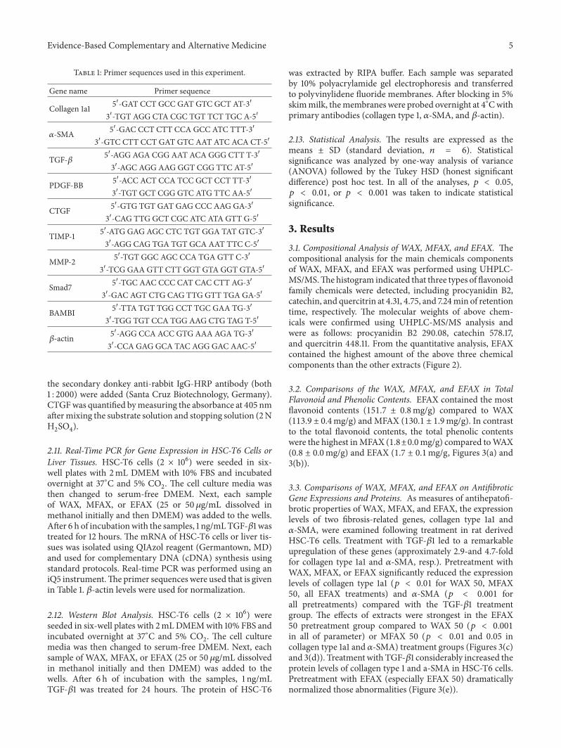