research article clinicopathological characteristics and...
TRANSCRIPT
Research ArticleClinicopathological Characteristics and Outcomes of ChinesePatients with Scanty Immune Deposits Lupus Nephritis: A LargeCohort Study from a Single Center
Qiuyu Li,1,2,3,4 Di Song,1,2,3,4 Fengmei Wang,1,2,3,4 Ying Tan,1,2,3,4
Feng Yu,1,2,3,4 and Minghui Zhao1,2,3,4
1 Renal Division, Department of Medicine, Peking University First Hospital, Beijing 100034, China2 Institute of Nephrology, Peking University, Beijing 100034, China3 Key Laboratory of Renal Disease, Ministry of Health of China, Beijing 100034, China4Key Laboratory of Chronic Kidney Disease Prevention and Treatment, Ministry of Education, Beijing 100034, China
Correspondence should be addressed to Feng Yu; [email protected]
Received 10 August 2013; Accepted 31 October 2013; Published 4 February 2014
Academic Editors: T. Gohda, Y. Iwata, and R. Liu
Copyright © 2014 Qiuyu Li et al.This is an open access article distributed under the Creative Commons Attribution License, whichpermits unrestricted use, distribution, and reproduction in any medium, provided the original work is properly cited.
Objective. To assess clinicopathological characteristics of lupus nephritis patients with scanty immune deposits.Methods.Thedata ofpatients with scanty immune deposits lupus nephritis were retrospectively analyzed. Plasma ANCA and complement componentswere detected. Results. Among 316 cases with renal biopsy-proven lupus nephritis, 40 cases were diagnosed as scanty immunedeposits. There were significantly higher value of serum creatinine (𝑃 = 0.002) and lower hemoglobin level (𝑃 = 0.009) andhigher score of cellular crescents (𝑃 = 0.015) in scanty immune deposits group compared with immune complex deposits group.The frequency of positive plasma ANCA was significantly higher in scanty immune deposits group than that in immune complexdeposits group (52.5% versus 10.1%, 𝑃 < 0.001). As for comparisons of plasma complement components, there were significantlyhigher levels of C1q (𝑃 = 0.005) and Bb (𝑃 = 0.02) and lower level of factor H (𝑃 = 0.003) in scanty immune deposits group.The ratio of treatment failure was significantly higher in scanty immune deposits group than that in immune deposits group(42.5% versus 19.20%, 𝑃 = 0.001). The renal outcomes were similar between the two groups. Conclusions. Patients with scantyimmune deposits lupus nephritis hadmore severe kidney damage. ANCA and activation of complement alternative pathwaymightbe involved in the pathogenesis of the disease.
1. Introduction
Systemic lupus erythematosus (SLE) is a prototypic autoim-mune disease characterized by the production of multipleautoantibodies. Renal involvement is common in SLE.The ty-pical feature in lupus nephritis is immune complex dep-osition, showed as “full house” under immunofluorescenceobservation. However, in previous reports, some pati-ents with lupus nephritis presented with “scanty immunedeposits,” that is, nonclassical glomerulonephritis, whichmight contribute to thrombotic microangiopathy (TMA) [1],ANCA-associated crescentic glomerulonephritis [2] podocy-topathy [3], and so forth. Here, the “scanty immune deposits”were indicated as a descriptive term to identify the specimenswith little or no staining for immunoglobulin and not nece-
ssarily for lesions with necrosis or crescents.The clinicopath-ological features, outcomes, and possible pathogenesis of sca-nty immune deposits lupus nephritis have not been well deli-neated and extensively studied.
This study is to assess clinical manifestations, laboratorycharacteristics, pathological features, and outcomes of patie-nts with scanty immune deposits in a large cohort of Chineselupus nephritis patients. Particularly, we further detect thedistribution of ANCA and complement activation profile inthe patients.
2. Methods
2.1. Patients. Renal histopathological data of 316 patientswith renal biopsy-proven lupus nephritis, diagnosed between
Hindawi Publishing Corporatione Scientific World JournalVolume 2014, Article ID 212597, 11 pageshttp://dx.doi.org/10.1155/2014/212597

2 The Scientific World Journal
January 2000 and July 2008 in Peking University FirstHospital, were reviewed and reclassified according to theInternational Society of Nephrology and Renal PathologySociety (ISN/RPS) 2003 classification [4]. Only biopsy speci-mens withmore than 10 glomeruli were included in the study.
On frozen sections of renal biopsy, at least two glomeruli,except for the sclerosed glomeruli, were evaluated by arenal pathologist. Scanty immune deposition was defined asnegative staining or 1+ positivity (on a scale of 0–4+) ofimmunoglobulins (IgG, IgA, and IgM) by direct immunoflu-orescence assay and no electron-dense deposit in glomeruli,tubular basement membrane, and vessels was observed byelectron microscopy assay. Immune complex deposits weredefined as (i) a score of 2+ or higher in staining for anykind of immunoglobulin observed by immunofluorescencemicroscopy and (ii) electron-dense deposits observed byelectron microscopy [5].
The patients fulfilled the 1997 American College ofRheumatology revised criteria for SLE [6].
2.2. Clinical Evaluation. The following clinical data were col-lected and analyzed: gender, fever, malar rash, photosensitiv-ity, oral ulcer, alopecia, arthritis, serositis, neurologic disor-der, anemia, leukocytopenia, thrombocytopenia, hematuria,and leukocyturia. The criteria for system involvement wereconsistent with the 1997 American College of Rheumatologyrevised criteria for SLE [6]. The clinical disease activitywas assessed by the Systemic Lupus Erythematosus DiseaseActivity Index (SLEDAI) [7, 8].
The renal response to the therapy includes completeremission, partial remission and treatment failure was deta-iled in previous studies [9–12].
A relapsewas defined as (1)nephritic relapse: a recent inc-rease of serum creatinine by >50%with active urinary sedim-ents; (2) proteinuric relapse: development of either a nep-hrotic syndrome (proteinuria >3.5 g/day and serum albumin<30 g/L) or proteinuria >1.5 g/day without other causes, inpreviously nonproteinuric patients [13, 14].
The patients were followed up in outpatient clinic speci-fied for patients with lupus nephritis. The primary end pointwas defined as death and the secondary end points weredefined as end-stage renal disease (ESRD) or doubling ofserum creatinine.
2.3. Laboratory Assessment. The following laboratory fea-tures were further detected using serum or plasma at the dayof renal biopsy.
Serum antinuclear antibodies (ANA) were detectedusing indirect immunofluorescence assay (EUROIMMUN,Lubeck, Germany) and anti-double-stranded DNA (ds-DNA) antibodies were detected using Crithidia luciliae indir-ect immunofluorescence test (EUROIMMUN, Lubeck, Ger-many). Anti-extractable nuclear antigen (ENA) antibodies,including anti-Sm, anti-SSA, anti-SSB, and anti-RNP antib-odies, were detected using immunodotting assay (EUROIM-MUN, Lubeck, Germany). Anti-cardiolipin antibodies andanti-𝛽
2GP-1 antibodies were detected using enzyme-linked
immunosorbent assay (ELISA) (EUROIMMUN, Lubeck,Germany).
2.3.1. Detection of ANCA. ANCA tests were performed byboth indirect immunofluorescence (IIF) assay and antigen-specific enzyme-linked immunosorbent assay (ELISA). Stan-dard IIF assay was performed using precooled ethanol fixednormal peripheral neutrophils as substrate according to themanufacturer (EUROIMMUN, Lubeck, Germany). The useof Hep-2 cell and paraformaldehyde-fixed neutrophils mayallow the distinction between ANA and p-ANCA. In antigen-specific ELISA, two highly purified known ANCA antigens,PR3 andMPO, purified as previously reported [15] were usedas solid-phase ligands.
2.3.2. Quantification of Plasma Complement Components.Plasma concentrations of major human complement com-ponents were determined by enzyme-linked immunoassays,including complement fragments C5a (Quidel Corporation,San Diego, CA), C3a (Quidel Corporation, San Diego, CA),Bb (Quidel Corporation, San Diego, CA), soluble C5b-9(SC5b-9, Quidel Corporation, San Diego, CA), properdin(Uscnk Life Science Inc., Wuhan, China), and C3 (QuidelCorporation, San Diego, CA). All the complement compo-nents were assayed according to the manufacturer’s instruc-tions. The principle of the assays was a four-step procedure:(I) microassay plates were precoated with murine mono-clonal antibodies binding specifically to the complementcomponents; (II) plasma samples were added according tothe optimal dilutions, incubation time, and temperature fromthe instructions; (III) horseradish peroxidase conjugatedantibodies binding to the complement components adsorbedon the plates were added; (IV) chromogenic substrate wasadded to determine the concentration of components.
The methods to detect plasma C4BP, C1q, and MBL(mannan-binding lectin) were the same as previouslydescribed with mild modification [16–18].
2.4. Renal Histopathology. The renal biopsy specimens wereexamined by light microscopy, direct immunofluorescence,and electron microscopy techniques.
2.4.1. Light Microscopy Examination. Renal biopsy speci-mens were fixed in 4.5% buffered formaldehyde for lightmicroscopy. Consecutive serial 3 𝜇m sections were usedfor histological staining. Stains employed included haema-toxylin and eosin (H&E), periodic acid-Schiff (PAS), silvermethenamine (Meth), and Masson’s trichrome.
Crescentic glomerulonephritis was defined as over half ofthe total glomeruli affected by large crescents (the crescenttakes up over half space in Bowman’s capsule) by lightmicroscope, which should be included in class IV-G lupusnephritis [5].
Renal thrombotic microangiopathy (TMA) was char-acterized by interlobular artery, arteriole, and glomerularcapillary lesions, including endothelial cell swelling, lumennarrowing or obliteration, and thrombi formation by lightmicroscopy. Swelling of glomerular endothelial cells, detach-ment from the glomerular basement membrane, and widen-ing of the subendothelial space were identified by electronmicroscopy [19].
The Scientific World Journal 3
(a) (b) (c)
(d) (e) (f)
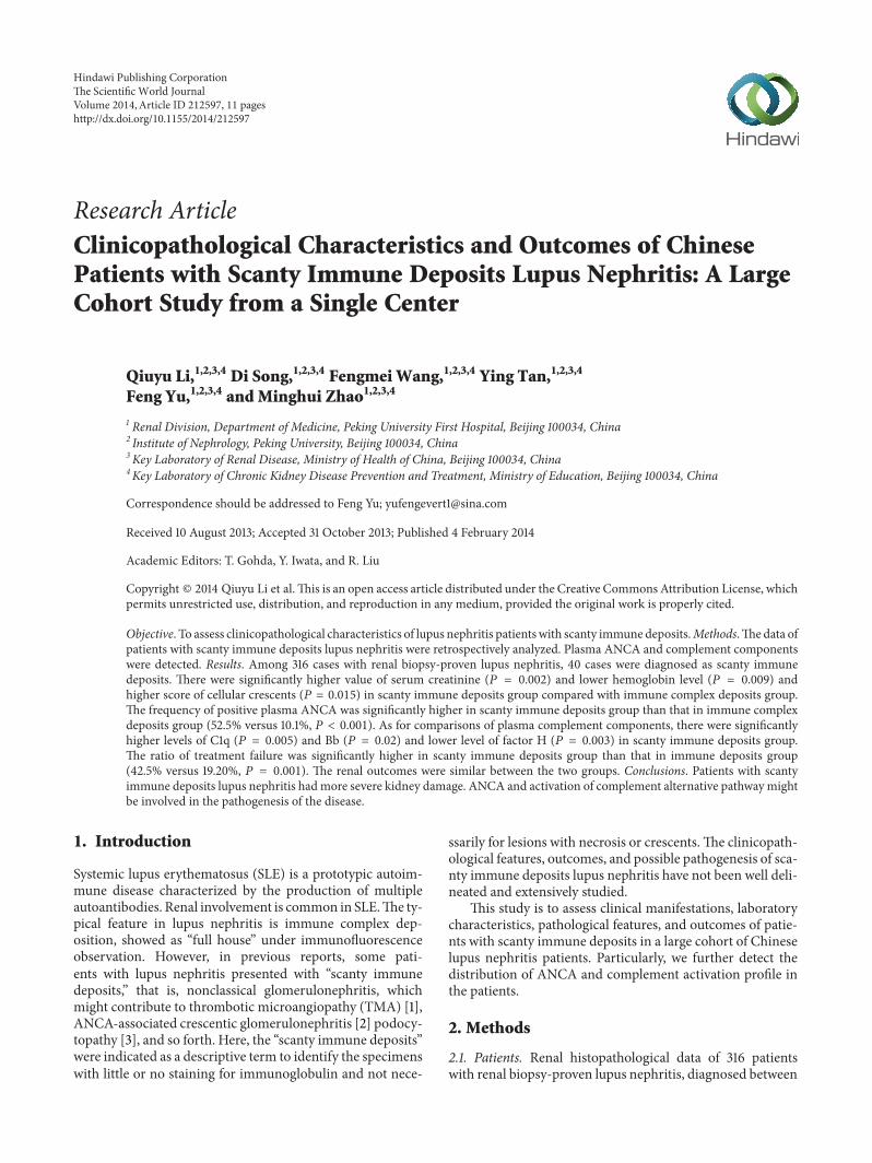
Figure 1: Electron micrographs of cases with scanty immune deposits lupus nephritis. (a)–(c) showed one case of mesangial proliferativelupus nephritis. No electron dense deposits were seen in mesangial area and glomerular basement membrane. Diffuse effacement of footprocesses was observed. (d)–(f) showed one case of diffuse proliferative lupus nephritis combined with renal thrombotic microangiopathy.Glomerular endothelial cell (black pointer) was swollen, with increasedmesangial matrix (d). Severe widening of subendothelial space (blackarrow) with fluffy material and irregular cell projections; few of electron dense deposits were identified at higher magnification ((e), (f)). ((a),(d), original mag. ×10000) ((b), (c), (e), and (f), original mag. ×20000).
Podocytopathy was defined as podocyte effacement. Bio-psy findings revealed either no glomerular immune depositsor sparse deposits, which were confined to the glomerularmesangium. The characteristic pathological glomerular abn-ormality was ultrastructural and resided in the visceral glo-merular epithelial cells. The glomerular lesions included idi-opathic minimal change glomerulopathy and focal and seg-mental glomerulosclerosis [3, 20].
Pathological parameters such as activity indices (AI) andchronicity indices (CI) were approached by renal patholo-gists using a modification of a previously reported systeminvolving semiquantitative scoring of specific biopsy features[21, 22].
2.4.2. Direct Immunofluorescence Examination. Direct imm-unofluorescence for immunoglobulin G (IgG), immunoglob-ulin A (IgA), immunoglobulin M (IgM), C3, C1q, and fibrindeposits was semiquantitatively graded from 0 to 4 accordingto the intensity of fluorescence. The glomeruli with sclerosiswere excluded.
2.4.3. Electron Microscopy Examination. Renal biopsy spec-imens were fixed in 2.5% paraformaldehyde for electronmicroscopy. After being embedded in epon, ultrathin sec-tions were mounted on metal grids and stained with uranylacetate before being viewed in a transmission electronmicro-scope (JEM-1230; JEOL, Tokyo, Japan).
2.5. Blood Samples. For detection ofANCAand complement,plasma samples were obtained from peripheral blood at thesame day of renal biopsy before initiation of immunosuppres-sive treatment. The blood samples of patients and controls
were drawn into EDTA tubes. The plasma was collectedimmediately by centrifugation at 2000 g for 15min at 4∘C.All plasma samples were stored at −80∘C until use. Repeatedfreeze/thaw cycles were avoided.
Informed consent was obtained for blood sampling andrenal biopsy from each patient. The research was in compli-ance of the Declaration of Helsinki. The design of this workwas approved by the local ethical committees.
2.6. Statistical Analysis. Statistical software SPSS 16.0 (SPSS,Chicago, IL, USA) was employed for statistical analysis.Quantitative data were expressed as mean ± SD, and medianwith range (minimum, maximum). For comparison of clini-cal and pathological features of patients, Student’s 𝑡-test, one-way ANOVA analysis of variance, and Chi-square test wereused. Kaplan-Meier curves were used to analyze patients’prognosis. Survival analysis was performed using the log-rank test. Results were expressed as hazard ratio (HR) with95% confidence intervals (CI). Statistical significance wasconsidered as 𝑃 < 0.05.
3. Results
3.1. General Data of Patients with Scanty Immune DepositsLupus Nephritis. Among the 316 lupus nephritis patientsenrolled in the study, 40 cases (12.66%) met the pathologicalcriteria of scanty immune deposits nephritis, which wereconfirmed by electron microscopy (Figure 1).
In the scanty immune deposits group, 6 were male and34 were female, with an average age of 39.78 ± 12.90 years atpresentation. The majority of the patients (80%) were withhematuria. Half of the patients were with leukocyturia and

4 The Scientific World Journal
Table 1: Comparison of clinical data between patients with scanty immune deposits and immune complex deposits lupus nephritis.
Scanty immune deposits Immune complex deposits 𝑃 valueNumber of patients 40 276Age (mean ± SD) (years) 39.78 ± 12.90 32.06 ± 10.94 <0.001Gender (male/female) 6/34 42/234 0.971Number with fever (noninfection) (%) 11 (27.5) 82 (29.7) 0.774Number with malar rash (%) 17 (42.5) 149 (54.0) 0.174Number with photosensitivity (%) 6 (15.0) 57 (20.7) 0.403Number with alopecia (%) 14 (35.0) 84 (30.4) 0.560Number with oral ulcer (%) 11 (23.9) 83 (30.1) 0.739Number with arthritis (%) 25 (62.5) 143 (51.8) 0.205Number with serositis (%) 5 (12.5) 46 (16.7) 0.503Number with neurologic disorder (%) 3 (7.5) 23 (8.3) 0.858Number with anemia (%) 31 (77.5) 183 (66.5) 0.165Number with leukocytopenia (%) 23 (57.5) 123 (44.6) 0.125Number with thrombocytopenia (%) 10 (25.0) 93 (33.8) 0.267Number with hematuria (%) 32 (80.0) 210 (76.1) 0.585Number with leukocyturia (noninfection) (%) 23 (57.5) 145 (52.5) 0.557Number with nephrotic syndrome (%) 25 (62.5) 161 (58.8) 0.653SLEDAI (median; interquartile range) 18, 13–23 17, 14–21 0.751𝑃 < 0.05, italic values refer to the significant value between the two groups.
62.5%with nephrotic syndrome.Themedian amount of urineprotein was 4.04 g/24 hours. The median value of serumcreatinine was 102mmol/dL (80.25–189.50mmol/dL) upondiagnosis. The mean level of SLEDAI was 17.92 ± 5.45.
According to the 2003 classification of lupus nephritis,6 patients were classified as class II (15%, including 1 casecombined with minimal change disease), 9 cases as classIII (22.5%), and 25 cases as class IV (62.5%, including1 case combined with TMA and 7 cases with crescenticglomerulonephritis).
All of the patients received oral prednisone therapy.The majority of patients completed treatment with oralcyclophosphamide (3/40) ormonthly intravenous cyclophos-phamide (600–800mg/month) (20/40). The other patientsreceived mycophenolate mofetil (3/40), leflunomide (8/40),and azathioprine (4/40). Two patients received prednisonealone. Twenty-six patients achieved clinical remission, 8 withcomplete remission and 18 with partial remission. Fourteenpatients presented with treatment failure.
We further compared the clinical and pathological char-acteristics of scanty immune deposits and immune complexdeposits patients with lupus nephritis.
3.2. Comparison of Clinical and Laboratory Parameters betwe-en Patients with Scanty Immune Deposits and Immune Com-plex Deposits Lupus Nephritis. The clinical and laboratoryfeatures of patients in the two groups were listed in Tables 1and 2.
The average age was significantly older in scanty immunedeposits group than that in immune complex deposits group(𝑃 < 0.001). There were no significant differences betweenthe two groups in other clinical indices.
In laboratory findings, there were significantly lowerhemoglobin level (𝑃 = 0.009) and higher value of serumcreatinine (𝑃 = 0.01) in scanty immune deposits group thanthose in immune complex deposits group.
Twenty-one out of the 40 patients (52.5%) in scantyimmune deposits group were ANCA positive including 15with p-ANCA and 2 with c-ANCA by IIF, 10 with anti-MPOantibodies, and 1with anti-PR3 antibodies by ELISA. 28 out ofthe 276 patients (10.1%) in immune complex deposits groupwere ANCA positive including 20 with p-ANCA by IIF and9 with anti-MPO by ELISA. The difference was significant(𝑃 < 0.001).
3.3. Comparison of Plasma Complement Components Levelsbetween Patients with Scanty Immune Deposits And ImmuneComplex Deposits Lupus Nephritis. The levels of plasmacomplement components of patients in the two groups werelisted in Table 3.
The normal levels of plasma MBL, C3a, C5a, and solubleC5b-9 were 1532 ± 1020 ng/mL, 100.87 ± 70.55 ng/mL, 9.32 ±7.88 ng/mL, and 467.41 ± 545.23 ng/mL, respectively. Theywere significantly higher in patients with scanty immunedeposits and immune complex deposits lupus nephritis thanthose in normal controls (𝑃 < 0.01, 𝑃 < 0.01; 𝑃 < 0.01,𝑃 < 0.01; 𝑃 < 0.01, 𝑃 < 0.01; 𝑃 < 0.01, 𝑃 < 0.01, resp.).
The normal levels of plasma C1q, properdin, Bb, C4BP,factor H, and C3 were 61.96 ± 10.50 𝜇g/mL, 22.58 ±9.67 𝜇g/mL, 0.69± 0.45 𝜇g/mL, 326.59 ± 87.25𝜇g/mL, 515.04± 134.08 𝜇g/mL, and 0.80 ± 0.17mg/mL, respectively. Theywere significantly lower in patients with scanty immunedeposits and immune complex deposits lupus nephritis thanthose in normal controls (𝑃 < 0.01, 𝑃 < 0.01; 𝑃 < 0.01,𝑃 < 0.01; 𝑃 < 0.01, 𝑃 < 0.01; 𝑃 < 0.01, 𝑃 < 0.01; 𝑃 < 0.01,𝑃 < 0.01; 𝑃 < 0.01, 𝑃 < 0.01, resp.).
3.3.1. Plasma Levels of C1q. C1q is the first component inthe classical pathway of complement activation. The level ofC1q was significantly higher in patients with scanty immunedeposits lupus nephritis than that in immune complexdeposits lupus nephritis (34.78 ± 5.65 𝜇g/mL versus 22.17 ±3.08 𝜇g/mL, 𝑃 = 0.005).

The Scientific World Journal 5
Table 2: Comparison of laboratory data between patients with scanty immune deposits and immune complex deposits lupus nephritis.
Scanty immunedeposits
Immune complexdeposits 𝑃 value
Number of patients 40 276Hemoglobin (g/L) (mean ± SD) 91.18 ± 24.39 102.16 ± 24.93 0.009Urine protein (g/24 hours)(median; interquartile range)
4.04,1.96–5.36
4.34,2.20–7.07 0.348
Serum creatinine (mmol/dL)(median; interquartile range)
102,80.25–189.50
81,67–126 0.01
Number with positive ANA (%) 39 (97.5) 273 (98.9) 1Number with positive anti-dsDNA (%) 25 (64.1) 191 (69.2) 0.521Number with positive anti-Sm (%) 13 (33.3) 69 (25.0) 0.267Number with positive anti-SSA (%) 16 (41.0) 124 (44.9) 0.646Number with positive anti-SSB (%) 3 (7.9) 32 (11.6) 0.493Number with positive anti-RNP (%) 12 (30.8) 84 (30.4) 0.966Number with positive anticardiolipin (%) 2 (2/34, 6.9%) 17 (17/223, 7.6%) 1Number with positive anti-𝛽2 GP-1 (%) 4 (4/34, 6.9%) 15 (15/223, 6.7%) 1Number with positive ANCA (%) 21 (52.5) 28 (10.1) <0.001𝑃 < 0.05, italic values refer to the significant value between the two groups.
Table 3: Comparison of plasma complement components levels betweenpatientswith scanty immunedeposits and immune complex depositslupus nephritis.
Scanty immunedeposits
Immune complexdeposits 𝑃 value Normal range
Number of patients 40 276C1q (ug/mL) (mean ± SD) 34.78 ± 5.65 22.17 ± 3.08 0.005 61.96 ± 10.50
MBL (ng/mL)(median; interquartile range)
2223575–3719
1768476–2879 0.663 1532 ± 1020
Properdin (ug/mL) (mean ± SD) 13.26 ± 6.32 15.39 ± 6.05 0.379 22.58 ± 9.67
Bb (ug/mL)(median; interquartile range)
1.810.62–2.54
1.010.7–1.74 0.02 0.69 ± 0.45
C4BP (ug/mL) (mean ± SD) 243.23 ± 131.54 221.56 ± 79.25 0.262 326.59 ± 87.25
Factor H (ug/mL) (mean ± SD) 302.19 ± 110.47 407.52 ± 181.44 0.002 515.04 ± 134.08
C3 (mg/mL)(median; interquartile range)
0.580.38–1.22
0.540.31–0.81 0.671 0.80 ± 0.17
C3a (ng/mL)(median; interquartile range)
4.091.5–801.27
3.151–562.32 0.321 100.87 ± 70.55
C5a (ng/mL)(median; interquartile range)
20.3211.26–30.79
17.2510.41–29.33 0.454 9.32 ± 7.88
Soluble C5b-9 (ng/mL)(median; interquartile range)
3.001–754.35
4.101–698.41 0.239 467.41 ± 545.23
𝑃 < 0.05, italic values refer to the significant value between the two groups.
3.3.2. Plasma Levels of MBL. MBL serves as a trigger for theactivation of lectin pathway. There was no significant differ-ence in MBL levels between patients with scanty immunedeposits and immune complex deposits lupus nephritis (𝑃 =0.675).
3.3.3. Plasma Levels of C4BP. C4BP is the main fluid-phaseinhibitor of the complement activation. It exerts inhibitoryfunction by enhancing the decay of classical/lectin pathwayC3 convertase, C4b2a [23], as well as the alternative pathwayC3 convertase, C3bBb [24].
There was no significant difference in C4BP levelsbetween patients with scanty immune deposits and immunecomplex deposits lupus nephritis (𝑃 = 0.389).
3.3.4. Plasma Levels of Bb, Properdin, and Factor H. Prop-erdin is critical in the stabilization of alternative pathwayC3 convertase. Bb is an activation fragment of factor B inthe alternative complement pathway. Complement factor His an abundant plasma complement regulator that inhibitsalternative pathway activation by inhibiting the formationand accelerating the decay of C3 convertase and acting

6 The Scientific World Journal
as a complement factor I cofactor, which inactivates C3bto iC3b. Measurement of properdin, Bb, and factor H inplasma provided evidence for the activation of the alternativecomplement pathway [25–27].
The level of Bb was significantly higher in patientswith scanty immune deposits lupus nephritis than thatin immune complex deposits lupus nephritis (1.81; 0.62–2.54 𝜇g/mL versus 1.01; 7–1.74𝜇g/mL, 𝑃 = 0.02). Thelevel of factor H was significantly lower in patients withscanty immune deposits lupus nephritis than that in immunecomplex deposits lupus nephritis (302.19 ± 110.47 𝜇g/mLversus 407.52 ± 181.44 𝜇g/mL, 𝑃 = 0.003). There was nosignificant difference in properdin level between patientswith scanty immune deposits lupus nephritis and immunecomplex deposits lupus nephritis (𝑃 = 0.357).
3.3.5. Plasma Levels of C3, C3a, C5a, and SC5b-9. Activationof complement results in the conversion of C3 to C3a andC3band then the formation of a C5 convertase multimolecularenzyme capable of cleaving C5 to C5a and C5b. The terminalcomplement complex (C5b-9) is generated by the assemblyof C5b through C9 as a consequence of activation of com-plement system. Therefore, we tested plasma C3, C3a, C5a,and soluble C5b-9 (SC5b-9) levels to reflect total complementactivation in circulation.
There were no significant differences in plasma concen-tration of C3, C3a, C5a, and SC5b-9 between patients withscanty immune deposits and immune complex deposits lupusnephritis (𝑃 = 0.541, 𝑃 = 0.134, 𝑃 = 0.446, and 𝑃 = 0.227,resp.).
3.4. Comparison of Renal Histopathologic Parameters betweenPatients with Scanty Immune Deposits and Immune ComplexDeposits Lupus Nephritis. The renal histopathological fea-tures of patients with and without immune deposits lupusnephritis were listed in Table 4.
In comparison with immune deposits group, patientswith scanty immune deposits group had significantly higherscores of cellular crescents (𝑃 = 0.015). There were nosignificant differences in other pathological indices betweenthe two groups. The ratios of thrombotic microangiopathy,crescentic glomerulonephritis, and podocytopathy were notsignificantly different between the two groups (2.17% versus8.33%, 𝑃 = 0.242; 15.22% versus 9.78%, 𝑃 = 0.052; 2.17%versus 0, 𝑃 = 0.307, resp.).
3.5. Comparison of Treatment and Outcomes between Patientswith Scanty Immune Deposits and Immune Complex DepositsLupus Nephritis. The treatment and outcomes of patientswith and without immune deposits lupus nephritis weredetailed in Table 5. There was no significant difference intreatment algorithm between the two groups. The rates ofcomplete remission and partial remission were not signif-icantly different. The incidence of treatment failure wassignificantly higher in scanty immune deposits group thanthat in immune complex deposits group (36.95% versus19.20%, 𝑃 = 0.007).
1.0
0.8
0.6
0.4
0.2
0.0
0 100 200 300 400Follow-up time (months)
Prob
abili
ty o
f ren
al su
rviv
al Group 1
Group 2
Log rank: P = 0.585
At risk:
Group 1
Group 2
40 5 1 0 0276 53 9 2 0
Figure 2: Comparison of renal outcomes between patients withscanty immune deposits (Group 1) and immune complex depositslupus nephritis (Group 2).
During a similar follow-up time (average for nearly 5years), the renal relapse rate was similar (13.79% versus12.56%, 𝑃 = 0.819) between the two groups.
Regarding long-term survival, there were no significantdifferences in mortality and renal outcomes between scantyimmune deposits and immune deposits groups (𝑃 = 0.598,𝑃 = 0.585, resp., Figure 2). In scanty immune depositsgroup, one patient died due to infection; 6 patients reachedthe secondary end point including 1 with doubling of serumcreatinine and 5 with ESRD. In immune complex depositsgroup, 2 patients died due to heart failure and cerebralhemorrhage, respectively; 35 patients reached the secondaryend point, all with ESRD.
We further compared renal outcomes between the scantyimmune deposits class III lupus nephritis patients with thoseof immune complex deposits class III lupus nephritis andbetween scanty immune deposits crescentic lupus nephritisand immune complex deposits class IV lupus nephritis. Inscanty immune deposits class III lupus nephritis group, 0 ofthe 9 patients reached the secondary end point. In immunecomplex deposits class III lupus nephritis group, one of the40 patients reached the secondary end point. The ratio didnot reach significant difference. In scanty immune depositscrescentic lupus nephritis group, 2 of the 7 patients reachedthe secondary end point. In immune complex deposits classIV lupus nephritis group, 30 of the 151 patients reached thesecondary end point. Survival analysis showed that scantyimmune deposits crescentic group had significantly worserenal outcome than that in immune complex deposits classIV group (𝑃 = 0.032, Figure 3).
Using the log-rank test for univariate survival analysis ofrenal prognosis in all the patients with lupus nephritis, wefound that scanty immune deposits nephritis was not a risk

The Scientific World Journal 7
Table 4: Comparison of renal pathological data between patientswith scanty immune deposits and immune complex deposits lupus nephritis.
Scanty immune deposits Immune complex deposits 𝑃 valueNumber of biopsies 40 276Class II (%) 6 (15) 13 (4.71)
0.195Class III (%) 9 (22.5) 46 (16.67)Class IV (%) 25 (62.5) 151 (54.7)Class V (%) 0 (0) 64 (23.2)Class VI (%) 0 (0) 2 (0.72)AI score(mean ± SD) 8.70 ± 4.58 7.50 ± 4.65 0.128
Endocapillary hypercellularity(median; interquartile range) 3, 1–3 3, 1–3 0.482
Cellular crescents(median; interquartile range) 2, 0–4 0, 0–2 0.015
Karyorrhexis/fibrinoid necrosis(median; interquartile range) 0, 0–2 0, 0–2 0.687
Subendothelial hyaline deposits(median; interquartile range) 1, 0–2 1, 0–2 0.913
Interstitial inflammation(median; interquartile range) 1, 1-2 1.1–1 0.067
Glomerular leukocyte infiltration(median; interquartile range) 1, 0-1 1, 0-1 0.694
CI score(mean ± SD) 3.10 ± 2.29 2.75 ± 1.99 0.31
Glomerular sclerosis(median; interquartile range) 0, 0-1 0, 0-1 0.717
Fibrous crescents(median; interquartile range) 0.0-0 0, 0-0 0.795
Tubular atrophy(median; interquartile range) 1, 1-2 1, 1-1 0.09
Interstitial fibrosis(median: interquartile range) 1, 1–1.75 1, 1-1 0.182
𝑃 < 0.05, italic values refer to the significant value between the two groups.
factor for renal outcome in lupus nephritis. Other univariaterisk factors included serum creatinine value, hemoglobinvalue, anti-SSB antibody, total activity indices score, cellu-lar crescents, interstitial inflammatory cell infiltration, totalchronicity indices score, fibrous crescents, and interstitialfibrosis (see Table 6 for details).
4. Discussion
It is well documented that various immunoglobulinsdeposited together with complements were found in theaffected glomeruli in lupus nephritis. Accordingly, lupusnephritis was considered to be the classical type of immunecomplex associated glomerulonephritis. It is rare forlupus nephritis to contain no immune complex deposits,although there have been several cases of scanty immunedeposits lupus nephritis reported previously [1–3, 28–30].The clinicopathological features, outcomes, and possiblepathogenesis of scanty immune deposits lupus nephritisshould be studied.
The data arising from our study showed that the scantyimmune deposits lupus nephritis was not uncommon, which
accounted for 13% of all the lupus nephritis in our center.Of course, the glomeruli under pathological detection withsclerosis were excluded, and all the patients did not acceptimmunosuppressive treatment when the renal biopsies weredone, which excluded the influences of therapy on theimmune complex deposits in kidneys in our study. Previousstudies indicated that the most possible explanation of scantyimmune deposits lupus nephritis was that it might overlapwith other scanty immune deposits-immune diseases, suchas TMA [1], true renal vasculitis [29], ANCA-associatednecrotizing and crescentic glomerulonephritis [2], glomeru-lar podocytopathy [3], and glomerular lesions in kidneytransplants [28]. Thus, we firstly focus on the presences ofabove disease in the scanty immune deposits group.We foundthat there were one patient with minimal change disease, onepatient with TMA, and seven patients with true crescenticglomerulonephritis based on the strict diagnostic criteria[3, 5, 9]. But there was no significant difference in the ratiosof the three pathological changes between scanty immunedeposits group and immune complex deposits group.
After further comparisons of clinical, laboratory, andpathological features between the two groups, we found that

8 The Scientific World Journal
Table 5: Comparison of treatment between patients with scanty immune deposits and immune complex deposits lupus nephritis.
Scanty immune deposits Immune complex deposits 𝑃 valueNumber of patients (%) 40 276Treatment
P 40 (100) 276 (100) 1CYC 23 (57.50) 156 (56.52) 0.907AZA 4 (10.00) 21 (7.60) 0.833MMF 3 (7.50) 17 (6.15) 1LEF 8 (20.00) 31 (11.23) 0.187
Treatment responseCR 8 (20.00) 78 (28.26) 0.560PR 18 (45.00) 145 (52.54) 0.373TF 14 (35) 53 (19.20) 0.001
Duration of followup (months) 38, 6–78 48, 8.5–84 0.952
Relapse rate4 (4/26, 15.38%, 3 with
nephritic relapse and 1 withproteinuric relapse)
28 (28/223, 12.56%, 20 withnephritic relapse and 8 with
proteinuric relapse)0.922
Note: P: oral prednisone; CYC: cyclophosphamide; AZA: azathioprine; MMF: mycophenolate mofetil; LEF: leflunomide; CR: complete remission; PR: partialremission; TF: treatment failure.𝑃 < 0.05, italic values refer to the significant value between the two groups.
Table 6: Univariate survival analysis of patients renal prognosis with lupus nephritis.
HR 95% confidence interval 𝑃 valueAge 0.240 0.056 1.031 0.055Sex 0.514 0.158 1.674 0.269C3 0.693 0.167 2.876 0.613Proteinuria 0.948 0.226 3.971 0.942Serum creatinine value 16.063 6.746 38.251 <0.001Hemoglobin 0.285 0.154 0.530 <0.001ANA 0.389 0.053 2.855 0.353Anti-ds-DNA antibody 1.376 0.710 2.670 0.345Anti-Sm antibody 0.972 0.476 1.984 0.938Anti-SSA antibody 0.786 0.417 1.479 0.455Anti-SSB antibody 2.878 1.365 6.067 0.005Anti-RNP antibody 0.721 0.361 1.441 0.355Anticardiolipin antibody 1.175 0.350 3.939 0.794SLEDAI 0.933 0.255 4.430 0.933Activity indices (AI) score 3.941 1.876 8.279 <0.001Endocapillary hypercellularity 1.814 0.803 4.099 0.152Cellular crescents 3.339 1.703 6.547 <0.001Karyorrhexis/fibrinoid necrosis 1.646 0.890 3.043 0.112Subendothelial hyaline deposits 0.848 0.445 1.616 0.721Interstitial inflammatory cell infiltration 5.492 2.859 10.547 <0.001Glomerular leukocyte infiltration 1.145 0.613 2.140 0.671Chronicity indices (CIs) score 1.379 1.230 1.545 <0.001Glomerular sclerosis 1.025 0.243 4.320 0.793Fibrous crescents 3.412 1.839 6.328 <0.001Tubular atrophy 25.129 0.497 1.271 0.107Interstitial fibrosis 9.222 1.268 67.070 0.028Scanty immune deposits or immune complex deposits 1.320 0.519 3.359 0.560𝑃 < 0.05, italic values refer to the significant value between the two groups.Bold values refers to that scanty immune deposits nephritis was not a risk factor for a renal outcome in lupus nephritis.

The Scientific World Journal 9
1.0
0.8
0.6
0.4
0.2
0.0
0.00 12.00 24.00 36.00 48.00 60.00 72.00Follow-up time (months)
Prob
abili
ty o
f ren
al su
rviv
al
Group 1
Group 2
Log rank: P = 0.032
At risk:
Group 1
Group 2
7 3 3 3 3 3 3
151 113 105 105 80 69 5
Figure 3: Comparison of renal outcomes between patients withscanty immune deposits crescentic lupus nephritis (Group 1) andimmune complex deposits class IV lupus nephritis (Group 2).
the patients with scanty immune deposits lupus nephritispresented with older age, higher value of serum creatinine,and lower value of hemoglobin. Furthermore, significantlyhigher score of cellular crescents was found in scanty immunedeposits group compared with immune complex depositsgroup evaluated by NIH scoring system. More importantly,by immunofluorescence assay and ELISA, we found thatthe positive ratio of ANCA was significantly higher inscanty immune deposits group than that in immune com-plex deposits group. Our previous study also showed thatcrescentic lupus nephritis presented with lower intensity ofimmunoglobulins and higher ratio of ANCA [31]. Thus, ithighlights the importance of ANCA in the pathogenesis ofscanty immune deposits crescentic lupus nephritis.
ANCA, as one of autoantibodies in SLE, might be impli-cated as a pathogenic factor for the development of scantyimmune deposits crescentic lupus nephritis. Nasr et al. rec-ently proposed that one of the two conditions (ANCA and lu-pus nephritis) may be creating fertile conditions for the seco-nd to develop [32]. It was suggested that lupus nephritis mig-ht facilitate the process of MPO autoantibody formation bypromoting neutrophil degranulation and priming neutrop-hils to increase surface expression of MPO. Besides, thepresence ofANCAwasmost closely associatedwith vasculiticlesions and the typical characteristic of ANCA-associatedrenal vasculitis was scanty immune deposits. So, it was sug-gested that the presence of ANCA in patients with SLEmightindicate overlaps of SLE andANCA-associated vasculitis [33],especially for thosewith the disproportionate necrotizing andcrescentic features in lupus nephritis [31].
On the other hand, over half of the patients with scantyimmune deposits lupus nephritis in our study were with
ANCA negative. Apart from ANCA, other mechanisms ofdeveloping scanty immune deposits lupus nephritis werethought of.The role of complement in SLEwas important andit was closely associated with immune complex formation.So, we further detected the plasma complement componentslevels, including classical, alternative, and mannose-bindinglectin (MBL) pathways in our patients with lupus nephritis.The results showed that there were significantly higherlevels of C1q and Bb and lower level of factor H in scantyimmune deposits group compared with immune complexdeposits group, which indicated the predominant activationof the complement alternative pathway in the pathogenesis ofscanty immune deposits lupus nephritis.
The role of complement activation played in SLE andlupus nephritis has been assumed for many years. It wasacknowledged that immune complexes formed by self-antigens and autoantibodies might activate the classicalpathway, generating inflammatorymediators and resulting intissue damage. However, convincing evidences indicated thatthe alternative pathway was also activated and participatedin the pathogenesis of SLE, especially in lupus nephritis [34–39]. The analysis of complement components in our studyshowed that all the three pathways might be activated andinvolved in the pathogenesis of lupus nephritis. However,the higher levels of C1q and Bb indicated that alternativepathway activation, other than classical pathway, was morenotable in scanty immune deposits lupus nephritis than thatin immune complex deposits group. In addition, lower levelof factor H, an important inhibitory factor in alternativepathway by accelerating the decay of the alternative pathwayC3-convertase (C3bBb), also supported that there mightexist overactivation of alternative pathway. A recent studyeven showed that factor H deficiency could accelerate thedevelopment of lupus nephritis in lupus-pronemiceMRL-lpr[40].
Interestingly, recent studies, including the mouse modelof anti-MPO IgG mediated glomerulonephritis and humanANCA associated vasculitis, suggested that complementalternative pathway activation was crucial for ANCA associ-ated vasculitis development [41–43]. Taken together, ANCAand complement alternative pathway activation might bothbe involved in the development of scanty immune depositslupus nephritis which need further investigation.
There is no well-established guideline for the treatment ofscanty immunedeposits lupus nephritis.The therapy betweenscanty immune deposits and immune complex deposits gro-ups in our patients was similar, both including immunosup-pressive treatment. Although the ratio of renal remission ratewas not different in the two groups, higher proportion of tre-atment failure was found in scanty immune deposits group. Itmight be related to the more severe clinical and pathologicalcharacteristics in scanty immune deposits group.
The 5-year survival rate and renal outcome between thetwo groups were similar. Scanty immune deposits nephritiswas not a risk factor for renal outcome by log-rank test forunivariate survival analysis of renal prognosis. However, asscanty immune deposits lupus nephritis consisted of a groupof pathological types, we further compared renal outcomesbetween different subgroups. Interestingly, we found that

10 The Scientific World Journal
scanty immune deposits crescentic group had significantlyworse renal outcome than that in immune complex depositsclass IV group, which needs further observation.
There were some limitations in this study: (I) despite be-ing a large cohort study, it was a retrospective analysis; (II) thestaining of complement components in kidney tissues, espe-cially in alternative pathway, is important; (III) longer dura-tion of followup and multicenter study are needed.
In conclusion, scanty immune deposits lupus nephritiswas not uncommon. Patients with scanty immune depositslupus nephritis had more severe kidney damage. ANCA-ass-ociated vasculitis and activation of complement alternativepathway might be involved in the pathogenesis of scantyimmune deposits lupus nephritis.
Conflict of Interests
The authors declare that there is no conflict of interests regar-ding the publication of this paper.
Authors’ Contribution
Qiu-yu Li and Di Song contributed equally to this work.
Acknowledgments
This work was supported by a Grant of Chinese 973 Project(no. 2012CB517702), National Natural Science Foundationof China to Innovation Research Group (no. 81021004),and National Natural Science Foundation of China (no.81100497).
References
[1] D. A. Charney, G. Nassar, L. Truong, and T. Nadasdy, “‘Pauci-immune’ proliferative and necrotizing glomerulonephritis withthrombotic microangiopathy in patients with systemic lupuserythematosus and lupus-like syndrome,”TheAmerican Journalof Kidney Diseases, vol. 35, no. 6, pp. 1193–1206, 2000.
[2] A. Fayaz, Y. Pirson, J. P. Cosyns, J. Yango, andM. Lambert, “Pau-ci-immune necrotizing and crescentic glomerulonephritis in apatient with systemic lupus erythematosus,” Clinical Nephrol-ogy, vol. 69, no. 4, pp. 290–293, 2008.
[3] S.W. Kraft, M.M. Schwartz, S. M. Korbet, and E. J. Lewis, “Glo-merular podocytopathy in patients with systemic lupus erythe-matosus,” Journal of the American Society of Nephrology, vol. 16,no. 1, pp. 175–179, 2005.
[4] J. J. Weening, V. D. D’Agati, M. M. Schwartz et al., “The classifi-cation of glomerulonephritis in systemic lupus ereythematosusrevisited,” Kidney International, vol. 65, pp. 521–530, 2004.
[5] R. J. Falk, J. C. Jennette, and P. H. Nachman, “Primary glomeru-lar disease,” in The Kidney, B. M. Brenner, Ed., pp. 1293–1380,Saunders, Philadelphia, Pa, USA, 7th edition, 2004.
[6] M.C.Hochberg, “Updating theAmerican college of rheumatol-ogy revised criteria for the classification of systemic lupus ery-thematosus,” Arthritis and Rheumatism, vol. 40, no. 9, article1725, 1997.
[7] C. Bombardier, D. D. Gladman,M. B. Urowitz, D. Caron, andC.H. Chang, “Derivation of the SLEDAI: a disease activity index
for lupus patients,” Arthritis and Rheumatism, vol. 35, no. 6, pp.630–640, 1992.
[8] M. H. Liang, S. A. Socher, M. G. Larson, and P. H. Schur, “Rel-iability and validity of six systems for the clinical assessment ofdisease activity in systemic lupus erythematosus,” Arthritis andRheumatism, vol. 32, no. 9, pp. 1107–1118, 1989.
[9] J. Wang, W. Hu, H. Xie et al., “Induction therapies for class IVlupus nephritis with non-inflammatory necrotizing vasculopa-thy: mycophenolate mofetil or intravenous cyclophosphamide,”Lupus, vol. 16, no. 9, pp. 707–712, 2007.
[10] E. M. Ginzler, M. A. Dooley, C. Aranow et al., “Mycophenolatemofetil or intravenous cyclophosphamide for lupus nephritis,”The New England Journal of Medicine, vol. 353, no. 21, pp. 2219–2228, 2005.
[11] T. M. Chan, K. C. Tse, C. S. O. Tang, K. N. Lai, and F. K. Li, “Lo-ng-term outcome of patients with diffuse proliferative lupusnephritis treated with prednisolone and oral cyclophosphamidefollowed by azathioprine,” Lupus, vol. 14, no. 4, pp. 265–272,2005.
[12] H. Y. Wang, T. G. Cui, F. F. Hou et al., “Induction treatment ofproliferative lupus nephritis with leflunomide combined withprednisone: a prospective multi-centre observational study,”Lupus, vol. 17, no. 7, pp. 638–644, 2008.
[13] G. Contreras, V. Pardo, B. Leclercq et al., “Sequential therapiesfor proliferative lupus nephritis,” The New England Journal ofMedicine, vol. 350, no. 10, pp. 971–980, 2004.
[14] C. Grootscholten, G. Ligtenberg, E. C. Hagen et al., “Azathio-prine/methylprednisolone versus cyclophosphamide in prolif-erative lupus nephritis. A randomized controlled trial,” KidneyInternational, vol. 70, no. 4, pp. 732–742, 2006.
[15] G. Xin, M. H. Zhao, and H. Y. Wang, “Detection rate and anti-genic specificities of antineutrophil cytoplasmic antibodies inChinese patients with clinically suspected vasculitis,” Clinicaland Diagnostic Laboratory Immunology, vol. 11, no. 3, pp. 559–562, 2004.
[16] A. F. Zadura, E.Theander, A. M. Blom, and L. A. Trouw, “Com-plement inhibitor C4b-binding protein in primary Sjogren’ssyndrome and its association with other disease markers,”Scandinavian Journal of Immunology, vol. 69, no. 4, pp. 374–380,2009.
[17] E. J. M. Nascimento, A. M. Silva, M. T. Cordeiro et al., “Alte-rnative complement pathway deregulation is correlated withdengue severity,”PLoSONE, vol. 4, no. 8, Article ID e6782, 2009.
[18] E. Forster-Waldl, L. Cokoja, O. Forster, and W. Maurer,“Mannose-binding lectin: comparison of two assays for thequantification of MBL in the serum of pediatric patients,”Journal of Immunological Methods, vol. 276, no. 1-2, pp. 143–146,2003.
[19] G. B. Appel, C. L. Pirani, and V. D’Agati, “Renal vascularcomplications of systemic lupus erythematosus,” Journal of theAmerican Society ofNephrology, vol. 4, no. 8, pp. 1499–1515, 1994.
[20] G. S. Markowitz and V. D. D’Agati, “Classification of lupusnephritis,” Current Opinion in Nephrology and Hypertension,vol. 18, no. 3, pp. 220–225, 2009.
[21] H. A. Austin III, D. T. Boumpas, E.M. Vaughan, and J. E. Balow,“Predicting renal outcomes in severe lupus nephritis: contribu-tions of clinical and histologic data,” Kidney International, vol.45, no. 2, pp. 544–550, 1994.
[22] H.A.Austin III, L. R.Muenz,K.M. Joyce, T. T.Antonovych, andJ. E. Balow, “Diffuse proliferative lupus nephritis: identificationof specific pathologic features affecting renal outcome,” KidneyInternational, vol. 25, no. 4, pp. 689–695, 1984.

The Scientific World Journal 11
[23] S. R. Barnum and B. Dahlback, “C4b-binding protein, a regula-tory component of the classical pathway of complement, is anacute-phase protein and is elevated in systemic lupus erythe-matosus,”Complement and Inflammation, vol. 7, no. 2, pp. 71–77,1990.
[24] A. M. Blom, L. Kask, and B. Dahlback, “CCP1-4 of the C4b-bin-ding protein 𝛼-chain are required for factor I mediated cleavageof complement factor C3b,”Molecular Immunology, vol. 39, no.10, pp. 547–556, 2003.
[25] I. Y. Pavlov, N. de Forest, and J. C. Delgado, “Specificity of EIAimmunoassay for complement factor Bb testing,” Clinical Lab-oratory, vol. 57, no. 3-4, pp. 225–228, 2011.
[26] H. Watanabe, E. Noguchi, K. Shio, H. Iwadate, H. Kobayashi,and H. Ohira, “Usefulness of complement split product, Bb, asa clinical marker for disease activity of lupus nephritis,” Fuk-ushima Journal of Medical Science, vol. 52, no. 2, pp. 103–109,2006.
[27] M. T. Ganter, K. Brohi,M. J. Cohen et al., “Role of the alternativepathway in the early complement activation following majortrauma,” Shock, vol. 28, no. 1, pp. 29–34, 2007.
[28] S.M.Meehan, A. Chang, A. Khurana, R. Baligan, P. V. Kadambi,and B. Javaid, “Pauci-immune and immune glomerular lesionsin kidney transplants for systemic lupus erythematosus,” Clini-cal Journal of the American Society of Nephrology, vol. 3, no. 5,pp. 1469–1478, 2008.
[29] A. A. Abdellatif, S. Waris, A. Lakhani, H. Kadikoy, W. Haque,and L. D. Truong, “True vasculitis in lupus nephritis,” ClinicalNephrology, vol. 74, no. 2, pp. 106–112, 2010.
[30] F. C. Li, D. Y. Hwang, C. C. Hung, andH. C. Chen, “Pauci-imm-une lupus nephritis: a case report,”Kaohsiung Journal ofMedicalSciences, vol. 24, no. 10, pp. 531–535, 2008.
[31] F. Yu, Y. Tan, G. Liu, S. X. Wang, W. Z. Zou, and M. H. Zhao,“Clinicopathological characteristics and outcomes of patientswith crescentic lupus nephritis,” Kidney International, vol. 76,no. 3, pp. 307–317, 2009.
[32] S. H. Nasr, V. D. D’Agati, H. R. Park et al., “Necrotizing andcrescentic lupus nephritis with antineutrophil cytoplasmic anti-body seropositivity,” Clinical Journal of the American Society ofNephrology, vol. 3, no. 3, pp. 682–690, 2008.
[33] N. N.Masani, L. J. Imbriano, V. D. D’Agati, andG. S.Markowitz,“SLE and rapidly progressive glomerulonephritis,” The Ameri-can Journal of Kidney Diseases, vol. 45, no. 5, pp. 950–955, 2005.
[34] J. P. Buyon, J. Tamerius, S. Ordorica, B. Young, and S. B. Abr-amson, “Activation of the alternative complement pathway acc-ompanies disease flares in systemic lupus erythematosus duringpregnancy,” Arthritis and Rheumatism, vol. 35, no. 1, pp. 55–61,1992.
[35] J. P. Buyon, J. Tamerius, H. M. Belmont, and S. B. Abramson,“Assessment of disease activity and impending flare in patientswith systemic lupus erythematosus: comparison of the use ofcomplement split products and conventional measurements ofcomplement,”Arthritis and Rheumatism, vol. 35, no. 9, pp. 1028–1037, 1992.
[36] L. D. Kerr, B. R. Adelsberg, P. Schulman, and H. Spiera, “Fac-tor B activation products in patients with systemic lupus eryth-ematosus. Amarker of severe disease activity,”Arthritis and Rh-eumatism, vol. 32, no. 11, pp. 1406–1413, 1989.
[37] N. Sato, I. Ohsawa, S. Nagamachi et al., “Significance of glom-erular activation of the alternative pathway and lectin pathwayin lupus nephritis,” Lupus, vol. 20, no. 13, pp. 1378–1386, 2011.
[38] P. Sanchez-Corral, D. Bellavia, L. Amico, M. Brai, and S. R. deCordoba, “Molecular basis or factor H and FHL-1 deficiency in
an Italian family,” Immunogenetics, vol. 51, no. 4-5, pp. 366–369,2000.
[39] G. Nagy, M. Brozik, L. Varga et al., “Usefulness of detection ofcomplement activation products in evaluating SLE activity,”Lupus, vol. 9, no. 1, pp. 19–25, 2000.
[40] L. Bao, M. Haas, and R. J. Quigg, “Complement factor H defi-ciency accelerates development of lupus nephritis,” Journal ofthe American Society of Nephrology, vol. 22, no. 2, pp. 285–295,2011.
[41] J. C. Jennette and R. J. Falk, “New insight into the pathogenesisof vasculitis associated with antineutrophil cytoplasmic autoan-tibodies,” Current Opinion in Rheumatology, vol. 20, no. 1, pp.55–60, 2008.
[42] H. Xiao, A. Schreiber, P. Heeringa, R. J. Falk, and J. C. Jennette,“Alternative complement pathway in the pathogenesis of dis-ease mediated by anti-neutrophil cytoplasmic autoantibodies,”American Journal of Pathology, vol. 170, no. 1, pp. 52–64, 2007.
[43] G. Q. Xing,M. Chen, G. Liu, X. Zheng, E. J. Jie, andM. H. Zhao,“Differential deposition of C4d and MBL in glomeruli ofpatients with ANCA-negative pauci-immune crescenticglomerulonephritis,” Journal of Clinical Immunology, vol. 30,no. 1, pp. 144–156, 2010.

Submit your manuscripts athttp://www.hindawi.com
Stem CellsInternational
Hindawi Publishing Corporationhttp://www.hindawi.com Volume 2014
Hindawi Publishing Corporationhttp://www.hindawi.com Volume 2014
MEDIATORSINFLAMMATION
of
Hindawi Publishing Corporationhttp://www.hindawi.com Volume 2014
Behavioural Neurology
EndocrinologyInternational Journal of
Hindawi Publishing Corporationhttp://www.hindawi.com Volume 2014
Hindawi Publishing Corporationhttp://www.hindawi.com Volume 2014
Disease Markers
Hindawi Publishing Corporationhttp://www.hindawi.com Volume 2014
BioMed Research International
OncologyJournal of
Hindawi Publishing Corporationhttp://www.hindawi.com Volume 2014
Hindawi Publishing Corporationhttp://www.hindawi.com Volume 2014
Oxidative Medicine and Cellular Longevity
Hindawi Publishing Corporationhttp://www.hindawi.com Volume 2014
PPAR Research
The Scientific World JournalHindawi Publishing Corporation http://www.hindawi.com Volume 2014
Immunology ResearchHindawi Publishing Corporationhttp://www.hindawi.com Volume 2014
Journal of
ObesityJournal of
Hindawi Publishing Corporationhttp://www.hindawi.com Volume 2014
Hindawi Publishing Corporationhttp://www.hindawi.com Volume 2014
Computational and Mathematical Methods in Medicine
OphthalmologyJournal of
Hindawi Publishing Corporationhttp://www.hindawi.com Volume 2014
Diabetes ResearchJournal of
Hindawi Publishing Corporationhttp://www.hindawi.com Volume 2014
Hindawi Publishing Corporationhttp://www.hindawi.com Volume 2014
Research and TreatmentAIDS
Hindawi Publishing Corporationhttp://www.hindawi.com Volume 2014
Gastroenterology Research and Practice
Hindawi Publishing Corporationhttp://www.hindawi.com Volume 2014
Parkinson’s Disease
Evidence-Based Complementary and Alternative Medicine
Volume 2014Hindawi Publishing Corporationhttp://www.hindawi.com