research article a comparative study on root canal...
TRANSCRIPT
Research ArticleA Comparative Study on Root Canal Repair Materials:A Cytocompatibility Assessment in L929 and MG63 Cells
Yuqing Jiang, Qinghua Zheng, Xuedong Zhou, Yuan Gao, and Dingming Huang
State Key Laboratory of Oral Diseases Department of Conservative Dentistry and Endodontics, West China Hospital of Stomatology,Sichuan University, Chengdu 610041, China
Correspondence should be addressed to Dingming Huang; [email protected]
Received 24 August 2013; Accepted 20 October 2013; Published 12 January 2014
Academic Editors: R. M. Love and S. Malkoc
Copyright © 2014 Yuqing Jiang et al. This is an open access article distributed under the Creative Commons Attribution License,which permits unrestricted use, distribution, and reproduction in any medium, provided the original work is properly cited.
Cytocompatibility of repair materials plays a significant role in the success of root canal repair. We conducted a comparative studyon the cytocompatibility among iRoot BP Plus, iRoot FS, ProRoot MTA, and Super-EBA in L929 cells and MG63 cells. The resultsrevealed that iRoot FS was able to completely solidify within 1 hour. iRoot BP Plus required 7-day incubation, which was muchlonger than expected (2 hours), to completely set. ProRoot MTA and Super-EBA exhibited a similar setting duration of 12 hours.All the materials except Super-EBA possessed negligible in vitro cytotoxicity. iRoot FS had the best cell adhesion capacity in bothL929 andMG63 cells. With rapid setting, negligible cytotoxicity, and enhanced cell adhesion capacity, iRoot FS demonstrated greatpotential in clinical applications. Future work should focus on longer-term in vitro cytocompatibility and an in vivo assessment.
1. Introduction
The selection of the repair material is critical to perform asuccessful apical root-end surgery or root perforation repair.As a root repair materials, thematerials should have excellentcharacteristics including acceptable biocompatibility, stabil-ity in physical and chemical property, radiopacity, set in awet environment, and good sealing capability. In additionto this traditional concept of the purpose, it has recentlybeen put forward that a root repair material should beable to actively stimulate tissue regeneration, especially aftersurgical procedures or apical pathosis. The relevant materialsshould be osteoconductive or osteoinductive [1–3]. So thecytocompatibility of the repair materials plays a significantrole in the success of root canal repair [4].
A colorimetric [3-(4,5-dimethyl-thyazol-2-yl)-2,5-di-phenyltetrazolium bromide] (MTT) assay is able todetermine cellular viability based on the production ofa colored formazan compound [5, 6], and this assay kit hasbeen well documented to be a simple and reliable methodfor the in vitro cytotoxicity evaluation of different rootcanal repair materials [7]. A number of fibroblast cell lines,including L929 and 3T3, have been widely used for MTTassays due to their availability and reproducible outcomes
[8–10]. Since there is direct contact between root canalrepair materials and periapical tissues, these materialsare expected to exhibit osteoinductive or osteoconductiveproperties that promote bone deposition and eventuallyroot canal repair [4, 11]. Thus, it is also important to assessthe cytocompatibility of root canal repair materials onosteoblast-like cells [12].
Various repair materials have been developed for rootcanal repair. Among these, Super-EBA and mineral trioxideaggregate (MTA) are the most commonly used materials inclinical applications [13–15], though some limitations exist inpractice. For example, Super-EBA exhibits cytotoxicity dueto the leaching of free eugenol [16–19]. In another variation,MTA is tissue-benign, but the long setting time and thedifficulty to maintain the consistency of the material stillremain issues [4, 20, 21].
Recently, a series of iRoot materials (iRoot BP Plus andiRoot FS) have emerged as a new generation of root canalrepair materials that have a shorter setting duration. Thesematerials are bioceramic-based and the main compositionsinclude calcium silicates and monobasic calcium phosphate,which facilitates the cytocompatibility of these materials[22]. Moreover, previous studies have demonstrated thatthe contact between osteoblasts and bioceramic components
Hindawi Publishing Corporatione Scientific World JournalVolume 2014, Article ID 463826, 8 pageshttp://dx.doi.org/10.1155/2014/463826
2 The Scientific World Journal
enhances the production of cytokines such as interleukinsand tumor necrosis factor [23]. The elevated expressionof these bone-resorptive cytokines has a beneficial effecton bone formation [24]. Thus, these materials show highpotential for root canal repair.
However, to the best of our knowledge, very few studieshave been reported regarding the cytocompatibility and cell-material interaction of these materials. Therefore, the aimof this study was to conduct a comparative assessment onthe surface morphology and the cell adhesion capacity ofiRoot BP Plus, iRoot FS, ProRoot MTA, and Super-EBA onboth fibroblast and osteoblast-like cellsmodels. Furthermore,the time-course in vitro cytotoxicity of these materials wasassessed.
2. Materials and Methods
2.1. Materials. The culture medium prepared was Dulbecco’smodified Eagle medium (DMEM, Hyclone) supplementedwith 10% fetal bovine serum (FBS, Hyclone) and antibiotics(Penicillin 100U/mL and Streptomycin 100 𝜇g/mL, Gibco).The osteoblast-like cells (MG63) and mouse fibroblast cells(L929) were supplied from State Key Laboratory of OralDiseases, Sichuan University, China. iRoot BP Plus and iRootFS were supplied from Innovative Bioceramix Inc. ProRootMTA was supplied from Dentsply Tulsa Dental. Super-EBAwas purchased from Bosworth Co.MTTwas purchased fromSigma.
2.2. Specimen Preparation. All repair materials (iRoot BPPlus, iRoot FS, ProRootMTA, and Super-EBA)were preparedunder aseptic conditions. iRoot BP Plus and iRoot FS werepremixed and packaged in paste forms. ProRoot MTA (inpowder form) was mixed with distilled water and Super-EBA was mixed with the working solution supplied bythe manufacturer. These materials were placed in sterilecustom-made Teflon cylindrical molds (10mm diameter and3mm thickness) at 37∘C under 100% humidity. iRoot FShas hardened with a 500 g load by Knoop Hardness Tester(Wilson Instruments, Norwood, MA) after being in the moldfor 1 hour. ProRoot MTA and Super-EBA exhibited a similarsetting duration of 12 hours. iRoot BP Plus was completelysolidified after 7 days.
All the samples were then covered with gauze andimmersed in distilled water for solidification at 37∘C under100% humidity for 7 days. The solid materials were subse-quently incubated in DMEM following a 1-hour ultravioletlight exposure at 37∘C with 5% CO
2.
2.3. Cytotoxicity Assay. The cytotoxicities of these materialswere determined as previously described [2, 22]. After 24-hour incubation in DMEM, elutes of each sample (with asurface area to volume ratio of 250mm2/mL) [25] at differenttime intervals (1, 3, 7, and 14 days) were extracted and filteredthrough a 0.22𝜇m filter (Millipore) to remove particulateimpurities. These elutes along with their dilutions in DMEM(50% and 25%, resp., without FBS) were subsequently usedfor cell culture. Fresh DMEM was examined as a control.
Cell suspensions (100 𝜇L/well) were transferred into 96-well plates at a concentration of 5 × 10⁴ cells/well andincubated for 24 hours. Then, the cells were removed and theelutes of different materials (200 𝜇L) were added for another24-hour incubation period. The relative quantities of cells(optical density (OD) at 490 nm) were evaluated by using acolorimetric (MTT) assay on a microplate reader (Bio-Rad).The relative cell viability was expressed as the ratio of the ODvalue of elutes at each condition (original elutes and theirdilutions) over the control (DMEM).
2.4. Surface Morphology and Element Analysis. Each samplewas pre-incubated in phosphate buffered saline (PBS) for 2weeks and the media was refreshed every day. The surfacemorphology of the prepared samples was examined by usinga scanning electron microscope (SEM, Hitachi, S-3000N).The element analysis was performed by a built-in energydispersive X-ray spectroscope (EDX).
2.5. In Vitro Cell Adhesion. Each sample was preincubated inPBS for 2 weeks and the medium was refreshed every day.Cells were loaded onto the samples at a concentration of 5× 10⁴ cells/sample and allowed to adhere for 24 hours. Afterincubation, the cell-adhered samples were washed with PBSthree times gently and fixed by 2.5% glutaraldehyde for 4hours. The fixed samples were then treated with a series ofgraded ethanol solutions (30%, 50%, 70%, 80%, 90%, 95%,and 100%, 15min each) and then examined by SEM.
3. Results
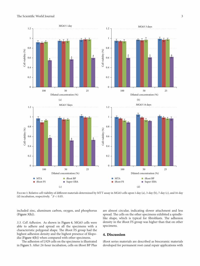
3.1. In Vitro Cytotoxicity. The in vitro cytotoxicities of thesematerials were compared using the MG63 model (Figure 1)and the L929 model (Figure 2), respectively. The resultsin the MG63 system were similar to those in the L929system. In both systems, elutes from the Super-EBA 7-daytreatment exhibited relative viabilities less than 60%. Therelative viability was dramatically enhanced (90%) by a 14-day culture. Serial dilutions of these elutes had no statisticallysignificant effect on the relative viability. On the other hand,the relative viabilities at different incubation durations (up to14 days) and different dilutions were all around 95% in theiRoot BP Plus, iRoot FS, and ProRoot MTA groups. Thesewere significantly higher than those of the Super-EBA group.
3.2. Surface Morphology and Element Analysis. The sur-face morphologies of the materials are illustrated in Fig-ures 3(a)–3(d). iRoot BP Plus and iRoot FS possessed asimilar morphology. Schistose and flaky crystals in variedsizes were observed (Figures 3(a) and 3(b)). The averagelength of flakes was 20𝜇m for iRoot BP Plus and 5𝜇m foriRoot FS, respectively. ProRoot MTA showed hexagonal-shaped granules with an average diameter of around 5𝜇m(Figure 3(c)). The EDX analysis revealed that iRoot BP Plus,iRoot FS, and ProRoot MTA possessed similar elementcompositions (calcium, carbon, oxygen, and phosphorus).Super-EBA showed a poorly crystallized morphology withlarge dendrites (∼100 𝜇m length), and themain compositions
The Scientific World Journal 3
0
0.2
0.4
0.6
0.8
1
1.2
100 50 25
Cel
l via
bilit
y (%
)MG63 1 day
∗
∗
∗
Diluted concentration (%)
(a)
0
0.2
0.4
0.6
0.8
1
1.2
100 50 25
Cel
l via
bilit
y (%
)
∗ ∗ ∗
MG63 3 days
Diluted concentration (%)
(b)
Diluted concentration (%)
MG63 7days
0
0.2
0.4
0.6
0.8
1
1.2
100 50 25
Cel
l via
bilit
y (%
)
MTAiRoot FS
iRoot BPSuper-EBA
∗
∗ ∗
(c)
MG63 14 days
0
0.2
0.4
0.6
0.8
1
1.2
100 50 25
Cel
l via
bilit
y (%
)
MTAiRoot FS
iRoot BPSuper-EBA
Diluted concentration (%)
(d)
Figure 1: Relative cell viability of different materials determined by MTT assay in MG63 cells upon 1-day (a), 3-day (b), 7-day (c), and 14-day(d) incubation, respectively. ∗𝑃 < 0.05.
included zinc, aluminum carbon, oxygen, and phosphorus(Figure 3(h)).
3.3. Cell Adhesion. As shown in Figure 4, MG63 cells wereable to adhere and spread on all the specimens with acharacteristic polygonal shape. The iRoot FS group had thehighest adhesion density and the highest presence of filopo-dia (Figure 4(b)) when compared with other specimens.
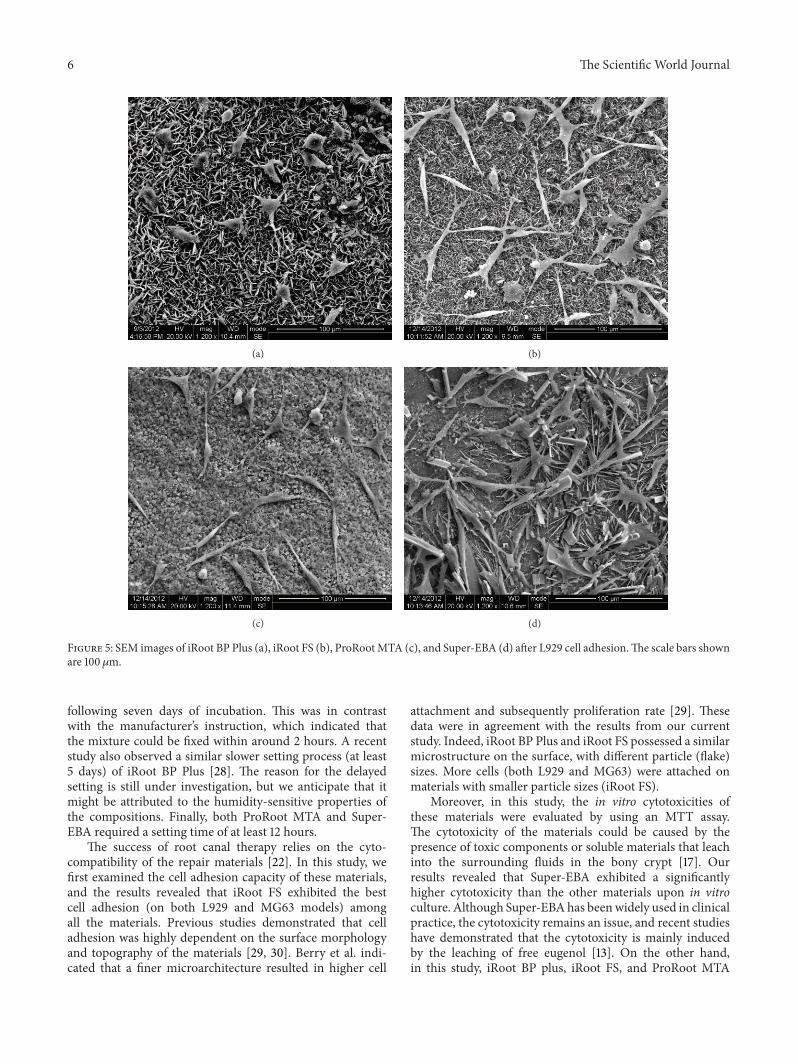
The adhesion of L929 cells on the specimens is illustratedin Figure 5. After 24-hour incubation, cells on iRoot BP Plus
are almost circular, indicating slower attachment and lessspread. The cells on the other specimens exhibited a spindle-like shape, which is typical for fibroblasts. The adhesiondensity in the iRoot FS group was higher than that on otherspecimens.
4. Discussion
iRoot series materials are described as bioceramic materialsdeveloped for permanent root canal repair applications with

4 The Scientific World Journal
L-929 1 day
0
0.2
0.4
0.6
0.8
1
1.2
100 50 25
Cel
l via
bilit
y (%
)
∗ ∗ ∗
Diluted concentration (%)
(a)
L-929 3 days
0
0.2
0.4
0.6
0.8
1
1.2
100 50 25
Cel
l via
bilit
y (%
)
∗
∗∗
Diluted concentration (%)
(b)
0
0.2
0.4
0.6
0.8
1
1.2
100 50 25
Cel
l via
bilit
y (%
)
MTAiRoot FS
iRoot BPSuper-EBA
∗
∗∗
Diluted concentration (%)
L-929 7 days
(c)
L-929 14 days
0
0.2
0.4
0.6
0.8
1
1.2
100 50 25
Cel
l via
bilit
y (%
)
MTAiRoot FS
iRoot BPSuper-EBA
Diluted concentration (%)
(d)
Figure 2: Relative cell viability of different materials determined by MTT assay in L929 cells upon 1-day (a), 3-day (b), 7-day (c), and 14-day(d) incubation, respectively. ∗𝑃 < 0.05.
improved handling properties and shorter setting times.Theyare ready-to-use white hydraulic premixed putty and arecomposed of calcium silicates, zirconium oxide, tantalumpentoxide, calciumphosphatemonobasic, andfiller agents. Inaddition, the manufacturer claims that there is no shrinkingduring the setting period and that the material is insoluble,radiopaque, and aluminum-free.
The ultimate goal of root canal repair procedure is topermanently seal infected or damaged root canals and topromote the healing of the repair area [1, 26]. For this
purpose, a number of materials have been developed andwidely used in clinics. In this study, we evaluated fourmarket-available repair materials (iRoot BP Plus, iRoot FS, ProRootMTA, and Super-EBA). One important criterion of thesematerials is the setting time [7, 27]. A reduction in settingduration has a beneficial effect on patient relief and reducingbacterial infection [5]. In this study, iRoot FS material wasable to completely solidify within one hour at 37∘C in 100%relative humidity given an extra load (500 g) to harden thematerial. Meanwhile, iRoot BP Plus formed a stable structure

The Scientific World Journal 5
iRoot BP Plus
(a)
iRoot FS
(b)
ProRoot MTA
(c)
Super-EBA
(d)
0200400600800
1000
0 2 4 6 8
C O
P Ca
(e)
CO
P Ca
0200400600800
1000
0 2 4 6 8
(f)
0
200
400
600
800
0 2 4 6 8
CO
P
Ca
(g)
C
O
Zn
ZnAl
P
0
200
400
600
0 2 4 6 8
(h)
Figure 3: SEM images and corresponding EDX spectrum of iRoot BP Plus ((a) and (e)), iRoot FS ((b) and (f)), ProRoot MTA ((c) and (g)),and Super-EBA ((d) and (h)). The scale bars shown are 100 𝜇m.
(a) (b)
(c) (d)
Figure 4: SEM images of iRoot BP Plus (a), iRoot FS (b), ProRoot MTA (c), and Super-EBA (d) after MG63 cell adhesion. The scale barsshown are 100 𝜇m.
6 The Scientific World Journal
(a) (b)
(c) (d)
Figure 5: SEM images of iRoot BP Plus (a), iRoot FS (b), ProRootMTA (c), and Super-EBA (d) after L929 cell adhesion.The scale bars shownare 100 𝜇m.
following seven days of incubation. This was in contrastwith the manufacturer’s instruction, which indicated thatthe mixture could be fixed within around 2 hours. A recentstudy also observed a similar slower setting process (at least5 days) of iRoot BP Plus [28]. The reason for the delayedsetting is still under investigation, but we anticipate that itmight be attributed to the humidity-sensitive properties ofthe compositions. Finally, both ProRoot MTA and Super-EBA required a setting time of at least 12 hours.
The success of root canal therapy relies on the cyto-compatibility of the repair materials [22]. In this study, wefirst examined the cell adhesion capacity of these materials,and the results revealed that iRoot FS exhibited the bestcell adhesion (on both L929 and MG63 models) amongall the materials. Previous studies demonstrated that celladhesion was highly dependent on the surface morphologyand topography of the materials [29, 30]. Berry et al. indi-cated that a finer microarchitecture resulted in higher cell
attachment and subsequently proliferation rate [29]. Thesedata were in agreement with the results from our currentstudy. Indeed, iRoot BP Plus and iRoot FS possessed a similarmicrostructure on the surface, with different particle (flake)sizes. More cells (both L929 and MG63) were attached onmaterials with smaller particle sizes (iRoot FS).
Moreover, in this study, the in vitro cytotoxicities ofthese materials were evaluated by using an MTT assay.The cytotoxicity of the materials could be caused by thepresence of toxic components or soluble materials that leachinto the surrounding fluids in the bony crypt [17]. Ourresults revealed that Super-EBA exhibited a significantlyhigher cytotoxicity than the other materials upon in vitroculture. Although Super-EBAhas beenwidely used in clinicalpractice, the cytotoxicity remains an issue, and recent studieshave demonstrated that the cytotoxicity is mainly inducedby the leaching of free eugenol [13]. On the other hand,in this study, iRoot BP plus, iRoot FS, and ProRoot MTA

The Scientific World Journal 7
Table 1: Characteristics of different root canal repair materials.
Specimen Initial form Setting time Surface morphology Particle sizeiRoot BP Plus Premixed paste 7 days∗ Schistose and flaky crystals 20 𝜇miRoot FS Premixed paste 1 hour Schistose and flaky crystals 5 𝜇mProRoot MTA Powder 4 hours Hexagonal granules 5 𝜇mSuper-EBA Powder 12 hours Dendrites 100 𝜇m∗Note: the expected setting time of iRoot BP Plus is around 2 hours according to manufacturer’s instruction.
exhibited negligible cytotoxicity.This can be explained by thenontoxic components, including calcium and phosphorus,of these materials. Furthermore, the presence of bioceramicfacilitated the formation of a hydroxyapatite or apatite-like layer (biomineralization), which further stabilizes thestructure and prevents the overdose of component leaching[31–33]. These results are in agreement with previous studies[22].
Taken together, iRoot FS demonstrated great potential infurther clinical applications due to its rapid setting, negligiblecytotoxicity, and enhanced cell adhesion capacity comparedwith other commonly used root canal repair materials(Table 1).
5. Conclusion
A comparative study was conducted on four root canal repairmaterials by using both L929 and MG63 cells. The resultsdemonstrated that iRoot FS exhibited the best cell adhesioncapacity, and only Super-EBA possessed in vitro cytotoxicity.Given the rapid solidification (within one hour) of iRootFS, this material showed high potential for further clinicalapplications. Future work should focus on long-term in vitrocytotoxicity and an in vivo assessment.
Conflict of Interests
The authors declare that there is no conflict of interestsregarding the publication of this paper.
Acknowledgments
The study was supported by the National Natural Sciencefoundation of China (Grant nos. 81070827 and 11272226) andThe Science and Technology support program of Sichuanprovince (no. 2011SZ0031).
References
[1] G. Bergenholtz, P. Hørsted-Bindslev, and C. Reit, Textbook ofEdodontology, vol. 193, Blackwell Publishing Company, Munks-gaard, Denmark, 2010.
[2] P. Xu, J. Liang, G. Dong, L. Zheng, and L. Ye, “Cytotoxicityof RealSeal on human osteoblast-like MG63 cells,” Journal ofEndodontics, vol. 36, no. 1, pp. 40–44, 2010.
[3] S. Desai andN. Chandler, “Calcium hydroxide-based root canalsealers: a review,” Journal of Endodontics, vol. 35, no. 4, pp. 475–480, 2009.
[4] J. Ma, Y. Shen, S. Stojicic, and M. Haapasalo, “Biocompatibilityof two novel root repair materials,” Journal of Endodontics, vol.37, no. 6, pp. 793–798, 2011.
[5] A. Z. AlAnezi, J. Jiang, K. E. Safavi, L. S. W. Spangberg, andQ. Zhu, “Cytotoxicity evaluation of endosequence root repairmaterial,” Oral Surgery, Oral Medicine, Oral Pathology, OralRadiology and Endodontology, vol. 109, no. 3, pp. e122–e125,2010.
[6] W. Zhang, Z. Li, and B. Peng, “Ex vivo cytotoxicity of anew calcium silicate-based canal filling material,” InternationalEndodontic Journal, vol. 43, no. 9, pp. 769–774, 2010.
[7] B. A. Damas, M. A. Wheater, J. S. Bringas, and M. M. Hoen,“Cytotoxicity comparison of mineral trioxide aggregates andendosequence bioceramic root repair materials,” Journal ofEndodontics, vol. 37, no. 3, pp. 372–375, 2011.
[8] S. Bouillaguet, J. C. Wataha, F. R. Tay, M. G. Brackett, and P. E.Lockwood, “Initial In vitro biological response to contemporaryendodontic sealers,” Journal of Endodontics, vol. 32, no. 10, pp.989–992, 2006.
[9] W. Geurtsen,W. Spahl, andG. Leyhausen, “Residual monomer/additive release and variability in cytotoxicity of light-curingglass-ionomer cements and compomers,” Journal of DentalResearch, vol. 77, no. 12, pp. 2012–2019, 1998.
[10] Z. Yilmaz, A. L. Dogan, O. Ozdemir, and A. Serper, “Evaluationof the cytotoxicity of different root canal sealers on L929 cellline by MTT assay,” Dental Materials Journal, vol. 31, no. 6, pp.1028–1032, 2012.
[11] B. Sagsen, Y. Ustun, K. Pala, and S. Demırbuga, “Resistance tofracture of roots filled with different sealers,” Dental MaterialsJournal, vol. 31, no. 4, pp. 528–532, 2012.
[12] S. Bonson, B. G. Jeansonne, and T. E. Lallier, “Root-end fillingmaterials alter fibroblast differentiation,” Journal of DentalResearch, vol. 83, no. 5, pp. 408–413, 2004.
[13] A. Samara, Y. Sarri, D. Stravopodis, G. N. Tzanetakis, E. G.Kontakiotis, and E. Anastasiadou, “A comparative study of theeffects of three root-end filling materials on proliferation andadherence of human periodontal ligament fibroblasts,” Journalof Endodontics, vol. 37, no. 6, pp. 865–870, 2011.
[14] E. Bodrumlu, “Biocompatibility of retrograde root filling mate-rials: a review,” Australian Endodontic Journal, vol. 34, no. 1, pp.30–35, 2008.
[15] S. Friedman, “Retrograde approaches in endodontic therapy,”Endodontics & Dental Traumatology, vol. 7, no. 3, pp. 97–107,1991.
[16] M.Asrari andD. Lobner, “In vitroneurotoxic evaluation of root-end-filling materials,” Journal of Endodontics, vol. 29, no. 11, pp.743–746, 2003.
[17] K. Keiser, C. Chad Johnson, and D. A. Tipton, “Cytotoxicity ofmineral trioxide aggregate using human periodontal ligamentfibroblasts,” Journal of Endodontics, vol. 26, no. 5, pp. 288–291,2000.

8 The Scientific World Journal
[18] O. R. Al-Sa’eed, A. S. Al-Hiyasat, and H. Darmani, “The effectsof six root-end filling materials and their leachable componentson cell viability,” Journal of Endodontics, vol. 34, no. 11, pp. 1410–1414, 2008.
[19] C.-P. Lin, Y.-J. Chen, Y.-L. Lee et al., “Effects of root-end fillingmaterials and eugenol onmitochondrial dehydrogenase activityand cytotoxicity to human periodontal ligament fibroblasts,”Journal of Biomedical Materials Research B, vol. 71, no. 2, pp.429–440, 2004.
[20] H. W. Roberts, J. M. Toth, D. W. Berzins, and D. G. Charlton,“Mineral trioxide aggregate material use in endodontic treat-ment: a review of the literature,”Dental Materials, vol. 24, no. 2,pp. 149–164, 2008.
[21] M. Parirokh andM. Torabinejad, “Mineral trioxide aggregate: acomprehensive literature review—part III: clinical applications,drawbacks, and mechanism of action,” Journal of Endodontics,vol. 36, no. 3, pp. 400–413, 2010.
[22] G. De-Deus, A. Canabarro, G. G. Alves, J. R. Marins, A. B.R. Linhares, and J. M. Granjeiro, “Cytocompatibility of theready-to-use bioceramic putty repair cement iRoot BPPluswithprimary human osteoblasts,” International Endodontic Journal,vol. 45, no. 6, pp. 508–513, 2012.
[23] M. Ciasca, A. Aminoshariae, G. Jin, T. Montagnese, and A.Mickel, “A comparison of the cytotoxicity and proinflammatorycytokine production of EndoSequence root repair material andProRoot mineral trioxide aggregate in human osteoblast cellculture using reverse-transcriptase polymerase chain reaction,”Journal of Endodontics, vol. 38, no. 4, pp. 486–489, 2012.
[24] F. de Paula and C. J. Rosen, “Bone remodeling and energymetabolism: new perspectives,” Bone Research, vol. 1, no. 1, pp.72–84, 2013.
[25] ISO 10993-5: 2009 Biological evaluation of medical devices part5: tests for in vitro cytotoxicity.
[26] I.Willershausen, A. Callaway, B. Briseno, and B.Willershausen,“In vitro analysis of the cytotoxicity and the antimicrobial effectof four endodontic sealers,” Head and Face Medicine, vol. 7, no.1, article 15, 2011.
[27] J.-S. Song, F. K. Mante, W. J. Romanow, and S. Kim, “Chemicalanalysis of powder and set forms of Portland cement, grayProRoot MTA, white ProRoot MTA, and gray MTA-Angelus,”Oral Surgery, OralMedicine, Oral Pathology, Oral Radiology andEndodontology, vol. 102, no. 6, pp. 809–815, 2006.
[28] M. R. Modareszadeh, P. M. di Fiore, D. A. Tipton, and N. Sala-mat, “Cytotoxicity and alkaline phosphatase activity evaluationof endosequence root repair material,” Jounal of Endodontics,vol. 38, no. 8, pp. 1101–1105, 2012.
[29] C. C. Berry, G. Campbell, A. Spadiccino, M. Robertson, andA. S. G. Curtis, “The influence of microscale topography onfibroblast attachment andmotility,” Biomaterials, vol. 25, no. 26,pp. 5781–5788, 2004.
[30] C. Ji, N. Annabi, A. Khademhosseini, and F. Dehghani, “Fab-rication of porous chitosan scaffolds for soft tissue engineeringusing dense gas CO
2
,”Acta Biomaterialia, vol. 7, no. 4, pp. 1653–1664, 2011.
[31] N. K. Sarkar, R. Caicedo, P. Ritwik, R. Moiseyeva, and I.Kawashima, “Physicochemical basis of the biologic propertiesof mineral trioxide aggregate,” Journal of Endodontics, vol. 31,no. 2, pp. 97–100, 2005.
[32] J. F. Reyes-Carmona, M. S. Felippe, andW. T. Felippe, “Biomin-eralization ability and interaction of mineral trioxide aggre-gate and white Portland cement with dentin in a phosphate-containing fluid,” Journal of Endodontics, vol. 35, no. 5, pp. 731–736, 2009.
[33] F. R. Tay, D. H. Pashley, F. A. Rueggeberg, R. J. Loushine, and R.N. Weller, “Calcium phosphate phase transformation producedby the interaction of the Portland cement component of whitemineral trioxide aggregate with a phosphate-containing fluid,”Journal of Endodontics, vol. 33, no. 11, pp. 1347–1351, 2007.

Submit your manuscripts athttp://www.hindawi.com
Hindawi Publishing Corporationhttp://www.hindawi.com Volume 2014
Oral OncologyJournal of
DentistryInternational Journal of
Hindawi Publishing Corporationhttp://www.hindawi.com Volume 2014
Hindawi Publishing Corporationhttp://www.hindawi.com Volume 2014
International Journal of
Biomaterials
Hindawi Publishing Corporationhttp://www.hindawi.com Volume 2014
BioMed Research International
Hindawi Publishing Corporationhttp://www.hindawi.com Volume 2014
Case Reports in Dentistry
Hindawi Publishing Corporationhttp://www.hindawi.com Volume 2014
Oral ImplantsJournal of
Hindawi Publishing Corporationhttp://www.hindawi.com Volume 2014
Anesthesiology Research and Practice
Hindawi Publishing Corporationhttp://www.hindawi.com Volume 2014
Radiology Research and Practice
Environmental and Public Health
Journal of
Hindawi Publishing Corporationhttp://www.hindawi.com Volume 2014
The Scientific World JournalHindawi Publishing Corporation http://www.hindawi.com Volume 2014
Hindawi Publishing Corporationhttp://www.hindawi.com Volume 2014
Dental SurgeryJournal of
Drug DeliveryJournal of
Hindawi Publishing Corporationhttp://www.hindawi.com Volume 2014
Hindawi Publishing Corporationhttp://www.hindawi.com Volume 2014
Oral DiseasesJournal of
Hindawi Publishing Corporationhttp://www.hindawi.com Volume 2014
Computational and Mathematical Methods in Medicine
ScientificaHindawi Publishing Corporationhttp://www.hindawi.com Volume 2014
PainResearch and TreatmentHindawi Publishing Corporationhttp://www.hindawi.com Volume 2014
Preventive MedicineAdvances in
Hindawi Publishing Corporationhttp://www.hindawi.com Volume 2014
EndocrinologyInternational Journal of
Hindawi Publishing Corporationhttp://www.hindawi.com Volume 2014
Hindawi Publishing Corporationhttp://www.hindawi.com Volume 2014
OrthopedicsAdvances in