renovascular hypertension in children - emeesy€¦renovascular hypertension in children martin...
TRANSCRIPT
Renovascular hypertension in children
Martin Christian
Consultant Paediatric Nephrologist
Nottingham Children’s Hospital
Perspective
• May account for up to 10% of childhood hypertension
• Most commonly caused by fibromuscular dysplasia
• Frequently bilateral • Some syndromic associations – particularly
neurofibromatosis • May be associated with extra-renal vascular
disease • Potentially curable!
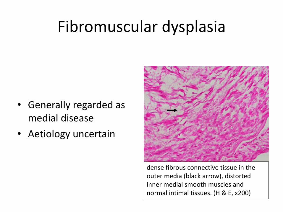
Fibromuscular dysplasia
• Generally regarded as medial disease
• Aetiology uncertain
dense fibrous connective tissue in the outer media (black arrow), distorted inner medial smooth muscles and normal intimal tissues. (H & E, x200)

Presentation Shroff et al, 2006; Pediatrics 118: 268-75.
Mode of Presentation No.
Incidental 9
Cardiac (congestive cardiac failure, palpitations, murmur) 7
Headache with or without vomiting or lethargy 6
Acute hypertensive encephalopathy 3
Cerebrovascular accident 2
Facial palsy 2
Poor feeding and failure to thrive 2
Screening for NF1 2
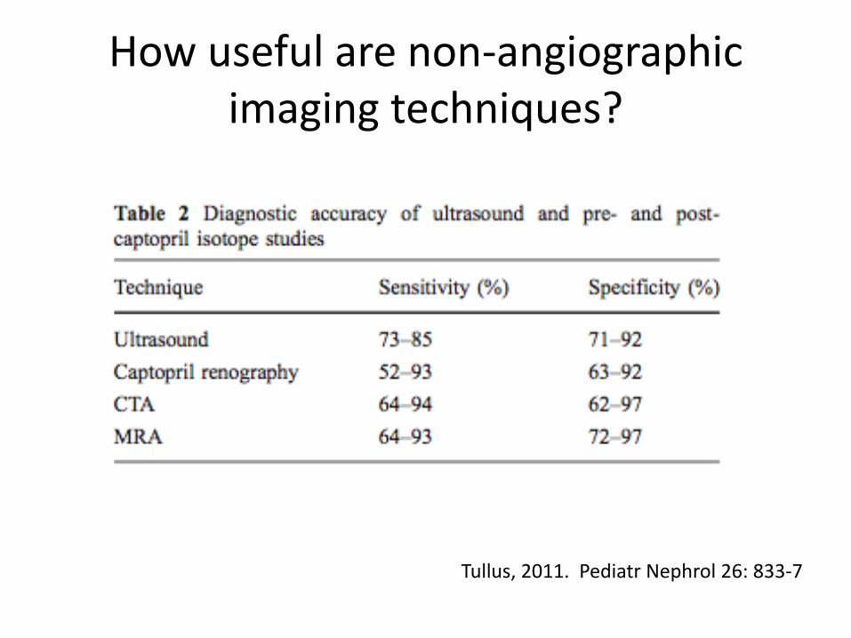
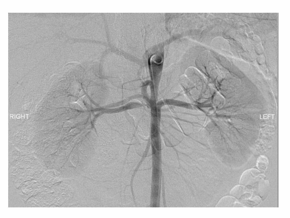
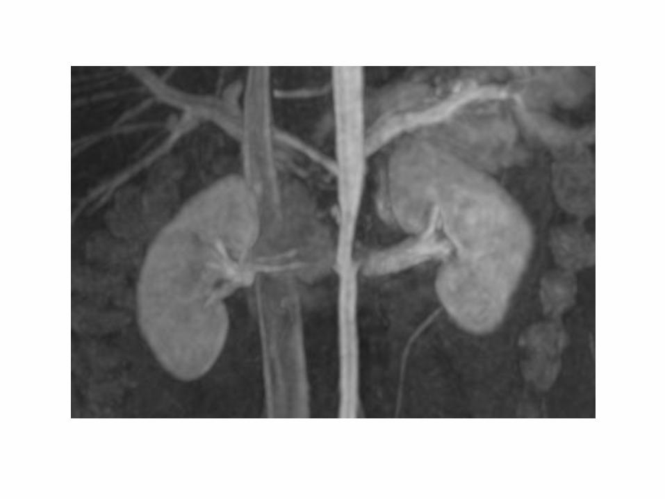
How useful are non-angiographic imaging techniques?
Tullus, 2011. Pediatr Nephrol 26: 833-7
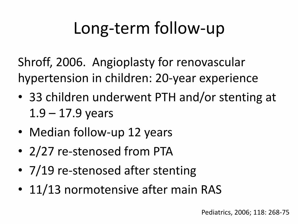
Long-term follow-up
Shroff, 2006. Angioplasty for renovascular hypertension in children: 20-year experience
• 33 children underwent PTH and/or stenting at 1.9 – 17.9 years
• Median follow-up 12 years
• 2/27 re-stenosed from PTA
• 7/19 re-stenosed after stenting
• 11/13 normotensive after main RAS
Pediatrics, 2006; 118: 268-75
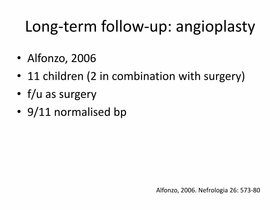
Long-term follow-up: angioplasty
• Alfonzo, 2006
• 11 children (2 in combination with surgery)
• f/u as surgery
• 9/11 normalised bp
Alfonzo, 2006. Nefrologia 26: 573-80
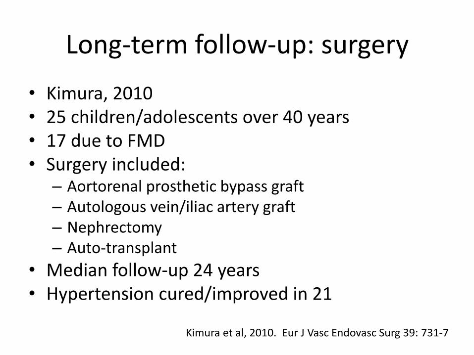
Long-term follow-up: surgery
• Kimura, 2010 • 25 children/adolescents over 40 years • 17 due to FMD • Surgery included:
– Aortorenal prosthetic bypass graft – Autologous vein/iliac artery graft – Nephrectomy – Auto-transplant
• Median follow-up 24 years • Hypertension cured/improved in 21
Kimura et al, 2010. Eur J Vasc Endovasc Surg 39: 731-7

Long-term follow-up: surgery
• Alfonzo, 2006
• 6 children, aged 2-18 y
• Mean follow-up 11.5 y
• All cured

Unilateral nephrectomy follow-up
• 4 cases presenting in early infancy and childhood
• Age at surgery: 0.1 – 2.5 years
• % Function 14 – 27%
• Follow-up: 5-16 years
• All normotensive off treatment with normal GFR
Hegde and Coulthard, 2007. Arch Dis Child Fetal Neonatal Ed; 92:F305-6
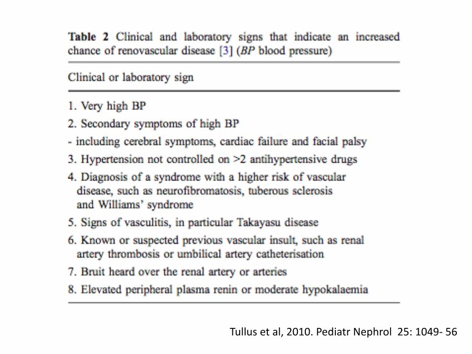
Tullus et al, 2010. Pediatr Nephrol 25: 1049- 56

R
P
Rc
Lateralisation: R/Rc >1.5
Suppression of contralateral kidney: Rc/P <1.3

25 year review of renal vein renin sampling. Goonasekara, 2002.
• 137 procedures reviewed
• 30% false positive rate in diagnosing RVD
• Useful in predicting response to intervention?
– High sensitivities but low specificities
Pediatr Nephrol 2002; 17: 943-9
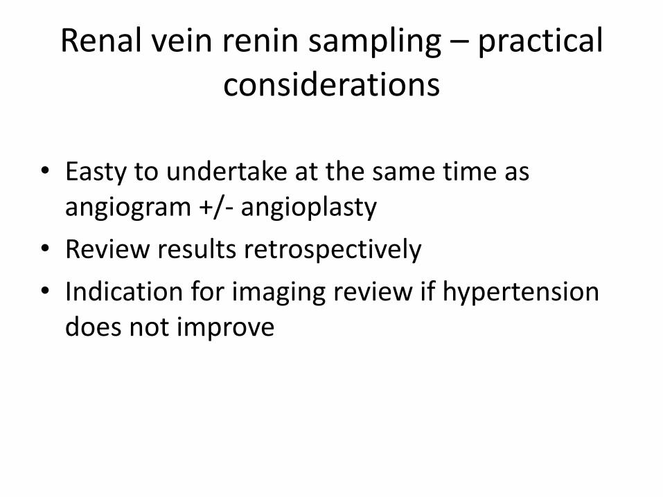
Renal vein renin sampling – practical considerations
• Easty to undertake at the same time as angiogram +/- angioplasty
• Review results retrospectively
• Indication for imaging review if hypertension does not improve

DISCUSSION OF CASES
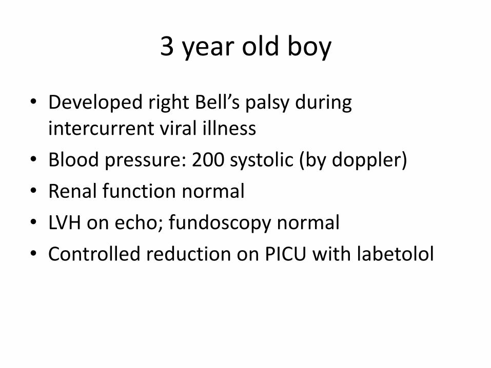
3 year old boy
• Developed right Bell’s palsy during intercurrent viral illness
• Blood pressure: 200 systolic (by doppler)
• Renal function normal
• LVH on echo; fundoscopy normal
• Controlled reduction on PICU with labetolol

Imaging
• USS and doppler – normal
• MAG3: 43% function on left
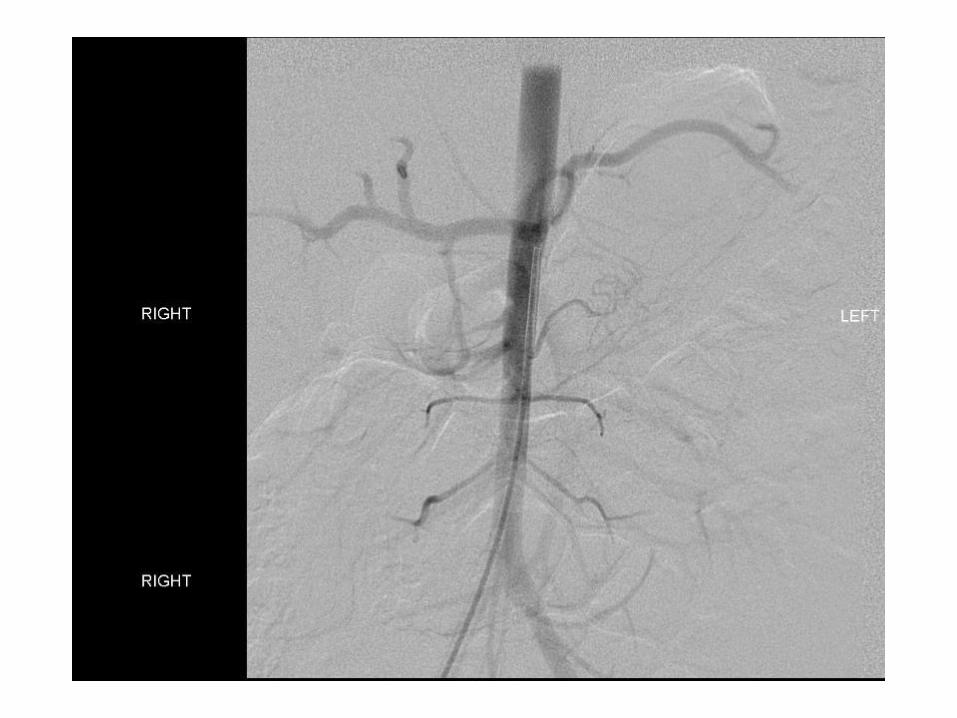
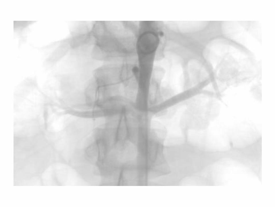
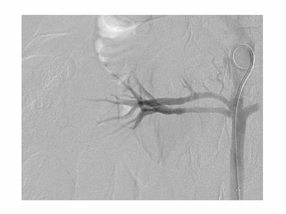
• Angiogram

Follow-up
• Good angioplasty result
• 2 antihypertensives at discharge
• Slow/near complete resolution of Bell’s palsy
• Atenolol finally stopped after 10 months

Learning points
• Importance of checking BP in the context of Bell’s palsy
• When to proceed to invasive hypertension investigations?
• Angioplasty effective but long-term follow-up needed
• BP can take months to normalise through vascular remodelling

13 month old girl
• Presented with left Bell’s palsy on a background of growth faltering
• Initial systolic blood pressure 240 mmHg
• Labetolol infusion to stabilise
• Required 5 oral agents to control, including captopril after definitive management decision
Imaging
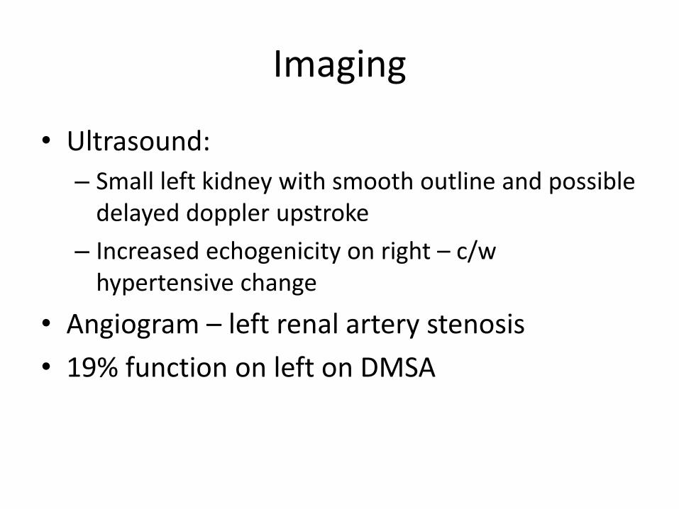
• Ultrasound:
– Small left kidney with smooth outline and possible delayed doppler upstroke
– Increased echogenicity on right – c/w hypertensive change
• Angiogram – left renal artery stenosis
• 19% function on left on DMSA
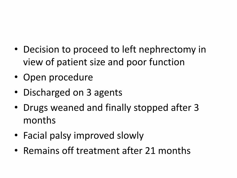
• Decision to proceed to left nephrectomy in view of patient size and poor function
• Open procedure
• Discharged on 3 agents
• Drugs weaned and finally stopped after 3 months
• Facial palsy improved slowly
• Remains off treatment after 21 months

Learning points
• Nephrectomy may be indicated rather than angioplasty in some cases
• Judicious use of ACEIs has a role in management of difficult cases
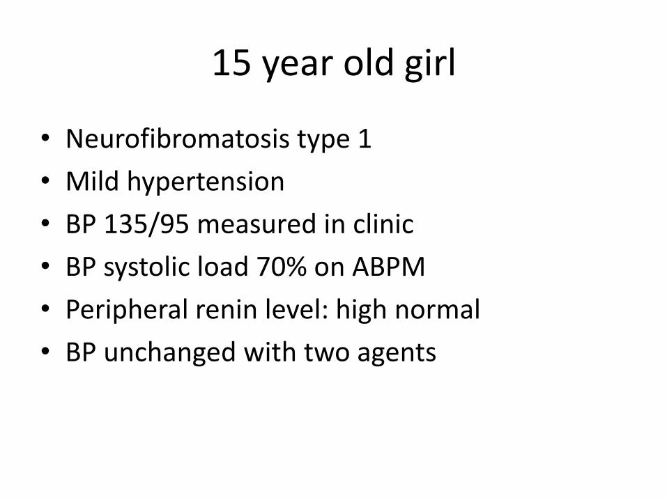
15 year old girl
• Neurofibromatosis type 1
• Mild hypertension
• BP 135/95 measured in clinic
• BP systolic load 70% on ABPM
• Peripheral renin level: high normal
• BP unchanged with two agents
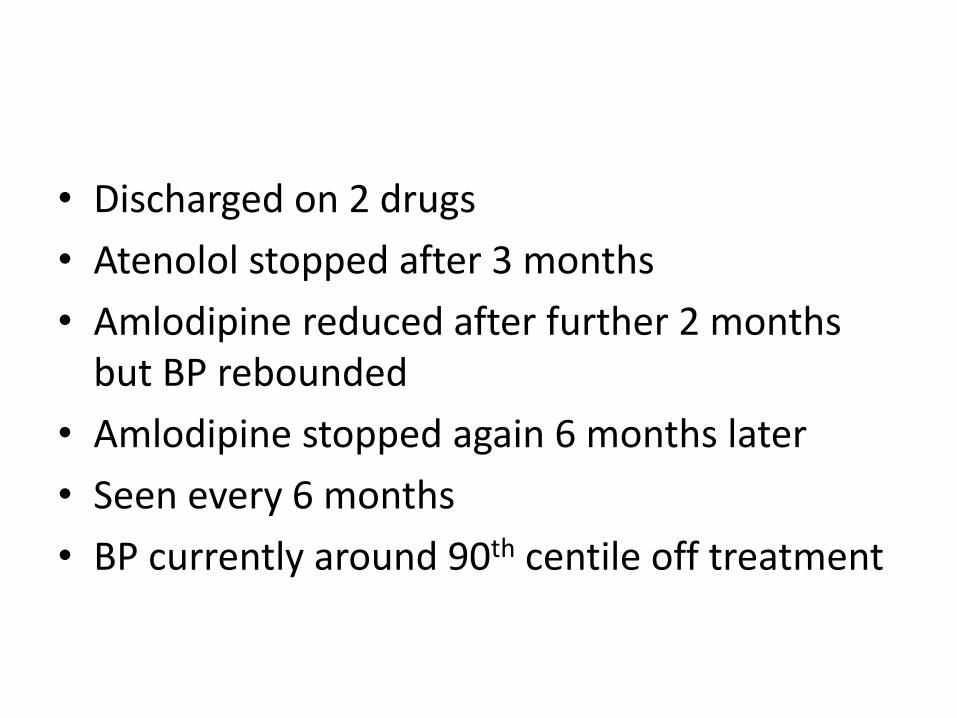
• Discharged on 2 drugs
• Atenolol stopped after 3 months
• Amlodipine reduced after further 2 months but BP rebounded
• Amlodipine stopped again 6 months later
• Seen every 6 months
• BP currently around 90th centile off treatment


R
P
Rc
Lateralisation: R/Rc >1.5
Suppression of contralateral kidney: Rc/P <1.3
235 113
105
1.7 = lateralised
1.1 = suppressed
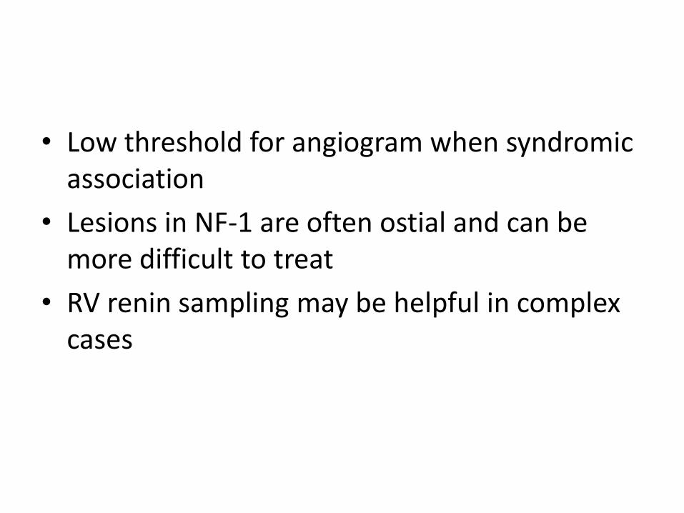
• Low threshold for angiogram when syndromic association
• Lesions in NF-1 are often ostial and can be more difficult to treat
• RV renin sampling may be helpful in complex cases

14 year old girl
• Incidental finding of hypertension
• BP uncontrolled in single agent
• Initial USS and DMSA normal
• Peripheral renin/aldosterone normal
• BP remained poorly controlled on two agents
• Decision to proceed to angiogram after 12 m

• Mid-aortic syndrome and left lower pole stenosis
• Decision to plan angioplasty / canvas opinion
• Angioplasty 4 months later with safety wire across aorta
• Both aorta and right lower pole artery angioplastied
• BP remained high on 2 agents

• Follow-up CT angiogram showed possible stenosed right upper pole vessel
• Repeat MR angiogram 6 m later – appearances unchanged
• Repeat angiogram/angioplasty

• BP again creeping up
• Further MR angiogram

• Now transitioned to adult care
• Remains on single agent with controlled BP
• Likely need for on-going imaging and possible repeat angiography/surgery
Learning points
• Indication for angiography when uncontrolled BP on two agents
• CT/MR angiography useful in follow-up imaging

Questions?