reliability of intraoperative ultrasound in margin status assessment in
TRANSCRIPT

RELIABILITY OF INTRAOPERATIVE ULTRASOUND
IN MARGIN STATUS ASSESSMENT IN WOMEN
UNDERGOING BREAST CONSERVING SURGERY
FINAL GRADE PROJECT
NOVEMBER 2014
AUTHOR: Joan Oliveras Andal
TUTOR: Dr. Ester Vila Camps
Facultat de Medicina

Reliability of intraoperative ultrasound in margin status assessment in women undergoing breast conserving surgery
2
I would like to thank Dr. Josep Trueta’s Hospital Breast Pathology Unit for their continued support and
encouragement: Dr. Ester Vila, my final grade project tutor; Dr. Miguel Alonso and Dr. Francesc Tuca.
I would also like to thank all the people who got involved with this project, without their generous
support it would not have been possible.

Reliability of intraoperative ultrasound in margin status assessment in women undergoing breast conserving surgery
3
INDEX
1. ABBREVIATIONS ......................................................................................................................................... 5
2. ABSTRACT ...................................................................................................................................................... 6
3. INTRODUCTION ........................................................................................................................................... 7
3.1. Background .......................................................................................................................................... 7
3.2. Justification ........................................................................................................................................ 17
4. BIBLIOGRAPHY .......................................................................................................................................... 19
5. QUESTION .................................................................................................................................................... 23
6. HYPOTHESIS ............................................................................................................................................... 23
7. OBJECTIVES ................................................................................................................................................. 23
8. MATERIAL AND METHODS ................................................................................................................... 24
8.1. Study Design ...................................................................................................................................... 24
8.2. Participants ........................................................................................................................................ 24
8.3. Inclusion criteria .............................................................................................................................. 24
8.4. Exclusion criteria ............................................................................................................................. 24
8.5. Sample selection .............................................................................................................................. 25
8.6. Sample size ......................................................................................................................................... 25
8.7. Variables .............................................................................................................................................. 25
8.7.1. Independent variables ......................................................................................................... 25
8.7.2. Dependant variable ............................................................................................................... 28
8.7.3. Covariables ............................................................................................................................... 28
8.8. Methods of data collection ........................................................................................................... 29
9. STATISTICAL ANALYSIS ......................................................................................................................... 30
10. ETHICAL ASPECTS ............................................................................................................................... 31
11. LIMITATIONS OF THE STUDY ......................................................................................................... 32
12. WORK PLAN ........................................................................................................................................... 33
13. TIME SCHEDULE ................................................................................................................................... 35
14. AVAILABLE MEANS TO CARRY OUT THE PROJECT .............................................................. 35

Reliability of intraoperative ultrasound in margin status assessment in women undergoing breast conserving surgery
4
15. BUDGET .................................................................................................................................................... 36
16. ANNEXES ................................................................................................................................................. 37
16.1. ANNEX I .......................................................................................................................................... 37
16.2. ANNEX II ......................................................................................................................................... 41
16.3. ANNEX III ....................................................................................................................................... 42
16.4. ANNEX IV ....................................................................................................................................... 44

Reliability of intraoperative ultrasound in margin status assessment in women undergoing breast conserving surgery
5
1. ABBREVIATIONS
BCS Breast Conserving Surgery
BCT Breast Conserving Therapy
CT-scan Computerized Tomography Scanner
DCIS Ductal Carcinoma In Situ
ER Oestrogen Receptor
FSA Frozen Section Analysis
HER2 Human Epidermal Growth Factor Receptor 2
IC Imprint Cytology
ICC Intraclass Coefficient
IMA Intraoperative Margin Assessment
IOUS Intraoperative Ultrasound
LR Local Recurrence
MRI Magnetic Resonance Imaging
PR Progesterone Receptor
ROLL Radio-guided Occult Lesion Localization
SERM Selective Oestrogen Receptor Modulator
SM Surgical Margins
SNOLL Sentinel Node and Occult Lesion Localization
US Ultrasound
WGL Wire-Guided Localization

Reliability of intraoperative ultrasound in margin status assessment in women undergoing breast conserving surgery
6
2. ABSTRACT
Background Effective treatment for breast cancer requires accurate preoperative
planning, developing and implementing a consistent definition of
margin clearance, and using tools that provide detailed real-time
intraoperative information on margin status. Intraoperative
ultrasound (IOUS) may fulfil these requirements and may offer few
advantages that other preoperative localization and intraoperative
margin assessment techniques may not.
Purpose The goal of the present work is to determine how accurate the
intraoperative ultrasound should be to acquire complete surgical
excision with negative histological margins in patients undergoing
Breast Conservative Surgery.
Design A diagnostic test study with a cross-sectional design carried out in a
tertiary referral hospital in Girona within a Breast Pathology Unit.
Participants Women diagnosed with breast cancer undergoing a Breast
Conservative Surgery in the Breast Pathology Unit at Hospital
Universitari de Girona Dr. Josep Trueta.
Key words Early Stage Breast Cancer; Breast Conserving Surgery;
Intraoperative Ultrasound; Margin Status.

Reliability of intraoperative ultrasound in margin status assessment in women undergoing breast conserving surgery
7
Graphic 1. Estimated age-standardised incidence and mortality rates of breast cancer in Spanish women
Resource: GLOBOCAN 2012: Estimated Cancer Incidence, Prevalence and Mortality Worldwide in 2012. [Internet]. Lyon: Internation Agency for Research on Cancer (IARC); 2014. p. 1–7. Available from: http://globocan.iarc.fr/Pages/fact_sheets_population.aspx
3. INTRODUCTION
3.1. Background
Breast Cancer is the most prevalent cancer in a wide majority of countries globally, being
the main type of cancer among women worldwide. Close to half of the prevalence burden
is in areas of high human development although it represents only one sixth of the world’s
population (1).
In Europe, there were over 3.4 million new cases of cancer (excluding non- melanoma skin
cancers) in 2012. The most common cancer globally was breast cancer, with an estimated
incidence of 464,000 cases, representing the 13.5% of all cancer cases and the 28.8% of
the cancers affecting women; moreover, breast cancer is the first cause of cancer death in
European women (2). In fact, Europe carries a significant load of the global burden; with
only one-ninth of the world’s population, it carries one quarter of the global burden of
cancer.
In 2012, as can be appreciated in the graphic 1, breast cancer was the most commonly
diagnosed cancer among females in Spain with an incidence of 25,215 cases representing
the 29% of the overall cancers in women. The five-year prevalence was 104,210 cases
representing the 40% of cancers diagnosed among Spanish women. When concerning
mortality, breast cancer stood for the 15.5% of cancer deaths (3).

Reliability of intraoperative ultrasound in margin status assessment in women undergoing breast conserving surgery
8
With reference to a local level, in the Sanitary Region of Girona, according to the CanGir
2007-9 report submitted by el Registre del Càncer de Girona, by the time of 2013-2014, it
is estimated an incidence of breast cancer of approximately 389 cases, being the most
common diagnosed cancer among women in Girona (4). All in all, breast cancer has
become the leading cause of cancer in terms of new cases according to the latest official
reports.
Updated data regarding cancer incidence and mortality in Europe is a crucial resource in
both planning and assessing the impact of cancer control programmes at a global and
regional level (2).
Due to the fact that breast cancer is the most common cancer in women, Health Care
Services from high developed countries have targeted this disease as an important issue to
be focused on. As a consequence of its high prevalence and incidence, lots of funds have
been invested in improving the medical approaches to diagnose and treat effectively
patients affected by this cancer. During the last decades, important screening programmes
have been designed to detect early stage breast cancer. The direct consequence of this
measure is the detection of a large amount of stage I-II breast cancers, currently
representing more than 80% of diagnosed cancers (5).
In fact, and, as a consequence of the early detection of breast cancer, its surgical treatment
has been in considerable evolution over the past decades. Previously, the radical
mastectomy introduced by Halsted, an American surgeon, was the standard treatment for
breast cancer of any size or type, regardless of the patient’s age. This surgical technique
cuts out the breast, axillary nodes and chest muscles including pectoralis major and minor.
Radical mastectomy was the surgical treatment choice for most of the 20th century and the
option of attempting a surgical procedure that would conserve the breast was not widely
considered during those years (6). It was during the 70s, when important clinical trials
were performed in order to update the management of early diagnosed breast cancers
with the aim of reducing the physical and psychological morbidity associated with this
aggressive procedure (7).
These randomized clinical trials concluded that there was no significant difference in the
disease-free survival, long-term survival and overall survival among women with early
stage breast cancer who underwent mastectomy and those who had gone through Breast
Conserving Therapy (BCT), which is the removal of the primary tumour and a margin of
surrounding tissue (lumpectomy) plus adjuvant radiotherapy; the surgical part is also
called Breast Conserving Surgery (BCS). The results of these studies showed that there
Reliability of intraoperative ultrasound in margin status assessment in women undergoing breast conserving surgery
9
was no decrease in overall survival after BCT (6,7). New less aggressive, safe and effective
standards of care in loco-regional and adjuvant systemic therapy were accepted in order
to reduce the extent of surgical procedures and to improve the outcome of patients with
early-stage breast cancer. Thus, surgical treatment shifted from mastectomy to breast
conserving surgery in conjunction with adjuvant therapies as first choice treatment for
early stage breast cancer (8).
Even though BCS has proved to be equivalent in terms of long-term survival when
compared to mastectomy (9), this surgical approach is not exempt from disadvantages.
BCS faces some limitations:
1. Localizing non-palpable tumours
2. The possibility of inadequate resection of the tumour during the initial
operation
3. Potential risk of local recurrence (LR)
1. Localizing non-palpable tumours
Regarding the first issue, that is the localization of non-palpable lesions, several
preoperative localization techniques have been developed (10,11):
Carbon Marking
Inert charcoal powder diluted with saline solution introduced through the skin,
marking the way to reach the lesion. It is introduced under image guidance such as
stereotactic or sonographic localization. The following surgical excision is guided
by the presence of the carbon in suspension. Due to charcoal stability, surgery can
be performed with delay. A potential disadvantage is the obstruction of needle tip
as a consequence of carbon powder particles precipitation.
Wire-Guided Localization (WGL)
It consists in the placement of one or more needle or flexible wire into the centre of
the lesion in order to guide the surgeon in excision. It can be guided through
mammographic, sonographic or CT image techniques. Despite the fact that WGL is
relatively simple and cost-effective, it may have some disadvantages such as wire
dislodgment leading to a difficult excision-guided surgery. Nowadays, it is
considered to be the gold standard approach for clinically non-palpable tumours.
Reliability of intraoperative ultrasound in margin status assessment in women undergoing breast conserving surgery
10
Radio-Guided Occult Lesion Localization (ROLL)
It is based on the preoperative injection of radioactive isotope 99mTc (radioactive
technetium) into the tumour under mammographic or sonographic guidance
followed by a scintigraphy scan of the breast in order to check the correct
inoculation of the tracer. The surgeon detects the tumour with the help of a gamma
probe and checks the adequacy of excision. Moreover, the tracer can be injected
near to the tumour to be drained in the sentinel node, which can be easily
identified by the gamma probe. This additional technique is called “sentinel node
and occult lesion localization” (SNOLL). A complication of this approach is the risk
of dispersion of the tracer among the breast tissue hindering the localization of the
lesion.
Intraoperative Ultrasound (IOUS)
It consists in the intraoperative ultrasound assessment of the lesion immediately
previous to the surgery. The tumour must be visualized by sonography for this
technique to be successful. It allows to plan the incision and reassess orientation in
real-time. Additionally, it may help in positioning the wire, injecting the dye or
marking on the skin. This approach avoids the requirement of the preoperative
localization of the tumour. In addition, the IOUS may guide the resection of the
tumour and assess the margin status.
Despite the fact that WGL is considered to be the gold standard approach for
preoperative localization, it has technical and scheduling drawbacks. This has led to
the development of alternative localization techniques. Among these techniques,
intraoperative sonographic localization has proved to be an accurate and simple
method of ensuring adequate excision of screen detected palpable and non-palpable
lesions. It is comparable to WGL, but avoids painful preoperative localization and
saves both radiology and theatre time. It may become the new gold standard for
image-guided surgery in BCS (12,13).
Many lesions are clearly visible on ultrasound, therefore allowing the localization of
the tumour and its limits; however, on the one hand, there are other breast tumours
that are scarcely visible on ultrasound such as DCIS, and on the other hand, there are
others owning image features that are not well detected through the ultrasound and
may not exhibit the typical criteria for malignancy; for instance, invasive lobular
carcinoma may not show posterior acoustic enhancement, simulating a benign lesion

Reliability of intraoperative ultrasound in margin status assessment in women undergoing breast conserving surgery
11
when, in fact, the real underlying character of the lesion is malignant (14). In these
cases, other preoperative localization techniques might be suitable.
As BCT has been established as the choice treatment for early stage breast cancers and
for those cancers with a satisfactory clinical response to post-neoadjuvant therapy, a
new challenge has arisen. That challenge is the adequacy of margin resection when
BCS is performed; a positive margin results in additional surgery. Over the last years,
the advances on the new preoperative localization techniques and methods of
intraoperative margin assessment have resulted in a reduction of positive margin
rates, reporting acceptable rates as low as 15-20% (15); however, BCS is still
associated to high re-excision rates ranging from 20 to 60% (16).
2. Possibility of inadequate resection of the tumour during the initial operation
Regarding the second downside of BCS, the probability of inadequate resection of the
tumour during the initial operation leads to a major risk of a positive margin in the
excised tumour; several intraoperative margin assessment (IMA) techniques have
been developed in order to avoid inadequate margins in BCS (11,17):
Standard Cavity Shaving
It consists in the resection of breast tissue from all 6 margins (anterior, posterior,
superior, inferior, medial and lateral) after the excision of the primary specimen, in
the same procedure. It allows to precisely asses which margin is involved in order
to calibrate the resection of the tumour.
Intraoperative Specimen Radiography
Once the specimen is excised, a digital mammography on the specimen is applied
to ensure that the entire lesion is removed. If abnormal tissue is detected close to
the surgical margin, the corresponding region of the cavity can be shaved.
Ultrasound-guided excision
Use of intraoperative US-guided excision consists in placing the ultrasound-probe
while BCS is undergone, hence allowing to reassess orientation in real-time and
confirm complete excision of the tumour. Its purpose is to obtain accurate margins.
Novel surgical techniques
Emerging technologies such as spectroscopy or optical coherence tomography
(OCT) may have limited clinical applicability and may require further research to
prove their reliability.

Reliability of intraoperative ultrasound in margin status assessment in women undergoing breast conserving surgery
12
Intraoperative Pathological Assessment:
- Frozen Section Analysis (FSA)
Once the specimen is excised, orientated with sutures, margins are inked and it
is thinly sliced; samples from any area of concern are cut, frozen with
embedding medium, and processed in less than 30 minutes.
- Imprint cytology (IC)
It consists in placing each of the six margins in contact with a glass slide to
determine if they are tumour-free. Cancer cells will adhere to the glass
whereas normal adipose cells will not. Once all the margins are imprinted, each
slide is fixed and stained. The process lasts for approximately 15 minutes.
The ideal technique for removing non-palpable and palpable invasive breast cancers
should be performed in a single intervention and should be simple, accurate and cost-
effective, and it should provide a good cosmetic result and be comfortable for the
patient. The ultrasound technique seems to be a suitable approach due to the fact that
can be used in both localizing palpable and non-palpable lesions and in guiding the
excision in BCS.

Reliability of intraoperative ultrasound in margin status assessment in women undergoing breast conserving surgery
13
In the following table are shown the advantages and disadvantages of IMA techniques:
ADVANTAGES DISADVANTAGES
Frozen
Sections
- Results within 30 minutes
- Decreases cost of treatment
- Requires an on-site breast
pathologist
- Labour intensive
- Tissue lost for permanent section
- Technically difficult
- Not suitable for DCIS and non-
palpable lesions
Imprint
Cytology
- Results within 15 minutes.
- Tissue not lost for
permanent section
- Decreases cost of treatment
- Requires an on-site breast
pathologist
- Does not distinguish between in situ
and invasive carcinoma
- Artefacts are common
Ultrasound-
guided
excision
- Dynamic 3D assessment of
margin status
- Probe differentiates invasive
carcinoma, benign
pathologies, and normal
tissue
- Suitable for palpable and
non-palpable tumours.
- Lower volume excised
relative to palpation-guided
surgery
- Quick performance time
- Not suitable for DCIS, multifocal
disease, or tumour with large areas
of microcalcifications
- Operator dependant
- Learning curve
Standard
Cavity Shaving
- Does not prolong operation
time
- Systematic surgical
procedure
- May overcome false
positives of lumpectomy
margin
- Depends on volume of surgical
specimen compared to the volume
of tumour
- May affect cosmetics
- Lengthens the operating time
Intraoperative
Specimen
Radiography
- Detects DCIS
- Reduces volume of tissue
excised
- Two dimensional image
- Surgery delay
- Incapable of detecting small, non-
calcified lesions
- High rate of non-specific findings
- Margins may be distorted
- Requires second method for the
detection of the lesion
Adapted from: Angarita FA, Nadler A, Zerhouni S, Escallon J. Perioperative measures to optimize
margin clearance in breast conserving surgery. Surg Oncol. 2014 Jun;23(2):81–91.

Reliability of intraoperative ultrasound in margin status assessment in women undergoing breast conserving surgery
14
Even if IOUS presents significant advantages as a margin assessment technique, limited
data is available regarding the role of specimen US in the assessment of surgical
margins. However, it is reported that specimen sonography may be restricted by both
false-positive and false-negative results (18). Related to false-positive results, IOUS
may overestimate margin involvement, a possible reason that may explain this is the
“pancake phenomenon” described as the compression applied during sonography
causing a flattening of the specimen contributing to underestimation of the normal-
appearing tissue volume located between the transducer superficially and the tumour
margin deeply, and might cause the tumour to appear falsely close to the specimen
surface (15). When concerning to false-negative rates, understood as underestimation
of the involved margins, a possible cause that might explain these results could be the
presence of intraductal component which is poorly visible on ultrasound.
In spite of IOUS limitations, especially the ones regarding its validity, in the present
study it will be assumed that a correction coefficient may be applied in order to rectify
these observed differences. Certainly, IOUS may correctly identify margins, but the
surgeon should be preoperatively aware of the presence of possible histological or
biological factors that may contribute to major false-positive or false-negative rates
(19).
3. Potential risk of local recurrence
Regarding the third drawback related to BCS, there is no doubt that acquiring negative
margins decreases the risk of local recurrence, for that reason, the priority of the
surgeons is to achieve the complete excision of the tumour with negative margins (11).
Actually, one of the most important predictors of local recurrence, as reported in the
literature, is margin status (20).
Positive margin rates after BCS for breast cancer and DCIS are 15-47% and 20-81%,
respectively. Nonetheless, re-excision rates range from 23-59% with a reported mean
of 26% depending on the treatment centre and the surgeons practice (11,21–23). The
fact that up to 60% of patients undergoing breast conserving surgery require re-
excision highlights the importance of optimizing margin clearance. However, patients
should not systematically undergo unexpected additional operations when there is a
positive margin due to poor cosmetic results, increased medical costs and patients’
anxiety (15,24). Hence, preoperative prediction of surgical margins status has recently
gained a key role in planning BCS.
Reliability of intraoperative ultrasound in margin status assessment in women undergoing breast conserving surgery
15
According to the reviewed literature, the most important factors that influence and
predict a positive margin status are: (a) presence of DCIS, (b) multifocal disease, (c)
large tumour size, (d) lobular histology, (e) microcalcifications on mammography, (f)
high grade breast cancer (10,25,26). Additionally, there are other factors that may
influence margin status, even though less evidence on them is found. These are:
younger age at diagnose and presence of limfovascular invasion (27).
The importance of accurately identify predictors of a positive margin after BCT is that
they may guide surgery. Surgical planning should be designed individually according
to patient and tumour characteristics, offering a wider excision or mastectomy to
avoid re-excision surgery in those patients at high risk of involved margins (26).
Even so, the definition on what constitutes the optimal margin width remains
controversial. Despite the long-term prognostic implications of margin status, no
consensus is established on what should be considered an adequate negative margin
in breast oncology. There is a higher risk of local recurrence when positive margins
are obtained; nevertheless, there are not significant differences in local recurrence
rates with margins of 1mm, 2mm, or 5mm (25,28). In an attempt to standardize the
adequate extent of tumour-free margins in BCS, the Annual Meeting of the American
Society of Breast Surgeons that took place in May 2014, concluded that an acceptable
margin for invasive breast cancer is ‘no ink on tumour’ when assessed by pathologists
(29,30). When regarding DCIS due to its difficulty on preoperatively localize lesions, a
wider margin width should be reckoned. A margin distance of 2mm or less is
associated to a higher risk of local recurrence (31).
The definitions about positive and negative margin status are summarized in the
following table:
For invasive carcinoma For in situ carcinoma
Positive
margin
Presence of ink on any cancer cells
(>0mm at the transected or inked
surgical margin)
Presence of tumour within less
than 2 mm of the resection
margin
Negative
margin No ink on tumour
Absence of tumour within less
than 2 mm of the resection
margin
Beyond discussing on what should be an adequate margin width such as close or
narrow, we should be adopting negative margins as the standard definition of an
Reliability of intraoperative ultrasound in margin status assessment in women undergoing breast conserving surgery
16
adequate margin. The subjectivity that implies the terms close or narrow should be
replaced with the measurement of the distance of the tumour cells from the inked
specimen surface, without further qualification or judgement (32).
Although margin status is considered to be a significant predictor of LR, there is a new
tendency reassessing the importance of margin width on the incidence of LR, in favour
of other prognostic factors such as the biological behaviour of the tumour (10,25,33).
The likelihood of LR is less related to the surgical margin width than to the underlying
tumour biology and to the availability of effective adjuvant therapy. With regards to
systemic treatment, the combination of chemotherapy and tamoxifen (a SERM that
acts as an oestrogen antagonist in the breast) is associated to a reduction in the LR
rate; concerning to tumour biology, triple negative tumours (lack of expression of the
oestrogen and progesterone receptors (ER and PR), as well as non-amplified HER2
status) are associated to a highest risk of LR. Another factor that should be taken into
account is the histological type; the likelihood of margin involvement of DCIS is
considerably higher when compared to invasive carcinoma.
To sum up, effective treatment requires accurate preoperative planning, developing and
implementing a consistent definition of margin clearance, and using tools that provide
detailed real-time intraoperative information on margin status. In this sense,
intraoperative ultrasound (IOUS) may fulfil these requirements and may offer few
advantages that other preoperative localization and intraoperative margin assessment
techniques may not:
Direct location avoiding the need of a preoperative localization of both palpable
and non-palpable lesions (34).
Acquisition of the three axes of the tumour resulting in a more accurate surgical
planning technique leading to a less extirpation of healthy breast tissue (35).
Awareness of the exact location of the lesion in relation to the overlying skin and
underlying fascia at all times during the operation
Patients will not have to undergo the unpleasant wire placement before surgery,
moreover, the specimen radiographs after excision are not required.
The adequacy of surgical margins is allowed by the ultrasound-guided excision.
When used in guiding surgery, it produces results immediately, therefore allowing
the decision to take further shaving with minimal impact on operating times.
The preoperative and intraoperative care processes are less complex, which saves
time and money due to a reduced use of radiology and nuclear medicine.

Reliability of intraoperative ultrasound in margin status assessment in women undergoing breast conserving surgery
17
3.2. Justification
As can be appreciated, the management of breast cancer remains an up-to-date issue in
hospitals’ breast pathology units; in spite of being the most common cancer among women
worldwide (1), it still generates discrepancies among physicians, especially with reference
to early stage breast cancers treated with BCT. The gold standard for localizing non-
palpable lesions, which is WGL, is currently being shifted by other diagnostic methods
much more valid and reliable (12). Additionally, the increasing use of intraoperative
margin assessment techniques indicates the significance of attaining negative margins in a
one-time intervention. Obtaining adequate margins is a crucial issue for adjusting the
volume of excision, for avoiding unnecessary resection of healthy breast tissue, and for
good cosmetic outcome (36). Nonetheless, the lack of standardization on what should be
an adequate margin width reflects that the matter in question is still controversial.
The need to develop this project lies on the fact that up to 26% of patients require a
second operation to ensure clear margins (23); a method capable of accurately assessing
intraoperative margins would potentially reduce the number of second operations as well
as the recurrence rate. Although the consequences of the additional re-excision
lumpectomy probably do not affect overall survival (33), it can potentially increase the
patients’ postoperative anxiety, resulting in worse cosmetic outcomes, delay in the
initiation of adjuvant chemotherapy and radiation therapy, increase the wound infection
risk and would suppose an additional expenditure for health care services (37).
In this sense, giving medical evidence of a reliable and accurate approach that provides
adequate information on what concerns margin status is the endpoint of this project. As
mentioned above, in our study we will consider the intraoperative ultrasound as a safe,
useful and efficient technique that will allow us to achieve a complete status of resection
(35). Furthermore, we aim to address how large US-measured margin should be to achieve
an adequate pathological margin. The exact distance in millimetres between the tumour
edges and the resection margin obtained through US will be compared with the
pathologically assessed margins. The confirmation that IOUS is a reliable and accurate
method for achieving adequate margin status will have a relevant positive impact on
breast pathology units and, above all, on patients’ management with the advantages that it
entails.

Reliability of intraoperative ultrasound in margin status assessment in women undergoing breast conserving surgery
18
Hence, this project could be used as a basis for clinical practice and further research
projects on this topic. We have to insist in the need of warranting the reliability of the
measurement tools used in the daily clinical practice and research so that inexact and not
dependable information is not acquired.

Reliability of intraoperative ultrasound in margin status assessment in women undergoing breast conserving surgery
19
4. BIBLIOGRAPHY
1. Bray F, Ren J-S, Masuyer E, Ferlay J. Global estimates of cancer prevalence for 27 sites in the adult population in 2008. Int J Cancer [Internet]. 2013 Mar 1 [cited 2014 Jul 9];132(5):1133–45. Available from: http://onlinelibrary.wiley.com/doi/10.1002/ijc.27711/pdf
2. Ferlay J, Steliarova-Foucher E, Lortet-Tieulent J, Rosso S, Coebergh JWW, Comber H, et al. Cancer incidence and mortality patterns in Europe: estimates for 40 countries in 2012. Eur J Cancer. 2013 Apr;49(6):1374–403.
3. GLOBOCAN 2012: Estimated Cancer Incidence, Prevalence and Mortality Worldwide in 2012. [Internet]. Lyon: Internation Agency for Research on Cancer (IARC); 2014. p. 1–7. Available from: http://globocan.iarc.fr/Pages/fact_sheets_population.aspx
4. Izquierdo A, Marcos-Gragera R, Vilardell ML, Buxó M FJ. El Càncer a Girona. Projeccions de la incidència 2013-2014. CanGir 2007-09 [Internet]. 2013;(4):3–22. Available from: http://ico.gencat.cat/web/.content/minisite/ico/professionals/documents/registre_cancer_girona/arxius/cangir_2007-9_projeccions_de_la_incidencia_2013-14.pdf
5. Lyratzopoulos G, Abel G a, Barbiere JM, Brown CH, Rous B a, Greenberg DC. Variation in advanced stage at diagnosis of lung and female breast cancer in an English region 2006-2009. Br J Cancer [Internet]. 2012 Mar 13 [cited 2014 Nov 6]; 106(6):1068–75. Available from: http://www.ncbi.nlm.nih.gov/pmc/articles/PMC3304409/pdf/bjc201230a.pdf
6. Veronesi U, Cascinelli N, Mariani L, Greco M, Saccozzi R, Luini A, et al. Twenty-year follow-up of a randomized study comparing breast-conserving surgery with radical mastectomy for early breast cancer. N Engl J Med [Internet]. 2002 Oct 17;347(16):1227–32. Available from: http://www.nejm.org/doi/full/10.1056/NEJMoa020989
7. Fisher B, Ander S, Bryant J, Margolese RG, Deutsch M, Fisher ER et al. Twenty-Year Follow-Up of a Randomized Trial Comparing Total Mastectomy, Lumpectomy, and Lumpectomy Plus Irradiation for the Treatment of Invasive Breast Cancer. N Engl J Med. 2002;347(16):1233–41.
8. Mamounas EP. NSABP Breast Cancer Clinical Trials: Recent Results and Future Directions. Clin Med Res. 2003;1(4):309–26.
9. Dongen JA Van, Voogd AC, Fentiman IS, Legrand C, Sylvester J, Tong D, et al. Long-Term Results of a Randomized Trial Comparing Breast-Conserving Therapy With Mastectomy : European Organization for Research and Treatment of Cancer. J Natl Cancer Inst. 2000;92(14):1143–50.
10. Corsi F, Sorrentino L, Bossi D, Sartani A, Foschi D. Preoperative Localization and Surgical Margins in Conservative Breast Surgery. Int J Surg Oncol [Internet]. 2013;1–9. Available from: http://www.hindawi.com/journals/ijso/2013/793819/

Reliability of intraoperative ultrasound in margin status assessment in women undergoing breast conserving surgery
20
11. Angarita F a, Nadler A, Zerhouni S, Escallon J. Perioperative measures to optimize margin clearance in breast conserving surgery. Surg Oncol. 2014 Jun;23(2):81–91.
12. Ahmed M, Douek M. Intra-operative ultrasound versus wire-guided localization in the surgical management of non-palpable breast cancers: systematic review and meta-analysis. Breast Cancer Res Treat [Internet]. 2013 Aug [cited 2014 Oct 20];140(3):435–46. Available from: http://link.springer.com/article/10.1007/s10549-013-2639-2
13. Eggemann H, Ignatov T, Beni A, Costa SD, Ignatov A. Ultrasonography-guided breast-conserving surgery is superior to palpation-guided surgery for palpable breast cancer. Clin Breast Cancer [Internet]. 2014 Feb [cited 2014 Oct 21]; 14(1):40–5. Available from: http://www.sciencedirect.com/science/article/pii/S1526820913001900
14. Wojcinski S, Stefanidou N, Hillemanns P, Degenhardt F. The biology of malignant breast tumors has an impact on the presentation in ultrasound: an analysis of 315 cases. BMC Womens Health [Internet]. 2013 Jan;13:47. Available from: http://www.ncbi.nlm.nih.gov/pmc/articles/PMC3840587/pdf/1472-6874-13-47.pdf
15. Londero V, Zuiani C, Panozzo M, Linda A, Girometti R, Bazzocchi M. Surgical specimen ultrasound: is it able to predict the status of resection margins after breast-conserving surgery? Breast [Internet]. 2010 Dec [cited 2014 Oct 19]; 19(6):532–7. Available from: http://www.sciencedirect.com/science/article/pii/S0960977610001517#
16. Waljee JF, Hu ES, Newman L a, Alderman AK. Predictors of re-excision among women undergoing breast-conserving surgery for cancer. Ann Surg Oncol [Internet]. 2008 May [cited 2014 Oct 31]; 15(5):1297–303. Available from: http://link.springer.com/article/10.1245/s10434-007-9777-x
17. Butler-Henderson K, Lee AH, Price RI, Waring K. Intraoperative assessment of margins in breast conserving therapy: a systematic review. Breast [Internet]. 2014 Apr [cited 2014 Oct 21];23(2):112–9. Available from: http://www.sciencedirect.com/science/article/pii/S0960977614000034
18. Mesurolle B, El-Khoury M, Hori D, Phancao J-P, Kary S, Kao E, et al. Sonography of postexcision specimens of nonpalpable breast lesions: value, limitations, and description of a method. AJR Am J Roentgenol [Internet]. 2006 Apr [cited 2014 Oct 21];186(4):1014–24. Available from: http://www.ajronline.org/doi/pdf/10.2214/AJR.05.0002
19. Eggemann H, Ignatov T, Costa SD, Ignatov A. Accuracy of ultrasound-guided breast-conserving surgery in the determination of adequate surgical margins. Breast Cancer Res Treat [Internet]. 2014 May [cited 2014 Oct 15];145(1):129–36. Available from: http://download.springer.com/static/pdf/40/art%3A10.1007%2Fs10549-014-2932-8.pdf?auth66=1415286781_c702e5834fc1ef55431cff038cc98742&ext=.pdf

Reliability of intraoperative ultrasound in margin status assessment in women undergoing breast conserving surgery
21
20. Shin H-C, Han W, Moon H-G, Cho N, Moon WK, Park I-A, et al. Nomogram for predicting positive resection margins after breast-conserving surgery. Breast Cancer Res Treat [Internet]. 2012 Aug [cited 2014 Oct 16];134(3):1115–23. Available from: http://link.springer.com/article/10.1007/s10549-012-2124-3
21. Dillon MF, Mc Dermott EW, O’Doherty A, Quinn CM, Hill AD, O’Higgins N. Factors affecting successful breast conservation for ductal carcinoma in situ. Ann Surg Oncol [Internet]. 2007 May [cited 2014 Oct 22];14(5):1618–28. Available from: http://link.springer.com/article/10.1245/s10434-006-9246-y
22. Azu M, Abrahamse P, Katz SJ, Jagsi R, Morrow M. What is an adequate margin for breast-conserving surgery? Surgeon attitudes and correlates. Ann Surg Oncol [Internet]. 2010 Feb [cited 2014 Oct 26];17(2):558–63. Available from: http://www.ncbi.nlm.nih.gov/pmc/articles/PMC3162375/pdf/nihms222868.pdf
23. Jaffré I, Campion L, Dejode M, Bordes V, Sagan C, Loussouarn D, et al. Margin width should not still enforce a systematic surgical re-excision in the conservative treatment of early breast infiltrative ductal carcinoma. Ann Surg Oncol [Internet]. 2013 Nov [cited 2014 Oct 21];20(12):3831–8. Available from: http://link.springer.com/article/10.1245/s10434-013-3063-x
24. Coopey S, Smith BL, Hanson S, Buckley J, Hughes KS, Gadd M, et al. The safety of multiple re-excisions after lumpectomy for breast cancer. Ann Surg Oncol [Internet]. 2011 Dec [cited 2014 Oct 23];18(13):3797–801. Available from: http://link.springer.com/article/10.1245/s10434-011-1802-4
25. Kurniawan ED, Wong MH, Windle I, Rose A, Mou A, Buchanan M, et al. Predictors of surgical margin status in breast-conserving surgery within a breast screening program. Ann Surg Oncol. 2008 Sep;15(9):2542–9.
26. Reedijk M, Hodgson N, Gohla G, Boylan C, Goldsmith CH, Foster G, et al. A prospective study of tumor and technical factors associated with positive margins in breast-conservation therapy for nonpalpable malignancy. Am J Surg [Internet]. 2012 Sep [cited 2014 Oct 21];204(3):263–8. Available from: http://www.sciencedirect.com/science/article/pii/S0002961012003042
27. Miles RC, Gullerud RE, Lohse CM, Jakub JW, Degnim AC, Boughey JC. Local recurrence after breast-conserving surgery: multivariable analysis of risk factors and the impact of young age. Ann Surg Oncol. 2012 Apr;19(4):1153–9.
28. Houssami N, Macaskill P, Marinovich ML, Dixon JM, Irwig L, Brennan ME, et al. Meta-analysis of the impact of surgical margins on local recurrence in women with early-stage invasive breast cancer treated with breast-conserving therapy. Eur J Cancer [Internet]. 2010 Dec [cited 2014 Oct 14];46(18):3219–32. Available from: http://www.sciencedirect.com/science/article/pii/S0959804910007537#
29. Harness JK, Giuliano AE, Pockaj B a, Downs-Kelly E. Margins: a status report from the annual meeting of the american society of breast surgeons. Ann Surg Oncol [Internet]. 2014 Oct [cited 2014 Oct 15];21(10):3192–7. Available from: http://download.springer.com/static/pdf/159/art%3A10.1245%2Fs10434-014-3957-2.pdf?auth66=1415043056_15a455ec575719a2eff9ee5ac3f615aa&ext=.pdf

Reliability of intraoperative ultrasound in margin status assessment in women undergoing breast conserving surgery
22
30. Houssami N, Morrow M. Margins in breast conservation: a clinician’s perspective and what the literature tells us. J Surg Oncol [Internet]. 2014 Jul [cited 2014 Oct 19];110(1):2–7. Available from: http://onlinelibrary.wiley.com/doi/10.1002/jso.23594/pdf
31. Rashtian A, Iganej S, Amy Liu I-L, Natarajan S. Close or positive margins after mastectomy for DCIS: pattern of relapse and potential indications for radiotherapy. Int J Radiat Oncol Biol Phys. 2008 Nov 15;72(4):1016–20.
32. Morrow M, Harris JR, Schnitt SJ. Surgical Margins in Lumpectomy for Breast Cancer - Bigger Is Not Better. N Engl J Med. 2012;367(1):79–82.
33. Bernardi S, Bertozzi S, Londero AP, Gentile G, Angione V, Petri R. Influence of surgical margins on the outcome of breast cancer patients: a retrospective analysis. World J Surg. 2014 Sep;38(9):2279–87.
34. Krekel NM a, Zonderhuis BM, Stockmann HB a C, Schreurs WH, van der Veen H, de Lange de Klerk ESM, et al. A comparison of three methods for nonpalpable breast cancer excision. Eur J Surg Oncol [Internet]. 2011 Feb [cited 2014 Oct 22]; 37(2):109–15. Available from: http://www.sciencedirect.com/science/article/pii/S0748798310006050#
35. Ramos M, Díaz JC, Ramos T, Ruano R, Aparicio M, Sancho M, et al. Ultrasound-guided excision combined with intraoperative assessment of gross macroscopic margins decreases the rate of reoperations for non-palpable invasive breast cancer. Breast [Internet]. 2013 Aug [cited 2014 Oct 21];22(4):520–4. Available from: http://www.sciencedirect.com/science/article/pii/S096097761200207X
36. Krekel NM a, Haloua MH, Lopes Cardozo AMF, de Wit RH, Bosch AM, de Widt-Levert LM, et al. Intraoperative ultrasound guidance for palpable breast cancer excision (COBALT trial): a multicentre, randomised controlled trial. Lancet Oncol [Internet]. 2013 Jan [cited 2014 Oct 21];14(1):48–54. Available from: http://www.sciencedirect.com/science/article/pii/S1470204512705272
37. Jacobs L. Positive margins: the challenge continues for breast surgeons. Ann Surg Oncol [Internet]. 2008 May [cited 2014 Oct 21];15(5):1271–2. Available from: http://www.ncbi.nlm.nih.gov/pmc/articles/PMC2277448/pdf/10434_2007_Article_9766.pdf
Reliability of intraoperative ultrasound in margin status assessment in women undergoing breast conserving surgery
23
5. QUESTION
Is the intraoperative ultrasound technique a reliable and accurate approach in assessing
the surgical margins in Breast Conservative Surgery when compared to histological
analysis?
6. HYPOTHESIS
Main hypothesis:
The intraoperative ultrasound is a reliable technique in guiding the gross status margins
in patients undergoing Breast Conservative Surgery in order to obtain negative margins.
Secondary hypothesis:
- The intraoperative ultrasound will overestimate the gross tumour margins when
compared to microscopic margin assessment.
- There may be possible confusion factors that may influence the expected findings
between the studied variables.
7. OBJECTIVES
Main objective:
The goal of the present work is to determine how accurate the intraoperative ultrasound
should be to acquire complete surgical excision with negative histological margins in
patients undergoing Breast Conservative Surgery. We aim to compare the measured
distance to the margin by the pathologist and the same margin by intraoperative
ultrasound.
Secondary objectives:
- To determine how and in what cases intraoperative ultrasound overestimate
status margins in patients undergoing breast conserving surgery when compared
to microscopic margin assessment.
- To evaluate if the relationship between intraoperative ultrasound margin
assessment and negative status margin is due to possible interactions of other
covariables.

Reliability of intraoperative ultrasound in margin status assessment in women undergoing breast conserving surgery
24
8. MATERIAL AND METHODS
8.1. Study Design
A diagnostic test study with a cross-sectional design carried out in a tertiary referral
hospital in Girona within the Breast Pathology Unit which integrates a multidisciplinary
team formed by gynaecologists, general surgeons, radiologists, pathologists, oncologists
and radiotherapists.
8.2. Participants
The study population is based on women diagnosed with breast cancer undergoing a
Breast Conservative Surgery in the Breast Pathology Unit at Hospital Universitari de
Girona Dr. Josep Trueta. A core needle biopsy will be used for preoperative diagnosis and
all patients will undergo preoperative evaluations using mammography, ultrasound (US)
and magnetic resonance imaging (MRI) according to the established protocol.
8.3. Inclusion criteria
Patients undergoing Breast Conservative Surgery (BCS)
Early stage Breast Cancer: stages I-II (see ANNEX I)
Palpable and non-palpable lesions
Ultrasound screen detectable lesions
8.4. Exclusion criteria
Multicentric lesions tributary to mastectomy
Tumour with large amount of calcifications and/or microcalcifications without an
associated mass
Preoperatively diagnosed primary or associated DCIS (Ductal Carcinoma In Situ)
Extensive intraductal component or exclusive DCIS
History of neo-adjuvant therapy (chemotherapy)
History of external radiotherapy on the chest wall

Reliability of intraoperative ultrasound in margin status assessment in women undergoing breast conserving surgery
25
8.5. Sample selection
A consecutive non-probabilistic sampling will be performed as women are diagnosed of
breast cancer tributary to BCS. The sample recruitment will take place in Hospital
Universitari Dr. Josep Trueta throughout a year and a half.
8.6. Sample size
Sample size calculations are based on software R 3.1.1 Library Sample Size. Accepting an
alpha risk of 0.05 and a beta risk of 0.2 in a two-sided test, 155 subjects are necessary to
recognize as statistically significant a difference greater than 1%. It is estimated that at Dr.
Josep Trueta’s Hospital approximately 90 breast conserving surgeries might take place
within a year. A dropout rate of 15% has been predicted.
8.7. Variables
Having into consideration the aim of the study, the variables are defined as it follows:
8.7.1. Independent variables:
The independent variables are represented by the two compared approaches used
for assessing margin status of breast tumours:
Intraoperative ultrasound
Margin distance in millimetres obtained within IOUS corresponds to a continuous
quantitative variable. The process that entails the acquisition of the margin
distance by the intraoperative ultrasound is detailed below.
Once the patient is diagnosed and she is considered to be a potential candidate for
the present study (fulfilling the inclusion criteria), she will be assessed by the
surgeon before undergoing the breast conserving surgery. Indeed, previously to
the surgical intervention, the lesion will be sonographically assessed at the
outpatient consulting room in order to verify whether the lesion is visible on
ultrasound or not, therefore, ensuring if the patient is a definitive candidate for the
study. Finally, patients will be scheduled for surgery.
At the operating room, an intraoperative ultrasound technique will be used. The
procedure is performed under general anaesthesia. The intraoperative ultrasound-
scanning will be performed using a portable 13.5MHz probe (Siemens VFX 13-5,
Multi-D) that is coupled to a mobile US unit (ACUSON AntaresTM Premium Edition,
Siemens AG).
Firstly, the tumour is going to be preoperatively localized and scanned by an
ultrasound probe covered by a sterile plastic sleeve and conductive gel in patients

Reliability of intraoperative ultrasound in margin status assessment in women undergoing breast conserving surgery
26
undergoing BCS. After sterile preparation and draping, the lesion will be carefully
localized in the breast by US before incision. The tumour size, the lesion-to-skin
distance, and the lesion-to fascia distance will be measured in millimetres by US.
After the localization of the tumour in the transverse and craniocaudal directions,
the tumour size and excision margins will be marked on the skin, then the incision
will be made straight down toward the chest wall.
After the incision, the skin overlying the lesion will be dissected from the
subcutaneous tissues, and the US probe will be positioned in the wound to
reassess the position of the lesion. The surgeon repeatedly will perform US, placing
the ultrasound probe in different positions in order to obtain clear surgical
margins.
Once the specimen is removed, an ex vivo US will be carried out by the surgeon in
order to determine the accuracy of the complete tumour resection. To orient
surgical specimen sutures or staples will be placed at the cut edge to mark two or
more of the six surfaces.
Then, the excised specimen will be placed into a bowl where it will be submerged
under saline serum in order to achieve better image quality. The resection margins
will be orientated in all six surfaces (anterior, posterior, superior, inferior, external
and internal). The distance between the hypoechoic tumour edge and the resection
margin will be measured by the surgeon in millimetres. The obtained sonographic
images will be printed along with the measured distances (in millimetres); such
information will be saved in the study file, which will only be accessible for the
research team. Furthermore, all data obtained during the procedure will be
recorded in the study database for the subsequent data analysis. If margin
involvement is detected through US, immediate cavity re-excision will take place.
Once the process is completed, the surgical specimen will be carefully orientated
on a polystyrene basement where a schematic drawing of the breast is
represented. The specimen will be attached to the support with needles and it will
be sent to the pathology department in order to be analysed.

Reliability of intraoperative ultrasound in margin status assessment in women undergoing breast conserving surgery
27
Pathology assessment
Margins in millimetres assessed by pathologists correspond to a continuous
quantitative variable.
Once the margin status from the excised specimen is assessed by the IOUS at the
operating room, it is send to the pathology service where it is evaluated and
processed by the pathologist. Nowadays, the pathologist evaluation of the excised
specimen is considered to be the gold standard for margin status assessment.
Firstly, the status of surgical margin will be assessed by applying ink to all six
surfaces of the lumpectomy specimen; each one will be inked in different colours.
Afterwards, the surgical specimen will be cut in 3-mm-thick slices.
Following that, the pathologist will assess the macroscopic margins of the sliced
specimen using an adapted ruler. Whether clearly affected margins are observed, it
will be reported at the operating room while the surgery is still being performed
and, in consequence re-excision of the involved margin will take place. Whether
macroscopic affection of any margins is observed, the microscopic assessment of
the margins will be effectuated.
Subsequently, the material will be fixed in neutral buffered formaldehyde and
processed in paraffin blocks according to standard procedure. Section 4 µm thick
will be cut and stained with haematoxylin-eosin. These samples will be analysed
through the microscope with the aim of assessing the microscopic margin width,
which is the closest distance of an inked surface to any tumour cells. Certainly, the
presence of cancer cells at a fixed distance will be the determining factor to
establish whether a positive or negative margin is observed.

Reliability of intraoperative ultrasound in margin status assessment in women undergoing breast conserving surgery
28
8.7.2. Dependant variable:
The dependant variable in this study is the assessment of the margin status which
is defined as the distance measured in millimetres between the edge cut and the
tumour in the excised specimen. The dependant variable is considered to be a
continuous quantitative variable.
According to the reviewed literature, and as specified in the background of the
project, there is some controversy on what it should be an adequate margin status,
especially when it is positive. The present study will take as reference the
following definitions for invasive carcinoma:
Positive margin will be considered as the presence of invasive cancer at any
distance detected at the inked surgical margin.
Negative margin will be considered when there is ‘no ink on the tumour’,
defined as the absence of tumour within the inked surgical margin.
8.7.3. Covariables
These variables are included because we want to describe our population. We are
also going to use them to make a multivariate analysis and its influence on the
correct diagnosis:
- Age: in years
- Histology: lobular or ductal carcinoma, presence of DCIS and LCIS. Based on
the WHO classification of breast tumours (see ANNEX I)
- Grade: stages 1,2 and 3 from Nottingham Histologic Score System (see ANNEX
II)
- Tumour size: measured in millimetres
- Microcalcifications detected on mammography
- Multifocal disease detected through image techniques priors to surgery

Reliability of intraoperative ultrasound in margin status assessment in women undergoing breast conserving surgery
29
8.8. Methods of data collection
For data collection, the surgeons of the breast pathology unit will inform the rest of the
members of the unit about the study that is being carried out. Special requirement will be
asked to pathology department due to the fact that they represent one of the most
important parts of the study concerning the microscopic margin status assessment.
Most of the data will be collected from the electronic medical records of the participating
women and will be reflected in the study database. Homogeneity in data collection must
be ensured. The information will be obtained from:
Radiologist report: the image techniques MRI, mammography and US will be used
for obtaining information about tumour size, nodal status, presence of
microcalcifications and multicentric lesions.
Pathologist report: the needle core biopsy will provide information about
histological type, grade and tumour biology (receptor expression and molecular
type). Additionally, the microscopic margin width (distance in mm) will be
calculated.
Surgeon report: intraoperative ultrasound will be used at the operating room in
order to localize the lesion and guide the surgery. Once the specimen is excised, its
margins will be measured in millimetres with the IOUS.
The following table summarizes the data collection process:
REPORT DATA
RESOURCE
COLLECTION PERIOD PRIOR TO SURGERY
DURING SURGERY
AFTER SURGERY
Radiologist MRI
Mammography US
Tumour size, nodal status, presence of microcalcifications
and multicentric lesions
Pathologist
Needle Core Biopsy
Histological type, grade and tumour
biology
Excised specimen
Margin width in
mm (microscopic)
Surgeon IOUS Localization of the
lesion
Margins width in mm
(sonographic)

Reliability of intraoperative ultrasound in margin status assessment in women undergoing breast conserving surgery
30
9. STATISTICAL ANALYSIS
Descriptive analysis:
For quantitative continuous variables, assuming a normal distribution, the mean and the
typical deviation will be estimated. If not possible to assume a normal distribution, the
median and the quartiles will be estimated.
Bivariate analysis:
Considering that intraoperative ultrasound and microscopic analysis measure
systematically different, we aim to determine the grade of concordance between these two
tests. The appropriate test for this purpose is to calculate the Intraclass Correlation
Coefficient (ICC). A variance analysis model ANOVA with repeated measures will be used
to calculate the ICC. Nevertheless, if it is a non-parametric variable a Kuskal-Wallis test
will be performed. A 95% confidence interval will be assumed. Moreover, each covariate
will be fitted with a stratified analysis univariately and jointly with margin status and
distance.
In order to accomplish the second endpoint, sensitivity, specificity and positive and
negative predictive values will be calculated for predicting positive histological margins. In
addition, positive and negative likelihood ratios will be calculated and a ROC curve will be
designed for the tested study variables.
Multivariate analysis:
For attaining the third endpoint, a multivariate analysis will be performed adjusting
covariables in order to detect possible confusion caused by the specified covariables that
may explain the relationship found between the independent and dependent variables.
Considering that our study variables are both quantitative, a multiple lineal regression will
be carried out. However, taking into consideration that we have repeated measures, an
aligned mixed model with Gaussian response and controlling for the dependence of
measures will be performed as well.
A p value of less than 0.05 will be used to determine statistical significance. A p value over
0.1 will be considered as weak evidence of association.
Statistical calculations will be performed using IBM Corp. Released 2013. IBM SPSS
Statistics for Windows, Version 22.0. Armonk, NY: IBM Corp.
Reliability of intraoperative ultrasound in margin status assessment in women undergoing breast conserving surgery
31
10. ETHICAL ASPECTS
This study will be evaluated by the Clinical Research Ethical Committee (CEIC, Comitè
d’Ètica d’Investigació Clínica) of Hospital Universitari Dr. Josep Trueta in Girona. It will
assess if the study fulfils the required criteria for being approved; moreover, the
recommendations given by the committee will be taken into account.
Related to the participants, they will be given the participant information sheet (see
ANNEX III) and they will be asked to sign the informed consent in order to be included in
the study (see ANNEX IV).
The project will be carried out according to the principles of the Helsinki Declaration and
in accordance with the Medical Research Involving Human Subjects Act (last revision in
64th WMA General Assembly, Fortaleza, Brazil, October 2013). Besides, we will take into
consideration the Spanish Organic Law 14/2007, de Investigación Biomédica, that regulates
biomedical investigation involving human beings in Spain.
Confidential rules will be respected and all study participants will be informed according
to article 5 from Spanish Organic Law 15/1999, de 13 de diciembre, de Regulación del
Tratamiento Automatizado de los Datos de Carácter Personal. The right of accessing to any
kind of information concerning the patient is guaranteed as well as the participants’ right
of consulting, modifying or erasing the personal data from their personal file. All data will
be managed anonymously.

Reliability of intraoperative ultrasound in margin status assessment in women undergoing breast conserving surgery
32
11. LIMITATIONS OF THE STUDY
Several limitations to this study need to be acknowledged:
Due to the fact that IOUS is an operator-dependent technique, it requires surgeons
to be trained in the use of US and close collaboration with radiologists and
pathologists. Indeed, a surgeons’ learning curve will be observed as it happens
with all technician dependent methods, it is required that surgeons become more
familiar with the use of intraoperative ultrasound. Hence, with appropriate
instruction and experience, breast surgeons may attain a level of competency that
will enable to perform US-guided BCS for both palpable and non-palpable breast
cancers.
Inability to report the impact of intraoperative ultrasound on subsequent breast
cancer events such as local recurrences or rate of re-excisions. In order to achieve
this goal, a longitudinal follow-up through a discontinued regression design is
required. The reported rates of re-excisions and local recurrences previous to
IOUS margin assessment will be compared with the re-excision and local
recurrence rates after the implementation of the intraoperative ultrasound
technique.
Taking into consideration that the present study will be carried out in a single-
institution centre, additional research will be required to confirm our findings.

Reliability of intraoperative ultrasound in margin status assessment in women undergoing breast conserving surgery
33
12. WORK PLAN
Investigators: Joan Oliveras (JO), Ester Vila (EV), Miguel Alonso (MA), Francesc Tuca (FT),
Eugeni Bonet (EB)
Collaborators: statistician, radiologists
The sequence of activities carried out by the research team is gathered in 5 phases:
1. Coordination phase (3 months)
It will involve all the investigators and collaborators.
The research operative protocol will be elaborated with a detailed definition of the
study variables. At the beginning of the study, an organizational meeting for the
research team will be performed, where the study chronogram will be scheduled and
the data collection circuits will be set up. Furthermore, before the definitive data
collection and the study setting up, a short pilot study will be undertaken in order to
correct or improve possible shortages or deficiencies from the study design. Problem
identification, suggestions, and final elaboration and evaluation of the research
protocol will be carried out. Additionally, surgeons training period will take place
within this phase. They will be taught and instructed by radiologists familiar to breast
sonography.
Every six months, a coordination meeting will be held and data quality controls will be
performed with the aim of evaluating the consistency of the collected data. Before
getting started, the research protocol will be submitted to the hospital’s ethical
committee in order to receive its approval for allowing the study to be carried out.
2. Field work (18 months)
It will involve all the investigators.
During a recruitment period of 18 months, a detailed field sampling will be executed
according to the inclusion and exclusion criteria specified above. With each recruited
patient: (A) patients will be asked to willingly join the study, once they agree to
participate, they will have to read carefully the information sheet and sign the consent
form; (B) during the surgical intervention, an intraoperative ultrasound margin
assessment of the excised specimen will be performed by the surgeon. Then, the
specimen will be send to pathology service in order to be analyzed. The margin’s
measured distances by both approaches, IOUS and pathologic assessment, will be

Reliability of intraoperative ultrasound in margin status assessment in women undergoing breast conserving surgery
34
recorded in the study database; (C) data from her medical history regarding
preoperative localization images and biopsy findings will be also introduced in the
same database. All the study variables and covariables will be taken into account
within the recorded data; (D) every participant will receive a postcard communicating
research team’s gratitude for their cooperation in the study.
3. Data extraction and processing database (9 months)
It will involve the statistical support.
Data will be entered in the created database by the statistician every 6 months
alongside with the periodic data quality control in order to subsequently analyse all
the collected data. Regularly, an analysis of data will be performed in order to control
its evolution.
4. Data analysis (3 months)
It will involve all the investigators and statistical consultant.
After processing the database, all data collected will be analysed using the appropriate
statistical test. Firstly, a descriptive and bivariate analysis will be conducted and,
secondary, a multivariate analysis using a multiple lineal regression and an aligned
mixed model will be performed.
5. Interpretation, publication and dissemination of the results (3 months)
It will involve all the investigators and collaborators.
A final report evaluation interpreting the outcomes will be written and the results will
be discussed among all investigators and collaborators. If the results of the study
conclude that intraoperative ultrasound is a reliable technique for assessing margin
status, we will try to spread the evidence-based knowledge by publishing scientific
articles in prestigious scientific journals.
It is important that the findings of this research are widely disseminated. The
dissemination strategy includes conference presentations, meetings, and training
sessions, among others.

Reliability of intraoperative ultrasound in margin status assessment in women undergoing breast conserving surgery
35
13. TIME SCHEDULE The proposed work plan has been divided into 5 phases spanning 24 months. The
schedule of project tasks is shown below:
TIME
PHASE PERSONAL
2015 2016
Jan-Mar
Apr-June
July-Sept
Oct-Dec
Jan-Mar
Apr-June
July-Sept
Oct-Dec
Coordination phase
All the team
Field work JO, EV, MA,
FT, EB
Data extraction and
processing database
Statistical support
Data analysis
Investigators and
statistical support
Interpretation, publication
and dissemination of the results
All the team
14. AVAILABLE MEANS TO CARRY OUT THE PROJECT
The project will take place at Hospital Universitari Dr. Josep Trueta in Girona, where the
centre will provide all means for developing the study. The breast pathology unit has in
coordination the radiology service where the image techniques are undertaken, the
gynaecology service that performs the surgery and the pathologic service that analysis the
tumour. Additionally, the hospital will provide the informatics equipment suitable for
processing database for the study development without additional cost. Nonetheless, the
sonographic probe and the statistician will be paid by the project.

Reliability of intraoperative ultrasound in margin status assessment in women undergoing breast conserving surgery
36
15. BUDGET
BUDGET PROPOSAL
Quantity Cost per unit Cost
Personnel
costs Statistician 1 35€/h 1680 €
Goods and
services
Consumables
- Saline serum
- Plastic sleeves for IOUS
probe (Microtex)
- Plastic sleeve for
ultrasound
160
160
160
0.50€
0.98€
1.20€
80€
156.8€
192€
Consumer durables
Ultrasound probe 1 7000€ 7000€
Travel and
subsistence
arrangements
Dissemination of the
results
Inscription to Congreso de
la Sociedad Española de
Senología y Patología
Mamaria (SESPM)
1
600€ 600€
Costs of the trip:
- Flights
- Accommodation
2
2
130€
170€
260€
340€
Publication Publishing fees 1 1500€ 1500€
TOTAL 11,808.8€

Reliability of intraoperative ultrasound in margin status assessment in women undergoing breast conserving surgery
37
16. ANNEXES
16.1. ANNEX I. Histology and staging
BREAST CANCER HISTOLOGY
The main histopathological breast cancer types are the following:
IN SITU CARCINOMAS INVASIVE CARCINOMAS
- NOS (not otherwise specified)
- Intraductal (DCIS, LCIS)
- Paget’s disease and intraductal
- NOS
- Ductal
- Inflammatory
- Medullary, NOS
- Medullary with lymphoid stroma
- Mucinous
- Papillary (predominantly
micropapillary pattern)
- Tubular
- Lobular
- Paget’s disease and infiltrating
- Undifferentiated
- Squamous cell
- Adenoid cystic
- Secretory
- Cribriform
Adapted from:
- Lakhani SR, Ellis IO, Schnitt SJ, Tan PH, van de Vijver MJ. WHO Classification of Tumours of the Breast. 4th
ed. Lyon: IARC; 2012.

Reliability of intraoperative ultrasound in margin status assessment in women undergoing breast conserving surgery
38
BREAST CANCER STAGING
The AJCC (American Joint Committee on Cancer) breast cancer classification is based
on TNM: (1) Primary Tumour; (2) Regional Lymph Node; (3) Distant Metastasis.
Primary Tumour (T)
Tx Primary tumour cannot be assessed
T0 No evidence of primary tumour
Tis Carcinoma in situ
Tis
(DCIS)
DCIS
Tis
(LCIS)
LCIS
Tis
(Paget)
Paget disease of the nipple NOT associated with invasive carcinoma
and/or carcinoma in situ (DCIS and/or LCIS) in the underlying breast
parenchyma. Carcinomas in the breast parenchyma associated with
Paget disease are categorized based on the size and characteristics of
the parenchymal disease, although the presence of Paget disease
should still be noted
T1 Tumour ≤20 mm in greatest dimension
T1 mi Tumour ≤1 mm in greatest dimension
T1a Tumour >1 mm but ≤5 mm in greatest dimension
T1b Tumour >5 mm but ≤10 mm in greatest dimension
T1c Tumour >10 mm but ≤20 mm in greatest dimension
T2 Tumour >20 mm but ≤50 mm in greatest dimension
T3 Tumour >50 mm in greatest dimension
T4 Tumour of any size with direct extension to the chest wall and/or to
the skin (ulceration or skin nodules)
T4a Extension to the chest wall, not including only pectoralis muscle
adherence/invasion
T4b Ulceration and/or ipsilateral satellite nodules and/or oedema
(including peau d'orange) of the skin, which do not meet the criteria
for inflammatory carcinoma
T4c Both T4a and T4b
T4d Inflammatory carcinoma

Reliability of intraoperative ultrasound in margin status assessment in women undergoing breast conserving surgery
39
Regional Lymph Node (N)
Nx Regional lymph nodes cannot be assessed (e.g., previously removed)
N0 No regional lymph node metastases
N1 Metastases to movable ipsilateral level I, II axillary lymph node(s)
N2 Metastases in ipsilateral level I, II axillary lymph nodes that are clinically
fixed or matted OR metastases in clinically detected ipsilateral internal
mammary nodes in the absence of clinically evident axillary lymph node
metastases
N2a Metastases in ipsilateral level I, II axillary lymph nodes fixed to one
another (matted) or to other structures
N2b Metastases only in clinically detected ipsilateral internal mammary nodes
and in the absence of clinically evident level I, II axillary lymph node
metastases
N3 Metastases in ipsilateral infraclavicular (level III axillary) lymph node(s)
with or without level I, II axillary lymph node involvement OR Metastases
in clinically detected ipsilateral internal mammary lymph node(s) with
clinically evident level I, II axillary lymph node metastases OR Metastases
in ipsilateral supraclavicular lymph node(s) with or without axillary or
internal mammary lymph node involvement
N3a Metastases in ipsilateral infraclavicular lymph node(s)
N3b Metastases in ipsilateral internal mammary lymph node(s) and axillary
lymph node(s)
N3c Metastases in ipsilateral supraclavicular lymph node(s)
Distant Metastasis (M)
M0 No clinical or radiographic evidence of distant metastases
cM0(i+)
No clinical or radiographic evidence of distant metastases, but deposits of
molecularly or microscopically detected tumour cells in circulating blood,
bone marrow, or other nonregional nodal tissue that are ≤0.2 mm in a
patient without symptoms or signs of metastases
M1 Distant detectable metastases as determined by classic clinical and
radiographic means and/or histologically proven >0.2 mm
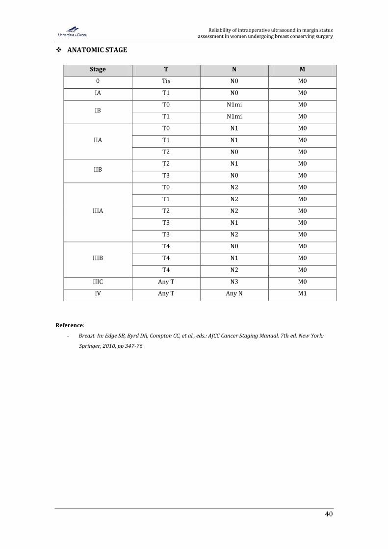
Reliability of intraoperative ultrasound in margin status assessment in women undergoing breast conserving surgery
40
ANATOMIC STAGE
Stage T N M
0 Tis N0 M0
IA T1 N0 M0
IB T0 N1mi M0
T1 N1mi M0
IIA
T0 N1 M0
T1 N1 M0
T2 N0 M0
IIB T2 N1 M0
T3 N0 M0
IIIA
T0 N2 M0
T1 N2 M0
T2 N2 M0
T3 N1 M0
T3 N2 M0
IIIB
T4 N0 M0
T4 N1 M0
T4 N2 M0
IIIC Any T N3 M0
IV Any T Any N M1
Reference:
- Breast. In: Edge SB, Byrd DR, Compton CC, et al., eds.: AJCC Cancer Staging Manual. 7th ed. New York:
Springer, 2010, pp 347-76

Reliability of intraoperative ultrasound in margin status assessment in women undergoing breast conserving surgery
41
16.2. ANNEX II. Histological Grade
The histological grade is determined by the Nottingham Histologic Score System (the
Elston-Ellis modification of Scarff-Bloom-Richardson grading system)
The grade of a tumour is determined by assessing morphologic features: (1) tubule
formation, (2) nuclear pleomorphism and (3) mitotic count.
1. Glandular (Acinar) / Tubular Differentiation
Score 1 >75% of tumour area forming glandular/tubular structures
Score 2 10% to 75% of tumour area forming glandular/tubular structures
Score 3 <10% of tumour area forming glandular/tubular structures
2. Nuclear pleomorphism
Score 1 Nuclei small with little increase in size in comparison with normal breast
epithelial cells, regular outlines, uniform nuclear chromatin, little variation in
size
Score 2 Cells larger than normal with open vesicular nuclei, visible nucleoli, and
moderate variability in both size and shape
Score 3 Vesicular nuclei, often with prominent nucleoli, exhibiting marked variation in
size and shape, occasionally with very large and bizarre forms
3. Mitotic Count (using a high power field diameter of 0.50 mm)
Score 1 Less than or equal to 7 mitoses per 10 high power fields
Score 2 8-14 mitoses per 10 high power fields
Score 3 Equal to or greater than 15 mitoses per 10 high power fields
Each of these features is assessed with a value of 1 (favourable) to 3 (unfavourable), and
the scores for all three categories are added together. A combine score of 3-5 points is
designated as grade 1; a combined score of 6-7 points is grade 2; a combined score of 8-9
points is grade 3.
Gx Grade cannot be assessed
G1 Low combined histological grade (favourable)
G2 Intermediate combined histological grade (moderately favourable)
G3 High combined histological grade (unfavourable)
Reference:
- Breast Cancer and Breast Pathology [Internet]. Baltimore: Johns Hopkins University, Faculty of
Medicine; 2012 [cited 2014 Oct 25]. Available from: http://pathology.jhu.edu/breast/grade.php
Reliability of intraoperative ultrasound in margin status assessment in women undergoing breast conserving surgery
42
16.3. ANNEX III. Information sheet
FULL D’INFORMACIÓ AL PACIENT
Nom de l’estudi:
FIABILITAT DE L’ECOGRAFIA INTRAOPERATÒRIA EN L’AVALUACIÓ DELS MARGES
QUIRÚRGICS EN DONES CANDIDATES A CIRURGIA CONSERVADORA DE MAMA
Agraïm el seu interès pel que fa a la seva col·laboració en l’estudi que estem duent a
terme des de la Unitat de Patologia Mamària (UPM) de l’Hospital Universitari Dr. Josep
Trueta de Girona. A continuació, li expliquem en que consisteix l’estudi. L’equip que en
forma part li respondrà qualsevol dubte o qüestió que li pugui sorgir.
Quin és l’objectiu de l’estudi?
Aquest estudi té com a principal objectiu avaluar si l’ecografia intraoperatòria és una
bona eina per obtenir uns adequats marges de resecció quirúrgica en dones que pateixen
un càncer de mama, i són candidates a una cirurgia conservadora de mama.
Actualment, el tractament d’elecció per les pacients diagnosticades de càncer de mama en
estadis inicials és la cirurgia conservadora de mama, és a dir, la preservació del pit
mitjançant l’extirpació únicament del tumor juntament amb una quantitat de teixit sa
circumdant. Aquest teixit sa, confereix el que anomenem marge de resecció quirúrgica i és
de capital importància pel que fa al risc de recurrència, és a dir, la reaparició del càncer.
Així, doncs, cal que els marges ressecats estiguin lliures de cèl·lules tumorals.
Què passarà si hi participo?
Vostè seguirà el procediment diagnòstic i terapèutic habitual segons el protocol establert
per la Unitat de Patologia Mamària de l’Hosipital Universitari Dr. Josep Trueta. Així,
doncs, aquest estudi no suposarà cap procediment diagnòstic ni terapèutic addicional. El
que pretenem avaluar és el tumor extirpat durant la intervenció quirúrgica un cop aquest
ja s’hagi extret.
D’acord amb l’objectiu del nostre estudi, realitzarem una ecografia intraoperatòria de la
peça extirpada en el mateix acte quirúrgic per tal de valorar els marges de resecció. Si
durant aquest procés detectem que hi ha un marge amb presència de tumor, procedirem
a ampliar-lo en el mateix acte quirúrgic, evitant, doncs, la necessitat d’haver-ho de fer en
una segona cirurgia.

Reliability of intraoperative ultrasound in margin status assessment in women undergoing breast conserving surgery
43
És obligatori participar-hi?
La participació a l’estudi és totalment voluntària. Si decideix participar-hi, se li demanarà
que signi el consentiment informat segons el qual vostè ha entès tot el que concerneix
participar en aquest estudi. Contràriament, si decideix no participar-hi, això no afectarà ni
modificarà el pla assistencial que ha de rebre.
Què he de fer per participar-hi?
Per dur a terme aquest estudi i atenent a les disposicions legals vigents li sol·licitem que
ompli la següent autorització. Abans i després de firmar el document de consentiment
informat, pot preguntar tot el que cregui convenient als metges i personal sanitari
responsable de l’estudi.
Les meves dades es manejaran de forma confidencial?
Sí. La informació recollida per aquest estudi serà tractada i regulada segons la Llei
Orgànica de Protecció de Dades de Caràcter Personal (15/1999) segons la qual, les seves
dades seran manejades de forma confidencial i només seran utilitzades amb finalitat
d’investigació. La seva identificació personal estarà codificada a través d’una sèrie
numèrica aleatoritzada.
Què passarà si canvio d’opinió en el decurs de l’estudi?
Si renuncia a seguir participant en l’estudi una vegada iniciat, no suposarà cap càstig ni
pèrdua de beneficis per vostè. Se li demanarà que segueixi els controls i seguiment
habituals.
Què se’n farà de la informació obtinguda a partir de l’estudi?
Els resultats seran publicats en revistes d’interès científic relacionades amb l’àrea de
coneixement corresponent a la patologia mamària per tal que altres centres i pacients
puguin aprofitar les troballes del nostre estudi. Recordi que totes les seves dades de
caràcter personal són confidencials i, per tant, seran manejades de forma anònima.
Moltes gràcies per la seva atenció,
Unitat de Patologia Mamària de l’Hospital Universitari Dr. Josep Trueta de Girona
Reliability of intraoperative ultrasound in margin status assessment in women undergoing breast conserving surgery
44
16.4. ANNEX IV. Informed consent
CONSENTIMENT INFORMAT
Declaració del pacient,
Declaro que he estat correctament informat pel membre responsable de l’equip
investigador a sota esmentat; sobre els objectius de l’estudi així com sobre el procés de
selecció de les dades personals; també declaro que he estat informat sobre l’ús de caire
científic que es farà de les meves dades personals, així com sobre el fet que la participació
en l’estudi és totalment voluntària, que puc formular les preguntes que jo consideri
oportunes i que puc sol·licitar la retirada i eliminació de les meves dades personals en
qualsevol moment de l’estudi. A més, he rebut una còpia d’aquest mateix document.
NOM, COGNOMS i DNI del pacient:
FIRMA: DATA:
NOM, COGNOMS i DNI del professional de salut que ha informat al pacient:
FIRMA: DATA:
Apartat per a la revocació del consentiment informat
Jo,
amb DNI revoco el consentiment de participar en
l’estudi anteriorment esmentat.
FIRMA: DATA: