regulationofmitochondrialapoptoticeventsby p53 … · 2010-07-02 · tein synthesis inhibitors,...
TRANSCRIPT

Regulation of Mitochondrial Apoptotic Events byp53-mediated Disruption of Complexes betweenAntiapoptotic Bcl-2 Members and Bim*□S
Received for publication, November 1, 2009, and in revised form, April 12, 2010 Published, JBC Papers in Press, April 19, 2010, DOI 10.1074/jbc.M109.081042
Jie Han‡1, Leslie A. Goldstein‡1, Wen Hou‡, Brian R. Gastman§, and Hannah Rabinowich‡¶2
From the ‡Department of Pathology, University of Pittsburgh School of Medicine, Pittsburgh, Pennsylvania 15213, the ¶Universityof Pittsburgh Cancer Institute, Pittsburgh, Pennsylvania 15213, and the §Department of Otolaryngology, University of MarylandMedical School, Baltimore, Maryland 21201
Multiple mechanisms have been proposed for the mitochon-drial function of p53 that are either dependent on or indepen-dent of its transcriptional activity. However, none of thesemechanisms involves Bim functioning downstreamof p53mito-chondrial translocation. Utilizing a p53 nuclear localization sig-nal mutant, whose nuclear import is completely abrogated, wedemonstrate that its apoptotic activity at the outer mitochon-drial membrane, which involves conformational changes in BaxandBak, ismediated byBim.We further demonstrate an inversecorrelation between the binding levels of p53 and Bim toMcl-1.Thus, enhanced binding of p53 toMcl-1 involves the disruptionof existing complexes betweenMcl-1 and Bim.We propose thatmitochondrial p53 functions as a Bim derepressor by releasingBim from sequestrating complexes with Mcl-1, Bcl-2, and Bcl-XL, and allowing its engagement in Bak/Bax activation.
Thepowerful apoptotic function of p53 is central to its tumorsuppressor activity (1). p53 can act as a transcription factor thatstimulates the expression of proapoptotic Bcl-2 family genesthat reside or act at the mitochondria, including NOXA, Puma,andBax (2–6). In addition to thewell established transcription-dependent mechanism of p53 apoptotic function, evidence hasalso accumulated for a p53 transcription-independent apopto-tic role at the mitochondria (7–19). In those studies, p53-de-pendent transcription was nullified by targeting p53 directly tothe mitochondria in p53-deficient tumor cells or by using pro-tein synthesis inhibitors, wheat germ agglutinin as a nuclearimport inhibitor, enucleated cytoplast preparations, or tran-scription-deficient p53 mutants (11, 13, 19). It was proposedthat such transcription-independent cell death wasmediated by aBH3-like activity of p53 that upon itsmitochondrial translocationcan bind Bcl-2, Bcl-XL, and Bak (12, 19, 20). Several potentialmechanisms were considered for the BH3-like activity of p53: (i)neutralization of antiapoptotic activity of Bcl-2 and Bcl-XL (19);(ii) conversionofBcl-2 activity fromantiapoptotic toproapoptotic
(21); (iii) displacement of Bak fromMcl-1 sequestration (20); (iv)direct activation of Bax (12, 13); and (v) competition with PUMAfor Bcl-XL binding (11). Thus, p53 may function as a direct Baxactivatororasaderepressor that relievesBakorBH3-onlyproteinsfrom repression by anti-apoptotic Bcl-2 proteins (13).Our recent studies have elucidated a central regulatory role
for the mitochondrial Mcl-1�Bim complex in the apoptoticresponse to granzyme B, TRAIL, and certain cytotoxic drugs(22–24). In the current study, we investigated the involvementof the Mcl-1�Bim complex in the p53-mediated mitochondrialapoptotic cascade.We demonstrate thatMcl-1 contributes sig-nificantly to the regulation of the mitochondrial response top53 and that Bim is directly involved in the execution of thisresponse upstream of Bax and Bak. In particular, inducedexpression of p53 results in disruption of complexes betweenBim and Mcl-1, Bcl-2, or Bcl-XL. Such a disruption may bemediated by de novo binding between antiapoptotic Bcl-2 pro-teins and p53 itself as well as by a selective binding of the p53transcriptional targets, NOXA and Puma, to their respectiveantiapoptotic Bcl-2 protein partners. Utilizing a p53 nuclearlocalization signal (NLS)3 mutant, we demonstrate the inde-pendence of p53 from the de novo transcription of Bax, NOXA,andPuma genes in its disruption of existing complexes betweenBim and Mcl-1 and in its mediation of Bim-dependent activa-tion of existing Bax and Bak pools. These findings extend pre-viously proposed models for mitochondrial p53 function toinclude a novel mechanism where p53 derepresses Bim fromthe repressive effects of Mcl-1, Bcl-2, and Bcl-XL. This p53derepressing activity sheds new light on the unexplained resis-tance of Bim�/� cells to �-irradiation (25, 26).
EXPERIMENTAL PROCEDURES
Reagents—Anti-human Mcl-1 Abs were from Oncogene(Boston, MA) and Santa Cruz Biotechnology, Inc. (Santa Cruz,CA). Anti-�-actin mAb (clone AC-15) was from Sigma; anti-Cox IV mAb was from Molecular Probes (Eugene, OR); anti-Bax mAb 6A7 was from BD Biosciences, and the N-20 Ab was
* This work was supported, in whole or in part, by National Institutes of HealthGrants RO1 CA109285 and RO1 CA111786 (to H. R.).
□S The on-line version of this article (available at http://www.jbc.org) containssupplemental Figs. S1–S5.
1 Both authors contributed equally to this work.2 To whom correspondence should be addressed: University of Pittsburgh
Cancer Institute, The Hillman Cancer Center, Research Pavilion Rm. G17c,5117 Centre Ave., Pittsburgh, PA 15213. Tel.: 412-623-3212; Fax: 412-623-1119; E-mail: [email protected].
3 The abbreviations used are: NLS, nuclear localization signal; Ab, antibody;mAb, monoclonal antibody; 5-FU, fluorouracil; CHAPS, 3-[(3-cholamido-propyl)dimethylammonio]-1-propanesulfonic acid; ORF, open readingframe; RNAi, RNA interference; siRNA, small interfering RNA; MIB, mito-chondrial buffer; MOPS, 4-morpholinepropanesulfonic acid; JC-1,1,1�,3,3�-tetraethylbenzamidazolocarbocyanin iodide; WT, wild-type; KD,knockdown.
THE JOURNAL OF BIOLOGICAL CHEMISTRY VOL. 285, NO. 29, pp. 22473–22483, July 16, 2010© 2010 by The American Society for Biochemistry and Molecular Biology, Inc. Printed in the U.S.A.
JULY 16, 2010 • VOLUME 285 • NUMBER 29 JOURNAL OF BIOLOGICAL CHEMISTRY 22473
by guest on April 7, 2020
http://ww
w.jbc.org/
Dow
nloaded from

from Santa Cruz Biotechnology, Inc.; Rabbit anti-Bak Abs werefromUpstate Biotechnology, Inc. (Lake Placid, NY), Santa CruzBiotechnology, Inc., and Oncogene (AB1); Rabbit anti-Bim andanti-Puma Abs were from ProSci (Poway, CA); an additionalBim-specific rat mAb was from Apoptech (San Diego, CA);anti-NOXA Ab was from Imgenex (San Diego, CA); Abs toBcl-2, Bcl-XL, p53 (clone DO-1), SMAC, and �-tubulin werefrom Santa Cruz Biotechnology, Inc.; Anti-Xpress mouse mAbfor lacZ-encoded protein was from Invitrogen; ProteinA-Sepharose beads and Protein G-Sepharose beads were fromAmersham Biosciences. TrueBlot reagent was from eBio-science (San Diego, CA), and fluorouracil (5-FU) was fromInvitrogen. Cell viability was measured by the CellTiter-Gloluminescent kit (Promega).Cell Lines, Cell Lysates, and Cell Extracts—All cell lines uti-
lized in this study were obtained from ATTC, apart fromHct116 and p53�/� Hct116 cells, which were generous giftsfrom Dr. Bert Vogelstein (The Johns Hopkins University). Jur-kat T leukemic cells were grown in RPMI 1640 medium con-taining 10% fetal calf serum, 20 mM HEPES, 2 mM L-glutamine,and 100 units/ml each of penicillin and streptomycin. Hct116,T-REx-293, MCF7, MDA-MB-231, and RPMI-8266 cell lineswere grown in Dulbecco’s modified Eagle’s medium containing15% fetal calf serum, 20mM L-glutamine, and 100 units/ml eachof penicillin and streptomycin. Transfected T-REx-293 clonalcell lines were maintained in the presence of blasticidin (5�g/ml) and zeocin (20 �g/ml). Cell lysates were prepared with1% CHAPS, 50 mM Tris-HCl, pH 7.4, 110 mM NaCl, 5 mM
EDTA, 1 mM phenylmethylsulfonyl fluoride, 10 �g/ml leupep-tin, and 10 �g/ml aprotinin. To prepare cell extracts, culturedcells were washed twice with phosphate-buffered saline andthen resuspended in ice-cold buffer (20 mM HEPES, pH 7.0, 10mM KCl, 1.5 mM MgCl2, 1 mM sodium EDTA, 1 mM sodiumEGTA, 1 mM dithiothreitol, 250 mM sucrose, and proteaseinhibitors). After incubation on ice for 20 min, cells (2.5 � 106/0.5 ml) were disrupted by Dounce homogenization. Nucleiwere removed by centrifugation at 650 � g for 10 min at 4 °C.Cellular extracts were obtained as the supernatants resultingfrom centrifugation at 14,000 � g at 4 °C for 30 min.Molecular Cloning of Tet-inducible p53 Expression Plasmid—
Total RNAwas isolated fromHct116 cells using RNASTAT-60Reagent (Tel-Test “B”, Inc.). Reverse transcription was carriedout with 5 �g of total RNA using an oligo(dT)12–18 primer andSuperScript II RNase H� Reverse Transcriptase (Invitrogen).PCRwas performed using the Expand LongTemplate PCR Sys-tem kit (RocheApplied Science). A p53 amplicon containing itsopen reading frame (ORF) was generated with the followingprimer pair (forward and reverse): 5�-CGCGGATCCACTGC-CATGGAGGAGCCG-3� (which extends 6 nucleotides into the5�-untranslated region (UTR)) and 5�-ACGCGTCGACT-CAGTCTGAGTCAGGCCC-3� (complementary to the C ter-minus of ORF). The putative p53 amplicon was size-selectedusing a 1% agarose gel, and DNA was purified with the QIA-quick gel extraction kit (Qiagen). The purified amplicon wasdigested with the restriction enzymes BamHI and SalI andligated into the Tet-inducible vector, pcDNA4/TO (Invitro-gen), that had been previously digested with BamHI and XhoI.Following transformation (Escherichia coli TOP 10F�, Invitro-
gen), plasmids from randomly picked colonies underwent auto-mated DNA sequence analysis (University of Pittsburgh DNASequencing Core Facility) to confirm sequence integrity. TheTet-inducible control plasmid pcDNA4/TO/lacZwas obtainedfrom Invitrogen.Generation of Tet-inducible p53 NLSMutant—The bipartite
p53 NLS mutant was generated in a two-step procedure byoverlap extension using the PCR method (27). First, using theWT p53 Tet-inducible plasmid described above, we mutatedpositions Lys319, Lys320, and Lys321 all toAla using the followingprimer pair (forward and reverse): 5�-CAGCCAGCTGCTG-CACCACTGGATGGAGAATATTTC-3� and 5�-CAGTGG-TGCAGCAGCTGGCTGGGGAGAGGAGCTGGT-3�. A re-combinant p53 K319A/K320A/K321A plasmid was producedas described above except that PCRwas carried out using Pfx50DNA polymerase (Invitrogen) and the mutant p53 ampliconwas ligated to the vector pCR3.1 (Invitrogen). The p53 bipartitemutant that converts Lys-305 and Arg-306 both to Ala wasgenerated from the K319A/K320A/K321A mutant plasmidusing the following primer pair (forward and reverse): 5�-AGC-ACTGCTGCAGCACTGCCCAACAACACCAGC-3� and 5�-CAGTGCTGCAGCAGTGCTCCCTGGGGGCAGCTC-3�. PCRwas performed with the Expand High Fidelity Kit (RocheApplied Science). All other procedures for generation of the Tet-inducible p53 NLSmutant were as described above forWT p53.Cell Transfection Procedures—The cell line T-REx-293
(Invitrogen) are human embryonic kidney cells that stablyexpress the Tet repressor. To generate stable clones that harborthe pcDNA4/TO vector control, pcDNA4/TO/lacZ expressioncontrol, pcDNA4/TO/p53, and pcDNA4/TO/p53 NLS, theseplasmids were linearized using the restriction enzymes FspI(pcDNA4/TO, pcDNA4/TO/p53, and pcDNA4/TO/p53NLS)and ScaI (pcDNA4/TO/lacZ). Transfection was carried outusing 30 �l of GenePorter transfection reagent (Gene TherapySystems Inc., San Diego, CA) and 5 �g of linearized plasmid in6-well plates with 0.8 � 106 cells/well (�80% confluence) thatwere maintained in the presence of 5 �g/ml blasticidin. At 48 hpost-transfection, zeocin was added to a final concentration of100–200 �g/ml. After �2 weeks, individual colonies wereselected, expanded, and harvested. Induction of p53 and lacZ-encoded protein expression by individual cloneswas confirmedby immunoblotting after the addition of 1 �g/ml tetracyclinefor 16 h. Confirmation of stable clones containing integratedpcDNA4/TO vector was obtained by reverse transcription-PCR of their total RNA.We utilized a primer pair that delimitsthe zeocin resistance gene ORF in pcDNA4/TO (forward andreverse): 5�-ACCATGGCCAAGTTGACCAGT-3� (corre-sponds to nucleotides 2247–2267) and 5�-GAAATCTCG-TAGCACGTGTCA-3� (complementary to nucleotides 2622–2642) (data not shown).T-REx-293 cells stably transfected with inducible p53 were
transfectedwithXmnI linearizedMcl-1 plasmid or pCR3.1 vec-tor using the GenePorter Transfection Reagent as describedabove. Geneticin-resistant cell lineswere grown in the presenceof G418 (1500 �g/ml). Geneticin-resistant clonal cell lineseither expressing Mcl-1 or harboring the vector control (seebelow)were generated by dakocytomation (1 cell/well) utilizingaMOFLO high speed cell sorter and Summit Software. Confir-
A Role for Bim in p53-mediated Mitochondrial Apoptosis
22474 JOURNAL OF BIOLOGICAL CHEMISTRY VOLUME 285 • NUMBER 29 • JULY 16, 2010
by guest on April 7, 2020
http://ww
w.jbc.org/
Dow
nloaded from

mation of stable clones containing the pCR3.1 vector wasobtained by reverse transcription-PCR using a primer pair thatdelimits the neomycin resistance geneORF in pCR3.1 (forwardand reverse): 5�-CGCATGATTGAACAAGATGGA-3� (com-plementary to nucleotides 3162–3142) and 5�-TCGCTTG-GTCGGTCATTT-3� (corresponding to nucleotides 2335–2352). All experiments were repeated with at least threeindividual clones orwith amixture of individual clones that hadbeen confirmed to express the transfected gene of interest.Transient transfections were carried out with Lipofectamine
2000 (Invitrogen) according to the manufacturer’s instructionsusing non-linearized plasmids. Cells were split in 24-well platesat densities of 2–8 � 104 cells/well 24 h prior to the transfec-tion. The cells were harvested and assessed 24–48 h after thetransfection.RNAi—NOXA, Mcl-1 and Bim siRNAs were obtained as
duplexes in purified and desalted form (Option C) from Dhar-macon/Thermo Fisher Scientific. These siRNAs had thefollowing sense strand sequences: NOXA, 5�-GUCGAGUGU-GCUACUCAACUdTdT-3�; Mcl-1, 5�-GAAACGCGGUAAU-CGGACUdTdT-3�; Bim, 5�-GACCGAGAAGGUAGACAAU-UGdTdT-3�. Also obtained from Dharmacon/Thermo FisherScientific as siGENOME SMARTpool reagents were Bak(M-003305-01), Bax (M-003308-00), Bim (M-004383-01),Mcl-1 (M-004501-04), NOXA (M-005275-01), and Puma(M-004380-01) siRNAs. The non-targeting siRNA control usedin our RNAi experiments with the Dharmacon siRNAs above isthe siCONTROL non-targeting siRNA 1 (D-001210–01). BimsiRNA was also obtained from Invitrogen as Stealth SelectsiRNA (HSS145411,412,413) and Stealth RNAi negative con-trol Med GC (12935-300). T-REx-293 cells (2.5 � 105) wereplated in a 6-well plate and, following 16 h (at �30% conflu-ence), were transfected with 10–50 nM siRNA in Opti-MEMmedium (Invitrogen) without fetal calf serum using Oligo-fectamine reagent (Invitrogen) according to themanufacturer’stransfection protocol. After 4 h, fetal calf serum was added to afinal concentration of 10%. At 40 h, the medium over the cellswas adjusted to 1 ml before the addition of an apoptotic agent.Subcellular Fractionation—To obtain an enriched mito-
chondrial fraction, Hct116 or T-REx-293 cells were suspendedin mitochondrial buffer (MIB) composed of 0.3 M sucrose, 10mM MOPS, 1 mM EDTA, and 4 mM KH2PO4, pH 7.4, and lysedby Dounce homogenization, as described previously (22).Briefly, nuclei and debris were removed by a 10-min centrifu-gation at 650 � g, and a pellet containing mitochondria wasobtained by two successive spins at 10,000 � g for 12 min. Toobtain the S-100 fraction, the postnuclear supernatant was fur-ther centrifuged at 100,000 � g for 1 h at 4 °C. To obtain theenriched mitochondrial fraction, the mitochondria-containingpellet was resuspended inMIB and layered on a Percoll gradientconsisting of four layers of 10, 18, 30, and 70% Percoll in MIB.After centrifugation for 30min at 15,000� g, themitochondrialfraction was collected at the 30/70 interface. Mitochondriawere diluted inMIB containing 1mg/ml bovine serum albumin(at least a 10-fold dilution required to remove Percoll). Themitochondrial pellet was obtained by a 40-min spin at 20,000�g and used immediately. Purity was assessed by electronmicroscopy and by enzyme marker analysis. For enzyme anal-
ysis, the following enzymes were assayed: aryl sulfatase (lyso-somes/granules); N-acetyl-�-D-glucosaminidase, �-L-fucosi-dase, and �-glucoronidase (lysosome); lactate dehydrogenase(cytosol); cytochrome oxidase or monoamine oxidase (mito-chondria); thiamine pyrophosphatase (Golgi); NADH oxidase(endoplasmic reticulum); dipeptidyl peptidase IV (plasmamembrane). The purity was assessed at 95%, with �5% or lesscontamination from the microsomal fraction.Flow Cytometry—Mitochondrial membrane potential depo-
larization was measured by utilizing a fluorescent cationic dye,1,1�,3,3�-tetraethylbenzamidazolocarbocyanin iodide (JC-1;Molecular Probes). The staining was performed according tothe manufacturer’s procedures, and loss of mitochondrialmembrane potential was quantified by flow cytometric analysis ofthe decrease in the 590-nm (red)/527-nm (green) fluorescenceintensity emission ratio utilizing a Beckman Coulter Epics XL-MCL and analyzed with the EXPO32 software. To detect Bax andBakconformational changesusinganti-Bax6A7andanti-BakAB1Abs, the cells were first fixed by paraformaldehyde and then per-meabilized by 0.15% Triton X-100 (28).Western Blot Analysis—Proteins in cell lysates, cell extracts,
mitochondria, or S-100were resolved by SDS-PAGE and trans-ferred to polyvinylidene difluoride membranes, as describedpreviously (29). Following probing with a specific primary Aband horseradish peroxidase-conjugated secondary Ab, the pro-tein bands were detected by enhanced chemiluminescence(Pierce).Immunoprecipitation—Cell lysates from 5–10 � 106 tumor
cells were made in 100 �l of lysis buffer (20 mM Tris, 137 mM
NaCl, 2 mM Na3VO4, 1% Nonidet P-40, 10% glycerol, and 1%protein inhibitors: aprotinin, leupeptin, phenylmethylsulfonylfluoride). The lysates were precleared with Protein A- orG-Sepharose beads and incubated with the immunoprecipitat-ing Abs (at 1:100–1:1000 dilution) at 4 °C for 4 h. The immunecomplexes were then precipitated with Protein A- or G-Sepha-rose beads at 4 °C overnight. The pellets were washed fourtimes with the appropriate lysis buffer and boiled for 5 min inSDS sample buffer.Confocal Microscopy—Cells were grown and treated on Lab-
Tek II chamber slides. After treatment, cells were fixed with 4%paraformaldehyde and permeabilized with 0.2% Triton X-100.Images were captured by a confocal Olympus Fluoview 1000system using a 100� oil immersion objective with the compan-ion FV10-ASW1.6 imaging software at 25 °C. The digitalimages were split andmerged using ImageJ, and image contrastand brightness were adjusted with Adobe Photoshop.
RESULTS
De Novo Generation of Complexes between Mcl-1 and Tet-induced p53 Is Associated with Disruption of Mcl-1�BimComplexes—Arole for Bim in p53-mediated cell death has beensuggested by studies performed with Bim knock-out lympho-cytes, whichwere somewhat refractory to�-irradiation (25, 26).However, themechanismunderlying the involvement of Bim inthe p53-mediated mitochondrial apoptotic cascade has not yetbeen elucidated. The binding of p53 to Bcl-2 and Bcl-XL hasbeen well established, but the possibility of consequential Bim-mediated mitochondrial changes has not yet been investigated.
A Role for Bim in p53-mediated Mitochondrial Apoptosis
JULY 16, 2010 • VOLUME 285 • NUMBER 29 JOURNAL OF BIOLOGICAL CHEMISTRY 22475
by guest on April 7, 2020
http://ww
w.jbc.org/
Dow
nloaded from

Because Bim constitutively binds antiapoptotic Bcl-2 familymembers, particularly Mcl-1, we investigated the possibilitythat such complexes are disrupted by induced expression ofp53. Co-precipitation of endogenous p53 with Mcl-1 wasobserved in multiple tumor cell types that express WT p53,including colon carcinoma Hct116, multiple myeloma RPMI-8266, and breast cancerMDA-MB-231 andMCF7 cells (Fig. 1).Because under quiescent conditions, the expression level ofendogenous p53 is low, we also assessed itsMcl-1 binding capa-bility following treatments known to induce its physiologicexpression level (30, 31). Thus, in Hct116 cells treated with5-FU (Fig. 1,D and E) or in UV-irradiatedMCF7 cells (Fig. 1F),the expression of endogenous p53 was significantly up-regu-lated, and a significant part of it co-immunoprecipitated withMcl-1. The ability of endogenousMcl-1 to bind p53was furtherdemonstrated in p53�/� Hct116 cells ectopically transfectedwith p53 (supplemental Fig. S1).
Endogenous levels of Mcl-1 are highly responsive to cellularstress and may be up-regulated or down-regulated in cell type-and treatment-dependent manners (32). Thus, in 5-FU-treatedHct116 cells, Mcl-1 is up-regulated (Fig. 1E, lane 4 versus lane1), whereas inUV-treatedMCF7 cells,Mcl-1 is down-regulated(Fig. 1F, lane 2 versus lane 1). Therefore, for further analysis ofthe relationship betweenMcl-1�Bim andMcl-1�p53 complexes,we utilized Tet-inducible p53 T-REx-293 cells, where theinduction of p53 does not significantly impact the expressionlevel ofMcl-1. T-REx-293 cells stably transfectedwith p53weretreated with tetracycline for 6, 12, or 24 h and then subjected toMcl-1 immunoprecipitation. Induced expression of p53 (Fig.2A, lanes 2–4 versus lane 1) did not significantly alter theexpression levels of either Mcl-1 or Bim. However, the levels ofMcl-1-boundp53were directly related to the levels of p53 in theoriginal extracts (Fig. 2A, lanes 10–12 versus lanes 2–4). Thus,the unchanged levels of endogenous Mcl-1 were sufficient toaccommodate the binding of increasing doses of p53. We andothers have previously reported that Mcl-1 is localized mainlyto the outer mitochondrial membrane, where it sequesters Bim(22, 33, 34). The binding of p53 to Mcl-1 was associated with areduction in the levels of Mcl-1�Bim complexes (Fig. 2, lanes10–12 versus lane 9) and increased levels of Mcl-1-free Bim inthe post-immunoprecipitation supernatants of cells expressinginduced p53 (Fig. 2A, lanes 6–8 versus lane 5). The inversecorrelation between Mcl-1�p53 and Mcl-1�Bim complexes mayindicate a direct disruption by p53 of theMcl-1�Bim complexes.However, an increased level of Mcl-1-free Bim may also resultfrom the de novo generation of complexes between p53-in-duced NOXA or p53-induced Puma and Mcl-1. Increasedexpression of p53 was associated with increased levels ofNOXA (Fig. 2A, lanes 2–4 versus lane 1). Furthermore, after24 h of the Tet induction of p53, an increased level of theNOXA�Mcl-1 complex was detected (Fig. 2A, lane 12). Pumainduction was also detected in cells with Tet-induced p53 (Fig.2A, lanes 2–4 versus lane 1), but neither NOXA�Mcl-1 norPuma�Mcl-1 complexes (Fig. 2A, lanes 9–12) exhibited aninverse correlationwith p53�Mcl-1 complexes. Previous studieshave established associations between mitochondria-translo-cated p53 and Bcl-2 or Bcl-XL (19). Because these Bcl-2 familymembers also form complexes with Bim under non-apoptoticconditions (35–37), we examined whether induction of p53results in increased levels of free Bim displaced from Bcl-2 andBcl-XL. To this end, we immunoprecipitated endogenous Bcl-2(Fig. 2B) and Bcl-XL (Fig. 2C) from Tet-induced p53 cells. Sim-ilar to observations made for Mcl-1, both Bcl-2 and Bcl-XLdemonstrated increased binding to p53 in a p53 dose-depen-dent manner (Fig. 2, B and C, lanes 7–9, top). Interestingly, thelevels of Bcl-2�Bim were inversely related to the levels of Bcl-2-bound p53 (Fig. 2B, lanes 8 and 9). Likewise, the levels of Bcl-XL�Bim were inversely related to the levels of Bcl-XL-boundp53 (Fig. 2C, lanes 8 and 9). Quantitation of all of the proteinbands of Fig. 2 is shown in the supplemental material(supplemental Fig. S2). These results suggest that the binding ofp53 to Mcl-1, Bcl-2, and Bcl-XL results in an increased level ofBim freed from sequestration by these antiapoptotic Bcl-2proteins.
FIGURE 1. Co-immunoprecipitation of endogenous p53 with Mcl-1 underquiescent or cellular stress conditions. A–C, immunoprecipitation of Mcl-1co-precipitates endogenous p53 in Hct116 (A), RPMI-8266 (B), and MDA-MB-231 cells (C). Controls and depleted supernatants represent 25% of the celllysate input, and the pellets represent 75% of input. In A, Hct116 cell lysateswere subjected to immunoprecipitation (IP) by mouse (m) or rabbit (r) Mcl-1-specific Abs. The depleted supernatants and the immunoprecipitated pelletswere assessed by immunoblotting for the expression of Mcl-1 (top). The samemembrane was stripped and reprobed with p53-specific mAb (DO-1) andTrueBlot reagent that eliminates the detection of the immunoprecipitatingAbs. Heavy and light chains of immunoprecipitating Abs are indicated by IgHand IgL, respectively. In B and C, the precipitation was performed with rabbitanti-Mcl-1-specific Ab, and the membranes were probed with p53-specificmAb. D–E, 5-FU significantly up-regulates the expression of p53 (D) and itsco-precipitation level with Mcl-1 (E). D, Hct116 cells were treated with 5-FU(150 �M) for the indicated time periods, and the expression levels of p53 wereassessed by immunoblotting. E, Hct116 cells were subjected to immunopre-cipitation of Mcl-1 following 5-FU treatment for 16 h. 5-FU treatment results inincreased expression of p53 (lane 4 versus lane 1) and its increased co-precip-itation with Mcl-1 (lane 6 versus lane 3). F, co-immunoprecipitation of p53 withMcl-1 in UV-treated MCF7 cells. MCF7 cells were exposed to 60 J/m2 UV-C, asdescribed previously (52) and assessed 16 h later. In response to UV exposure,p53 is up-regulated, and Mcl-1 is down-regulated (lane 2 versus lane 1). Nev-ertheless, the low level Mcl-1 still co-immunoprecipitates a significant part ofthe induced p53 (lane 6). The asterisks indicate unidentified protein bands.Each of the panels represents results that were confirmed in at least threeindependent experiments.
A Role for Bim in p53-mediated Mitochondrial Apoptosis
22476 JOURNAL OF BIOLOGICAL CHEMISTRY VOLUME 285 • NUMBER 29 • JULY 16, 2010
by guest on April 7, 2020
http://ww
w.jbc.org/
Dow
nloaded from

Involvement of Mcl-1, Bim, NOXA, and Puma in Mitochon-drial Depolarization Mediated by WT p53—To investigate arole for Mcl-1 in protection against p53-mediated mitochon-drial changes, we utilized T-REx-293 clonal cell lines stablytransfected with both Mcl-1 (constitutive) and Tet-induciblep53. p53-mediated mitochondrial depolarization was assessedby flow cytometry of the mitochondrial membrane potentialdye JC-1 (Fig. 3A). JC-1 is a lipophilic fluorochrome that, uponmitochondrial binding, emits a green fluorescent signal, whichis further processed to an additional red fluorescent signal onlyin mitochondria with preserved membrane potential. Thus,mitochondria with intact membrane potential emit high greenand high red fluorescence, whereas loss ofmitochondrialmem-brane potential results in reduced emission of red fluorescencewhile maintaining the high green fluorescence. As assessed bychanges in JC-1 red fluorescence, Tet induction of p53, but notof lacZ-encoded protein, resulted in a significant loss in themitochondrial membrane potential, which was largely abro-gated by concomitant overexpression ofMcl-1 (Fig. 3,A andB).To investigate the involvement of Mcl-1 binding partners inp53-mediated mitochondrial depolarization, we assessed theeffects of their knockdown (KD) on mitochondrial JC-1 stain-ing (Fig. 3, C–F). Whereas Mcl-1 KD enhanced the p53-medi-ated loss of mitochondrial ��m, Bim KD inhibited the induceddepolarization. The inhibitory effect of Bim RNAi was morepronounced than that of either Bax or Bak RNAi, potentiallydue to the ability of Bax and Bak to substitute for each other(38). NOXA and Puma also function upstream of Bak and Baxin p53-mediated mitochondrial apoptotic events (26, 39).Indeed, knockdown of either Bim, NOXA, or Puma signifi-cantly inhibited p53-mediated mitochondrial depolarization(Fig. 3, E and F). However, knockdown of Bim appeared tomediate an additive inhibitory effect to that observed withsiRNA targeting of NOXA, Puma, or a combination of NOXAand Puma (Fig. 3E) (data not shown). These results suggest thatin p53-mediated mitochondrial depolarization, Bim functions,at least in part, independently of NOXA and Puma.Induction of Mitochondrial Apoptotic Events by p53 NLS
Mutant—To further distinguish between the contribution ofBim (which is not a transcriptional target of p53) (26) and thep53 transcriptional targets NOXA and PUMA in the observedmitochondrial apoptotic events, we generated a p53 NLSmutant. p53 contains a bipartite NLS consisting of two basicamino acid groups, Lys305-Arg306 and Lys319-Lys320-Lys321,separated by a spacer of 12 amino acid residues. Because eachpart of the bipartite NLS sequences can function individually asa weaker NLS (40), we generated an NLS mutant in which allfive amino acids were replaced with Ala. Tetracycline treat-
FIGURE 2. Competitive binding of p53 and Bim to Mcl-1, Bcl-2, and Bcl-XL.A, inverse relationship between p53 and Bim binding to Mcl-1. T-REx-293clonal cells stably transfected with Tet-inducible p53 were treated with tetra-cycline (1 �g/ml) for 6, 12, or 24 h. The cells were then lysed and subjected toMcl-1 immunoprecipitation (IP). The cell lysate (25% of input; lanes 1– 4), thedepleted supernatant (25% of input; lanes 5– 8), and immunoprecipitatedpellet (75% of input; lanes 9 –12) proteins were resolved by SDS-PAGE, and theindicated proteins were detected by immunoblotting. The membranes weresuccessively probed with primary Abs to the indicated proteins and witheither horseradish peroxidase-conjugated secondary Ab or TrueBlot. Induced
expression of p53 is associated with enhanced expression of NOXA and Puma(lanes 2– 4 versus lane 1). Neither NOXA nor Puma exhibits an inverse relation-ship to p53 in Mcl-1 binding. Tet treatment of control T-REx-293 cells stablytransfected with Tet-inducible lacZ is shown in Fig. 3B. B and C, co-immuno-precipitation of Tet-induced p53 with endogenous Bcl-2 or Bcl-XL. T-REx-293clonal cells stably transfected with Tet-inducible p53 were treated with tetra-cycline for 6 or 16 h and then subjected to immunoprecipitation of Bcl-2 (B)and Bcl-XL (C), as described for Mcl-1 in A. The asterisks indicate unidentifiedprotein bands. Quantitation of the protein bands for this figure is shown insupplemental Fig. S2. Similar results were obtained in at least three indepen-dent experiments.
A Role for Bim in p53-mediated Mitochondrial Apoptosis
JULY 16, 2010 • VOLUME 285 • NUMBER 29 JOURNAL OF BIOLOGICAL CHEMISTRY 22477
by guest on April 7, 2020
http://ww
w.jbc.org/
Dow
nloaded from

FIGURE 3. p53-mediated mitochondrial depolarization is inhibited by overexpression of Mcl-1 or KD of Bim, NOXA, or Puma. A and B, p53-mediated lossin ��m is regulated by Mcl-1. Tet-inducible p53 T-REx-293 clonal cell line, Tet-inducible lacZ control T-REx-293 cell line, and Tet-inducible p53 T-REx-293 clonalcell line that is also stably transfected with constitutively expressed Mcl-1 were treated with tetracycline (1 �g/ml) for the indicated time periods and assessedfor emission of red JC-1 by flow cytometry. Treatment by carboxyl cyanide m-chlorophenyl hydrazone (CCCP) served as a positive control for mitochondrialdepolarization. The expression levels of Mcl-1 and p53 of the utilized cells are shown in B. C and D, p53-mediated mitochondrial depolarization is enhanced byMcl-1 KD and inhibited by Bim KD. Tet-inducible lacZ control and Tet-inducible p53 T-REx-293 clonal cell lines were treated with the indicated siRNA for 40 h.The cells were then treated with tetracycline (1 �g/ml) for 16 h and assessed for JC-1 staining by flow cytometry. Immunoblot analyses for these cells are shownin D. E and F, Bim RNAi increases the inhibitory effects of NOXA and Puma RNAi on p53-mediated mitochondrial depolarization. Tet-inducible p53 cells weretreated with various siRNAs and combinations of siRNAs, including Bim, NOXA, Puma, NOXA � Puma, NOXA � Bim, Puma � Bim, and NOXA � Puma � Bim, asdescribed in C. Controls included Tet-inducible p53 cells transfected with the various siRNA combinations without tetracycline treatment and Tet-induciblelacZ cells transfected with the various siRNA combinations with or without tetracycline treatment. These controls are not shown because no changes in JC-1staining were detected (all negative controls were similar in staining to Tet-inducible lacZ cells treated with tetracycline and non-targeting siRNA shown on theleft, bottom). Immunoblot analyses of Tet-induced p53 cells treated with NOXA, Puma, Bim, or non-targeting siRNAs that were utilized in this experiment areshown in F. To avoid redundancy, immunoblot analyses of control Tet-inducible lacZ cells (treated with siRNAs), as shown in B, are not included for thisexperiment. The same membrane was successively probed with the indicated Abs. �-Actin serves as an equal loading control. The asterisks indicate uniden-tified protein bands. Similar results were obtained in at least three independent experiments.
A Role for Bim in p53-mediated Mitochondrial Apoptosis
22478 JOURNAL OF BIOLOGICAL CHEMISTRY VOLUME 285 • NUMBER 29 • JULY 16, 2010
by guest on April 7, 2020
http://ww
w.jbc.org/
Dow
nloaded from

ment of T-REx-293 cells stably transfectedwith a Tet-induciblep53 NLS mutant resulted in the cytosolic accumulation of thep53mutant and its complete exclusion from the nucleus (Fig. 4,A and B, and supplemental Fig. S3). In contrast, Tet-inducedWT p53 localized mainly to the nucleus, and only a small frac-tion was detected in the cytoplasm (supplemental Fig. S3). Asexpected,WTp53 and theNLSmutant p53were also distinct intheir transcriptional activity. Upon their transient transfectioninto p53�/� Hct116 cells, only WT p53, and not the NLSmutant p53, induced the expression of p21 and NOXA(supplemental Fig. S4). A biochemical subcellular fractionationfurther demonstrated that both the p53 NLS mutant and WTp53were present in the S-100 andmitochondrial fractions (Fig.4, C and D). Both WT and the p53 NLS mutant induced mito-chondrial release of cytochrome c and SMAC and, ultimately,cell death (Fig. 5, A and B), but the efficiency of the release ofmitochondrial apoptogenic proteins and the kinetics of celldeath appear to be reduced with the Tet-induced p53 NLSmutant as compared with its WT counterpart. p53 has beenreported to induce conformational changes in Bax and Bak thattrigger their oligomerization and subsequent mitochondrialpermeabilization (12, 20). To detect such changes, we utilizedspecific anti-Bax (6A7) and anti-Bak (AB1) antibodies docu-mented to detect apoptosis-mediated N-terminal conforma-tional alterations in these proteins (41–43). As assessed by con-focal microscopy and flow cytometry, only WT p53 enhancedthe overall expression of Bax as detected by the N20 Ab (totalBax) (Fig. 5, C and F), whereas both WT and the p53 NLSmutant induced conformational changes in either Bax or Bakdetected by the binding of the 6A7 or AB1 Abs, respectively(Fig. 5, D–F).Involvement of Bim in theMitochondrial Apoptotic Activity of
p53—To confirm that the disruption of Mcl-1�Bim complexesis mediated by p53 itself, independent of NOXA and Puma, we
immunoprecipitated Mcl-1 fromTet-inducible p53 NLS mutantT-REx-293 cells treated with tetra-cycline for 24 or 48 h. Similar toresults obtained with WT p53 (Fig.2A), the level of p53 NLS mutantthat co-precipitated withMcl-1 wasinversely correlated with the levelof co-precipitated Bim (Fig. 6A).Quantitation of these protein bandsis shown in the supplementalmaterial (supplemental Fig. S5).These results show that the inverserelationship is directly related to theexpression levels of p53 indepen-dent of NOXA and Puma. Also, theinduced p53 NLS mutant mediatedmitochondrial depolarization thatwas Bim-dependent; it was signifi-cantly inhibited by Bim KD and sig-nificantly enhanced by Mcl-1 KD(Fig. 6, B and C). To further investi-gate a role for Bim upstream of p53-mediated Bax and Bak conforma-
tional changes, we assessed the effect of Bim knockdown (Fig. 6,D–G). Indeed, Bim knockdown (evident by its lost expression)was associated with a loss in the up-regulated expression of6A7-detectable Bax and of AB1-detectable Bak, whenmediatedby either WT or the p53 NLS mutant. These findings providefurther evidence for placingBimupstreamof Bax andBak in theexecution of mitochondrial apoptotic events mediated by p53.
DISCUSSION
A role for Bim in p53-mediated apoptosis has been indicatedby the partial resistance to �-irradiation acquired by Bimknock-out lymphocytes (25, 26). The exact mechanism for Biminvolvement remained unresolved despite significant advancesin the current understanding of the mitochondrial apoptoticfunction of p53. In this study, we demonstrate a direct involve-ment of Bim in the mitochondrial response to p53 and provideevidence for disruption of complexes between Bim and anti-apoptotic Bcl-2 proteins in a p53-dependent manner.Progress in elucidating the p53-mitochondrial apoptotic cas-
cade has been achieved through the discovery of NOXA andPUMA as p53 transcriptional targets (3–5, 26) and the recentidentification of complexes between mitochondria-translo-cated p53 and Bcl-2 or Bcl-XL (11, 19, 44). The current studyhas identified a heretofore-unexplored association betweenp53 and Mcl-1. Like other antiapoptotic Bcl-2 proteins, Mcl-1prevents the permeabilization of the mitochondrial outermembrane, which requires the oligomerization of either Bax orBak (38). Although it has been established that activation of Baxand Bak is mediated by certain BH3-only proteins, the exactmechanisms involved remain controversial (45–47). Two prin-cipal mechanisms have been proposed, focusing on the seques-tration by antiapoptotic Bcl-2 proteins of either Bax and Bak orBH3-only proteins. According to the neutralization model,upon receipt of an apoptotic signal, activated BH3-only pro-
FIGURE 4. Differential subcellular localization of WT p53 and p53 NLS mutant. A and B, WT p53 co-localizeswith nuclei, whereas p53 NLS mutant surrounds nuclei with no co-localization. T-REx-293 clonal cell lines stablytransfected with Tet-inducible WT p53 (left panels) or with Tet-inducible p53 NLS mutant (right panels) weretreated with tetracycline (1 �g/ml, 16 h) and assessed by confocal microscopy for co-localization of p53 (green)with either mitochondria (red) or nuclei (blue). Scale bar, 40 �m (A) and 10 �m (B). Both NLS mutant and WT p53partly co-localize with mitochondria; p53 NLS mutant exclusively surrounds the nucleus, whereas WT p53primarily co-localizes with the nucleus. C and D, subcellular localization of Tet-induced WT p53 and Tet-in-duced p53 NLS mutant as assessed by biochemical cellular fractionation. T-REx-293 clonal cell lines stablytransfected with Tet-inducible WT p53 (C) or Tet-inducible p53 NLS mutant (D) were treated with tetracycline(1 �g/ml, 16 h). Control and treated cells were Dounce homogenized and, after removal of nuclei, were sub-jected to subcellular fractionation to obtain extract, S-100, and mitochondria. The proteins were resolved bySDS-PAGE and detected by immunoblotting. Expression of �-actin and Cox IV are included as markers of therespective subcellular fractions. Cyt C, cytochrome c; DAPI, 4�,6-diamidino-2-phenylindole.
A Role for Bim in p53-mediated Mitochondrial Apoptosis
JULY 16, 2010 • VOLUME 285 • NUMBER 29 JOURNAL OF BIOLOGICAL CHEMISTRY 22479
by guest on April 7, 2020
http://ww
w.jbc.org/
Dow
nloaded from

teins remove Bax and Bak from the constraints of prosurvivalBcl-2 proteins (45). According to the alternative model, activa-tor BH3-only proteins, including Bid, Bim, and Puma, are capa-ble of direct activation of Bax and Bak unless kept in check byprosurvival proteins (39, 48–50). In contrast to direct activatorBH3-only proteins, derepressor/sensitizer BH3-only proteins(e.g. Bad) function indirectly by displacing the sequestered acti-
vators from their complex with Bcl-2, Bcl-XL, or Mcl-1. Thefirst biophysical evidence for a direct activation of Bax by Bimhas only recently been reported, utilizing NMR to prove thebinding between stapled Bim BH3 peptide and Bax (51). Therecently identified complexes between p53 and antiapoptoticBcl-2 proteins expand the current perspectives on p53 mito-chondrial function to include several models that are similar in
FIGURE 5. Assessment of mitochondrial apoptotic events in T-REx-293 cells overexpressing WT or NLS mutant p53. A, WT p53 exhibits acceleratedkinetics of cell death in comparison with p53 NLS mutant. Cell viability was assessed by the CellTiterGlo luminescent kit, and the results are mean � S.D. oftriplicate determinations in one of at least five experiments with similar results. B, Tet-induced expression of either WT p53 or p53 NLS mutant results inmitochondrial release of cytochrome c (Cyt c) and SMAC, but the release kinetics for the p53 NLS mutant are slower. T-REx-293 clonal cell lines stably transfectedwith Tet-inducible WT p53 (left lanes) or with Tet-inducible p53 NLS mutant (right lanes) were treated with tetracycline for the indicated time periods. Controland treated cells were Dounce homogenized and fractionated to heavy membrane and cytosolic fractions. The cytosolic fractions were assessed by immuno-blotting for the expression of the indicated proteins. C–E, confocal microscopic evidence for conformational and expression level changes in Bax and Bakfollowing induction of WT or p53 NLS mutant. Scale bars, 40 �m. C, increased expression of cellular Bax (N20 Ab) is detected mainly following Tet induction ofWT p53. D, increased levels of conformationally altered Bax (6A7 Ab) are detected in cells with induced expression of either WT p53 or p53 NLS mutant.E, increased detectability of Bak is mediated by WT p53 and to a lesser extent by p53 NLS mutant. F, flow cytometry evidence for conformational and expressionlevel changes in Bax and Bak following induction of WT or p53 NLS mutant. T-REx-293 clonal cell lines stably transfected with Tet-inducible WT p53 (left lanes)or with Tet-inducible p53 NLS mutant (right lanes) were treated with tetracycline for 16 h and assessed by flow cytometry for changes in mean fluorescenceintensity (MFI) of Bax (N20 Ab), active Bax (6A7 Ab), and active Bak (AB1 Ab). Whereas induction of WT p53 results in marked increase in MFI of Bax (N20), Bax(6A7), and Bak (AB1), induction of NLS mutant p53 results in milder increase in MFI of Bax (6A7) and Bak (AB1) but no increase in Bax (N20). Similar results wereobtained in at least three independent experiments. DAPI, 4�,6-diamidino-2-phenylindole.
A Role for Bim in p53-mediated Mitochondrial Apoptosis
22480 JOURNAL OF BIOLOGICAL CHEMISTRY VOLUME 285 • NUMBER 29 • JULY 16, 2010
by guest on April 7, 2020
http://ww
w.jbc.org/
Dow
nloaded from

FIGURE 6. Functional relationship between p53 NLS mutant and Bim. A, inverse relationship between p53 NLS mutant and Bim binding to Mcl-1. IncreasedMcl-1 binding to p53 NLS mutant is associated with reduced Mcl-1 binding to Bim. Tet-inducible p53 NLS mutant T-REx-293 cells were treated with tetracyclinefor the indicated time periods. The cells were then lysed and subjected to Mcl-1 immunoprecipitation (IP). The left panels of cell lysate (lanes 1–3) and depletedsupernatant (lanes 4 – 6) represent 25% of input, and the corresponding films were exposed for 20 s; the immunoprecipitated (IP) pellet (lanes 7–9) represents75% of input, and the corresponding films were exposed for 5 s. The asterisks indicate unidentified protein bands. Quantitation of the protein bands of thisfigure is shown in supplemental Fig. S5. B and C, mitochondrial depolarization mediated by the p53 NLS mutant is enhanced by Mcl-1 KD and inhibited by BimKD. Tet-inducible p53 NLS mutant T-REx-293 cells were treated with non-target control, Mcl-1, or Bim siRNAs for 40 h. The cells were then left untreated (control)or treated with tetracycline for 24 h. Mitochondrial depolarization was assessed by flow cytometry of JC-1-stained cells. Efficacy of Bim and Mcl-1 RNAi is shownin C. D–G, changes in patterns of expression of Bax (D and E) or Bak (F and G) following induction of WT or NLS mutant p53s are inhibited by Bim KD. Scale bars,40 �m. Tet-inducible WT p53 (D and F) and Tet-inducible p53 NLS mutant (E and G) T-REx-293 cells were treated with the indicated siRNAs and tetracycline asdescribed above. The cells were assessed by confocal microscopy for expression of Bim (red), conformationally altered Bax (6A7 Ab; green), conformationallyaltered Bak (AB1 Ab; green), and 4�,6-diamidino-2-phenylindole (DAPI) (blue). The increased expression of conformationally altered Bax and Bak by either WTp53 or p53 NLS mutant is blocked in cells treated with Bim siRNA.
A Role for Bim in p53-mediated Mitochondrial Apoptosis
JULY 16, 2010 • VOLUME 285 • NUMBER 29 JOURNAL OF BIOLOGICAL CHEMISTRY 22481
by guest on April 7, 2020
http://ww
w.jbc.org/
Dow
nloaded from
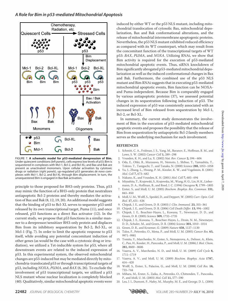
principle to those proposed for BH3-only proteins. Thus, p53may mimic the function of a BH3-only protein that neutralizesantiapoptotic Bcl-2 proteins and thereby mediates the activa-tion of Bax and Bak (8, 12, 19, 20). An additionalmodel suggeststhat the binding of p53 to Bcl-XL serves to sequester p53 untilreleased by its own transcriptional target, Puma (11), and oncereleased, p53 functions as a direct Bax activator (12). In thecurrent study, we propose that p53 functions in a similar man-ner to a derepressor/sensitizer BH3-only protein and displacesBim from its inhibitory sequestration by Bcl-2, Bcl-XL, orMcl-1 (Fig. 7). In order to limit the apoptotic response to p53itself, while avoiding any potential concomitant induction ofother genes (as would be the case with a cytotoxic drug or irra-diation), we utilized a Tet-inducible system for p53, where alldownstream events are related to the induced expression ofp53. In this experimental system, the observed mitochondrialchanges are p53-induced butmay bemediated directly bymito-chondria-translocated p53 or through transcriptional targets ofp53, includingNOXA, PUMA, and BAX (6, 26). To exclude theinvolvement of p53 transcriptional targets, we utilized a p53NLS mutant whose nuclear localization is completely blocked(40).Qualitatively, similarmitochondrial apoptotic eventswere
induced by either WT or the p53 NLS mutant, including mito-chondrial translocation of cytosolic Bax, mitochondrial depo-larization, Bax and Bak conformational alterations, and therelease ofmitochondrial intermembrane apoptogenic proteins.Nevertheless, the p53NLSmutant exhibited reduced efficiencyas compared with its WT counterpart, which may result fromthe concomitant function of the transcriptional targets of WTp53: BAX, PUMA, and NOXA. Utilizing RNAi, we show thatBim activity is required for the execution of p53-mediatedmitochondrial apoptotic events. Thus, siRNA knockdown ofBim significantly abrogated p53-mediatedmitochondrial depo-larization as well as the induced conformational changes in Baxand Bak. Furthermore, the combined use of the p53 NLSmutant and BimRNAi suggests that in executing p53-mediatedmitochondrial apoptotic events, Bim function can be NOXA-and Puma-independent. Because Bim is comparably engagedby various antiapoptotic proteins (37), we assessed potentialchanges in its sequestration following induction of p53. Theinduced expression of p53 was consistently associated with anincreased level of Bim released from sequestration by Mcl-1,Bcl-2, or Bcl-XL.In summary, the current study demonstrates the involve-
ment of Bim in the execution of p53-mediated mitochondrialapoptotic events and proposes the possibility that the release ofBim from sequestration by antiapoptotic Bcl-2 familymembersserves as the underlying mechanism for such involvement.
REFERENCES1. Schmitt, C. A., Fridman, J. S., Yang, M., Baranov, E., Hoffman, R. M., and
Lowe, S. W. (2002) Cancer Cell 1, 289–2982. Vousden, K. H., and Lu, X. (2002) Nat. Rev. Cancer 2, 594–6043. Oda, E., Ohki, R., Murasawa, H., Nemoto, J., Shibue, T., Yamashita, T.,
Tokino, T., Taniguchi, T., and Tanaka, N. (2000) Science 288, 1053–10584. Yu, J., Zhang, L., Hwang, P. M., Kinzler, K. W., and Vogelstein, B. (2001)
Mol. Cell 7, 673–6825. Nakano, K., and Vousden, K. H. (2001)Mol. Cell 7, 683–6946. Miyashita, T., Krajewski, S., Krajewska,M.,Wang,H.G., Lin,H.K., Lieber-
mann, D. A., Hoffman, B., and Reed, J. C. (1994) Oncogene 9, 1799–18057. Erster, S., and Moll, U. M. (2005) Biochem. Biophys. Res. Commun. 331,
843–8508. Moll, U.M.,Wolff, S., Speidel, D., andDeppert,W. (2005)Curr. Opin. Cell
Biol. 17, 631–6369. Chipuk, J. E., and Green, D. R. (2003) J. Clin. Immunol. 23, 355–36110. Chipuk, J. E., and Green, D. R. (2006) Cell Death Differ. 13, 994–100211. Chipuk, J. E., Bouchier-Hayes, L., Kuwana, T., Newmeyer, D. D., and
Green, D. R. (2005) Science 309, 1732–173512. Chipuk, J. E., Kuwana, T., Bouchier-Hayes, L., Droin, N. M., Newmeyer,
D. D., Schuler, M., and Green, D. R. (2004) Science 303, 1010–101413. Green, D. R., and Kroemer, G. (2009) Nature 458, 1127–113014. Talos, F., Petrenko, O., Mena, P., and Moll, U. M. (2005) Cancer Res. 65,
9971–998115. Tomita, Y., Marchenko, N., Erster, S., Nemajerova, A., Dehner, A., Klein,
C., Pan, H., Kessler, H., Pancoska, P., andMoll, U. M. (2006) J. Biol. Chem.281, 8600–8606
16. Vaseva, A. V., Marchenko, N. D., and Moll, U. M. (2009) Cell Cycle 8,1711–1719
17. Vaseva, A. V., and Moll, U. M. (2009) Biochim. Biophys. Acta 1787,414–420
18. Wolff, S., Erster, S., Palacios, G., and Moll, U. M. (2008) Cell Res. 18,733–744
19. Mihara, M., Erster, S., Zaika, A., Petrenko, O., Chittenden, T., Pancoska,P., and Moll, U. M. (2003)Mol. Cell 11, 577–590
20. Leu, J. I., Dumont, P., Hafey, M., Murphy, M. E., and George, D. L. (2004)
FIGURE 7. A schematic model for p53-mediated derepression of Bim.Under quiescent conditions (left panels), cells express low levels of p53; Bim issequestered in complexes with Mcl-1, Bcl-2, and Bcl-XL; and Bax and Bak arepresent as unactivated monomers. Upon cellular activation by cytotoxicdrugs or radiation (right panels), up-regulated p53 generates de novo com-plexes with Mcl-1, Bcl-2, and Bcl-XL through Bim displacement. In turn, theunsequestered Bim is engaged in Bax�Bak activation.
A Role for Bim in p53-mediated Mitochondrial Apoptosis
22482 JOURNAL OF BIOLOGICAL CHEMISTRY VOLUME 285 • NUMBER 29 • JULY 16, 2010
by guest on April 7, 2020
http://ww
w.jbc.org/
Dow
nloaded from

Nat. Cell Biol. 6, 443–45021. Kolluri, S. K., Zhu, X., Zhou, X., Lin, B., Chen, Y., Sun, K., Tian, X., Town,
J., Cao, X., Lin, F., Zhai, D., Kitada, S., Luciano, F., O’Donnell, E., Cao, Y.,He, F., Lin, J., Reed, J. C., Satterthwait, A. C., and Zhang, X. K. (2008)Cancer Cell 14, 285–298
22. Han, J., Goldstein, L. A., Gastman, B. R., Froelich, C. J., Yin, X. M., andRabinowich, H. (2004) J. Biol. Chem. 279, 22020–22029
23. Han, J., Goldstein, L. A., Gastman, B. R., Rabinovitz, A., and Rabinowich,H. (2005) J. Biol. Chem. 280, 16383–16392
24. Han, J., Goldstein, L. A., Gastman, B. R., and Rabinowich, H. (2006) J. Biol.Chem. 281, 10153–10163
25. Erlacher, M., Michalak, E. M., Kelly, P. N., Labi, V., Niederegger, H., Coul-tas, L., Adams, J. M., Strasser, A., and Villunger, A. (2005) Blood 106,4131–4138
26. Villunger, A., Michalak, E. M., Coultas, L., Mullauer, F., Bock, G., Ausser-lechner, M. J., Adams, J. M., and Strasser, A. (2003) Science 302,1036–1038
27. Ho, S. N., Hunt, H. D., Horton, R. M., Pullen, J. K., and Pease, L. R. (1989)Gene 77, 51–59
28. Griffiths, G. J., Dubrez, L., Morgan, C. P., Jones, N. A., Whitehouse, J.,Corfe, B. M., Dive, C., and Hickman, J. A. (1999) J. Cell Biol. 144, 903–914
29. Wang, G. Q.,Wieckowski, E., Goldstein, L. A., Gastman, B. R., Rabinovitz,A., Gambotto, A., Li, S., Fang, B., Yin, X. M., and Rabinowich, H. (2001) J.Exp. Med. 194, 1325–1337
30. Bunz, F., Hwang, P.M., Torrance, C.,Waldman, T., Zhang, Y., Dillehay, L.,Williams, J., Lengauer, C., Kinzler, K.W., and Vogelstein, B. (1999) J. Clin.Invest. 104, 263–269
31. Hall, P. A., McKee, P. H., Menage, H. D., Dover, R., and Lane, D. P. (1993)Oncogene 8, 203–207
32. Ricci, M. S., Kim, S. H., Ogi, K., Plastaras, J. P., Ling, J., Wang, W., Jin, Z.,Liu, Y. Y., Dicker, D. T., Chiao, P. J., Flaherty, K. T., Smith, C. D., andEl-Deiry, W. S. (2007) Cancer Cell 12, 66–80
33. Zhu, Y., Swanson, B. J.,Wang,M., Hildeman, D. A., Schaefer, B. C., Liu, X.,Suzuki, H.,Mihara, K., Kappler, J., andMarrack, P. (2004)Proc. Natl. Acad.Sci. U.S.A. 101, 7681–7686
34. Opferman, J. T., Letai, A., Beard, C., Sorcinelli, M. D., Ong, C. C., andKorsmeyer, S. J. (2003) Nature 426, 671–676
35. O’Connor, L., Strasser, A., O’Reilly, L. A., Hausmann, G., Adams, J. M.,
Cory, S., and Huang, D. C. (1998) EMBO J. 17, 384–39536. Cheng, E.H.,Wei,M.C.,Weiler, S., Flavell, R. A.,Mak, T.W., Lindsten, T.,
and Korsmeyer, S. J. (2001)Mol. Cell 8, 705–71137. Chen, L., Willis, S. N., Wei, A., Smith, B. J., Fletcher, J. I., Hinds, M. G.,
Colman, P.M., Day, C. L., Adams, J. M., andHuang, D. C. (2005)Mol. Cell17, 393–403
38. Wei,M.C., Zong,W.X., Cheng, E.H., Lindsten, T., Panoutsakopoulou, V.,Ross, A. J., Roth, K. A., MacGregor, G. R., Thompson, C. B., and Kors-meyer, S. J. (2001) Science 292, 727–730
39. Certo, M., Del Gaizo Moore, V., Nishino, M., Wei, G., Korsmeyer, S.,Armstrong, S. A., and Letai, A. (2006) Cancer Cell 9, 351–365
40. O’Keefe, K., Li, H., and Zhang, Y. (2003)Mol. Cell. Biol. 23, 6396–640541. Hsu, Y. T., and Youle, R. J. (1998) J. Biol. Chem. 273, 10777–1078342. Griffiths, G. J., Corfe, B. M., Savory, P., Leech, S., Esposti, M. D., Hickman,
J. A., and Dive, C. (2001) Oncogene 20, 7668–767643. Zhu, Y., Liu, X., Hildeman,D., Peyerl, F.W.,White, J., Kushnir, E., Kappler,
J., and Marrack, P. (2006) J. Exp. Med. 203, 1147–115244. Petros, A. M., Gunasekera, A., Xu, N., Olejniczak, E. T., and Fesik, S. W.
(2004) FEBS Lett. 559, 171–17445. Youle, R. J., and Strasser, A. (2008) Nat. Rev. Mol. Cell Biol. 9, 47–5946. Willis, S. N., Fletcher, J. I., Kaufmann, T., van Delft, M. F., Chen, L.,
Czabotar, P. E., Ierino, H., Lee, E. F., Fairlie,W. D., Bouillet, P., Strasser, A.,Kluck, R.M., Adams, J.M., andHuang, D. C. (2007) Science 315, 856–859
47. Merino, D., Giam, M., Hughes, P. D., Siggs, O. M., Heger, K., O’Reilly,L. A., Adams, J. M., Strasser, A., Lee, E. F., Fairlie, W. D., and Bouillet, P.(2009) J. Cell Biol. 186, 355–362
48. Deng, J., Carlson, N., Takeyama, K., Dal Cin, P., Shipp, M., and Letai, A.(2007) Cancer Cell 12, 171–185
49. Letai, A., Bassik, M. C., Walensky, L. D., Sorcinelli, M. D., Weiler, S., andKorsmeyer, S. J. (2002) Cancer Cell 2, 183–192
50. Kuwana, T., Bouchier-Hayes, L., Chipuk, J. E., Bonzon, C., Sullivan, B. A.,Green, D. R., and Newmeyer, D. D. (2005)Mol. Cell 17, 525–535
51. Gavathiotis, E., Suzuki, M., Davis, M. L., Pitter, K., Bird, G. H., Katz, S. G.,Tu, H. C., Kim, H., Cheng, E. H., Tjandra, N., and Walensky, L. D. (2008)Nature 455, 1076–1081
52. Han, J., Goldstein, L. A., Hou,W., and Rabinowich, H. (2007) J. Biol. Chem.282, 16223–16231
A Role for Bim in p53-mediated Mitochondrial Apoptosis
JULY 16, 2010 • VOLUME 285 • NUMBER 29 JOURNAL OF BIOLOGICAL CHEMISTRY 22483
by guest on April 7, 2020
http://ww
w.jbc.org/
Dow
nloaded from

Jie Han, Leslie A. Goldstein, Wen Hou, Brian R. Gastman and Hannah RabinowichComplexes between Antiapoptotic Bcl-2 Members and Bim
Regulation of Mitochondrial Apoptotic Events by p53-mediated Disruption of
doi: 10.1074/jbc.M109.081042 originally published online April 19, 20102010, 285:22473-22483.J. Biol. Chem.
10.1074/jbc.M109.081042Access the most updated version of this article at doi:
Alerts:
When a correction for this article is posted•
When this article is cited•
to choose from all of JBC's e-mail alertsClick here
Supplemental material:
http://www.jbc.org/content/suppl/2010/04/19/M109.081042.DC1
http://www.jbc.org/content/285/29/22473.full.html#ref-list-1
This article cites 52 references, 21 of which can be accessed free at
by guest on April 7, 2020
http://ww
w.jbc.org/
Dow
nloaded from