regulation of pathogenic spore germination by cgrac1 in ... · virulence (25). in candida albicans,...
TRANSCRIPT
EUKARYOTIC CELL, Aug. 2011, p. 1122–1130 Vol. 10, No. 81535-9778/11/$12.00 doi:10.1128/EC.00321-10Copyright © 2011, American Society for Microbiology. All Rights Reserved.
Regulation of Pathogenic Spore Germination by CgRac1 in the FungalPlant Pathogen Colletotrichum gloeosporioides�‡
Iris Nesher,1§ Anna Minz,1§ Leonie Kokkelink,2† Paul Tudzynski,2 and Amir Sharon1*Department of Molecular Biology and Ecology of Plants, Tel Aviv University, Tel Aviv 69978, Israel,1 and Molekularbiologie und
Biotechnologie der Pilze Institut fuer Biologie und Biotechnologie der Pflanzen Schlossgarten 3 D-48149 Muenster, Germany2
Received 19 December 2010/Accepted 26 March 2011
Colletotrichum gloeosporioides is a facultative plant pathogen: it can live as a saprophyte on dead organicmatter or as a pathogen on a host plant. Different patterns of conidial germination have been recognized undersaprophytic and pathogenic conditions, which also determine later development. Here we describe the role ofCgRac1 in regulating pathogenic germination. The hallmark of pathogenic germination is unilateral formationof a single germ tube following the first cell division. However, transgenic strains expressing a constitutivelyactive CgRac1 (CA-CgRac1) displayed simultaneous formation of two germ tubes, with nuclei continuing todivide in both cells after the first cell division. CA-CgRac1 also caused various other abnormalities, includingdifficulties in establishing and maintaining cell polarity, reduced conidial and hyphal adhesion, and formationof immature appressoria. Consequently, CA-CgRac1 isolates were completely nonpathogenic. Localizationstudies with cyan fluorescent protein (CFP)-CgRac1 fusion protein showed that the CgRac1 protein is abun-dant in conidia and in hyphal tips. Although the CFP signal was equally distributed in both cells of agerminating conidium, reactive oxygen species accumulated only in the cell that produced a germ tube,indicating that CgRac1 was active only in the germinating cell. Collectively, our results show that CgRac1 isa major regulator of asymmetric development and that it is involved in the regulation of both morphogenesisand nuclear division. Modification of CgRac1 activity disrupts the morphogenetic program and prevents fungalinfection.
Asymmetric growth is perhaps the most significant principlein development. For asymmetric growth to occur, a polar axismust be established, which defines cell and organ orientationand along which growth will take place. One of the mostpronounced forms of cellular asymmetry coupled to growth ishyphal elongation in filamentous fungi (20).
Hyphal growth initiates with spore germination, which marksthe transition from a resting state to active development. It isa crucial stage in the fungal life cycle, and therefore fungi haveevolved mechanisms to ensure that spores will respond tospecific signals that are indicative of favorable growth condi-tions. In most cases, free sugars are necessary and sufficient forgermination (29). In plant pathogens, germination can also beinduced by plant-derived substances or signals, such as contactwith a solid hydrophobic surface or presence of cuticular waxes(7, 23, 30). In these species, spores have evolved differentpatterns of germination according to the signals that activate it(1, 12).
All types of spore germination are associated with cell po-larization. First, a polar site is defined inside the cell (polarityestablishment), and then growth occurs along the polar axis
(polarity maintenance). These processes are regulated by spe-cialized proteins: septins, which are present at the growth sites,serve as positional landmarks for recruitment and activationof the polarity-establishing machinery (15). The Spitzenkor-per, an accumulation of vesicles present at sites of polarizedgrowth, plays an important role in hyphal polarity, acting as avesicle supply center for the growing tip (20). Actin and themicrotubule cytoskeleton allow continuous flow of secretedvesicles to the growing tip, thereby enabling germ tube exten-sion (36). Regulation of all these activities, particularly cyto-skeleton organization and vesicle trafficking, involves Rho-typeGTPases (22, 27, 41).
Rho GTPases constitute a distinct subfamily within the su-perfamily of Ras-related small GTPases. Similar to other typesof small GTPases, Rho GTPases act as binary molecularswitches. They are found in all eukaryotic cells and are in-volved in various aspects of cellular development, particularlymorphogenesis, cell cycle, and vesicular trafficking (6, 42).The best-studied members of the Rho GTPase subfamily areRhoA, Rac1, and Cdc42 (43). Budding and fission yeasts lackRac homologs, and the Cdc42 protein is essential for theirsurvival (14). Filamentous fungi usually have homologs of bothCdc42 and Rac (most often termed Rac1). In most filamentousspecies, Cdc42 proteins play a relatively minor role, whereasRac1 proteins are central players in fungal development, par-ticularly in the regulation of polarized growth, pathogenicity,sporulation, and spore germination (5, 8–10, 26, 32, 34, 40).Due to the central role of Rac1, deletion mutants are eithernonviable or severely defective in growth and development. Tocircumvent this limitation, constitutively active (CA) isoformsare commonly used in functional analyses of Rac1, as well as in
* Corresponding author. Mailing address: Department of MolecularBiology and Ecology of Plants, Tel Aviv University, Tel Aviv 69978,Israel. Phone: 972 3 640 6741. Fax: 972 3 640 5498. E-mail: [email protected].
§ These authors contributed equally to the article.† Present address: Botanisches Institut, Universitat zu Koln, D-50674
Cologne, Germany.‡ Supplemental material for this article may be found at http://ec
.asm.org/.� Published ahead of print on 1 April 2011.
1122
on April 1, 2020 by guest
http://ec.asm.org/
Dow
nloaded from
other small GTPases. An amino acid substitution of Val forGly in the GTP hydrolysis site of small GTPases gives rise to aconstitutively activated phenotype because the mutated pro-tein is unable to hydrolyze GTP and therefore constitutivelyinteracts with downstream effectors (4). Because the transitionbetween an active and inactive state is essential for properactivity of small GTPases, including Rac, these mutations areuseful in the determination of Rac-regulated processes. Forexample, a CA-Rac strain of Colletotrichum trifolii had aber-rant hyphal morphology and reduced rates of conidial germi-nation (8). Interestingly, the CA-Rac mutation restored thedefects in hyphal morphology that are observed in a strain thatexpresses a CA-Ras isoform, placing Rac1 downstream of Rasin this fungus. In Ustilago maydis, Rac1 is required for patho-genicity and normal hyphal morphology (26). Deletion of Rac1in this fungus affected cellular morphology and hyphal growth,whereas overexpression of wild-type Rac1 induced filamentformation in haploid cells. Expression of CA-Rac1 in this fun-gus was lethal. In Claviceps purpurea, Rac1 interacts with thekinase Cla4, and both proteins have an impact on growth,vegetative differentiation, and pathogenicity (32). In Magna-porthe oryzae, Rac1 is necessary for conidial formation andcontributes to pathogenicity, since strains expressing CA-Racdisplay defects in infection-related growth (10). Deletion ofRac1 in Aspergillus fumigatus results in a reduced growth rateand abnormal conidial formation; however, it does not affectvirulence (25). In Candida albicans, Rac1 is not necessary forviability or serum-induced hyphal growth, but it is essential forfilamentous growth when cells are embedded in a matrix (3).
The fungus Colletotrichum gloeosporioides f. sp. aeschynomene(referred to herein as C. gloeosporioides) is pathogenic on thelegume weed Aeschynomene virginica. It was previously shownthat conidia (asexual spores) of C. gloeosporioides germinate intwo different patterns, which were termed “saprophytic” (ornonpathogenic) and “pathogenic” (1). Pathogenic germinationoccurs on plants and can be induced by stimuli that are com-mon to plant surfaces. It is characterized by instant activationof cell division and formation of a single germ tube, which laterdifferentiates into an infection structure (appressorium). Al-though the morphogenetic events are closely associated withnuclear divisions, progression of the cell cycle is dispensablefor completion of pathogenic germination, as blocking of thecell cycle does not prevent germination or appressorium for-mation (28).
In order to gain molecular insight into pathogenic conidialgermination, particularly the regulation of asymmetric germtube formation following the first conidial cell division, weisolated and studied CgRAC1, a homolog of RAC1 in C. gloeospo-rioides. Here we show that conidial symmetry is already brokenduring the first cell division and that this early asymmetricdevelopment is regulated by CgRac1, which is also required forsubsequent stages of pathogenic development. We also showthat CgRac1 is involved in nuclear division during pathogenicgermination and thus provides a possible link between mor-phogenesis and the cell cycle.
MATERIALS AND METHODS
Fungi, growth conditions, and transformation. Emerson’s YpSs (EMS), re-generation (REG), and pea extract (PE) media were prepared as previouslydescribed (1). Colletotrichum gloeosporioides f. sp. aeschynomene 3.1.3 wild-type
and transgenic strains were cultured on EMS agar plates at 28°C with continuouslight. Conidia were obtained from 5-day-old cultures by washing the plates withsterile water. To obtain mycelium, the fungus was cultured in 50 ml REGmedium with constant agitation at 180 rpm, and the mycelium was collected after48 h by centrifugation. Genetic transformation was performed by electroporationof germinated conidia according to the method of Robinson and Sharon (31). Atleast 16 independent hygromycin-resistant isolates were obtained from each typeof transgenic strain and analyzed by PCR to confirm the presence of the trans-formed DNA.
DNA and RNA manipulations. Genomic DNA was isolated according to themethod of Barhoom and Sharon (2). Plasmid DNA was isolated using theGenElute plasmid miniprep kit (Sigma). Total RNA was extracted from driedmycelium using the GenElute mammalian total RNA miniprep kit (Sigma). Forreverse transcriptase PCR (RT-PCR), cDNA sequences were generated with theReverse-iT first-strand synthesis kit (ABgene), and PCR amplification was per-formed with the RT-PCR product and the appropriate primers using Taq DNApolymerase (Fermentas). Ex Taq polymerase (TaKaRa) was used for proofread-ing, according to the manufacturer’s instructions.
Cloning of CgRAC1 and generation of transgenic strains. Primers for ampli-fication of CgRAC1 sequences were designed according to the CtRAC1 sequenceof C. trifolii (GenBank accession no. AAP89013). Genomic and cDNA sequencesof CgRAC1 were amplified by primers 1 and 2 (see Table S1 in the supplementalmaterial for primer sequences). The PCR amplification products were clonedinto the pTZ57R/T vector (Fermentas). The constitutive active (CA) form ofCgRac1 was generated by site-directed mutagenesis according to the gene SOE-ing method (21). Primer 3, which carries a point mutation and insertion of anAatII restriction site, was used to produce the mutant clone from the vectorpTZ57R-CgRac1. CA-CgRAC1 was excised from pTZ57R/T with NcoI/BamHIand introduced into pAHG4 and Ksh52-1 vectors between the Aspergillus nidu-lans GPDA promoter and TRPC terminator to form the pAHG4-CA-CgRac1and Ksh-CA-CgRac1 vectors, respectively. The pAHG4 vector carries the doubleselection marker HPH-green fluorescent protein (HPH-GFP) fusion protein,whereas Ksh52-1 does not contain a selection marker and therefore was used incotransformation with p57-gpd-HPH, which contains a hygromycin resistancecassette. For nuclear labeling, cotransformation was performed with pMF-280(16), which carries the egfp gene fused to the C terminus of the histone E11-encoding gene hH1 from Neurospora crassa under the control of the N. crassaCCG-1 promoter and p57-gpd-HPH. To obtain GFP-labeled nuclei in a CA-CgRAC1 genetic background, simultaneous transformation was performed withthe three vectors, pMF-280, Ksh-CA-CgRac1, and p57-gpd-HPH. For proteinlocalization studies, an N-terminal cyan fluorescent protein (CFP)-CgRac1 fu-sion protein was generated. The cfp gene was amplified from pECFP-N1 (Clon-tech) by PCR using primers 4 and 5 and cloned into the pTZ57-CgRac1 vectorin the EcoRI and XbaI sites. The CFP-CgRAC1 cassette was cut with NcoI andBamHI and inserted into Ksh52-1 to produce the Ksh-CFP-CgRac1 plasmid. A1-kb fragment upstream of the CgRAC1 open reading frame (ORF) was ampli-fied from genomic DNA using primers 6 and 7 and introduced at the NotI andNcoI sites of Ksh-CFP-CgRac1, replacing the GPDA promoter with the endog-enous CgRAC1 promoter. CA-CgRAC1 was amplified by PCR from the pAHG4-CA-CgRac1 vector with primers 8 and 9 and inserted in place of the nativeCgRAC1 at the EcoRV and BamHI sites, yielding Ksh-CFP-CA-CgRac1.
Pathogenicity tests. Whole-plant inoculations were performed by spraying10-day-old A. virginica plants with conidial suspensions as previously described(1). Plants were sprayed to runoff with 103 to 105 conidia/ml and kept in a humidchamber for 24 h. Symptoms were evaluated after 5 to 7 days. Detached leaves(Pisum sativum cv. White sugar) and onion epidermis inoculations were per-formed according to the method of Nesher et al. (28). Detached pea leaves wereinoculated with 5 to 10 �l of 102 to 104 conidia/ml with 0.01% Tween 20. Forinoculation of wounded leaves, superficial scratches were made with a needle,and the leaves were inoculated with a 5-�l droplet containing conidia suspendedin water without Tween 20.
Conidial adhesion and germination. Germination and adhesion assays wereperformed as described by Chaky et al. (7). Droplets of the conidial suspension(106 conidia/ml in PE) were placed on the surface of a petri dish (hydrophobic)or on a glass slide (hydrophilic) and incubated at 28°C in a humid chamber.Conidia were counted every 30 min, washed by rinsing three times in water, andthen counted again, and the percentage of attached conidia was calculated.
Microscopy. Fluorescent and light microscopies were performed with a ZeissAxioImager M1 microscope. Images were captured with a Zeiss AxioCam MRmcamera and analyzed using the AxioVision Rel 4.5 software package. Confocalmicroscopy was performed with a Zeiss CLSM510 confocal microscope. Scan-ning electron microscopy was performed with a Hitachi S-3000N scanning elec-tron microscope as described by Giesbert et al. (17). Samples were fixed in a
VOL. 10, 2011 ROLE OF Rac1 IN GERMINATION AND PATHOGENICITY 1123
on April 1, 2020 by guest
http://ec.asm.org/
Dow
nloaded from
glutaraldehyde solution and then dehydrated in ethanol. Critical-point dryingwas carried out with a K850 critical-point dryer (Emitech, Germany) and goldsputtered with vacuum sputter device K550X (Emitech).
Staining procedures. Nuclei were stained with 4�,6-diamidino-2-phenylindoledihydrochloride (DAPI; Sigma) as previously described (1). Reactive oxygenspecies (ROS) were detected by staining with 2�,7�-dichlorodihydrofluoresceindiacetate (H2DCFDA; Invitrogen). Conidia were germinated on a glass slide forvarious lengths of time. H2DCFDA (10 mM stock solution in dimethyl sulfoxide[DMSO]) was added to a final concentration of 10 �M, and the samples wereincubated for an additional 20 min at 28°C.
Sequence analysis. Sequence analysis was performed using the Vector NTIAdvance Suite 9 software package (InforMax, Inc.). Sequence searches wereperformed using BLAST algorithms. Multiple sequence alignments were per-formed using ClustalW.
RESULTS
Isolation and characterization of CgRAC1. Genomic(GenBank accession no. EU364808) and cDNA (GenBank ac-cession no. EU348861) clones of CgRAC1 were isolated andsequenced. The 199-amino acid (aa)-long predicted proteinwas highly homologous to Rac1 proteins from other fungi (seeFig. S1 in the supplemental material). All of the conservedmotifs characteristic of Rac1 proteins were identified in thepredicted CgRac1 sequence: GTP binding and hydrolysis sites(GDGAVGKT, residues 15 to 22, and DTAG, residues 62 to65, respectively), effectors’ interaction site (TVFDNY, resi-dues 40 to 45), GDP-GTP exchange site (TKLD, residues 120to 123), and plasma membrane association site (CTIL, residues196 to 199). The GDP-GTP exchange site, TKLD, is unique toRac proteins and differs from the motifs found in Cdc42(TQXD) and the related Rho GTPase (NKXD). In order tostudy the regulation of pathogenic germination by CgRac1, wegenerated a strain that expresses a CA-CgRac1 isoform (G17replaced with V) under the control of the strong Aspergillusnidulans GPDA promoter. Expression of the CA-CgRAC1gene was verified in 16 independent hygromycin-resistanttransgenic strains (not shown). The CA-CgRac1 colonies ex-hibited a typical morphology on plates, which distinguished
them from wild-type cultures (Fig. 1A). Detailed analyses wereconducted with isolates CA19 and CA27, and the two strainsshowed similar phenotypes. For convenience, only results fromstrain CA27 are presented.
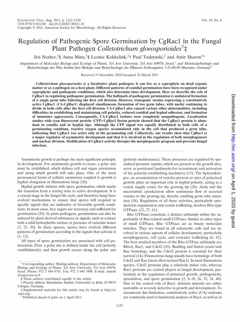
CgRac1 is important for growth and morphology. The CA-CgRac1 strains exhibited reduced growth rate on plates (34 �2 mm) and produced less biomass in liquid culture (10 � 2 mg)than the wild type (40 � 2 mm and 15 � 2 mg, respectively). Inaccordance with the role of Rac1 in determining cell polarity,cells of the CA-CgRac1 strains had polarity defects and a morerounded shape than wild-type cells. In particular, CA-CgRac1conidia were swollen and had near-isometric dimensions (ratiobetween width and length, �0.6), unlike the polar conidia ofthe wild-type strain (ratio between width and length, 0.333 �0.096) (Fig. 1B). The CA-CgRac1 strains also formed swollen,round, hyphal cells (Fig. 1C), as well as a class of structuresthat resembled immature appressoria, but instead of a singleappressorium at the end of a germ tube, these appressorium-like structures seemed to bud from random sites along thehyphae and formed bundles of cells (Fig. 1D and E).
CgRac1 regulates pathogenic germination. Conidia of theCA-CgRac1 strains showed delayed pathogenic germination:only 25% of conidia of the CA-CgRac1 strain germinated after4 h on a hydrophobic surface in PE medium (conditions thatinduce pathogenic germination) compared with 75% germina-tion rates after 4 h for the wild-type conidia (Fig. 2A). Germi-nation rates of the CA-CgRac1 strains reached wild-type levelsafter 8 h of incubation, suggesting that germination in theformer was delayed but not blocked. CA-CgRac1 conidia wereseverely compromised in their ability to form a close contactwith the surface, as revealed by an adhesion assay: after 150min of incubation, less than 20% of the CA-CgRac1 conidiaremained attached to the surface of a petri dish, compared toover 65% of the wild-type conidia (Fig. 2B). Close contact withthe surface is essential for pathogenic germination, and there-fore defects in adhesion might account for the low germination
FIG. 1. Morphology of CA-CgRac1 strains. (A) Colony morphology of wild-type (WT) and CA-CgRac1 (CA27) strains. Fungi were culturedfor 7 days on EMS agar medium. Plates were incubated at 28°C with continuous fluorescent light. (B) Images of wild-type and CA-CgRac1 conidia(light microscope; magnification, �1,000). Insets show enlarged images of a representative cell in each strain. For quantitative estimation ofconidial morphology, the dimensions of 100 wild-type and CA-CgRac1 conidia were measured. Wild-type conidia had an average ratio betweenwidth and length of 0.333 � 0.096. Only 7% of the conidia of the CA-CgRac1 strain had a wild-type phenotype (ratio � 0.333), and the rest hada higher ratio, with 56% of the conidia having a ratio greater than 0.6. (C) Images of hyphae following 24 h of germination on a glass slide (confocalmicroscope; magnification, �630). Appressorium formation is shown on polypropylene after 24 h (D) and on onion epidermis after 16 h (scanningelectron microscope) (E). Arrows point to appressoria (wild type) and appressorium-like structures (CA-CgRac1) that are presented as insets.
1124 NESHER ET AL. EUKARYOT. CELL
on April 1, 2020 by guest
http://ec.asm.org/
Dow
nloaded from

rates of CA-CgRac1 strains during the first hours. Nonpatho-genic germination, which is slower but unaffected by surfacecontact, might account for the later increase in conidial ger-mination in this strain (Fig. 2A).
During pathogenic germination, the unicellular conidiumdivides, and then only one of the resulting cells germinates.Nuclear division in the other cell is arrested, and the cellremains inactive but can resume growth at a later stage (28).Remarkably, this pattern was disrupted in the CA-CgRac1strain: up to 20% of the CA-CgRac1 conidia germinated bysimultaneous formation of two germ tubes, one from each cell(Fig. 2C).
Taken together, these results show the importance ofCgRac1 for proper morphogenesis during pathogenic germina-tion. It might also be that cell cycle progression is altered inCA-CgRac1 strains because of the tight link between morpho-genesis and nuclear division (28).
Appressorium formation is distorted in CA-CgRac1 mutants.When germinated on leaves or a hard surface, C. gloeosporioidesdifferentiates large melanized appressoria, which are necessaryfor penetration of the host cuticle. Appressorium formationrepresents a distinct morphogenetic stage and is associatedwith a switch from polar to isotropic growth. The cell cycle isalso arrested, and nuclear division resumes only when a pen-etration peg is formed at the base of the appressorium (28).CA-CgRac1 strains failed to form normal appressoria. Instead,partly melanized, rounded cells were formed in different loca-tions along the hyphae. Chains of single rounded cells orpatches of several cells that seemed to bud from one anotherwere observed (Fig. 1C and 3B). Scanning electron microscopy(SEM) observations showed that the cells might have lost to-pographical orientation, thus forming piles of such organs (Fig.1D and E). These cells usually contained several nuclei, unlikenormal appressoria, which always contain a single nucleus (seeFig. 4A). Thus, the CA-CgRac1 strains do not form normalappressoria, and the abnormal rounded cells, which might rep-resent defective appressoria, seem to be nonfunctional.
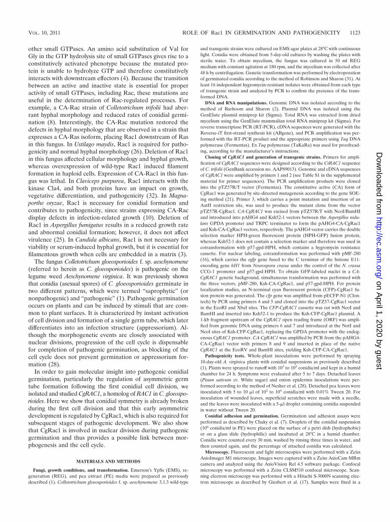
CgRac1 is necessary for fungal pathogenicity. Infection as-says showed that the CA-CgRac1 strains are unable to causedisease on A. virginica plants (Fig. 3A). The multiple defects inearly stages of pathogenic development, particularly the re-duced attachment of conidia and the abnormal appressoria,suggested that the mutants would be compromised in theirability to penetrate the host cuticle. On onion epidermis, theCA-CgRac1 strains developed abnormal, swollen hyphae andbundles of appressorium-like structures (Fig. 3B). Thesestructures were compromised in their ability to penetratethe epidermal cell layer, as indicated by the absence of
FIG. 2. Conidial germination in CA-CgRac1 strains. Conidia weresuspended in PE medium, and 50-�l droplets of conidial suspensionwere applied to a glass slide and incubated in a humid chamber at
28°C. The percentages of conidial germination (A), attached conidia(B), and conidia that produced two germ tubes (C) were evaluated attime intervals during a period of up to 8 h. (D) Pictures of conidia after5 h of incubation. Wild-type (WT) conidia produced a single germ tubethat is attached to the surface and differentiate an appressorium.Conidia of CA-CgRac1 (CA27) strains are swollen; they produce twogerm tubes that might not be attached to the surface and do notdifferentiate appressoria. Results in panels A to C are average resultsfor two independent experiments, each with 3 replications per treat-ment. Mean values (A to C) and standard errors (A) are presented.
VOL. 10, 2011 ROLE OF Rac1 IN GERMINATION AND PATHOGENICITY 1125
on April 1, 2020 by guest
http://ec.asm.org/
Dow
nloaded from
subcellular hyphae in the CA-CgRac1 strain, whereas abun-dant subepidermal primary hyphae developed in wild-type-inoculated samples (Fig. 3B). These results confirmed thatthe CA-CgRac1 strain is impaired in host penetration.
We inoculated wounded pea leaves to determine whether theCA-CgRac1 strain is also affected in invasive growth. Severenecroses developed in wounded leaves following inoculation withconidia (103 conidia/ml) of the wild-type strain (Fig. 3C). In con-trast, no symptoms developed when leaves were inoculated with asimilar concentration of CA-CgRac1 conidia. These results showthat the CA-CgRac1 strain has defects in host penetration as wellas in later stages of pathogenic development.
CgRac1 regulates nuclear and cell division. Nuclear divisionwas followed in transgenic strains expressing a histone-GFP(H1-GFP) fusion protein, which produces GFP-labeled nuclei.Normally, resting conidia of C. gloeosporioides contain a singlenucleus and the cell cycle is arrested. Following induction ofgermination, the conidium divides, and then nuclear divisioncontinues in only one of the resulting cells (28). We found thatcell cycle arrest was less stringently maintained during thesedevelopmental stages in the CA-CgRac1 strain than in the wildtype. About 10% of conidia collected from CA-CgRac1 cultureplates contained two or more nuclei, compared with less than1% of wild-type conidia having more than a single nucleus. Inaccordance with the formation of two germ tubes instead ofone, nuclear division progressed in both cells following the firstcell division and all resulting nuclei progressed through the cellcycle, even when only a single germ tube was produced (Fig.4A and B). A similar phenomenon was observed in the hyphae:all nuclei divided more than once, unlike in the wild type, inwhich only nuclei in apical cells of the hyphal tips divided. Asa result, instead of having a single nucleus per compartment asin the wild type, hyphae and appressoria of the CA-CgRac1strains were swollen and multinucleated (Fig. 4A).
Taken together, these results suggest that CgRac1 is neces-sary for resumption of both the cell cycle and growth duringtransitions between different developmental stages.
CgRac1 affects production and intracellular localization ofROS. Levels and cellular localization of ROS were examinedduring conidial germination. In the wild type, ROS accumu-lated in the apical cell of elongating germ tubes forming agradient toward the back of the cell (Fig. 5a). ROS also accu-mulated in the conidial cell in which germination occurred butnot in the other cell that did not germinate. In CA-CgRac1strains, the two conidial cells accumulated equal amounts ofROS, regardless of whether one or both of the cells germinated(Fig. 5b). ROS also accumulated in the germ tube, but unlikein the wild type, there was no gradient, and uniform stainingwas detected in all of the apical as well as subapical cells. Theseresults show that production and cellular localization of ROS isdisrupted in the CA-CgRac1 strain, which might account forsome of the observed defects, especially in polarization, andhence loss of asymmetric germination.
Cellular localization of the CgRac1 protein. Cellular local-ization of the CgRac1 protein was determined in the wild-typeand CA-CgRac1 genetic backgrounds. In germinating conidia,CgRac1 was detected in rapidly moving vesicles, throughoutthe cytoplasm, and at the sites designated for polar growth,such as the germ tube and hyphal tips (Fig. 6). The signal wasalso visible in the septa. Surprisingly, no significant localization
FIG. 3. Infection assays. (A) Infection of whole plants. A. virginicaplants were inoculated by spraying to runoff with 105 conidia/ml. Pic-tures were taken 7 days postinoculation. (B) Onion epidermis infectionassays. Conidia of the wild type (wt) and CA-CgRac1 strain 27 (CA27)were applied to onion epidermis. Images were captured 24 h postin-oculation. The top left panel shows a wild-type conidium (cn) thatproduces a germ tube (gt) and an appressorium (ap). The top rightpanel shows development of subcuticular primary hyphae (ph) under-neath the appressorium. (C) Invasive growth assay of wounded pealeaves. Leaves were scratched using a needle, and conidia (103 conidia/ml) were applied to the wounded area. The inoculated leaves wereincubated for 48 h in a humid chamber. Images were captured after48 h using a fluorescent stereoscope.
1126 NESHER ET AL. EUKARYOT. CELL
on April 1, 2020 by guest
http://ec.asm.org/
Dow
nloaded from

differences were observed between the wild-type and CA-Cg-Rac1 strains, unlike the distribution of ROS, which was dras-tically altered in the CA-CgRac1 strain. This result suggeststhat CgRac1 activity is regulated by activation/inactivation ofthe protein and not by protein levels or subcellular localization.Measurement of transcript levels showed constant expressionunder various conditions (data not shown), further supportingthis possibility.
DISCUSSION
Fungal Rac1 proteins are required for polarized growth, andhence, they are involved in and necessary for various processes,such as conidial germination, conidial formation, and patho-genesis (20). Here we showed that CgRac1 affects asymmetric
fungal development by controlling both morphogenesis andcell cycle progression. Impairment of CgRac1 activity by theexpression of a constitutive active form of the protein disruptsthe developmental scheme during early stages of pathogenicdevelopment, and as a consequence, CA-CgRac1 strains arecompletely nonpathogenic.
The connection between cell cycle and morphogenesis infungi is well established. In Saccharomyces cerevisiae, the cellcycle is tightly linked with morphogenesis: polarity establish-ment, polar growth, and isotropic expansion all take placeduring bud formation and depend on cell cycle progression(24). In M. oryzae, blockage of mitosis prevents completion ofgermination and appressorium formation (39). Genetic analy-sis has shown that commitment to form an appressorium is
FIG. 4. CgRac1 effect on cell cycle and nuclear division. (A) Nuclei division in germinating conidia and in developing hyphae. Cells weregerminated for 6 h (left) and 24 h (right). In the wild type (wt), the nucleus does not divide in the conidial cell that does not germinate (top left,arrow) or in subapical hyphal cells (top right, arrow), and wild-type appressoria always contain a single nucleus (top right, arrowhead). InCA-CgRac1 (CA27) strains, the nuclei in the conidial cell that does not germinate (bottom left, arrow) and in subapical hyphal cells (bottom right,arrow) continue to divide, resulting in multinucleated cells. Appressorium-like structures may contain numerous nuclei (bottom right, arrowhead).(B) Live-cell imaging of nuclear division during pathogenic germination of CA-CgRac1 for 6 h after germination induction. Merged differentialinterference contrast (DIC) and GFP channels (top), GFP channel (bottom). Simultaneous divisions of all nuclei can be seen in the second (imagesa and b) and third (images d and e) mitoses. Following germination (images c and d), 3 of the 4 nuclei migrate into the elongating germ tube, andthen all three nuclei divide (e). Three nuclei return to the original conidial cell, and the three others disperse along the germ tube. The time intervalbetween images a and b and between images d and e is 7 min. Two nuclei in the cell did not germinate (arrow).
VOL. 10, 2011 ROLE OF Rac1 IN GERMINATION AND PATHOGENICITY 1127
on April 1, 2020 by guest
http://ec.asm.org/
Dow
nloaded from

determined early in the cell cycle, between S and G2 (33). In U.maydis, spores expressing constitutive active Rac1 are arrestedwith a single nucleus, indicating that Rac1 might interfere withthe mitotic cell cycle (26). During pathogenic germination in C.gloeosporioides, nuclear division always precedes the next mor-phogenetic event, suggesting a similar interplay between thecell cycle and developmental morphogenesis. However, mor-phogenesis continues when the cell cycle is blocked and acti-vation of morphogenetic changes occurs before nuclear divi-sion (28). Furthermore, the developmental changes that takeplace during pathogenic germination are all preceded by arrestof the cell cycle, which resumes when new organs start to form.The developmental plan and coordination of cell cycle arrestwith morphogenetic switches are disrupted in CA-CgRac1strains, suggesting that CgRac1 is involved in regulating theexit from cell cycle arrest, an activity that is coordinated withresumption of growth when a certain developmental stage hasbeen completed. Because in CA-CgRac1 strains the cell cycleis less tightly controlled and morphogenesis continues withoutthe necessary adaptation time, multinucleate cells and mal-formed organs (e.g., multinucleated appressorium-like struc-tures) are apparent.
Nuclear division in fungal hyphae can follow different patterns.In some species, such as A. nidulans, nuclei divide throughout thehypha in a coordinated manner (11), whereas in other species,such as Ashbya gossypii, nuclear division in the hypha is asynchro-nous (18). In C. gloeosporioides, only the nucleus that is closest tothe hyphal tip (apical cell) divides, similar to the situation in C.albicans (19). Our results suggest that in pathogenic germination,a polar axis is established already after the first cell division.Accordingly, one of the resulting cells is defined as the apical side,and this cell will divide and produce the germ tube. The other cell,which represents the subapical side of the incipient hypha, doesnot continue to develop, and its nucleus is arrested. BecauseCgRac1 is essential for polarity, strains that express CA-CgRac1lose the ability to form a polar axis, nuclei in both cells continueto divide, and both cells have the capacity to produce a germ tube.Notably, the effect of CgRac1 on the cell cycle is probably sepa-rate from its effect on morphogenesis, since in the CA-CgRac1strain, both nuclei can divide, even when only one of the cellsgerminates.
ROS production and distribution are important for polar-ized growth and fungal pathogenicity (8, 13, 35, 38). In fila-mentous fungi, NADPH oxidase (NOX) and its associated
FIG. 5. Accumulation and cellular distribution of ROS during pathogenic germination. Conidia were germinated for 4 h on glass slides withPE medium and then stained with H2DCFDA. In the wild type (WT), ROS accumulated in the germinating cell and hyphal tips (a) and indeveloping appressoria (c). In the CA-CgRac1 (CA27) strain, ROS accumulated in both conidial cells and germ tubes (b). Staining was detectedin both conidial cells, even when only one of the cells produced a germ tube (d). No signal was detected in unstained wild-type (e) or CA-CgRac1(f) control samples.
1128 NESHER ET AL. EUKARYOT. CELL
on April 1, 2020 by guest
http://ec.asm.org/
Dow
nloaded from

protein complex, including Rac1, are a major source of ROS(37). In M. oryzae, ROS are found in apical cells and in ap-pressoria (13). In C. gloeosporioides, ROS were most abundantin the hyphal tips, forming a gradient with lower concentra-tions toward the back of the hypha than toward the front (Fig.5). In addition, ROS were highly abundant in developing or-gans, for example, in the germinating conidial cell and in ap-pressoria. In the CA-CgRac1 strains, ROS were evenly distrib-uted in the conidia, germ tubes, and short hyphae. ROS alsoaccumulated to similar levels in both of the conidial cells fol-lowing the first cell division and throughout germination. Onthe other hand, we could not determine significant differences
in the localization of the CgRac1 protein in wild-type versusCA-CgRac1 strains. These results suggest that the protein lev-els and localization are similar in developing and arrested cellsand that the activity of CgRac1 does not depend on the pres-ence of the protein but rather on CgRac1-regulating elementssuch as guanine nucleotide exchange factors (GEFs) andGTPase-activating proteins (GAPs). Further investigation will beneeded to elucidate the molecular regulation of CgRac1 andits downstream effectors.
ACKNOWLEDGMENT
This research was supported by grant number 50/15-02 from the DFG.
FIG. 6. Cellular localization of CgRac1. Protein localization was followed using a confocal microscope in transgenic strains expressing aCFP-Rac1 fusion in wild-type (CgRAC1) and CA-CgRAC1 genetic backgrounds. (a and b) In resting conidia and following the first cell divisionafter 2 h of incubation, the protein is localized in small vesicles in both cells of the conidium. (c and d) Larger vesicles are observed duringgermination, and protein can be detected in the elongating germ tube after 3 h of incubation. Signal intensities and distributions are similar in thewild type (a and c) and CA-CgRac1 (b and d). No signal is detected in wild-type or CA-CgRac1 strains after 2 and 3 h of incubation, respectively(e and f), whereas an evenly distributed signal is observed throughout the cytoplasm in strains expressing a cytoplasmic GFP in wild-type andCA-CgRac1 backgrounds after 2 h of incubation (g and h).
VOL. 10, 2011 ROLE OF Rac1 IN GERMINATION AND PATHOGENICITY 1129
on April 1, 2020 by guest
http://ec.asm.org/
Dow
nloaded from

REFERENCES
1. Barhoom, S., and A. Sharon. 2004. cAMP regulation of “pathogenic” and“saprophytic” fungal spore germination. Fungal Genet. Biol. 41:317–326.
2. Barhoom, S., and A. Sharon. 2007. Bcl-2 proteins link programmed celldeath with growth and morphogenetic adaptations in the fungal plant patho-gen Colletotrichum gloeosporioides. Fungal Genet. Biol. 44:32–43.
3. Bassilana, M., and R. A. Arkowitz. 2006. Rac1 and Cdc42 have different rolesin Candida albicans development. Eukaryot. Cell 5:321–329.
4. Bishop, A. L., and A. Hall. 2000. Rho GTPases and their effector proteins.Biochem. J. 348:241–255.
5. Boyce, K., M. Hynes, and A. Andrianopoulos. 2003. Control of morphogen-esis and actin localization by the Penicillium marneffei RAC homolog. J. CellSci. 116:1249–1260.
6. Bustelo, X., V. Sauzeau, and I. Berenjeno. 2007. GTP-binding proteins of theRho/Rac family: regulation, effectors and functions in vivo. Bioessays 29:356–370.
7. Chaky, J., K. Anderson, M. Moss, and L. Vaillancourt. 2001. Surface hydro-phobicity and surface rigidity induce spore germination in Colletotrichumgraminicola. Phytopathology 91:558–564.
8. Chen, C., and M. Dickman. 2004. Dominant active Rac and dominant neg-ative Rac revert the dominant active Ras phenotype in Colletotrichum trifoliiby distinct signalling pathways. Mol. Microbiol. 51:1493–1507.
9. Chen, C., Y.-S. Ha, J.-Y. Min, S. D. Memmott, and M. B. Dickman. 2006.Cdc42 is required for proper growth and development in the fungal pathogenColletotrichum trifolii. Eukaryot. Cell 5:155–166.
10. Chen, J., et al. 2008. Rac1 is required for pathogenicity and Chm1-depen-dent conidiogenesis in rice fungal pathogen Magnaporthe grisea. PLoS Pat-hog. 4:e1000202.
11. Clutterbuck, A. 1970. Synchronous nuclear division and septation in Asper-gillus nidulans. J. Gen. Microbiol. 60:133–135.
12. Doehlemann, G., P. Berndt, and M. Hahn. 2006. Different signalling path-ways involving a G� protein, cAMP and a MAP kinase control germinationof Botrytis cinerea conidia. Mol. Microbiol. 59:821–835.
13. Egan, M., Z. Wang, M. Jones, N. Smirnoff, and N. Talbot. 2007. Generationof reactive oxygen species by fungal NADPH oxidases is required for riceblast disease. Proc. Natl. Acad. Sci. U. S. A. 104:11772–11777.
14. Etienne-Manneville, S. 2004. Cdc42—the centre of polarity. J. Cell Sci.117:1291–1300.
15. Fischer, R., N. Zekert, and N. Takeshita. 2008. Polarized growth in fungi—interplay between the cytoskeleton, positional markers and membrane do-mains. Mol. Microbiol. 68:813–826.
16. Freitag, M., P. Hickey, N. Raju, E. Selker, and N. Read. 2004. GFP as a toolto analyze the organization, dynamics and function of nuclei and microtu-bules in Neurospora crassa. Fungal Genet. Biol. 41:897–910.
17. Giesbert, S., H. B. Lepping, K. B. Tenberge, and P. Tudzynski. 1998. Thexylanolytic system of Claviceps purpurea: cytological evidence for secretion ofxylanases in infected rye tissue and molecular characterization of two xyla-nase genes. Phytopathology 88:1020–1030.
18. Gladfelter, A., A. Hungerbuehler, and P. Philippsen. 2006. Asynchronousnuclear division cycles in multinucleated cells. J. Cell Biol. 172:347–362.
19. Gow, N. 1997. Germ tube growth of Candida albicans. Curr. Top. Med.Mycol. 8:43–55.
20. Harris, S. 2006. Cell polarity in filamentous fungi: shaping the mold. Int.Rev. Cytol. 251:41–77.
21. Horton, R., Z. Cai, S. Ho, and L. Pease. 1990. Gene splicing by overlapextension: tailor-made genes using the polymerase chain reaction. Biotech-niques 8:528–535.
22. Jaffe, A., and A. Hall. 2005. Rho GTPases: biochemistry and biology. Annu.Rev. Cell Dev. Biol. 21:247–269.
23. Kolattukudy, P., L. Rogers, D. Li, C. Hwang, and M. Flaishman. 1995.Surface signaling in pathogenesis. Proc. Natl. Acad. Sci. U. S. A. 92:4080–4087.
24. Lew, D. 2003. The morphogenesis checkpoint: how yeast cells watch theirfigures. Curr. Opin. Cell Biol. 15:648–653.
25. Li, H., et al. 2011. The small GTPase RacA mediates intracellular reactiveoxygen species production, polarized growth, and virulence in the humanfungal pathogen Aspergillus fumigatus. Eukaryot. Cell 10:174–186.
26. Mahlert, M., L. Leveleki, A. Hlubek, B. Sandrock, and M. Bolker. 2006. Rac1and Cdc42 regulate hyphal growth and cytokinesis in the dimorphic fungusUstilago maydis. Mol. Microbiol. 59:567–578.
27. Momany, M. 2002. Polarity in filamentous fungi: establishment, maintenanceand new axes. Curr. Opin. Microbiol. 5:580–585.
28. Nesher, I., S. Barhoom, and A. Sharon. 2008. Cell cycle and cell death arenot necessary for appressorium formation and plant infection in the fungalplant pathogen Colletotrichum gloeosporioides. BMC Biol. 6:9.
29. Osherov, N., and G. May. 2001. The molecular mechanisms of conidialgermination. FEMS Microbiol. Lett. 199:153–160.
30. Podila, G. K., L. M. Rogers, and P. E. Kolattukudy. 1993. Chemical signalsfrom avocado surface wax trigger germination and appressorium formationin Colletotrichum gloeosporioides. Plant Physiol. 103:267–272.
31. Robinson, M., and A. Sharon. 1999. Transformation of the bioherbicideColletotrichum gloeosporioides f. sp. aeschynomene by electroporation of ger-minated conidia. Curr. Genet. 36:98–104.
32. Rolke, Y., and P. Tudzynski. 2008. The small GTPase Rac and the p21-activated kinase Cla4 in Claviceps purpurea: interaction and impact on po-larity, development and pathogenicity. Mol. Microbiol. 68:405–423.
33. Saunders, D., Y. Dagdas, and N. Talbot. 2010. Spatial uncoupling of mitosisand cytokinesis during appressorium-mediated plant infection by the riceblast fungus Magnaporthe oryzae. Plant Cell 22:2417–2428.
34. Scheffer, J., C. Chen, P. Heidrich, M. B. Dickman, and P. Tudzynski. 2005.A CDC42 homologue in Claviceps purpurea is involved in vegetative differ-entiation and is essential for pathogenicity. Eukaryot. Cell 4:1228–1238.
35. Segmuller, N., et al. 2008. NADPH oxidases are involved in differentiation andpathogenicity in Botrytis cinerea. Mol. Plant Microbe Interact. 21:808–819.
36. Steinberg, G. 2007. Hyphal growth: a tale of motors, lipids, and the Spitzen-korper. Eukaryot. Cell 6:351–360.
37. Takemoto, D., A. Tanaka, and B. Scott. 2007. NADPH oxidases in fungi:diverse roles of reactive oxygen species in fungal cellular differentiation.Fungal Genet. Biol. 44:1065–1076.
38. Tanaka, A., D. Takemoto, G. Hyon, P. Park, and B. Scott. 2008. NoxAactivation by the small GTPase RacA is required to maintain a mutualisticsymbiotic association between Epichloe festucae and perennial ryegrass. Mol.Microbiol. 68:1165–1178.
39. Veneault-Fourrey, C., M. Barooah, M. Egan, G. Wakley, and N. Talbot. 2006.Autophagic fungal cell death is necessary for infection by the rice blastfungus. Science 312:580–583.
40. Virag, A., M. Lee, H. Si, and S. Harris. 2007. Regulation of hyphal morpho-genesis by Cdc42 and Rac1 homologues in Aspergillus nidulans. Mol. Micro-biol. 66:1579–1596.
41. Wendland, J., and P. Philippsen. 2001. Cell polarity and hyphal morpho-genesis are controlled by multiple rho-protein modules in the filamentousascomycete Ashbya gossypii. Genetics 157:601–610.
42. Wennerberg, K., K. Rossman, and C. Der. 2005. The Ras superfamily at aglance. J. Cell Sci. 118:843–846.
43. Zohn, I., S. Campbell, R. Khosravi-Far, K. Rossman, and C. Der. 1998. Rhofamily proteins and Ras transformation: the RHOad less traveled gets con-gested. Oncogene 17:1415–1438.
1130 NESHER ET AL. EUKARYOT. CELL
on April 1, 2020 by guest
http://ec.asm.org/
Dow
nloaded from