regional chromatin decompaction in cornelia de lange syndrome
TRANSCRIPT

Regional chromatin decompaction in Cornelia deLange syndrome associated with NIPBL disruptioncan be uncoupled from cohesin and CTCF
Leisha D. Nolen, Shelagh Boyle, Morad Ansari, Emily Pritchard and Wendy A. Bickmore∗
MRC Human Genetics Unit, Institute of Genetics and Molecular Medicine, University of Edinburgh, Crewe Road,
Edinburgh EH4 2XU, UK
Received April 3, 2013; Revised and Accepted June 3, 2013
Cornelia de Lange syndrome (CdLS) is a developmental disorder caused by mutations in NIPBL, a protein whichhas functionally been associated with the cohesin complex. Mutations in core cohesin complex componentshave also been reported in individuals with CdLS-like phenotypes. In addition to its role in sister chromatidcohesion, cohesin is thought to play a role in regulating gene expression during development. The mechanismof this gene regulation remains unclear, but NIPBL and cohesin have been reported to affect long-range chromo-somal interactions, both independently and through interactions with CTCF. We used fluorescence in situhybridization to investigate whether the disruption of NIPBL affects chromosome architecture. We show thatcells from CdLS patients exhibit visible chromatin decompaction, that is most pronounced across gene-richregions of the genome. Cells carrying mutations predicted to have a more severe effect on NIPBL functionshow more extensive chromatin decompaction than those carrying milder mutations. This cellular phenotypewas reproduced in normal cells depleted for NIPBL with siRNA, but was not seen following the knockdown ofeither the cohesin component SMC3, or CTCF. We conclude that NIPBL has a function in modulating chromatinarchitecture, particularly for gene-rich areas of the chromosome, that is not dependent on SMC3/cohesinor CTCF, raising the possibility that the aetiology of disorders associated with the mutation of core cohesincomponents is distinct from that associated with the disruption of NIPBL itself in classical CdLS.
INTRODUCTION
Cornelia de Lange syndrome (CdLS; OMIM 122470) is a geneticdisorder characterized by characteristic facial features, abnor-mal upper limb development, delayed growth and cognitive re-tardation (1). These diverse clinical features are indicative of adevelopmental disorder affecting the expression of multiplegenes. Interestingly, all causative mutations identified in casesdefined as CdLS have been in genes encoding proteins in thecohesin complex or in proteins that interact with this complex.These include NIPBL (2,3), SMC1A and SMC3 (4–7), RAD21(8) and HDAC8 (9). The cohesin complex was initially identifiedfor its role in keeping sister chromatids together during cell div-ision until anaphase; however, recent studies have expanded therole of this complex outside of mitosis and meiosis.
The core components of cohesin are SMC1/SMC3, Scc1/Mcd1/Rad21 and Scc3/stromalin/SA/stag. Together these
proteins form a ring-like structure that is responsible forholding sister chromatids together (10). While mutations ingenes encoding cohesin complex proteins have been identifiedin a small subset of patients diagnosed with CdLS, up to 60%of CdLS mutations, and 80% of mutations in the most severeforms of the disease, involve NIPBL (nipped-B-like) which isnot a core component of cohesin (3,11–13). NIPBL (Scc2 inSaccharomyces cerevisiae) is important for loading thecohesin complex onto chromatin during S-phase (14). Interest-ingly, another clinical syndrome, Roberts-SC phocomelia, hasbeen found to have mutations in a modulator of cohesin,ESCO2. While sharing some similar clinical characteristics,this syndrome has features that are distinct from CdLS (1), sug-gesting that subtle changes in the regulation of cohesin functioncan result in different phenotypes.
Individuals with CdLS are heterozygous for the mutantNIPBL allele. Most mutant alleles are predicted to result in
∗To whom correspondence should be addressed. Tel: +44 1313322471; Fax: +44 1314678456; Email: [email protected]
# The Author 2013. Published by Oxford University Press.This is an Open Access article distributed under the terms of the Creative Commons Attribution License (http://creativecommons.org/licenses/by/3.0/),which permits unrestricted reuse, distribution, and reproduction in any medium, provided the original work is properly cited.
Human Molecular Genetics, 2013, Vol. 22, No. 20 4180–4193doi:10.1093/hmg/ddt265Advance Access published on June 10, 2013
Dow
nloaded from https://academ
ic.oup.com/hm
g/article/22/20/4180/626684 by guest on 17 February 2022

either a complete absence of protein or the production of aseverely truncated one and thus are considered to be loss offunction alleles, though missense mutations have also beenreported (3,11,13). Mouse models that are heterozygous formutant Nipbl alleles have some of the phenotypes characteristicof CdLS individuals, including growth retardation, craniofacialabnormalities, heart defects and behavioural changes (15).NIPBL mRNA expression levels in both CdLS human cellsand mouse models are 60–70% of the normal level, indicatingan up-regulation of the wild-type copy (15).
Given that the mutated proteins associated with CdLS areknown to be involved in sister chromatid cohesion, one wouldexpect patients to have disorders related to mitosis andmeiosis. However, CdLS cell lines do not consistently exhibitpremature sister chromatid separation (16) and the phenotypesobserved in CdLS individuals suggest a defect in gene regulationrather than in chromatid cohesion during cell division. This mayindicate that a single functional copy of NIPBL is sufficient toallow chromatid cohesion, but insufficient to perform interphasefunctions related to developmental gene regulation. Studies ofnon-dividing cells have demonstrated that components of thecohesin complex do indeed have a role outside of cell division(17–19).
In cell lines from CdLS human patients, as well as cells fromCdLS mouse models, a large number of genes are misexpressedat moderate levels when NIPBL is mutated (15,20). The misex-pressed genes are more likely to be those that are normally boundby cohesin, and a correlation between disease severity and thedegree of change in gene expression was found in CdLS indivi-duals (20). Both of these results support the idea that NIPBL isdirectly regulating gene expression and that it is the disruptionof this function that results in CdLS.
In Drosophila Nipped-B, the fly homologue of NIPBL, affectsthe control of transcription elongation (21) and insulator andenhancer–promoter interactions (18,22–24). These results ledto a model in which Nipped-B and the cohesin complex canpromote long-range chromatin structures that are involved inbringing enhancers and promoters together to regulate geneexpression.
A well-characterized protein involved in DNA looping, insu-lation and enhancer function is the CCCTC-binding factor(CTCF) zinc finger protein. In mammalian cells, most cohesin-binding sites captured by chromatin immunoprecipitation(ChIP) were first reported to be DNaseI hypersensitive sitesalso bound by CTCF (25–28). Cohesin is reported to be involvedin long-range cis associations detected between CTCF sites atthe mammalian IFNg, APO, IgH, TCR, b-globin, HoxA andimprinted gene loci (29–35). However, further studies have
shown that NIPBL and cohesin can bind at promoters and enhan-cers, alongside the Mediator complex, transcription factors andRNA polymerase II (Rpol II), independently of CTCF (36,37).
Given the role of NIPBL and cohesin in long-range chromatininteractions, it is possible that cells from CdLS patients have dis-rupted higher-order chromatin structure. Here, using interphasefluorescence in situ hybridization (FISH) to assay higher-orderchromatin compaction (38), we reveal that there is a visibledecompaction of chromatin in cells derived from CdLS indivi-duals carrying NIPBL mutant alleles. This decompaction iswidespread but is most pronounced at specific genomicregions and in cells with the most severe mutations. Chromatindecompaction was also observed in cells depleted of NIPBLby siRNA. Genomic regions with a high gene density weremore likely to be decompacted in both patient cells and inNIPBL knockdown cells than gene-poor regions. Surprisingly,SMC3 knockdown resulted in detectable chromatin decompac-tion at only one of the tested loci and not at the loci that weremost susceptible to NIPBL mutation or knockdown. Moreover,the knockdown of CTCF did not result in visible chromatindecompaction at any of the tested loci.
We conclude that NIPBL plays an important role in chromatincompaction, having the most impact in regions of the genomewith the highest gene density and the highest density of CTCFand cohesin binding sites. We suggest that the effects on chroma-tin compaction are independent of CTCF and indeed also largelyindependent of SMC3. This raises the possibility that the clinicalphenotypes of CdLS individuals with NIPBL mutation mightresult from the perturbation of a function of NIPBL that isindependent of its role in cohesion or cohesin biology.
RESULTS
Nuclear size in CdLS cell lines
To determine whether there are global changes in chromatincompaction associated with the mutation of NIPBL, we firstanalysed nuclear size in fixed lymphoblastoid cell lines (LCLs)from individuals with a severe CdLS phenotype. Many factorsaffect nuclear size within a particular cell type, but chromatindecompaction is known to result in nuclear swelling (39–41).
Cell lines were chosen that carry different types of mutationsin NIPBL with predicted differing effects on the resultingprotein. CdL223P carries an NIPBL allele with a deletion ofexons 2–17 that removes the start codon for NIPBL (Fig. 1Aand Table 1) (42). CdL125P carries an allele with a very earlyframeshift (11) that is the most common mutation found inCdLS (43) and is expected to create no, or a severely truncated,
Table 1. CdLS cell lines
Cell line Mutation type Location of mutation in NIPBL Amino acid change in NIPBL Karyotype Reference
CdL 223 P Microdeletion Deletion of exons 2–17 No start codon 46XX (42)CdL 125 P Frameshift 2479_2480delAG R827GfsX2 50% 46XX, 50% 44XX + fusion (poss 1;16) (11,43)AG0088 Frameshift 7306_7307 ins G S2435X 46XY (2)AG0805 Splice site 5575–2 A . G No exon 30 46XX (2)
The type of mutation in NIPBL in the four CdLS LCL cell lines studied is indicated as is the predicted effect on NIPBL protein. Patient phenotypes were reported in theindicated references. Karyotypes for three of the four cell lines were normal. For CdL125P cell line, 50% of metaphases were normal 46XX but in 50% of cells therewas a fusion chromosome. This did not appear to involve any of the regions analysed in this paper.
Human Molecular Genetics, 2013, Vol. 22, No. 20 4181
Dow
nloaded from https://academ
ic.oup.com/hm
g/article/22/20/4180/626684 by guest on 17 February 2022

Figure 1. Characterization of NIPBL cell lines. (A) Representation of NIPBL protein with the main protein domains shown below and the location of CdLS mutationsin LCLs indicated above. Orange represents a microdeletion, red a frameshift, blue a splice site mutation and green a nonsense mutation. (B) Box plots showing thedistribution of nuclear sizes (mm2) in fixed nuclei from control and CdLS LCLs. Data from CdLS lines CdL223P and CdL125 are shown on the left together with awild-type unaffected sibling (WT). AGO805 and AG0088 are shown on the right together with an independent WT control (575). Horizontal lines show the means andthe boxed area is the interquartile range (IQR). Whiskers show 1.5 IQR of the upper and lower quartile. n . 100 loci each, P , 0.001. (C) Sample images ofDAPI-stained nuclei from wild-type and patient cells demonstrating differences in nuclear size. Scale bar ¼ 10 mm. (D) Fluorescence-activated cell sorting(FACS) analysis of DNA content from PI stained WT (575), AG0805 and AG0088 cells.
4182 Human Molecular Genetics, 2013, Vol. 22, No. 20
Dow
nloaded from https://academ
ic.oup.com/hm
g/article/22/20/4180/626684 by guest on 17 February 2022

protein. Two other cell lines have point mutations within(AG0805), or just after (AG0088), the conserved C-terminalHEAT domains (2), which have been shown to be necessary torecruit NIPBL to sites of DNA damage (44) as well as mediatingprotein–protein interactions (45,46). By imaging 4′,6-diami-dino-2-phenylindole (DAPI) stained fixed nuclei, we foundthat CdLS cell lines had a significant increase in nuclear size dis-tribution relative to wild-type LCLs (P , 0.001; Fig. 1B and C).
Nuclear size increases during the cell cycle, so one possibilitywas that CdLS cell lines have an altered cell cycle profile,perhaps resulting from aberrant sister chromatid cohesion.However, fluorescence-activated cell sorting (FACS) analysisshowed that the CdLS LCLs have a normal cell cycle distributionrelative to control LCLs (Fig. 1D). Therefore, we conclude thatthe increased nuclear size we observe in CdLS cells is not theresult of abnormal cell cycle dynamics and may instead reflectaltered chromatin compaction.
Regional chromatin decompaction in CdLS cell linescorrelates with NIPBL mutation
To determine whether NIPBL mutations affect higher-order chro-matincompactionatparticular loci,weselectedspecific regionsofthe human genome to examine in more detail. The five regionswere chosen to represent a variety of different genomic character-istics (Table 2). Because of the reported binding of NIPBL at genepromoters in association with Rpol II and Mediator (36), weselected two regions of low gene and CpG island (CGI) density,11p12 and 18q22.2 (Fig. 2A and B), one of moderate genedensity, 18p11.3 (Fig. 2C), and two regions, 11q13.3 and19q13.3, of high gene and CGI density (Fig. 2D and E). Moreover,11q13.3 is also a region of the human genome (RIDGEs) wheredensely packed genes are also expressed to a high level (47).
Rpol II binding was also ascertained from the ChIP data forLCLs established by ENCODE (48). No Rpol II binding isdetected at either of the low gene-density regions, while peaksof binding are found scattered throughout the other threeregions. In agreement with this, gene expression data fromhuman LCLs show transcripts originating from the high andmoderate gene-density regions, while no transcripts are detectedin the regions of low gene density (49).
The selected regions also have different CTCF binding pro-files that largely follow the trends in gene density consistentwith the location of many CTCF sites close to the transcriptionstart sites of genes (50,51). Finally, and as expected given thecorrelation between CTCF and cohesin subunit ChIP signals(25–28), cohesin density (SMC3 ChIP peaks) also variesacross the selected genomic regions in line with gene density(Fig. 2 and Table 2).
We assayed higher-order chromatin compaction in controland CdLS LCLs at these five selected genomic regions byFISH using probe pairs separated by 250 kb (Fig. 2 andTable 2). The mean-squared interprobe distance (d2) betweenhybridization signals for such probe pairs is known to have alinear relationship with genomic separation (kb) over this sizerange and can be used to measure changes in chromatin compac-tion both between different regions of the same genome (52,53)and between different cell types—e.g. during differentiation anddevelopment (54,55). Moreover, such analysis can determine therole of specific histone modifications and proteins in chromatincompaction in wild-type and mutant cells (38). In order to detecteffects that are locus-specific as opposed to those that are just areflection of genome-wide chromatin decompaction andincreased nuclear size, data were normalized to the nuclearradius (r2), as described previously (38).
FISH analysis at regions of low to moderate gene density(Fig. 3) showed that in three of the four CdLS cell lines, therewas no significant change (P . 0.05, Table 3) in regional chro-matin compaction compared with wild-type cells. There wasalso no altered chromatin compaction detected at the 18q22locus in CdLS lines AG0805 and AG0088 with a probe pair sepa-rated by a larger (400 kb) genomic distance (data not shown).The only cell line where there was a significant decrease in re-gional chromatin compaction (increase in d2/r2) in regions oflow gene density, relative to control cells, was CdL223P, thecell line that is expected to have the most profound impairmentof NIPBL function.
In contrast, at the two regions of high gene and high CTCF/SMC3 density, significant chromatin decompaction was seenacross the 250 kb size range in both CdL223P and CdL125Pcell lines (Fig. 4A, B and D). Moreover, for CdL223P cells,the extent of decompaction measured at these loci relative to
Table 2. Characteristics of Fosmid probe locations
Chromosome Whiteheadprobe name
Other probe name Start (bp) End Midpoint Separationbetween probe pairmidpoints (bp)
Genes/100 kb
CGI/100 kb
CTCF/100 kb
SMC3/100 kb
11p12 WI2-1843D17 G248P86589B9 40 702 638 40 738 514 40 720 576 256 605.0 ,1 0 0 0WI2-0676N14 G248P80235G7 40 441 561 40 486 381 40 463 971
18q22.2 WI2-1702P7 G248P87869H4 64 134 988 64 175 629 64 155 309 238 833.5 0 0 1.6 1WI2-502C21 G248P8988B11 63 893 119 63 939 831 63 916 475
18p11.3 WI2-0672M24 G248P80018G12 3 467 167 3 506 775 3 486 971 250 195.0 3 1.5 5.5 3.5WI2-1795M06 G248P86030G3 3 217 757 3 255 795 3 236 776
11q13.3 WI2-671I21 G248P80020E11 64 768 160 64 811 897 64 790 029 249 082.0 9 9 9 10WI2-1737E8 G248P86034C4 65 019 283 65 058 937 65 039 110
19q13.3 WI2-1832N17 G248P86553G9 46 105 895 46 147 486 46 126 691 241 287.0 7 7 12 13WI2-1336P19 G248P84001H10 45 865 675 45 905 133 45 885 404
Probe names are from the Whitehead Fosmid database (http://bacpac.chori.org/library.php?Id=275). Alternative probe names can be used to view fosmids on theUCSC genome browser. All genome locations are reported as hg19 coordinates. Gene, CGI, CTCF and SMC3 peak densities are estimates based on UCSC genomebrowser and Encode datasets and averaged for a 100 kb region.
Human Molecular Genetics, 2013, Vol. 22, No. 20 4183
Dow
nloaded from https://academ
ic.oup.com/hm
g/article/22/20/4180/626684 by guest on 17 February 2022

Figure 2. Genomic regions used for FISH analyses. UCSC genome browser images showing the location of FISH probes in the five tested genomic regions. Mapposition (Mb) and RefSeq gene annotations are from the February 2009 (hg19) assembly of the human genome (http://genome.ucsc.edu). Positions of UCSCgenes and CGIs are also indicated. ChIP peaks for CTCF, Rpol II and SMC3 are from ENCODE data for GM12878 LCLs. (A) Chromosome 11p12 region withlow gene density. (B) Chromosome 18q22.2 region with low gene density. (C) Chromosome 18p11.3 region with moderate gene density. (D) Chromosome11q13.3 region with high gene density. (E) Chromosome 19q13.3 region with high gene density.
4184 Human Molecular Genetics, 2013, Vol. 22, No. 20
Dow
nloaded from https://academ
ic.oup.com/hm
g/article/22/20/4180/626684 by guest on 17 February 2022

wild-type cells was greater (2.4-fold for 11q13.3 and 3.7-fold for19q13.3) than that at the regions of moderate to low gene density(,2-fold; Table 3). The 11q13.3 region was also analysed inAG0805 and AG0088 CdLS cell lines (Fig. 4A). Significantchromatin decompaction was seen in AG0805, but not inAG0088 which potentially has the mildest mutation, a nonsensemutation at the extreme C-terminus of NIPBL (Fig. 1A). Signifi-cant chromatin decompaction was, however, seen in AG0088cells when the probe pair separation was increased to 500 kb(Fig. 4C).
To exclude that the increased nuclear distances seen by FISHin CdLS patients could be due to a genomic alteration, e.g. copynumber variation, in the regions being tested, we used arraycomparative genome hybridization (array CGH) on genomicDNAs prepared from AG0805, AG0088 and CdL125P. Theresults showed no indication of gross genomic rearrangementsat a genome-wide level in any of the three samples analysed.Detailed inspection of the regions examined by FISH revealedno significant changes in genomic copy number in thoseregions (Supplementary Material, Fig. S1).
Fromourdata,weconclude that lossofNIPBLfunctioncausesalarge-scale unfolding of higher-order chromatin structure that canbe detected by FISH and that this occurs to the greatest extentin genomic regions with the highest gene density and thehighest density of CTCF and cohesin binding sites. Our dataalso suggest that different mutations of NIPBL impact on theextent of chromatin decompaction, with the most severe cellularphenotype seen in the cases where the mutation is predicted tohave themostsevereeffectonNIPBLproteinandprotein function.
Chromatin decompaction after knockdown of NIPBL
Because NIPBL expression levels are reported to be reduced toonly 60–70% of wild-type levels in CdLS (15,20), we analysedthe effects on chromatin compaction of more severely reducedlevels of NIPBL, achieved by siRNA knockdown in HT1080human fibrosarcoma cells. Knockdown of NIPBL at both themRNA and the protein level was confirmed by quantitativereverse transcription PCR (rtPCR) and western blot (Fig. 5A
Figure 3. Chromatin compaction at gene-poor regions in CdLS. Box plots show the distribution of interprobe distances2 (d2) normalized for nuclear radius2 (r2) inFISH across 250 kb regions in unaffected and CdLS patient cell lines. The shaded boxes show the median and interquartile range of the data; asterisks indicate outliers.The statistical significance of differences between unaffectedsibling and the CdLS cell lines were determinedby the Mann–WhitneyU-tests. All P-values are reportedin relation to unaffected cells in Table 3 and significant values are indicated below the graphs. (A) 11p12 gene and CTCF low region, n . 95. (B) 18q22.2 gene andCTCF low region, n . 97. (C) 18p11.3 region with moderate gene and CTCF density, n . 87. (D) Example FISH images from WT, CdL223P and CdL125P cells forthe 11p12 region. Bar represents 5 mm.
Human Molecular Genetics, 2013, Vol. 22, No. 20 4185
Dow
nloaded from https://academ
ic.oup.com/hm
g/article/22/20/4180/626684 by guest on 17 February 2022

and B). We estimate that NIPBL protein and mRNA levels arereduced to �20% of those in untreated cells (Fig. 5A and C).
FISH was then performed on these cells to assess the conse-quence of NIPBL knockdown on chromatin compaction at thesame five specific genomic regions that had been examined inCdLS cells. At the gene/CTCF/cohesin poor region on chromo-some 11 (Fig. 2A), there was no detectable change in relativechromatin compaction in NIPBL knockdown cells comparedwith either untransfected cells or a random siRNA control(Fig. 6A and Table 4).
However, at all other loci tested, significant chromatin decom-paction was seen after NIPBL knockdown compared with eitherthe untreated cells or the random siRNA control (Fig. 6B–E andTable 4). As in CdLS cells, the extent of chromatin decompac-tion after NIPBL knockdown was greater at the gene-denseregions than at gene poor loci.
Absence of chromatin decompaction after the knockdownof CTCF and SMC3
NIPBL could exert its effect on chromatin compaction throughits role in cohesin loading. This would be consistent with thefact that some cases diagnosed as CdLS are associated withmutations in the cohesin complex itself. In order to evaluatethe role of other components of the cohesin complex in chroma-tin compaction, we knocked down SMC3 in the HT1080 cells.Knockdown was confirmed by rtPCR and western blot(Fig. 5D–F). Expression of SMC3 was reduced to 55% thelevel in untreated cells after specific siRNA treatment.
FISH on SMC3 knockdown cells did not reveal the same ex-tensive chromatin decompaction across the tested genomicregions as was seen with NIPBL knockdown (Fig. 6 andTable 4). This is particularly significant since SMC3 knockdown
did appear to reduce NIPBL protein levels to a small degree(Fig. 5C). There was a small effect (,2-fold) on chromatin com-paction at 18q22.2 after SMC3 knockdown, although paradoxic-ally there are no annotated SMC3/cohesin binding sites at thisgenomic region (Fig. 2B), but there was no chromatin decom-paction detected at the gene-dense regions with the highestdensity of SMC3 ChIP peaks and which are the regions alsomost affected by NIPBL knockdown (Fig. 2D and E). This sug-gests that the effects of NIPBL on chromatin compaction may beindependent of the cohesin complex itself.
Since many NIPBL sites have been reported to be coincidentwith CTCF binding sites (25–28) and CTCF has been suggestedto affect higher-order chromatin conformation through chroma-tin compaction (32) and ‘looping’ (56,57), we also assayedhigher-order chromatin compaction after CTCF knockdown bysiRNA. CTCF RNA and protein levels were reduced to �60%of wild-type (Fig. 5G–I).
The only significant change in chromatin compaction detectedby FISH after CTCF knockdown was at the 11q13 locus, and infact this was a decrease in d2/r2 and d2 values compared with un-treated cells (Table 4), i.e. an increase in chromatin compaction.Although NIPBL knockdown appears to significantly reducelevels of CTCF as well as NIPBL (Fig. 5I), given the absenceof detectable chromatin decompaction upon CTCF knockdown,we conclude that the decompaction of chromatin seen in NIPBLdepleted or NIPBL mutant cells is attributable to NIPBL itselfand is not due to the association of NIPBL with a cohesincomponent (SMC3) or with CTCF.
DISCUSSION
The most common gene mutated in individuals diagnosed asCdLS is NIPBL. However, the link between the genotype of
Table 3. Statistical results from FISH analysis of CdLS cell lines
11p12 18q22.2 18p11.3 11q1.3 19q13.3
Normalized interprobe distance (d2/r2)Unaffected sibling 0.00162 0.00161 0.00206 0.00295 0.00206CdL223P 0.00324 0.0022 0.00365 0.00713 0.00758
P ¼ 0.0054 P ¼ 0.0388 P ¼ 0.0031 P ¼ 0.0006 P , 0.0001CdL125P 0.00155 0.00122 0.00278 0.00552 0.00575
P ¼ 0.8593 P ¼ 0.0822 P ¼ 0.1022 P ¼ 0.0085 P , 0.0001WT (575) 0.002 0.0036AG0805 0.0013 0.0043
P ¼ 0.688 P ¼ 0.007AG0088 0.002 0.0035
P ¼ 0.787 P ¼ 0.178Squared interprobe distance (d2)Unaffected sibling 0.1698 0.1782 0.1991 0.3305 0.1991CdLS 223P 0.3548 0.1726 0.4181 0.7442 0.7598
P ¼ 0.0014 P ¼ 0.7038 P ¼ 0.0001 P ¼ 0.0016 P , 0.0001CdLS patient 125 0.2003 0.1251 0.3429 0.8425 0.8723
P ¼ 0.4797 P ¼ 0.1336 P ¼ 0.0035 P , 0.0001 P , 0.0001WT (575) 0.15 0.41AG0805 0.25 1.04
P ¼ 0.118 P ¼ 0.001AG0088 0.16 0.49
P ¼ 0.495 P ¼ 0.697
Mean normalized distance (d2/r2) and squared interprobedistances (d2) for FISH data from control and CdLS LCLs at five genomic loci, using probe pairs separated by250 kb. P-values were generated using the Mann–Whitney U non-parametric test comparing CdLS to wild-type controls. Significant P-values (,0.05) are indicatedby shaded boxes.
4186 Human Molecular Genetics, 2013, Vol. 22, No. 20
Dow
nloaded from https://academ
ic.oup.com/hm
g/article/22/20/4180/626684 by guest on 17 February 2022

these individuals and their biological phenotypes remainsunclear. It is accepted that the CdLS clinical phenotypes asso-ciated with NIPBL mutation are not the result of mitotic/sister chromatid cohesion defects, but instead result from thealtered regulation of gene expression. This is consistent withthe reported genomic localization of NIPBL with componentsof the transcriptional machinery (36). How perturbation ofNIPBL affects gene expression has not been established.
Here, we have demonstrated that there is a decompaction ofhigher-order chromatin structure at a subset of genomicregions in cells from CdLS individuals and that this is seento the greatest degree at regions that are gene-rich andcharacterized by a high density of binding sites for cohesinand CTCF as assessed by ChIP (Figs 3 and 4). We provide evi-dence that the extent to which chromatin compaction is per-turbed is linked to the likely severity of the NIPBL mutation,and this is further supported by the extensive chromatindecompaction we report after NIPBL knockdown by siRNA(Figs 5 and 6). These observations are consistent with thechromatin decondensation seen in budding yeast with
mutations analogous to CdLS mutations, in Scc2—the yeasthomologue of NIPBL (58).
Given that both NIPBL and cohesin affect long-range chroma-tin interactions in genetic assays, and cohesin affects chromatinconformation as measured in 3C cross-linking assays (29,30,32),we evaluated chromatin compaction in cells after the knock-down of the cohesin component SMC3. This did not phenocopythe effect seen either with NIPBL knockdown or inCdLS-associated NIPBL mutant cells.
Despite the fact that many NIPBL sites appear coincident withCTCF binding sites (25–28) and CTCF has been suggested toaffect higher-order chromatin conformation (32,56,57), wealso did not see chromatin decompaction after CTCF knock-down, even at genomic regions with the highest density ofCTCF binding sites. We note that we were unable to decreasethe levels of CTCF below 60% by siRNA treatment—CTCF isrequired for cell viability and division and is notoriously hardto knockdown (59). However, we did paradoxically observe anincrease in chromatin compaction at the 11q13.3 locus afterCTCF knockdown, indicating that knockdown was significant
Figure 4. Chromatin compaction at gene-rich regions in CdLS. As in Figure 3 but for (A) 11q13.3 gene- and CTCF-rich region, n . 100 and (B) 19q13.3 gene- andCTCF-richregion, n . 95.All P-valuesare reported in relation to unaffectedsibling in Table 3 and significantvalues are indicatedbelow the graphs. (C) 11q13.3gene-and CTCF-rich region, assayed with probes separated by 400 kb, n . 100. (D) Example FISH images from WT, CdL223P and CdL125P cells for 19q13.3. Bar repre-sents 5 mm.
Human Molecular Genetics, 2013, Vol. 22, No. 20 4187
Dow
nloaded from https://academ
ic.oup.com/hm
g/article/22/20/4180/626684 by guest on 17 February 2022

Figure 5. Knockdown of NIPBL, SMC3 and CTCF. (A) Real-time quantification of NIPBL mRNA levels relative to those for the mRNA of NADH dehydrogenase(ubiquinone) 1 alpha (NDUFA1) in untreated cells, in mock transfected cells and in cells transfected with random siRNAs or with NIPBL-specific siRNA. All valuesare normalized to levels in untreated cells (value of 1). All analysis performed in triplicate. (B) Western blot for NIPBL and actin in untreated cells and in cells aftersiRNA knockdown for CTCF, NIPBL and SMC3. Data for transfection with a random siRNA control are also shown. (C) Quantification of NIPBL protein levels basedon western results. Samples normalized with actin levels and relative to the ratio in untreated cells. (D) As in (A) but for SMC3 knockdown. (E) As in (B) but for SMC3and PCNA. (F) As in C but SMC3 protein levels normalized to those of PCNA. (G, H and I) As in (D), (E) and (F) but for CTCF knockdown.
4188 Human Molecular Genetics, 2013, Vol. 22, No. 20
Dow
nloaded from https://academ
ic.oup.com/hm
g/article/22/20/4180/626684 by guest on 17 February 2022

Figure 6. Regional chromatin compaction after NIPBL, SMC3 and CTCF knockdown. Box plots show the distribution of interprobe distances2 (d2) normalized fornuclear radius2 (r2) in untreated cells and after knockdown with siRNAs against NIPBL, SMC3, CTCF and with random siRNAs. All P-values are reported in relationto untreated cells (Table 4) and are indicated here by ∗P , 0.05 and .0.01, ∗∗P , 0.01 and .0.001 and ∗∗∗P , 0.001 and .0.0001. Analysis at (A) 11p12 gene- andCTCF-poor region, n . 105; (B) 18q22.2 gene- and CTCF-poor region, n . 100; (C) 18p11.3 region with moderate gene and CTCF density, n . 110; (D) 11q13.3gene- and CTCF-rich region, n . 100; (E) 19q13.3 gene- and CTCF-rich region, n . 100.
Human Molecular Genetics, 2013, Vol. 22, No. 20 4189
Dow
nloaded from https://academ
ic.oup.com/hm
g/article/22/20/4180/626684 by guest on 17 February 2022
enough to have an impact on chromatin structure, albeit not in theexpected direction.
Our data, therefore, suggest that NIPBL can affect higher-orderchromatin folding, independent of cohesin and CTCF. This raisesthe possibility that the underlying aetiology of disorders asso-ciated with the mutation of core cohesin components is differentfrom that associated with the disruption of NIPBL. Individualsidentified with mutations of SMC1A and SMC3 have relativelymild phenotypes, often with atypical facial features and fewerlimb and digit abnormalities than those which characterizeclassicCdLSwithNIPBLmutation(6,13,43).Thishasbeenattrib-uted to the fact that the SMC1A/SMC3 mutations have a predictedmodest affect on protein structure—more severe mutations areassumed to be incompatible with birth—but it could also reflect
a fundamental difference in the cellular functions of NIPBL andcohesin and a different aetiology for NIPBL-associated CdLSand the CdLS-like diseases associated with mutations in cohesincomponents. This would be consistent with the differences indysregulated gene expression seen in zebrafish morphants fornipbl and those depleted in cohesin components (60).
Future work to dissect the regional differences in susceptibil-ity to chromatin decompaction associated with NIPBL mutation,to establish how this level of chromatin structure is linked to thedisruption of gene expression and to examine this in different celltypes and at different developmental stages in animal models ofCdLS has the potential to provide new insight into the mechan-isms of CdLS-like diseases.
MATERIALS AND METHODS
Cell culture
LCLs were cultured in RPMI-1640 supplemented with 0.2 mM
L-glutamine, 100 U/ml penicillin, 100 mg/ml streptomycin and20% fetal calf serum (FCS). HT1080 fibrosarcomma cells (61)were cultured in Dulbecco’s modified Eagle medium (Invitro-gen) supplemented with 0.2 mM L-glutamine, 100 U/ml penicil-lin, 100 mg/ml streptomycin and 10% FCS. For cell cycleanalysis, harvested cells were resuspended in 50% FCS/phosphate buffered saline (PBS), 3× volume 70% ice-coldethanol was added and cells were stored at 48C. Cells were
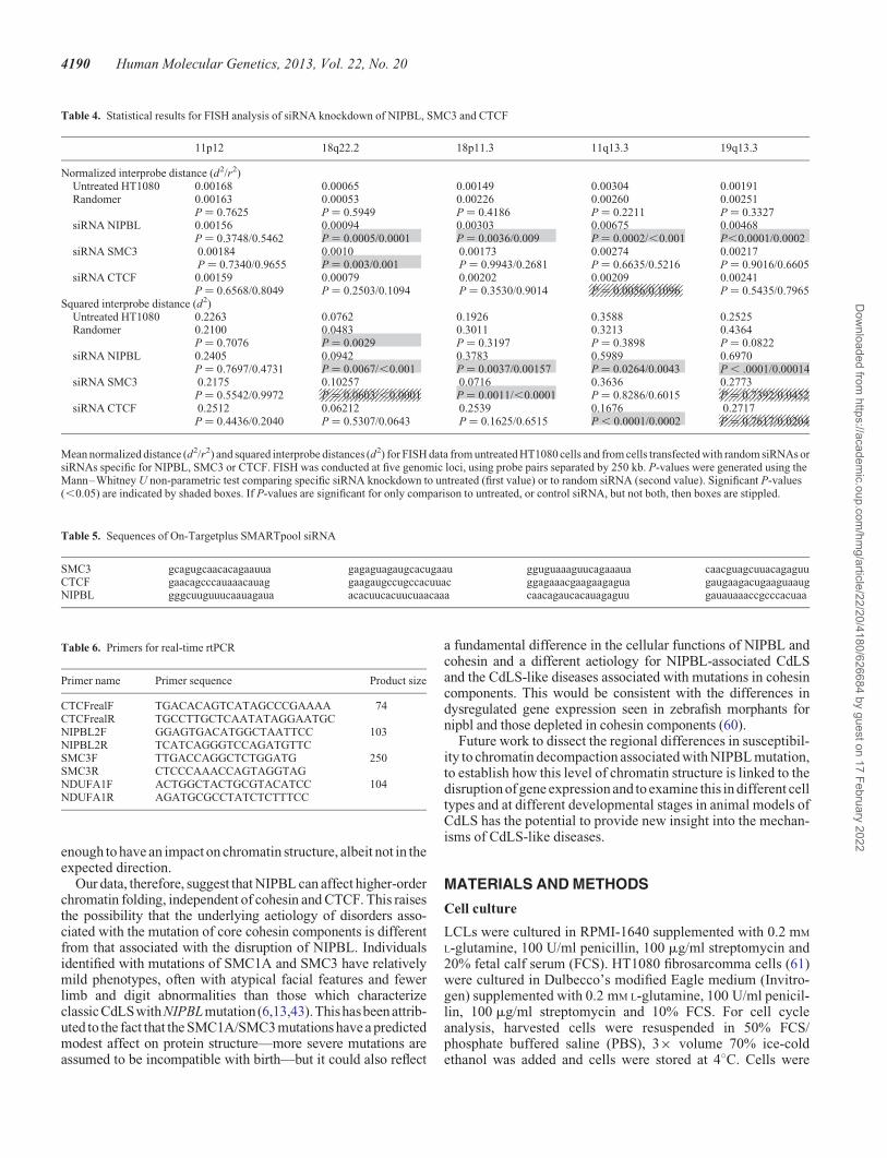
Table 4. Statistical results for FISH analysis of siRNA knockdown of NIPBL, SMC3 and CTCF
Mean normalized distance (d2/r2) and squared interprobe distances (d2) for FISH data from untreated HT1080 cells and from cells transfected with random siRNAs orsiRNAs specific for NIPBL, SMC3 or CTCF. FISH was conducted at five genomic loci, using probe pairs separated by 250 kb. P-values were generated using theMann–Whitney U non-parametric test comparing specific siRNA knockdown to untreated (first value) or to random siRNA (second value). Significant P-values(,0.05) are indicated by shaded boxes. If P-values are significant for only comparison to untreated, or control siRNA, but not both, then boxes are stippled.
Table 5. Sequences of On-Targetplus SMARTpool siRNA
SMC3 gcagugcaacacagaauua gagaguagaugcacugaau gguguaaaguucagaaaua caacguagcuuacagaguuCTCF gaacagcccauaaacauag gaagaugccugccacuuac ggagaaacgaagaagagua gaugaagacugaaguaaugNIPBL gggcuuguuucaauagaua acacuucacuucuaacaaa caacagaucacauagaguu gauauaaaccgcccacuaa
Table 6. Primers for real-time rtPCR
Primer name Primer sequence Product size
CTCFrealF TGACACAGTCATAGCCCGAAAA 74CTCFrealR TGCCTTGCTCAATATAGGAATGCNIPBL2F GGAGTGACATGGCTAATTCC 103NIPBL2R TCATCAGGGTCCAGATGTTCSMC3F TTGACCAGGCTCTGGATG 250SMC3R CTCCCAAACCAGTAGGTAGNDUFA1F ACTGGCTACTGCGTACATCC 104NDUFA1R AGATGCGCCTATCTCTTTCC
4190 Human Molecular Genetics, 2013, Vol. 22, No. 20
Dow
nloaded from https://academ
ic.oup.com/hm
g/article/22/20/4180/626684 by guest on 17 February 2022

then washed with PBS and resuspended in propidium iodide so-lution (50 mg/ml PI + 100 mg/ml RNAse) for 1 h prior to FACSanalysis on a BD FACSAria2 SORP. Cells were excited at488 nm and measured at 562–588 nm. Data were analysedusing BD FACSDiva Software version 6.1.3.
FISH, image capture and analysis
Nuclei were isolated in hypotonic buffer (0.25% KCl) and fixedwith 3:1 v/v methanol/acetic acid. Fosmid clones (Table 2) wereprepared and labelled with digoxigenin-11-dUTP or withbiotin-16-dUTP as described previously (55). After hybridiza-tion, digoxigenin-labelled probes were detected using Rhoda-mine anti-digoxigenin and Texas Red anti-sheep IgG (VectorLaboratories). Biotin-labelled probes were detected usingfluorescein–streptavidin and biotinylated anti-avidin (VectorLaboratories).
Slides were analysed as described previously (55) except thata Chroma #83000 triple band pass filter set (Chroma TechnologyCorporation, Rockingham, VT, USA) and a motorized filterwheel (Prior Scientific Instruments, Cambridge, UK) were used.
Array CGH
Genomic DNA was prepared from LCLs using a Nucleon DNAextraction kit (Tepnel Life Sciences, UK). DNA was quantifiedby NanoDrop spectrophotometry (Thermo Scientific).
Genome-wide analysis of DNA copy number aberrations wascarried out using the Roche Nimblegen 135k whole-genomearray (median probe spacing of �12 kb) according to the manu-facturer’s instructions, as described previously (62). Afterwashing, slides were scanned using the Roche MS200 scannerand analysed using NimbleScan software (Roche Nimblegen).The CGH-segMNT module of NimbleScan was used for theanalysis with a minimum segment length of 5 probes and an aver-aging window of 130 kb. The results were analysed by obtainingthe log2 ratios of the case (labelled with Cy-5) compared with thecontrol (labelled with Cy-3). A heterozygous deletion and aheterozygous duplication are expected to result in log2 ratiosof 21.0 and 0.58, respectively. Similarly, a homozygous dele-tion and a homozygous duplication are expected to result inlog2 ratios of 22.0 and 1.0, respectively. Genomic coordinateswere converted from hg19 to hg18 for analysis, using theUCSC Lift Genome Annotations utility.
SiRNA knockdown
SiRNA-mediated knockdown was performed using On-Targetplus SMARTpool siRNA mixes (Thermo Scientific),designed with dual-strand modification that decreases off targeteffects and containing a mix of four individual siRNAsTable 5). The randomer was the ON-TARGETplus Non-targetingControl Pool. siRNAs were diluted as directed by the manufactur-er to a stock concentration of 100 mM in the supplied 1× siRNAbuffer. A 10 cm dish of HT1080 cells at 90% confluence was lipo-fected with 165 pmol siRNAs using Lipofectamine 2000. Cellswere harvested for RNA and protein 50 h post-transfection.
Real-time PCR
RNA was isolated using Qiagen RNA Easy Kit. 500 ng of RNAwere reverse transcribed in a 20 ml reaction using Superscript II.1 ml of cDNA was amplified using specific primers (Table 6)and SYBER green real-time master mix (Life Technologies).Real-time analysis was performed using a Roche LightCycler480. The programme for amplification was 958C × 3 min,then 45 cycles of 958C × 15 s, 62oC × 15 s and 72oC × 30 s.The melting curve programme was 958C × 5 s and 658C ×1 min, and then readings were acquired while increasing the tem-perature by 0.118C/s to 978C.
Immunoblotting
Protein was isolated from a 6-well plate of cells. 100 ml of4× SDSLB (2.0 ml 1 M Tris–HCl, pH 6.8, 0.8 g sodiumdodecyl sulphate SDS), 4.0 ml of 100% glycerol, 0.4 ml of14.7 M b-mercaptoethanol, 1.0 ml of 0.5 M ethylenediaminete-traacetic acid 8 mg bromophenol blue) and 300 ml PBS wereadded to the cells. The cells and liquid were collected from thewells, boiled for 5 min and then sonicated for 20 s (BioruptorNextGen Diagenode) prior to loading directly onto gels. Foranalysis of NIPBL, samples were run on a NuPage TrisAcetate3–8% gel (Life Technologies) using the NuPage runningbuffer. For CTCF and SMC3, samples were run on a standard10% SDS polyacrylamide gel. Gels were transferred onto polyvi-nylidene fluoride membranes by semidry transfer at 10 V for 1 h.The membrane was blocked for 1 h in 5% milk PBST (0.1%Tween-20 in Dulbecco A PBS; Oxoid). NIPBL was detectedwith the anti-IND3 rat monoclonal antibody (AbCAM#131913) diluted 1:500. For Smc3, anti-SMC3 rabbit polyclonalantibody (Bethyl A300-060A) was used at a 1:1000 dilution. ForCTCF, anti-CTCF rabbit polyclonal antibody (Upstate 07-729)was used at a 1:2000 dilution. Membranes were incubated withantibodies overnight in 1.5% milk PBST at 48C, washed 3×with PBST and incubated with the appropriate horseradishperoxidase-conjugated secondary antibody at a 1:10 000 dilutionat room temperature for 1 h. Signal was detected by ChemiGlowWest (Alpha Innotech) and imaged using Image Quant LAS4010(Version 1, Build 1.0.0.52; GE Healthcare).
SUPPLEMENTARY MATERIAL
Supplementary Material is available at HMG online.
ACKNOWLEDGEMENTS
We thank Tom Strachan (Newcastle) and Matt Deardorff andIan Krantz (Children’s Hospital of Philadelphia) for their giftsof CdLS LCLs. At the MRC Human Genetics Unit, we are grate-ful to Elisabeth Freyer for FACs analysis, Anne Seawright forcell culture assistance and Paul Perry for help with image ana-lysis. We thank David Fitzpatrick (MRC HGU, Edinburgh) forcritical reading of the manuscript and for discussions about theclinical features of CdLS and CdLS-like syndromes.
Conflict of Interest statement. None declared.
Human Molecular Genetics, 2013, Vol. 22, No. 20 4191
Dow
nloaded from https://academ
ic.oup.com/hm
g/article/22/20/4180/626684 by guest on 17 February 2022

FUNDING
This work was supported by an EMBO long-term fellowship anda Medical Research Council Centenary award to L.D.N. and aPhD studentship from the Medical Research Council to E.P.W.A.B. is supported by the Medical Research Council and byan ERC advanced grant (249956). Funding to pay the OpenAccess publication charges for this article was provided byMedical Research Council UK.
REFERENCES
1. Liu, J. and Krantz, I.D. (2008) Cohesin and human disease. Ann. Rev.Genomics Hum. Genet., 9, 303–320.
2. Tonkin, E.T., Wang, T.J., Lisgo, S., Bamshad, M.J. and Strachan, T. (2004)NIPBL, encoding a homolog of fungal Scc2-type sister chromatid cohesionproteins and fly Nipped-B, is mutated in Cornelia de Lange syndrome. Nat.Genet., 36, 636–641.
3. Krantz, I.D., McCallum, J., DeScipio, C., Kaur, M., Gillis, L.A., Yaeger, D.,Jukofsky, L., Wasserman, N., Bottani, A., Morris, C.A. et al. (2004) Corneliade Lange syndrome is caused by mutations in NIPBL, the human homolog ofDrosophila melanogaster Nipped-B. Nat. Genet., 36, 631–635.
4. Liu, J., Feldman, R., Zhang, Z., Deardorff, M.A., Haverfield, E.V., Kaur, M.,Li, J.R., Clark, D., Kline, A.D., Waggoner, D.J. et al. (2009) SMC1Aexpression and mechanism of pathogenicity in probands with X-linkedCornelia de Lange syndrome. Hum. Mutat., 30, 1535–1542.
5. Musio, A., Selicorni,A., Focarelli, M.L.,Gervasini, C.,Milani, D., Russo, S.,Vezzoni, P. and Larizza, L. (2006) X-linked Cornelia de Lange syndromeowing to SMC1L1 mutations. Nat. Genet., 38, 528–530.
6. Deardorff, M.A., Kaur, M., Yaeger, D., Rampuria, A., Korolev, S., Pie, J.,Gil-Rodriguez, C., Arnedo, M., Loeys, B., Kline, A.D. et al. (2007)Mutations in cohesin complex members SMC3 and SMC1A cause a mildvariant of Cornelia de Lange syndrome with predominant mentalretardation. Am. J. Hum. Genet., 80, 485–494.
7. Borck, G., Zarhrate, M., Bonnefont, J.P., Munnich, A., Cormier-Daire, V.and Colleaux, L. (2007) Incidence and clinical features of X-linked Corneliade Lange syndrome due to SMC1L1 mutations. Hum. Mutat., 28, 205–206.
8. Deardorff, M.A., Wilde, J.J., Albrecht, M., Dickinson, E., Tennstedt, S.,Braunholz, D., Monnich, M., Yan, Y., Xu, W., Gil-Rodriguez, M.C. et al.(2012) RAD21mutations cause a humancohesinopathy. Am. J. Hum. Genet.,90, 1014–1027.
9. Deardorff, M.A., Bando, M., Nakato, R., Watrin, E., Itoh, T., Minamino, M.,Saitoh, K., Komata, M., Katou, Y., Clark, D. et al. (2012) HDAC8 mutationsin Cornelia de Lange syndrome affect the cohesin acetylation cycle. Nature,489, 313–317.
10. Nasmyth, K. and Haering, C.H. (2009) Cohesin: its roles and mechanisms.Ann. Rev. Genet., 43, 525–558.
11. Gillis, L.A., McCallum, J., Kaur, M., DeScipio, C., Yaeger, D., Mariani, A.,Kline, A.D., Li, H.-h., Devoto, M., Jackson, L.G. et al. (2004) NIPBLmutational analysis in 120 individuals with Cornelia de Lange syndrome andevaluation of genotype-phenotype correlations. Am. J. Hum. Genet., 75,610–623.
12. Bhuiyan, Z.A., Klein, M., Hammond, P., van Haeringen, A., Mannens,M.M., Van Berckelaer-Onnes, I. and Hennekam, R.C. (2006)Genotype-phenotype correlations of 39 patients with Cornelia De Langesyndrome: the Dutch experience. J. Med. Genet., 43, 568–575.
13. Rohatgi, S., Clark, D., Kline, A.D., Jackson, L.G., Pie, J., Siu, V., Ramos,F.J., Krantz, I.D. and Deardorff, M.A. (2010) Facial diagnosis of mild andvariant CdLS: insights from a dysmorphologist survey. Am. J. Med. Genet.Part A, 152A, 1641–1653.
14. Ciosk, R., Shirayama, M., Shevchenko, A., Tanaka, T., Toth, A. andNasmyth, K. (2000) Cohesin’s binding to chromosomes depends on aseparate complex consisting of Scc2 and Scc4 proteins. Mol. Cell, 5,243–254.
15. Kawauchi, S., Calof, A.L., Santos, R., Lopez-Burks, M.E., Young, C.M.,Hoang, M.P., Chua, A., Lao, T., Lechner, M.S., Daniel, J.A. et al. (2009)Multiple organ system defects and transcriptional dysregulation in theNipbl(+/-) mouse, a model of Cornelia de Lange syndrome. PLoS Genet., 5,e1000650.
16. Castronovo, P., Gervasini, C., Cereda, A., Masciadri, M., Milani, D., Russo,S., Selicorni, A. and Larizza, L. (2009) Premature chromatid separation is
nota useful diagnosticmarker for Corneliade Lange syndrome. Chrom. Res.,17, 763–771.
17. Schuldiner, O., Berdnik, D., Levy, J.M., Wu, J.S., Luginbuhl, D., Gontang,A.C. and Luo, L. (2008) piggyBac-based mosaic screen identifies apostmitotic function for cohesin in regulating developmental axon pruning.Dev. Cell, 14, 227–238.
18. Pauli, A., Althoff, F., Oliveira, R.A., Heidmann, S., Schuldiner, O., Lehner,C.F., Dickson, B.J. and Nasmyth, K. (2008) Cell-type-specific TEV proteasecleavage reveals cohesin functions in Drosophila neurons. Dev. Cell, 14,239–251.
19. Seitan, V.C., Hao, B., Tachibana-Konwalski, K., Lavagnolli, T.,Mira-Bontenbal, H., Brown, K.E., Teng, G., Carroll, T., Terry, A., Horan, K.et al. (2011) A role for cohesin in T-cell-receptor rearrangement andthymocyte differentiation. Nature, 476, 467–471.
20. Liu, J., Zhang, Z., Bando, M., Itoh, T., Deardorff, M.A., Clark, D., Kaur, M.,Tandy, S., Kondoh, T., Rappaport, E. et al. (2009) Transcriptionaldysregulation in NIPBL and cohesin mutant human cells. PLoS Biol., 7,e1000119.
21. Schaaf, C.A., Kwak, H., Koenig, A., Misulovin, Z., Gohara, D.W., Watson,A., Zhou, Y., Lis, J.T. and Dorsett, D. (2013) Genome-wide control of RNApolymerase II activity by cohesin. PLoS Genet., 9, e1003382.
22. Dorsett, D., Eissenberg, J.C., Misulovin, Z., Martens, A., Redding, B. andMcKim, K. (2005) Effects of sister chromatid cohesion proteins on cut geneexpression during wing development in Drosophila. Development, 132,4743–4753.
23. Rollins, R.A., Morcillo, P. and Dorsett, D. (1999) Nipped-B, a Drosophila
homologue of chromosomal adherins, participates in activation by remoteenhancers in the cut and Ultrabithorax genes. Genetics, 152, 577–593.
24. Rollins, R.A., Korom, M., Aulner, N., Martens, A. and Dorsett, D. (2004)Drosophila nipped-B protein supports sister chromatid cohesion andopposes the stromalin/Scc3 cohesion factor to facilitate long-rangeactivation of the cut gene. Mol. Cell. Biol., 24, 3100–3111.
25. Parelho, V., Hadjur, S., Spivakov, M., Leleu, M., Sauer, S., Gregson, H.C.,Jarmuz, A., Canzonetta, C., Webster, Z., Nesterova, T. et al. (2008) Cohesinsfunctionally associate with CTCF on mammalian chromosome arms. Cell,132, 422–433.
26. Rubio, E.D., Reiss, D.J., Welcsh, P.L., Disteche, C.M., Filippova, G.N.,Baliga, N.S., Aebersold, R., Ranish, J.A. and Krumm, A. (2008) CTCFphysically links cohesin to chromatin. Proc. Natl Acad. Sci. USA, 105,8309–8314.
27. Stedman, W., Kang, H., Lin, S., Kissil, J.L., Bartolomei, M.S. andLieberman, P.M. (2008) Cohesins localize with CTCF at the KSHV latencycontrol region and at cellular c-myc and H19/Igf2 insulators. EMBO J., 27,654–666.
28. Wendt, K.S., Yoshida, K., Itoh, T., Bando, M., Koch, B., Schirghuber, E.,Tsutsumi, S., Nagae, G., Ishihara, K., Mishiro, T. et al. (2008) Cohesinmediates transcriptional insulation by CCCTC-binding factor. Nature, 451,796–801.
29. Hadjur, S., Williams, L.M., Ryan, N.K., Cobb, B.S., Sexton, T., Fraser, P.,Fisher, A.G. and Merkenschlager, M. (2009) Cohesins form chromosomalcis-interactions at the developmentally regulated IFNG locus. Nature, 460,410–413.
30. Mishiro,T., Ishihara, K., Hino, S., Tsutsumi, S., Aburatani, H., Shirahige, K.,Kinoshita, Y. and Nakao, M. (2009) Architectural roles of multiplechromatin insulators at the human apolipoprotein gene cluster. EMBO J., 28,1234–1245.
31. Nativio, R., Wendt, K.S., Ito, Y., Huddleston, J.E., Uribe-Lewis, S.,Woodfine, K., Krueger, C., Reik, W., Peters, J.M. and Murrell, A. (2009)Cohesin is required for higher-order chromatin conformation at theimprinted IGF2-H19 locus. PLoS Genet., 5, e1000739.
32. Degner, S.C., Verma-Gaur, J., Wong, T.P., Bossen, C., Iverson, G.M.,Torkamani, A., Vettermann, C., Lin, Y.C., Ju, Z., Schulz, D. et al. (2011)CCCTC-binding factor (CTCF) and cohesin influence the genomicarchitecture of the Igh locus and antisense transcription in pro-B cells. Proc.
Natl Acad. Sci. USA, 108, 9566–9571.33. Chien, R., Zeng, W., Kawauchi, S., Bender, M.A., Santos, R., Gregson, H.C.,
Schmiesing, J.A., Newkirk, D., Kong, X., Ball, A.R. et al. (2011) Cohesinmediates chromatin interactions that regulate mammalian b-globinexpression. J. Biol. Chem., 286, 17870–17878.
34. Kim, Y.J., Cecchini, K.R. and Kim, T.H. (2011) Conserved,developmentally regulated mechanism couples chromosomal looping andheterochromatin barrier activity at the homeobox gene A locus. Proc. NatlAcad. Sci. USA, 108, 7391–7396.
4192 Human Molecular Genetics, 2013, Vol. 22, No. 20
Dow
nloaded from https://academ
ic.oup.com/hm
g/article/22/20/4180/626684 by guest on 17 February 2022

35. Seitan, V.C. and Merkenschlager, M. (2012) Cohesin and chromatinorganisation. Curr. Opin. Genet. Dev., 22, 93–100.
36. Kagey, M.H., Newman, J.J., Bilodeau, S., Zhan, Y., Orlando, D.A.,van Berkum, N.L., Ebmeier,C.C., Goossens, J., Rahl,P.B., Levine, S.S. et al.
(2010) Mediator and cohesin connect gene expression and chromatinarchitecture. Nature, 467, 430–435.
37. Schmidt, D., Schwalie, P.C., Ross-Innes, C.S., Hurtado, A., Brown, G.D.,Carroll, J.S., Flicek, P. and Odom, D.T. (2010) A CTCF-independent rolefor cohesin in tissue-specific transcription. Genome Res., 20, 578–588.
38. Eskeland, R., Leeb, M., Grimes, G.R., Kress, C., Boyle, S., Sproul, D.,Gilbert, N., Fan, Y., Skoultchi, A.I., Wutz, A. et al. (2010) Ring1B compactschromatin structure and represses gene expression independent of histoneubiquitination. Mol. Cell, 38, 452–464.
39. Gurdon, J.B. (1976) Injected nuclei in frog oocytes: fate, enlargement, andchromatin dispersal. J. Embryol. Exp. Morphol., 36, 523–540.
40. Shen, X., Yu, L., Weir, J.W. and Gorovsky, M.A. (1995) Linker histones are
not essential and affect chromatin condensation in vivo. Cell, 82, 47–56.
41. Mazumder, A., Roopa, T., Basu, A., Mahadevan, L. and Shivashankar, G.V.(2008) Dynamics of chromatin decondensation reveals the structuralintegrity of a mechanically prestressed nucleus. Biophys. J., 95,
3028–3035.
42. Pehlivan, D., Hullings, M., Carvalho, C.M., Gonzaga-Jauregui, C.G., Loy,E., Jackson, L.G., Krantz, I.D., Deardorff, M.A. and Lupski, J.R. (2012)NIPBL rearrangements in Cornelia de Lange syndrome: evidence for
replicative mechanism and genotype-phenotype correlation. Genet. Med.,14, 313–322.
43. Pie, J., Gil-Rodriguez, M.C., Ciero, M., Lopez-Vinas, E., Ribate, M.P.,Arnedo, M., Deardorff, M.A., Puisac, B., Legarreta, J., de Karam, J.C. et al.
(2010) Mutations and variants in the cohesion factor genes NIPBL, SMC1A,and SMC3 in a cohort of 30 unrelated patients with Cornelia de Langesyndrome. Am. J. Med. Genet. A, 152A, 924–929.
44. Oka, Y., Suzuki, K., Yamauchi, M., Mitsutake, N. and Yamashita, S. (2011)Recruitment of the cohesin loading factor NIPBL to DNA double-strandbreaks depends on MDC1, RNF168 and HP1gamma in human cells.Biochem. Biophys. Re.s Commun., 411, 762–767.
45. Neuwald, A.F. and Hirano, T. (2000) HEAT repeats associated withcondensins, cohesins, and other complexes involved in chromosome-relatedfunctions. Genome Res., 10, 1445–1452.
46. Jahnke, P., Xu, W., Wulling, M., Albrecht, M., Gabriel, H.,
Gillessen-Kaesbach, G. and Kaiser, F.J. (2008) The cohesin loading factorNIPBL recruits histone deacetylases to mediate local chromatinmodifications. Nucleic Acids Res., 36, 6450–6458.
47. Caron, H., van Schaik, B., van der Mee, M., Baas, F., Riggins, G., van Sluis,P., Hermus, M.C., van Asperen, R., Boon, K., Voute, P.A. et al. (2001) Thehuman transcriptome map: clustering of highly expressed genes inchromosomal domains. Science, 291, 1289–1292.
48. The ENCODE consortium (2012) An integrated encyclopedia of DNAelements in the human genome. Nature, 489, 57–74.
49. Cheung, V.G., Spielman, R.S., Ewens, K.G., Weber, T.M., Morley, M. andBurdick, J.T. (2005) Mapping determinants of human gene expression byregional and genome-wide association. Nature, 437, 1365–1369.
50. Barski, A., Cuddapah, S., Cui, K., Roh, T.Y., Schones, D.E., Wang, Z., Wei,G., Chepelev, I. and Zhao, K. (2007) High-resolution profiling of histonemethylations in the human genome. Cell, 129, 823–837.
51. Vavouri, T. and Lehner, B. (2012) Human genes with CpG island promotershave a distinct transcription-associated chromatin organization. GenomeBiol., 13, R110.
52. Yokota, H., Singer, M.J., van den Engh, G.J. and Trask, B.J. (1997) Regionaldifferences in the compaction of chromatin in human G0/G1 interphasenuclei. Chrom. Res., 5, 157–166.
53. Gilbert, N., Boyle, S., Fiegler, H., Woodfine, K., Carter, N.P. and Bickmore,W.A. (2004) Chromatin architecture of the human genome: gene-richdomains are enriched in open chromatin fibers. Cell, 118, 555–566.
54. Chambeyron, S. and Bickmore, W.A. (2004) Chromatin decondensation andnuclear reorganization of the HoxB locus upon induction of transcription.Genes Dev., 18, 1119–1130.
55. Morey, C., Da Silva, N.R., Perry, P. and Bickmore, W.A. (2007)Nuclear reorganisation and chromatin decondensation are conserved, butdistinct, mechanisms linked to Hox gene activation. Development, 134,909–919.
56. Guo, Y., Monahan, K., Wu, H., Gertz, J., Varley, K.E., Li, W., Myers, R.M.,Maniatis, T. and Wu, Q. (2012) CTCF/cohesin-mediated DNA looping isrequired for protocadherin alpha promoter choice. Proc. Natl Acad. Sci.USA, 109, 21081–21086.
57. Junier, I., Dale, R.K., Hou, C., Kepes, F. and Dean, A. (2012)CTCF-mediated transcriptional regulation through cell type-specificchromosome organization in the beta-globin locus. Nucleic Acids Res., 40,7718–7727.
58. Gard, S., Light, W., Xiong, B., Bose, T., McNairn, A.J., Harris, B., Fleharty,B., Seidel, C., Brickner, J.H. and Gerton, J.L. (2009) Cohesinopathymutations disrupt the subnuclear organization of chromatin. J. Cell Biol.,187, 455–462.
59. Monahan, K., Rudnick, N.D., Kehayova, P.D., Pauli, F., Newberry, K.M.,Myers, R.M. and Maniatis, T. (2012) Role of CCCTC binding factor(CTCF) and cohesin in the generation of single-cell diversity ofProtocadherin-alpha gene expression. Proc. Natl Acad. Sci. USA, 109,9125–9130.
60. Muto, A., Calof, A.L., Lander, A.D. and Schilling,T.F. (2011) Multifactorialorigins of heart and gut defects in nipbl-deficient zebrafish, a model ofCornelia de Lange syndrome. PLoS Biol., 9, e1001181.
61. Rasheed, S., Nelson-Rees, W.A., Toth, E.M., Arnstein, P. and Gardner, M.B.(1974) Characterization of a newly derived human sarcoma cell line(HT-1080). Cancer, 33, 1027–1033.
62. Gerth-Kahlert, C., Williamson, K., Ansari, M., Rainger, J.K., Hingst, V.,Zimmermann, T., Tech, T., Guthoff, R.F., van Heyningen, V. andFitzPatrick, D.R. (2013) Clinical and mutation analysis of 51 probands withanophthalmia and/or severe microphthalmia from a single center. Mol.Genet. Genomic Med., 1, 15–31.
Human Molecular Genetics, 2013, Vol. 22, No. 20 4193
Dow
nloaded from https://academ
ic.oup.com/hm
g/article/22/20/4180/626684 by guest on 17 February 2022