refractive surgery - ileyemd.org · refractive surgery an intro to the ... vision correction...
TRANSCRIPT
3/13/2015
1
Refractive SurgeryAn Intro to the Basics of Screening
and Post Operative Care
Neema Nayeb-Hashemi
Loyola University Medical Center
3/6/2015
LASER VISION CORRECTION
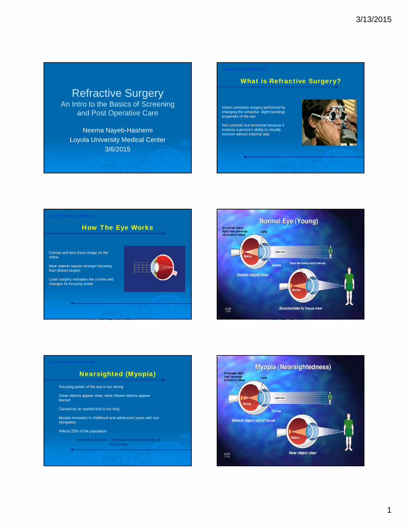
What is Refractive Surgery?
Vision correction surgery performed by changing the refractive (light bending) properties of the eye
Not cosmetic but functional because it restores a person’s ability to visually function without external aids
How The Eye Works
LASER VISION CORRECTION
Cornea and lens focus image on the retina
Near objects require stronger focusing than distant targets
Laser surgery reshapes the cornea and changes its focusing power
Nearsighted (Myopia)
LASER VISION CORRECTION
Focusing power of the eye is too strong
Close objects appear clear, while distant objects appear blurred
Caused by an eyeball that is too long
Myopia increases in childhood and adolescent years with eye elongation
Affects 25% of the population
Refractive Solution: Decrease the central power of the cornea
3/13/2015
2
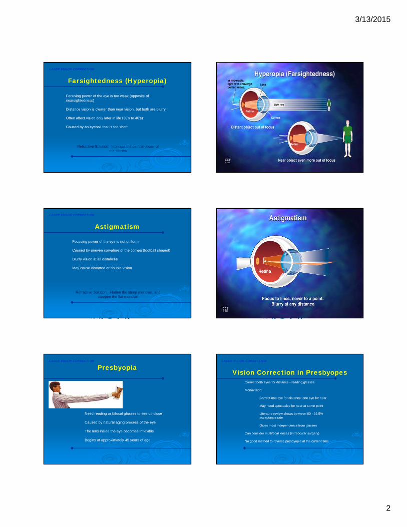
Farsightedness (Hyperopia)
LASER VISION CORRECTION
Focusing power of the eye is too weak (opposite of nearsightedness)
Distance vision is clearer than near vision, but both are blurry
Often affect vision only later in life (30’s to 40’s)
Caused by an eyeball that is too short
Refractive Solution: Increase the central power of the cornea
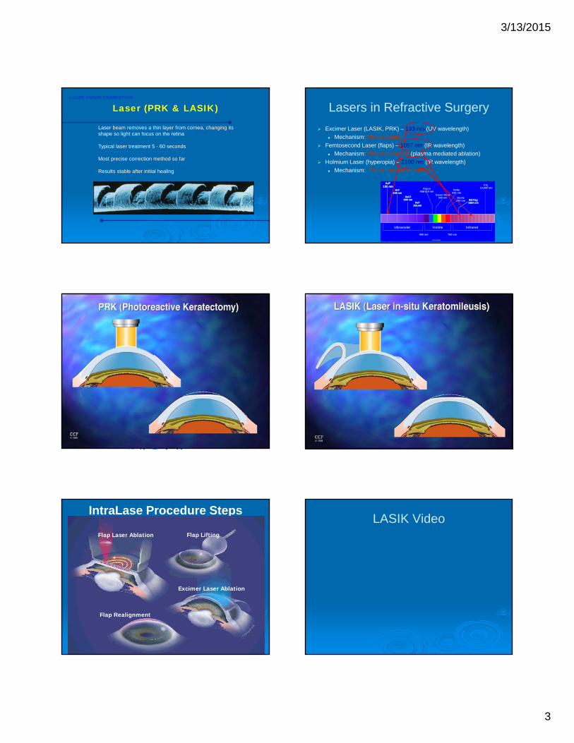
Astigmatism
LASER VISION CORRECTION
Focusing power of the eye is not uniform
Caused by uneven curvature of the cornea (football shaped)
Blurry vision at all distances
May cause distorted or double vision
Refractive Solution: Flatten the steep meridian, and steepen the flat meridian
PresbyopiaLASER VISION CORRECTION
Need reading or bifocal glasses to see up close
Caused by natural aging process of the eye
The lens inside the eye becomes inflexible
Begins at approximately 45 years of age
Vision Correction in Presbyopes
LASER VISION CORRECTION
Correct both eyes for distance - reading glasses
Monovision:
Correct one eye for distance; one eye for near
May need spectacles for near at some point
Literaure review shows between 80 - 92.5% acceptance rate
Gives most independence from glasses
Can consider multifocal lenses (intraocular surgery)
No good method to reverse presbyopia at the current time
3/13/2015
3
Laser (PRK & LASIK)LASER VISION CORRECTION
Laser beam removes a thin layer from cornea, changing its shape so light can focus on the retina
Typical laser treatment 5 - 60 seconds
Most precise correction method so far
Results stable after initial healing
Lasers in Refractive Surgery
Excimer Laser (LASIK, PRK) – 193 nm (UV wavelength)
Mechanism: Photo-ablation
Femtosecond Laser (flaps) – 1057 nm (IR wavelength)
Mechanism: Photo-disruption (plasma mediated ablation)
Holmium Laser (hyperopia) – 2100 nm (IR wavelength)
Mechanism: Photo-thermal shrinkage
Nd:Yag1064 nmNd:Yag1064 nm
Argon488-514 nm
Green HeNe543 nm
CO210,600 nm
Diode680 nm
ArF193 nm
ArF193 nm
KrF248 nm
KrF248 nm
XeCl308 nm
XeCl308 nm
XeF351nm
XeF351nm
HeNe632 nm
Ultraviolet InfraredVisible
750 nm400 nm
Not to Scale
IntraLase Procedure Steps
Flap Laser Ablation Flap Lifting
Excimer Laser Ablation
Flap Realignment
LASIK Video
3/13/2015
4
Screening Candidates for Refractive Surgery
Neema Nayeb-Hashemi MD
Loyola University Medical Center
3/6/2015
Components of the Evaluation
Patient History
Examination
Ancillary Testing
Patient History
Key aspects requiring evaluation: Patient Expectations
Social History
Medical History
Pertinent Ocular History
Patient Age, Presbyopia, and Monovision
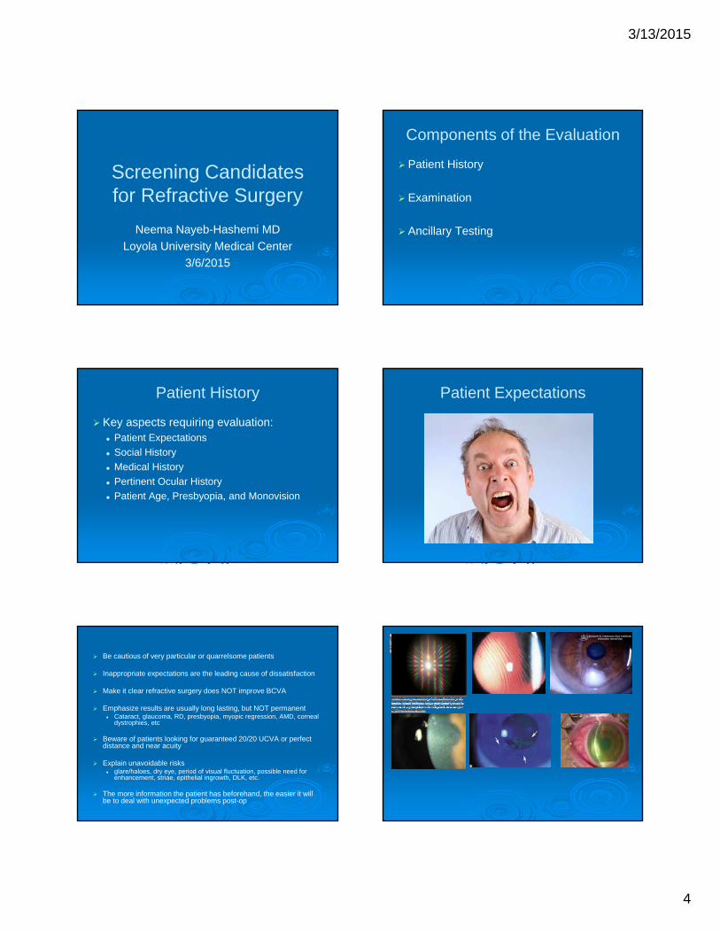
Patient Expectations
Be cautious of very particular or quarrelsome patients
Inappropriate expectations are the leading cause of dissatisfaction
Make it clear refractive surgery does NOT improve BCVA
Emphasize results are usually long lasting, but NOT permanent Cataract, glaucoma, RD, presbyopia, myopic regression, AMD, corneal
dystrophies, etc
Beware of patients looking for guaranteed 20/20 UCVA or perfect distance and near acuity
Explain unavoidable risks glare/haloes, dry eye, period of visual fluctuation, possible need for
enhancement, striae, epithelial ingrowth, DLK, etc.
The more information the patient has beforehand, the easier it will be to deal with unexpected problems post-op
3/13/2015
5
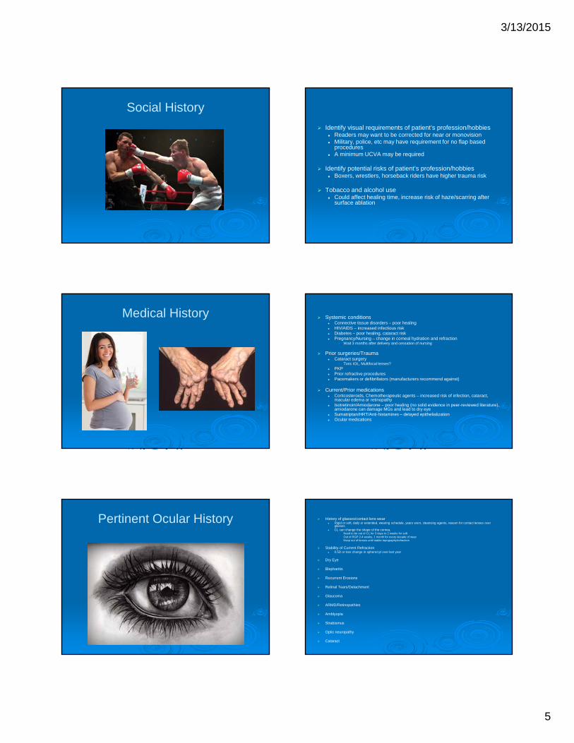
Social History
Identify visual requirements of patient’s profession/hobbies Readers may want to be corrected for near or monovision Military, police, etc may have requirement for no flap based
procedures A minimum UCVA may be required
Identify potential risks of patient’s profession/hobbies Boxers, wrestlers, horseback riders have higher trauma risk
Tobacco and alcohol use Could affect healing time, increase risk of haze/scarring after
surface ablation
Medical History Systemic conditions Connective tissue disorders – poor healing HIV/AIDS – increased infectious risk Diabetes – poor healing, cataract risk Pregnancy/Nursing – change in corneal hydration and refraction
• Wait 3 months after delivery and cessation of nursing
Prior surgeries/Trauma Cataract surgery
• Toric IOL, Multifocal lenses? PKP Prior refractive procedures Pacemakers or defibrillators (manufacturers recommend against)
Current/Prior medications Corticosteroids, Chemotherapeutic agents – increased risk of infection, cataract,
macular edema or retinopathy Isotretinoin/Amiodarone – poor healing (no solid evidence in peer-reviewed literature),
amiodarone can damage MGs and lead to dry eye Sumatriptan/HRT/Anti-histamines – delayed epithelialization Ocular medications
Pertinent Ocular History History of glasses/contact lens wear Rigid or soft, daily or extended, wearing schedule, years worn, cleansing agents, reason for contact lenses over
glasses CL can change the shape of the cornea,
• Need to be out of CL for 3 days to 2 weeks for soft• Out of RGP 2-4 weeks, 1 month for every decade of wear• Keep out of lenses until stable topography/refraction
Stability of Current Refraction 0.5D or less change in sphere/cyl over last year
Dry Eye
Blepharitis
Recurrent Erosions
Retinal Tears/Detachment
Glaucoma
ARMD/Retinopathies
Amblyopia
Strabismus
Optic neuropathy
Cataract
3/13/2015
6
Patient Age, Presbyopia, Monovision
Loss of near vision should be predicted and discussed based on age
Discuss specific tasks that may be affected Reading, shaving/makeup, computer reading, etc
Patient MUST be willing to accept this reality before surgery
Monovision discussion vs readers Target: depends on visual requirements
• Anywhere from -0.75 to -2.50, • Best tolerated/functional typically -1.50 to -1.75• Watch out for loss of depth perception, intolerable anisometropia
Could try contact lens trial, check muscle imbalance Traditionally dominant for distance, reverse mono also well
accepted by most• Dominance testing with bilateral viewing through aperture, and then
closing eye
Examination Uncorrected Visual Acuity, Manifest/Cycloplegic Refraction
Measure UCVA, Glasses Rx and Acuity with Rx
MRx pushing plus (negate accomodation)
Best acuity attainable is the goal (beyond 20/20 if possible)
Automated refraction with autorefractor or abberometer helpful
Tropicamide or cyclopentolate 1% for CRx• Neutralize sphere (not cyl) from MRx
If difference between MRx and CRx high, likely MRx was overminused
Laser program can be with either MRx or CRx (depends on age, myopia vs hyperopia, visual requirements, etc)
• Treating too much minus may push to hyperopia, tolerable in young but not older patients near presbyopia
Components of Examination
Visual Acuity and Refraction
Pupillary Examination
Ocular Motility, Confrontation Fields, Ocular Anatomy
Intraocular Pressure
Slit Lamp Exam
Fundus Exam
Pupillary Exam
3/13/2015
7
Pupil size in bright light and dim light
Check for APD
Techniques Near card in dim light at distance Light amplification pupillometer Infrared pupillometer
Large pupil size MAY be risk factor for glare/haloes Pupil size greater than optical zone may increase risk due
to greater HOAs Size of effective optical zone and level of refractive error
may be greater risk factor
Ocular Motility, Confrontation Fields, Ocular Anatomy
Asymptomatic tropia or phoria may become manifest after surgery Ex. Exotropia after accommodation from hyperopic
Rx lost
Trial of contact lenses may be helpful to predict post operative problems
CVF should be assessed to determine if any glaucomatous or intracranial origins of VF loss
Small palpebral fissures or large brows may pose problem for flap creation in LASIK
Intraocular Pressure
Should be checked after the MRx and topography
Glaucoma patients should be warned of high pressures with suction in LASIK
Topical corticosteroids post op could increase IOP
Implications for IOP checks in glaucoma Thinner corneas yield falsely low Ga IOP
Slit Lamp Examination
3/13/2015
8
Eyelids Check for blepharitis/meibomitis, tear lake
Conjunctiva Check for scarring, chalasis
Cornea Check for signs of keratoconus: thinning, steepening
• Contraindication to surgery Surface anomalies: decreased TFBUT, PEE, ABMD
• Dry eye needs pre op treatment if significant Endothelial anomalies
• Edema generally a contraindication for refractive surgery Stromal and Bowman’s Membrane Dystrophies
• Granular and Avellino can lead to interface opacities post LASIK• Reis Buckler and Thiel Benke dystrophy can lead to severe surface scarring after PRK
Anterior Chamber, Iris, Lens Shallow chamber is contraindication to certain phakic IOLs Cataracts are a relative contraindication, should mention that IOL power calculations
less predictable post LASIK Cataract extraction, with/without toric or multifocal options, could be better options Good idea to give patients their records of preoperative refractions and keratometry ,
amount of laser ablation performed, and post op refraction***
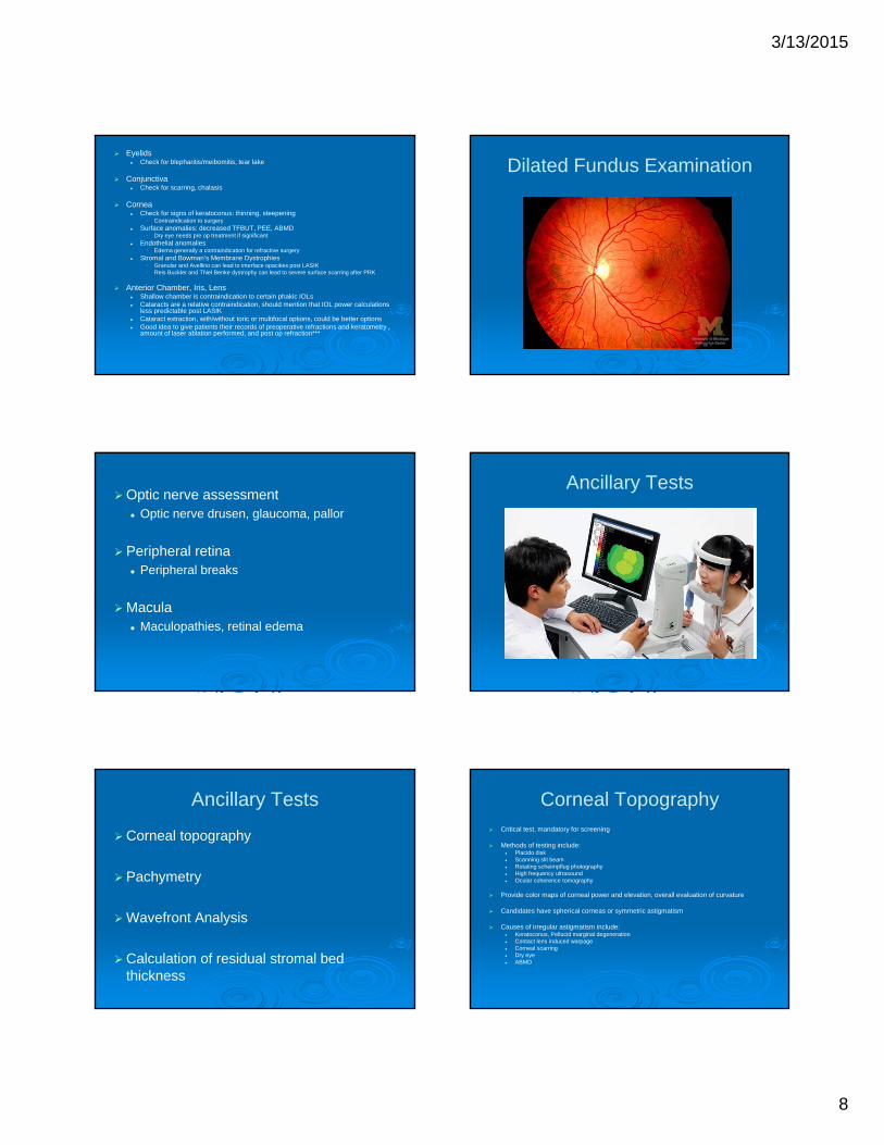
Dilated Fundus Examination
Optic nerve assessment Optic nerve drusen, glaucoma, pallor
Peripheral retina Peripheral breaks
Macula Maculopathies, retinal edema
Ancillary Tests
Ancillary Tests
Corneal topography
Pachymetry
Wavefront Analysis
Calculation of residual stromal bed thickness
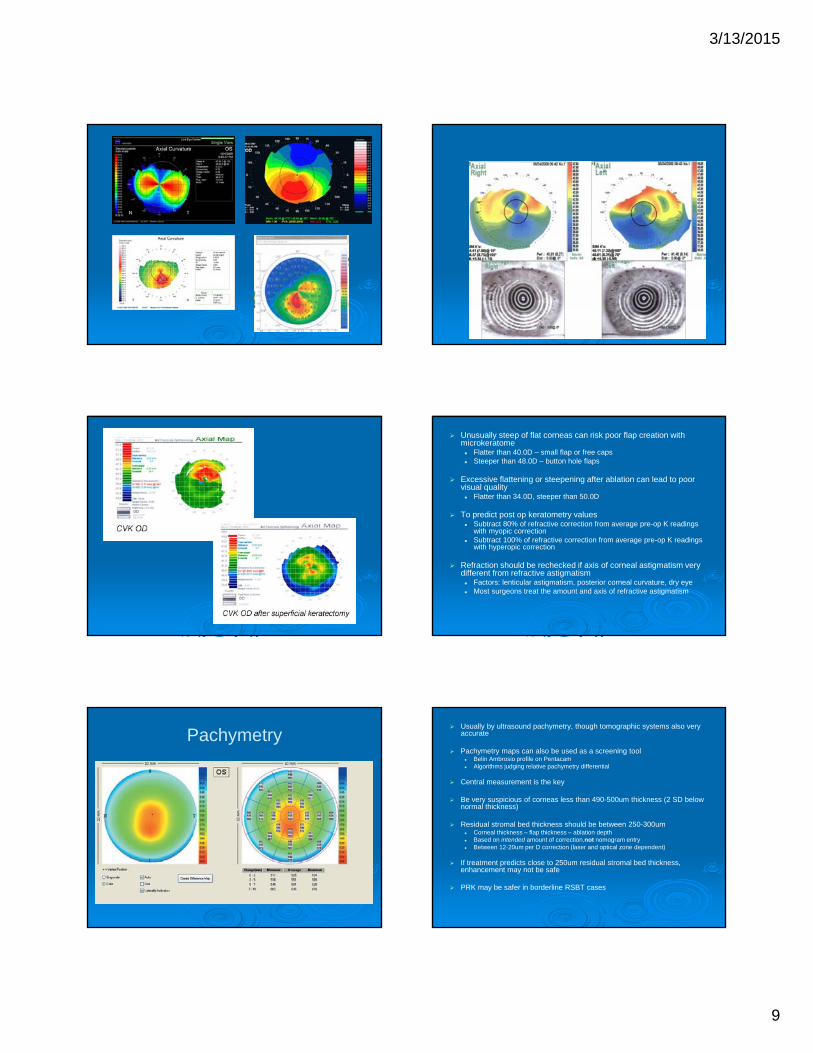
Corneal Topography Critical test, mandatory for screening
Methods of testing include: Placido disk Scanning slit beam Rotating scheimpflug photography High frequency ultrasound Ocular coherence tomography
Provide color maps of corneal power and elevation, overall evaluation of curvature
Candidates have spherical corneas or symmetric astigmatism
Causes of irregular astigmatism include: Keratoconus, Pellucid marginal degeneration Contact lens induced warpage Corneal scarring Dry eye ABMD
3/13/2015
9
Unusually steep of flat corneas can risk poor flap creation with microkeratome Flatter than 40.0D – small flap or free caps Steeper than 48.0D – button hole flaps
Excessive flattening or steepening after ablation can lead to poor visual quality Flatter than 34.0D, steeper than 50.0D
To predict post op keratometry values Subtract 80% of refractive correction from average pre-op K readings
with myopic correction Subtract 100% of refractive correction from average pre-op K readings
with hyperopic correction
Refraction should be rechecked if axis of corneal astigmatism very different from refractive astigmatism Factors: lenticular astigmatism, posterior corneal curvature, dry eye Most surgeons treat the amount and axis of refractive astigmatism
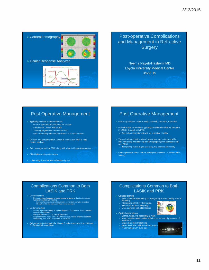
Pachymetry Usually by ultrasound pachymetry, though tomographic systems also very
accurate
Pachymetry maps can also be used as a screening tool Belin Ambrosio profile on Pentacam Algorithms judging relative pachymetry differential
Central measurement is the key
Be very suspicious of corneas less than 490-500um thickness (2 SD below normal thickness)
Residual stromal bed thickness should be between 250-300um Corneal thickness – flap thickness – ablation depth Based on intended amount of correction,not nomogram entry Between 12-20um per D correction (laser and optical zone dependent)
If treatment predicts close to 250um residual stromal bed thickness, enhancement may not be safe
PRK may be safer in borderline RSBT cases

3/13/2015
10
Wavefront Analysis
Information can be used for “guided” ablations
Information can give preoperative or postoperative higher order aberrations
Can give objective refraction measurements
If MRx and wavefront refractions are not similar, patient needs rerefraction and may not be candidate for “guided” treatment
Typically more tissue removed than in “standard” treatments
Correction Limits for Refractive Surgery
LASIK Between -10.00 to +4.00, 4D cyl for most lasers
PRK Between -10.00 to +4.00, 4D cyl for most lasers
ICRS Between -1.00 to -3.00, no cyl correction
Phakic IOL Between -3.00 to -20.00D, no cyl correction (with current FDA
approved lenses)
Refractive lens exchange All ranges, but cyl correction only up to 4.11D at corneal plane
for current FDA approved lenses
Discussion of Findings and Informed Consent
Risk of: Decreased BCVA Glare/haloes Dry Eye Change in vision quality Flap displacement, DLK, ingrowth, striae Incomplete, decentered, or buttonhole flap Decentered ablation Need for possible enhancement Infection
3/13/2015
11
Corneal tomography
Ocular Response Analyzer
Post-operative Complications and Management in Refractive
Surgery
Neema Nayeb-Hashemi MD
Loyola University Medical Center
3/6/2015
Post Operative Management
Typically involves a combination of:
4th or 5th generation quinolone for 1 week
Steroids for 1 week with LASIK
Tapering regimen of steroids for PRK
Non steroidal ophthalmic medication in some instances
Contact lens placement for 1 week in the case of PRK to help hasten healing
Pain management for PRK, along with vitamin C supplementation
Shield/glasses to protect eyes
Lubricating drops for post refractive dry eye
Post Operative Management
Follow up visits at 1 day, 1 week, 1 month, 3 months, 6 months
Full refractive correction is typically considered stable by 3 months in LASIK, 6 month with PRK
Any enhancement must wait for refractive stability
Typically at each visit starting 1 week post op, vision and MRxobtained along with staining and topography (once contact is out with PRK) If complaining of glare despite good acuity, may also need abberometry
Gentle pressure check can be attempted between 1-4 weeks after surgery
Complications Common to Both LASIK and PRK
Overcorrection Overcorrection happens in older people in general due to decreased
hydration and slower healing• Beware of excessive stromal dehydration or hydration during the procedure• Humidity and temperature controlled room is important
Undercorrection Occurs more commonly at higher degrees of correction due to greater
severity of regression May partially respond to steroid treatment Regression and haze after PRK makes reccurrence after retreatment
more likely, and MMC may need to be used
Enhancement rates typically 1% per D spherical correction, 10% per D of astigmatic correction
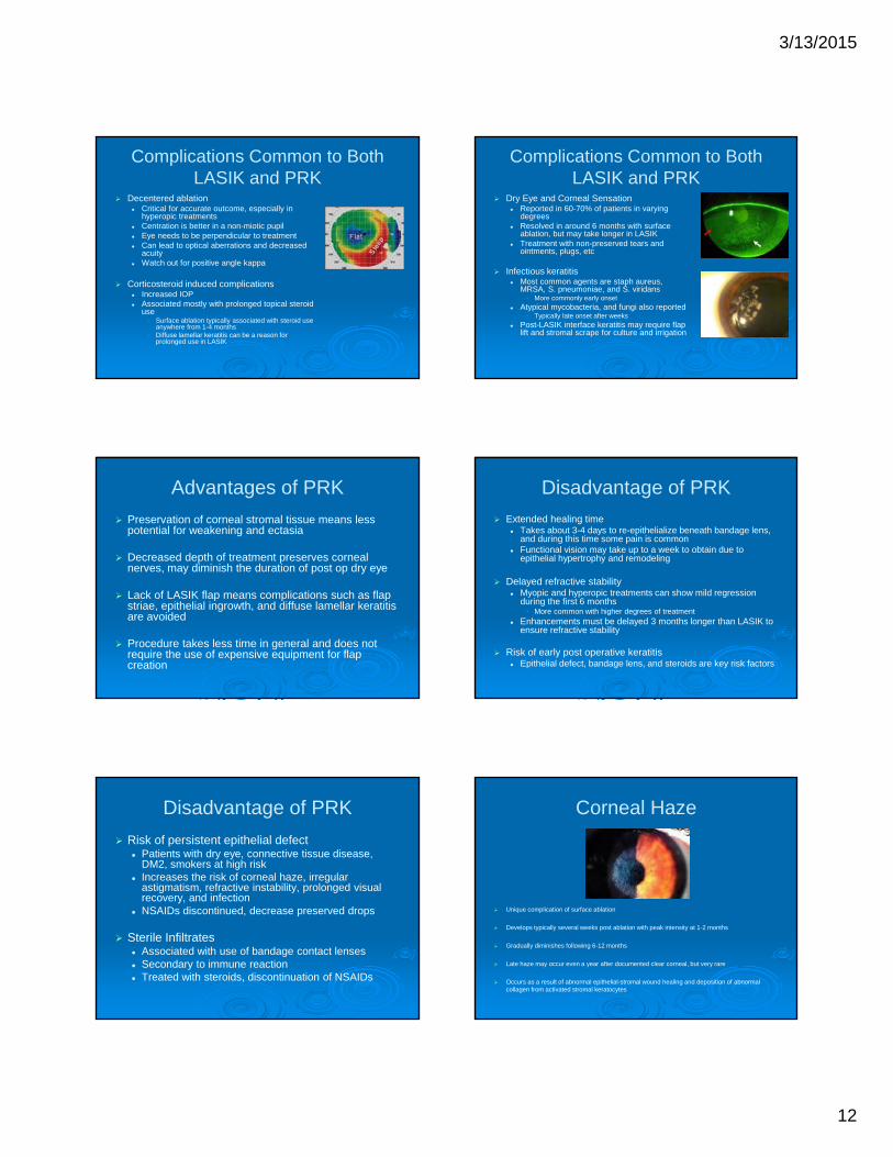
Central islands Area of central steepening on topography surrounded by area of
flattening Steepening of 1D in >1mm area Results in poor visual quality More common with older lasers
Optical aberrations Glares, halos, etc especially at night Most prevalent with smaller ablation zones and higher order of
treatment Exacerbated in dim lighting HOAs evaluated with wavefront mapping ? Correlation with pupil size
Complications Common to Both LASIK and PRK
3/13/2015
12
Decentered ablation Critical for accurate outcome, especially in
hyperopic treatments Centration is better in a non-miotic pupil Eye needs to be perpendicular to treatment Can lead to optical aberrations and decreased
acuity Watch out for positive angle kappa
Corticosteroid induced complications Increased IOP Associated mostly with prolonged topical steroid
use• Surface ablation typically associated with steroid use
anywhere from 1-4 months• Diffuse lamellar keratitis can be a reason for
prolonged use in LASIK
Complications Common to Both LASIK and PRK
Dry Eye and Corneal Sensation Reported in 60-70% of patients in varying
degrees Resolved in around 6 months with surface
ablation, but may take longer in LASIK Treatment with non-preserved tears and
ointments, plugs, etc
Infectious keratitis Most common agents are staph aureus,
MRSA, S. pneumoniae, and S. viridans• More commonly early onset
Atypical mycobacteria, and fungi also reported• Typically late onset after weeks
Post-LASIK interface keratitis may require flap lift and stromal scrape for culture and irrigation
Complications Common to Both LASIK and PRK
Advantages of PRK
Preservation of corneal stromal tissue means less potential for weakening and ectasia
Decreased depth of treatment preserves corneal nerves, may diminish the duration of post op dry eye
Lack of LASIK flap means complications such as flap striae, epithelial ingrowth, and diffuse lamellar keratitis are avoided
Procedure takes less time in general and does not require the use of expensive equipment for flap creation
Disadvantage of PRK
Extended healing time Takes about 3-4 days to re-epithelialize beneath bandage lens,
and during this time some pain is common Functional vision may take up to a week to obtain due to
epithelial hypertrophy and remodeling
Delayed refractive stability Myopic and hyperopic treatments can show mild regression
during the first 6 months• More common with higher degrees of treatment
Enhancements must be delayed 3 months longer than LASIK to ensure refractive stability
Risk of early post operative keratitis Epithelial defect, bandage lens, and steroids are key risk factors
Risk of persistent epithelial defect Patients with dry eye, connective tissue disease,
DM2, smokers at high risk Increases the risk of corneal haze, irregular
astigmatism, refractive instability, prolonged visual recovery, and infection
NSAIDs discontinued, decrease preserved drops
Sterile Infiltrates Associated with use of bandage contact lenses Secondary to immune reaction Treated with steroids, discontinuation of NSAIDs
Disadvantage of PRK Corneal Haze
Unique complication of surface ablation
Develops typically several weeks post ablation with peak intensity at 1-2 months
Gradually diminishes following 6-12 months
Late haze may occur even a year after documented clear corneal, but very rare
Occurs as a result of abnormal epithelial-stromal wound healing and deposition of abnormal collagen from activated stromal keratocytes
3/13/2015
13
Corneal Haze
Associated with greater amounts of correction and smaller ablation zones
Ultraviolet light (UVB) may play a critical role in prolonging the stromal healing process, leading to haze
UV blocking glasses and hats for 1 year are recommended
Prolonged steroids may be beneficial, particularly in patients with haze and undercorrection
Haze can be addressed surgically with superficial keratectomy or PTK coupled with MMC 0.02%
Reablation should be delayed at least 6-12 months to allow for clearance of haze
LASIK Complications Buttonhole flaps
Free flaps
Epithelial Defects
Corneal Perforation
LASIK flap tear
Microstriae/Macrostriae
Flap dislocation
Diffuse lamellar keratitis
Infectious lamellar keratitis
Epithelial ingrowth
Interface Debris
Post refractive ectasia
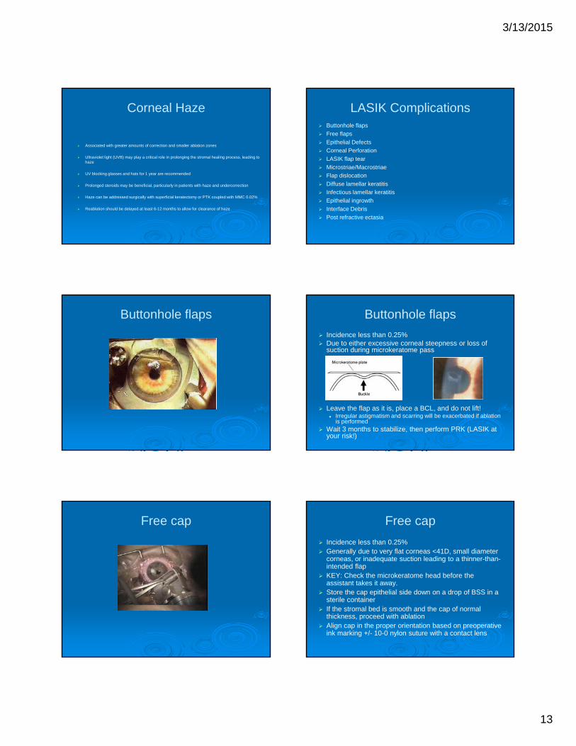
Buttonhole flaps Buttonhole flaps
Incidence less than 0.25% Due to either excessive corneal steepness or loss of
suction during microkeratome pass
Leave the flap as it is, place a BCL, and do not lift! Irregular astigmatism and scarring will be exacerbated if ablation
is performed Wait 3 months to stabilize, then perform PRK (LASIK at
your risk!)
Free cap Free cap
Incidence less than 0.25% Generally due to very flat corneas <41D, small diameter
corneas, or inadequate suction leading to a thinner-than-intended flap
KEY: Check the microkeratome head before the assistant takes it away.
Store the cap epithelial side down on a drop of BSS in a sterile container
If the stromal bed is smooth and the cap of normal thickness, proceed with ablation
Align cap in the proper orientation based on preoperative ink marking +/- 10-0 nylon suture with a contact lens
3/13/2015
14
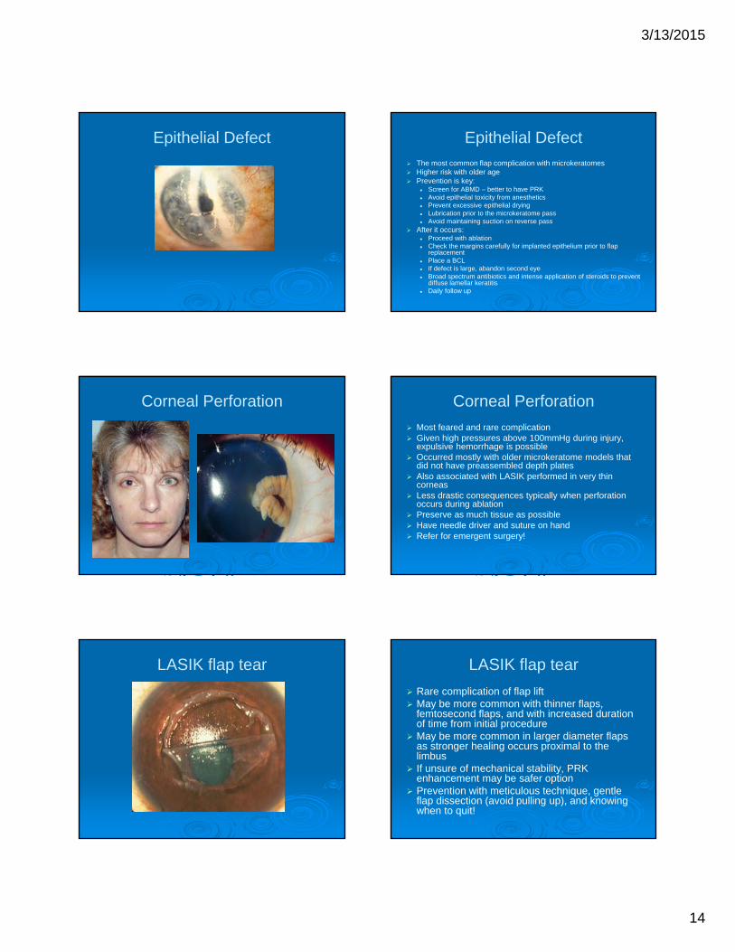
Epithelial Defect Epithelial Defect The most common flap complication with microkeratomes Higher risk with older age Prevention is key:
Screen for ABMD – better to have PRK Avoid epithelial toxicity from anesthetics Prevent excessive epithelial drying Lubrication prior to the microkeratome pass Avoid maintaining suction on reverse pass
After it occurs: Proceed with ablation Check the margins carefully for implanted epithelium prior to flap
replacement Place a BCL If defect is large, abandon second eye Broad spectrum antibiotics and intense application of steroids to prevent
diffuse lamellar keratitis Daily follow up
Corneal Perforation Corneal Perforation
Most feared and rare complication Given high pressures above 100mmHg during injury,
expulsive hemorrhage is possible Occurred mostly with older microkeratome models that
did not have preassembled depth plates Also associated with LASIK performed in very thin
corneas Less drastic consequences typically when perforation
occurs during ablation Preserve as much tissue as possible Have needle driver and suture on hand Refer for emergent surgery!
LASIK flap tear LASIK flap tear
Rare complication of flap lift May be more common with thinner flaps,
femtosecond flaps, and with increased duration of time from initial procedure
May be more common in larger diameter flaps as stronger healing occurs proximal to the limbus
If unsure of mechanical stability, PRK enhancement may be safer option
Prevention with meticulous technique, gentle flap dissection (avoid pulling up), and knowing when to quit!
3/13/2015
15
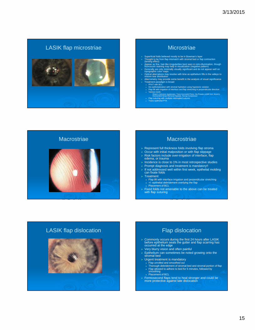
LASIK flap microstriae Microstriae Superficial folds believed mostly to be in Bowman’s layer Thought to be from flap mismatch with stromal bed or flap contraction
(tenting effect) Appear as fine, hair-like irregularities best seen in retro-illumination, though
fluoroscein staining may help in visualization (negative pattern) Generally are only minimally visually significant and do not appear well on
topographic color maps Optical aberrations may resolve with time as epithelium fills in the valleys to
restore tear distribution Aberrometry may provide some benefit in the analysis of visual significance Treatment paradigm is broad:
PFAT with BCL De-epithelialization with stromal hydration using hypotonic solution Flap lift with irrigation of interface and flap stretching in perpendicular direction Various tools:
• Rhein's Johnston Applanator, Tress Kornmehl Press, the Pineda LASIK Iron, Acorn's Donnenfeld Striae Removal Spatula, Herzig Compressor
Flap suturing with multiple interrupted sutures Trans-epithelial PTK
Macrostriae Macrostriae
Represent full thickness folds involving flap stroma Occur with initial malposition or with flap slippage Risk factors include over-irrigation of interface, flap
edema, or trauma Incidence is close to 1% in most retrospective studies Prompt diagnosis and treatment is mandatory!! If not addressed well within first week, epithelial molding
can fixate folds Treatment
Flap lift with interface irrigation and perpendicular stretching +/- epithelial debridement overlying the flap Placement of BCL
Fixed folds not amenable to the above can be treated with flap suturing
LASIK flap dislocation Flap dislocation
Commonly occurs during the first 24 hours after LASIK before epithelium seals the gutter and flap scarring has occurred at the edge
Very blurry vision and often painful Epithelium can sometimes be noted growing onto the
stromal bed Urgent treatment is mandatory
Flap unrolled and smoothed out Thorough debridement of stromal bed and stromal portion of flap Flap allowed to adhere to bed for 5 minutes, followed by
smoothing Placement of BCL
Femtosecond flaps tend to heal stronger and could be more protective against late dislocation
3/13/2015
16
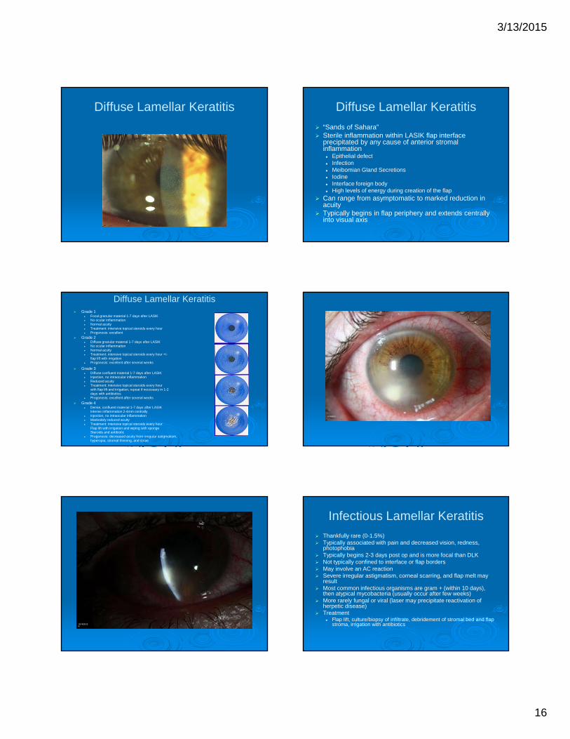
Diffuse Lamellar Keratitis Diffuse Lamellar Keratitis
“Sands of Sahara” Sterile inflammation within LASIK flap interface
precipitated by any cause of anterior stromal inflammation Epithelial defect Infection Meibomian Gland Secretions Iodine Interface foreign body High levels of energy during creation of the flap
Can range from asymptomatic to marked reduction in acuity
Typically begins in flap periphery and extends centrally into visual axis
Diffuse Lamellar Keratitis Grade 1
Focal granular material 1-7 days after LASIK No ocular inflammation Normal acuity Treatment: intensive topical steroids every hour Progsnosis: excellent
Grade 2 Diffuse granular material 1-7 days after LASIK No ocular inflammation Normal acuity Treatment: intensive topical steroids every hour +/-
flap lift with irrigation Progsnosis: excellent after several weeks
Grade 3 Diffuse confluent material 1-7 days after LASIK Injection, no intraocular inflammation Reduced acuity Treatment: intensive topical steroids every hour
with flap lift and irrigation, repeat if necessary in 1-2days with antibiotics
Progsnosis: excellent after several weeks
Grade 4 Dense, confluent material 1-7 days after LASIK
Intense inflammation 2-4mm centrally Injection, no intraocular inflammation Markedely reduced acuity Treatment: intensive topical steroids every hour
Flap lift with irrigation and wiping with spongeSteroids and antibiotic
Progsnosis: decreased acuity from irregular astigmatism,hyperopia, stromal thinning, and striae
Infectious Lamellar Keratitis Thankfully rare (0-1.5%) Typically associated with pain and decreased vision, redness,
photophobia Typically begins 2-3 days post op and is more focal than DLK Not typically confined to interface or flap borders May involve an AC reaction Severe irregular astigmatism, corneal scarring, and flap melt may
result Most common infectious organisms are gram + (within 10 days),
then atypical mycobacteria (usually occur after few weeks) More rarely fungal or viral (laser may precipitate reactivation of
herpetic disease) Treatment
Flap lift, culture/biopsy of infiltrate, debridement of stromal bed and flap stroma, irrigation with antibiotics

3/13/2015
17
Epithelial Ingrowth Epithelial Ingrowth Growth of epithelium within interface either from the flap edge or
from implanted epithelium during flap manipulation More commonly occurs with ABMD, patients of older age, epithelial
defects, after multiple enhancements, or with misalignment of flap edge
Peripheral 1-2mm usually inconsequential Pattern of growth includes nests, strands, pearls, or sheets Growth into axis can generate some irregular astigmatism Rarely, nutritional deprivation of flap stroma can precipitate melt Treatment in progressive or symptomatic cases requires flap lift,
scraping of stromal bed as well as flap stroma Flap suturing or gluing may be employed in recurrent cases,
particularly if a melt has occurred at the edge
Keratolysis after epithelial ingrowth Interface Debris
Interface debris
Commonly seen Origins include lint from clothing, metal particles from
surgical instruments, blood from limbal bleeding, and meibomian secretions
Generally well tolerated and not visually significant Materials that generate significant inflammatory
reactions, ie dense blood or large fibers, need to be removed via flap lift to prevent DLK
Fibers noted at the flap edge which could provide a path for epithelial ingrowth need to be removed immediately
Prevention is key: Proper draping of lids and lashes Decreased surgical time Adequate interface irrigation prior to flap alignment Sterile/clean operating suite
Postoperative Keratectasia
3/13/2015
18
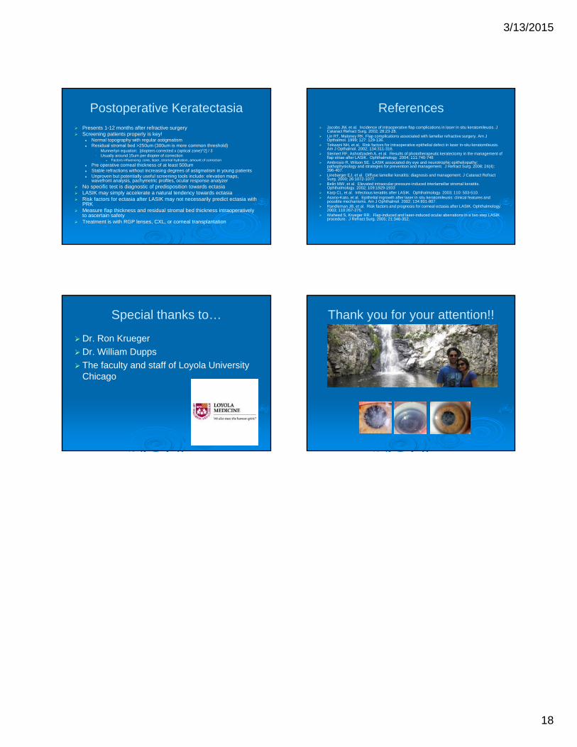
Postoperative Keratectasia Presents 1-12 months after refractive surgery Screening patients properly is key!
Normal topography with regular astigmatism Residual stromal bed >250um (300um is more common threshold)
• Munnerlyn equation: [diopters corrected x (optical zone)^2] / 3• Usually around 15um per diopter of correction
Factors influencing: zone, laser, stromal hydration, amount of correction
Pre operative corneal thickness of at least 500um Stable refractions without increasing degrees of astigmatism in young patients Unproven but potentially useful screening tools include: elevation maps,
wavefront analysis, pachymetric profiles, ocular response analyzer No specific test is diagnostic of predisposition towards ectasia LASIK may simply accelerate a natural tendency towards ectasia Risk factors for ectasia after LASIK may not necessarily predict ectasia with
PRK Measure flap thickness and residual stromal bed thickness intraoperatively
to ascertain safety Treatment is with RGP lenses, CXL, or corneal transplantation
References Jacobs JM, et al. Incidence of intraoperative flap complications in laser in situ keratomileusis. J
Cataract Refract Surg. 2002; 28:23-28. Lin RT, Maloney RK. Flap complications associated with lamellar refractive surgery. Am J
Opthalmol. 1999; 127: 129-136. Tekwani NH, et al. Risk factors for intraoperative epithelial defect in laser in-situ keratomileusis.
Am J Opthalmol. 2002; 134:311-316. Steinert RF, Ashrafzadeh A, et al. Results of phototherapeutic keratectomy in the management of
flap striae after LASIK. Ophthalmology. 2004; 111:740-746 Ambrosio R, Wilson SE. LASIK associated dry eye and neurotrophic epitheliopathy:
pathophysiology and strategies for prevention and management. J Refract Surg. 2008; 24(4): 396-407.
Linebarger EJ, et al. Diffuse lamellar keratitis: diagnosis and management. J Cataract Refract Surg. 2000; 26:1072-1077.
Belin MW, et al. Elevated intraocular pressure-induced interlamellar stromal keratitis. Ophthalmology. 2002; 109:1929-1933
Karp CL, et al. Infectious keratitis after LASIK. Ophthalmology. 2003; 110: 503-510. Asano-Kato, et al. Epithelial ingrowth after laser in situ keratomileusis: clinical features and
possible mechanisms. Am J Ophthalmol. 2002; 134:801-807. Randleman JB, et al. Risk factors and prognosis for corneal ectasia after LASIK. Ophthalmology.
2003; 110:267-275. Waheed S, Krueger RR. Flap-induced and laser-induced ocular aberrations in a two step LASIK
procedure. J Refract Surg. 2005; 21:346-352.
Special thanks to…
Dr. Ron Krueger
Dr. William Dupps
The faculty and staff of Loyola University Chicago
Thank you for your attention!!