reelin mouse mutants as models of cortical development disorders
TRANSCRIPT
www.elsevier.com/locate/yebeh
Epilepsy & Behavior 8 (2006) 81–90
Review
Reelin mouse mutants as models of cortical development disorders
Gabriella D’Arcangelo *
The Cain Foundation Laboratories, Texas Children�s Hospital, Departments of Pediatrics,
Neuroscience and Programs in Developmental Biology and Translational Biology and Molecular Medicine,
Baylor College of Medicine, Houston, TX 77030, USA
Received 17 June 2005; revised 9 September 2005; accepted 10 September 2005Available online 2 November 2005
Abstract
Developmental defects in neuronal positioning and synaptic connectivity are commonly found in neurological diseases, and they arebelieved to underlie many cognitive and affective disorders. Several mouse mutants are currently available that model at least someaspects of human developmental brain disorders. With the identification of the genes mutated in these animals and the study of the cel-lular basis of the phenotypes, we have taken significant strides toward an understanding of the mechanisms controlling proper braindevelopment and the consequences of their dysfunction. In particular, mouse mutants deficient in the Reelin gene have provided valuableinsights into the mechanisms of cortical development. Absence of Reelin expression in the spontaneous mutant mouse reeler leads toextensive defects in neuronal position and dendrite development. In humans, loss of Reelin results in a type of lissencephaly with severecortical and cerebellar malformation. Genetic and biochemical studies using mouse mutants suggest that the Lis1 protein may participatein the Reelin signaling pathway controlling cortical development. Reduced levels of Reelin are also present in postmortem brains ofpatients with schizophrenia, suggesting a possible link with this cognitive disorder. The regulation of the Reelin gene may thus provideinsights into the mechanisms of this disease.� 2005 Elsevier Inc. All rights reserved.
Keywords: Reeler; Disabled-1; Very low density lipoprotein receptor; Apolipoprotein E receptor 2; Platelet-activating factor acetylhydrolase 1b1;Hydrocephalus; Epilepsy; Cerebral cortex; Schizophrenia
1. Introduction
Homozygous mutations in the Reelin (Reln) gene resultin a severe disruption of brain development in humans andin rodents. The gene encodes a large extracellular glycopro-tein that bears little similarity to other known proteins,reflecting its unique properties in the control of brain devel-opment. The mouse mutant reeler has been studied exten-sively as a model for understanding the molecular andcellular mechanisms governing the orderly developmentof cortical structures (see [1–3] for comprehensive reviews).In this mutant, cellular layer formation in the cerebral cor-tex, hippocampus, and cerebellum goes awry and ectopic
1525-5050/$ - see front matter � 2005 Elsevier Inc. All rights reserved.
doi:10.1016/j.yebeh.2005.09.005
* Fax: +1 832 825 4217.E-mail address: [email protected].
neurons can be found in most anatomical structures ofthe central nervous system. The loss of cellular organiza-tion in the cerebellum of reeler mice results in severe hypo-plasia, which in turn causes an ataxic phenotypecharacterized by tremors, dystonia, and a reeling gate. Sim-ilar behavioral and anatomical traits are present in mutantmice in which essential components of the Reelin signalingpathway are disrupted. This pathway includes two high-af-finity Reelin receptors, the very low density lipoproteinreceptor (VLDLR) and the apolipoprotein E receptor 2(ApoER2) [4,5], and an adapter molecule called Dis-abled-1 (Dab1) (Fig. 1). Binding of Reelin to its receptorscauses clustering [6] and promotes the phosphorylation ofDab1 on specific tyrosine residues through the activationof src-family kinases [7–11]. Additional components ofthe signaling cascade have recently been identified, buttheir role in mediating Reelin function in cortical layer
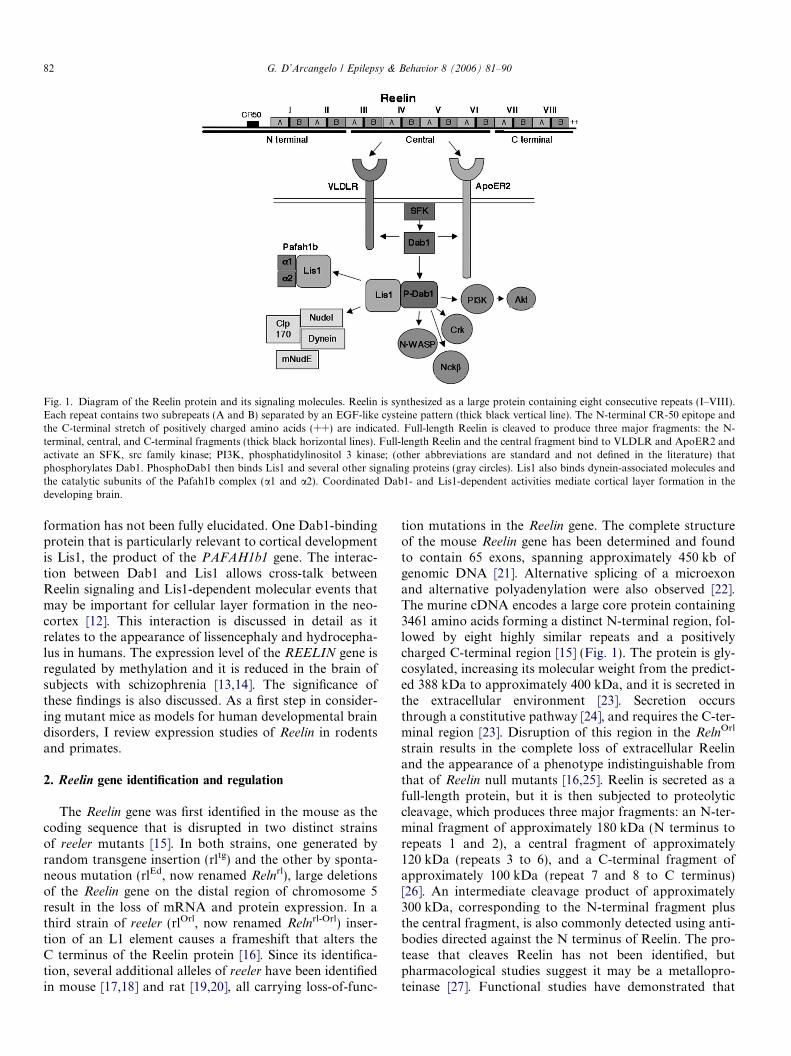
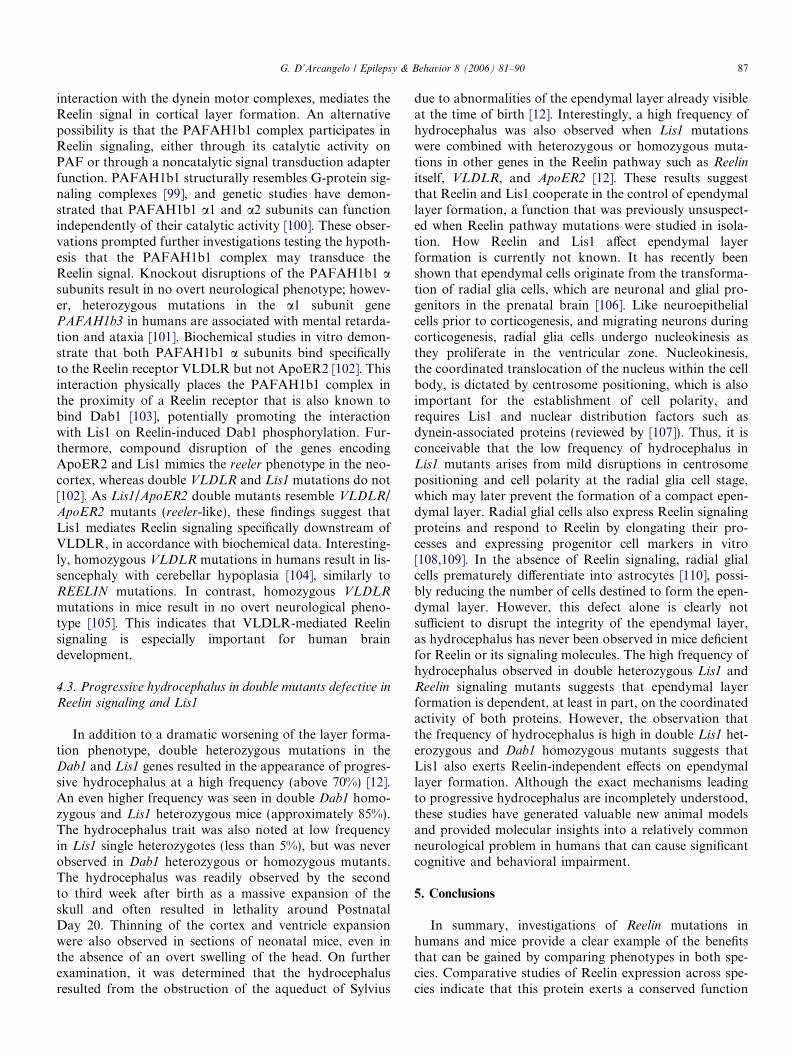
Fig. 1. Diagram of the Reelin protein and its signaling molecules. Reelin is synthesized as a large protein containing eight consecutive repeats (I–VIII).Each repeat contains two subrepeats (A and B) separated by an EGF-like cysteine pattern (thick black vertical line). The N-terminal CR-50 epitope andthe C-terminal stretch of positively charged amino acids (++) are indicated. Full-length Reelin is cleaved to produce three major fragments: the N-terminal, central, and C-terminal fragments (thick black horizontal lines). Full-length Reelin and the central fragment bind to VLDLR and ApoER2 andactivate an SFK, src family kinase; PI3K, phosphatidylinositol 3 kinase; (other abbreviations are standard and not defined in the literature) thatphosphorylates Dab1. PhosphoDab1 then binds Lis1 and several other signaling proteins (gray circles). Lis1 also binds dynein-associated molecules andthe catalytic subunits of the Pafah1b complex (a1 and a2). Coordinated Dab1- and Lis1-dependent activities mediate cortical layer formation in thedeveloping brain.
82 G. D’Arcangelo / Epilepsy & Behavior 8 (2006) 81–90
formation has not been fully elucidated. One Dab1-bindingprotein that is particularly relevant to cortical developmentis Lis1, the product of the PAFAH1b1 gene. The interac-tion between Dab1 and Lis1 allows cross-talk betweenReelin signaling and Lis1-dependent molecular events thatmay be important for cellular layer formation in the neo-cortex [12]. This interaction is discussed in detail as itrelates to the appearance of lissencephaly and hydrocepha-lus in humans. The expression level of the REELIN gene isregulated by methylation and it is reduced in the brain ofsubjects with schizophrenia [13,14]. The significance ofthese findings is also discussed. As a first step in consider-ing mutant mice as models for human developmental braindisorders, I review expression studies of Reelin in rodentsand primates.
2. Reelin gene identification and regulation
The Reelin gene was first identified in the mouse as thecoding sequence that is disrupted in two distinct strainsof reeler mutants [15]. In both strains, one generated byrandom transgene insertion (rltg) and the other by sponta-neous mutation (rlEd, now renamed Relnrl), large deletionsof the Reelin gene on the distal region of chromosome 5result in the loss of mRNA and protein expression. In athird strain of reeler (rlOrl, now renamed Relnrl-Orl) inser-tion of an L1 element causes a frameshift that alters theC terminus of the Reelin protein [16]. Since its identifica-tion, several additional alleles of reeler have been identifiedin mouse [17,18] and rat [19,20], all carrying loss-of-func-
tion mutations in the Reelin gene. The complete structureof the mouse Reelin gene has been determined and foundto contain 65 exons, spanning approximately 450 kb ofgenomic DNA [21]. Alternative splicing of a microexonand alternative polyadenylation were also observed [22].The murine cDNA encodes a large core protein containing3461 amino acids forming a distinct N-terminal region, fol-lowed by eight highly similar repeats and a positivelycharged C-terminal region [15] (Fig. 1). The protein is gly-cosylated, increasing its molecular weight from the predict-ed 388 kDa to approximately 400 kDa, and it is secreted inthe extracellular environment [23]. Secretion occursthrough a constitutive pathway [24], and requires the C-ter-minal region [23]. Disruption of this region in the RelnOrl
strain results in the complete loss of extracellular Reelinand the appearance of a phenotype indistinguishable fromthat of Reelin null mutants [16,25]. Reelin is secreted as afull-length protein, but it is then subjected to proteolyticcleavage, which produces three major fragments: an N-ter-minal fragment of approximately 180 kDa (N terminus torepeats 1 and 2), a central fragment of approximately120 kDa (repeats 3 to 6), and a C-terminal fragment ofapproximately 100 kDa (repeat 7 and 8 to C terminus)[26]. An intermediate cleavage product of approximately300 kDa, corresponding to the N-terminal fragment plusthe central fragment, is also commonly detected using anti-bodies directed against the N terminus of Reelin. The pro-tease that cleaves Reelin has not been identified, butpharmacological studies suggest it may be a metallopro-teinase [27]. Functional studies have demonstrated that
G. D’Arcangelo / Epilepsy & Behavior 8 (2006) 81–90 83
the central fragment of Reelin is sufficient to confer biolog-ical and biochemical activity. Addition of a recombinantprotein corresponding to Reelin repeats 3–6 to the culturemedium was capable of inducing Dab1 phosphorylation indissociated cortical neurons and partially rescued layer for-mation in reeler cortical slices [28]. However, the full-lengthprotein appeared to be more active than the central frag-ment alone. Because an N-terminal epitope recognized bythe CR-50 epitope mediates the formation of large Reelinaggregates [23,29], it is conceivable that this region confersmaximum biological activity, perhaps by facilitating Reelinreceptor clustering [6], which may lead to a stronger signalto the target cells.
Soon after identification of the murine gene, the humanREELIN gene was isolated and sequenced [30]. The generesides on chromosome 7q22 and it encodes a protein com-posed of 3460 amino acids that is 94% identical to themouse protein, strongly suggesting a conserved function.Based on its sequence similarity to the mouse 5 0UTR, thehuman REELIN promoter region was cloned [13]. Thisregion contains canonical GC boxes, which are bindingsites for basal transcription factor such as Sp1, and T-box elements capable of binding Tbr1. This factor caninduce expression from the REELIN promoter in the pres-ence of the membrane-associated guanylate kinase CASK.As Tbr1 mutant mice exhibit reduced Reelin expressionand reeler-like cortical layer defects [31], it is likely thatthese regulatory elements are crucial for gene expressionin vivo.
The REELIN promoter also contains a large CpGisland, suggesting that it may be regulated by methylation.Indeed, functional studies in culture demonstrated that thelevels of mRNA transcription inversely correlate with lev-els of promoter methylation, with high levels of REELINexpression detected in transfected cells in the presence ofmethylation inhibitors, and in neural progenitor cells treat-ed with retinoic acid to induce neuronal differentiation [13].Methionine treatment also was found to suppress Reelingene expression in mice in vivo, strongly suggesting thatthis promoter is under epigenetic control [32]. A similarmechanism also appears to control expression of theGAD67 gene in adult inhibitory cortical neurons. Down-regulation of the Reelin and GAD67 promoters in theseneurons was shown to be mediated by the methylatingenzyme DNA methyltransferase 1 (Dnmt1) [33]. Thesefindings are intriguing because downregulation of REELINand GAD67 gene expression was reported in GABAergiccortical interneurons of the postmortem brains of personswith schizophrenia, which also expressed increased levelsof Dnmt1 [14,34]. Proteins that bind methylated DNAare known to recruit repressor complexes and the histonedeacetylase complex (HDAC). Interestingly, HDAC inhib-itors, including the mood-stabilizing drug valproic acid,were found to induce Reelin expression [32]. These findingssuggest a link between schizophrenia and Reelin gene regu-lation in GABAergic neurons. Indeed, a recent study dem-onstrated that hypermethylation of the CpG islands in the
REELIN promoter is present at significantly higher levelsin postmortem brains of schizophrenic patients, thus pro-viding a mechanism for the observed decrease in geneexpression [35]. Although at present it is not clear whetherdecreased REELIN gene expression represents a risk factorfor the disease or is a symptom of interneuron dysfunctionin schizophrenia, these gene expression studies point topromoter methylation in GABAergic neurons as a poten-tial disease mechanism and may lead to new treatmentoptions for this disorder (reviewed by [36]).
The Reelin gene is conserved among vertebrates butabsent in invertebrates and plants. To date, homologousReelin sequences have been identified and their expressionhas been characterized in the brain of developing and adultrodents and primates [37–39]. In addition, Reelin expres-sion has been documented in the brain of a number ofother vertebrate species including adult ferret [40], zebrafish [41], sea lamprey [42], and embryonic chicken, croco-dile, lizard, and turtle [43–46]. Comparative studies haveindicated that Reelin is expressed by a variety of neuronsin the adult vertebrate pallium, suggesting that, in additionto playing a crucial role in embryonic cortical development,Reelin may also function in the adult neocortex [38,47].The cell identity and laminar position of Reelin-expressingcells in the cortex vary somewhat among species, althoughall species express high levels of Reelin in marginal layers, apattern that probably originated in ancestral amniotes. Theevolutionary significance of these findings may lie in thecorrelation of Reelin expression with brain architectonicpatterns. For example, amplification of Reelin-expressingneuronal populations in mammals may reflect a need tofacilitate the formation of a synaptic circuitry in a cortexthat is developing according to an inside-out gradient,unlike in lower vertebrates [48,49]. In this review, I focuson the expression pattern of Reelin in the cerebral cortexof rodents and primates.
3. Reelin expression in the cortex of primates and rodents
3.1. Corticogenesis
Reelin mRNA expression has been systematically stud-ied in the developing mouse brain. In situ hybridizationstudies using radioactive [50] or digoxygenin-labeled ribo-probes [51] revealed that in the mouse brain, Reelin is firstexpressed around Embryonic Day 10 in differentiatingfields of the telencephalon, diencephalon, and rhomboen-cephalon, and it is not expressed in germinal layers. Duringcorticogenesis in the mouse (from Embryonic Day 11 to theearly postnatal period) Reelin is expressed prominently inthe marginal zone [15]. The identification of Cajal-Retziuscells as the major Reelin-producing cells in the marginalzone of the cerebral cortex and hippocampus in mice andrats is based on their location, time of appearance, distinc-tive horizontal bipolar morphology, co-expression ofcalretinin and p75 markers [51,52], and immunodetectionwith CR-50, a monoclonal antibody directed against the
84 G. D’Arcangelo / Epilepsy & Behavior 8 (2006) 81–90
N-terminal region of Reelin [23,53]. It has recently beendemonstrated that Cajal-Retzius cells originate mostlyfrom the caudomedial wall of the telencephalon, includingthe cortical hem [54], and invade the marginal zone of theneocortex by tangential migration.
In mice and rats, Reelin expression is fairly similar; how-ever, in the rat cerebral cortex, the expression patternappears to be more complex [52]. The rat marginal zonecontains, in addition to Cajal-Retzius cells, subpial granu-lar layer (SGL) cells, a cell population that is more abun-dant and better characterized in the marginal zone of theprimate cortex. Both cell types were found to expressReelin during corticogenesis. In the primate and humancortex, the contribution of SGL cells to Reelin expressionis even more significant. In humans, REELIN expressionis detectable in Cajal-Retzius cells from Gestational Week(GW) 11 [55,56]. From GW14 onward, however, SGL cellsinvade the marginal zone and express significant amountsof Reelin in this layer [55]. A similar pattern is observedin the cortex of monkeys, where Reelin is expressed inthe marginal zone by large Cajal-Retzius cells at early stag-es and by smaller SGL cells at later stages of corticogenesis[57]. The presence of an abundant SGL population express-ing Reelin in the marginal zone of higher vertebrates maycompensate for the gradual loss of Cajal-Retzius cellsand enable neuronal organization during the prolongedperiod of corticogenesis in these species.
The detailed analysis of Reelin expression, in combina-tion with in vitro assays, provides an essential frameworkto understand its function. Because Reelin is expressedmostly in the most superficial layer of the neocortex duringcorticogenesis, it has been suggested that it may regulatethe development of the cortical plate by acting as a stopsignal for radially migrating neurons. This view is support-ed by functional studies indicating that the addition ofexogenous Reelin induces neuron migration arrest anddetachment from radial glia [58]. However, in reeler late-born neurons terminate ectopically in deep layers of thecortex without invading the marginal zone, and in wildtype, lower levels of Reelin mRNA are also detected indeeper cortical layers (prospective layer V–VI) at lateembryonic and early postnatal ages, when late-born neu-rons are still migrating toward the upper II/III layers [51](see also [59]). Similarly, ectopic expression of Reelin
mRNA driven from the nestin promoter in the ventricularzone of transgenic mice did not cause migration arrest andpartially rescued the reeler cortical layer phenotype [60].Together, these observations raise doubts that Reelinmay function as a stop signal, although expression levelsof Reelin protein in the middle of the developing corticalplate or in the ventricular zone of transgenic mice are solow as to fall below the detection level of common immu-nological techniques. Recent studies using the powerful inutero electroporation technique [61] have shown that theReelin signal transducer Dab1 is required in migrating neu-rons to extend the leading edge across the cortical plate[62]. These findings strongly suggest that Reelin is a permis-
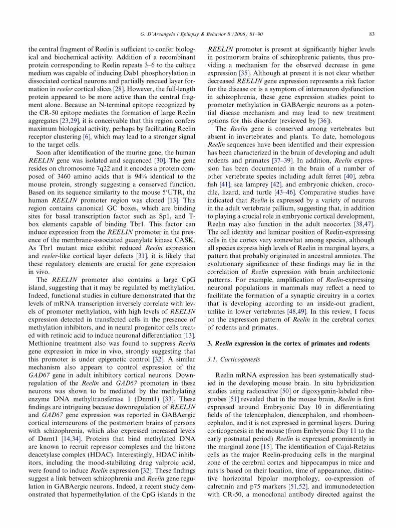
sive factor promoting neuronal migration toward the sur-face of the cortex. Taking into account all currentlyavailable data, I propose that a gradient of extracellularReelin may be present in the developing cortex and thatReelin may exert different cellular effects depending on itsconcentration (Fig. 2). At low levels present within the cor-tical plate, Reelin may favor the extension of the neuronleading edge and thus promote upward migration. At highlevels present only in the marginal zone, Reelin may causedetachment from radial glia and thus neuronal migrationarrest. Thus, the behavior of a cortical neuron may dependon the distribution of extracellular Reelin in the radialdimension of the cortex. Direct visualization of the Reelingradient with more sensitive approaches than simple immu-nofluorescence techniques is necessary to corroborate thisidea.
3.2. Postnatal and adult expression
Reelin expression persists in the postnatal and adult cor-tex well after most Cajal-Retzius cells have disappearedand corticogenesis is completed. In the late postnatal andadult mouse cortex, Reelin mRNA is detected exclusivelyin GABAergic neurons throughout all cortical layers [51].Variable fractions of the Reelin-expressing neurons alsoco-express other markers of interneuron subpopulations,such as calbindin, calretinin, neuropeptide Y, and somato-statin. Similarly, in the prenatal and adult rat cortex,Reelin is detected in many, but not all, GABAergic neu-rons, which were classified by morphological criteria ashorizontal cells in layer I, bitufted neurons in layers II–V,and occasionally Martinotti cells in deep layers [52,63,64].Detailed studies of Reelin expression in the adult humanand nonhuman primate cortex revealed that, similarly torodents, cells expressing high levels of Reelin were presentin layer I, corresponding to surviving Cajal-Retzius andSGL cells [37,56]. Surprisingly, however, many pyramid-shaped principal neocortical neurons in the primate cortexwere found to be lightly Reelin-immunoreactive [38]. In themacaque cortex, Reelin was shown to be present in the neu-ropil as well as in some axonal pathways and their terminalarborizations, suggesting that it can be axonally transport-ed over long distances [37]. Reelin expression in someaxonal tracts was also previously noted in the mouse [51].Unlike rodents, however, in the primate cortex Reelin isco-expressed with its transducing molecule Dab1 in someprincipal cortical neurons [56] and Cajal-Retzius cells[65], suggesting a possible autocrine function that may beunique to primate neurons. The function of Reelin in thepostnatal cortex is not known, but its expression and neu-ropil localization suggest a possible function in neuronalmaturation or in the development and maintenance ofthe synaptic circuitry of the brain. It has long been knownthat dendrites are severely disrupted in ectopic cortical,hippocampal, and cerebellar neurons of reeler mice [66–68]. This phenotype is attributable in part to the abnormalposition and the consequent abnormal innervation of these

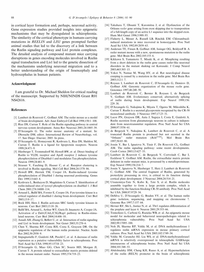
Fig. 2. Hypothetical model of Reelin function in corticogenesis. During embryonic corticogenesis, Cajal-Retzius cells (CR) in the marginal zone of normalmice or humans secrete Reelin in the extracellular environment. Extracellular Reelin may form a gradient in the radial dimension of the developingneocortex. High concentrations of Reelin are present in the marginal zone, whereas low concentrations are present in the middle of the developing corticalplate due to diffusion of Reelin or expression by cell types other than Cajal-Retzius cells. Migrating neurons (in red) in wild type or reeler mice begin theirupward migration normally along radial fibers (step 1). In the wild-type cortex, neurons extend their leading edge in response to low levels of Reelin, thusmoving efficiently toward the surface, whereas in the reeler cortex they fail to do so (step 2). When neurons in the normal cortex approach the marginalzone they encounter high concentrations of Reelin that induce their detachment from radial fibers and cause migration arrest (step 3). This step also fails inthe reeler cortex, and neurons remain in close apposition to the radial fibers. Finally, neurons in the normal cortex retain the attachment to the Reelin-richmarginal layer that enables them to maintain proper orientation and elaborate complex apical dendrites (step 4). Instead, in the reeler cortex, neurons losetheir orientation and develop poor dendrites due to the lack of Reelin.
G. D’Arcangelo / Epilepsy & Behavior 8 (2006) 81–90 85
neurons in mutant cortical structures. Recent studies inhippocampal cultures demonstrated that Reelin also direct-ly promotes dendrite maturation and growth and, thus,may enable the formation of a normal complement of syn-aptic contacts [69]. In the hippocampus, a reduction in thenumber of synaptic contacts between entorhinal axon ter-minals and their target cells was reported [70], althoughthis is likely to be a secondary consequence of the reducedbranching of this particular projection pathway in thedeveloping reeler hippocampus [71]. However, reducedspine density was also noted in heterozygous reeler mice,in the absence of cellular ectopia [72]. Despite these tanta-lizing reports, the ability of Reelin to directly promote syn-apse development has not yet been demonstrated. The factthat Reelin is most highly expressed in brain regions whereadult plasticity takes place, such as the olfactory bulb andthe dentate gyrus, suggests that Reelin may directly regu-late synaptogenesis or synaptic function. Electron micros-copy studies in nonhuman primates have furthersuggested a role of postnatal Reelin in synaptic develop-ment or function, as extracellular Reelin was found toaccumulate at the postsynaptic density [73]. In mice hippo-campal slices, addition of exogenous Reelin was found topromote long-term potentiation (LTP), a learning para-digm [74]. Lately, it was demonstrated that Reelin also pro-motes associative learning, and that this function ismediated through a specific isoform of ApoER2 capable
of interacting with the NMDA receptor [75]. These dataraise the possibility that Reelin modulates synaptic func-tion throughout the postnatal brain, and may have poten-tial implications for schizophrenia, a disease that, asdiscussed above, is associated with lower levels of Reelinexpression.
4. Reelin and lissencephaly
4.1. reeler phenotypes in mice and men
Disruption of the REELIN gene in humans results in lis-sencephaly with cerebellar hypoplasia (LCH) [76]. Thesepatients display severe ataxia, epilepsy, and cognitive delay.The human phenotype is recessive and is reminiscent of thatof homozygous reeler mice, particularly with respect to theappearance of the cerebellum and cerebellum-related atax-ia. However, there are notable differences. In the humancerebral cortex, REELIN mutations result in lissencephaly(reduction in the number of convolutions), but detailedanalysis of layer formation is currently not available. Thecortical lissencephaly phenotype cannot be assessed in micebecause the cortex of these animals normally has no convo-lutions. Furthermore, reeler mice have no seizures,although this could be due to the fact that the strain is main-tained on a genetic background particularly resistant to sei-zures. Despite their limitations, molecular studies described
86 G. D’Arcangelo / Epilepsy & Behavior 8 (2006) 81–90
below clearly demonstrate that reeler mice are valid animalmodels for understanding both neuronal migration and theorigin of human lissencephaly.
Mutations in the LIS1 gene in humans (also known asPAFAH1b1) result in isolated lissencephaly sequence(ILS), an autosomal dominant disease characterized byreduction in the number of cortical gyri, but no major cer-ebellar involvement, associated with severe epilepsy andcognitive delay [77,78]. Lissencephaly can occur in combi-nation with craniofacial abnormalities in the Miller–Diekersyndrome, which is due to a deletion of a region of chromo-some 17 including the LIS1 and 14–3–3 genes [79,80]. Ana-tomically, the lissencephaly in heterozygous LIS1 humanpatients is thought to result from reduced migration of cor-tical neurons from the ventricular zone, where they areborn, into the cortical plate. Unlike humans, heterozygousLis1 null mice display no apparent disruption in corticaldevelopment and no seizures, and homozygous mice areearly embryonic lethal [81]. Thus, heterozygous Lis1
mutant mice do not exactly recapitulate the phenotypeobserved in humans. However, compound null Lis1
mutants expressing less than 35% of the normal proteinand heterozygous mice expressing a truncated Lis1 protein(sLis) similar to that found in some human patients do dis-play overt neuronal migration defects in the cortex [81–83].Also, abnormalities in the rate of neuronal migration areobserved in vitro using cerebellar granule cells obtainedfrom heterozygous Lis1 mice. Therefore, these mutantscan be used to investigate the migration defects that under-lie the etiology of human lissencephaly. It is estimated thatapproximately 60% of cases of classic lissencephaly are dueto deletions or mutations in the LIS1 gene [84]. Theremaining cases are attributable to mutations in the X-linked Doublecortin gene or other unidentified genes. Dou-blecortin mutations in males cause lissencephaly, similar toLIS1 mutations. In females, however, Doublecortin muta-tions result in neuronal heterotopia, which appears as adouble cortex [85]. The subcortical band heterotopia isdue to random inactivation of the X chromosome, whichgives rise to neurons that either migrate normally to thecortical plate or terminate prematurely in the ectopic band.Unfortunately, this defect cannot be modeled in animals,as doublecortin knockout mice display no apparent pheno-type in the neocortex [86].
4.2. Reelin and Lis1 come together
Anatomically, the cerebral cortexes in patients with het-erozygous Lis1 and homozygous REELIN mutations areremarkably similar in appearance, suggesting that thetwo proteins may share common functions in neuronalmigration. The similarity of the cortical phenotype inhumans was not predicted based on the analysis of corre-sponding animal models, due in part to the fact that Lis1homozygous mutants die prior to gastrulation and thuscould not be compared with reeler homozygous mice. Also,neither heterozygous Lis1 nor heterozygous Reelin mouse
mutants have defects in cortical layer formation. However,genetic and biochemical studies demonstrated Reelin andLis1 activities are indeed functionally linked. Genetic inter-action between the Reelin effector Dab1 and Lis1 was dem-onstrated by the appearance of a cortical layer phenotypein Lis1 and Dab1 double heterozygous mice that is notapparent in single mutants [12]. Biochemical experimentsfurther clarified the nature of this interaction and demon-strated that Lis1 binds to Dab1 in a Reelin-dependentfashion [12] (Fig. 1). These proteins can be co-immunopre-cipitated from the brain lysates of normal mice, in whichReelin induces phosphorylation of Dab1 on several tyro-sine residues [7–10]. Far less co-immunoprecipitation wasdetected from brain lysates of reeler mice in which Dab1protein levels were elevated, but levels of tyrosine phos-phorylation on Reelin-dependent sites was drasticallyreduced. In vitro experiments further demonstrated thatDab1 binding to Lis1 can be induced by src-induced phos-phorylation of Dab1, and that two of the three knownReelin-dependent tyrosine residues on Dab1 [8,11], resi-dues 198 and 220, are required for Lis1 interaction [12].These findings have significant implications for under-standing the molecular mechanisms controlling neuronalmigration. From the Reelin signaling perspective, thesestudies identified a downstream Dab1-dependent event thatis physiologically relevant to cortical layer formation andthat potentially links the Reelin signal to cytoskeletondynamics (see below). Other proteins capable of bindingphospho-Dab1 have in fact been identified [11,87–90](Fig. 1), but the role of these proteins in Reelin activity isnot understood. From the Lis1 perspective, interactionwith Dab1 indicates participation in a novel biochemicalcomplex. Lis1 was originally identified as a component ofthe platelet-activating factor (PAF) acetyl hydrolase 1b(PAFAH1b1) complex, in which two related a subunitscatalyze PAF hydrolysis and Lis1 acts as a noncatalyticregulatory subunit [91]. In recent years Lis1 was also foundto interact with the microtubule-associated cytoplasmicdynein/dynactin-motor complex, in which Lis1 binds thecytoplasmic dynein heavy chain [92], mNudE [93], andthe plus-end tracking protein CLIP-170 [94] (Fig. 1). Whichof these complexes, if any, is important for Lis1-dependentregulation of neuronal migration? Several studies havefocused on the role of the dynein/dynactin complexbecause it seems likely to affect migration by acting onmicrotubule structure and function. It is known that neuro-nal migration in the cortex consists of two phases, anextension of the leading edge followed by translocation ofthe nucleus (nucleokinesis) and thus the cell body (somaltranslocation) toward the pial surface or toward a branchpoint of the same leading edge [95–97]. The dynein complexand Lis1 are part of an evolutionarily conserved pathwaythat controls nucleokinesis from fungi to mice (reviewedby [98]). The pathway was first described in Aspergillusnidulans, where it controls nuclear distribution (NUD) dur-ing spore production. Despite the attractiveness of themodel, there is currently no evidence that Lis1, through
G. D’Arcangelo / Epilepsy & Behavior 8 (2006) 81–90 87
interaction with the dynein motor complexes, mediates theReelin signal in cortical layer formation. An alternativepossibility is that the PAFAH1b1 complex participates inReelin signaling, either through its catalytic activity onPAF or through a noncatalytic signal transduction adapterfunction. PAFAH1b1 structurally resembles G-protein sig-naling complexes [99], and genetic studies have demon-strated that PAFAH1b1 a1 and a2 subunits can functionindependently of their catalytic activity [100]. These obser-vations prompted further investigations testing the hypoth-esis that the PAFAH1b1 complex may transduce theReelin signal. Knockout disruptions of the PAFAH1b1 asubunits result in no overt neurological phenotype; howev-er, heterozygous mutations in the a1 subunit genePAFAH1b3 in humans are associated with mental retarda-tion and ataxia [101]. Biochemical studies in vitro demon-strate that both PAFAH1b1 a subunits bind specificallyto the Reelin receptor VLDLR but not ApoER2 [102]. Thisinteraction physically places the PAFAH1b1 complex inthe proximity of a Reelin receptor that is also known tobind Dab1 [103], potentially promoting the interactionwith Lis1 on Reelin-induced Dab1 phosphorylation. Fur-thermore, compound disruption of the genes encodingApoER2 and Lis1 mimics the reeler phenotype in the neo-cortex, whereas double VLDLR and Lis1 mutations do not[102]. As Lis1/ApoER2 double mutants resemble VLDLR/ApoER2 mutants (reeler-like), these findings suggest thatLis1 mediates Reelin signaling specifically downstream ofVLDLR, in accordance with biochemical data. Interesting-ly, homozygous VLDLR mutations in humans result in lis-sencephaly with cerebellar hypoplasia [104], similarly toREELIN mutations. In contrast, homozygous VLDLR
mutations in mice result in no overt neurological pheno-type [105]. This indicates that VLDLR-mediated Reelinsignaling is especially important for human braindevelopment.
4.3. Progressive hydrocephalus in double mutants defective inReelin signaling and Lis1
In addition to a dramatic worsening of the layer forma-tion phenotype, double heterozygous mutations in theDab1 and Lis1 genes resulted in the appearance of progres-sive hydrocephalus at a high frequency (above 70%) [12].An even higher frequency was seen in double Dab1 homo-zygous and Lis1 heterozygous mice (approximately 85%).The hydrocephalus trait was also noted at low frequencyin Lis1 single heterozygotes (less than 5%), but was neverobserved in Dab1 heterozygous or homozygous mutants.The hydrocephalus was readily observed by the secondto third week after birth as a massive expansion of theskull and often resulted in lethality around PostnatalDay 20. Thinning of the cortex and ventricle expansionwere also observed in sections of neonatal mice, even inthe absence of an overt swelling of the head. On furtherexamination, it was determined that the hydrocephalusresulted from the obstruction of the aqueduct of Sylvius
due to abnormalities of the ependymal layer already visibleat the time of birth [12]. Interestingly, a high frequency ofhydrocephalus was also observed when Lis1 mutationswere combined with heterozygous or homozygous muta-tions in other genes in the Reelin pathway such as Reelin
itself, VLDLR, and ApoER2 [12]. These results suggestthat Reelin and Lis1 cooperate in the control of ependymallayer formation, a function that was previously unsuspect-ed when Reelin pathway mutations were studied in isola-tion. How Reelin and Lis1 affect ependymal layerformation is currently not known. It has recently beenshown that ependymal cells originate from the transforma-tion of radial glia cells, which are neuronal and glial pro-genitors in the prenatal brain [106]. Like neuroepithelialcells prior to corticogenesis, and migrating neurons duringcorticogenesis, radial glia cells undergo nucleokinesis asthey proliferate in the ventricular zone. Nucleokinesis,the coordinated translocation of the nucleus within the cellbody, is dictated by centrosome positioning, which is alsoimportant for the establishment of cell polarity, andrequires Lis1 and nuclear distribution factors such asdynein-associated proteins (reviewed by [107]). Thus, it isconceivable that the low frequency of hydrocephalus inLis1 mutants arises from mild disruptions in centrosomepositioning and cell polarity at the radial glia cell stage,which may later prevent the formation of a compact epen-dymal layer. Radial glial cells also express Reelin signalingproteins and respond to Reelin by elongating their pro-cesses and expressing progenitor cell markers in vitro[108,109]. In the absence of Reelin signaling, radial glialcells prematurely differentiate into astrocytes [110], possi-bly reducing the number of cells destined to form the epen-dymal layer. However, this defect alone is clearly notsufficient to disrupt the integrity of the ependymal layer,as hydrocephalus has never been observed in mice deficientfor Reelin or its signaling molecules. The high frequency ofhydrocephalus observed in double heterozygous Lis1 andReelin signaling mutants suggests that ependymal layerformation is dependent, at least in part, on the coordinatedactivity of both proteins. However, the observation thatthe frequency of hydrocephalus is high in double Lis1 het-erozygous and Dab1 homozygous mutants suggests thatLis1 also exerts Reelin-independent effects on ependymallayer formation. Although the exact mechanisms leadingto progressive hydrocephalus are incompletely understood,these studies have generated valuable new animal modelsand provided molecular insights into a relatively commonneurological problem in humans that can cause significantcognitive and behavioral impairment.
5. Conclusions
In summary, investigations of Reelin mutations inhumans and mice provide a clear example of the benefitsthat can be gained by comparing phenotypes in both spe-cies. Comparative studies of Reelin expression across spe-cies indicate that this protein exerts a conserved function
88 G. D’Arcangelo / Epilepsy & Behavior 8 (2006) 81–90
in cortical layer formation and, perhaps, neuronal activity.Gene expression studies provided insights into epigeneticmechanisms that may be dysregulated in schizophrenia.The similarity of the cortical phenotype in humans carryingmutations in REELIN and LIS1 genes prompted furtheranimal studies that led to the discovery of a link betweenthe Reelin signaling pathway and Lis1 protein complexes.The detailed analysis of compound mutant mice carryingdisruptions in genes encoding molecules involved in Reelinsignal transduction and Lis1 led to the genetic dissection ofthe mechanisms underlying neuronal migration and to abetter understanding of the origin of lissencephaly andhydrocephalus in human patients.
Acknowledgment
I am grateful to Dr. Michael Sheldon for critical readingof the manuscript. Supported by NIH/NINDS Grant R01NS42616.
References
[1] Lambert de Rouvroit C, Goffinet AM. The reeler mouse as a modelof brain development. Adv Anat Embryol Cell Biol 1998;150:1–108.
[2] Rice DS, Curran T. Role of the Reelin signaling pathway in centralnervous system development. Annu Rev Neurosci 2001;24:1005–39.
[3] D�Arcangelo G. The reeler mouse: anatomy of a mutant. In:Dhossche DM, editor. International Review of Neurobiology, vol.17. San Diego: Elsevier; 2005. In press.
[4] D�Arcangelo G, Homayouni R, Keshvara L, Rice DS, Sheldon M,Curran T. Reelin is a ligand for lipoprotein receptors. Neuron1999;24:471–9.
[5] Hiesberger T, Trommsdorff M, Howell BW, et al. Direct binding ofReelin to VLDL receptor and ApoE receptor 2 induces tyrosinephosphorylation of Disabled-1 and modulates Tau phosphorylation.Neuron 1999;24:481–9.
[6] Strasser V, Fasching D, Hauser C, et al. Receptor clustering isinvolved in Reelin signaling. Mol Cell Biol 2004;24:1378–86.
[7] Howell BW, Herrick TM, Cooper JA. Reelin-induced tyrosinephosphorylation of Disabled 1 during neuronal positioning. GenesDev 1999;13:643–8.
[8] Keshvara L, Benhayon D, Magdaleno S, Curran T. Identification ofreelin-induced sites of tyrosyl phosphorylation on disabled 1. J BiolChem 2001;276:16008–114.
[9] Arnaud L, Ballif BA, Forster E, Cooper JA. Fyn tyrosine kinase is acritical regulator of disabled-1 during brain development. Curr Biol2003;13:9–17.
[10] Bock HH, Herz J. Reelin activates SRC family tyrosine kinases inneurons. Curr Biol 2003;13:18–26.
[11] Ballif BA, Arnaud L, Arthur WT, Guris D, Imamoto A, Cooper JA.Activation of a Dab1/CrkL/C3G/Rap1 pathway in Reelin-stimu-lated neurons. Curr Biol 2004;14:606–10.
[12] Assadi AH, Zhang G, Beffert U, et al. Interaction of reelin signalingand Lis1 in brain development. Nat Genet 2003;35:270–6.
[13] Chen Y, Sharma RP, Costa RH, Costa E, Grayson DR. On theepigenetic regulation of the human reelin promoter. Nucleic AcidsRes 2002;30:2930–9.
[14] Impagnatiello F, Guidotti AR, Pesold C, et al. A decrease of reelinexpression as a putative vulnerability factor in schizophrenia. ProcNatl Acad Sci USA 1998;95:15718–23.
[15] D�Arcangelo G, Miao GG, Chen SC, Soares HD, Morgan JI,Curran T. A protein related to extracellular matrix proteins deletedin the mouse mutant reeler. Nature 1995;374:719–23.
[16] Takahara T, Ohsumi T, Kuromitsu J, et al. Dysfunction of theOrleans reeler gene arising from exon skipping due to transpositionof a full-length copy of an active L1 sequence into the skipped exon.Hum Mol Genet 1996;5:989–93.
[17] Flaherty L, Messer A, Russell LB, Rinchik EM. Chlorambucil-induced mutations in mice recovered in homozygotes. Proc NatlAcad Sci USA 1992;89:2859–63.
[18] Andersen TE, Finsen B, Goffinet AM, Issinger OG, Boldyreff B. Areeler mutant mouse with a new, spontaneous mutation in the reelingene. Mol Brain Res 2002;105:153–6.
[19] Kikkawa S, Yamamoto T, Misaki K, et al. Missplicing resultingfrom a short deletion in the reelin gene causes reeler-like neuronaldisorders in the mutant shaking rat Kawasaki. J Comp Neurol2003;463:303–15.
[20] Yokoi N, Namae M, Wang HY, et al. Rat neurological diseasecreeping is caused by a mutation in the reelin gene. Mol Brain Res2003;112:1–7.
[21] Royaux I, Lambert de Rouvroit C, D�Arcangelo G, Demirov D,Goffinet AM. Genomic organization of the mouse reelin gene.Genomics 1997;46:240–50.
[22] Lambert de Rouvroit C, Bernier B, Royaux I, de BergeyckV, Goffinet AM. Evolutionary conserved, alternatively splicingof reelin during brain development. Exp Neurol 1999;156:229–38.
[23] D�Arcangelo G, Nakajima K, Miyata T, Ogawa M, Mikoshiba K,Curran T. Reelin is a secreted glycoprotein recognized by the CR-50monoclonal antibody. J Neurosci 1997;17:23–31.
[24] Lacor PN, Grayson DR, Auta J, Suguya I, Costa E, Guidotti A.Reelin secretion from glutamatergic neurons in culture is indepen-dent from neurotransmitter regulation. Proc Natl Acad Sci USA2000;97:3556–61.
[25] de Bergeyck V, Nakajima K, Lambert de Rouvroit C, et al. Atruncated Reelin protein is produced but not secreted in the‘‘Orleans’’ reeler mutation (Relnrl-Orl). Mol Brain Res1997;50:85–90.
[26] Jossin Y, Bar I, Ignatova N, Tissir F, De Rouvroit CL, GoffinetAM. The reelin signaling pathway: some recent developments.Cereb Cortex 2003;13:627–33.
[27] Lambert de Rouvroit C, de Bergeyck V, Cortvrindt C, Bar I,Eeckhout Y, Goffinet AM. Reelin, the extracellular matrix proteindeficient in reeler mutant mice, is processed by a metalloproteinase.Exp Neurol 1999;156:214–7.
[28] Jossin Y, Ignatova N, Hiesberger T, Herz J, Lambert de RouvroitC, Goffinet AM. The central fragment of Reelin, generated byproteolytic processing in vivo, is critical to its function duringcortical plate development. J Neurosci 2004;24:514–21.
[29] Utsunomiya-Tate N, Kubo K, Tate S, et al. Reelin moleculesassemble together to form a large protein complex, which isinhibited by the function-blocking CR-50 antibody. Proc Natl AcadSci USA 2000;97:9729–34.
[30] DeSilva U, D�Arcangelo G, Braden VV, et al. The human reelingene: isolation, sequencing, and mapping on chromosome 7.Genome Res 1997;7:157–64.
[31] Hevner RF, Shi L, Justice N, et al. Tbr1 regulates differentiation ofthe preplate and layer 6. Neuron 2001;29:353–66.
[32] Tremolizzo L, Carboni G, Ruzicka WB, et al. An epigenetic mousemodel for molecular and behavioral neuropathologies related toschizophrenia vulnerability. Proc Natl Acad Sci USA2002;99:17095–100.
[33] Noh JS, Sharma RP, Veldic M, et al. DNA methyltransferase 1regulates reelin mRNA expression in mouse primary corticalcultures. Proc Natl Acad Sci USA 2005;102:1749–54.
[34] Veldic M, Caruncho HJ, Liu WS, et al. DNA-methyltransferase1mRNA is selectively overexpressed in telencephalic GABAergicinterneurons of schizophrenia brains. Proc Natl Acad Sci USA2004;101:348–53.
[35] Abdolmaleky HM, Cheng KH, Russo A, et al. Hypermethylationof the reelin (RELN) promoter in the brain of schizophrenic

G. D’Arcangelo / Epilepsy & Behavior 8 (2006) 81–90 89
patients: a preliminary report. Am J Med Genet B NeuropsychiatrGenet 2005;134:60–6.
[36] Costa E, Chen Y, Davis J, et al. REELIN and Schizophrenia: adisease at the interface of the genome and the epigenome. MolInterv 2002;2:47–57.
[37] Martinez-Cerdeno V, Galazo MJ, Cavada C, Clasca F. Reelinimmunoreactivity in the adult primate brain: intracellular locali-zation in projecting and local circuit neurons of the cerebral cortex,hippocampus and subcortical regions. Cereb Cortex2002;12:1298–311.
[38] Martinez-Cerdeno V, Clasca F. Reelin immunoreactivity in theadult neocortex: a comparative study in rodents, carnivores, andnon-human primates. Brain Res Bull 2002;57:485–8.
[39] Rodriguez MA, Caruncho HJ, Costa E, Pesold C, Liu WS, GuidottiA. In Patas monkey, glutamic acid decarboxylase-67 and reelinmRNA coexpression varies in a manner dependent on layers andcortical areas. J Comp Neurol 2002;451:279–88.
[40] Martinez-Cerdeno V, Galazo MJ, Clasca F. Reelin-immunoreactiveneurons, axons, and neuropil in the adult ferret brain: evidence foraxonal secretion of reelin in long axonal pathways. J Comp Neurol2003;463:92–116.
[41] Costagli A, Kapsimali M, Wilson SW, Mione M. Conserved anddivergent patterns of Reelin expression in the zebrafish centralnervous system. J Comp Neurol 2002;450:73–93.
[42] Perez-Costas E, Melendez-Ferro M, Santos Y, Anadon R, RodicioMC, Caruncho HJ. Reelin immunoreactivity in the larval sealamprey brain. J Chem Neuroanat 2002;23:211–21.
[43] Bernier B, Bar I, Pieau C, Lambert de Rouvroit C, Goffinet AM.Reelin mRNA expression during embryonic brain development inthe turtle Emys orbicularis. J Comp Neurol 1999;413:463–79.
[44] Bernier B, Bar I, D�Arcangelo G, Curran T, Goffinet AM. ReelinmRNA expression during embryonic brain development in thechick. J Comp Neurol 2000;422:448–63.
[45] Goffinet AM, Bar I, Trujillo C, Raynaud A, Meyer G. Reelinexpression during embryonic brain development in lacertian lizards.J Comp Neurol 1999;414:533–50.
[46] Tissir F, Lambert De Rouvroit C, Sire JY, Meyer G, Goffinet AM.Reelin expression during embryonic brain development in Croco-
dylus niloticus. J Comp Neurol 2003;457:250–62.[47] Perez-Garcia CG, Gonzalez-Delgado FJ, Suarez-Sola ML, et al.
Reelin-immunoreactive neurons in the adult vertebrate pallium. JChem Neuroanat 2001;21:41–51.
[48] Aboitiz F. Evolution of isocortical organization. A tentativescenario including roles of reelin, p35/cdk5 and the subplate zone.Cereb Cortex 1999;9:655–61.
[49] Bar I, Lambert de Rouvroit C, Goffinet AM. The evolution ofcortical development: an hypothesis based on the role of the Reelinsignaling pathway. Trends Neurosci 2000;23:633–8.
[50] Schiffmann SN, Bernier B, Goffinet AM. Reelin mRNA expres-sion during mouse brain development. Eur J Neurosci1997;9:1055–71.
[51] Alcantara S, Ruiz M, D�Arcangelo G, et al. Regional and cellularpatterns of reelin mRNA expression in the forebrain of thedeveloping and adult mouse. J Neurosci 1998;18:7779–99.
[52] Meyer G, Soria JM, Martinez-Galan JR, Martin-Clemente B,Fairen A. Different origins and developmental histories of transientneurons in the marginal zone of the fetal and neonatal rat cortex. JComp Neurol 1998;397:493–518.
[53] Ogawa M, Miyata T, Nakajima K, Yagyu K, et al. The reeler gene-associated antigen on Cajal–Retzius neurons is a crucial moleculefor laminar organization of cortical neurons. Neuron1995;14:899–912.
[54] Takiguchi-Hayashi K, Sekiguchi M, Ashigaki S, et al. Generationof reelin-positive marginal zone cells from the caudomedial wall oftelencephalic vesicles. J Neurosci 2004;24:2286–95.
[55] Meyer G, Goffinet AM. Prenatal development of Reelin-immuno-reactive neurons in the human neocortex. J Comp Neurol1998;397:29–40.
[56] Deguchi K, Inoue K, Avila WE, et al. Reelin and disabled-1expression in developing and mature human cortical neurons. JNeuropathol Exp Neurol 2003;62:676–84.
[57] Zecevic N, Rakic P. Development of layer I neurons in the primatecerebral cortex. J Neurosci 2001;21:5607–19.
[58] Dulabon L, Olson EC, Taglienti MG, et al. Reelin binds alpha3be-ta1 integrin and inhibits neuronal migration. Neuron 2000;27:33–44.
[59] Soriano E, Del Rio JA. The cells of Cajal-Retzius: still a mystery onecentury after. Neuron 2005;46:389–94.
[60] Magdaleno S, Keshvara L, Curran T. Rescue of ataxia and preplatesplitting by ectopic expression of Reelin in reeler mice. Neuron2002;33:573–86.
[61] Tabata H, Nakajima K. Efficient in utero gene transfer system to thedeveloping mouse brain using electroporation: visualization ofneuronal migration in the developing cortex. Neuroscience2001;103:865–72.
[62] Sanada K, Gupta A, Tsai LH. Disabled-1-regulated adhesion ofmigrating neurons to radial glial fiber contributes to neuronalpositioning during early corticogenesis. Neuron 2004;42:197–211.
[63] Pesold C, Impagnatiello F, Pisu MG, et al. Reelin is preferentiallyexpressed in neurons synthesizing c-aminobutyric acid in cortex andhippocampus of adult rats. Proc Natl Acad Sci USA1998;95:3221–6.
[64] Pesold C, Liu WS, Guidotti A, Costa E, Caruncho HJ. Corticalbitufted, horizontal, and Martinotti cells preferentially express andsecrete reelin into perineuronal nets, nonsynaptically modulatinggene expression. Proc Natl Acad Sci USA 1999;96:3217–22.
[65] Meyer G, De Rouvroit CL, Goffinet AM, Wahle P. Disabled-1mRNA and protein expression in developing human cortex. Eur JNeurosci 2003;17:517–25.
[66] Pinto Lord MC, Caviness Jr VS. Determinants of cell shape andorientation: a comparative Golgi analysis of cell–axon interrela-tionships in the developing neocortex of normal and reeler mice. JComp Neurol 1979;187:49–69.
[67] Stanfield BB, Cowan WM. The morphology of the hippocampusand dentate gyrus in normal and reeler mice. J Comp Neurol1979;185:393–422.
[68] Mikoshiba K, Nagaike K, Kosaka S, Takamatsu K, Aoki E,Tsukada Y. Developmental studies on the cerebellum from reelermutant mice in vivo and in vitro. Dev Biol 1980;79:64–80.
[69] Niu S, Renfro A, Quattrocchi CC, Sheldon M, D�Arcangelo G.Reelin promotes hippocampal dendrite development through theVLDLR/ApoER2-Dab1 pathway. Neuron 2004;41:71–84.
[70] Borrell V, del Rio JA, Alcantara S, et al. Reelin regulates thedevelopment and synaptogenesis of the layer-specific entorhino-hippocampal connections. J Neurosci 1999;19:1345–58.
[71] Del Rio JA, Heimrich B, Borrell V, et al. A role for Cajal-Retziuscells and reelin in the development of hippocampal connections.Nature 1997;385:70–4.
[72] Liu WS, Pesold C, Rodriguez MA, et al. Down-regulation ofdendritic spine and glutamic acid decarboxylase67 expressions in thereelin haploinsufficient heterozygous reeler mouse. Proc Natl AcadSci USA 2001;98:3477–82.
[73] Rodriguez MA, Pesold C, Liu WS, et al. Colocalization of integrinreceptors and reelin in dendritic spine postsynaptic densities of adultnonhuman primate cortex. Proc Natl Acad Sci USA 2000;97:3550–5.
[74] Weeber EJ, Beffert U, Jones C, et al. Reelin and ApoE receptorscooperate to enhance hippocampal synaptic plasticity and learning.J Biol Chem 2002;277:39944–52.
[75] Beffert U, Weeber EJ, Durudas A, et al. Modulation of synapticplasticity and memory by Reelin involves differential splicing of thelipoprotein receptor ApoER2. Neuron 2005;47:567–79.
[76] Hong SE, Shugart YY, Huang DT, et al. Autosomal recessivelissencephaly with cerebellar hypoplasia is associated with humanRELN mutations. Nat Genet 2000;26:93–6.
[77] Reiner O, Carrozzo R, Shen Y, et al. Isolation of a Miller–Diekerlissencephaly gene containing G protein beta-subunit-like repeats.Nature 1993;364:717–21.
90 G. D’Arcangelo / Epilepsy & Behavior 8 (2006) 81–90
[78] Lo Nigro C, Chong CS, Smith AC, Dobyns WB, Carrozzo R,Ledbetter DH. Point mutations and an intragenic deletion in LIS1,the lissencephaly causative gene in isolated lissencephaly sequenceand Miller–Dieker syndrome. Hum Mol Genet 1997;6:157–64.
[79] Yingling J, Toyo-Oka K, Wynshaw-Boris A. Miller–Dieker syn-drome: analysis of a human contiguous gene syndrome in themouse. Am J Hum Genet 2003;73:475–88.
[80] Toyo-oka K, Shionoya A, Gambello MJ, et al. 14-3-3epsilon isimportant for neuronal migration by binding to NUDEL: amolecular explanation for Miller–Dieker syndrome. Nat Genet2003;34:274–85.
[81] Hirotsune S, Fleck MW, Gambello MJ, et al. Graded reduction ofPafah1b1 (Lis1) activity results in neuronal migration defects andearly embryonic lethality. Nat Genet 1998;19:333–9.
[82] Gambello MJ, Darling DL, Yingling J, Tanaka T, Gleeson JG,Wynshaw-Boris A. Multiple dose-dependent effects of Lis1 oncerebral cortical development. J Neurosci 2003;23:1719–29.
[83] Cahana A, Escamez T, Nowakowski RS, et al. Targeted mutagen-esis of Lis1 disrupts cortical development and LIS1 homodimeriza-tion. Proc Natl Acad Sci USA 2001;98:6429–34.
[84] Clark GD. The classification of cortical dysplasias through molec-ular genetics. Brain Dev 2004;26:351–62.
[85] Gleeson JG, Allen KM, Fox JW, et al. Doublecortin, a brain-specific gene mutated in human X-linked lissencephaly and doublecortex syndrome, encodes a putative signaling protein. Cell1998;92:63–72.
[86] Corbo JC, Deuel TA, Long JM, et al. Doublecortin is required inmice for lamination of the hippocampus but not the neocortex. JNeurosci 2002;22:7548–57.
[87] Bock HH, Jossin Y, Liu P, et al. PI3-Kinase interacts with theadaptor protein Dab1 in response to Reelin signaling and is requiredfor normal cortical lamination. J Biol Chem 2003;278:38772–9.
[88] Pramatarova A, Ochalski PG, Chen K, et al. Nck beta interactswith tyrosine-phosphorylated disabled 1and redistributes in Reelin-stimulated neurons. Mol Cell Biol 2003;23:7210–21.
[89] Huang Y, Magdaleno S, Hopkins R, Slaughter C, Curran T,Keshvara L. Tyrosine phosphorylated Disabled 1 recruits Crkfamily adapter proteins. Biochem Biophys Res Commun2004;318:204–12.
[90] Suetsugu S, Tezuka T, Morimura T, et al. Regulation of actincytoskeleton by mDab1 through N-WASP and ubiquitination ofmDab1. Biochem J 2004;384:1–8.
[91] Hattori M, Adachi H, Tsujimoto M, Arai N, Inoue K. Miller–Dieker lissencephaly gene encodes a subunit of brain platelet-activating factor acetylhydrolase. Nature 1994;370:216–8.
[92] Niethammer M, Smith DS, Ayala R, et al. NUDEL is a novel Cdk5substrate that associates with LIS1 and cytoplasmic dynein. Neuron2000;28:697–711.
[93] Feng Y, Olson EC, Stukenberg PT, Flanagan LA, Kirschner MW,Walsh CA. LIS1 regulates CNS lamination by interacting withmNudE, a central component of the centrosome. Neuron2000;28:665–79.
[94] Coquelle FM, Caspi M, Cordelieres FP, et al. LIS1, CLIP-170�s keyto the dynein/dynactin pathway. Mol Cell Biol 2002;22:3089–4102.
[95] Nadarajah B, Brunstrom JE, Grutzendler J, Wong RO, PearlmanAL. Two modes of radial migration in early development of thecerebral cortex. Nat Neurosci 2001;4:143–50.
[96] Noctor SC, Martinez-Cerdeno V, Ivic L, Kriegstein AR. Corticalneurons arise in symmetric and asymmetric division zones andmigrate through specific phases. Nat Neurosci 2004;7:136–44.
[97] Tabata H, Nakajima K. Multipolar migration: the third mode ofradial neuronal migration in the developing cerebral cortex. JNeurosci 2003;23:9996–10001.
[98] Wynshaw-Boris A, Gambello MJ. LIS1 and dynein motor functionin neuronal migration and development. Genes Dev 2001;15:639–51.
[99] Ho YS, Swenson L, Derewenda U, et al. Brain acetylhydrolase thatinactivates platelet-activating factor is a G-protein-like trimer.Nature 1997;385:89–93.
[100] Yan W, Assadi AH, Wynshaw-Boris A, Eichele G, Matzuk MM,Clark GD. Previously uncharacterized roles of platelet-activatingfactor acetylhydrolase 1b complex in mouse spermatogenesis. ProcNatl Acad Sci USA 2003;100:7189–94.
[101] Nothwang HG, Kim HG, Aoki J, et al. Functional hemizygosity ofPAFAH1B3 due to a PAFAH1B3-CLK2 fusion gene in a femalewith mental retardation, ataxia and atrophy of the brain. Hum MolGenet 2001;10:797–806.
[102] Zhang G, Assadi AH, McNeil RS, et al. PAFAH1B complex actsdownstream of VLDLR in brain development. Program No. 932.7.Abstract Viewer. Washington, DC: Society for Neuroscience; 2004.Online.
[103] Trommsdorff M, Gotthardt M, Hiesberger T, et al. Reeler/Dis-abled-like disruption of neuronal migration in knockout micelacking the VLDL receptor and ApoE receptor 2. Cell1999;97:689–701.
[104] Boycott KM, Flavelle S, Bureau A, et al. Homozygous deletion ofthe very low density lipoprotein receptor gene causes autosomalrecessive cerebellar hypoplasia with cerebral gyral simplification.Am J Hum Genet 2005;77:477–83.
[105] Frykman PK, Brown MS, Yamamoto T, Goldstein JL, Herz J.Normal plasma lipoproteins and fertility in gene-targeted micehomozygous for a disruption in the gene encoding very low densitylipoprotein receptor. Proc Natl Acad Sci USA 1995;92:8453–7.
[106] Spassky N, Merkle FT, Flames N, Tramontin AD, Garcia-VerdugoJM, Alvarez-Buylla A. Adult ependymal cells are postmitotic andare derived from radial glial cells during embryogenesis. J Neurosci2005;25:10–8.
[107] Tsai LH, Gleeson JG. Nucleokinesis in neuronal migration. Neuron2005;46:383–8.
[108] Luque JM, Morante-Oria J, Fairen A. Localization of ApoER2,VLDLR and Dab1 in radial glia: groundwork for a new model ofreelin action during cortical development. Dev Brain Res2003;140:195–203.
[109] Hartfuss E, Forster E, Bock HH, et al. Reelin signaling directlyaffects radial glia morphology and biochemical maturation. Devel-opment 2003;130:4597–609.
[110] Hunter-Schaedle KE. Radial glial cell development and transfor-mation are disturbed in reeler forebrain. J Neurobiol1997;33:459–72.