recent advances in structure-based drug design · recent advances in structure-based drug design....
TRANSCRIPT

Woody ShermanVice President, Applications Science
Recent Advances in Structure-Based Drug Design
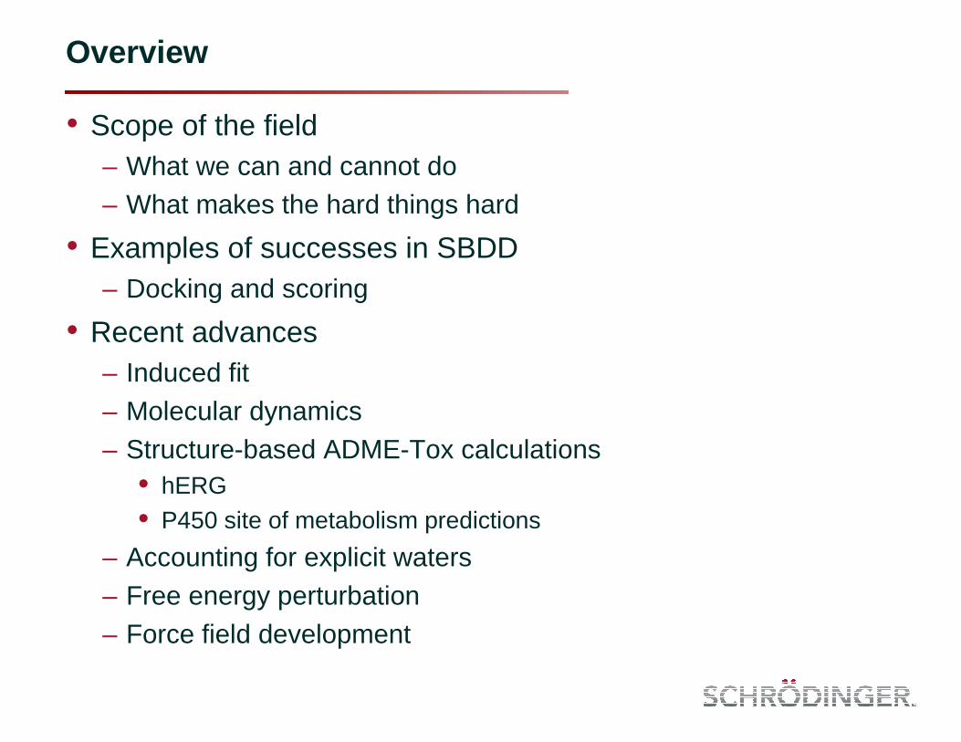
Overview
• Scope of the field– What we can and cannot do– What makes the hard things hard
• Examples of successes in SBDD– Docking and scoring
• Recent advances– Induced fit– Molecular dynamics– Structure-based ADME-Tox calculations
• hERG• P450 site of metabolism predictions
– Accounting for explicit waters– Free energy perturbation– Force field development
100001111
0101111110
010011112
000011118
000011117
001100003
111001005
111100009
111100006
111100004
65318742
100001111
0101111110
010011112
000011118
000011117
001100003
111001005
111100009
111100006
111100004
65318742
Pharmacophore ModelingCheminformaticsScoring Functions
Protein Structure Prediction
Molecular Dynamics Workflows
Core Technologies in Computer-Aided
Drug Design
DockingVisualization and Analysis

100001111
0101111110
010011112
000011118
000011117
001100003
111001005
111100009
111100006
111100004
65318742
100001111
0101111110
010011112
000011118
000011117
001100003
111001005
111100009
111100006
111100004
65318742
Pharmacophore ModelingCheminformaticsScoring Functions
Protein Structure Prediction
Molecular Dynamics Workflows
Core Technologies in Computer-Aided
Drug Design
DockingVisualization and Analysis
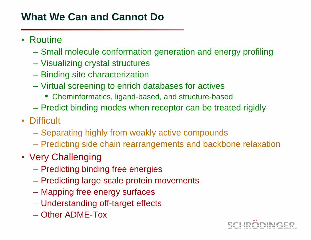
What We Can and Cannot Do
• Routine– Small molecule conformation generation and energy profiling– Visualizing crystal structures– Binding site characterization– Virtual screening to enrich databases for actives
• Cheminformatics, ligand-based, and structure-based– Predict binding modes when receptor can be treated rigidly
• Difficult– Separating highly from weakly active compounds– Predicting side chain rearrangements and backbone relaxation
• Very Challenging– Predicting binding free energies– Predicting large scale protein movements– Mapping free energy surfaces– Understanding off-target effects– Other ADME-Tox

What Makes the Difficult Things Difficult?
• Force fields are approximate– Quantum mechanics would be better, but is too computationally
expensive for most tasks
• Conformational sampling can be limiting– Typical drug like molecules can have many thousands of local
minima that must be evaluated– Proteins have a significantly larger accessible conformational space
• The solution– Focus on specific problems– Know the limits of your method– Keep up with current methods
• Methods are always improving• New resources can make old problems accessible
– Cloud Computing– GPGPU

Structure-based Virtual Screening Example 1
• Researchers at Vernalis used docking to screen commercially available compounds; found 10 novel inhibitors to Chk1 kinase
• Novel hinge interaction motifs were discovered
• Crystal structures were obtained for 4 inhibitors– The others were docked
Foloppe, N., et al. Identification of chemically diverse Chk1 inhibitors by receptor-based virtual screening. Bioorg Med Chem 2006 (14) 4792–4802
Proposed binding modes from docking
Binding mode from one of the crystal structures

Structure-based Virtual Screening Example 2
• Researchers at Vertex used docking to supplement experimental HTS and found 4 novel hits for Pim-1 kinase
• Used special aromatic CH��O hydrogen-bond constraint to the hinge
• Enrichment of actives 14x over HTS
Pierce, A.C., et al. Docking study yields four novel inhibitors of the protooncogenePim-1 kinase. J Med Chem 2008 (51) 1972-1975
Crystal structure used for docking (PDB code 3BGQ)

Glide Enrichments – Including Epik State Penalty
• Simply including many states degrades enrichments
• Need energetic penalty of the ionization/tautomer states
• Using the state penalty improves enrichments

Fragment Docking
• Docking can generate accurate poses for fragments
• 12 cases[1]
– Maximum RMSD 1.3 Å– Most cases less than 0.5 Å RMSD– Accounting for tautomer/ionization
state energies is key
• Loving K, et al., J Comput Aided Mol Des 2009 23:541–554
• Cross docking can be considerably more challenging due to induced-fit, but we are making progress
• More data is needed
[1] Congreve M, et al. J Med Chem 2008 51:3661

Loving K, et al., J Comput Aided Mol Des 2009 23:541–554
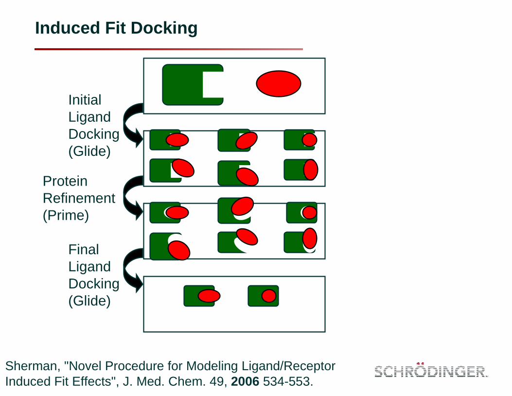
Induced Fit Docking
Sherman, "Novel Procedure for Modeling Ligand/Receptor Induced Fit Effects", J. Med. Chem. 49, 2006 534-553.
Initial Ligand Docking (Glide)
Protein Refinement (Prime)
Final Ligand Docking (Glide)

Induced Fit Docking: Performance
1RMSD of 2nd ranked IFD structure that has nearly identical composite score as top ranked structure2RMSD excluding 13 atoms in solvent exposed methylphenyloxazole tail of the ligand 3RMSD excluding 10 atoms in solvent exposed methyl-2-pyridinylamino tail of the ligand4RMSD excluding 6 atoms in the quasi-symmetric di-carboxylate that are flipped 180°
2nd most sited J Med Chempublication from 2006 (>150 citations, meaning this is working in the real world)
• Average ligand RMSD for docking to a flexible receptor for the 21 pairs is 1.4 Å
• RMSD ≤1.8 Å for 18 cases
• For the 3 cases with RMSD >1.8 Å, the core of the ligand is properly docked and all key protein/ligand interactions are captured
• Still, a substantially larger validation set is needed
Ligand RMSD (Å) Target Receptor Ligand
From: Rigid Receptor Docking Induced Fit
Docking Aldose Reductase 2acr:_ 1ah3 6.5 0.9 Antibody DB3 1dba:H 1dbb 7.6 0.3 CDK2 1dm2:A 1aq1 6.2 0.8 CDK2 1aq1:_ 1dm2 0.6 0.8 CDK2 1buh:A 1dm2 6.4 1.1 COX-2 3pgh:A 1cx2 11.1 1.0 COX-2 1cx2:A 3pgh 6.6 1.0 (0.51) Estrogen Receptor 3ert:A 1err 2.3 1.4 (1.01) Estrogen Receptor 1err:A 3ert 5.3 1.0 Factor Xa 1ksn:A 1xka 9.3 1.5 Factor Xa1 1xka:C 1ksn 5.3 1.5 HIV-RT 1rth:A 1c1c 2.5 1.3 HIV-RT 1c1c:A 1rth 12.0 2.5 Neuraminidase 1nsc:A 1a4q 3.9 0.8 Neuraminidase 1a4q:A 1nsc 1.0 1.7 PPAR-γ 2prg:A 1fm9 9.8 3.0 (1.52) PPAR-γ 1fm9:D 2prg 9.1 1.8 (0.43) Thermolysin 1kr6:A 1kjo 1.1 1.3 Thermolysin 1kjo:A 1kr6 3.5 3.2 (1.64) Thymidine Kinase 1kim:A 1ki4 4.7 0.4 Thymidine Kinase 1ki4:A 1kim 0.5 1.2

Induced Fit Docking Application
• PPAR-γ is a highly flexible target– See superposition of PDB structures
– Most nuclear receptors are flexible
• Researchers at the University of Sydney identified novel PPAR-γ agonists from a natural product library– Flexible ligand docking to a rigid receptor of
known active compounds produced inconsistent poses (see top right)
– Receptor flexibility was required to get good and consistent poses (see bottom right)
• IFD has been used in this project to find new PPAR-γ inhibitors and novel IP
Salam, N.K., et al. Novel PPAR-gamma agonists identified from a natural product library: A virtual screening, induced-fit docking and biological assay study. Chem Biol Drug Des 2008 (71) 51-70
Rigid receptor docking
Flexible receptor docking

Molecular Dynamics
• Probing protein flexibility
• Generation of structural ensembles
• Visualization molecular processes
• Estimation binding energies– Solvation free energies– Binding free energies– Conformational free energies

G-protein coupled receptors
• Largest gene family in the human genome• Represent the target for >30% of drugs• Structural data has historically been scarce
G-protein-coupled receptors and cancer. Robert T. Dorsam and J. Silvio Gutkind. Nature Reviews Cancer 2007 7, 79-94
Target validation of G-protein coupled receptors. Wise A, Gearing K, Rees S. Drug Discov Today. 2002 Feb 15;7(4):235-46.

Milestones in structure determination
0
100
200
300
400
500
600
700
800
900
1000
2009
2008
2007
2006
2005
2004
2003
2002
2001
2000
1999
1998
1997
1996
1995
1994
1993
1992
1991
1990
1989
1988
1987
1986
1985
1984
1983
1982
1981
1980
1979
1978
1977
1976
1975
1974
1973
1972
1971
1970
`
PubMed search: molecular model G-protein coupled receptor
Henderson et al., . Three-dimensional model of purple membraneobtained by electron microscopy . Nature. 1975.
Grigorieff et al., Electron-crystallographic refinement of the structure of bacteriorhodopsin . J Mol Biol. 1996
Warne et al., Structure of a beta1-adrenergicG-protein-coupled receptor. Nature. 2008
Jaakola et al., The 2.6 angstrom crystal structureof a human A2A adenosine receptor bound to an antagonist. Science. 2008
Cherezov et al., High-resolution crystal structureof an engineered human beta2-adrenergic G protein-coupled receptor . Science. 2007
Palczewski et al., Crystal structure of rhodopsin : A G protein-coupled receptor. Science. 2000
Henderson et al., Model for the structure of bacteriorhodopsinbased on high-resolution electron cryo-microscopy(3.5Å). J Mol Biol. 1990
Unger et al., Arrangement of rhodopsintransmembrane alpha-helices. Nature. 1997
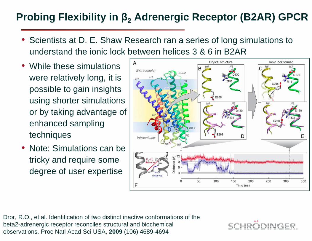
Probing Flexibility in β2 Adrenergic Receptor (B2AR) GPCR
• Scientists at D. E. Shaw Research ran a series of long simulations to understand the ionic lock between helices 3 & 6 in B2AR
Dror, R.O., et al. Identification of two distinct inactive conformations of the beta2-adrenergic receptor reconciles structural and biochemical observations. Proc Natl Acad Sci USA, 2009 (106) 4689-4694
• While these simulations were relatively long, it is possible to gain insights using shorter simulations or by taking advantage of enhanced sampling techniques
• Note: Simulations can be tricky and require some degree of user expertise

Induced Fit Docking in GPCR Dock 2008
Application to the A2A receptor
“With the aim of evaluating the current status of GPCR structure prediction and ligand docking, a community-wide, blind prediction assessment — GPCR Dock 2008 — was conducted in coordination with the publication of the crystal structure of the human adenosine A2A receptor bound to the ligand ZM241385. Twenty-nine groups submitted 206 structural models before the release of the experimental structure, which were evaluated for the accuracy of the ligand binding mode and the overall receptor model compared with the crystal structure.” Michino et al., Nat. Rev. Drug Disc., 2009
Software used: Autodock, DOCKER, Fred, Glide,Gold, ICM, IFD, Membstruck,Modeller, Moe, Q-dock, Rosetta, Tasser
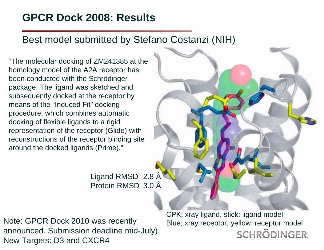
GPCR Dock 2008: Results
Best model submitted by Stefano Costanzi (NIH)
“The molecular docking of ZM241385 at the homology model of the A2A receptor has been conducted with the Schrödinger package. The ligand was sketched and subsequently docked at the receptor by means of the “Induced Fit” docking procedure, which combines automatic docking of flexible ligands to a rigid representation of the receptor (Glide) with reconstructions of the receptor binding site around the docked ligands (Prime).”
Ligand RMSD 2.8 ÅProtein RMSD 3.0 Å
CPK: xray ligand, stick: ligand modelBlue: xray receptor, yellow: receptor modelNote: GPCR Dock 2010 was recently
announced. Submission deadline mid-July).New Targets: D3 and CXCR4

Predicting P450 Sites of Metabolism
• hERG predictions were previously successful– Farid, R. et al. "New insights about HERG blockade obtained from protein
modeling, potential energy mapping, and docking studies", Bioorg. & Med. Chem., 2006, 14, 3160-3173
– Induced fit structure available for download
• Goal:– Method to accurately predict all sites of metabolism– Also get poses, to help with design– Can account for difficult cases, like stereoisomers
• Methods:– Structure-based combined with intrinsic reactivity– If an atom has high intrinsic reactivity and can get an atom close to the
reactive iron, then it is considered to be a site of metabolism– More sophisticated free energy-based scoring is being investigated
• Next steps– Reactivity rates– Inhibition

Aceclofenac SOM in CYP2C9 – Value of IFD
• Highlighted atom in light green is SOM, but has poor reactivity (2.0) vs non-SOM in dark green (-3.6)
• Many IFD derived poses have the SOM close to the heme. It has the best overall score
• The non-SOM is far from the heme in all poses

Tamoxifen SOM in CYP2D6 – Value of IFD
• Highlighted atom in light green is SOM, but has poor reactivity (0.9) vs non-SOM in dark green (-5.5)
• Many IFD derived poses have the SOM close to the heme. It has the 2nd best overall score
• The non-SOM is far from the hemein all poses

The Role of Waters in Drug Discovery
• “Hydrophobic effect” is driven by release of water molecules• Explicit nature of water is essential to describe water behavior• Water molecules have a great deal of mobility, making them ideal
chemical probes with real physical implications– Water molecules are competing with ligands– We can learn from water locations and interactions
• Recent advances in methods and algorithms have allowed for the quantification of water molecule thermodynamics
• Lazaridis, T. (1998) J Phys Chem. B 102:3531–3541– Inhomogeneous fluid approach to solvation thermodynamics
• Young, T., et al. (2007) PNAS 104:808-813– Initial validation to describe hydrophobic enclosure motif
• Abel, R., et al. (2008) J Am Chem Soc 130:2817-2831– Predicting affinity for congeneric pairs of factor Xa inhibitors
• Beuming, T., et al. (2009) Prot Sci 18:1609-1619– Affinity and selectivity insights for PDZ domains
• Robinson, D., et al. (2010) ChemMedChem 5:618-627– Kinase selectivity
• Guimaraes, C., et al. (2010) J Chem Inf Model 50:547–559– Scoring of factor Xa and CDK2 ligands
• Higgs, C., et al. (2010) Med Chem Lett (accepted)– Affinity predictions for a series of A2A GPCR inhibitors
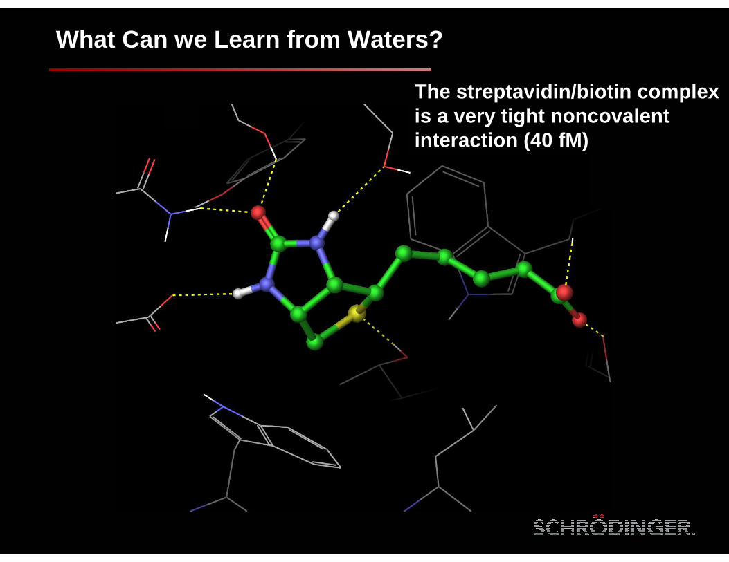
What Can we Learn from Waters?
The streptavidin/biotin complex is a very tight noncovalentinteraction (40 fM)

Clathrate ice-like water structure persists through an MD simulation
Biotin displaces these five waters and makes back the key H-bond interactions
Streptavidin/biotin complex
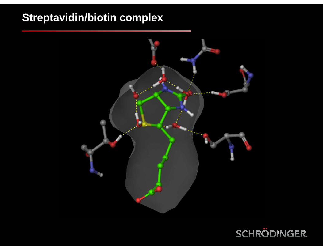
Streptavidin/biotin complex

+2.9
+3.4
+2.5
+4.6
Streptavidin/biotin complex
Waters with ∆G > 2.5 kcal/mol
Biotin displaces highly unstable waters in the streptavidin binding site
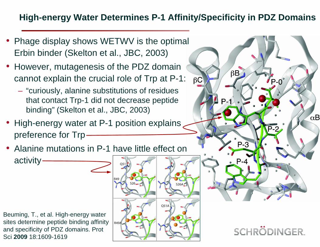
High-energy Water Determines P-1 Affinity/Specifici ty in PDZ Domains
• Phage display shows WETWV is the optimal Erbin binder (Skelton et al., JBC, 2003)
• However, mutagenesis of the PDZ domain cannot explain the crucial role of Trp at P-1:– “curiously, alanine substitutions of residues
that contact Trp-1 did not decrease peptide binding” (Skelton et al., JBC, 2003)
• High-energy water at P-1 position explains preference for Trp
• Alanine mutations in P-1 have little effect on activity
Beuming, T., et al. High-energy water sites determine peptide binding affinity and specificity of PDZ domains. Prot Sci 2009 18:1609-1619
Free Energy Methods
• A long history of academic literature suggests we can accuratelypredict binding free energies
– We are not really there
• FEP (and related methods) feel like they are parameter free– There are many parameters associated with the protocol
• We have started a large-scale relative binding free energy FEP validation study
– 10+ people– 20+ targets– Pharmaceutically relevant perturbations
• Goals– Define scope of applicability and limitations– Understand what can be expected from FEP
– Identify areas for improvements (force fields, sampling time, λ, etc.)
Overview of Absolute Solvation Free Energy Work
• Used existing dataset from Shivakumar et al.*
– Allows for direct comparison with other programs and force fields
• First objective was to test Desmond FEP implementation– Overall results with default parameters worked well
• OPLS_2005 • SPC water model• 12 lambda windows
• Looked into sensitivity of various parameters– Force field– Solvent model– Charges– Lambda schedule
• Paper now available in JCTC– J. Chem. Theory Comput. 2010, 6, 1509–1519– “Prediction of Absolute Solvation Free Energies using Molecular Dynamics
Free Energy Perturbation and the OPLS Force Field”
* Shivakumar D, Deng Y, Roux B; J. Chem. Theory Comput. 5; 919; 2009
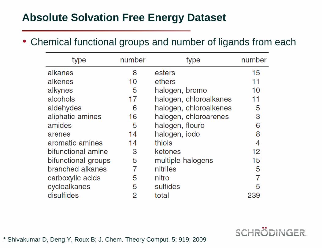
Absolute Solvation Free Energy Dataset
• Chemical functional groups and number of ligands from each
* Shivakumar D, Deng Y, Roux B; J. Chem. Theory Comput. 5; 919; 2009

Overall ASFE Results and Comparison
AM1-BCC/GAFFR2=0.87
ChelpG/CHARMm-MSIR2=0.71
OPLS_2005R2=0.94
Shivakumar et al., J. Chem. Theory Comput. 2010 (ASAP)
Shivakumar et al., J. Chem. Theory Comput. 5; 919; 2009

What Makes Binding Free Energy Methods Difficult
• We really do not know– Not enough data has been generated
• We come back to same possible sources of errors– Sampling
• We know this is a major problem– Force field
• We cannot determine how much of a problem this is until we address the sampling problem
• At least we have over come the human time needed to setup FEP calculations– What used to take an expert days or weeks can now be done I
minutes
• Enhanced sampling is a key step in moving forward– In addition to general methods, intelligent sampling can be done
along a path if the endpoints are known

• Abelson Tyrosine Kinase (4 ligands)• Acetylcholinesterase (18 ligands)• Aldose reductase (3 ligands)• Aurora Kinase (3 ligands)• cAMP Dependant Protein Kinase (9 ligands)• CHK1 Checkpoint Homolog (6 ligands)• Estrogen Receptor (2 ligands)• Estrogen Related Receptor (2 ligands)• Extra-Cellular Signal Regulated Kinase 2 (5 ligands)• Factor VIIa (2 ligands)• Factor Xa (4 + 24 + 24 ligands)• HIV-1 Protease (9 ligand)• HIV-1 Reverse Transcriptase (21 ligands)• Ionotropic Glutamate Receptor (14 ligands)• Lymphocyte-specific Tyrosine Kinase (20 ligands) • P38 Map Kinase (16 ligands)• Phosphodiesterase 4 (5 ligands)• Pim-1 Kinase (4 ligands)• Thrombin (2 + 8 + 5 ligands)• Urokinase-type Plasminogen Activator (2 ligands)
Relative Binding Free Energies – Datasets
20 targets200+ ligands

Force Field Development
• Many limitations existing in current force fields
• Some are just issues of parameterization
• Others have to do with more fundamental methodological issues
• We have 5 people working over the past 5 years on the next generation OPLS force field– Over 1 million commercially available compounds searched for
torsion types• Over 12,000 unique torsion types
– Using CM1A-BCC charges– Special non-bond rules for water– Preliminary data suggests the new force field will help with free
energy calculations
Conclusions
• Some tasks are relatively well validated– Pose prediction for rigid receptors– Virtual screening enrichment– Local protein structure rearrangements
• Significant work is still needed for certain tasks– Binding free energy predictions– Large-scale protein structure predictions
• Advances in method and increased compute resources are allowing us to address challenging problems– Prediction of water locations and thermodynamic properties– GPCR modeling– Structure-based predictions of ADME properties
• hERG
• P450
Final Thoughts
• The field needs more accurate lead optimization methods– Current empirical models have proven to be valuable for virtual
screening, but are lacking for lead optimization
• Accurate scoring cannot be achieved without accurate prediction of the structure
• Confidence/error estimates are important– It is okay to only be right 50% of the time if you can be confident
about when you will be right
• Small molecule energy predictions are only a small part of CADD– ADME/Tox– Selectivity/pathways– Kinetics (yes, biology is not happening at equilibrium)– Antibodies– Other biologics

AcknowledgementsDevelopersApplications ScientistsAcademic Collaborators
