advances in pharmacology volume 43 drug drug interactions scientific and regulatory perspectives
TRANSCRIPT


t
ADVANCES IN PHARMACOLOGY
SERtAL EDITORS
J. Thomas August Baltimore, Maryland
M. W. Anders Rochester, New York
Ferid Murad Houston, Texas
Joseph T. Coyle Belmont, Massachusetts
ADVISORY BOARD
R. Wayne Alexander Floyd E. Bloom Boston, Massachusetts La Jolla, California
Thomas F. Burke Houston, Texas
Anthony R. Means Durham, North Carolina
John A. Thomas San Antonio, Texas
Leroy Liu Piscataway, New Jersey
G. Alan Robison Houston, Texas
Thomas C. Westfall St. Louis, Missouri
DRUGIDRUG INTERACTIONS: SCIENTIFIC AND
REGULATORY PERSPECTIVES
Edited by
Albert P. Li In Vitro Technologies Inc.
University of Maryland Technology Center Baltimore, Maryland
ACADEMIC PRESS San Diego London Boston New York Sydney Tokyo Toronto
This book is printed on acid-free paper. @
Copyright 0 1997 by ACADEMIC PRESS
All Rights Reserved. No pan of this publication may be reproduced or transmitted in any form or by any
means, electronic or mechanical, including photocopy, recording, or any information storage and retrieval system, without permission in writing from the Publisher,
The appearance of the code at the bottom of the first page of a chapter in this book indicates the Publisher's consent that copies of the chapter may be made for personal or internal use of specific clients. This consent is given on the condition, however. that the copier pay the stated per copy fee through the Copyright Clearance Center, Inc. (222 Rosewood Drive, Danvers, Massachusetts 01923). for copying beyond that permitted by Sections 107 or 108 of the U.S. Copyright Law. This consent does not extend to other kinds of copying, such as copying for general distribution, for advertising or promotional purposes, for creating new collective works, or for resale. Copy fees for pre-1997 chapters are as shown on the title pages, if no fee code appears on the title page, the copy fee is the same as for current chapters. 1054-3589197 $25 .OO
Academic Press u di\ision of Harcourt Bruce & Compunj 525 B Street, Suite 1900. San Diego, California 92101-4495. USA http://www.apnet.com
Academic Press Limited 24-28 Oval Road, London NWl 7DX. UK http://www.hbuk.co.uk/ap/
International Standard Book Number: 0- 12-032944-1
PRINTED IN THE UNlTED STATES OF AMERICA 97 98 9 9 0 0 01 0 2 E B 9 8 7 6 5 4 3 2 1

Contributors
Numbers in parentheses indicate the pages on which the authors’ contributions begin.
John Dikran Balian (231) Office of Clinical Pharmacology and Biopharma- ceutics, Center for Drug Evaluation and Research, U.S. Food and Drug Administration, Rockville, Maryland 20857
Klaus Brendel (131) Department of Pharmacology, University of Arizona, Tucson, Arizona 85724
Charles L. Crespi ( 171) GENTEST Corporation, Woburn, Massachusetts 01801
James L. Ferrero (131) Drug Metabolism Department, Pharmaceutical Prod- ucts Division, Abbott Laboratories, Abbott Park, Illinois 60064
Frank J . Gonzalez (255) National Cancer Institute, Bethesda, Maryland 20892
F. Peter Guengerich (7) Department of Biochemistry and Center in Molecu- lar Toxicology, Vanderbilt University School of Medicine, Nashville, Tennessee 37232
Lhanoo Gunawardhana (37) Department of Pharmacology and Toxicology, University of Arizona, Tucson, Arizona 85721
Dwayne A. Hill (37) Department of Pharmacology and Toxicology, Univer- sity of Arizona, Tucson, Arizona 85721
Malle Jurima-Romet (1,239) Bureau of Drug Research, Drugs Directorate, Department of National Health and Welfare, Canada, Banting Research Centre, Ottawa, Ontario, Canada K1A OL2
Gregory L. Kedderis (189) Chemical Industry Institute of Toxicology, Re- search Triangle Park, North Carolina 27709
xiii

xiv Contributors
Albert P. Li (1, 103) In Vitro Technologies, Inc., University of Maryland Technology Center, Baltimore, Maryland 21227
Bruce W. Penman ( 171 ) GENTEST Corporation, Woburn, Massachusetts 01801
Atiqur Rahman (23 1) Office of Clinical Pharmacology and Biopharmaceu- tics, Center for Drug Evaluation and Research, U.S. Food and Drug Administration, Rockville, Maryland 20857
A. David Rodrigues (65) Drug Metabolism Department, Pharmaceutical Products Division, Abbott Laboratories, Abbott Park, Illinois 60064
John-Michael Sauer (37) Department of Pharmacology and Toxicology, University of Arizona, Tucson, Arizona 85721
I. Glenn Sipes (37) Department of Pharmacology and Toxicology, University of Arizona, Tucson, Arizona 85721
Eric R. Stine (37) Department of Pharmacology and Toxicology, University of Arizona, Tucson, Arizona 55721
Thomas N. Thompson (205) Department of Drug Metabolism, North Amer- ican Pharmacokinetics, Hoechst Marion Roussel, Inc., Kansas City, Mis- souri 64137
Shekman L. Wong (65) Drug Metabolism Department, Pharmaceutical Products Division, Abbott Laboratories, Abbott Park, Illinois 60064

Foreword
For decades, drug metabolism has been an area of pharmacology that has been interesting in its own right, drawing on the instrumental expertise of analytical chemistry, the elegance of enzymology, and the emerging tools of molecular biology. While serving its primary role as an independent discipline, drug metabolism has also produced a number of important contri- butions to the development and therapeutics of specific agents. In the area of pharmacogenetics, several deficiencies of drug-metabolizing enzymes have been reported (e.g., N-acetyltransferase for metabolism of isoniazid). Interac- tions between coadministered drugs have been described based on either induction of the metabolizing enzymes (e.g., by anticonvulsants) or inhibi- tion (e.g., quinidine and CYP2D6).
As the variety of new drugs has increased and polypharmacy has become commonplace, drug-drug interactions have been gaining in visibility. In less than five years, we have seen an impressively swift change in the paradigm of applications for these studies. All of the elegance and academic interest remain, but there is also a fundamental retargeting of efforts toward predict- ive approaches rather than retrospective evaluations. Although the identifi- cation of metabolic pathways is still a central event for each new drug, there is an immediate emphasis on interpreting these pathways in terms of understanding, anticipating, and avoiding adverse drug-drug interactions.
As described in this volume, the set of tools is impressively arrayed for the exploration of metabolic-based interactions, ranging from the classics of microsomes, hepatocytes, or slices, and then intersecting with the realm of molecular biology via recombinant human enzymes and transgenic animal systems. The organized ventures that procure, process, and distribute human-derived material have overcome the major barriers of availability that hindered past studies. However, this large tool chest in itself does not
xv

xvi Foreword
produce data that are useful to drug development and regulation. The users of the tools, clinical pharmacologists in academia, industry, and regulatory agencies, must focus on those experiments that yield the greatest insight into therapeutics.
Once a drug-drug metabolic interaction has been discovered, the impli- cations for further development and regulation can follow one of several scenarios. Most drug-drug interactions change the therapeutic index of one or both agents. In general, we are concerned about either loss of therapeutic effect or amplification of adverse effects. However, not all interactions are automatically problematic. A few are even intentional, such as the treatment of methanol poisoning with ethanol. Either the parent drug or its metabo- lite(s) may have therapeutic and/or undesirable pharmacological effects. Thus, the interpretation of the consequences for an interaction depends on the relative therapeutic index of each pharmacologically active species. For example, if the parent compound has a more desirable therapeutic index than the metabolite, then inhibition might be advantageous (although the dose probably needs to be lowered). On the other hand, induction of metabo- lism of the parent to the metabolite could adversely affect therapy in this situ- ation.
The task of applied methodology development is not complete. By far the greatest gains have come with the family of cytochrome P450 enzymes. We need to develop a similar level of sophistication and to apply the same principles to other reactions of therapeutic interest, such as the various transferases of Phase 2 metabolism. While the most recent work has focused on the evaluation of drugs as potential inhibitors of major metabolic path- ways, promising work has also been reported on approaches to evaluating whether a new agent has the potential to induce the metabolism of itself andlor other concomitantly administered drugs.
Dr. A1 Li and the contributors to this volume have summarized where we are and how we got here, while pointing us toward where we are going. There is a consensus among academic, industrial, and regulatory scientists that this journey has been highly rewarding thus far. We look forward to continuing to work together to harvest further returns on our careful investments in this field.
Jerry M. Collins

Preface
During drug development, each new drug is tested vigorously for phar- macokinetic, pharmacodynamic, and toxicological properties. On the basis of this information, the safety and efficacy of the drug will be evaluated. An acceptable drug is one that can be used at a safe dose to produce the desirable therapeutic effects.
In the reality of drug administration, multiple drugs are frequently administered to the same patients, either to treat multiple diseases (as in the case of the elderly patients) or as a multiple modality treatment of a single disease (as in the case of HIV patients). It is important to know whether the safety and efficacy of a drug are affected by coadministered drugs.
Because drug dosage can affect both safety and efficacy, the most critical drug-drug interactions are the effects of drugs on the plasmahissue levels of coadministered drugs, a phenomenon called pharmacokinetic drug-drug interaction. Although the major determinants of the body burden of a drug involve the processes of absorption, distribution, metabolism, and clearance, interference with the metabolism process appears to be the most important mechanism for drug-drug interactions.
The chapters in this book present a comprehensive review of the scientific and regulatory aspects of drug-drug interactions from the point of view of academia, industry, and government agencies. The topics covered include drug metabolism enzymes, toxicology, and in vitro mechanistic approaches, as well as the regulatory perspectives of drug-drug interactions. This book is intended for professionals in the pharmaceutical industry and in the health- care and governmental regulatory agencies who are interested in the mecha- nistic understanding of drug-drug interactions, the prediction of the drug-
xvii

xviii Preface
drug interaction potential of new drugs, and the avoidance of clinically significant drug-drug interaction in patients. This book should be of interest as well to students and researchers in the areas of pharmacology, toxicology, pharmacokinetics, and medicine.
Albert P . Li

Albert P. Li* Malle Jurima-Romet?
*In Vitro Technologies, lnc, University of Maryland
Technology Center Baltimore, Maryland 2 I227
tBureau of Drug Research, Drugs Directorate Department of National Health Canada
Banting Research Center Ottawa, Ontario Canada KIA OL2
Overview: Pharmacokinetic Drug-Drug Interactions
1. Clinical Significance of Drug-Drug Interactions
Drug-drug interactions-the effects of one drug on the efficacy and/or toxicity of another drug-have become an important issue in health care. The majority of patients are treated with more than one drug simultaneously Reasons for the treatment with multiple drugs include the treatment of multiple ailments in the same patient and the use of multiple drugs for a single ailment. With the increasing median age of the population, and the now known effectiveness of multiple-therapy regimens for viral diseases (e.g., HIV), cancer, cardiovascular diseases, and infectious diseases, exposura of a patient to multiple drugs is a common rather than rare occurrence.
The need to understand the interaction potential of drugs is necessitated by events observed in the human patient population. A comprehensive review of specific cases of drug-drug interactions can be seen in Hansten and Horq (1 993). Examples of drug-drug interactions with significant toxicological and pharmacological consequences are illustrated below.
A. Terfenadine Cardiotoxicity
The nonsedating antihistamine, terfenadine, has been associated with cardiotoxicity. Terfenadine-related cardiotoxicity is manifested as torsades de pointes, a form of polymorphic ventricular tachycardia associated with prolongation of the QT interval of the electrocardiograph, and can be a
Aduances in Pharmacology, VoLme 43 Copyrtghr D 1997 by Academic Press. All rights of reproduction in any form reserved. 1054-3589/97 $25.00

2 Albert P. Li and Malle Jurima-Romet
lethal event. Blockage of cardiac potassium currents (Kato et al., 1996) and prolongation of action potential duration (Crumb et al., 1995) are consid- ered to be possible mechanisms of terfenadine-associated cardiotoxicity. Terfenadine cardiotoxicity has been observed when the drug is coadminis- tered with other drugs. These interacting drugs include ketoconazole (Mona- han et d., 1990; Honig et d., 1993), erythromycin (Honig et al., 1992), and itraconazole (Pohjola-Sintonen et al., 1993). Where drug interaction occurs, excessive levels of parent terfenadine have been observed. It is be- lieved that the interacting drugs inhibit terfenadine metabolism, thereby leading to the elevation of parent terfenadine to a cardiotoxic level. These findings led to FDA’s requirement for the manufacturer of terfenadine to inform the medical community of potentially serious interactions. Because of the warning issued, coprescription of terfenadine and the known interacting drugs erythromycin and ketoconazole dramatically declined but nonetheless continue to occur (Carlson and Morris, 1996).
B. Sorivudine-Fluoropyrimidine Lethality
Sixteen deaths occurred among patients orally administered the new antiviral drug sorivudine within weeks after the drug’s approval for use in Japan. The mortality occurred in patients who were coadministered both sorivudine and fluoropyrimidines. Sorivudine is a potent inhibitor of hepatic dihydropyrimidine dehydrogenase (DPD). DPD is the enzyme responsible for the catabolism of fluoropyrimidines. It is believed that in these patients, the fluoropyrimidine levels were elevated to lethal levels due to the inhibition of DPD by sorivudine (Okuda et al., 1995).
C. Oral Contraceptive Failures
It was noted that extraordinary rates of birth-control failure occurred in several populations of women: patients administered rifampin, griseoful- vin, and anticonvulsants. The patients experienced transient intermenstrual bleeding, amenorrhea, and unintended pregnancies. It is believed that these interacting drugs act through the induction of metabolizing enzymes of the active ingredients of oral contraceptives. Bolt et al. (1977) showed that rifampin accelerated the elimination of radioactive ethinylestradiol from plasma and concluded that the increased elimination is a result of increased steroid hydroxylation. Griseofulvin and anticonvulsants are believed to act via similar mechanisms (Van Dijke and Weber, 1984). Induction of drug- metabolizing enzymes appears to be the common mechanism for these drugs that could lead to failures of oral contraceptives.
The pharmacological and toxicological consequences of drug-drug in- teractions are detailed in the chapters by J. M. Sauer etal. and G. L. Kedderis.

Overview 3
II. Mechanism of Pharmacokinetic Drug-Drug Interactions
The majority of serious cases of drug-drug interactions, as exemplified by the three specific drugs just discussed, are a result of the interference of the metabolic clearance of one drug by a coadministered drug. The interfer- ence can occur via inhibition or induction of metabolic enzymes. It is of no surprise that most known mechanisms of drug-drug interactions involve the major enzyme system for xenobiotic metabolism, the cytochrome P450 (CYP) isozymes. Although many isoforms of CYP have been discovered, most drugs are metabolized in humans by CYP isoforms belonging to the subfamilies lA, 2A, 2C, 2D, 2E, and 3A. Of the isozymes, CYP3A, 2D6, and 2C contribute in the metabolism of the greatest number of drugs. A detailed discussion of CYP is presented in the chapter by F. P. Guengerich.
A number of drug-drug interactions related to CYP involve inhibition or induction of CYP3A activity. The CYP3A subfamily is probably the most important isozyme for human drug metabolism. CYP3A4 is found universally in human and is abundant in both the liver and the intestinal mucosa. In the human liver, approximately one-third of the total CYP is CYP3A. CYP3A is believed to be responsible of the metabolism of over 50% of known human drugs (Li etal., 1996). The previously mentioned drug-drug interactions with terfenadine, and with oral contraceptives, are results of interference with CYP3A4 activity. Terfenadine is metabolized by CYP3A4 to noncardiotoxic metabolites. Drugs such as ketoconazole, erythromycin, and itraconazole are potent inhibitors of CYP3A4; therefore, if one of them is coadministered with terfenadine, it may significantly inhibit terfenadine metabolism, thereby ele- vating the plasma level of terfenadine to a cardiotoxic level (Jurima-Romet et al., 1996). CYP3A4 is also an inducible CYP isozyme. Rifampin, dexametha- sone, and anticonvulsants are known inducers of CYP3A4 and have been found to lower the efficacy of drugs that are CYP3A4 substrates, which include oral contraceptives, cyclosporin, and erythromycin. Other forms of CYP3A include CYP3A5, which is found in only 10-30% of human livers, and CYP3A7, which is found predominantly in fetal liver. Rat liver has predomi- nantly CYP3A2, which is dramatically different from the human CYP3A4 in response to enzyme inducers. The potent inducer of human CYP3A4, rifam- pin, for instance, is only a weak inducer of CYP3A2 in the rat (see chapter by A. P. Li).
Polymorphisms of CYP isozymes, as observed with CYP2C and CYP2D, illustrate the importance of isozyme activity and drug adverse effects. CYP2D6 polymorphism was initially characterized by debrisoquine metabo- lism. In the Caucasian population, the incidence of the poor metabolizing phenotype is approximately S-lO%, whereas in Asians, the incidence is only around 1%. A different ethnic variation is observed for the CYP2C19 polymorphism as characterized by S-mephenytoin metabolism. For

4 Albert P. Li and Malle jurima-Romer
CYPC19, poor metabolizers are found in 20% of the Asian population, but only in 3-5% of the Caucasian population. Because of impaired drug metabolism, the poor metabolizers are found to suffer adverse effects of drugs known to be metabolized by the isozyme they lack, as dosages are established based on the majority of the population, the extensive metabo- lizers. Adverse effects observed with poor metabolizers illustrate the clinical significance of CYP inhibitors. An individual who is an extensive metabo- lizer, if administered a drug that would inhibit that specific isozyme (e.g., quinidine, a potent inhibitor of CYP2D6), would metabolize other substrates of the isozyme like a poor metabolizer, leading to adverse effects.
111. Prediction of Drug-Drug Interactions
Prediction of the drug-drug interaction potential is presently an impor- tant aspect of drug development. In addition to in vivo experimentation with animals and clinical trials with key drugs (e.g., theophylline, ethynyles- tradiol), human in vitro metabolic systems have become important experi- mental tools (FDA, 1997). The following mechanistic approaches are now becoming common in evaluating the drug-drug interaction potential.
A. Definition of CYP lsozyme Specificity of Metabolism
Understanding which CYP isozyme(s) is responsible for metabolism is important in the evaluation of drug-drug interaction potential. A drug that is the substrate of a specific isozyme may compete with other drugs that are also substrates. Furthermore, drug-drug interaction is likely to occur between such a drug and known inhibitors or inducers of that specific isozyme. The definition of CYP isozyme specificity is studied using human hepatic microsomes and cDNA-expressed microsomes. With microsomes, the common approach is to evaluate whether specific inhibitors of individual isozymes would inhibit metabolism of the drug. With cDNA-expressed mi- crosomes, microsomes containing individual isozymes are used to evaluate which isozyme would have the highest affinity and capacity for the drug (see chapters by A. D. Rodrigues and S. L. Wong and by C. L. Crespi and B. W. Penman).
B. Evaluation of CYP Inhibition Potential
If a drug is an inhibitor of a specific CYP isozyme, it would have the poten- tial to inhibit the metabolism of other substrates of the isozyme. Therefore, a clear understanding of the CYP inhibitory potential of a drug on individual CYP isozymes will allow one to predict whether the drug would inhibit the metabolism of drugs with defined CYP isozyme specificity. The inhibition po-
Overview 5
tential is usually evaluated using human hepatic microsomes or cDNA- expressed microsomes. The inhibitory effect of the drug on the rate of metabo- lism of isozyme-specific substrates is usually expressed as apparent K,. Using the K , value, the in vivo effect can be predicted based on the known plasma and/or tissue concentration. Because of the potential differences between plasma and intracellular concentrations, an intact cell system such as primary hepatocytes may offer an advantage over cell-free systems such as microsomes. As plasma concentration values are used to estimate in vivo effects, K, values derived from cell-free systems are only relevant if the plasma concentration and hepatocyte concentration are similar (see chapters by A. D. Rodrigues and S. L. Wong; J. L. Ferrero and K. Brendel; and A. P. Li).
C. Evaluation of CYP Induction Potential
A drug that induces a CYP isozyme can cause drug-drug interactions with drugs that are substrates of the same isozyme, leading to enhanced clearance, Primary human hepatocytes are the preferred experimental system for the evaluation of this aspect of drug-drug interactions. The validity of the primary human hepatocyte system as an assay for CYP induction is supported by two lines of observation: (1) Known in vivo inducers of CYP, rifampin, phenobarbital, dexamethasone, phenytoin, 3 -methylcholanthrene, and so on, have been found to be potent inducers of CYP in primary hepatocytes; and (2) known in vivo species differences in sensitivity to in- ducers, e.g., differences between human and rat in response to rifampin (a potent inducer of CYP3A4 in humans but significantly less potent in rats), are observed in primary hepatocytes (see chapter by A. P. Li).
IV. Conclusions and Future Directions
The clinical importance of drug-drug interaction is well recognized. Recent major advances in this field have been (1) a better understanding of the mechanism of interactions and (2) the application of the mechanistic knowledge toward developing logical and scientifically acceptable ap- proaches for evaluating new and existing drugs for their potential to interact with other drugs. This mechanistic approach is an important scientific ad- vancement. It would be impossible to evaluate experimentally the interaction of a new drug with all existing drugs, and it would not be prudent to wait until significant drug interactions are observed in the clinic. The logical prediction of drug-drug interactions based on knowledge of the metabolic pathway and the ability of the drugs in question to inhibit or induce key metabolic enzymes remains the most promising approach (see chapters by T. N. Thompson; J. D. Balian and A. Rahman; and M. Jurima-Romet).
We need to keep on refining our experimental systems as well as our approaches for extrapolating data from the laboratory to the clinic. For drugs with both experimental and clinical data, we need to critically evaluate

6 Albert P. Li and Malle Jurima-Romet
under what circumstances, and via which mechanism, do data differ. Objec- tive evaluation of experimental and clinical data, and conscientious efforts toward understanding the similarities and differences between experimental and clinical data can direct our effort to further advance technology, experi- mental approaches, and data analysis. Sound scientific approaches, coupled with effective communication between drug manufacturers and health pro- fessionals, will minimize the incidence of drug-drug interactions in the human population.
References
Bolt, H. M., Bolt, M., and Kappus, H. (1977). Interaction of rifampacin treatment with pharmacokinetics and metabolism of ethinyloestradiol in man. Acta Endocr. (Copenha- gen) 85 , 189-197.
Carlson, A. M., and Morris, L. S. (1996). Coprescription of terfenadine and erythromycin or ketoconazole: An assessment of potential harm.]. Am. Pharm. Assoc. [N.S.] 36,263-269.
Crumb, W. J. Jr., Wible, B., Arnold, D. J., Payne, J. P., and Brown, A. M. (1995). Blockade of multiple human cardiac potassium currents by the antihistamine terfenadine: Possible mechanism for terfenadine-associated cardiotoxicity. Mol. Pharmacol. 47, 181-190.
F D A ( 1997). Drug metabolism/drug interaction studies in the drug developing process: Studies in vitro. Guide for Industry, April 1997.
Hansten, P. D., and Horn, J. R. (1993). “Drug Interactions & Updates.” Applied Therapeutics, Inc., Vancouver, WA.
Honig, P. K., Woosley, R. L., Zamani, K., Conner, D. P., and Cantilena, L. R. (1992). Changes in the pharmacokinetics and electrocardiographic pharmacodynamics of terfenadine with concomitant administration of erythromycin. Clin. Pharnracol. Ther. 52, 231-238.
Honig, P. K., Wortham, D. C., Zamani, K., Conner, D. P., Mullin, J. C., and Cantilena, L. R. ( 1 993). Terfenadine-ketoconazole interaction. Pharmacokinetic and electrocardiographic consequences. ]AMA J . Am. Med. Assoc. 269, 1550-1552.
Jurima-Romet, M., Huang, H. S., Beck, D. J., and Li, A. P. (1996). Evaluation of drug interactions in intact hepatocytes: Inhibitors of terfenadine metabolism. Toxicol. In Vitro
Kato, Y., IMori, T., Ohmori, K., and Ichimura, M. (1996). Effect of terfenadine and KW-4679, a novel antiallergic compound, on action potential of guinea pig ventricular myocytes. ]pn. J. Pharmacol. 70, 199-202.
I& A. P., Kaminski, D. L., and Rasmussen, A. (1995). Substrates of human hepatic cytochrome
Monahan, B. P., Ferguson, C . L., Killeavy, E. S., Lloyd, B. K., Troy, J., and Cantilena, L. R. (1990). Torsades de pointes occurring in association with terfenadine use. J A M A J. Am. Med. Assoc. 264, 2788-2790.
Okuda, H., Nishiyama, T., Ogura, K., Nagayama, S., Ikeda, K., Yamagouchi, S., Nakamura, Y ., and Watabe, T. (1995). Mechanism of lethal toxicity exerted by simultaneous adminis- tration of the new antiviral sorivudine, and the antitumor agent tefafur. Xenobiotic Metub. Dispos. 10, Suppl. S166-Sl69.
Pohjola-Sintonen, S., Vitassalo, M., Tolvonen, L., and Neuvonen, P. (1993). Itraconazaole prevents terfenadine metabolism and increases risk of torsades de pointes ventricular tachycardia. Eur. J. Clin. Pharmacol. 45, 19 1-193.
Van Dijke, C. P. H., and Weber, J. C. P. (1984). Interaction between oral contraceptives and griseofulvin. Br. Med. J. 288, 1125-1 126.
10,655-663.
P450 3A4. T o x i c o l o ~ 104, 1-8.

F. Peter Guengerich Department of Biochemistry and Center in Molecular Toxicology
Vanderbilt University School of Medicine Nashville, Tennessee 37232-0 146
Role of Cytochrome P450 Enzymes in Drug-Drug Interactions
1. Introduction
Cytochrome P450 (P450) enzymes were discovered in independent stud- ies on the metabolism of drugs, carcinogens, and steroids (Guengerich, 1993). These enzymes are components of mixed-function oxidase systems that catalyze reactions of the overall stoichiometry
NAD(P)H + H+ + 0, + R + NAD(P)+ + HzO + RO,
where R is an organic substrate (e.g., drug in our discussion here). At the present time the conventional wisdom is that there are -40 different P450 enzymes expressed in each mammalian species, including humans (Nelson et al., 1993; Guengerich, 1995). Many of these P450 enzymes have specific roles in the anabolism of steroids. The P450 enzymes that oxidize drugs are localized in the liver, although some of this group are also found in sites
Advances in Pharmacology, Volunre 43 Copyright 0 1997 by Academic Press. All rights of reproduction in any form reserved. 1054-3589/97 $25.00 7

8 F. Peter Guengerich
such as lung and small intestine, where an appreciable contribution to overall metabolism of a drug can occur depending on the route of administration.
Fortunately, it appears that the metabolism of most drugs can be ac- counted for by a relatively small subset of the P450s. One estimate is that 290% of human drug oxidation can be attributed to six enzymes: P450s lA2,2C9/10,2C19,2D6,2El, and 3A4 (Guengerich, 1995; Wrighton and Stevens, 1992). Further, most could probably be attributed to P450s 1A2, 2C9/10, 2D6, and 3A4 and, by some estimates, half can be attributed to P450 3A4 (Guengerich, 1995; Guengerich et al., 1994a). This view is based primarily on (in vitro) microsomal studies done with drugs studied to date and may change somewhat with time. For instance, the fraction of drugs oxidized by P450 2D6 may be too high in current estimates because of the ease of identifying these and the attention that has been given to this particu- lar enzyme. Nevertheless, the concept that most drug oxidations are cata- lyzed primarily by a small number of P450 enzymes is important in that the approaches to identifying drug-drug interactions are feasible, both in vitro and in vtvo.
This chapter operates from the premise that many significant drug-drug interactions can be understood in terms of P450s. However, drug-drug interactions are more complex for at least two reasons. First, some drug- drug interactions can be attributed to pharmacokinetic differences due to other enzymes such as monoamine oxidases, flavin-containing monooxygen- ases, UDP-glucuronosyl transferases, and sulfotransferases. These and other so-called “drug-metabolizing” (or “xenobiotic-metabolizing”) enzymes also show the characteristics of induction and inhibition by drugs that are associ- ated with P450s, although most have not yet been studied as extensively. The other aspect of drug-drug interactions is that some of these are probably pharmacodynamic instead of pharmacokinetic. For instance, drugs can com- pete for binding to a receptor directly related to the pharmacological re- sponse.
II. Potential Consequences of Drug-Drug Interactions
From a pharmacokinetic standpoint, the major effects of drug-drug interactions can be understood in terms of causing the disposition of a durg to be unusually slow or fast. The major consequence is a high or low plasma and tissue level of the drug.
If the metabolism of a drug is impeded due to enzyme inhibition, then a high plasma level may follow (Fig. 1). One of the major effects will be increased pharmacological activity, and this may or may not be a problem, depending on the therapeutic window. Of course, not only the desired effect may be increased but also any undesirable side effects. If activation of a pro-drug is inhibited, then a lower level of therapeutic effectiveness might

P450 and Drug-Drug Interactions
4
c
9
Normal Metabolism
be anticipated. Another possibility is that when the major pathway of metab- olism of a drug is blocked, secondary pathways may become more favorable. This can be a problem if the secondary pathway leads to a toxic product. An example of this is seen with the analgesic phenacetin (no longer on the U.S. market). If 0-deethylation (P450 1A2) is slow, then other pathways are favored that lead to quinoneimine formation and methemoglobinemia (Fischbach and Lenk, 1985; Klehr et al., 1987). Another possibility is that the increased level of a drug due to inhibition of the P450 involved in its oxidation may lead to inhibition of another P450. Although direct evidence for such a situation has not been presented, one could postulate that accumu- lation of quinidine due to P450 3A4 inhibition might lead to inhibition of P450 2D6, an enzyme for which quinidine is an inhibitor but not a substrate (Guengerich et al., 1986b; Otton et al., 1984).
When levels of P450 (or, for that matter, another enzyme) are induced, the major consequence is a lack of therapeutic effectiveness. Although this might seem to be a common event, the number of real clinical situations in which this has been a problem are rather limited. Two of the best documented examples are cyclosporin and 17a-ethynylestradiol (vide infru). Another possibility with a pro-drug is that activation may be too rapid and a seriously high level of active drug could result. This could be a problem, as one of the primary reasons for developing pro-drugs is to avoid a transiently high level of the active drug. However, no good examples of clinical problems resulting from a phenomenon of this type are known yet.
There are two other possibilities that can be considered in regard to issues of drug-drug interactions. One involves metabolism of chemical car- cinogens. Most of the P450s that transform drugs can also oxidize chemical carcinogens (Guengerich and Shimada, 1991; Guengerich, 1995). The possi- bility exists that a P450 induced by a drug could lead to enhanced levels

10 F. Peter Guengerich
of DNA-carcinogen adducts due to increased carcinogen activation. The induction of human P450s 1Al and 1A2 by omeprazole was postulated to present a risk due to such considerations (Diaz et al., 1990). Whether or not this is a serious issue is unknown, as levels of P450 1A2 are only one of many factors linked to cancer risk from known carcinogenic substrates for the enzyme (Lang et al., 1994). Nevertheless, most pharmaceutical com- panies and the Food and Drug Administration (FDA) would rather avoid drugs that induce P450 1A subfamily enzymes, which have been suggested to be related to cancer development (Ioannides and Parke, 1990, 1993). Again, it should be emphasized that increased cancer risk due to P450 (1A or other) induction is still a hypothesis. Another matter to consider is that some P450s are involved in the detoxication of potential carcinogens and that induction or inhibition might have an impact on this process, as well as the activation (Guengerich and Shimada, 1991; Richardson et al., 1952; Nebert, 1989).
The other possibility involves the influence of drugs on P450s involved in the transformation of endogenous compounds, i.e., those normally found in the body. This matter has not been extensively investigated, but some possibilities exist. Depending on the tissue, 17P-estradiol is oxidized by 1’450 3A4 (Brian et al., 1990; Guengerich et al., 1986a), P450 1A2 (Guo et al., 1994), or P450 1B1 (Liehr et al., 1995). It is not known what the impact of changes in these enzymes is on total body levels. Testosterone is a substrate for P450 3A4 (Guengerich et al., 1986a; Waxman et al., 1988). Another case to consider is animals devoid of bilirubin UDP-glucuronosyl transferase activity, who can be administered P450 1A inducers to lower their levels of bilirubin to nontoxic levels (Kapitulnik and Gonzalez, 1993; Kapitulnik and Ostrow, 1978).
A final point to consider is that some drugs may affect the disposition of chemicals in foods and beverages through P450 interactions. For instance, the drug disulfiram (Antabuse) inhibits P450 2E1. This would affect ethanol oxidation by P450 2E1, although the more serious effect is on aldehyde dehydrogenase (Guengerich et al., 1991). The P450 1A2 inhibitor furafylline blocks caffeine N’-demethylation to the point where severe insomnia is associated with drinking coffee (Sesardic et al., 1990; Kunze and Trager, 1993).
Ill . Use of Information about Human P450s
Much of the information abot drug metabolism by human P450s has been acquired in the past decade. Induction was first seen in clinical settings in the 1950s (Remmer, 1959) and there were many in vitro studies with human tissue samples (Distlerath and Guengerich, 1987). By 1980 some purification work had commenced and several of what are now recognized

P450 and Drug-Drug Interactions I I
as the major P450 2C and 3A subfamily proteins were isolated (Kitada and Kamataki, 1979; Beaune et al., 1979; Wang et al., 1980). Further work led to the isolation of more of the major human P450s (Guengerich, 1989), and cDNA cloning methods were used to obtain DNA sequences for the human P450s (Gonzalez, 1989).
In recent years the access to human tissue samples in the United States and Europe has facilitated characterization of P450 reactions catalyzed by human P450s. The availability of the recombinant human P450s expressed in various systems has also facilitated studies on their catalytic selectivity (Gonzalez et al., 1991a,b; Guengerich et al., 1996). Thus, it is now relatively straightforward to use in vitro studies to determine which P450s oxidize a particular drug and which drugs can inhibit oxidations catalyzed by this P450. The in vitro determination of inducibility is not as easily done, but a number of possibilities exist with cultures of human hepatocytes (Guillouzo et al., 1993; Loretz et al., 1989) (also see chapter by A. P. Li).
It is also possible to do logical in vivo studies to test the relevance of in vitro findings. For instance, individuals known to be high or low in a particular P450 from the use of other noninvasive assays can be examined with regard to the pharmacokinetics of the new drug to see if there is a match. In some cases, inducers or inhibitors of a specific P450 can be given safely to people to verify that a P450 is involved in the oxidation of a drug. Also, the drug under consideration can be given to people to determine if it affects the pharmacokinetics of other drugs through enzyme induction.
The acquisition of the in vitro information about a new drug can be extremely useful. In many cases, the FDA now expects in vitro information on the P450s involved in the oxidation of a drug early in the registration process. The in vitro information can be used to guide the more expensive and time-consuming in vivo studies. In particular, potential adverse drug interactions due to pharmacokinetics can be predicted and the number of in vivo interaction studies can be restricted, as some of those historically done with all new drugs may be found irrelevant. The in vitro procedures, if used early in the drug development process, may be used to select from a series of potential candidates, in terms of which will be least likely to cause problems with drug-drug interactions. Another point is that the in vitro studies can be used as a guide in predicting bioavailability, simply by screening candidate drugs for resistance to oxidation by the major known human P450s.
IV. Mechanisms of Drug Interactions Attributable
A. Induction
This is a phenomenon first identified a half century ago in in vivo studies with humans, primarily in the laboratories of Remmer and Brodie (Remmer,

I 2 F. Peter Guengerich
1959; Brodie et al., 1958). Individuals who were administered certain drugs developed a certain “tolerance,” in that increasing doses were needed to produce the same effect. Work with experimental animals demonstrated that the effect could be reproduced. For instance, animals treated with barbiturates decreased their “sleeping time,” a parameter indicating how long a certain dose of a barbiturate would keep animals sedated (Burke, 1981). Other studies on chemical carcinogenesis reinforced the concept of enzyme induction (Conney et al., 1956; Conney, 1967), particularly with what are now termed the P450 1A subfamily genes.
The mechanism of P450 1A induction is perhaps the most well character- ized in this field (for reviews, see Hankinson, 1993; Denison and Whitlock, 1995). Although barbiturate induction was also discovered early, mechanis- tic studies on this phenomenon are not as well developed and there is not general agreement regarding observations in different laboratories (Rangara- jan and Padmanaban, 1989; Liang et al., 1995; Ramsden et al., 1993). Nevertheless, there seems to be a rather general agreement that most human P450 2C and 3A subfamily proteins are induced by barbiturates (Zilly et al., 1975; Morel eta)., 1990). Studies with experimental animals indicate that subfamily 2B proteins are induced by barbiturates (Burke, 198 1; Guengerich, 1987), but direct information on inducibility in humans is not available. Evidence indicates that human P450 2E1 is inducible by ethanol and isonia- zid, although the mechanism of the process is complex (Perrot et al., 1989; Kim et al., 1994). Compounds (including drugs) that cause peroxisomal proliferation induce P450s in the 4A subfamily in experimental animals (Muerhoff et al., 1992; Rao and Reddy, 1991; Gibson, 1993); presumably this can also happen in humans, although the system is suspected to be less responsive (Bell et al., 1993). The mechanism involves the interactions of ligand-bound peroxisomal proliferation activation receptor (PPAR): retinoid X receptor (RXR) heterodimers with upstream recognition sequences (Lee et al., 1993), and compounds such as fatty acids and retinoids may be involved in this response.
The effect of induction is simply to increase the amount of the P450 present and make oxidation and clearance of a drug faster. As mentioned earlier, details of mechanisms remain to be understood. The overall situation is complicated because even in situations where a response element can be identified, there are probably interactions with other response systems that must be considered. However, knowledge of such phenomena will be useful in the further development of in vitro systems that can be used to screen new drug candidates for their potential as inducers of P450s and other enzymes.
B. Inhibition
Inhibition is decreased enzyme activity due to direct interaction with a drug (or other chemical). In a sense, this can be considered more serious

P450 and Drug-Drug Interactions 13
than enzyme induction because inhibition happens rather immediately and does not take time to develop, in the manner that induction does. Further, there seem to be more reported incidences of drug-drug interaction problems that can be attributed to inhibition rather than induction. There are different types of enzyme inhibition, and the clinical effects are influenced by the basic mechanisms.
The first type of inhibition is competitive, where the inhibitor and sub- strate compete for the same binding site on an enzyme. In the situations under consideration here, the inhibitor and the substrate would be drugs, competing for the binding site of a P450. Insofar as is currently known, P450s are thought to have a single substrate-binding site (aside from some possible allosteric situations, vide infra) (Raag and Poulos, 1991; Cupp- Vickery and Poulos, 1995), although the sizes and flexibility of the micro- soma1 P450s we are concerned with here are not known. The inhibitor may be a substrate itself. For examples, see the section on P450 2D6 (vide infra). This type of inhibition is easily identified by the classic intersecting plots seen in in vitro studies (Kuby, 1991).
Another type of inhibition has precedent in the classical studies of enzymology. The two situations are called noncompetitive inhibition, where the inhibitor binds at a site on the enzyme distinct from the substrate, and uncornpetitive inhibition, where the inhibitor binds only to the enzyme- substrate complex (Kuby, 1991). Actually, neither of these have many clear examples in the literature of drug-drug interactions or in drug metabolism in general. (An example of a noncompetitive inhibitor would be a reagent that modifies sulfhydryl groups remote from the substrate-binding site to attenuate the activity of an enzyme.)
A fairly common mechanism of inhibition related to drug-drug interac- tions is mechanism-based, or suicide, inhibition (Silverman, 1988, 1995; Ortiz de Montellano and Correia, 1983; Ortiz de Montellano and Reich, 1986; Halpert and Guengerich, 1997). In the strict definition of the mecha- nism, a substrate (the inhibitor) is transformed by the enzyme in the normal course used for other substrates and an intermediate is formed, which usually has a fleeting but finite half-life. This intermediate can partition between reaction with the enzyme (at the active site), to inactivate the enzyme, or undergo a different transformation (e.g., reaction with water or proton loss) to yield a stable product. The ratio of the two processes (latter: former) is termed the partition ratio and is used to compare the efficiencies of different mechanism-based inactivators. Mechanism-based inactivators are character- ized in vitro by a number of properties, including time-dependent loss of enzyme activity, requirement for normal enzyme cofactors, blockage by noninhibitory substrates, saturation kinetics, and (usually) single irreversible modification of the protein or prosthetic group (Silverman, 1988). Examples of this type are seen in the P450 drug metabolism literature with compounds such as secobarbital (Levin et al., 1973), gestodene (Guengerich, 1990a),

14 F. Peter Guengerich
furafylline (Kunze and Trager, 1993), and disulfiram (Guengerich et al., 1991; Brady etal., 1991), and many clinical interactions may be understood in these terms. The inhibition of specific enzymes by mechanism-based inacti- vators is an approach used in the design of new drugs. In principle, a substrate can be designed as a mechanism-based inactivator of a single enzyme. This approach has been used to attenuate monoamine oxidase (Thull and Testa, 1994). The only good examples of development of drugs to specifically inhibit P450s deal with P450 19, the steroid aromatase, which is a target in breast and ovarian tumors because of its role in estrogen production (Brodie, 1994). Nevertheless, there are many examples of experi- mental compounds that are selective inactivators of individual P450 enzymes in vitro and in experimental animals (Ortiz de Montellano and Reich, 1986). These can be used in a diagnostic manner (to help identify P450s involved in various reactions) (Guengerich and Shimada, 1991) to label enzyme active sites (Roberts et al., 1994; Yun et al., 1992) and to identify a drug target in a complex mixture of proteins (Rando, 1984).
Several other types of irreversible enzyme inhibition are related to mechanism-based inactivation but can be distinguished. In one case, there IS rime-dependent inhibition at the active site by reaction of a substrate (or analog) with the protein, unrelated to the normal enzyme mechanism. A good example is not available for P450, except perhaps a substrate such as acrylonitrile that reacts rather nonselectively with all protein sulfhydryls but is oxidized by P450 2E1. A slow-binding inhibitor of testosterone 5a- reductase is the prostate growth inhibitor finasteride (Proscar) in which the enzyme bonds with the drugs a t a slow rate, competitive with normal ste- roids, and irreversibly inactivates the enzyme (Tian et al., 1995).
Another case involves the conversion of a substrate to a product that is reactive enough to modify the protein. An example of this latter case is chloramphenicol, which is oxidized by P450 to an acyl chloride (Halpert et a/., 1985). The acyl chloride is not an enzyme intermediate in the strict sense. The product can be readily hydrolyzed by water. It would also leave the protein and modify other proteins; however, the similarity of the molecule to the substrate seems to keep it in the active site so that it will label groups there. Distinguishing inhibitors of this type from true mechanism-based inactivators may not be easy; one test is to determine if a scavenger such as glutathione (which would not enter the active site of the enzyme) can block inhibition. Another test is to find a certain P450 enzyme (“P450 1”) that is not inactivated when incubated with the drug (plus normal cofactors). This P450 (“P450 1”) can be mixed with the drug, cofactors, and another P450 known to be inactivated (“P450 2”). If P450 1 is now inactivated, then the most direct explanation is that a reactive product has migrated from P450 2 to P450 1. Although the mechanistic distinction may seem subtle, these properties influence the selectivity of inhibitors of P450s. An

P450 and Drug-Drug Interactions 15
example of this type of inhibition involves 4-alkyl dihydropyridines oxidized by P450 3A4 that inhibit P450 2C9 (Bocker and Guengerich, 1986).
Sometimes the product of a P450 reaction may inhibit by liganding to the heme iron instead of covalent modification of amino acid residues. For instance, many amines are oxidized to nitroso compounds that form spectral complexes with absorbance maxima at 455 nm (Jonsson and Lindeke, 1992; Mansuy et al., 1983). A classical case in pesticide biochemistry is the synergist piperonyl butoxide, which is oxidized to a carbene that binds the heme (Ortiz de Montellano and Reich, 1986). Evidence shows that mechanisms of this type may be important in inhibition under physiological conditions (Bensoussan et al., 1995).
C. Stimulation
Enzyme stimulation refers to the process by which direct addition of one compound to an enzyme enhances the rate of reaction of the substrate. This phenomenon has been observed in a number of cases with P450s (Halpert and Guengerich, 1997; Huang et al., 1981).
Distinguishing enzyme induction and stimulation in vivo is not easy because some of the compounds that seem most effective in P450 stimulation are also enzyme inducers, e.g., flavonoids. One approach used was the treatment of rats with a substrate in which product formation was accompa- nied by the release of tritiated water, for a short period of time (15 min), in the absence or presence of flavone (Lasker et al., 1982). The increase in product formation observed (in total body radioactive water) in the presence of flavone provides evidence that stimulation occurred in a time frame before significant enzyme induction could have occurred.
In our laboratory we have been studying the effect of a-naphthoflavone on the oxidation of the carcinogen aflatoxin B1 by P450 3A4 (Raney et al., 1992; Ueng et al., 1995). a-Naphthoflavone has the interesting effect of inhibiting the 3a-hydroxylation of aflatoxin B1 but stimulating the 8,9- epoxidation, and our current working hypothesis is that an allosteric mecha- nism is involved (Ueng et al., 1995; Guengerich et al., 1994b). In line with this view, plots of rates of these reactions versus substrate concentration are sigmoidal in the absence of a-naphthoflavone but hyperbolic in the presence of a-naphthoflavone (Ueng et al., 1995). There is also evidence in the literature that sigmoidal kinetics are observed in the in vjtro oxidation of drugs [e.g., carbamazepine (Kerr et al., 1994) and possibly acetaminophen (Lee et al., 1991)] and steroids [e.g., progesterone and 17/3-estradiol (Schwab et al., 1988)], usually with P450 3A subfamily enzymes.
The in vivo relevance of these phenomena to drug metabolism remains to be established, as does the mechanism(s).

16 F. Peter Guengerich
V. Examples of P450-Based Interactions
A. Cimetidine
Cimetidine (Tagemet, Fig. 2) is a drug that inhibits antihistamine H2 receptor binding and is used in the treatment of gastric ulcers. There is considerable literature on the inhibition of drug metabolism by cimetidine in both animal and human models (Gerber et al., 1985). A similar H2 receptor antagonist, ratinidine (Zantac), was developed by another company and was devoid of the inhibitory properties, a point that was exploited in marketing.
Analysis of the scientific literature indicates that cimetidine is a relatively weak P450 inhibitor (Knodell et al., 1991). No serious acute episodes of adverse health have been attributed to cimetidine despite long use in many patients, many of whom are undoubtedly using other drugs.
The mechanism of inhibition appears to involve the imidazole ring of cimetidine, which is not present in ranitidine. Cimetidine shows selectivity for inhibiting reactions catalyzed by P450s 2D6 and 3A4 (Knodell et al., 1991 ). The inhibition has generally been regarded as due to competitive binding of cimetidine, possibly through interaction of the imidazole with the P450 heme. However, some evidence for mechanism-based inactivation of P450 has also been published (Coleman etal., 1991), although a chemical basis has not been established.
B. P450206
P450 2D6 inhibitors and substrates have attracted considerable concern. In the early 1970s Smith personally experienced an adverse response in a clinical trial of the antihypertensive agent debrisoquine. This episode led him to study the basis in more detail, and the work led to the identification of a subset of the population (-7% Caucasians) as “poor metabolizers,” who hydroxylated the drug at a much slower rate than the rest of the population (Mahgoub et al., 1977).
Subsequent work led to characterization of this enzyme, P450 2D6, by purification, cDNA cloning, and genetic analysis (Gonzalez et al., 1988; Gonzalez and Meyer, 1991). P450 2D6 is now recognized to be involved in the oxidation of >30 drugs. Some of these show relatively narrow thera-
FIGURE 2 Structure of cimetidine.

P450 and Drug-Drug Interactions 17
peutic windows and adverse side effects have been reported. The case of debrisoquine itself has already been mentioned. The accumulation of perhex- iline in poor metabolizers has been reported to cause peripheral neuropathy (Shah et al., 1982). However, P450 2D6-deficient individuals do not convert the pro-drug encainide to its active form as effectively as the rest of the population (Woosley et al., 1981). A number of drugs are also potent inhibitors of P450 2D6 (Fig. 3) (Strobl et al., 1993). Prominent among these are alkaloids such as quinidine and the ajmalicine derivatives (Strobl et al., 1993; Fonne-Pfister and Meyer, 1988).
It is now relatively easy to identify P450 2D6 substrates and inhibitors in vitvo early in the development process. Strong P450 2D6 inhibitors are generally avoided. An issue can be raised, though, as to how serious a P450 2D6 inhibitor really is. Because -5% of the population (depending on the country) is already deficient in P450 2D6, the effect of the inhibitor is to extend this group of individuals. The problem would be slow metabolism of P450 2D6 substrates, but this may not be a serious issue.
Ajmaiiclne and derivatives YohimMne and dsrivntives OH
Qulnldindqulnine and derlvatlves
Smpervlrlns Indals d.rivativ.r
Wni-
Qulnollne and derivatives cn, Harman and derivntives
FIGURE 3 Some inhibitors of P450 2D6 (Strobl et al., 1993).

18 F. Peter Guengerich
The issue of development of P450 2D6 substrates has been a more serious matter, and some pharmaceutical companies had developed policies of dropping these from development. A realistic way of addressing the issue is to test candidate drugs in vitro to determine if they are substrates and then proceed to examine them in vivo to establish the pharmacokinetics and the therapeutic window. The majority of P450 2D6 substrates can probably be tolerated reasonably well even by P450 2D6-deficient indi- viduals.
The molecular basis of the P450 2D6 polymorphism has been described in detail, and there are a number of alleles that contribute to cause both unusually slow and also unusually rapid oxidation (Broly et al., 1991; Johansson et al., 1993). The P450 2D6 substrates and inhibitors all seem to share a basic nitrogen group, which is positioned 5-7 A away from the site of hydroxyiation. The carboxylate anionic moiety of Asp 301 has been suggested to interact with the basic nitrogen of the substrate, on the basis of modeling and site-directed mutagenesis work (Ellis et al., 1995). Sub- strates and inhibitors have been used to develop pharmacophore models of P450 2D6 (Strobl et al., 1993; Islam et al., 1991; Koymans et al., 1992). The strong inhibitors of P450 2D6 (e.g., quinidine) are not readily oxidized (Strobl et al., 1993; Guengerich et al., 1986b), and the conclusion has been reached that the basic nitrogen in these binds to the same protein anion as the substrates (Asp 301) but no atoms that can be oxidized are accessible to the FeO complex (Islam et al., 1991). The model does not explain the oxidation of deprenyl by P450 2D6 (Grace et al., 1994). A modification involves the transient deprotonation of the amine and electron transfer (Grace et al., 1994; Guengerich, 1995).
6. I7a-Ethynylestradiol
This is a classic example of a drug-drug interaction and one of the few attributed to induction, instead of inhibition. In the early 1970s several German reports indicated that women who were using oral contraceptives began spotting or became pregnant after using rifampicin or barbiturates (Reimers and Jezek, 1971; Nocke-Finck et al., 1973; Janz and Schmidt, 1974). The major estrogen in oral contraceptives is 17a-ethynylestradiol (Fig. 4), which is metabolized via 2-hydroxylation, plus other pathways (Bolt et al., 1973; Guengerich, 1990b). Administration of rifampicin resulted in the faster elimination of 17a-ethynylestradiol in volunteers (Bolt et al., 1977).
Subsequently, P450 3A4 was shown to be a major enzyme involved in the (2-)hydroxylation of 17a-ethynylestradiol (Guengerich, 1988). P450 3A4 can also be induced by rifampicin or barbiturates in cultured human hepatocytes (Morel et al., 1990), and in vivo induction has also been reported (Watkins et al., 1985).

P450 and Drug-Drug Interactions 19
OH
17a-Ethynylestradioi Desogestrel
Gestodene Norethisterone
Levonorgestrel
1 1-CHrNorethisterone
3-Ketodesogestrei
OH
0
1 1 -CH~Al~-Norethisterone
FIGURE 4 tives (Guengerich, 1990a,b).
Structures of 17c~-ethynylestradioI and several progestins used in oral contracep-
The ineffectiveness of oral contraceptives due to P450 3A4 induction can be explained in these terms. There could also be contributions of induced conjugating enzymes (e.g., UDP-glucuronosyltransferases), but these have not been documented. This phenomenon of lack of efficacy of oral contracep- tives is still a problem because of the low doses of 17a-ethynylestradiol used (to prevent unwanted effects of estrogens) and the sensitivity to changes due to variations in P450 3A4.
In the course of work with 17a-ethynylestradiol and oral contraceptives, the progestin gestodene (Fig. 4) was found to be a relatively effective and

20 F. Peter Guengerich
selective mechanism-based inactivator of P450 3A4 in in vitro experiments (Guengerich, 1990a). This inactivation is due in part to the presence of an ethynyl moiety, which is also a part of many P450 inactivators (Ortiz de Montellano et al., 1979; Gan et ul., 1984). However, most of the pro- gestins used in oral contraceptives have 17a-ethynyl groups (Fig. 4), and other features of gestodene are apparently responsible for the inactivation (Guengerich, 1990a). This inactivation phenomenon has been postulated to account for the increased levels of estradiol and cortisol in women using oral contraceptives (Jung-Hoffmann and Kuhl, 1990), although it is not clear that the dose of gestodene is sufficient to inhibit a large fraction of hepatic or intestinal P450 3A4 (Guengerich, 1990a).
D. Terfenadine
Terfenadine is a component of the antihistamine formulation Seldane. It is rapidly oxidized by P450 3A4 to two products, acyclinol and an alcohol derived from oxidation of a t-butyl methyl group (Fig. 5) (Yun etal., 1993). Acyclinol is inactive. The alcohol is further oxidized to a carboxylic acid by either P450 3A4 (Rodrigues et al., 1995) or by dehydrogenases; the relative contributions of the two enzyme systems are not known (Fig. 6 ) . This carboxylic acid, like terfenadine itself, binds to the HI histamine receptor and should produce relief of allergy symptoms. However, the acid is a zwitterion and does not readily cross the blood-brain barrier and does not cause
\ Acyclinol
Terfenadine
no j + : c n & r ~ \ CH20H
FIGURE 5 Major routes of oxidation of terfenadine.

P450 and Drug-Drug Interactions 21
Erythromycin Ketoconazoie
I x
Terfenadine
(Acycllnol)
Arrhythmia, QT does not cross bloodlbrain
barrier interval problems
CO,.
Antagonists of HI receptor
(antihistamines)
FIGURE 6 P450 3A4 (Yun et al., 1993).
Scheme depicting role of terfenadine as a pro-drug and effects of influences on
drowsiness. Terfenadine is metabolized very rapidly in most individuals and the levels in the plasma account for <1% of the administered dose (Yun et al., 1993; Kivisto et al., 1994). Thus, terfenadine fits the description of a pro-drug and the carboxylic acid is the effective agent (Kivisto et al., 1994).
The oxidation of terfenadine by P450 3A4 can be inhibited by agents such as ketoconazole and erythromycin (Kivisto et al., 1994; Woosley et al., 1993). High plasma levels of terfenadine have been associated with cardiac problems, including arrhythmias, torsade de pointes, and abnormal heart ven- tricular rhythms (Woosley et al., 1993; Kivisto etal., 1994). For this reason, the package insert for Seldane warns against the concomitant use of erythro- mycin or ketoconazole. Ingestion of grapefruit juice (vide infra) has also been reported to increase the plasma level of terfenadine, although apparently not to a point considered serious (D. G. Bailey, personal communication).
E. Grapefruit Juice
The inhibitory effect of grapefruit juice was discovered rather serendipi- tously in an interaction study with ethanol and felodipine (Bailey et al., 1991). The in vivo effect can be quite dramatic, with a single large glass of grapefruit juice producing fivefold increases in the plasma C,,, and AUC parameters for dihydropyridines (Bailey et al., 1991, 1993, 1994). Effects have also been demonstrated for other P450 oxidations, e.g., terfenadine (vide supra) and cyclosporin. Several lines of investigation indicate that the effect is on the oxidation itself, probably by intestinal P450 3A4 (Bailey, 1995).
22 F. Peter Guengerich
The identity of the components(s) responsible for the inhibition is not known. One problem is that not all individuals seem to respond, and the incidence of response is not directly linked to rates of oxidation (Bailey et al., 1993). One clue to the active component is that orange juice does not show this inhibitory effect. Presumably the inhibitor is grapefruit specific. The major grapefruit-specific flavonoid is naringin, which can account for up to 10% of the dry weight. Naringin did not inhibit P450 3A4 in vitro but its aglycone naringenin did (Guengerich and Kim, 1990). The effect may not be strong enough to explain the in vivo results. Naringin administered in vivo did not show the inhibitory effect (Bailey et al., 1993), but there are questions about the bioavailability of an aqueous formation and also the stereochemistry of the naringin relative to that in the grapefruit. To date there have been no reports of attempts to use grapefruit juice extracts in in vitro assays.
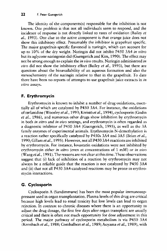
F. Erythromycin
Erythromycin is known to inhibit a number of drug oxidations, essen- tially all of which are catalyzed by P450 3A4. For instance, the oxidations of terfanidine (Woosley et al., 1993; Kivisto et al., 1994), cyclosporin (Godin et al., 1986), and numerous other drugs show inhibition by erythromycin in both in vitro and in vivo settings, and erythromycin is often regarded as a diagnostic inhibitor of P450 3A4 (Guengerich, 1995), as well as the 3A family enzymes of experimental animals. Erythromycin N-demethylation is a reaction rather specifically catalyzed by P450s 3A4 and 3A5 (Brian et al., 1990; Gillam etal., 1995). However, not all P450 3A4 reactions are inhibited by erythromycin. For instance, lovastatin oxidations were not inhibited by erythromycin either in vitro (even at concentrations of 1 mM) or in vivo (Wang et al., 1991). The reasons are not clear a t this time. These observations suggest that ( i ) lack of inhibition of a reaction by erythromycin may not always be a reliable guide that the reaction is not catalyzed by P450 3A4 and (ii) that not all P450 3A4-catalyzed reactions may be prone to erythro- mycin interactions.
G. Cyclosporin
Cyclosporin A (Sandimmune) has been the most popular immunosup- pressant used in organ transplantation. Plasma levels of this drug are critical because high levels lead to renal toxicity but low levels can lead to organ rejection. In contrast to chronic diseases where there is an opportunity to adjust the drug dosage, the first few days after organ transplant are usually critical and there is often not much opportunity for dose adjustment in this period. The major pathway of cyclosporin metabolism is via P450 3A4 (Kronbach et al., 1988; Combalbert et al., 1989; Aoyama et al., 1989), with

P450 and Drug-Drug Interactions 23
three major metabolites formed (Aoyama et al., 1989). Another complication in a liver transplant is that the level of P450 3A4 in the donor liver must be considered as well as in the recipient’s liver, small intestine, and other tissues.
The effect of modulations on cyclosporin levels was apparently first noted by Godin et al. (1986). Rifampicin treatment led to lower plasma levels of cyclosporin and erythromycin treatment led to higher levels. Subse- quently the involvement of P450 3A4 in the oxidation was clearly demon- strated (Combalbert et al., 1989; Kronbach et al., 1988; Aoyama et al., 1989). Similar cases were seen by Watkins and associates (Lucey et al., 1990), where plasma levels rose and fell. This group was also able to demon- strate that humans are oxidizing cyclosporin during the period when no liver is present in the body, arguing for a significant role of intestinal (or other extrahepatic) P450 3A4 in the process (Kolars et al., 1991). Because of the serious nature and high cost of organ transplants, efforts are being made to use noninvasive assays for hepatic and intestinal P450 3A4 to estimate what doses of cyclosporin will be most appropriate for individuals about to undergo transplantation surgery (Watkins et al., 1990).
Other immunosuppressants have been introduced and two of these, tacrolimus (FK506) and rapamycin, have been studied. Both are metabolized primarily by P450 3A4 (Sattler et al., 1992; Vincent et al., 1992). Tacrolimus is a more potent immunosuppressant but is not without toxic side effects. Evidence shows that the metabolites may retain more of the biological activity of the parent drug than in the case of cyclosporin.
H. Omeprazole
Omeprazole is an acid pump inhibitor used in the treatment of gastric ulcers. In general, it is considered to be a relatively safe drug and little in the way of adverse episodes have been reported. There have been two concerns about omeprazole, induction and inhibition.
Maurel and associates reported that omeprazole could induce P450s 1 A l and 1A2 in cultured human hepatocyte cultures (Diaz et al., 1990). This observation was considered of interest because many procarcinogens are known to be activated to genotoxic forms by P450s 1Al and 1A2 (Ioannides and Parke, 1987; Guengerich and Shimada, 1991), although it should be emphasized that there is a 40-fold variation in P450 1A2 among humans and there is not strong evidence to date that differences have a major influence in the risk of any cancers (Butler et al., 1989; Lang et al., 1994). Further studies with noninvasive assays of P450 1A2 showed that induction by omeprazole could be demonstrated, but only in individuals deficient in P450 2C19 (Rost et al., 1992). This phenomenon is due to the involvement of P450 2C19 [along with P450 3A4 (Andersson et al., 1990, 1993)] in the metabolism of omeprazole.

24 F. Peter Guengerich
The induction of P450 1A family enzymes by omeprazole was unusual in the sense that such induction of the orthogous P450s was not seen in mice or rats, in vitro or in vivo (Diaz et al., 1990). Omeprazole did not displace known radiolabeled ligands bound to the Ah receptor, and a non- Ah receptor mechanism has been proposed (Lesca et a[., 1995; Diaz et al., 1990). However, Tukey and associates have shown that a sensitive construct containing an Ah receptor enhancer element connected to a reporter gene can be stimulated in cell culture in the presence of very high doses of omeprazole (Quattrochi and Tukey, 1993). Thus, the exact mechanism of P450 1A induction by omeprazole has not been definitively established. However, we can conclude that (i) very high doses of omeprazole itself are needed for induction and (ii) the general significance of a small amount of P450 1A induction in humans has yet to be established.
Omeprazole also appears to be a competitive inhibitor of P450 2C19, an enzyme involved in its metabolism (Unge et al., 1992). Such competition may be a reflection of the low amount of P450 2C19 present in the body (de Morais et af., 1994), especially those individuals showing the deficient genetic polymorphism (Guengerich, 1995).
VI. Other Issues
Two other issues will be mentioned with regard to interactions. Neither can be considered a direct problem in the sense of human drug-drug interac- tions, but both bear on studies in this area and need to be considered.
A. Barbiturate and Peroxisome Proliferation Inducers
Chemicals in these two categories are of interest in the pharmaceutical industry. We have already considered how barbiturates can induce human P450 enzymes such as P450 3A4 and 2C9 and lead to more rapid elimina- tion of drugs (e.g., 17a-ethynylestradiol, vide strpra). Induction of enzymes by the peroxisome proliferation agents does not seem to affect the clearance of drugs, as the only P450s that seem to be induced are in the 4A subfamily and these are apparently not extensively involved in the metabolism of drugs.
A major concern with both of these groups of compounds is the correla- tion between enzyme induction and tumor promotion (Lubet et al., 1989; Rao and Reddy, 1991). There is a concern that a compound in either of these groups (i.e., that shows, e.g., P450 2B or 4A induction in animals) might also be a tumor promoter and be positive in a long-term animal cancer bioassay. The barbiturate group includes a variety of barbiturates, hydantoins, and miscellaneous compounds (Diwan et al., 1988). The peroxi- some proliferation group includes plasticizers, phthalates, and many phar-

P450 and Drug-Drug Interactions 25
maceutical groups such as leukotriene receptor antagonists (Rao and Reddy, 1991; Bars et al., 1993).
The correlation between tumor promotion and induction is certainly not very strict in either group of compounds. Further, the relevance to humans and human health is still a matter of speculation. A considerable amount of epidemiology evidence is available from people who have used barbiturates for epilepsy on a long-term basis, and no real increase in tumors was seen (Olsen et al., 1989). It would appear that even the doses adminis- tered to humans in such settings are not enough to show the hepatocyte proliferation observed when animals are treated with high doses.
Good evidence has been obtained that humans have a functional PPAR system, but actual in vivo induction has not been demonstrated in humans, and even those individuals using clofibrate, a known inducer of this response in animals, have not shown any evidence of liver tumors. It is possible that rodents contain genes relevant to cancer that are activated by the PPAR. Of interest are recent observations that the PPAR system is a mixture of different receptors that interact with individual members of the RXR recep- tor family to activate PPAR-associated genes. Also, certain fatty acids have actually proven to be better inducers than any of the prototypic synthetic ligands; the possibility exists for highly active metabolites of fatty acids.
B. In Vitrolin Vivo Comparisons
One general problem involves approaches to searching for possible drug-drug interactions. Ideally this should be done first in in vitro settings to expedite the process, and some approaches looking for enzyme inhibition and induction have been mentioned (vide supra). However, there is the matter of validation of in vivo results in in vivo settings.
In principle, one can readily look for the inhibition and induction if an appropriate noninvasive assay is available. The compound under investiga- tion can be administered to humans, and the alteration of pharmacokinetics of another drug can be observed. This process seems to work reasonably well in some cases. For instance, the induction and inhibition of human P450 1A2 can be inferred from changes in the N3-demethylation of caffeine (Butler et al., 1992; Kalow and Tang, 1993). Other noninvasive assays in which a generally acceptable degree of validation has been obtained include P450 2A6 and coumarin 7-hydroxylation (Cholerton et al., 1992), P450 2C9 and tolbutamide hydroxylation and warfarin 7-hydroxylation (Breimer et al., 1978; Rettie et al., 1992, 1994; Knodell et al., 1987), P450 2D6 and debrisoquine 4-hydroxylation (Mahgoub et al., 1977) and several other assays (Evans et al., 1989; Eichelbaum et al., 1979), and now P450 2E1 and chlorzoxazone 6-hydroxylation (Kim et al., 1995). However, there seems to still be a problem with P450 3A4, which is unfortunate as this appears to be the main human P450 involved in the oxidation of drugs (Kinirons et
26 F. Peter Guengerich
al., 1993). The differences among individuals are not genetic, and temporal intraindividual changes in in vivo parameters are observed. A number of reactions (of different substrates) that clearly seem to be associated with P450 3A4 in vitro do not show good in vivo corrections (Kinirons et al., 1993). There are several possible reasons, including the balance between hepatic and small intestine oxidation with individual compounds, the pres- ence of inhibitors in the diet (Bailey et al., 1994), and possibly the contribu- tion of the MDRl and other pump proteins in influencing cellular concentra- tions (Schuetz et al., 1995).
The problem has been choosing the most appropriate in vivo assay for P450 3A4, and the merits and disadvantages of several probe drugs have been discussed (Kinirons et al., 1993; Lown et al., 1992; Ged et al., 1989; Thummel et al., 1993). Currently the list of possibilities includes nifedi- pine and felodipine oxidation, erythromycin conversion to C02, 6p- hydroxycortisol production, midozolam 4-hydroxylation, lidocaine N- deethylation, and dapsone N-hydroxylation.
VII. Summary and Conclusions
Many adverse drug-drug interactions are attributable to pharmacoki- netic problems and can be understood in terms of alterations of P450- catalyzed reactions. Much is now known about the human P450 enzymes and what they do, and it has been possible to apply this information to issues related to practical problems. A relatively small subset of the total number of human P450s appears to be responsible for a large fraction of the oxidation of drugs. The three major reasons for drug-drug interactions involving the P450s are induction, inhibition, and possibly stimulation, with inhibition appearing to be the most important in terms of known clinical problems.
With the available knowledge of human P450s and reagents, it is possible to do in vitro experiments with drugs and make useful predictions. The results can be tested in vivo, again using assays based on our knowledge of human P450s. This approach has the capability of not only improving predictions about which drugs might show serious interaction problems, but also decreasing the number of in vivo interaction studies that must be performed. These approaches should improve with further refinement and technical advances.
Acknowledgments
Research in this area in the author’s laboratory has been supported in part by United States Public Health Service grants CA44353 and ES00267. Thanks are extended to E. Rochelle for assistance in preparation of the manuscript.
P450 and Drug-Drug Interactions 27
References
Andersson, T., Regirdh, C. G., Dahl-Puustinen, M. L., and Bertilsson, L. (1990). Slow omepra- zole metabolizers are also poor S-mephenytoin hydroxylators. Thera. Drug Monit. 12,
Andersson, T., Miners, J. O., Veronese, M. E., Tassaneeyakul, W., Meyer, U. A., and Birkett, D. J. (1993). Identification of human liver cytochrome P450 isoforms mediating omepra- zole metabolism. BY. J. Clin. Pharmacol. 36, 521-530.
Aoyama, T., Yamano, S., Waxman, D. J., Lapenson, D. P., Meyer, U. A., Fischer, V., Tyndale, R., Inaba, T., Kalow, W., Gelboin, H. V., and Gonzalez, F. J. (1989). Cytochrome P- 450 hPCN3, a novel cytochrome P-450 IIIA gene product that is differentially expressed in adult human liver. J. Biol. Chem. 264, 10388-10395.
Bailey, D. G. (1995). Food-drug interactions: Emphasis on grapefruit juice effects. Can. J. Clin.
Bailey, D. G., Spence, J. D., Munoz, C., and Arnold, J. M. 0. (1991). Interaction of citrus juices with felodipine and nifedipine. Lancet 337, 268-269.
Bailey, D. G., Arnold, J. M. O., Strong, H. A., Munoz, C., and Spence, J. D., (1993). Effect of grapefruit juice and naringin on nisoldipine pharmacokinetics. Clin. Pharmacol. Ther.
Bailey, D. G., Arnold, J. M. O., and Spence, J. D., (1994). Grapefruit juice and drugs: how significant is the interaction? Clin Pharmacokinet. 26, 91-98.
Bars, R. G., Bell, D. R., and Elcombe, C. R. (1993). Induction of cytochrome P450 and peroxisomal enzymes by clofibric acid in vivo and in uitro. Biochem. Pharmacol. 45,2045- 2053.
Beaune, P., Dansette, P., Flinois, J. P., Columelli, S., Mansuy, D., and Leroux, J. P. (1979). Partial purification of human liver cytochrome P-450. Biochem. Biophys. Res. Commun.
Bell, D. R., Plant, N. J., Rider, C. C., Na, L., Brown, S., Ateitalla, I., Acharya, S. K., Davies, M. H., Elias, E., Jenkins, N. A., Gilbert, D. J., Copeland, N. G., and Elcombe, C. R. (1993). Species-specific induction of cytochrome P-450 4A RNAs: PCR cloning of partial guinea-pig, human and mouse CYP4A cDNAs. Biochem. J . 294, 173-180.
Bensoussan, C., Delaforge, M., and Mansuy, D. (1995). Particular ability of cytochromes P450 3A to form inhibitory P45O-iron-metabolite complexes upon metabolic oxidation of amino drugs. Biochem. Pharmacol. 49, 591-602.
Bocker, R. H., and Guengerich, F. P. (1986). Oxidation of 4-aryl- and 4-alkyl-substituted 2,6- dimethyl-3,5-bis(alkoxycarbonyl)-l,4-dihydropyridines by human liver microsomes and immunochemical evidence for the involvement of a form of cytochrome P-450. J. Med. Chem. 29,1596-1603.
Bolt, H. M., Kappus, H., and Remmer, H. (1973). Studies on the metabolism of ethynylestradiol in vitro and in vivo: The significance of 2-hydroxylation and the formation of polar products. Xenobiotica 3 , 773-785.
Bolt, H. M., Bolt, M., and Kappus, H., (1977). Interaction of rifampicin treatment with pharmacokinetics and metabolism of ethinyloestradiol in man. Acta Endocrinol. (Copen- hagen) 85, 189-197.
Brady, J. F., Xiao, F., Wang, M. H., Li, Y., Ning, S. M., Gapac, J. M., and Yang, C. S. (1991). Effects of disulfiram on hepatic P45OIIE1, other microsomal enzymes, and hepatotoxicity in rats. Toxicol. Appl. Pharmacol. 108, 366-373.
Breimer, D. D., Zilly, W., and Richter, E. (1978). Influence of corticosteroids on hexobarbital and tolbutamide disposition. Clin. Pharmacol. Ther. 24,208-212.
Brian, W. R., Sari, M.-A., Iwasaki, M., Shimada, T., Kaminsky, L. S . , and Guengerich, F. P. (1990). Catalytic activities of human liver cytochrome P-450 IIIA4 expressed in Saccharo- myces cereuisiae. Biochemistry 29, 11280-1 1292.
415-416.
Pharmacol. 2, 10-14.
54,589-594.
88, 826-832.

28 F. Peter Guengerich
Brodie, A. M. H. (1994). Aromatase inhibitors in the treatment of breast cancer. J . Steroid Biochem. Mol. Biol. 49, 281-287.
Brodie, B. B. Gillette, J. R., and LaDu, B. N. (1958). Enzymatic metabolism of drugs and other foreign compounds. Annu. Reo. Biochem. 27, 427-454.
Broly, F., Gaedigk, A., Heim, M., Eichelbaum, M., Morike, K., and Meyer, U. A. (1991). Debrisoquinekparteine hydroxylation genotype and phenotype: Analysis of common mu- tations and alleles of CYP2D6 in a European population. D N A Cell Biol. 10, 545-558.
Burke, M. D. (1981). Cytochrome P-450: A pharmacological necessity or a biochemical curios- ity? Biochem. Pharmacol. 30, 181-187.
Butler, M. A., Iwasaki, M., Guengerich, F. P., and Kadlubar, F. F. (1989). Human cytochrome P-45OPA (P-4501.42), the phenacetin 0-deethylase, is primarily responsible for the hepatic 3-demethylation of caffeine and N-oxidation of carcinogenic arylamines. Proc. Natl. Acad. Sci. U.S.A. 86, 7696-7700.
Butler, M. A., Lang, N. P., Young, J. F., Caporaso, N. E., Vineis, P., Hayes, R. B., Teitel, C. H., Massengill, J. P., Lawsen, M. F., and Kadlubar, F. F. (1992). Determination of CYPlA2 and NAT2 phenotypes in human populations by analysis of caffeine urinary metabolites. Phamacogenetics 2, 116-127.
Cholerton, S., Idle, M. E., Vas, A., Gonzalez, F. J., and Idle, J. R. (1992). Comparison of a novel thin-layer chromatographic-fluorescence detection method with a spectrofluorome- tric method for the determination of 7-hydroxycoumarin in human urine. J. Chromatogr.
Coleman, M. D., Tingle, M. D., and Park, B. K. (1991). Inhibition of dapsone-induced meth- aemoglobinaemia by cimetidine in the rat during chronic dapsone administration. J . Pharm. Phamacol. 43, 186-190.
Cornbalberr, J,, Fabre, I., Fabre, G., Dalet, I., Derancourt, J., Cano, J. P., and Maurel, P. (1989). Metabolism of cyclosporin A. IV. Purification and identification of the rifampicin- inducible human liver cytochrome P-450 (cyclosporin A oxidase) as a product of P45OIIIA gene subfamily. Drug Metab. Dispos. 17, 197-207.
Conney, A. H. ( 1967). Pharmacological implications of microsomal enzyme induction. Pharma- col. Rev. 19, 317-366.
Conney, A. H., Miller, E. C., and Miller, J. A. (1956). The metabolism of methylated aminoazo dyes. V. Evidence for induction of enzyme synthesis in the rat by 3-methylcholanthrene. Cancer Res. 16,450-459.
Cupp-Vickery, J. R., and Poulos, 7. L. (1995). Structure of cytochrome P4SOeryF involved in erythromycin biosynthesis. Nature Struct. Biol. 2, 144-153.
de Morais, S. M. F., Wilkinson, G. R., Blaisdell, J., Nakamura, K., Meyer, U. A., and Goldstein, J. A. (1994). The major genetic defect responsible for the polymorphism of S-mephenytoin metabolism in humans. J. Biol. Chem. 269, 15419-15422.
Denison, M. S., and Whitlock, J. P. (1995). Xenobiotic-inducible transcription of cytochrome P450 genes. J . Biol. Chem. 270, 18175-18178.
Diaz, D., Fabre, I., Daujat, M., Saintaubert, B., Bories, P., Michel, H., and Maurel, P. (1990). Omeprazole is an aryl hydrocarbon-like inducer of human hepatic cytochrome-P450. Gastroenterology 99, 737-747.
Distlerath, L. M. and Guengerich, F. P. (1987). Enzymology of human liver cytochromes P- 450. In “Mammalian Cytochromes P-450” (F. P. Guengerich, ed.), Vol. 1, pp. 133-198. CRC Press, Boca Raton, FL.
Diwan, B. A., Rice, J. M., Nims, R. W., Lubet, R. A., Hu, H., and Ward, J. M. (1988). P- 450 enzyme induction by 5-ethyl-5-phenylhydantoin and 5,5-diethylhydantoin, analogues of barbiturate tumor promoters phenobarbital and barbital, and promotion of liver and thyroid carcinogenesis initiated by N-nitrosodiethylamine in rats. Cancer Res. 48,2492- 2497.
Eichelbaum, M., Spannbrucker, N., Steincke, B., and Dengler, H. J. (1979). Defective N- oxidation of sparteine in man: A new pharmacogenetic defect. Eur. J. Clin. Pharmacol.
575, 325-330.
16.183-187.

P450 and Drug-Drug Interactions 29
Ellis, S. W., Hayhurst, G. P., Smith, G., Lightfoot, T., Wong, M. M. S., Simula, A. P., Ackland, M. J., Sternberg, M. J. E., Lennard, M. S., Tucker, G. T., and Wolf, C. R. (1995). Evidence that aspartic acid 301 is a critical substrate-contact residue in the active site of cytochrome P450 2D6. J. Biol. Chem. 270,29055-29058.
Evans, W. E., Relling, M. V., Petros, W. P., Meyer, W. H., Mirro, J., Jr., and Crom, W. R. (1989). Dextromethorphan and caffeine as probes for simultaneous determination of debrisoquin-oxidation and N-acetylation phenotypes in children. Ctin. Pbarmacol. Ther.
Fischbach, T., and Lenk, W. (1985). Additional routes in the metabolism of phenacetin. Xenobiotica 15, 149-164.
Fonne-Pfister, R., and Meyer, U. A. (1988). Xenobiotic and endobiotic inhibitors of cytochrome P45Odbl function, the target of the debrisoquine/sparteine type polymorphism. Biochem. Pharmacol. 37,3829-3835.
Gan, L. S . L., Acebo, A. L., and Alworth, W. L. (1984). 1-Ethynylpyrene, a suicide inhibitor of cytochrome P-450 dependent benzo[a]pyrene hydroxylase activity in liver microsomes. Biochemistry 23, 3827-3836.
Ged, C., Rouillon, J. M., Pichard, L., Combalbert, J., Bressot, N., Bories, P., Michel, H., Beaune, P., and Maurel, P. (1989). The increase in urinary excretion of 6~-hydroxycortisol as a marker of human hepatic cytochrome P45OIIIA induction. Br. J. Clin. Pharmacol.
Gerber, M. C . , Tejwani, G. A., Gerber, N., and Bianchine, J. R. (1985). Drug interactions
Gibson, G. G. (1993). Peroxisome proliferators: Paradigms and prospects. Toxicol. Lett.
Gillam, E. M., J., Guo, Z., Ueng, Y.-F., Yamazaki, H., Cock, I., Reilly, P. E. B., Hooper, W. D., and Guengerich, F. P. (1995). Expression of cytochrome P450 3A5 in Escherichia coli: Effects of 5’ modifications, purification, spectral characterization, reconstitution conditions, and catalytic activities. Arch. Biochem. Biophys. 317, 374-384.
Godin, J. R. P., Sketris, I. S., and Belitsky, P. (1986). Erythromycin-cyclosporine interaction. Drug Intell. Ciin. Pharm. 20, 504-505.
Gonzalez, F. J. (1989). The molecular biology of cytochrome P450s. Pharmacol. Rev. 40,
Gonzalez, F. J., and Meyer, U. A. (1991). Molecular genetics of the debrisoquin-sparteine polymorphism. Clin. Pharmacol. Ther. 50, 233-238.
Gonzalez, F. J., Skoda, R. C., Kimura, S., Umeno, M., Zanger, U. M. Nebert, D. W., Gelboin, H. V., Hardwick, J. P., and Meyer, U. A. (1988). Characterization of the common genetic defect in humans deficient in debrisoquine metabolism. Nature (London) 331,442-446.
Gonzalez, F. J., Aoyama, T., and Gelboin, H. V., (1991a). Expression of mammaliancytochrome P450 using vaccinia virus. In “Methods in Enzymology” (M. R. Waterman and E. F. Johnson, eds.), Vol. 206, pp 85-92. Academic Press, San Diego, CA.
Gonzalez, F. J., Kimura, S., Tamura, S., and Gelboin, H. V., (1991b). Expression of mammalian cytochrome P450 using baculovirus. In “Methods in Enzymology” (M. R. Waterman and E. F. Johnson, eds.), Vol. 206, pp 93-99. Academic Press, San Diego, CA.
Grace, J. M., Kinter, M. T., and Macdonald, T. L. (1994). Atypical metabolism of deprenyl and its enantiomer, (S)-( + )-N,a-dimethyl-N-propynylphenethylamine, by cytochrome P450 2D6, Chem. Res. Toxicol. 7,286-290.
Guengerich, F. P. (1987). Enzymology of rat liver cytochromes P-450. In “Mammalian Cyto- chromes P-450” (F. P. Guengerich, ed.) Vol. 1, pp. 1-54. CRC Press, Boca Raton, FL.
Guengerich, F. P. (1988). Oxidation of 17a-ethynylestradiol by human liver cytochrome P- 450. Mol. Pharmacol. 33, 500-508.
Guengerich, F. P. (1 989). Characterization of human microsomal cytochrome P-450 enzymes. Annu. Rev. Pharmacol. Toxicol. 29, 241-264.
45, 568-573.
28,373-387.
with cimetidine: An update. Pharmacol. Ther. 27, 353-370.
68, 193-201.
243-288.

30 F. Peter Guengerich
Guengerich, F. P. (1 990a). Mechanism-based inactivation of human liver cytochrome P-450
Guengerich, F. P. (1990b). Metabolism of 17a-ethynylestradiol in humans. Life Sci. 47,1981-
Guengerich, F. P. (1993). Cytochrome P450 enzymes. Am. Sci. 81,440-447. Guengerich, F. P. (1995). Human cytochrome P450 enzymes. In “Cytochrome P450” (P. R.
Ortiz de Montellano, ed.). 2nd ed., pp. 473-535. Plenum, New York. Guengerich, F. P., and Kim, D.-H. (1990). In uitro inhibition of dihydropyridine oxidation
and aflatoxin B, activation in human liver microsomes by naringenin and other flavonoids. Carcznogenesis (London) 11, 2275-2279.
Guengerich, F. P., and Shimada, T. (1991). Oxidation of toxic and carcinogenic chemicals by human cytochrome P-450 enzymes. Chem. Res. Toxicol. 4, 391-407.
Guengerich, F. P., Martin, M. V., Beaune, P. H., Kremers, P., Wolff, T., and Waxman, D. J. (1986a). Characterization of rat and human liver microsomal cytochrome P-450 forms involved in nifedipine oxidation, a prototype for genetic polymorphism in oxidative drug metabolism. J . Biol. Chem. 261, 5051-5060.
Guengerich, F. P., Miiller-Enoch, D., and Blair, I. A. (1986b). Oxidation of quinidine by human liver cytochrome P-450. Mol. Phamacol. 30, 287-295.
Guengerich, F. P., Kim, D.-H., and Iwasaki, M. (1991). Role of human cytochrome P-450 IIEl in the oxidation of many low molecular weight cancer suspects. Chem. Res. Toxicol.
Guengerich, F. P., Gillam, E. M. J., Martin, M. V., Baba, T., Kim, B.-R., Shimada, T., Raney, K. D., and Yun, C.-H. (1994a). The importance of cytochrome P450 3A enzymes in drug metabolism. In “Schering Foundation Workshop 13, Assessment of the Use of Single Cytochrome P450 Enzymes in Drug Research” (M. R. Waterman and M. Hildebrand, eds.), pp. 161-186. Springer-Verlag, Berlin.
Guengerich, F. P., Kim, B.-R., Gillam, E. M. J., and Shimada, T. (1994b). Mechanisms of enhancement and inhibition of cytochrome P450 catalytic activity. In Proceedings of the Eighth International Conference on Cytochrome P450: Biochemistry, Biophysics, and Molecular Biology” (M. C . Lechner, ed.), pp. 97-101. John Libbey Eurotext, Paris
Guengerich, F. P., Gillam, E. M. J., and Shimada, T. (1996). New applications of bacterial systems to problems in toxicology. Crit. Rev. Toxicol. 26, 551-583.
Guillouzo, A., Morel, F., Fardel, O., and Meunier, B. (1993). Use of human hepatocyte cultures for drug metabolism studies. Toxicology 82, 209-219.
Guo, Z. , Gillam, E. M. J., Ohmori, S., Tukey, R. H., and Guengerich, F. P. (1994). Expression of modified human cytochrome P450 1Al in Escherichia coli: Effects of 5‘ substitution, stabilization, purification, spectral characterization, and catalytic properties. Arch. Bio- chem. Biophys. 312,436-446.
Halpert, J. A., and Guengerich, F. P. (1997). Enzyme inhibition and stimulation. In “Biotransfor- mation” (F. P. Guengerich, ed.), Vol. 3. pp. 21-35. Pergamon, New York.
Halpert, J. A., Miller, N. E., and Gorsky, L. D. (1985). On the mechanism of the inactivation of the major phenobarbital-inducible isozyme of rat liver cytochrome P-450 by chloram- phenicol. J. Biol. Chem. 260, 8397-8403.
Hankinson, 0. (1993). Research on the aryl hydrocarbon (dioxin) receptor is primed to take off. Arch. Biochem. Biophys. 300, 1-5.
Huang, M. T., Johnson, E. F., Miiller-Eberhard, U., Koop, D. R., Coon, M. J., and Conney, A. H. (1981). Specificity in the activation and inhibition by flavonoids of benzol[a]pyrene hydroxylation by cytochrome P-450 isozymes from rabbit liver microsomes. J . Biol. Chem.
Ioannides, C., and Parke, D. V. (1987). The cytochromes P-488-a unique family of enzymes involved in chemical toxicity and carcinogenesis. Biochem. Phamacol. 36, 4197-4207.
Ioannides, C., and Parke, D. V. (1990). The cytochrome P450 1 gene family of microsomal hemoproteins and their role in the metabolic activation of chemicals. Drug Metab. Rev.
IIIA4 by gestodene. Chem. Res. Toxicol. 3, 363-371.
1988.
4, 168-179.
256,10897-10901.
22. 1-85.

P450 and Drug-Drug Interactions 31
Ioannides, C., and Parke, D. V. (1993). Induction of cytochrome P4501 as an indicator of potential chemical carcinogenesis. Drug Metab. Rev. 25, 485-501.
Islam, S . A., Wolf, C. R., Lennard, M. S . , and Sternberg, M. J. E. (1991). A three-dimensional molecular template for substrates of human cytochrome P450 involved in debrisoquine 4-hydroxylation. Carcinogenesis (London) 12, 221 1-2219.
Janz, D., and Schmidt, D. (1974). Anti-epileptic drugs and failure of oral contraceptives. Lancet 1, 1113.
Johansson, I., Lundqvist, E., Bertilsson, L., Dahl, M. L., SjBqvist, F., and Ingelman-Sundberg, M. (1993). Inherited amplification of an active gene in the cytochrome P450 CYP2D locus as a cause of ultrarapid metabolism of debrisoquine. Proc. Natl. Acad. Sci. U.S.A.
Jansson, K. H., and Lindeke, B. (1992). Cytochrome P-455 nm complex formation in the metabolism of phenylalkylamines. XII. Enantioselectivity and temperature dependence in microsomes and reconstituted cytochrome P-450 systems from rat liver. Chirality
Jung-Hoffmann, C., and Kuhl, H. (1990). Interaction with the pharmacokinetics of ethinyles- tradiol of progestogens contained in oral contraceptives. Contraception 40, 299-312.
Kalow, W., and Tang, B. K. (1993). The use of caffeine for enzyme assays: A critical appraisal. Clin. Pharmacol. Ther. 53, 503-514.
Kapitulnik, J., and Gonzalez, F. J. (1993). Marked endogenous activation of the CYPlAl and CYPlA2 genes in the congenitally jaundiced Gum rat. Mol. Pharmacol. 43, 722-725.
Kapitulnik, J., and Ostrow, J. D. (1978). Stimulation of bilirubin catabolism in jaundiced Gunn rats by an inducer of microsomal mixed-function monooxygenases. Proc. Natl. Acad. Sci. U.S.A. 75, 682-685.
Kerr, B. M., Thummel, K. E., Wurden, C. J., Klein, S . M., Kroetz, D. L., Gonzalez, F. J., and Levy, R. H. (1994). Human liver carbamazepine metabolism: Role of CYP3A4 and CYP2C8 in 10,l I-epoxide formation. Biochem. Pharmacol. 47, 1969-1979.
Kim, R. B., O’Shea, D., and Wilkinson, G. R. (1994). Relationship in healthy subjects between CYP2E1 genetic polymorphism and the 6-hydroxylation of chlorzoxazone: A putative measure of CYP2E1 activity. Pharmacogenetiw 4, 162-165.
Kim, R. B., O’Shea, D., and Wilkinson, G. R. (1995). Interindividual variability of chlorzoxa- zone 6-hydroxylation in men and women and its relationship to CYP2E1 genetic polymor- phisms. Clin. Pharmacol. Ther. 57, 645-655.
Kinirons, M. T., O’Shea, D., Downing, T. E., Fitzwilliam, A. T., Joellenbeck, L., Groopman, J. D., Wilkinson, G. R., and Wood, A. J. J. (1993). Absence of correlations among 3 putative in vivo probes of human cytochrome P4503A activity in young healthy men. Clin. Pharmacol. Ther. 54, 621-629.
Kitada, M., and Kamataki, T. (1979). Partial purification and properties of cytochrome P450 from homogenates of human fetal livers. Biochem. Pharmacol. 28, 793-797.
Kivisto, K. T., Neuvonen, P. J., and Klotz, U. (1994). Inhibition of terfenadine metabolism: Pharmacokinetic and pharmacodynamic consequences. Clin. Pharmacokinet. 27, 1-5.
Klehr, H., Eyer, P., and Schafer, W. (1987). Formation of 4-ethoxy-4’-nitrosodiphenylamine in the reaction of the phenacetin metabolite 4-nitrosophenetol with glutathione. Biol. Chem. Hoppe-Seyler 368, 895-902.
Knodell, R. G., Hall, S. D., Wilkinson, G. R., and Guengerich, F. P. (1987). Hepatic metabolism of tolbutamide: Characterization of the form of cytochrome P-450 involved in methyl hydroxylation and relationship to in vivo dispositi0n.f. Pharmacol. E x p . Ther. 241,1112- 1119.
Knodell, R. G., Browne, D., Gwodz, G. P., Brian, W. R., and Guengerich, F. P. (1991). Differential inhibition of human liver cytochromes P-450 by cimetidine. Gastroenterology
Kolars, J. C., Awni, W. M., Merion, R. M., and Watkins, P. B. (1991). First-pass metabolism
90,11825-11829.
4,469-477.
101,1680-1691.
of cyclosporin by the gut. Lancet 338, 1488-1490.

32 F. Peter Guengerich
Koymans, L., Vermeulen, N. P. E., van Acker, S. A. B. E., te Koppele, J. M., Heykants, J. J. P., Lavrijsen, K., Meuldermans, W., and DonnC-Op den Kelder, G. M. (1992). A predictive model for substrates of cytochrome P45O-debrisoquine (2D6). Chem. Res. Toxicol. 5, 211-219.
Kronbach, T., Fischer, V., and Meyer, U. A. (1988). Cyclosporine metabolism in human liver: Identification of a cytochrome P-450IIl gene family as the major cyclosporine-metabolizing enzyme explains interactions of cyclosporine with other drugs. Clin. Phurrnacol. Ther.
Kuby, S. A. (1991). “A Study of Enzymes,” Vol. 1, pp. 253-281. CRC Press, Boca Raton, FL. Kunze, K. L., and Trager, W. F. (1993). Isoform-selective mechanism-based inhibition of human
cytochrome P450 1A2 by furafylline. Chem. Res. Toxicol. 6, 649-656. Lang, N. P., Butler, M. A., Massengill, J., Lawson, M., Stotts, R. C., Maurer-Jensen, M., and
Kadlubar, F. F. (1994). Rapid metabolic phenotypes for acetyltransferase and cytochrome P4501A2 and putative exposure to food-borne heterocyclic amines increase the risk for colorectal cancer or polyps. Cancer Epidemiol. Biomurkers Prev. 3, 675-682.
Lasker, J. M., Huang, M.-T., and Conney, A. H. (1982). In vivo activation of zoxazolamine metabolism by flavone. Science 216, 1419-1421.
Lee, C. A., Thummel, K. E., Kalhorn, T. F., Nelson, S. D., and Slattery, J. T. (1991). Activation of acetaminophen-reactive metabolite formation by methylxanthines and known cyto- chrome D-450 activators. Drug Metab. Dispos. 19, 966-971.
Lee, M. S., Kliewer, S. A., Provencal, j., Wright, P. E., and Evans, R. M. (1993). Structure of rhe retinoid X receptor a DNA binding domain: A helix required for homodimeric DNA binding. Science 260, 1117-1121.
Lesca, P., Peryt, B., Larrieu, G., Alvinerie, M., Galtier, P., Daujat, M., Maurel, P., and Hoogen- boom, L. (1995). Evidence for the ligand-independent activation of the AH receptor. Biochem. Biophys. Res. Commun. 209,474-482.
Levin, W., Jacobson, IM., Sernatinger, E., and Kuntzman, R. (1973). Breakdown of cytochrome P-450 heme by secobarbital and other allyl-containing barbiturates. Drug Metub. Dispos.
Liang, Q., and Fuico, A. J. ( 1995). Transcriptional regulation of the genes encoding cytochrornes P450BM., and P4508M.3 in Bacillus megaterium by the binding of Bm 3R1 repressor to Barbie box elements and operator sites. J . Biol. Chem. 270, 18606.
Liehr, J. G., Ricci, M. J., Jefcoate, C. R., Hannigan, E. V.,Hokanson, J. A., and Zhu, B. T. (1995). 4-hydroxylation of estradiol by human uterine myometrium and myoma microsomes: Implications for the mechanism of uterine tumorigenesis. Proc. Natl. Acud. Sci. U.S.A.
Loretz, L. J., Li, A. P., Flye, M. W., and Wilson, A. G. E. (1989). Optimization of cryopreserva- tion procedures for rat and human hepatocytes. Xenobiotica 19, 489-498.
Lown, K., Kolars, J., Turgeon, K., Merion, R., Wrighton, S. A., and Watkins, P. B. (1992). The erythromycin breath test selectively measures P45OIIIA in patients with severe liver disease. Clin. Pharmacol. Ther. 51, 229-238.
Lubrt, R. A., Nims, R. W., Ward, J. M., Rice, J. M., and Diwan, B. A. (1989). Induction of cytochrome P-450b and its relationship to liver tumor promotion. J. Am. Coll. Toxicol.
Lucey, M. R., Kolars, J. C., Merion, R. M., Campbell, D. A., Aldrich, M., and Watkins, P. B. (1990). Cyclosporin toxicity at therapeutic blood levels and cytochrome P-450 IIIA. Lancet 335, 11-15.
Mahgoub, A., Idle, J. R., Dring, L. G., Lancaster, R., and Smith, R. L. (1977). Polymorphic hydroxylation of debrisoquine in man. Lancet 2, 584-586.
Mansuy, D., Battioni, P., Chottard, J. C., Riche, C., and Chiaroni, A. (1983). Nitrosoalkane complexes of iron-porphyrins: Analogy between the bonding properties of nitrosoalkanes and dioxygen. I. Am. Chem. SOC. 105,455-463.
43, 630-635.
1,275-285.
92,9220-9224.
8, 259-268.

P450 and Drug-Drug Interactions 33
Morel, F., Beaune, P. H., Ratanasavanh, D., Flinois, J.-P., Yang, C.-S., Guengerich, F. P., and Guillouzo, A. (1990). Expression of cytochrome P-450 enzymes in cultured human hepatocytes. Eur. J. Biochem. 191,437-444.
Muerhoff, A. S. , Griffin, K. J., and Johnson, E. F. (1992). Characterization of a rabbit gene encoding a clofibrate-inducible fatty acid o-hydroxylase: CYP4A6. Arch. Biochem. Bio-
Nebert, D. W. (1989). The Ah locus: Genetic differences in toxicity, cancer, mutation, and birth defects. Crit. Rev. Toxicol. 20, 153-174.
Nelson, D. R., Kamataki, T., Waxman, D. J., Guengerich, F. P., Estabrook, R. W., Feyereisen, R., Gonzalez, F. J., Coon, M. J., Gunsalus, I. C., Gotoh, O., Okuda, K., and Nebert, D. W. (1993). The P450 superfamily: Update on new sequences, gene mapping, accession numbers, early trivial names of enzymes, and nomenclature. DNA Cell Biol. 12, 1-51.
Nocke-Finck, L., Brewer, H., and Reimers, D. (1973). Wirkung van Rifampicin auf den Mens- trationszylkus und die Ostrogenausscheidung bei Einnahme oraler Kontrazeptive. Dtsch. Med. Wochenschr. 98, 1521-1523.
Olsen, J. H., Boice, J. D., Jr., Jensen, J. P. A., and Fraumeni, J. P., Jr. (1989). Cancer among epileptic patients exposed to anticonvulsant drugs. J. Natl. Cancer Inst. 81, 803-808.
Ortiz de Montellano, P. R., and Correia, M. A. (1983). Suicidal destruction of cytochrome P- 450 during oxidative drug metabolism. Annu. Rev. Pharmacol. Toxicol. 23, 481-503.
Ortiz de Montellano, P. R., and Reich, N. 0. (1986). Inhibition of cytochrome P-450 enzymes. In “Cytochrome P-450” (P. R. Ortiz de Montellano, ed.), pp. 273-314. Plenum, New York.
Ortiz de Montellano, P. R., Kunze, K. L., Yost, G. S . , and Mico, B. A. (1979). Self-catalyzed destruction of cytochrome P-450: Covalent binding of ethynyl sterols to prosthetic heme. Proc. Natl. Acad. Sci. U.S.A. 76, 746-749.
Otton, S. V., Inaba, T., and Kalow, W. (1984). Competitive inhibition of sparteine oxidation in human liver by P-adrenoceptor antagonists and other cardiovascular drugs. Life Sci.
Perrot, N., Nalpas, B., Yang, C . S . , and Beaune, P. H. (1989). Modulation of cytochrome P450 isozymes in human liver, by ethanol and drug intake. Eur. J. Clin. Invest. 19,549-555.
Quattrochi, L. C., and Tukey, R. H. (1993). Nuclear uptake of the Ah (dioxin) receptor in response to omeprazole: Transcriptional activation of the human CYPl A1 gene. Mol. Pharmacol. 43, 504-508.
Raag, R., and Poulos, T. L. (1991). Crystal structures of cytochrome P-450cAM complexed with camphane, thiocamphor and adamatane: Factors controlling P-450 substrate hydrox- ylation. Biochemistry 30, 2674-2684.
Ramsden, R., Sommer, K. M., and Omiecinski, C. J. (1993). Phenobarbital induction and tissue-specific expression of the rat CYP2B2 gene in transgenic mice. J. Biol. Chem.
Rando, R. R. (1984). Mechanism-based enzyme inactivators. Phamacol. Rev. 36, 11 1-142. Raney, K. D., Shimada, T., Kim, D.-H., Groopman, J. D., Harris, T. M., and Guengerich,
F. P. (1992). Oxidation of aflatoxins and sterigmatocystin by human liver microsomes: Significance of aflatoxin Q1 as a detoxication product of aflatoxin B1. Chem. Res. Toxicol. 5,202-210.
Rangarajan, P. N., and Padmanaban, G. (1989). Regulation of cytochrome P-45Obk gene expression by a heme- and phenobarbitone-modulated transcription factor. Proc. Natl. Acad. Sci. U.S.A. 86, 3963-3967.
Rao, M. S., and Reddy, J. K. (1991). An overview of peroxisome proliferator-induced hepatocar- cinogenesis. Environ. Health Perspect. 93, 205-209.
Reimers, D., and Jezek, A. (1971). Rifampicin und andere Antituberkulotika bei gleichzeitiger oraler Kontrzeption. Prm. Pneumol. 25,255-262.
Remmer, H. (1959). The acceleration of evipan oxidation and the demethylation of methylami- nopyrine by barbiturates. Arch. Exp. Pathol. Pharmakol. 237, 296-307.
phys. 296, 66-72.
34,73-80.
268, 21722-21726.

34 F. Peter Guengerich
Rettie, A. E., Korzekwa, K. R., Kunze, K. L., Lawrence, R. F, Eddy, A. C., Aoyama, T., Gelboin, H. V., Gonzalez, F. J., and Trager, W. F. (1992). Hydroxylation of warfarin by human cDNA-expressed cytochrome P-450: A role for P-4502C9 in the etiology of (S)-warfarin-drug interactions. Chem. Res. Toxicol. 5 , 54-59.
Rettie, A. E., Wienkers, L. C., Gonzalez, F. J., Trager, W. F., and Korzekwa, K. R. (1994). Impaired (S)-warfarin metabolism catalysed by the R144C allelic variant of CYP2C9. Pbarmacagenetics 4, 39-42.
Richardson, H. L., Stier, A. R., and Borsos-Nachtnebel, E. (1952). Liver tumor inhibition and adrenal histologic responses in rats to which 3’-methyl4-dimethylaminoazobenzene and 20-methylcholanthrene were simultaneously administered. Cancer Res. 12, 356-361.
Roberts, E. S., Hopkins, N. E., Zaluzec, E. J., Gage, D. A., Alworth, W. L., and Hollenberg, P. F. (1994). Indentification of active-site peptides from ‘H-labeled 2-ethynylnaphthalene- inactivated P450 2Bl and 2B4 using amino acid sequencing and mass spectrometry. Biochemistry 33,3766-3771.
Rodrigues, A. D., Mulford, D. J., Lee, R. D., Surber, B. W., Kukulka, M. J., Ferrero, J. L., Thomas, S . B., Shet, M. S., and Estabrook, R. W. (1995). In vifro metabolism of terfenadine by a purified recombinant fusion protein containing cytochrome P450 3A4 and NADPH- P450 reductase. Drug Metab. Dispos. 23, 765-775.
Rost, K. L., Brosicke, H., Brockmoller, J., Scheffler, M., Helg, H., and Roots, I. (1992). Increase of cytochrome P450IA2 activity by omeprazole: Evidence by the ”C-[N-3-methyl]-caffeine breath test in poor and extensive metabolizers of S-mephenytoin. Clin. Pharmacol. Tber.
Kost, K. I.., Brosicke, H., Heinemeyer, G., and Roots, 1. (1994). Specific and dose-dependent enzyme induction by omeprazole in human beings. Hepatology 20, 1204-1212.
Sattler, M., Guengerich, F. P., Yun, C.-H., Christians, U., and Sewing,K.-F. (1992). Cytochrome P450 3A enzymes are responsible for biotransformation of FK506 and rapamycin in man and rat. Drug Metab. Dispos. 20, 753-761.
Schuetz, E. G., Schuetz, J. D., Thompson, M. T., Fisher, R. A., Madariage, J. R., and Strom, S. C. (1995). Phenotypic variability in induction of P-glycoprotein rnRNA by aromatic hydrocarbons in primary human hepatocytes. Mol. Carcinog. 12, 61-65.
Schwab, G. E., Raucy, J. L., and Johnson, E. F. (1988). Modulation of rabbit and human hepatic cytochrome P-450-catalyzed steroid hydroxylations by a-naphthoflavone. Mol. Pharmacol. 33, 493-499.
Sesardic, D., Boobis, A., Murray, B., Murray, S., Segura, J., De La Torre, R., and Davies, D. (1990). Furafylline is a potent and selective inhibitor of cytochrome P450 1A2 in man. BY. 1. Clin. Pharmacol. 29, 651-663.
Shah, R. R., Oates, N. S., Idle, J. R., Smith, R. L., and Lockhart, J. D. F. (1982). Impaired oxidation of debrisoquine in patients with perhexiline neuropathy. Br. Med. J. 284,
Silverman, R. 8. (1988). “Mechanism-based Enzyme Inactivation: Chemistry & Enzymology.” CRC Press, Boca Raton, FL.
Silverman, R. B. (1995). Mechanism-based enzyme inactivators. In “Methods in Enzymology,” Vol. 249, pp. 240-283. Academic Press, San Diego, CA.
Srrobl, G. R., von Kruedener, S., Stockigt, J., Guengerich, F. P., and Wolff, T. (1993). Deveiop- ment of a pharrnacophore for inhibition of human liver cytochrome P-450 2D6: Molecular modeling and inhibition studies, I . Med. Chem. 36, 1136-1145.
Thull, U., and Testa, B. (1994). Screening of unsubstituted cyclic compounds as inhibitors of monoamine oxidases. Biocbem. Pharmacol. 47,2307-2310.
Thummel, K. E., Shen, D. D., Carithers, R. L., Jr., Hartwell, P., Podoll, T. D., Trager, W. F., and Kunze, K. L. (1993). Prediction of in vivo midazolam clearance from hepatic CYP3A content and midazolam 1 ‘-hydroxylation activity in liver transplant patients. Abstr., North Am. International Society for tbe Study of Xenobiotics Meet., Stb, Tucson, AZ, 1993, Vol. 4, p. 235.
52, 170-180.
295-299.

P450 and Drug-Drug Interactions 35
Tian, G. C., Mook, R. A., Moss, M. L., and Frye, S. V. (1995). Mechanism of time-dependent inhibition of 5 a-reductases by A( 1)-4-azasteroids: Toward perfection of rates of time- dependent inhibition by using ligand-binding energies. Biochemistry 34, 13453-13459.
Ueng, Y.-F., Shimada, T., Yamazaki, H., and Guengerich, F. P. (1995). Oxidation of aflatoxin B1 by bacterial recombinant human cytochrome P450 enzymes. Chem. Res. Toxicol.
Unge, P., Svedberg, L. E., Nordgren, A., Blom, H., Andersson, T., and Lagerstrom, P. 0. (1992). A study of the interaction of omeprazole and warfarin in anticoagulated patients. Br. J. Clin. Pharmacol. 34, 509-512.
Vincent, S. H., Karanam, B. V., Painter, S. K., and Chiu, S. H. L. (1992). In vitro metabolism of FK-506 in rat, rabbit, and human liver microsomes: Identification of a major metabolite and of cytochrome P450 3A as the major enzymes responsible for its metabolism. Arch. Biochem. Biophys. 294, 454-460.
Wang, P., Mason, P. S., and Guengerich, F. P. (1980). Purification of human liver cytochrome P-450 and comparison to the enzyme isolated from rat liver. Arch. Biochem. Biophys.
Wang, R. W., Kari, P. H., Lu, A. Y. H., Thomas, P. E., Guengerich, F. P., and Vyas, K. P. (1991). Biotransformation of lovastatin. IV. Identification of cytochrome P-450 3A proteins as the major enzymes responsible for the oxidative metabolism of lovastatin in rat and human liver microsomes. Arch. Biochem. Biophys. 290, 355-361.
Watkins, P. B., Wrighton, S. A., Maurel, P., Schuetz, E. G., Mendez-Picon, G., Parker, G. A., and Guzelian, P. S. (1985). Identification of an inducible form of cytochrome P-450 in human liver. Proc. Natl. Acad. Sci. U.S.A. 82, 6310-6314.
Watkins, P. B., Hamilton, T. A., Annesley, T. M., Ellis, C. N., Kolars, J. C., and Voorhees, J. J. (1990). The erythromycin breath test as a predictor of cyclosporine blood levels. Clin. Pharmacol. Ther. 48, 120-129.
Waxman, D. J., Attisano, C., Guengerich, F. P., and Lapenson, D. P. (1988). Cytochrome P- 450 steroid hormone metabolism catalyzed by human liver microsomes. Arch. Biochem. Biophys. 263,424-436.
Woosley, R. L., Roden, D. M., Duff, H. J., Carey, E. L., Wood, A. J. J., and Wilkinson, G. R. (1981). Co-inheritance of deficient oxidative metabolism of encainide and debriso- quine. Am. Soc. Clin. Invest. 29, 501A.
Woosley, R. L., Chen, Y., Freiman, J. P., and Gillis, R. A. (1993). Mechanism of the cardiotoxic actions of terfenadine. JAMA, J. Am. Med. Assoc. 269, 1532-1536.
Wrighton, S. A., and Stevens, J. C. (1992). The human hepatic cytochromes P450 involved in drug metabolism. Crit. Rev. Toxicol. 22, 1-21.
Yun, C.-H., Hammons, G. J., Jones, G., Martin, M. V., Hopkins, N. E., Alworth, W. L., and Guengerich, F. P. (1992). Modification of cytochrome P450 1A2 enzymes by the mechanism-based inactivator 2-ethynylnaphthalene and the photoaffinity label 4- azidobiphenyl. Biochemistry 31, 10556-10563.
Yun, C.-H., Okerholm, R. A., and Guengerich, F. P. (1993). Oxidation of the antihistaminic drug terfenadine in human liver microsomes: Role of cytochrome P450 3A(4) in N- dealkylation and C-hydroxylation. Drug Metab. Dispos. 21,403-409.
Zilly, W., Breimer, D. D., and Richter, E. (1975). Induction of drug metabolism in man after rifampicin treatment measured by increased hexobarbital and tolbutamide clearance. Eur. 1. Clin. Pharmacol. 9, 219-227.
8,218-225.
199,206-219.

John-Michael Sauer Eric R. Stine
Lhanoo Gunawardhana Dwayne A. Hill
1. Glenn Sipes Department of Pharmacology and Toxicology
University of Arizona Tucson, Arizona 8572 I
The Liver as a Target for Chemical-Chemical Interactions
1. Chemically Induced Tissue Injury
At some dose all substances are poisons and capable of causing cellular injury and even death. Furthermore, all organs and organ systems within the body can be injured and their functions disrupted by exposure to toxic chemicals. Over the past decades, toxicologists have shown that chemically induced tissue injury and cell death are complex multifactorial processes. In the simplest case, a chemical would be directly cytocidal on cell contact by inhibiting vital cellular functions. However, in many instances, chemically induced tissue injury is not the simple case; it involves several elements which in and of themselves are complex.
In the 1970s the critical role of biotransformation in hepatic and extrahe- patic chemically induced tissue injury was established (Reid et al., 1971; Mitchell etal., 1976; Boyd, 1977; Boyd etal., 1980). Although it was known that foreign compounds are biochemically modified (biotransformation) to
Advances in Pharmacology, Volume 43 Copyright 9 1997 by Academic Press. All rights of reproduction in any form reserved. 1054-3589/97 $25.00 37

38 john-Michael Sauer et a/.
more water-soluble forms by specialized cellular processes, it became appar- ent that biotransformation may lead to the formation of reactive intermedi- ates which initiate events that result in tissue damage. Furthermore, other processes (conjugation) were shown to detoxify reactive intermediates and protect the tissue from injury (Brodie et al., 1971; Mitchell et al., 1973). In the late 1980s the idea that several other factors, besides metabolic activation and detoxification, influence the ability of a chemical to cause tissue injury gained acceptance. For example, the rate of tissue repair, as well as the infiltration of inflammatory cells, were found to play an important roles in some basic models of chemically induced hepatotoxicity (Bell et al., 1988; Laskin and Pilaro, 1986; Sipes eta!., 1989). These concepts gathered further support in the 1990s when several pharmacological interventions, which modulate the cellular immune response or cellular replication, were shown to alter the progression and final outcome of chemical-induced tissue injury (Sipes et al., 1991; Gunawardhana et al., 1993; ElSisi et al., 1993c; Edwards et al., 1993; Laskin and Pendino, 1995; Mehendale, 1995). A diagrammatic view of the various stages of tissue injury during acute toxicity just described is illustrated in Fig. 1 .
Modulation of the immune system, cell replication, and metabolizing systems each represent areas where one chemical may influence the toxicity of another. Understanding mechanisms of “interactive toxicology” is im-
FIGURE I Stages in the progression of chemically induced tissue injury. Currently it is accepted that there are various stages involved with chemical-induced tissue injury. These stages include the bioactivation of the chemical to a toxic metabolite, the induction of an inflammatory response, the resolution of inflammatory response, and the repair of tissue following toxic injury. The model shown illustrates these various stages over time.

Liver as a Target for Chemical-Chemical Interactions 39
portant, as humans and animals are not exposed to individual chemicals. More commonly, they are exposed to combinations of chemicals that can have differing effects on individual cells or organ systems. Interactive toxicol- ogy has been defined by the National Research Council (1988) as “a circum- stance in which exposure to two or more chemicals results in an altered biological response relative to that predicted from the actions of a single chemical. The multi-chemical exposure may be simultaneous or sequential in time and the altered response may result in enhanced (potentiation or synergism) or reduced (antagonism) toxicity.”
Because of its fundamental anatomy, high metabolic potential, and ability to clear xenobiotics from the blood, the liver represents an organ with a high susceptibility to the toxic effects of chemical-chemical interactions. Therefore, this chapter focuses on the liver as a target organ for chemical toxicity and presents examples of how chemicals interact biologically to alter toxic liver injury.
II. The Liver as a Target Organ for Chemically Induced Toxicity
The liver is a complex organ, composed of various cell types that interact with each other, but are adapted to performing specialized functions. Paren- chymal cells (hepatocytes) comprise approximately 80% of the total volume of the liver with 250 billion hepatocytes found in normal human adults (Arias et al., 1988). The sinusoidal space, and cells associated with the sinusoids, comprises the remaining liver volume. The endothelial cells total 44% of the sinusoidal cell volume, whereas 22% are Ito cells and 33% are Kupffer cells (Arias et al., 1988; Blouin et al., 1977). Lymphocytes and pit (natural killer) cells comprise the remaining 1 % of the sinusoidal cell volume.
Although it is impossible to assign orders of importance to specific cell types, the hepatocytes or parenchymal cells carry out the functions generally associated with the liver. Hepatocytes extract nutrients and other chemicals from the blood, produce bile to aid the digestive process and promote the absorption of lipids, secrete metabolic products into bile, synthesize proteins for hepatic and nonhepatic use, store energy-rich products such as fat and glycogen, and, among many other functions, metabolize drugs and chemi- cals. The nonparenchymal cells also have important properties. Kupffer cells are the resident macrophages of the liver and serve to phagocytize blood- borne toxicants and particulates from the circulation. The endothelial cells form a leaky barrier between the parenchymal cells and the hepatic blood flow. These fenestrated cells act as a sieve to prevent red blood cells and other cellular components from interacting with hepatocytes while allowing plasma components intimate access to microvilli of the hepatocyte. The fat-
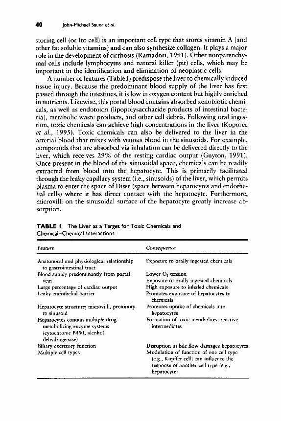
40 John-Michael Sauer et 01.
storing cell (or Ito cell) is an important cell type that stores vitamin A (and other fat soluble vitamins) and can also synthesize collagen. It plays a major role in the development of cirrhosis (Ramadori, 1991). Other nonparenchy- ma1 cells include lymphocytes and natural killer (pit) cells, which may be important in the identification and elimination of neoplastic cells.
A number of features (Table I ) predispose the liver to chemically induced tissue injury. Because the predominant blood supply of the liver has first passed through the intestines, it is low in oxygen content but highly enriched in nutrients. Likewise, this portal blood contains absorbed xenobiotic chemi- cals, as well as endotoxin (lipopolysaccharide products of intestinal bacte- ria), metabolic waste products, and other cell debris. Following oral inges- tion, toxic chemicals can achieve high concentrations in the liver (Koporec et al., 1995). Toxic chemicals can also be delivered to the liver in the arterial blood that mixes with venous blood in the sinusoids. For example, compounds that are absorbed via inhalation can be delivered directly to the liver, which receives 29% of the resting cardiac output (Guyton, 1991). Once present in the blood of the sinusoidal space, chemicals can be readily extracted from blood into the hepatocyte. This is primarily facilitated through the leaky capillary system (i.e., sinusoids) of the liver, which permits plasma to enter the space of Disse (space between hepatocytes and endothe- lial cells) where it has direct contact with the hepatocyte. Furthermore, microvilli on the sinusoidal surface of the hepatocyte greatly increase ab- sorption.
TABLE I Chemical-Chemical Interactions
The Liver as a Target for Toxic Chemicals and
Feature Consequence ~ ~~~
Anatomical and physiological relationship
Blood supply predominantly from portal
Large percentage of cardiac output Leaky endothelial barrier
Hepatocyte structure; microvilli, proximity
Hepatocytes contain multiple drug-
Exposure to orally ingested chemicals
Lower O2 tension Exposure to orally ingested chemicals High exposure to inhaled chemicals Promotes exposure of hepatocytes to
Promotes uptake of chemicals into
Formation of toxic metabolites, reactive
to gastrointestinal tract
vein
chemicals
to sinusoid hepatocytes
metabolizing enzyme systems intermediates (cytochrome P4S0, alcohol dehydrogenase)
Biliary excretory function Multiple cell types
Disruption in bile flow damages hepatocytes Modulation of function of one cell type
(e.g., Kupffer cell) can influence the response of another cell type (e.g., hepatocyte)

Liver as a Target for Chemical-Chemical Interactions 41
Another feature of the liver that predisposes it to chemical-induced injury is its ability to biotransform or metabolize chemicals. This important process is catalyzed by numerous enzymes that convert lipophilic compounds into more hydrophilic metabolites. These metabolites can be more readily excreted in the urine and feces. The liver is the most important organ of biotransformation, due largely to its high content and large diversity of enzymes capable of metabolizing foreign, as well as endogenous, chemicals. These enzymes include UDP-glucuronosyltransferases, g1utathione-S- transferases, cytochromes P450, and FAD-containing monooxygenases, as well as others. However, with respect to biotransformation the cytochromes P450 represent the most important family of enzymes in the liver. The main function of this family of enzymes is to add or expose functional groups on a substrate. Cytochrome P450, by its ability to reduce oxygen to a highly reactive chemical form, introduces oxygen into a wide variety of chemicals. The resulting products can be excreted or further metabolized (usually conju- gated with endogenous molecules such as glutathione, glucuronic acid, and sulfate). On occasion, a chemical can accept electrons from cytochrome P450. The resulting product may be a highly reactive free radical (i.e., - CC13-). Although biotransformation of chemicals is a critical process that works to prevent the accumulation of lipophilic chemicals in the body, the process of biotransformation is not innocuous. Toxic metabolites, some of which are chemically reactive, often can be produced.
Toxic or reactive metabolites can initiate a series of events that ultimately result in liver injury. Such events include initiation of lipid peroxidation, covalent modification of critical cellular molecules, consumption and ulti- mate depletion of important cellular components, mutations in DNA, and inhibition of protein synthesis among others (Hinson et al., 1994). At low rates of formation, reactive metabolites can be detoxified by conjugation with endogenous molecules or their damage repaired. If the rate of utilization of endogenous molecules exceeds their synthesis, they will ultimately be depleted. At this stage, the hepatocyte becomes extremely vulnerable to damage caused by reactive metabolites of chemicals (or reactive oxygen species). Repair processes are overwhelmed and extensive toxicity ensues.
111. Chemical-Chemical Interactions in the Liver
Chemical-chemical interactions can be classified into two broad catego- ries: those that involve an alteration in the disposition of the chemical (i.e., toxicokinetics) and those that involve an alteration in the response of a tissue to the chemical and/or the injury it causes (i.e., toxicodynamics). Specifically, toxicokinetic interactions involve the modification of factors that influence the disposition of a toxic chemical (i.e., absorption, distribu- tion, metabolism, excretion). These processes are primarily mediated by

42 john-Michael Suer et 01.
the enhancement or inactivation of enzyme systems that govern xenobiotic metabolism. For example, a compound that induces hepatic cytochrome P450 2E1 (i.e., chronic ethanol treatment) could significantly potentiate the toxicity of a compound that is bioactivated by this enzyme (i-e., acetamino- phen, CCI,). Thus, the enhancement of bioactivation by one xenobiotic can result in a greater amount of toxicity caused by another (Sato et al., 1981; Coon et al., 1984). Glutathione represents an endogenous compound capa- ble of altering both the toxicokinetics and the toxicodynamics of a xenobi- otic. This three amino acid peptide can alter the toxicokinetics of a chemical by conjugating with it (or its reactive intermediate metabolites) to neutralize its toxic effects. Thereby, the depletion or induction of glutathione can serve to alter chemically induced toxicity through a toxicokinetic mechanism. For example, a chemical that consumes hepatic glutathione (i.e., 1,3-dichloro- benzene) can significantly increase the hepatotoxicity of a xenobiotic that requires glutathione for its detoxification (i.e., acetaminophen). Further- more, glutathione can act to quench reactive oxygen species generated by phagocytic cells via its peroxidase activities. This detoxification of phagocyte-derived reactive oxygen species is unrelated to the bioactivation of a xenobiotic and does not affect its disposition. Instead, this alteration of toxicity by glutathione is mediated by an alteration in the toxicodynamics of the xenobiotic.
Much less is known about toxicodynamic interactions, but examples include induction or depletion of tissue protective factors (i.e., glutathione and vitamin E), alteration in inflammation, enhanced or diminished tissue repair, or alterations in hemodynamics (i.e., increased or decreased tissue perfusion). For example, a compound that inhibits hepatic macrophage (Kupffer cell) function (i.e., dextran sulfate) could significantly attenuate the toxicity of a compound in which its toxic injury is dependent on Kupffer cell function (i.e., acetaminophen). Thus, the inhibition of release of cyto- toxic and chemotactic factors by Kupffer cells results in a reduced inflamma- tory response, as well as associated injury to the liver (Laskin and Pendino, 1995). Furthermore, a compound that inhibits cell division or tissue repair in the liver (i.e., chlordecone) could significantly potentiate the hepato- toxicity of a chemical that causes rapid hepatocyte death (i.e., carbon tetra- chloride). Thus, the inhibition of tissue repair results in a significantly greater amount of liver injury caused by a second chemical (Mehendale, 1995).
The remainder of this chapter discusses several types of chemical- chemical interactions in detail. These will include examples of both toxicoki- netic and toxicodynamic interaction studied by our laboratory.
A. The Interaction of Carbon Tetrachloride and I ,2-Dichiorobenzene (Toxicokinetic Antagonism)
An example of a toxicokinetic interaction that results in antagonism with respect to liver injury is that between carbon tetrachloride (CCl.,)

Liver as a Target for Chemical-Chemical Interactions 43
7000- =! ’ 6000- .- 0
5000- 2 >
and 1,2-dichlorobenzene (1,2-DCB). For both compounds, bioactivation by cytochrome P450 is known to be an important event in the initiation of liver injury (Valentovic et al., 1993a; Sipes et al., 1977). Furthermore, these chemicals have been shown to cause damage to hepatocytes in the centrilobu- lar region of several species of animals (Valentovic et al., 1993b). However, unlike CCl,, 1,2-DCB-induced liver damage is dependent on hepatic glutathi- one content, which serves to conjugate and detoxify its reactive intermediates (Fisher et al., 1991). This section describes studies examining the interactive hepatotoxicity of 1,2-DCB and ccl4 in male Fischer-344 rats.
Administration of 1,2-DCB (1.8 or 2.7 mmolkg, ip) to rats caused moderate to severe hepatotoxicity, whereas Ccl4 (1 .O mmolkg, ip) resulted in only a minor amount of hepatocyte injury. Interestingly, the concomitant administration of cC1, and 1,2-DCB resulted in an almost complete attenua- tion of the hepatotoxicity normally seen following a dose of 1,2-DCB alone. Indeed, concomitant exposure to these two agents results in a decrease in plasma alanine aminotransferase (ALT) activity and morphological changes to approximately the levels seen following treatment with CC14 alone (Fig. 2). This antagonism was an unexpected finding, as intuitively one would expect at least an additive response. Ultimately, it was shown that CCl,
8000 0 Carbon Tetrachloride gOOO1 Vehicle
2 4oooj (II
3000
h 2000
looo[ 0 b . b +
i
Vehicle 0.9 1.8 2.7
1,2-Dichlorobenzene (mmoledKg)
FIGURE 2 Effect of coadministration of carbon tetrachloride (CCI,) on 1,2-dichlorobenzene (1,2-DCB)-induced liver injury. Male Fischer-344 rats received 1,2-DCB (0.9, 1.8, and 2.7 mmolkg, ip) alone or concomitantly with CCI4 (1.0 mmol/kg, ip). Twenty-four hours after administration, rats were killed and plasma ALT activity was evaluated. Bars represent means ? SEM (n = 3). 5.ignificantly different from respective vehicle control. bSignificantly different from respective CCl, vehicle group.

44 John-Michael S u e r et 01.
altered the disposition and metabolism of 1,2-DCB. Animals that received 1,2-DCB alone exhaled 1,2-DCB over a period of 8 hr following exposure, at which time its elimination by this route plateaued (3.5% of total dose). In contrast, animals receiving both 1,2-DCB and CCl, showed an increase in the expiration of 1,2-DCB over the entire 24-hr postexposure monitoring period. By 24 hr, these animals had expired approximately 15% of the administered dose of 1,2-DCB (Fig. 3). Accompanying this increase in exhalation of 1,2-DCB was a decrease in the concentration of 1,2-DCB metabolites in the liver and urine (Fig. 4). Thus, it appears that CCl, is attenuating the hepatotoxicity of 1,2-DCB by inhibiting its bioactivation. Previous studies have shown that cc14 can significantly reduce the activity of cytochrome P450 in the liver by acting as a suicide substrate (Glende, 1972; Manno et al., 1995). The mechanism of cytochrome P450 loss involves the destruction of the heme moiety, which is likely mediated by a reactive metabolite of eel, (Manno et al., 1995). In light of the role of cytochrome P450 in the bioactivation of 1,2-DCB, the antagonistic interaction between CCI, and 1,2-DCB likely results from the CC1,- mediated destruction of cytochrome P450 and a reduction in the bioactiva- tion of 1,2-DCB. A diagrammatic illustration of this chemical-chemical interaction between 1,2-DCB and CCl, is shown in Fig. 5.
P ] % 15
Lu .E I
- 10 3 I- d /
1,P-DCB
-ft 1,2-DCB + CC14 a
I
I
0 3 6 9 12 15 18 21 24 Time (Hours)
FIGURE 3 Effect of coadministration of carbon tetrachloride (CCI,) on the exhalation of 1,2-dichlorobenzene (IZ-DCB). Male Fischer-344 rats received [“C] 1,2-DCB (0.9 mmoYkg, 50 pCi/Kg, ip) alone or concomitantly with CCI4 (1.0 mmolkg, ip). Over a 24-hr period, the exhalation air was measured for [“C] 1,2-DCB. Symbols represent means 2 SEM ( n = 3). “Significantly different from respective CC14 vehicle group.

Liver as a Target for Chemical-Chemical Interactions 45
Urinary 1,P-DCB
Urinary 1,P-DCB + CCI4
-A- Fecal I ,2-DCB
. -4 . . 0
0
/ /
/
-A - _ - - _ _
0 3 6 9 12 15 18 21 24 Time (Hours)
FIGURE 4 Effect of coadministration of carbon tetrachloride (CC14) on the urinary and fecal elimination of 1,2-dichlorobenzene (1,2-DCB). Male Fischer-344 rats received ["C] 1,2- DCB (0.9 mmokg, 50 pCiKg, ip) alone or concomitantly with CC& (1.0 mmoVkg ip). Over a 24-hr period, urine and feces were collected and measured for ['4C]1,2-DCB metabolites. Symbols represent means 2 SEM (n = 3).
Interestingly, this antagonistic interaction between 1,2-DCB and CCl, was also observed following the pretreatment of rats with low levels of CCl, in drinking water. The hepatotoxicity of 1,2-DCB (1.8 mmolkg, ip) was significantly attenuated in animals treated for 7 days with CCl, at concentra- tions of either 5 or 20 mM. Similar to the simultaneous administration studies, animals pretreated with CCI4 in their drinking water showed rela- tively mild elevations in ALT activity following 1,2-DCB treatment com- pared to rat that received 1,2-DCB alone (Fig. 6 ) . These findings indicate that this antagonistic effect is demonstrable following routes more applicable to human exposures.
However, caution must be exercised in the prediction of an antagonistic interaction between Cc1, and those agents requiring metabolic bioactivation. An important example is the hepatotoxicity observed following the coadmin- istration of CCl, and chloroform (CHC13). Concomitant administration of Cc1, (1.0 mmoYkg, ip) and CHC13 (25.0 mmolkg, ip) elevates plasma ALT activity to approximately 3100 U/liter, 24 hr after treatment. This activity is approximately 10-fold higher than that produced by either agent alone (each chemical, at these doses, produces ALT activities of approximately 300 Ufliter). In light of the antagonistic interaction between CCl, and 1,2- DCB, the results of this study are surprising. Both of these compounds
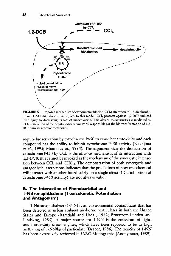
46 John-Michael Sauer et 01.
Inhibition of P-450 by CCI, - - - CCI, 1,IL-DCB /
Reactive 1,2-DCB
FIGURE 5 Proposed mechanism of carbon tetrachloride (CCI,) alteration of 1,2-dichlorobe- nzene (1,2-DCB)-induced liver injury. In this model, CCl, protects against 1,2-DCB-induced liver injury by decreasing its rate of bioactivation. This altered toxicokinetics is mediated by CCI, destruction of the hepatic cytochrome P450 responsible for the biotransformation of 1,2- DCB into its reactive metabolite.
require bioactivation by cytochrome P450 to cause hepatotoxicity and each compound has the ability to inhibit cytochrome P450 activity (Nakajima et al., 1995; Manno et al., 1995). The argument that the destruction of cytochrome P450 by CCI, is the obvious mechanism of its interaction with 1,2-DCB, this cannot be invoked as the mechanism of the synergistic interac- tion between CCl, and CHC13. The demonstration of both synergistic and antagonistic interactions indicates that the predictions of how one chemical will interact with another based solely on a single effect (CCL inhibition of cytochrome P450 activity) are not always valid.
8. The Interaction of Phenobarbital and I -Nitronaphthalene (Toxicokinetic Potentiation and Antagonism)
1 -Nitronaphthalene ( 1-NN) is an environmental contaminant that has been detected in urban ambient air-borne particulates in both the United States and Europe (Ramdahl and Urdal, 1982; Brorstrom-Lunden and Lindskog, 1985). A major source for 1-NN is the emissions of light- and heavy-duty diesel engines, which have been reported to be as high as 0.7 mg of 1-NNkg of particulate (Draper, 1986). The toxicity of 1-NN has been extensively reviewed in IARC Monographs (Anonymous, 1989).

Liver as a Target for Chemical-Chemical Interactions 47
10000-
2 8000 - 3
2 > .- CI ' 6000-
2 m $, 4000-
h m
2000-
0
120001 Vehicle
0 1,2-DichIorobenzene
b T
a, b 8, b I a a
-
FIGURE 6 Effect of exposure to drinking water containing carbon tetrachloride (ccl4) on 1,2-dichlorobenzene (1 &DCB)-induced liver injury. Male Fischer-344 rats were exposed to drinking water containing cc14 (5 or 20 mM) for 1 week, subsequent to administration of 1,2-DCB (1.8 mmolkg, ip). Twenty-four hours after the administration of 1,2-DCB, rats were killed and plasma ALT activity was evaluated. Bars represent means 2 SEM (n = 3). "Signifi- cantly different from respective vehicle control. bSignificantly different from respective CCI, vehicle group.
Although environmental exposure to 1-NN would be through inhalation, 1-NN is also a systemic lung toxicant. When given systemically to rats, 1-NN causes acute bronchiolar epithelial injury in the lung, as well as parenchymal and bile duct epithelial cell injury in the liver (Johnson et al., 1984). However, the lung appears to be the primary target organ of 1-NN in the rat.
At 24 hr after 1-NN administration, extensive lesions are observed in the lungs of rats. These lesions are usually restricted to the bronchioles and consisted of Clara cell and ciliated cell necrosis with exfoliation from the basement membrane. Severe interstitial and perivascular edema, as well as moderate pneumonitis, is also observed. These rats also exhibit increases of protein content, as well as lactate dehydrogenase (LDH) and y-glutamyl transpeptidase (GGT) activities in their bronchioalveolar lavage fluid (BALF). Although less severe than the 1-"-induced pulmonary injury, these rats also develop hepatotoxicity. This injury is characterized by mild multifocal necrosis, typically limited to a few hepatocytes in the centrilobular and periportal regions, as well as bile duct epithelial cell necrosis. Associated with the histopathological changes are increases in plasma ALT activity and total bilirubin (Sauer et aE., 1995).

48 John-Michael Sauer et of.
100-
a
75 2 2. .- 2'
P 50
5
> .- .c.
I
U
25 3
0
1250
b
2 1000 E z cn 1 1
c
750 3 C
3 C Q) .- * 500 2 n
;;If 250
LL A
a
a
T H Vehicle
0 Phenobarbital
Vehicle
Vehicle
0 Phenobarbital
b
i 1 -Nitronaphthalene
a T
b
I Vehicle 1-Nitronaphthalene
FIGURE 7 Effect of phenobarbital on 1-nitronaphthalene-induced lung injury. Male Sprague-Dawley rats received phenobarbital (80 mgkgld, ip) for 4 days followed by I-NN (100 mgikg, ip) on day five. Twenty-four hours after 1-NN administration, rats were killed and markers of lung injury were evaluated. (a) BALF L D H activity, (b) BALF protein content, and (c) BALF GGT activity. Bars represent means -t SEM (n = 9 ) . "Significantly different from respective vehicle control. bSignificantly different from respective phenobarbital vehicle group (bronchioalveolar lavage fluid, BALF).

Liver as a Target for Chemical-Chemical Interactions 49
C
5-
Vehicle
0 Phenobarbital a
I
T
b
1 Vehicle 1 -Nitronaphthalene
FIGURE 7 Continued
Bioactivation has been shown to be important in the elicitation of lung and liver toxicity for 1-NN (Johnson et al., 1984; Rasmussen et al., 1986; Verschoyle and Dinsdale 1990; Verschoyle et al., 1993). In vitro studies using lung microsomes, lung slices, and isolated lung cells have shown that 1-NN is metabolized by cytochrome P450 enzymes via an oxidative pathway that results in macromolecular binding. Autoradiographic studies have indi- cated that the majority of binding occurs in the bronchiolar epithelium (Rasmussen et al., 1986). Using inhibitors of specific cytochrome P450 isoenzymes, Verschoyle and Dinsdale (1 990) showed l-"-induced pulmo- nary toxicity to be correlated with cytochrome P450 2B1 activity in the lung. In the noninduced rat liver it appears that 1-NN is bioactivated by cytochromes P450 1 A l and 1A2 (Verschoyle et al., 1993). However, in phenobarbital-pretreated rats, bioactivation by the liver is associated with cytochrome P450 2B1. Under aerobic conditions using rat liver supernatant, 1 -NN undergoes ring oxidation, resulting in the production of dihydrodiols and nitronaphthol metabolites (El-Bayoumy and Hecht, 1982). When per- formed under anaerobic conditions, the reductive product, l-naphthylamine, was detected (Poirier and Weisburger, 1974). In an in vivo study, Johnson and Cornish (1978) reported the presence of l-naphthylamine in the urine of male Sprague-Dawley rats administered 1-NN.
Pretreatment of rats with phenobarbital causes a significant attenuation in the pulmonary injury caused by 1-NN (Fig. 7). No gross or light micro- scopic changes in the lungs were observed. However, pretreatment with
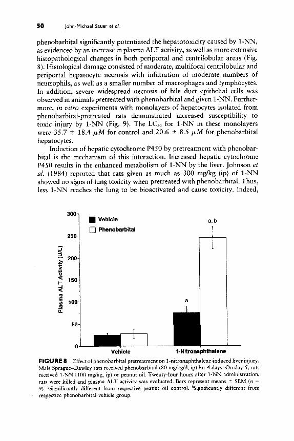
50 John-Michael Sauer et a/.
2 2. 200- ZI c > .- c) ' 150- !i a
phenobarbital significantly potentiated the hepatotoxicity caused by 1-NN, as evidenced by an increase in plasma ALT activity, as well as more extensive histopathological changes in both periportal and centrilobular areas (Fig. 8). Histological damage consisted of moderate, multifocal centrilobular and periportal hepatocyte necrosis with infiltration of moderate numbers of neutrophils, as well as a smaller number of macrophages and lymphocytes. In addition, severe widespread necrosis of bile duct epithelial cells was observed in animals pretreated with phenobarbital and given 1-NN. Further- more, in vitro experiments with monolayers of hepatocytes isolated from phenobarbital-pretreated rats demonstrated increased susceptibility to toxic injury by 1-NN (Fig. 9). The LCjo for 1-NN in these monolayers were 35.7 2 18.4 p M for control and 20.6 2 8.5 p.M for phenobarbital hepatocytes.
Induction of hepatic cytochrome P450 by pretreatment with phenobar- bital is the mechanism of this interaction. Increased hepatic cytochrome P450 results in the enhanced metabolism of 1-NN by the liver. Johnson et al. (1984) reported that rats given as much as 300 mg/kg (ip) of 1-NN showed no signs of lung toxicity when pretreated with phenobarbital. Thus, less 1-NN reaches the lung to be bioactivated and cause toxicity. Indeed,
300 1 Vehicle
17 Phenobarbital 250
1
Vehicle 1-Nitronaphthalene
FIGURE 8 Effect of phenobarbital pretreatment on 1-nitronaphthalene-induced liver injury. Male Sprague-Dawley rats received phenobarbital (80 mg/kg/d, ip) for 4 days. On day 5, rats received 1-NN (100 mg/kg, ip) or peanut oil. Twenty-four hours after 1-NN administration, rats were killed and plasma ALT activity was evaluated. Bars represent means ? SEM (n =
9). 5ignificantly different from respective peanut oil control. "Significantly different from respective phenobarbital vehicle group.

Liver as a Target for Chemical-Chemical Interactions 51
Control LC50 = 35 * 18 pM
0 Phenobarbital LCso = 20 t 9 pM t
pretreatment of rats with phenobarbital has been shown to reduce the amount of parent compound (1-NN) which reaches the lungs (Dankovic and Cornish, 1982). However, the enhanced bioactivation of 1-NN by the liver results in greater hepatotoxicity. A diagrammatic illustration of this chemical-chemical interaction is shown in Fig. 10.
C. The Interaction of I ,2-Dichlorobenzene and I ,3-Dichlorobenzene (Toxicokinetic or Toxicodynamic Potentiation)
As described previously, 1,2-DCB is a potent hepatotoxicant in male Fischer-344 rats. Interestingly, the 1,3-DCB isomer is considerably less toxic to the liver at equal molar doses but, like 1,2-DCB, also causes a rapid loss of hepatic glutathione (Kimura et a/., 1992; Allis etal., 1992). Because both of these isomers of DCB are found together in the environment, coexposure of humans to these pollutants is a potential health hazard. However, the interactive hepatotoxicity of these two chemicals has only been investigated recently (Sipes et al., 1994). Several possible outcomes could result from the interaction of 1,2-DCB and 1,3-DCB. For example, the competition for cytochrome P450 by both isomers of DCB could inhibit their bioactivation,

52 John-Michael Suer er ol.
Non-Induced Induced (Vehicle)
( J / parent to lung)
1 -Nitronaphthalene FIGURE I0 Proposed mechanism of phenobarbital alteration of 1-nitronaphthalene ( 1-")-induced lung and liver injury. In this proposed model, phenobarbital potentiates 1 -"-induced liver injury by increasing its rate of hepatic clearance and bioactivation. This altered toxicokinetics is mediated by phenobarbital induction of hepatic cytochrome P450 2B1 and results in increased hepatotoxicity. In the lung, the toxicity caused by 1-NN is reduced by phenobarbital because less I-NN (parent) reaches the lung to be bioactivated and initi- ate toxicity.
thereby antagonizing their hepatotoxicity. However, the interaction of 1,2- DCB and 1,3-DCB could cause a synergistic depletion of glutathione, result- ing in reduced detoxification and enhanced hepatotoxicity. Studies described here were performed to determine the interactive hepatotoxicity of 1,2-DCB and 1,3-DCB in the male Fischer-344 rat.
Concomitant administration of 1,2-DCB (0.9 mmol/kg, ip) and 1,3- DCB (1.8 mmol/kg, ip) resulted in a significant potentiation of the hepatotox- icity as compared to that following a dose of either 1,2-DCB or 1,3-DCB alone (Fig. 11). When 1,3-DCB was administered either 6 hr prior to or 6 hr post-1,2-DCB treatment, the hepatotoxicity was greatly potentiated. However, administration of 1,3-DCB 12 hr after 1,2-DCB treatment did not produce potentiation. Thus, it appears that events occurring within the first 12 hr post-1,2-DCB administration are important for this chemical- chemical interaction. Because the interaction occurs only at early time points, during the metabolic activation of both 1,2-DCB and 1,3-DCB, it is likely that the altered hepatotoxicity is due to enhanced bioactivation and/or re- duced detoxification of the DCB isomers.
As shown in Fig. 12, both 1,2-DCB and 1,3-DCB deplete glutathione at 5 hr postdosing. Interestingly, concomitant administration of 1,2-DCB

Liver as a Target for Chemical-Chemical Interactions 53
4000-
3500-
3000-
a, b
I 1OOO-
500- a
0
I
a
rl Vehicle 1 ,2-DCB 1 ,SDCB 1,2-DCB 1,P-DCB 1,2-DCB 1,2-DCB
+ + + + 1 W C B 1 W C B 1 W C B 1 W C B (-6hr) (Ohr) (+6hr) (+12hr)
FIGURE I I Effect of 1,3-dichlorobenzene (1,3-DCB) on 1,2-dichlorobenze (1,2-DCB)- induced liver injury. Male Fischer-344 rats received 1,3-DCB (1.8 mmol/kg, ip) concomitantly, 6 hr prior, 6 post, or 12 hr post 1,2-DCB (0.9 mrnol/kg, ip) administration. Twenty-four hours after 1,2-DCB administration, rats were killed and plasma ALT activity was evaluated. Bars represent means rf: SEM (n = 3). "Significantly different from respective vehicle control. bSignifi- cantly different from respective DCB alone group.
and 1,3-DCB resulted in no greater depletion of glutathione than either 1,2-DCB or 1,3-DCB alone. However, hepatic glutathione levels at 24 hr postcoadministration of 1,2-DCB and 1,3-DCB appeared to be less than that when 1,2-DCB was administered alone (Fig. 13). Therefore, it is possible that the prolonged depletion of glutathione by both isomers contributes to the potentiated hepatotoxicity. Thus, because there is less glutathione to detoxify the reactive metabolites produced during the biotransformation of the DCB isomers, significantly more hepatic injury occurs. Furthermore, the depletion of glutathione, a factor that has been implicated in the modulation of macrophage function (Buchmuller-Rouiller et al., 1995), may act to in- crease the activity of hepatic macrophages (Kupffer cells). It has been pre- viously reported (Gunawardhana et al., 1993) that Kupffer cells play a key role in the progression of 1,2-DCB-induced hepatotoxicity. Thus, there is the possibility that this chemical-chemical interaction is mediated by alter-

54 John-Michael Luer et al.
0
m Q 0) I
.- CI
6.0
2.0
0.0 Vehicle
a 1
a a
T
1,P-DCB 1,3-DCB 1,2-DCB +
1,3-DCB
FIGURE I 2 Effect of coadrninistration of 1,3-dichlorobenzene (1,3-DCB) and 1,2-dichloro- benzene (1,2-DCB) on hepatic glutathione. Male Fischer-344 rats received 1,2-DCB (0.9 mmol/ kg, ip) and 1,3-DCB (1.8 mmolikg, ip) alone or concomitantly. Five hours after administration, rats were killed and hepatic glutathione was evaluated. Bars represent means t SEM ( n = 3). %Significantly different from respective vehicle control.
ations in both toxicodynamic (i.e., altered Kupffer cell response) and toxico- kinetic events (i.e., reduced glutathione conjugation). A diagrammatic illus- tration of this chemical-chemical interaction between 1,2-DCB and 1,3-DCB is shown in Fig. 14.
D. The Interaction of All-trans-Retinol and Bromotrichloromethane (Toxicodynamic Potentiation)
Bromotrichloromethane ( BrCCI3), an analog of Ccl4, is a volatile, color- less, noncombustible chemical used industrially as a degreasing solvent, a stabilizer in waxes, and a flame retardant, as well as an antimicrobial fumi- gant. Several studies have shown that exposure to BrCCI3 causes toxicities that are similar to those resulting from CCI, exposure, with the primary target tissue being the liver (Faroon and Mehendale, 1990; Waller et al., 1983). However, BrCC13 is approximately 100 times more toxic than CCI4 (Waller et al., 1983). Although this difference in toxicity is not completely understood, several lines of evidence suggest that it is due to the more labile bromine-carbon bond. This bond is more readily cleaved by cytochrome P450, resulting in a more rapid production of the highly toxic trichloro- methyl radical (Sipes et al., 1977; Brattin et al., 1985; Mehendale, 1991).
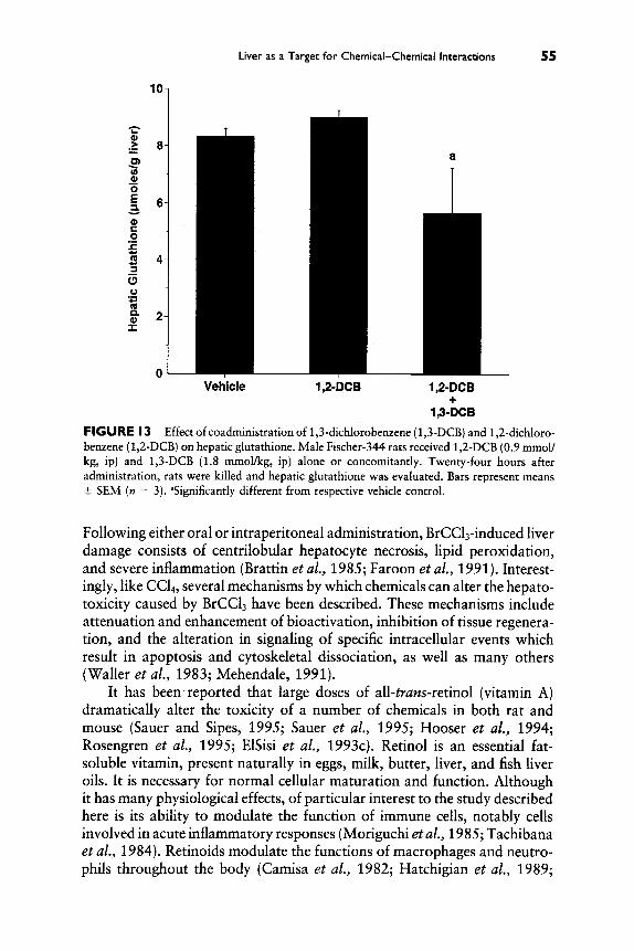
Liver as a Target for Chemical-Chemical Interactions 55
a T
0 I Vehicle 1 ,%-DCB 1,2-DCB
1,3-DCB +
FIGURE I 3 Effect of coadministration of 1,3-dichlorobenzene (1,3-DCB) and 1,2-dichloro- benzene (1,2-DCB) on hepatic glutathione. Male Fischer-344 rats received 1,2-DCB (0.9 mmoll kg, ip) and 1,3-DCB (1.8 mmofig, ip) alone or concomitantly. Twenty-four hours after administration, rats were killed and hepatic glutathione was evaluated. Bars represent means 2 SEM (n = 3). ‘Significantly different from respective vehicle control.
Following either oral or intraperitoneal administration, BrCCL-induced liver damage consists of centrilobular hepatocyte necrosis, lipid peroxidation, and severe inflammation (Brattin et al., 1985; Faroon et al., 1991). Interest- ingly, like CC14, several mechanisms by which chemicals can alter the hepato- toxicity caused by BrCCI3 have been described. These mechanisms include attenuation and enhancement of bioactivation, inhibition of tissue regenera- tion, and the alteration in signaling of specific intracellular events which result in apoptosis and cytoskeletal dissociation, as well as many others (Waller et al., 1983; Mehendale, 1991).
It has been reported that large doses of all-trans-retinol (vitamin A) dramatically alter the toxicity of a number of chemicals in both rat and mouse (Sauer and Sipes, 1995; Sauer et al., 1995; Hooser et al., 1994; Rosengren et al., 1995; ElSisi et al., 1993~) . Retinol is an essential fat- soluble vitamin, present naturally in eggs, milk, butter, liver, and fish liver oils. It is necessary for normal cellular maturation and function. Although it has many physiological effects, of particular interest to the study described here is its ability to modulate the function of immune cells, notably cells involved in acute inflammatory responses (Moriguchi et al., 1985; Tachibana et al., 1984). Retinoids modulate the functions of macrophages and neutro- phils throughout the body (Camisa et al., 1982; Hatchigian et al., 1989;

56 John-Michael Sauer et ol.
He patotoxicity a
Reactive - ~ oxygenspecies \ \ \ \
Reactive P450 111111) Metaboliter w
7 (4 P450 Reactive idc, Metabolites
t Macromolecular
Binding
f Glutathione I Depletion of Conjugation Glutathione
@ /
Reactive @
oxygen species
Enhanced Kupffer Cell Function
He patotoxicity
FIGURE I 4 Proposed mechanism of 1,3-dichlorobenzene (1,3-DCB) alteration of 1,2- dichlorobenzene (1,2-DCB)-induced liver injury. In this proposed model, 1,3-DCB interacts with I,2-DCB by synergistically decreasing hepatic glutathione. This reduction in hepatic glutathone content could result in toxicokinetic interaction (decreased conjugation by glutathi- one) or in a toxicodynamic interaction (enhancement of Kupffer cell function). ROS, reactive oxygen species.
Hemila and Wikstrom, 1985; Fumarulo et a/., 1991). For example, increased phagocytic activity and production of reactive oxygen species have been observed in these phagocytes following in vivo retinol pretreatment (Guzman et al., 1991; Lison et ai., 1990). This section describes the chemical-chemical interactions between retinol and BrCCI,.
Bromotrichloromethane (0.2 mmoYkg, ip) administration to control rats caused minor hepatotoxicity. However, 7 days of oral dosing with retinol(75 mg/kg/day) greatly increases the hepatotoxicity of BrCCI3. Indeed, pretreatment with retinol markedly elevated levels of plasma ALT and histo- pathological changes at 24 hr postadministration of BrCCI3 (Fig. 15). ElSisi et al. (1993a) have proposed a mechanism for retinol potentiation of chemi- cally induced hepatotoxicity in which retinol increases the activity of liver macrophages (Kupffer cells) and their ability to generate reactive oxygen species during liver injury. Evidence that activated Kupffer cells are involved in retinol potentiation of BrCCI,-induced toxicity in the liver was provided by studies with gadolinium chloride (GdC13), a compound that destroys Kupffer cells (Hustzik et a/., 1980). Importantly, pretreatment with GdCI, markedly attenuated the potentiated liver damage at 24 hr postadministra- tion of BrCCI3 in retinol-pretreated rats (Fig. 16). Furthermore, methyl palmitate, another inhibitor of Kupffer cell function, also attenuated the

Liver as a Target for Chemical-Chemical Interactions 57
4000
3500
2 3000
p 2500 2.
.- - 2 2000
3 1500
UJ m = 1000
500
0
Vehicle
0 All-trans-Retinol P
a
Vehicle BkCl3
FIGURE I 5 Effect of all-trans-retinol pretreatment on bromotrichloromethane (BrCC1,)- induced liver injury. Male Sprague-Dawley rats received retinol (75 mg/kg/d, PO) for 7 days. One day after the last dose of retinol, rats received BrCCI3 (0.2 mmol/kg, ip) or corn oil. Twenty-four hours after BrCC1, administration, rats were killed and plasma ALT activity was evaluated. Bars represent means 2 SEM (n = 5). "Significantly different from respective corn oil control. bSignificantly different form respective retinol vehicle group.
potentiated liver damage. Because both GdC4 and methyl palmitate pretreat- ment protected against retinoi-potentiated hepatotoxicity caused by BrCCI3, it appears that retinol pretreatment increased BrCCI3-induced hepatocyte injury through a Kupffer cell-dependent mechanism.
Characterization of isolated Kupffer cells from animals pretreated with a similar regimen of retinol showed that phagocytosis by these cells was enhanced, as was their ability to produce reactive oxygen species (Mobley et al., 1991; ElSisi et al., 1993b). A subpopulation of Kupffer cells isolated from retinol-treated rats were highly primed to release increased quantities of superoxide after stimulation with zymosan, whereas other Kupffer cells isolated from the same rat did not respond to zymosan activation. They released superoxide anion at amounts comparable to those of Kupffer cells isolated from control rats (Mobley et al., 1991). Thus, retinol pretreatment primed a subpopulation of Kupffer cells. These primed Kupffer cells mediate the events that ultimately result in a potentiation of BrCCI3 liver injury.
Figure 17 presents a model for retinol alteration of chemically induced liver injury in which Kupffer cells and inflammatory phagocytes play a key role. Important to the model is the fact that retinol pretreatment primes Kupffer cells and, when activated by the hepatic injury caused by BrCCI3,

5a john-Michael Sauer et a/.
5000
4500
4000 2 2 3500
3 ‘5 .- 3000 ‘ 2500 Y
3 : 2000
E 2 1500 n
lO00-l
Vehicle a, b
All-trans-Retinol 1
a T
5001 , a 0
C
Vehicle BrCCl BrCCl BrCCl
Methyl + +
-13 Palmitate
FIGURE I 6 Effect of gadolinium chloride and methyl palmitate pretreatment on all-trans- retinol-potentiated bromotrichloromethane (BrCClJ-induced liver injury. Male Sprague- Dawley rats received retinol (75 mg/kg/d, PO) for 7 days, and on day 7 rats were given GdCli (10 mg/kg, iv) via the tail vein. For the methyl palmitate blockade on days 5, 6, and 7, rats were given methyl palmitate (300 mg/kg, iv) via the tail vein. On day 8 rats received BrCC13 (0.2 mmoYkg, ip) or corn oil. Twenty-four hours after BrCC13 administration, rats were killed and plasma ALT activity was evaluated. Bars represent means 2 SEM (n = 5). 5ignificantly different from respective corn oil control. bSignificantly different from respective retinol vehicle group. ‘Significantly different from respective retinol and BrCCI3 group.
they respond with an enhanced release of mediators and toxic products (i.e., tumor necrosis factor-a, superoxide anion, etc.). From the results of these experiments described in this chapter, it can be concluded that retinol poten- tiates BrCCI,-induced liver injury by increasing the associated inflammatory response and that these events are at least in part mediated by Kupffer cells.
IV. Conclusions
Because of its basic physiology and biochemistry, the liver is a major target organ for chemical-chemical interactions. By nature the liver is re- sponsible for the clearance of a multitude of compounds from the blood, thus concentrating potentially toxic chemicals in its tissue. In addition, it possesses the machinery (i.e,, enzymes) that can lead to bioactivation of chemicals. Although in many instances high doses of xenobiotics are required

Liver as a Target for Chemical-Chemical Interactions 59
(Hepatocytes) (Kupffer Cell)
cytotox/c
factors - - b
(Primed Kupffer Cell)
(Damaged Hepatocytes) I A
0 I Chemokines
1 0
(Extrahepatlc Inflammatory Cells)
FIGURE I 7 Proposed mechanism of all-trans-retinol alteration of BrCCI3-induced liver injury. In this proposed model of retinol potentiation of BrCC13-induced liver injury, the initial toxicity caused by bioactivation (initial hepatocyte injury) is similar in both retinol vehicle and retinol-pretreated animals. At later time points, the progression of hepatic injury (Kupffer cell activity and infiltration of inflammatory cells) is increased in animals pretreated with retinol. Thus, it appears that retinol alters chemical-induced hepatotoxicity by increasing the associated inflammatory responses and not altering the initial injury caused by the bioactivation of the hepatotoxicant.
to cause hepatotoxicity, the actions of other chemicals can shift the dose- response curve for a particular chemical. Thus, for some chemicals the toxic threshold can be achieved at much lower doses, whereas for other chemicals the threshold may be elevated. The two general mechanisms by which chemi- cal-chemical interactions occur are via alterations in toxicokinetic or toxico- dynamic effects. Humans are exposed to a myriad of natural, as well as synthetic, toxicants over a lifetime. Thus the potential for toxic interactions to occur is real. Examples of high doses of a modulating chemical that may cause chemical-chemical interactions are consumption of alcohol, smoking, and occupational exposure. In addition, pharmacological agents are taken at doses that cause biological effects. Such increased risk factors may predis- pose a person to the toxicity caused by a normally nontoxic dhemical ex- posure.
The examples illustrated in this chapter indicate that the predictions of how one chemical will interact with another, based solely on a single factor, are not always valid. Thus, the knowledge of an interaction does not always

60 John-Michael Sauer et 01
allow prediction or extrapolation from one chemical to another or from one species to another. However, in many cases toxicokinetic interactions are usually much easier to predict than toxicodynamic interactions. This relates to a lack of knowledge to many of the events associated with the initiation and progression of injury. Therefore, a better understanding of mechanisms of chemical-chemical interactions is required for the appro- priate assessment of risk during situations of multichemical exposures in humans and animals.
Acknowledgments
This research was supported by a grant from NIEHS (ES-06095) and the NIEHS-sponsored Southwest Environmental Health Sciences Center (P30-ES-06694), as well as the Graduate Training Program in Environmental Toxicology Fellowship (T32-ES-07091-17).
References
Allis, J. W., Simmons, J. E., House, D. E., Robinson, B. L., and Berman, E. (1992). The differential hepatotoxicity and cytochrome P450 responses of Fischer-344 rats to the three isomers of dichlorobenzene. 1. Biochem. Toxicol. 7, 257-264.
Anonymous (1989). 1-Nitronaphthalene. IARC Monogr. Eval. Curcino. Risks Hum. 46,
Arias, 1. M., Jakohy, W. B., Popper, H., Schachter, D., and Shafritz, D. A. (1988). The cells A. The hepatocyte organization. In “The Liver: Biology and Pathobiology” (I. M. Aias, W. B. Jakoby, H. Popper, D. Schachter, and D. A. Shafritz, eds.), 2nd ed., pp. 9-10. Raven Press, New York.
Bell, A. N., Young, R. A., Lockard, V. G., and Mehendale, H. M. (1988). Protection of chlordecone-potentiated carbon tetrachloride hepatotoxicity and lethality by partial hepa- tectomy. Arch. Toxicol. 61, 392-405.
Blouin, A., Bolender, R. P., and Weibel, E. R. (1977). Distribution of organelles and membranes between hepatocytes and nonhepatocytes in the rat liver parenchyma. A stereological study. /. Cell Biol. 72, 441-455.
Boyd, M. R. [ 1977). Evidence for the Clara cell as a site of cytochrome P450-dependent mixed- function oxidase activity in lung. Nature (London) 269, 713-715.
Boyd, M. R., Statham, C. N., and Longo, N. S . (1980). The pulmonary Clara cell as a target for toxic chemicals requiring metabolic activation; studies with carbon tetrachloride. 1. Pharmacol. Exp. Ther. 212, 109-114.
Brattin, W. J., Glende, E. A., Jr., and Recknagel, R. 0. (1985). Pathological mechanisms in carbon tetrachloride hepatotoxicity. /. Free Radicals Biol. Med. 1, 27-38.
Brodie, B. B., Reid, W. D., Cho, A. K., Sipes, G., Krishna, G., and Gillette, J. R. (1971). Possible mechanism of liver necrosis caused by aromatic organic compounds. Proc. Nut. Acad. Sci. U.S.A. 68, 160-164.
Brorstrom-Lunden, E., and Lindskog, A. (1985). Characterization of organic compounds on airborne particles. Environ. Int. 11, 183-188.
Buchmuller-Rouiller, Y., Corrandin, S. B., Smith, J., Schneider, P., Ransijn, A., Jongened, C. V., and Mauel, J. (1995). Role of glutathione in macrophage activation: Effect of cellular glutathione depletion on nitrite production and leishmanicidal activity. Cell. Immunol. 164, 73-80.
303-312.
Liver as a Target for Chemical-Chemical Interactions 61
Camisa, C., Eisenstat, B., Ragaz, A., and Weissmann, G. (1982). The effects of retinoids on neutrophil functions in vitro. J. Am. Acad. Dermatol. 6 , 620-629.
Coon, M. J., Koop, D. R., Reeve, L. E., and Crump, B. L. (1984). Alcohol metabolism and toxicity: role of cytochrome P-450. Fundam. Appl. Toxicol. 4, 134-43.
Dankovic, D., and Cornish, H. H. (1982). Phenobarbital alteration of 1-nitronaphthalene levels in target organs of toxicity. Toxicologist 2, 129.
Draper, M. W. (1986). Quantitation of nitro- and dinitropolycyclic aromatic hydrocarbons in diesel exhaust particulate matter. Chemosphere 15, 437-447.
Edwards, M. J., Keller, B. J., Kauffman, F. C., and Thurman, R. G. (1993). The involve- ment of Kupffer cells in carbon tetrachloride toxicity. Toxicol. Appl. Pharmacol. 119,
El-Bayoumy, K., and Hecht, S. S. (1982). Comparative metabolism in vitro of 5-nitroacenph- thene and 1-nitronaphthalene. In “Polynuclear Aromatic Hydrocarbons: Physical and Biological Chemistry” (M. Cooke, A. J. Dennis, and G. L. Fisher, eds.), p. 263. Battelle Press, Columbus, OH.
EISisi, A. E., Hall, P., Sim, W. L., Earnest, D. L., and Sipes, I. G. (1993a). Characterization of vitamin A potentiation of carbon tetrachloride-induced liver injury. Toxicol. Appl. Pharmacol. 119,280-288.
ElSisi, A. E., Earnest, D. L., and Sipes, I. G. (1993b). Vitamin A potentiation of carbon tetrachloride hepatotoxicity: Enhanced lipid peroxidation without enhanced biotransfor- mation. Toxicol. Appl. Pharmacol. 119, 289-294.
EISisi, A. E., Earnest, D. L., and Sipes, I. G. (1993b). Vitamin A potentiation of carbon tetrachloride hepatotoxicity: Role of liver macrophages and active oxygen species. Toxicol. Appl. Pharmacol. 119,295-301.
Faroon, 0. M., and Mehendale, H. M. (1990). Bromotrichloromethane hepatotoxity. The role of stimulated hepatocellular regeneration in recovery: Biochemical and histophatological studies in control and chlordecone pretreated male rats. Toxicol. Pathol. 18, 667-677.
Faroon, 0. M., Henry, R. W., Soni, M. G., and Mehendale, H. M. (1991). Potentiation of BrCC13 hepatotoxicity by chlordecone: Biochemical and ultrastructural study. Toxicol. Appl. Pharmacol. 110, 185-197.
Fisher, R., McCarthy, S. , Sipes, I. G., Hanzlik, R. P., and Brendel, K. (1991). Metabolism of dichlorobenzenes in organ cultured liver slices. Adv. Exp. Med. Biol. 283, 717-723.
Fumarulo, R., Conese, M., Riccardi, S., Giordano, D., Montemurro, P., Colucci, M., and Semeraro, N. (1991). Retinoids inhibit the respiratory burst and degradation of stimu- lated human polymorphonuclear leukocytes. Agents Actions 34, 339-344.
Glende, E. A., Jr. (1972). On the mechanism of carbon tetrachloride toxicity-coincidence of loss of drug-metabolizing activity with peroxidation of microsomal lipid. Biochem. Pharmacol. 21,2131-2138.
Gunawardhana, L., Mobley, S. A., and Sipes, I. G. (1993). Modulation of 1,2-dichlorobenzene hepatotoxicity in the Fischer-344 rat by a scavenger of superoxide anions and an inhibitor of Kupffer cells. Toxicol. Appl. Pharmacol. 119, 205-213.
Guyton, A. C. (1991). The circulation. In “Textbook of Medical Physiology” (M. J. Wonsiewicz, ed.), pp. t183. Saunders, Philadelphia.
Guzman, J. J., Marks, J. F., and Caren, L. D. (1991). In vivo and in vitro studies on the effects of vitamin A on the chemiluminescent response of murine peritoneal exudate cells. Toxicol. Lett . 57, 125-137.
Hatchigian, E. A., Santos, J. L., Broitman, S. A., and Vitale, J. J. (1989). Vitamin A supplementa- tion improves macrophage function and bacterial clearance during experimental salmo- nella infection. Proc. SOC. Exp. Biol. Med. 191, 47-54.
Hemila, H., and Wikstrom, M. (1985). Retinoids activate superoxide production by polymor- phonuclear leukocytes. Scand. J. Immunol. 21, 227-234.
Hinson, J. A., Pumford, N. R., and Nelson, S. D. (1994). The role of metabolic activation in drug toxicity. Drug Metab. Rev. 26, 395-412.
275-279.

62 John-Michael Sauer et a/.
Hooser, S . B., Rosengren, R. J., Hill, D. A., Mobley, S. A., and Sipes, I. G. (1994). Vitamin A modulation of xenobiotic-induced hepatotoxicity in rodents. Environ. Health Perspect.
Huseik, E., Lazar, G., and Parduez, A. (1980). Electron microscopic study of Kupffer-cell phagocytosis blockade induced by gadolinium chloride. Br. J. Exp. Pathol. 61,624-630.
Johnson, D. E., and Cornish, H. H. (1978). Metabolic conversion of 1- and 2-nitronaphthalene to 1- and 2-naphthylamine in the rat. Toxicol. Appl. Phamacol. 46, 549-573.
Johnson, D. E., Riley, M. G., and Cornish, H. H. (1984). Acute target organ toxicity of 1- nitronaphthalene in the rat. 1. Appl. Toxicol. 4, 253-257.
Kimura, R., Ohishi, N., Kato, Y., Yamada, S., and Sato, M. (1992). Identification of biliary metabolites of m-dichlorobenzene in rats. Drug Metab. Dispos. 20, 161-171.
Koporec, K. P., Kim, H. J., MacKenzie, W. F., and Bruckner, J. V. (1995). Effect of oral dosing vehicles on the subchronic hepatotoxicity of carbon tetrachloride in the rat. 1. Toxicol. Environ. Health 44, 13-27.
Laskin, D. L., and Pendino, K. J. (1995). Macrophages and inflammatory mediators in tissue injury. Annu. Rev. Pharmacol. Toxicol. 35, 655-677.
Laskin, D. L., and Pilaro, A. M. (1986). Potential role of activated macrophages in acetamino- phen hepatotoxicity. I. Isolation and characterization of activated macrophages from rat liver. Toxicol. Appl. Pharmacol. 86, 204-215.
Lison, D., Raguzzi, F., and Lauwerys, R. (1990). Comparison of the effects of auranofin, heavy metals and retinoids on protein kinase C in vitro and on a protein kinase C mediated response in macrophages. Pharmacol. Toxicol. 67, 239-242.
Manno, M., Tolando, R., Ferrara, R., Rezzadore, M., and Cazzaro, S . (1995). Suicidal inactiva- tion of haemoproteins by reductive metabolites of halomethanes: A structure-activity relationship study. Toxicology 100, 175-1 83.
Mehendale, H. M. (1 991). Role of hepatocellular regeneration and hepatolobular healing in the final outcome of liver injury. A two-stage model of toxicity. Biochem. Pharmacol. 42,1155-1162.
Mehendale, H. M. (1995). Toxicodynamics of low level toxicant interactions of biological significance: Inhibition of tissue repair. Toxicology 105, 251-266.
Mitchell, J. R., Jollow, I). J., Potter, W. Z . , Gillette, J. R., and Brodie, B. B. (1973). Acetamino- phen-induced hepatic necrosis. IV. Protective role of glutathione. J. Pharmacol. Exp. Theru. 187, 211-217.
Mitchell, J. R., Snodgrass, W. R., and Gillette, J. R. (1976). The role of biotransformation in chemical-induced liver injury. Environ. Health Perspect. 15, 27-38.
Mobley, D. L., Lantz, R. C., Sipes, 1. G., and Earnest, S. A. (1991). Differential production of superoxide anion by single Kupffer cells isolated from rats with hypervitaminosis A. In “Cells of the Hepatic Sinusoid” (E. Wisse, D. L. Knook, and K. Decker, eds.), Vol. 3, pp. 106-11 1. Kupffer Cell Foundation, Rijswijk.
Moriguchi, S . , Werner, L., and Watson, R. R. (1985). High dietary vitamin A (retinyl palmitate) and cellular immune functions in mice. Immunology 56, 169-177.
Nakajima, T., Elovaara, E., Okino, T., Gelboin, H. V., Klockars, M., Riihimaki, V., Aoyama, T., and Vainio, H. (1995). Different contributions of cytochrome P450 2E1 and P450 2B1/2 to chloroform hepatotoxicity in rat. Toxicol. Appl. Pharmacol. 133, 215-222.
National Research Council (1988). “Complex Mixtures.” National Academy Press, Washing- ton, DC.
Poirier, L. A., and Weisburger, J. H. (1974). Enzymic reduction of carcinogenic aromatic nitro compounds by rat and mouse liver fractions. Biochem. Pharmacol. 23, 661-669.
Ramadori, G. (1991). The stellate cell (Ito-cell, fat-storing cell, lipocyte, perisinusoidal cell] of the liver. New insights into pathophysiology of an intriguing cell. Virchows Arch. B
Ramdahl, T., and Urdal, K. (1982). Determination of nitrated polycyclic aromatic hydrocarbons by fused silica capillary gas chromatographylnegarive ion chemical ionization mass spec- trometry. Anal. Chem. 54, 2256-2260.
102, Suppl. 9, 39-43.
61,147-157.

Liver as a Target for Chemical-Chemical Interactions 63
Rasmussen, R. E., Do, D. H., Kim, T. S., and Dearden, L. C. (1986). Comparative cytotoxicity of naphthalene and its monomethyl- and mononitro-derivatives in the mouse lung. J. Appl. Toxicol. 6 , 13-20.
Reid, W. D., Christie, B., Krishna, G., Mitchell, J. R., Moskowitz, J., and Brodie, B. B. (1971). Bromobenzene metabolism and hepatic necrosis. Pharmacology 6, 41-55.
Rosengren, R. J., Sauer, J. M., Hooser, S. B., and Sipes, I. G. (1995). The interactions between retinol and five different hepatotoxicants in the Swiss Webster mouse. Fundam. Appl. Toxicol. 25, 281-292.
Sato, C., Matsuda, Y., and Lieber C. S. (1981). Increased hepatotoxicity of acetaminophen after chronic ethanol consumption in the rat. Gastroenterology 80, 140-148.
Sauer, J. M., and Sipes, 1. G. (1995). Modulation of chemical-induced lung and liver toxicity by all-trans-retinol in the male Sprague-Dawley rat. Toxicology 105, 237-249.
Sauer, J. M., Hooser, S. B., and Sipes, I. G. (1995). All-trans-retinol alteration of l-nitronaphtha- lene-induced pulmonary and hepatic injury by modulation of associated inflammatory responses in the male Sprague-Dawley rat. Toxicol. Appl. Pharmacol. 133, 139-149.
Sipes, I. G., Krishna, G., and Gillette, J. R. (1977). Bioactivation of carbon tetrachloride, chloroform and bromotrichloromethane: Role of cytochrome P-450. Life Sci. 20, 1541- 1548.
Sipes, I. G., EISisi, A. E. D., Sim, W. L., Mobley, S. A., and Earnest, D. L. (1989). Role of reactive oxygen species secreted by activated Kupffer cells in the potentiation of carbon tetrachloride hepatotoxicity by hypervitaminosis A. In “Cells of the Hepatic Sinusoid” (E. Wisse, D. L. Knook, and K. Decker eds.), Vol. 2, pp. 376-379. Kupffer Cell Founda- tion, Rijswijk.
Sipes, I. G., EISisi, A. E., Sim, W. L., Mobley, S. A., and Earnest, D. L. (1991). Reactive oxygen species in the progression of CC14-induced liver injury. Adv. Exp. Med. Biol.
Sipes, I. G., Kramer, K., Kriesel, D., and Gunawardhana, L. (1994). Interactive hepatotoxicity of 1,2- and 1,3-dichlorobenzene in the Fischer-344 rat. Toxicologist 14, 701.
Tachibana, K., Sone, S., Tsubura, E., and Kishino, Y. (1984). Stimulatory effect of vitamin A on tumoricidal activity of rat alveolar macrophages. BY. J . Cancer 49, 343-348.
Valentovic, M. A., Ball, J. G., Anestis, D., and Madan, E. (1993a). Modification of P450 activity and its effect on 1,2-dichlorobenzene toxicity in Fischer 344 rats. Toxicology
Valentovic, M. A., Ball, J. G., Anestis, D., and Madan, E. (1993b). Acute hepatic and renal toxicity of dichlorobenzene isomers in Fischer 344 rats. J. Appl. Toxicol. 13, 1-7.
Verschoyle, R. D., and Dinsdale, D. (1990). Protection against chemical-induced lung injury by inhibition of pulmonary cytochrome P-450. Environ. Health Perspect. 85, 95-100.
Verschoyle, R. D., Carthew, P., Wolf, C. R., and Dinsdale, D. (1993). 1-Nitronaphthalene toxicity in rat lung and liver: Effects of inhibiting and inducing cytochrorne P-450 activity. Toxicol. Appl, Pharmacol. 122,208-213.
Waller, R. L., Glende, E. A., Jr., and Recknagel, R. 0. (1983). Carbon tetrachloride and bromotrichloromethane toxicity. Dual role of covalent binding of metabolic cleavage products and lipid peroxidation in depression of microsomal calcium sequestration. Bio- chem. Pharmacol. 32, 1613-1617.
283,489-497.
79, 169-180.

A. David Rodrigues and Shekman L. Wong Drug Metabolism Department
Pharmaceutical Products Division Abbott Laboratories
Abbott Park, Illinois 60064-3500
Application of Human Liver Microsomes in Metabolism-Based Drug-Drug Interactions: In Vitro-in Vivo Correlations and the Abbott Laboratories Experience
1. Introduction
Our knowledge of the various human drug-metabolizing enzyme (DME) systems continues to expand. In recent years, this expansion has been fueled by advances in molecular biology, the increased availability of human tissue, and the development of model systems and sensitive assay methods (e.g., liquid chromatography-mass spectrometry, LC-MS) for studying human drug metabolism in vitro (Rodrigues, 1994; Ball et al., 1995; Tucker, 1992; Miners et al., 1994; Wrighton et al., 1993; Hayes et al., 1995). It is now known that most of the DME systems present in the human liver (e.g., cytochrome P450, CYP; UDP-glucuronosyltransferase, UDPGT; N- acetyltransferase, NAT; and NADPH-dependent flavin-containing monoox- ygenase, FMO) are composed of two or more enzymes or “ i s ~ f o r m s ~ ~ that
Advancer in Pharmacology, Volume 43 Copyright 0 1997 by Academic Press. All rights of reproduction in any form reserved. 1054-3589197 $25.00 65

66 A. David Rodrigues and Shekrnan L. Wong
can differentially interact with any number of drugs (Burchell et al., 1991; Nelson et al., 1993; Vatsis et al., 1995; Lawton et al., 1994). If multiple drugs interact with the same DME, then the likelihood of a potentially hazardous drug-drug interaction is greatly increased, especially if one of the drugs exhibits a relatively narrow therapeutic index or a steep dose-response profile (Fig. 1) . In a safety- and cost-conscious drug development environ- ment, many pharmaceutical companies have seized on this type of informa- tion and are now routinely using data from in vitro human metabolism studies as part of their submissions to various regulatory agencies. In turn, these agencies have acknowledged the utility of this information, both in terms of assessing drug safety and approvability (Temple, 1993; Peck et al., 1993).
For a given new drug entity (NDE), it is now apparent that knowledge of the “DME interaction potential” can also lead to a marketing edge. Currently, this is evident with the lucrative and highly competitive serotonin reuptake inhibitor (SSRI), macrolide antibiotic, antihistamine, and antacid markets (Steinijans et al., 1994; Rodrigues and Freston, 1994; Amsden, 1995; Honig et al., 1994; Brosen, 1993; DeVane, 1994). Nonetheless, this information should be viewed in light of the fact that many regulatory agencies presently adhere to a policy of “class labeling” in drug package inserts. Well-documented examples include fluconazole, sertraline, and azi- thromycin. All three compounds exhibit relatively weak interactions with terfenadine and/or the tricyclic antidepressants (Honig et al., 1993a; Harris et al., 1995; Preskorn et al., 1994), but have at one time or another been
Superfarnib P-450 Q Families J’ 8 Subfamilies
Members 1A2 2C9 2D6 3A4l5 Thwphylline (S)-Warfarin Imipramine Terfenadine Imipramine Phenytoin Desipramine Nifedipine
Carbamazepine Amitriptyline Midazolam Nomipcyline Imipramine Tbioridazine Carbamazepine
Cyclosporin
FiGURE I Examples of drugs exhibiting a relatively narrow therapeutic index or steep dose-response profile. The metabolism of each drug has been ascribed to one or more forms of CYP.

In Vitro-in Vivo Correlations 67
assigned “imidazole antifungal,” “SSRI,” and “macrolide” class labeling, respectively, by the Food and Drug Administration.
Presently, there is also a growing awareness that it may become possible to “manage” drug-drug interactions in a “proactive” rather than a “retroac- tive” manner. A well-managed drug-drug interaction, as in the case of ketoconazole and cyclosporin, can potentially be of economic benefit to the patient (Albengres and Tillement, 1992; Keogh et al., 1995). Likewise, a potent interaction with an enzyme such as CYP can lead to autoinhibition of metabolism and superior pharmacokinetics, as has recently been observed with ritonavir (ABT-538; Fig. 2), an orally active peptidomimetic human immunodeficiency virus (HIV-1) protease inhibitor currently in development
ABT-538
A-152184
Zileuton Naproxen
FIGURE 2 position of chiral center. Arrows indicate the sites of CYP-dependent oxidation.
Structure of ABT-538, A-152184, zileuton, and naproxen. Asterisks denote the

68 A. David Rodrigues and Shekman L. Wong
for the treatment of AIDS (Kempf, 1994; Kempf et al., 1995; Kumar et al., 1996). Furthermore, as endogenous substrates for the different human DME systems are discovered, drug companies may begin to view these proteins as potential therapeutic targets. For instance, CYP2C8 is thought to play a role in the metabolism of arachidonic acid (Zeldin et al., 1995; Rifkind et al., 1995).
II. The Utility of in Vitro Human Drug-Drug Interaction Studies in Drug Discovery and Development
In vitro drug-drug interaction screening is fast becoming a standardized part of the drug development process and, depending on the particular pharmaceutical company and/or NDE, can be performed prospectively (e.g., pre-IND or investigational new drug application), concurrently, or retro- spectively (e.g., post-NDA or new drug application). Because human carbon- 14 absorption-distribution-meta bolism-excretion (ADME) and drug-drug interaction studies are carried out relatively late in the drug development process, most pharmaceutical companies have recognized that the informa- tion obtained, if applied correctly and in a timely manner, can make clinical programs better guided, safer, and more cost effective (Rodrigues, 1994). For instance, once a clinical drug candidate is chosen, the information can be indicative of which in vivo interaction studies need to be performed, in addition to those involving often-evaluated drugs such as theophylline, phenytoin, warfarin, oral contraceptives, and cimetidine (Schentag, 1993). With polymorphically expressed enzymes, such as CYP2D6, CYP2C19, and NAT2, in vitro data can indicate if subject phenotyping is necessary (Rodrigues, 1994; Tucker, 1992; Miners et al., 1994; Wrighton et al., 1993).
In early or “discovery” stages of drug development, in vitro drug-drug interaction studies can be used to rapidly screen a large number of develop- ment candidates. This may become especially useful if coupled to the bur- geoning efforts of combinatorial chemists (Eichler et al., 1995; Kubinyi, 1995). The information can be used as part of the “lead candidate” selection process, alongside more conventional data such as oral bioavailability and pharmacologic efficacy in animal models. In the long run, it may be expedient for drug metabolism investigators to alert project chemists and pharmacolo- gists of the potential pitfalls of synthesizing compounds with incriminating imidazole, methylenedioxyaryl, benzofuran, and/or acetylenic moieties, or compounds that are metabolized by polymorphically expressed enzymes (Breimer, 1994; Tucker, 1994; Brosen and Gram, 1989a; Bertilsson, 1995). However, the in vitro “screening out” of drugs should be performed judi- ciously. One only has to glance through the “Physicians’ Desk Reference” (PDR) to realize that there are a number of efficacious and widely prescribed drugs on the market that are known to inhibit enzymes such as CYP, most

In Vitro-in Vivo Correlations 69
notably gestodene, paroxetine, and cimetidine (Wilde and Balfour, 1995; Meltzer, 1993; Somogyi and Muirhead, 1987). Judicial screening should also extend to those drugs that are shown to be metabolized in vitro by polymorphically expressed enzymes, especially when information concern- ing the complete in vivo metabolic profile and/or therapeutic index of the drug is lacking. In many instances, drugs that are metabolized by enzymes such as CYP2D6 (e.g., dextromethorphan) exhibit a relatively wide thera- peutic index, and increased plasma drug concentrations in “poor metabo- h e r ” (PM) subjects are of no clinical concern.
111. In Vitro Drug-Drug Interaction Studies
In most cases, in vitro studies focus on the human liver microsomal CYP “superfamily” of proteins, as our present understanding of this system exceeds that of other DME systems (Guengerich, 1992, 1995; Wrighton and Stevens, 1992). In the future, however, other enzyme systems (e.g., UDPGT) will also have to be incorporated into the screening process, as the database expands to include more clinically relevant therapeutic agents (Bock, 1995; Burchell et al., 1995; Batt et al., 1994). Most of these studies are focused on establishing which form(s) of CYP is involved in the metabo- lism of the NDE, so-called “reaction phenotyping” (Rodrigues, 1994; Wrighton et al., 1993; Ball et al., 1995; Tucker, 1992; Miners et al., 1994). The information most sought after is whether or not a drug candidate is metabolized by CYP3A subfamily members, or polymorphically expressed CYP2D6 and/or CYP2C19, as there is a certain amount of “baggage” associated with each of these CYP forms. If a NDE is metabolized by CYP3A4, then one might expect extensive first-pass metabolism, due to the presence of high levels of the enzyme in the gut wall and liver. One might also expect decreased metabolic clearance with potent CYP3A inhibitors such as ketoconazole and/or nitrosoalkane complex-forming macrolides. Furthermore, one would predict that the metabolism of the NDE is going to be significantly increased when administered to subjects that have been exposed to CYP3A-inducing agents such as phenytoin, rifampicin, and/ or phenobarbital (Guengerich, 1992, 1995; Wrighton and Stevens, 1992). Likewise, metabolism of a drug candidate by polymorphically expressed CYP2D6 and/or CYP2C19 may lead to elevated plasma drug concentrations in subjects characterized as “poor metabolizers” (Breimer, 1994; Tucker, 1994; Brosen and Gram, 1989a; Bertilsson, 1995), although the situation is now complicated by recent findings concerning the existence of allelic variants of CYP forms such as CYP2C9 and CYP2C18 (Goldstein and de Morais, 1994). However, as the procedures for phenotyping and/or genotyp- ing of clinical subjects become more readily available, in vivo confirmation
70 A. David Rodrigues and Shekman L. Wong
of these polymorphic oxidations will be greatly facilitated (Gonzalez and Idle, 1994).
Identifying which CYP form(s) is involved in the metabolism of a given N D E is only part of an investigation. The second facet of in vitro studies involves screening the NDE as an inhibitor of different CYP forms. There are a number of instances where a given compound is metabolized by one CYP form but inhibits a second form of the enzyme. Two well-documented examples include the CYP3A substrates quinidine and 7,8-benzoflavone, which are known to be potent inhibitors of CYP2D6 and CYPlA2, respec- tively (Rodrigues, 1994; Guengerich et al., 1986; Lee et al., 1994). In some cases, as with zileuton, this phenomenon may be extended to include non- CYP enzymes. Zileuton (Fig. 2), an orally active 5-lipoxygenase inhibitor, is largely metabolized (- 80%) by UDPGT and has been shown to alter the pharmacokinetics of theophylline and antipyrine in vivo (Braeckman et al., 1989; Granneman et al., 1995; St. Peter et al., 1995). The interaction with theophylline is considered particularly relevant, as zileuton is currently in development for the treatment of asthma and theophylline has a narrow therapeutic index (Meyer, 1994; Machinist et al., 1995).
A. Models
It is evident from the literature that there is no all-encompassing in vitro and/or in vivo animal model for predicting drug-drug interactions in humans. For instance, many of the members of the mouse, rat, and dog liver microsomal CYP pool differ from those present in the human liver, while the characterization of the various nonhuman primate models is still in its infancy (Rodrigues et al., 1995b; Sharer et al., 1995; Smith, 1991). Therefore, because the liver is considered to be the major site of metabolism, most investigators now find themselves routinely performing in vitro human metabolism studies with various combinations of primary cultured hepato- cytes, precision-cut liver slices, banks of liver microsomes, and/or cDNA- expressed enzymes (Rodrigues, 1994; Rodrigues et al., 1994; Wrighton et at., 1993; Ball et al., 1995). In future years, it is probable that drug-drug interaction screening will also include the use of transgenic animal models and computer-aided predictions. At the present time, pharmacophoric mod- els exist for enzymes such as CYP2D6 (Strobl et al., 1993), and progress is slowly being made toward an understanding of the structural requirements needed for drug interactions with enzymes such as CYP2C9 and CYP2A6 (Smith and Jones, 1992; Mancy et al., 1995; Lewis and Lake, 1995). The ability to heterologously express large amounts of modified (soluble) forms of mammalian DMEs, as in the case of murine CYP2a4 (Sueyoshi et al., 1995), may one day lead to the ultimate goal of obtaining the high-resolution crystal structure of enzymes such as CYP3A4 and CYP2D6.

In Vitro-in Vivo Correlations 71
Although a few groups have employed primary cultures of hepatocytes and/or precision-cut liver slices (Pichard et al., 1990; Rodrigues et al., 1995a), the majority of investigators have utilized human liver microsomes and/or cDNA-expressed enzymes as models for predicting in vivo drug-drug interactions (Rodrigues et al., 1995a; Kumar et al., 1996; von Moltke et al., 1994a; Back and Tjia, 1991; Wrighton and Ring, 1994). Whenever possible, however, it may be advantageous to use all of the models in a coordinated fashion (Rodrigues, 1994). Such an “integrated approach” enables one to exploit the strengths of each model system (Fig. 3).
Intact cell models offer several advantages: (1) all of the DME systems are coupled, which can be difficult to achieve with subcellular fractions such as microsomes; (2) because overall oxidative and conjugative metabolism is coupled, one has the potential of studying metabolic switching; and (3) one can study the effect of DME induction on metabolism. However, one of the major problems with slices and isolated hepatocytes is that DME inhibition screening has to be performed with fresh tissue, as cryopreserva- tion methods are still being optimized (Coundouris et al., 1993). This may become rate limiting if high-throughput screening is required. Furthermore, the issue of cellular toxicity, differential drug uptake into the cells, and/or lack of drug solubility in culture media may also have to be addressed. All of these factors will complicate data interpretation, especially when determining various kinetic constants. However, many of these problems can be circumvented, or at least minimized, with microsomes. Furthermore, because microsomal suspensions can be stored almost indefinitely at - 80°C, they can serve as a convenient source of enzyme(s). Invariably, the only major drawback is that one has to study inhibition of CYP in the absence of phase I1 metabolism and vice versa.
B. New Drug Entity as Substrate
One of the principal factors governing the magnitude and clinical out- come of a given drug-drug interaction is the fraction of the dose that is metabolized by a particular pathway ( f m ) . If a pathway significantly con- tributes to a drug’s total metabolic clearance, then the potential for a signifi- cant drug-drug interaction is great (Rowland and Matin, 1973; Rowland et al., 1974). This is especially true when metabolism is mediated by a single enzyme and the drug exhibits a narrow therapeutic index. Some drugs are almost exclusively metabolized by one form of CYP ( f m 2 0.5) and the metabolic profile obtained in vivo can be modeled in vitro with human liver microsomes and/or the appropriate cDNA-expressed enzyme, e.g., terfena- dine and midazolam (Rodrigues et al., 1995a; Gorski et al., 1994). On the one hand, it should be stated that primary oxidative products can themselves often serve as substrates for a variety of phase I1 enzymes (e.g., UDPGT). On the other hand, many drugs are metabolized by CYP and direct conjugative

72 A. David Rodrigues and Shekman L. Wong
Confirm metabolism: Phase I andlor Phase II
I I I
I Identify enzymes involved in metabolism of NDE I
Clinical U predictions
FIGURE 3 Integrated scheme for determining which form(s) of a given enzyme is involved in the metabolism of a NDE, where f,, K , V,,,, and CL,., represent fraction metabolized by a particular pathway, apparent Michaelis constant, apparent maximal initial reaction velocity, and hepatic intrinsic clearance ( Vmah/Km), respectively.
metabolism, e.g., naproxen and propranolol (Vree et al., 1993; Walle et al., 1985). With these types of drug, the overall metabolic profile can only be obtained with coupled whole-cell models such as precision-cut liver slices.
When considering a NDE as the potential inhibitee, it is critical to obtain information concerning its overall metabolic profile. If no in vivo human data are available, then one has to rely on whole-cell models, so that data

In Vtro-in Vivo Correlations 73
obtained with subcellular fractions can be viewed in perspective. It may be prudent to carry out initial studies with rat, dog, and monkey liver slices, and/or hepatocyte suspensions,, and then perform the appropriate in vivo ADME studies. In this way, a cross:species in vztro-in vivo correlation can be obtained, which strengthens the predictions made with human liver tissue (Rodrigues, 1994). For a given NDE, once the overall pattern of metabolism has been established with a degree of certainty, one can begin to identify the enzyme(s) involved in its metabolism and obtain estimates of apparent K , (Michaelis constant), Vm,, (maximal initial reaction velocity), and intrinsic clearance (V,,,,JK,) for each metabolic pathway (Fig. 3).
In the case of the CYP system, “reaction phenotyping” can be carried out with microsomes, which entails multivariant or univariant regression analysis and the use of CYP form-selective chemical inhibitors and/or inhibi- tory antibodies (Fig. 3). Heterologously (cDNA)-expressed CYP proteins can be subsequently used in a confirmatory manner (Rodrigues et al., 1994; Rodrigues, 1994; Wrighton et a/., 1993). Regression analysis involves corre- lating the metabolism of the NDE with CYP selective monooxygenase activi- ties (Table I ) in a panel of human liver microsomes, where the statistical significance of the correlation coefficient is determined in each case. Comple- mentary inhibitor studies are carried out with both reversible (e.g., CYP3A, ketoconazole; CYP2C9110, sulfaphenazole; CYP2D6, quinidine; CYPZEl, 4-methylpyrazole) and/or mechanism-based (e.g., CYP3A, troleandomycin; CYP2C9/10, tienilic acid; CYPlA2, furafylline) inhibitors (Rodrigues, 1994; Rodrigues et al., 1994,1996). At Abbott Laboratories, we have successfully
TABLE I Various CYP-Dependent Monooxygenase Activities in Human Liver Microsomes
Activity CYP selectivity K , (PMY
Dextromethorphan 0-demethylase 2D6 5 2 0 (EM)* 2 4 0 (PM)”
Coumarin hydroxylase 2A6 0.5 p-Nitrophenol hydroxylase 2E1 30 Terfenadine hydroxylase 3A 13
(S )-Mephenytoin 4’-hydroxylase 2C19 8 Od Tolbutamide hydroxylase 2C8, 9, 10 100
7-Ethoxyresorufin 0-deethylase 1 A2 0.2
li Michaelis constants (K,) were obtained with human liver microsomes and are representative. In all cases, data conformed to single enzyme Michaelis-Menten ki- netics.
* Subjects expressing CYP2D6 ( Vmax/Km ratio 2 1.0 mffhrlmg) were characterized as “extensive metabolizers” (EM). Subjects lacking CYP2D6 (VmaJK,,, 5 0.01 ml/hr/mg) were classified as “poor metabolizers” (PM). Only EM subjects so far detected in our panel in microsomes.

74 A. David Rodrigues and Shekman L. Wong
reaction phenotyped a number of development compounds and a partial listing is presented in Table 11. Most of the NDEs are primarily metabolized by CYP, a large percentage of which are solely or partly metabolized by CYP3A. A few have also been shown to be partly metabolized by polymor- phically expressed CYP2D6 or CYP2C19, which has been confirmed in vivo with phenotyped subjects (R. Granneman and M. Karol, unpublished observations).
1. Studies with Zileuton
The in vivo metabolic profile of zileuton (N-hydroxy glucuronidation, f,,, - 0.8; CYP-dependent oxidation, f m - 0.1) has been confirmed with precision-cut human liver slices (J. Machinist, unpublished observations), and the results of reaction phenotyping studies with human liver microsomes have suggested that sulfoxidation is mediated by CYP3A and CYP2C9, whereas benzthiophene ring hydroxylation is catalyzed by CYPlA2 and CYP2C9 (Fig. 2). It appears that CYP2C19, CYP2D6, CYP2E1, and CYP2A6 do not play a major role in the oxidative metabolism of the drug (Machinist et al., 1995). Therefore, metabolism of zileuton by CYPlA2 may partly explain the observed interaction with theophylline in vivo (Granne- man et al., 1995). The power of in vitro reaction phenotyping is illustrated
TABLE 11 Metabolism of Various Compounds in the Presence of Human Liver Microsomes
Compound" CYP formsls) K , (pMJb
Zileuton' 1A2, 2C9110, 3A A-66193' 1A2, 2C9110, 3A ABT-538 2D6,3A B 2C19*
3A C 2D6d
3A D 3A F' 3A, 2C9110
100-1 100 170-500
-20 10
100 ?
100-200 71-182 >500'
~~ ~
Compounds at various stages of development at Abbott Laboratories. Apparent K, obtained with human liver micro- somes. Data conformed to single enzyme Michae- 11s-Menren kinetics. For compound "B", data con- formed to a two-enzyme model.
Polymorphism confirmed in vivo. Apparent K , exceeded the maximum solubility of drug in human liver microsomes.
' CYP and UDPGT substrates.

In Vitro-in Vivo Correlations 75
in Table 111, where comparative data for zileuton and “compound F” are presented. The latter undergoes only minimal metabolism by CYPlA2 in vitro and elicits a minor effect on the pharmacokinetics of theophylline. At the same time, it should be noted that the therapeutic plasma levels of zileuton and “compound F” are similar (W. Awni, unpublished observa- tions). Interestingly, we have obtained data showing that naproxen (Fig. 2) also undergoes CYP2CS/CYPlA2-dependent oxidation (0-demethylation) at a site that is analogous to the 6-position of the benzthiophene ring of zileuton. However, there are no reports of a theophylline-naproxen drug interaction in the literature, so these findings still have to be confirmed in vivo (Rodrigues et al., 1996).
C. New Drug Entity as Inhibitor
The inhibitory effect of a particular NDE can be readily investigated with subcellular fractions or cDNA-expressed enzymes, as the extent and/ or potency of the interaction is usually independent of its metabolic profile ( f m ) . A proposed scheme is presented in Fig. 4. In the case of CYP, the NDE can be systematically incubated with a number of CYP form-selective substrates (Table I). Many of these marker substrates can be radiolabeled, which circumvents the problems of interfering compounds and makes the assays simpler and more rapid (Rodrigues, 1994). One can often do away with the need for chromatographic procedures all together by simply 0- methyl or N-methyl carbon-14 labeling the substrate of choice, e.g., [N3- methyl 14C]caffeine and [O-methyl-14C]dextromethorphan (Rodrigues et al., 1994; Bloomer et al., 1995). Thus, metabolism of the probe drug can be
TABLE 111 Metabolism of Three Drugs by Human Liver Microsomal CYPlA2
Parameterlactivity Zileuton Compound F Naproxen ~
ERODase correlation (T)‘ 0.762“ 0.311 0.677‘
Metabolism by cDNA-expressed CYPlA2?‘ Yes No Yes Inhibition ( % ) b 45 52.0 54
AUC (% increase)d 92““* 4.0 ?
a Hydroxylation (zileuton and “compound F”) or 0-demethylation (naproxen) activity was correlated with ethoxyresorufin 0-deethylase (ERODase) activity in a panel of microsomes ( n = 11 subjects). For zileuton (“P < 0.01) and naproxen (“P < 0.05), the correlation was statistically significant. Inhibition of hydroxylation in the presence of the CYPlA2-selective, mechanism-based inhibi- tor furafylline. Drugs were singularly incubated in the presence of human B-lymphoblastoid microsomes containing cDNA-expressed CYPlA2. Effect on theophylline area under the plasma concentration vs time curve (AUC) in human subjects (Granneman et al., 1995). Effect of zileuton was statistically significant (“*‘P < 0.001).
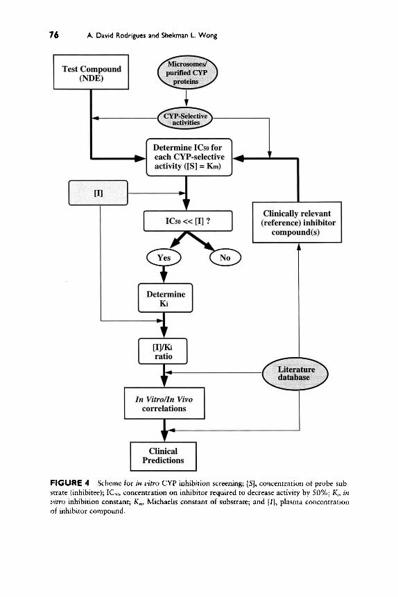
76 A. David Rodrigues and Shekman L. Wong
In V i t r o h Vivo
I Clinical Predictions I
FIGURE 4 Scheme for i f z vitro CYP inhibition screening; [S], concentration of probe sub- strate (inhibitee); IC,,, concentration on inhibitor required to decrease activity by 50%; K,, in vitio inhibition constant; K,, blichaelis constant of substrate; and [ I ] , plasma concentration of inhibitor compound.

In Vitro-in Vivo Correlations 77
followed by measuring the production of [14C]formaldehyde and assays can be run in a “batch extraction” format. In addition, it is imperative that clinically relevant positive controls, such as ketoconazole (CYP3A), sulfa- phenazole (CYP2C9/10), fluvoxamine (CYPlA2), and quinidine (CYP2D6), are run in parallel with the test compound (Rodrigues, 1994).
Inhibition studies are usually geared toward obtaining a measure of inhibitory potency, such as ICso (concentration of NDE required to inhibit activity by 50%) and/or the inhibition constant (K,) . The former is easier to obtain, at a fixed concentration of substrate ( [ S ] ) , and can be a useful parameter for screening a large number of compounds. However, when determining ICso for a given CYP activity, it is important to have some knowledge of the apparent K, of the substrate (Table I). When substrate concentration equals apparent K,, assuming competitive, noncompetitive, or uncompetitive inhibition (Segel, 1993), an initial estimate of K , can be obtained (Fig. 5 ) . The formal determination of K, entails considerably more work and involves studying inhibition at different concentrations of inhibitor and substrate. Data are then analyzed by linear (e.g., Dixon and Cornish- Bowden plots) or nonlinear transformations (Segel, 1993; von Moltke et al., 1994a,b), which also yield information concerning the mechanism of inhibition. Irrespective of the substrate being studied, the K, can be regarded as a measure of the affinity of the inhibitor for the enzyme and is an intrinsic property of that inhibitor. A third parameter that is often used is “fractional inhibition” ( i ) , which relates the degree of inhibition to both the K, of the inhibitor and its concentration ([A) in the assay (Fig. 5). For a competitive, noncompetitive, or mixed (competitivehoncompetitive) inhibitor (Segel, 1993; Webb, 1963), if one assumes that the concentration of substrate ( [ S ] ) is below its apparent K,, fractional inhibition is described by the relationship [ I] /K, + [A (Fig. 5) . This is a useful relationship, as most compounds exhibit first-order or linear pharmacokinetics ( [ S ] < K,) in uiuo (Leeman and Dayer, 1995). In the case of uncompetitive inhibition, fractional inhibition will be governed by [ S ] and will increase as [ S ] increases (Fig. 5). Therefore, it may be useful to screen for in uitro inhibition at higher concentrations of substrate ([S] > K,), although the clinical relevance of uncompetitive inhibition is debatable when [S] is low ([S] < K,) (Segel, 1993).
1. Studies with ABT-538 (Ritonavir)
At the present time, ABT-538 has been identified as the most potent inhibitor of CYP in our library of compounds. Therefore, a considerable amount of effort has been spent characterizing this inhibition. Data have been used by clinical investigators to establish the priority of drug-drug interaction trials.
0. CYP Inhibition. The effect of ABT-538 (Fig. 2) on a number of different CYP-dependent monooxygenase activities (Table I ) was studied using hu-
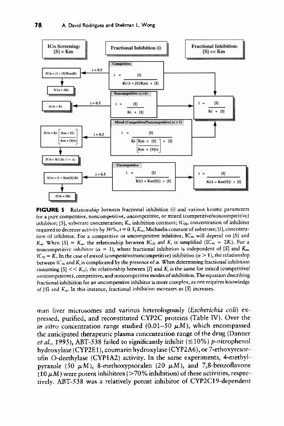
78 A. David Rodrigues and Shekman L. Wong
FIGURE 5 Relationship between fractional inhibition ( 1 ) and various kinetic parameters for a pure competitive, noncompetitive, uncompetitive, or mixed (competitivehoncompetitive) inhibitor; [S], substrate concentration; K,, inhibition constant; KjO, concentratlon of inhibitor required to decrease activity by SO%, 1 = 0.5; K , Michaelisconstant of substrate; [ I ] , concentra- tion of inhibitor. For a competitive or uncompehtive inhibitor, ICjO will depend on [ S ] and K,. When IS] = K,, the relationship between ICY, anf K, is simplified (ICS0 = 2KJ. For a noncompetitive inhibitor (a = l ) , where fractional inhibition is independent of [S] and K,, IC,, = K,. In the case of mixed (competitive/noncompetitive) inhibition (a l) , the relationship between ICsO and K, is complicated by the presence of (Y. When determining fractional inhibition (assuming [S] << K,), the relationship between [I] and K, is the same for mixed (competitive/ noncompetitive), competitive, and noncompetitive modes of inhibition. The equation describing fractional inhibition for an uncompetitive inhibitor is more complex, as one requires knowledge of [ S ] and K,. In this instance, fractional inhibition increases as [S] increases.
man liver microsomes and various heterologously (Escherichia coli) ex- pressed, purified, and reconstituted CYP2C proteins (Table IV). Over the in vitro concentration range studied (0.01-50 pM ), which encompassed the anticipated therapeutic plasma concentration range of the drug (Danner et al., 1995), ABT-538 failed to significantly inhibit (510%) p-nitrophenol hydroxylase (CYP2El ), coumarin hydroxylase (CYP2A6), or 7-ethoxyresor- ufin 0-deethylase (CYPlA2) activity. In the same experiments, 4-methyl- pyrazole (50 pM), 8-methoxypsoralen (20 pM), and 7,8-benzoflavone (10 pM) were potent inhibitors (>70% inhibition) of these activities, respec- tively. ABT-538 was a relatively potent inhibitor of CYP2C19-dependent

In Vitro-in Vivo Correlations 79
TABLE IV Inhibition of Various Human Liver Microsomal CYP-Dependent Monooxygenase Activities by ABT-538
Activity CYP selectivity ICJO (pM)Y
Microsomes Dextromethorphan 0-demethylase Coumarin hydroxylase p-Nitrophenol hydroxylase Terfenadine hydroxylase Tolbutamide hydroxylase (S)-Mephenytoin 4’-hydroxylase 7-Ethoxyresorufin 0-deethylase
Naproxen 0-demethylase Naproxen 0-demethylase Naproxen 0-demethylase
cDNA-expressed CYP2Cb
2D6 2A6 2E1 3A 2C8, 9, 10 2C19 1 A2
2C8 2C9 2C19
2.5 >50 >50
0.14 8.0
13 >50
50
30 8.0
a Concentration of ABT-538 required to inhibit activity by 50% (IC,,) was obtained graphically from a plot of percentage activity remaining (relative to incubations containing methanol alone) vs log,, of ABT-538 concentration (0.01-50 p M ) . All incubations were performed under similar assay conditions, where the substrate concentration was equal to K,. ABT-538 was dissolved in methanol, such that the final concentration in the assay was 0.5% (v/v). Various CYP2C proteins were purified from E. coli cell lysate and were reconstituted with NADPH-CYP reductase, cytochrome bJ, and dilaurylphosphatidylcholine.
(S)-mephenytoin 4’-hydroxylase (ICso = 13 pM) and CYP2C9/10- dependent tolbutamide hydroxylase (ICso = 8.0 pM) activity, which was confirmed with reconstituted CYP2C19 and CYP2C9 (Table IV). However, the most potent inhibition (ICs0 5 5.0 pM) was observed with dextrometh- orphan 0-demethylase (CYP2D6) and terfenadine hydroxylase (CYP3A) activity, yielding an ICso of 2.5 and 0.14 pM, respectively. In accordance, ABT-538 was shown to be a CYP2D6 and CYP3A substrate (Kumar et al., 1996). Therefore, data implied that the drug was a noncompetitive inhibitor of CYP2C activity and that the rank order of CYP inhibition is CYP3A B CYP2D6 > CYP2C % CYPlA2 = CYP2A6 = CYP2E1. Linear transforma- tion of data, using both Dixon (Fig. 6) and Cornish-Bowden (data not shown) plots, has revealed that ABT-538 is a competitive inhibitor of dextro- methorphan 0-demethylase activity (K , = 1.0 pM) and is a very potent mixed (competitive/noncompetitive) inhibitor (Ki = 0.02 pM ) of terfenadine hydroxylase activity (Table V). The marked inhibitory effect of ABT-538 on CYP3A activity more than likely reflects its hydrophobicity (log P = 4.0) and large size (molecular weight -721) (Kumar et al., 1996; D. Kempf, unpublished observations). In this regard, ABT-538 is similar to ketocona- zole (Ki = 0.02-0.7 pM). However, in the case of ketoconazole, the mecha- nism of inhibition (competitive vs noncompetitive) may depend on the sub- strate being investigated (Wrighton and Ring, 1994; Back and Tjia, 1991;

A. David Rodrigues and Shekman L. Wong
a, a 0 5;
z
b
CI 0
e 0 X
5 z z
a
.I 0
s 0 x
0.4 1
0.00 0.05 0.10 0.15 0.20
l/[Dextromethorphan] (pM)
0.0 0.1 0 . 2 0.3 0 .4
l/[Terfenadine] (pM) FIGURE 6 The effect of ABT-538 on dextromethorphan 0-demethylase and terfenadine hydroxylase activity in human liver microsomes. Replot of Dixon plot slopes for the inhibition of (A) dextromethorphan 0-demethylase and (B) terfenadine hydroxylase activity. In the case of dextromethorphan, the effect of ABT-538 was consistent with competitive inhibition. Data indicate that ABT-538 was either a mixed or a noncompetitive inhibiror of terfenadine hydroxy- lase activity, as the plot failed to pass through the origin. Dixon and Cornish-Bowden plots confirmed that ABT-538 was a mixed inhibitor of terfenadine hydroxylase activity (Kumar et al., 1996).

In Vitro-in Vivo Correlations 81
TABLE V inhibition of Dextrornethorphan 0-Demethylase (CYP2D6) and Terfenadine Hydroxylase (CYP3A) Activity in Human Liver Microsornes by ABT-538
Activity
Parametera CYP2D6 CYP3A
1 .o
Competitive
0.02 0.13 6.25 Mixedb
a Inhibition constants were obtained from Dixon and Cornish-Bowden plots. Apparent K, is defined as the dissociation constant of enzyme-inhibitor complex (absence of substrate). Apparent K,,, represents the dissociation constant of the enzyme-inhibitor-substrate complex. Factor (Y represents K, (dissociation constant of enzyme-substrate complex) changes when the inhibitor occupies the enzyme. Competitivehoncompetitive inhibition (a > 1; K,, > K,). For competitive inhibition, K,,, and 01 are equal to m.
Pichard et al., 1990; von Moltke et al., 1 9 9 4 ~ ) . Furthermore, unlike ABT- 538, ketoconazole does not appreciably inhibit (510%) CYP2D6 activity in human liver microsomes at low ( < l o p M ) concentrations (A. D. Rodrigues, unpublished observations).
b. Spectral Binding Studies. Subsequent studies have revealed that ABT- 538 is similar to ketoconazole, an "-substituted imidazole, because it elicits a type I1 difference spectrum (A,,, - 424 nm; Amin - 396 nm) when added to human liver microsomes (Fig. 7). This type of spectral perturbation indicates that the drug is able to reversibly bind to the ferric heme of CYP (Rodrigues et al., 1987). Because the 4-substituted analog of ABT-538 (A- 152184; Fig. 2) fails to elicit a type I1 spectrum (Fig. 7), heme binding probably occurs via the sterically unhindered nitrogen on the 5-substituted thiazole moiety (Smith and Wilkinson, 1978). However, the remainder of the molecule, or backbone, is also involved in binding to CYP (Rodrigues et al., 1987). This is evidenced by the observation that a type I spectrum (A,,, - 390 nm; Amin - 415 nm) is obtained when A-152184 is added to human liver microsomes (Fig. 7). In addition, despite the loss of heme binding, A-152184 is still a relatively potent inhibitor (ICso = 2.0 p M ) of terfenadine hydroxylase activity in human liver microsomes (Fig. 7).
c. Clinical Significance of CYP Inhibition. Inhibition of CYP by ABT-538 will be of clinical significance because most AIDS patients are exposed to a large number of therapeutic agents (Kumar et al., 1996, and references therein). These range from reverse transcriptase inhibitors (e.g., AZT) to tricyclic antidepressants and anti-infectives (e.g., macrolide antibiotics and

82 A. David Rodrigues and Shekman L. Wong
A B ABT-538 A-152184 120 1
M fi fi m
." .- f d
.01 .1 1 10 100
Wavelength (nm) Wavelength (nm) Concentration (pM) FIGURE 7 The binding of ABT-538 and A-152184 to human liver microsomes and inhibi- tion of terfenadine hydroxylase activity. (A) Difference spectra (350-500 nrn) were recorded atter the addition of ABT-538 or A-152184 to human liver microsornes, under aerobic condi- tions at room temperature. Washed rnicrosornes were diluted with 0.1 M potassium phosphate buffer ( p H 7.4), containing 0.1 m M EDTA, so that the final concentration of CYP was 1.0 p M (1.53 mg proteidml). ABT-538 and A-152184 were singularly added to the diluted microsomes at a final (saturating) concentration of 5.0 pM. Both compounds were dissolved in spectral grade dirnethyl sulfoxide, the final volume of which never exceeded 1.0% (vh) . (B) inhibition of terfenadine hydroxylase activity in human liver rnicrosornes by ABT-538 (0) and A-152184 (0). Data are expressed relative to controls (solvent alone). The concentration of terfenadine was 10 pM ( -K , , , ) .
imizadole/triazole antifungals). Moreover, at the present time, it is accepted that the successful treatment of AIDS may require the administration of various combinations of AZT and one or more protease inhibitors (D. Kempf, personal communication). Drug-drug interactions between ABT- 538 and other protease inhibitors are likely, as a number of these (e.g., SC- 52151, indinavir, and saquinavir) have been shown to be CYP3A substrates (Farrar et al., 1994; Hensleigh et a/., 1995; Roy et al., 1995). Preliminary data indicate that ABT-538 has a pronounced effect on the pharmacokinetics of drugs, such as saquinavir in both the rat and the dog, and markedly inhibits the metabolism of saquinavir in human liver microsomes (Kempf et al., 1997; K. Marsh, unpublished observations). Therefore, it may be possible to enhance the oral bioavailability of saquinavir in humans by coadministration with ABT-538, which may prove to be clinically advanta- geous (Kempf et al., 1997; Kempf, 1994).

In Virro-in Vivo Correlations 83
IV. In Vitro-in Vivo Correlations
Although in vitro inhibition studies can be performed with relative ease, it is important to recognize that currently employed in vitro-in vivo correlation ( M V C ) approaches are only very useful for predicting “lack of inhibition” or “potent inhibition” of DMEs in vivo. For drugs that exhibit “intermediary potency” it may be difficult to predict clinical outcomes, without the use of more sophisticated approaches. Furthermore, one should not lose sight of the fact that two drugs may interact via multiple mechanisms in vivo, e.g., DME inhibition and/or induction, protein-binding interactions, and/or absorption effects. The presence of inhibitory metabolites, as in the case of norsertraline and norfluoxetine (Otton et al., 1993; von Moltke et al., 1994b), and extrahepatic metabolism (e.g., CYP3A in the gut) may further complicate the situation in vivo. An additional problem that is often faced by investigators is the lack of information concerning the complete metabolic profile of the inhibitee drug ( f,), as much of the data is based solely on the urinary metabolic profile, and in many cases only parent drug levels in plasma are quantitated (Olkkola et al., 1994; Vanakoski et al., 1995; Heizmann and Ziegler, 1981; Eberts e t al., 1981; Varhe et al., 1994; Garteiz et al., 1982). Many of these factors may confound and undermine IVIVC methods.
Despite the challenges, and as the in vitro database continues to grow, it is probable that one of the next great explosions in the field of drug metabolism will be the development of quantitative methods for predicting in vivo drug-drug interactions. For the present moment, however, no con- sensus has been reached concerning which parameters are most appropriate for the accurate prediction of in vivo drug-drug interactions, by analogy with the “scaling factors” used in predicting metabolic clearance from in vitro data (Houston, 1994). Although a few approaches have been docu- mented (von Moltke et a/., 1995b; Leeman and Dayer, 1995; Tucker, 1992), it is not known if these are applicable in all situations. For the most part, currently employed IVIVC approaches can best be described as empirical, leading to “ball park” or rough estimates of clinical outcomes.
An example of such an “emprical” approach is presented in Table VI, where in vitro inhibition data obtained with seven structurally diverse Abbott compounds are shown. In most cases, except for ABT-538, the ICso for the inhibition of terfenadine hydroxylase (CYP3A) activity in human liver microsomes was high (> 100 pM) and exceeded the therapeutic concentra- tion of total (free and protein bound) drug in plasma (I 25 p M ) . The CYP3A-dependent metabolism of most of the drugs was characterized by a relatively high K,,, (2 100 p M ; Table 11), and the relatively low affinity ( K , r 100 p M ) inhibition of terfenadine hydroxylase activity ( [ S ] = K , ) was assumed to be competitive (inhibitor/cosubstrate K , - K,; Boobis, 1995). When compared to ketoconazole (ICso = 0.20 pM; plasma C,,, =

84 A. David Rodrigues and Shekman L. Wong
TABLE VI Terfenadine Metabolism
In Vkro and in Vivo Effects of Various Compounds on
Therapeutic plasma Terfenadine AUC Compound” ICw ( F M / ~ concentration (wh4)’ (% increase)d
Zileuton B C D E ABT-538 G” Ketoconazole
> 100 >loo >lo0 > lo0
25 0.14 6.5 0.20
25 36‘ 6.0 12’ 0.22 48‘ 0.80 ?‘ 1.5 ,r
5-2 1 > ? ?
5.0-16 2 1700’
,i Drugs at various stages of development, except for ketoconazole. Concentration of drug required to inhibit human liver microsomal terfenadine hydroxylase activity by 50%. Activity was measured employing a final terfenadine concentration that approximated apparent K,. Typical plasma concentration attained in subjects receiving therapeutic doses. Effect on area under the plasma concentration vs time curve (AUC) for parent terfenadine.
” Parent terfenadine was measured using a sensitive liquid chromatography-mass spectromet- ric method.
‘ Although no studies have been carried out with terfenadine, it has no effect on the pharmacoki- netics of the CYP3A substrate diltiazem (L. Gustavson, personal communication). No studies have been conducted with terfenadine. However, compound “E” contains a methylenedioxyaryl moiety, exhi bits nonlinear pharrnacokinetics, and forms a CYP complex upon incubation with human liver microsomes. The drug was incubated with human liver microsomes, and NADPH, prior to the addition of terfenadine. Benzimidazole-containing compound.
’ Data obtained from Honig et al. (1993b).
5-16 pM), the effect of these drugs on the plasma AUC of parent terfenadine was minimal (548% increase vs 2 1700% increase) and was comparable to that observed when grapefruit juice is coadrninistered with other CYP3A substrates such as midazolarn and triazolam (Hukkinen et al., 1995; Kupfer- Schmidt et al., 1995; Honig et al., 1993b). However, some degree of caution is needed when studying mechanism-based inhibitors (e.g., erythromycin and cimetidine), where the degree of inhibition in vivo can often be underesti- mated in vitro (Wrighton and Ring, 1994). We have noted that many of these types of compound often exhibit in vitro K , values that exceed the concentration of drug in plasma. One of the drugs that we have studied (“compound E”; Table VI) is an example of a mechanism-based inhibitor of CYP3A and, based on the nonlinear pharmacokinetics exhibited in vivo (R. Granneman and J. Ferrero, unpublished observations), it is higly likely that the extent of CYP inhibition was underestimated in vitro. Therefore, the in vitro study design should be structured so that the possibility of mechanism-based inhibition is ruled ou t as early as possible.
In Vitro-in Vivo Correlations 85
Because most investigators in the drug industry are faced with a large array of clinical candidates, often exhibiting markedly different structures, physicochemical properties and pharmacokinetics, and so on, it may be very difficult to develop a single IVIVC approach that encompasses all classes of drug. However, it is generally accepted that the ratio of inhibitor concentra- tion to its K , ([IIlK,) is one of the most important parameters defining the potency of inhibition (Boobis, 1995; Leeman and Dayer, 1995; Tucker, 1992; Rowland et al., 1994; Rowland and Matin, 1973). This presents investigators with three problems: (1) one has to estimate the concentration of drug ( [ I l ) at the site of metabolism (e.g., the liver); (2) one may have to consider drug binding to plasma and/or tissue protein (i.e., fraction unbound; fu) in the IVIVC; and ( 3 ) one has to relate in vitro K , to changes in the gross pharmacokinetic parameters of the inhibitee in vivo. These parameters include plasma area under the concentration vs time curve (AUC), half-life ( tIl2), steady-state concentration (C,,), and systemic clearance (Leeman and Dayer, 1995; Tucker, 1992; Rowland and Matin, 1973). With respect to the first and second problems, many investigators are currently employing the concentration of total (free and protein bound) drug in plasma as a measure of [ I ] in the liver, although a number of workers have also opted to include estimates of liver tissue/plasma or liver tissue/water ratios in their IVIVC procedures (von Moltke et al., 1994a,b,cY1995b; Leeman and Dayer, 1995; Tucker, 1992). Steady-state inhibitor plasma concentrations are often employed, whereas maximal concentrations ( C,,,) are considered after single doses. However, it may be prudent to utilize a time-averaged plasma concen- tration (AUCo - &), as the plasma concentration of the inhibitor will often decrease with time between doses.
A. Inhibition Index
When considering the most common types of inhibition (e.g., competi- tive, noncompetitive, and mixed), the ratio [I]/K, can be related to changes in the (hepatic) intrinsic clearance (CLi,,) of the inhibitee in a straightforward manner [Eqs. (1-3), Fig. 81. However, relating changes in CLinr to changes in plasma AUC and/or Css may be difficult because the relationship between CLinr and plasma clearance (CL,) has to be considered [Eqs. (4 and 5), Fig. 81 and may require more complicated models (Leeman and Dayer, 1995; Wilkinson, 1987). This is especially true in those cases where the plasma clearance in the absence of the inhibitor [CL,,,,] approaches plasma flow ( Q h ) to the liver (-900 mumin). Only when CL,,,, is low [cL,,,,/Qh I 0.051 can CLi,, and AUC be directly related with some degree of certainty (Leeman and Dayer, 1995). This is possible with substrates such as warfarin and tolbutamide, which are cleared slowly [CL,,,, < 20 ml/min] and are primarily metabolized in the liver (Houston, 1994). In fact, the relationship proposed by Rowland and Matin (1973), which is very similar to Eq. (3) (Fig. 8),


In Vitro-in Vivo Correlations 87
modeled the effect of sulfaphenazole on the steady-state plasma concentra- tion (CJ of tolbutamide. For drugs that exhibit higher (>lo0 mYmin) values of CL,,,, (e.g., desipramine, terfenadine, midazolam, etc) it may be more difficult to predict changes in plasma AUC based on [II/K, ratios. This is especially true after intravenous drug administration or in the case of orally administered drugs exhibiting a high gut extraction (EG).
At the present time our laboratory uses the concentration of total drug (free and protein bound) in plasma as a measure of [q in the liver, knowing that this may lead to an overestimation of inhibition in those cases where drugs are highly protein bound. Once the K, for a given form of CYP is obtained in vitro, an “inhibition index” ( R ) can be calculated [Eq. ( 3 ) , Fig. 81. This parameter describes the effect of the inhibiting NDE on the intrinsic clearance of the inhibitee and no direct estimates of changes in AUC are made, unless CL,,,, is low (5 20 mumin). For a given inhibitee, calculation of an inhibition index assumes that one has prior knowledge of the fraction of the dose ( f m ) that is metabolized by the relevant form of CYP. For CYP3A substrates such as clarithromycin ( f m = 0.35), terfenadine ( f m - 0.95), midazolam and triazolam ( f m - 0.85), and CYP2D6 substrates such as desipramine ( f m = 0.85), nominal estimates of f m can be obtained from the literature (Ferrero et al., 1990; Garteiz et al., 1982; Brosen and Gram, 1988, 1989b; Brosen et al., 1986, 1993; Heizmann and Ziegler, 1981; Eberts et al., 1981; Fabre et al., 1988). However, many drugs are metabolized by parallel pathways, each mediated by different CYP forms (e.g., propranolol and warfarin). In these instances, one has to consider the in vitro K, values for each form of CYP and the fraction of the dose that is metabolized via each pathway (von Moltke et al., 1995b).
In order for this approach to be useful, one determines the inhibition index for a number of clinically relevant positive and negative controls,
FIGURE 8 Predicting the effect of an inhibitor on intrinsic clearance. Equation( 1) is derived from the relationships described in Fig. 5, assuming competitive, noncompetitive, or mixed (competitivehoncompetitive) inhibition. Protein binding in microsomal suspensions is consid- ered to be minimal for both inhibitor and inhibitee, is., intrinsic clearance of free drug (CL,,,) is considered. Equation (2) can be related to Eq. (1) with the introduction of the term fm (fraction of inhibitee dose metabolized by the inhibited pathway), due to the fact that multiple pathways exist in vivo. The unbound fraction in plasma (inhibitor, f*’; inhibitee, fa) is also factored into the equation. In turn, Eq. ( 2 ) can be rearranged to give Eq. ( 3 ) , which describes the effect of an inhibitor on inhibitee intrinsic clearance (CL,,,) as a ratio ( R ) or “inhibition index”: CL.,,,(,, and CL,znc(,l represent intrinsic clearance in the absence and presence of inhibitor, respectively. Equation (5) can be derived from Eq. ( 3 ) and the “well-stirred model” [Eq. (4); Wilkinson, 19871, where C L , CLh, and Q h represent inhibitee plasma (systemic) clearance, hepatic clearance, and plasma flow to the liver (-900 mumin), respectively. For a given inhibitee, assuming that plasma clearance in the absence of the inhibitor [CL,,,,] is less than/ equal to 5% of Qb changes in CL,., can be directly equated to changes in the area under the plasma concentration vs time curve (AUC); AUCi, inhibitee plasma AUC in the presence of inhibitor; AUCc, inhibitee plasma AUC in the absence of inhibitor. It is assumed that renal clearance is negligible (is., CL, = CLL).

a% A. David Rodrigues and Shekman L. Wong
where clinical outcomes have already been documented, and relates these to the inhibition index of the NDE. A useful database is the SSRI series and quinidine (Table VII), where representative in vitro K, data are readily available (Otton et al., 1993; Broly et al., 1989; von Moltke et al., 1994b, 1995a; Crewe eta/., 1991), and the effects on the pharmacokinetics of drugs such as desipramine have been reported (Brosen et al., 1993; Brosen and Gram, 1989b; Steiner et al., 1987; Bergstrom et al., 1992; Preskorn et al., 1994). Despite the fact that the extent of protein binding in plasma varies considerably among this set of compounds (e.g., sertraline, 99%; fluvoxa- mine, 77%; Grimsley and Jann, 1992; Van Harten, 1993), a reasonably good correlation ( r = 0.829) can be obtained between the in vitro-derived inhibition index and the observed increases in desipramine plasma AUC (Fig. 9). It should be stated, however, that we had to take into account the plasma concentrations of norsertraline and norfluoxetine, as both metabo- lites exhibit a K, value similar to that of parent drug (Otton et al., 1993; von Moltke et al., 1994b). With time, it may be possible to obtain correla- tions with larger numbers of compounds and for each form of CYP.
B. Correlations with ABT-538 (Ritonavir)
ABT-538 posed a number of problems, as it was shown to be an inhibitor of multiple CYP forms in vitro (Table IV) and was extensively protein bound (-98%) in plasma (J. Denissen, unpublished observations). However, the decision was made to go ahead with the IVIVC assuming that [I] equated with the concentration of total drug ([I]tota,) in the plasma (range = 8.0- 16 pM; Danner et al., 1995). It should also be stated that the IVIVC procedures described later were carried out prospectively, prior to the avail- ability of clinical drug-drug interaction data. Emphasis was placed on CYP3A and CYP2D6, as the lowest ICso values ( [ S ] = K,) were obtained with these two forms of CYP. Subsequent studies have revealed that ABT- 538 does not markedly reduce the plasma clearance of theophylline in vivo (A. Hsu, unpublished observations), suggesting the lack of inhibition of CYPlA2, in agreement with in vitro data. In addition, there are some prelimi- nary reports that ABT-538 does not inhibit CYP2C9 in vivo, as no reduction in phenytoin plasma clearance has been noted (A. Hsu, personal communi- cation).
I . CYP3A Inhibition
Based on our in vitro findings, it was anticipated that the greatest effect would be observed with CYP3A, given the fact that the K , was low ( K , < [a) and fm was known to be high (> 0.7) for a large number of substrates. It was estimated that CYP3A activity would be markedly inhibited (298%0), or abolished (Fig. lo), a t clinically relevant plasma concentrations. There- fore, changes in CL,,, would be solely governed by whatever fraction of the

TABLE VII Effect of Various Compounds on the Pharmacokinetics of Desipramine (DMI): Correlation with in Vitro CYP2D6 Inhibitory Potency
In vivo"
Test compound DMI In vitrob
Dose Plasma Plasma 2-OH-DMI Fractional
Compound mglda y No. of days concentration (pM) A UCilA UCc (% decrease) K , (PM) inhibition (i)
Quinidine Paroxetine Fluoxetine A Fluoxetine B Fluoxetine C Sertraline Fluvoxamine EM vs PM'
200 20 60 20 60 50
100 -
12 20
1 21
8 21 10 -
1.20 0.14 0.1 8d 0.70' 0.87' 0.22' 0.60 -
6.0 5.0 2.0 4.8 6.4 1.2 1.1
24.0
97 66 58
90 -
- - 2 90
0.03 0.06 0.17 0.17 0.17 1.50 1.8
0.98 0.70 0.51 0.80 0.84 0.13 0.25 -
a Subjects were given a single dose (SO-100 mg) of desipramine in the presence or absence of test compound. There are three dosing regimens for fluoxetine (A, B, and C). The fold-increase in plasma AUC of desipramine was determined in each case, where AUCi and AUCc represent desipramine AUC in the presence and absence of inhibitor, respectively. With some of the compounds, the effect on the urinary levels of 2-hydroxydesipramine (2-OH-DMI) was also measured. The plasma concentration of compound (free and protein bound) attained in plasma is also indicated (Brosen et al., 1993; Brosen and Gram, 1988, 1989b; Kaye et al., 1989; Steiner et a[., 1987; Bergstrom et al., 1992; Preskorn et al., 1994; Van Harten, 1993; Spina et al., 1993; Grimsley and Jann, 1992). Plasma concentrations were determined at steady state, except for single-dose fluoxetine (fluoxetine A), which represents maximal plasma concentration ( CmJ. Except for paroxetine, K, was obtained with human liver microsomes employing dextromethorphan 0-demethylase (DEXase) as the CYP2D6 marker activity (Otton et al., 1993; Broly et al., 1989). The K, for the inhibition of DEXase activity by paroxetine is anticipated to be 0.06 p M (fluoxetine Kj3), based on data obtained with sparteine and desipramine (von Moltke et al., 1994b, 1995a; Crewe et al., 1991). Fractional inhibition of desipramine metabolism was calculated using the equation [plasma]/K, + [plasma], where the in vitro K, was related to the plasma concentration of the test compound. Increase in plasma AUC of desipramine and decrease in urinary 2-OH-DMI levels, when poor metabolizers (PM) are compared to extensive metabolizer (EM) subjects (Spina et al., 1984; Brosen et al., 1986, 1993; Brosen and Gram, 1988, 1989b). Sum of fluoxetine and norfluoxetine plasma concentrations, as the in vitro K, for both is similar (Otton et al., 1993). Sum of sertraline and norsertraline plasma concentrations, as the tn vitro K, for both is similar (von Moltke et al., 1994b).

90 A. David Rodrigues and Shekman L. Wong
7 -
6 -
5 - 0 Fluox B
0
y = 0.65470 + 1.1439~: r = 0.829
sdn I OOFluvox 1 1 t I I
1 2 3 4 5 6
Inhibition Index (R) (In Vitro)
FIGURE 9 The effect of various CYP2D6 inhibitors on the pharmacokinetics of desipramine: in uitro-in vivo correlation. The clinically observed increases in desipramine AUC (Table VII) were correlated ( Y = 0.829) with valuesof “R” [Eq. (3); Fig. 81 for a number of well-documented CYP2D6 inhibitors. In each case, the in uitro K, and plasma (total) drug concentrations (f”’ = 1; [I],,,) listed in Table VII were used. The fraction of the desipramine dose ( f m ) metabolized by CYP2D6 was assumed to be 0.85 (Brosen and Gram, 1988, 1989b; Brosen et al., 1986, 1993). QND, quinidine; fluox, fluoxetine; parox, paroxetine; sert, sertraline; fluvox, fluvoxa- mine; AUCi, desipramine plasma AUC in the presence of inhibitor; AUCc, desipramine plasma AUC in the absence of inhibitor.
dose was metabolized by CYP3A [when [I] + K, , R = l/l-fm; Eq. (3); Fig. 81. Subsequent clinical data obtained with clarithromycin (Table VIII) were in agreement with this conclusion, as we have shown that the 14-hydroxyla- tion of clarithromycin is primarily catalyzed by CYP3A in human liver microsomes (A. D. Rodrigues, unpublished observations). By comparison, “compound B” (Table VI) has been shown to have no effect on the pharma- cokinetics or metabolism of clarithromycin (L. Gustavson, unpublished re- sults). Furthermore, we have determined the CYP3A inhibition index ( R ) of ABT-538, at different values of fm, and found that the profife is very similar to that of ketoconazole, although fluconazole was also included in the analyses (Fig. 11) . Therefore, it is expected that ABT-538 will bring about pronounced elevations (2 17-fold) in the plasma AUC of CYP3A substrates such as terfenadine, midazolam, and triazolam (Honig et al., 1993a,b; Olkkola et al., 1994; Varhe et al., 1994; Vanakoski et al., 1995). As discussed previously, this is of clinical significance because a number of

In Vitro-in Vivo Correlations 91
0.6 -I Fractional
Inhibition (i) of Total CLint o.4
0.0 Om2 I .001 .01 .1 1 10 100
[ABT-5381 (WM) FIGURE I0 Fractional inhibition of CYP2D6 and CYP3A by ABT-538. The curves were generated using Eq. ( 2 ) (Fig. 8), with the in vitro K , values describing the inhibition of CYP2D6- dependent dextromethorphan 0-demethylase ( K - = 1 .O p M ) and CYP3A-dependent terfena- dine hydroxylase ( K , = 0.02 pM) activity. The simplest case is modeled, where the drug is metabolized completely by CYP3A or CYP2D6 ( f m = 1) and protein binding of ABT-538 is considered negligible ( f. - 1). The shaded area encompasses the anticipated therapeutic plasma concentration range of ABT-538.
TABLE Vlll Time Curve (AUC) of Clarithromycin and Formation of 14-[R]-Hydroxyclarithromycin
Effect of ABT-538 on the Area under the Plasma Concentration vs
AUCo.2, ,,, (pg ' hrlrnly
Minus ABT-538 Plus ABT-538 Ratio
Clarithromycin 49.0 t 14.1 86.9 2 20.4 1.78 Hydroxyclarithromycin 15.7 t 4.53 0.04 2 O . l l b 0.003
il Data represent mean t SD of 24 subjects. Clarithromycin (500 mg q 12 hr, PO) was dosed in the absence or presence of ABT-538 (200 mg q 8 hr, PO) for 4 days. Plasma concentrations were measured on day 4. For ABT-538, mean plasma C,,, and Cmin were 6.7 pglml(9.0 p M ) and 2.1 pglrnl (4.2 p M ) , respectively. Represents 99.7% inhibition of metabolism.

92 A. David Rodrigues and Shekman L. Wong
43 -
37 -
31 -
25 -
19 -
13 -
7-
I t
R Midazolam Triazolam
Ctarithronrycin
- __-_-____-_-_-___-------- 1 I
1
49 4 Fluconazole ----- ABT-538 ... ,......... *... Ketoconazoie -
Terfenadine
1.
0.0 0.2 0.4 0.6 0.8 1.0
Fraction of Dose Metabolized by CYP3A (fm)
FIGURE I I Predicting the effect of ABT-538, ketoconazole, and fluconazole on the pharma- cokinetics of CYP3A substrates. For a given value of fm, the effect on substrate (inhibitee) CL,,, was calculated ( R ) using Eq. (3) (Fig. 8). For ABT-538, ketoconazole, and fluconazole, the in vitro K, used was 0.02,0.2, and 60 p M , respectively (Wrighton and Ring, 1994; von Moltke et al., 1994c; Back and Tjia, 1991). Therapeutic plasma concentrations of each drug ( f.’ =
1; [&,,.,) were I0 pM (ABT-5381, 16 p M (ketoconazole), and 20 pM (fluconazole). For clarithrornycin, terfenadine, rnidazolarn, and triazolarn, the fraction of the dose metabolized by CYI’3A i s indicated.
peptidomimetic HIV-1 protease inhibitors (e.g., SC-52151, saquinavir, and indinavir ) are almost exclusively metabolized by CYP3A and exhibit exten- sive ( f,,, 2 0.8) first-pass metabolism (Kempf, 1994; Roy et al., 1995; Hen- sleigh et al., 1995; Farrar et al., 1994). The abolishment of CYP3A activity may also tead to marked “metabolic switching” or “shunting” in those cases were CYP3A substrates are also metabolized by parallel pathways involving different (noninhibited) enzymes (e.g., UDPGT, CYP2C, and/or CYPl A). This may potentially complicate the interpretation of clinical findings, espe- cially with drugs exhibiting a relatively narrow therapeutic index.
2. CYPZD6 Inhibition
Using the SSRVquinidine correlation described previously (Fig. 9), we at- tempted to predict the effect of ABT-538 on the pharmacokinetics of desipra-

In Vitro-in Vivo Correlations 93
7 -
6 -
5 -
4 -
3 -
2-
mine. In contrast to amitriptyline and imipramine, desipramine is almost ex- clusively metabolized by CYP2D6 and serves as a useful in vivo probe (Ketter etal., 1995). Initial data predicted that, unlike CYP3A, the extent of CYP2D6 inhibition was going to be dependent on the plasma concentration of ABT- 538 (Fig. 10). Therefore, an accurate prediction of the clinical outcome would be difficult to attain. However, based on the rank order of in vitro inhibition (CYP3A + CYP2D6 > CYP2C > CYPlA2), it was anticipated that the magni- tude of the effect of ABT-538 on CYP2D6 would be less pronounced than that observed with CYP3A. Data in Fig. 12 show that ABT-538 was predicted to increase the plasma AUC of desipramine by 5.0- to 6.5-fold over a plasma concentration range of 7 to 18 p M . This meant that the effect of ABT-538 on the pharmacokinetics of desipramine was going to be similar to that of quinidine (- 6.0-fold increase in desipramine plasma AUC) and that ABT-
Observed (wtlwt)
Observed (Blwt)
0 0 0
0 0
0 .- c)
d
1 0
1 I I I
0 10 20 30
Plasma [ABT-5381 (pM) FIGURE I2 Predicting the effect of ABT-538 on the pharmacokinetics of desipramine. Plot represents the relationship between plasma concentration of ABT-538 ( f,’ = 1; [IItoml) and the predicted changes (fold-increase) in the plasma AUC of desipramine. At each concentration of ABT-538, an inhibition index ( R ) was calculated [Eq. (3); Fig. 81 using a K , of 1.0 p M ( fm = 0.85). Each value of R (x) was then inserted into the equation described in Fig. 9, which yielded estimates of AUCUAUCc ( y) . Filled circles represent data obtained with 14 CYP2D6 genotyped subjects, all of the individuals used in the study were classified as “extensive metabo- lizers.” Subjects received a single dose of desipramine (100 mg, PO) on day 1 of the study and plasma levels of the drug were monitored. Following an 8-day washout period, the subjects received ABT-538 (500 mg q 12 hr, PO) for 3 days. On the third day of ABT-538 treatment, the subjects received a second single dose of desipramine (100 mg, PO).

94 A. David Rodrigues and Shekman L. Wong
538 would convert EM subjects to PM subjects. Subsequent data obtained with 14 EM subjects indicated that the IVIVC procedure overestimated the effect of ABT-538 on the pharmacokinetics of desipramine. In fact, a 1.5- to 5-fold (mean = 2.7-fold) increase in plasma AUC was observed, although 6 of the subjects (42%) exhibited greater than 3-fold increases in AUC. In gen- eral, the effect of ABT-538 was similar to that observed with fluoxetine (60 mg/day) at single doses (Bergstrom et aL, 1992). Whether or not long-term treatment with ABT-538 will have a more pronounced effect on the pharmaco- kinetics of desipramine is presently not known. Interestingly, if the concentra- tion of free ABT-538 in plasma (0.1-0.3 p M ) had been employed in our WIVC procedure ( [I]rree < K,), we would have predicted no inhibition of desi- pramine metabolism.
V. Conclusions
The “potential” of in vitro methods for predicting in vivo drug-drug interactions has already been realized by most investigators in the field of drug metabolism. In recent years, emphasis has been placed on the “develop- ment” of in vttro models for studying human drug metabolism and the identification of clinically relevant DMEs (Rodrigues, 1994; Wrighton et al., 1993; Tucker, 1992; Miners et al., 1994; Ball et al., 1995). However, we are now rapidly entering the “application” phase, albeit empirically, wherein data are being used by pharmaceutical companies to establish the priority of drug interaction trials and as part of IND, NDA, and internal review board (IRB) submissions. Moreover, as combinatorial methods be- come established (Eichler et al., 1995; Kubinyi, 1995), it is highly likely that the number of lead compounds will increase greatly. High-throughput in vitro drug interaction methods may play an ancillary role in the screening of these compounds, so that only potentially favorable clinical candidates are chosen for development.
In general, however, it is acknowledged that a considerable amount of work still remains to be done. This is evident in the case of the non-CYP DMEs such as UDPGT, where we are only now beginning to understand the metabolism of various drugs in terms of the specific enzyme subtypes (Burchell et al., 1995; Bock, 1995; Batt et al., 1994). Undoubtedly, the availability of heterologously expressed UDPGT enzymes will greatly facili- tate this process. The second major challenge facing investigators is the development of more refined, and widely applicable, IVIVC procedures for predicting drug-drug interactions. With the eventual development of rapid automated assays and the in vitro screening of larger numbers of structurally diverse and clinically relevant compounds, perhaps coupled to statistical experimental design or “virtual kinetics” (Bronson et al., 1995), it may be possible to construct an in vitro database. Such a database would include

In Vitro-in Vivo Correlations 95
potent (K; 6 [plasma]), intermediate (K; - [plasma]), and weak (K; s=- [plasma]) inhibitor compounds. Once generally acceptable and proven IVIVC “scaling factors’’ are established, it may be feasible to translate these in vitro data into clinically meaningful information. One possible strategy would be to use such a database as input or “training data” for an artificial neural network (Erb, 1995, and references therein).
Acknowledgments
The author thanks the following personnel at Abbott Laboratories: Drs. R. Granneman, A. Hsu, W. Awni, D. Ouellet, M. Karol, and R. Bertz for providing the appropriate pharmacoki- netic data; M. Mayer, M. Kukulka, P. Johnson, and E. Roberts for their technical support; and Drs. J. Ferrero, D. Mulford, J. Machinist, D. Kempf, and G. Kumar for their contributions. The author also thanks Drs. M. Shet and R. Estabrook (University of Texas Southwestern Medical Center, Dallas, TX), J. Lasker (Mount Sinai Medical School, New York, NY), and A. Parkinson and R. Pearce (University of Kansas Medical Center, Kansas City, KS) for very productive collaborations.
References
Albengres, E., and Tillement, J. P. (1992). Cyclosporin and ketoconazole, drug interaction or therapeutic association? Int. J. Clin. Pharmacol. Ther. Toxicol. 30,555-570.
Amsden, G. W. (1995). Macrolides versus azalides: A drug interaction update. Ann. Pharma- cother. 29, 906-917.
Back, D. J., and Tjia, J. F. (1991). Comparative effects of the antimycotic drugs ketoconazole, fluconazole, itraconazole and terbinafine on the metabolism of cyclosporin by human liver microsomes. Br. J. Clin. Pharmacol. 32, 624-626.
Ball, S. E., Scatina, J. A., Sisenwine, S . F., and Fisher, G. L. (1995). The application of in vitro models of drug metabolism and toxicity in drug discovery and drug development. Drug Chem. Toxicol. 18, 1-28.
Batt, A. M., Magdalou, J., Vincent-Viry, M., Ouzzine, M., Fournel-Gigleux, S., Galteua, M. M., and Siest, G. (1994). Drug metabolizing enzymes related to laboratory medicine: Cytochromes P450 and UDP-glucuronosyltransferases. Clin. Chim. Acta 226, 171-190.
Bergstrorn, R. F., Peyton, A. L., and Lemberger, L. (1992). Quantification and mechanism of fluoxetine and tricyclic antidepressant interaction. Clin. Pharmacol. Ther. 51,239-248.
Bertilsson, L. (1995). GeographicaVinterracial differences in polymorphic drug oxidation. Clin. Pharmacokinet. 29, 192-209.
Bloomer, J. C., Clarke, S. E., and Chenery, R. J. (1995). Determination of P4501A2 activity in human liver microsomes using [3-’4C-methyl]caffeine. Xenobiotica 25, 91 7-927.
Bock, K. W. (1995). Human UDP-glucuronosyltransferases: Classification and properties of isozymes. In “Advances in Drug Metabolism in Man” (G. M. Pacifici and G. N. Fracchia, eds.), pp. 291-309. European Commission, Brussels, Belgium.
Boobis, A. R. (1995). Predicting of inhibitory drug-drug interactions by studies in vitro. In “Advances in Drug Metabolism in Man” (G. M. Pacifici and G. N. Fracchia, eds.), pp. 515-539. European Commission, Brussels, Belgium.
Braeckman, R. A., Granneman, G. R., Rubin, P. R., and Kesterson, J. W. (1989). Pharmacokinet- ics and metabolism of the new 5-lipoxygenase inhibitor A-64077 after a single oral administration in man (abstr.). J. Clin. Pharmacol. 29, A22.

96 A. David Rodrigues and Shekrnan L. Wong
Breimer, D. D. (1994). Genetic polymorphisms in drug metabolism: Clinical implications and consequences in ADME studies. In “The Relevance of Ethnic Factors in the Clinical Evaluation of Medicines” (S. Walker, C . Lumley, and N. McAuslane, eds.), pp. 13-26. Kluwer Academic, Boston.
Broly, F., Lihersa, C. , Lhermitte, M., Bechtel, P., and Dupuis. B. (1989). Effect of quinidine on the dextromethorphan 0-demethylase activity of microsomal fractions from human liver. Br. 1. Clin. Phamacof. 28, 29-36.
Bronson, D. D., Daniels, D. M., Dixon, J. T., Redick, C. C., and Haaland, P. D. (1995). Virtual kinetics: Using statistical experimental design for rapid analysis of enzyme inhibitor mechanisms. Biochem. Pharmacol. 50, 823-831.
Brosen, K. (1993). The pharmacogenetics of the selective serotonin reuptake inhibitors. Clin. Invest. 71, 1002-1009.
Brosen, K., and Gram, L. F. (1988). First-pass metabolism of imipramine and desipramine: Impact of the sparteine oxidation phenotype. Clin. Pharmacol. Ther. 43, 400-406.
Brosen, K., and Gram, L. F. (1989a). Clinical significance of the sparteine/debrisoquine oxida- tion polymorphism. Eur. I . Clin. Pharmacol. 36, 537-547.
Brosen, K., and Gram, L. F. (1989b). Quinidine inhibits the 2-hydroxylation of imipramine and desipramine hut not the demethylation of imipramine. Eur. 1. Clin. Pharmacol.
Brosen, K., Otton, S. V., and Gram, L. F. (1986). Imipramine demethylation and hydroxylation: Impact of the sparteine oxidation phenotype. Clin. Pharmacol. Ther. 40, 543-549.
Brosen, K., Hansen, J. G., Nielsen, K. K., Sindrup, S. H., and Gram, L. F. (1993). Inhibition by paroxetine of desipramine metabolism in extensive but not poor metabolizers of sparteine. Eur. J . Clin. Pharmacol. 44, 349-355.
Burchell, B., Nebert, D. W., Nelson, D. R., Bock K. W., Iyanagi, T., Jansen, P. L. M., Lancet, D., Mulder, G. J., Chowdhury, J. R., Siest, G., Tephly, T. R., and Mackenzie, P. I. (1991). The UDP-glucuronosyltransferase gene superfamily: Suggested nomenclature based on evolutionary divergence. DNA Cell Biol. 10, 487-494.
Burchell, B., Brierley, C. H., and Rance, D. (1995). Specificity of human UDP-glucuronosyltrans- ferases and xenobiotic glucuronidation. Life Sci. 57, 1819-1831.
Coundouris, J. A., Grant, M. H., Engeset, J., Petrie, J. C., and Hawksworth, G. M. (1993). Cryopreservation of human adult hepatocytes for use in drug metabolism and toxicity. Xenobiotica 23, 1399-1409.
Crewe, H. K., Lennard, M. S. , Tucker, G. T., Woods, F. R., and Haddock, R. E. (1991). The effect of paroxetine and other specific 5-HT re-uptake inhibitors on cytochrome P450IID6 activity in human liver rnicrosomes. Br. I . Clin. Pharmacol. 32, 658.
Danner, S. h., Carr, A., Leonard, J. M., Lehman, L. M., Gudiol, F., Gonzalez, J., Raventos, A,, Rubio, R., Bouza, E., Pintado, V., Aguado, A. G., Garcia De Lomas, J., Delgado, R., Borleffs, J. C. C., Hsu, A., Valdes, J. M., Boucher, C . A. EL, and Cooper, D. A. (1995). A short-term study of the safety, pharrnacokinetics, and efficacy of ritonavir, an inhibitor of HIV-1 protease. N. Engl. 1. Med. 333, 1528-1533.
DeVane, C. L. (1994). Pharmacogenetics and drug metabolism of newer antidepressant agents. I . Clin. Psychiatry 55, 38-45.
Eberts, F. S., Philopoulos, Y., Reineke, L. M., and Vliek, R. W. (1981). Triazolam disposition. Clin. Pharmacol. Ther. 29, 81-93.
Eichler, J., Appel, J. R., Blondelle, S. E., Dooley, C. T., Dorner, B., Ostresh, J. M., Perez-Paya, E., Pinilla, C., and Houghten, R. A. (1995). Peptide, peptidomimetic, and organic synthetic combinatorial libraries. Med. Res. Rev. 15, 481-496.
Erb, R. J. (1995). The backpropagation neural network-a Bayesian classifier. Clin. Pharma- cokinet, 29,69-79.
Fabre, G., Rahmani, R., Placidi, M., Combalbert, J., Covo, J., Cano, J.-P., Coulange, C . , Ducros, M., and RampaI, M. (1988). Characterization of midazolam metabolism using
37, 155-160.

97 In Vitro-in Vivo Correlations
human microsomal fractions and hepatocytes in suspension obtained by perfusing whole human livers. Biochem. Pharmacol. 37,4389-4397.
Farrar, G., Mitchell, A. M., Hooper, H., Stewart, F., and Malcolm, S. L. (1994). Prediction of potential drug interactions of saquinavir (Ro 31-8959) from in vitro data (abstr.). Br. J . Clin. Pharmacol. 38, 162P.
Ferrero, J. L., Bopp, B. A., Marsh, K. C., Quigley, S. C., Johnson, M. J., Anderson, D. J., Lamm, J. E., Tolman, K. G., Sanders, S. W., Cavanaugh, J. H., and Sonders, R. C. (1990). Metabolism and disposition of clarithromycin in man. Drug Metab. Dispos. 18,441-446.
Garteiz, D. A., Hook, R. H., Walker, B. J., and Okerholm, R. A. (1982). Pharmacokinetics and biotransformation studies of terfenadine in man. Arzneim.-Forsch. 32, 1185-1 190.
Goldstein, J. A., and de Morais, S. M. F. (1994). Biochemistry and molecular biology of the human CYP2C subfamily. Pharmacogenetics 4,285-299.
Gonzalez, F. J., and Idle, J. R. (1994). Pharmacogenetic phenotyping and genotyping: Present status and future potential. Clin. Pharmucokinet. 26, 59-70.
Gorski, J. C., Hall, S. D., Jones, D. R., VandenBranden, M., and Wrighton, S. A. (1994). Regioselective biotransformation of midazolam by members of the human cytochrome P450 3A (CYP3A) subfamily. Biochem. Phamzacol. 47, 1643-1653.
Granneman, G. R., Braeckman, R. A., Locke, C. S., Cavanaugh, J. H., Dube, L. M., and Awni, W. M. (1995). Effect of zileuton on theophylline pharmacokinetics. Clin. Pharmacokinet.
Grimsley, S . R., and Jann, M. W. (1992). Paroxetine, sertraline, and fluvoxamine: New selective
Guengerich, F. P. (1992). Characterization of human cytochrome P450 enzymes. FASEB J .
Guengerich, F. P. (1995). Cytochromes P450 of human liver. Classification and activity profiles of the major enzymes. In “Advances in Drug Metabolism in Man” (G. M. Pacifici and G. N. Fracchia, eds.), pp. 181-231. European Commission, Brussels, Belgium.
Guengerich, F. P., Muller-Enoch, D., and Blair, I. A. (1986). Oxidation of quinidine by human liver cytochrome P450. Mol. Pharmacol. 30, 287-295.
Harris, S., Hilligoss, D. M., Colangelo, P. M., Eller, M., and Okerholm, R. (1995). Azithromycin and terfenadine: Lack of drug interaction. Clin. Pharmacol. Tber. 58, 310-315.
Hayes, R. N., Pool, W. F., Sinz, M. W., and Woolf, T. F. (1995). Recent developments in drug metabolism methodology. In “Pharmacokinetics: Regulatory, Industrial, Academic Perspectives” (P. G. Welling and F. L. S. Tse, eds.), pp. 201-234. Dekker, New York.
Heizmann, P., and Ziegler, W. H. (1981). Excretion and metabolism of ‘‘C-midazolam in humans following oral dosing. Arzneirn.-Forsch. 31, 2220-2223.
Hensleigh, M., Chiba, M., and Lin, J. H. (1995). Hepatic and intestinal metabolism of MK- 639, an HIV protease inhibitor, in rats and human. Pharm. Res. 12, S374.
Honig, P. K., Wortham, D. C., Zamani, K., Mullin, J. C., Conner, D. P., and Cantilena, L. R. (1993a). The effect of fluconazole on the steady-state pharmacokinetics and electro- cardiographic pharmacodynamics of terfenadine in humans. Clin. Pharmacol. Ther.
Honig, P. K., Wortham, D. C., Zamani, K., Conner, D. P., Mullin, J. C., and Cantilena, L. R. (1 993b). Terfenadine-ketoconazole interaction: Pharmacokinetic and electrocardio- graphic consequences. JAMA, J. Am. Med. Assoc. 269, 1513-1518.
Honig, P. K., Wortham, D. C., Zamani, K., and Cantilena, L. R. (1994). Comparison of the effect of the macrolide antibiotics erythromycin, clarithromycin and azithromycin on terfenadine steady-state pharmacokinetics and electrocardiographic parameters. Drug Invest. 7 , 148-156.
Houston, J. B. (1994). Utility of in vitro drug metabolism data in predicting in vivo metabolic clearance. Biocbem. Pharmacol. 47, 1469-1479.
Hukkinen, S. K., Varhe, A,, Olkkola, K. T., and Neuvonen, P. J. (1995). Plasma concentrations of triazolam are increased by concomitant ingestion of grapefruit juice. Clin. Pharmacol. Ther. 58, 127-131.
29, SUPPI. 2, 77-83.
serotonin reuptake inhibitors. Clin. Pharm. 11, 930-957.
6,745-748.
53,630-636.

98 A. David Rodrigues and Shekman L. Wong
Kave, C. M., Haddock, R. E., Langley, P. F., Mellows, G., Tasker, T. C. G., Zussaman, B. D., and Greb, W. H. (1989). A review of the metabolism and pharmacokinetics of paroxetine in man. Acta Psychiatr. Scand. 80, 60-75.
Kempf, D. J. (1994). Progress in the discovery of orally bioavailable inhibitors of HIV protease. Perspect. Drug Discovery Des. 2, 427-436.
Kempf, D. J., Marsh, K. C., Denissen, J. F., McDonald, E., Vasavanonda, S . , Flentge, C. A., Green B. E., Fino, L., Park, C. H., Kong, X.-P., Wideburg, N. E., Saldivar, A., Ruiz, L., Kati, W. M., Sham, H. L., Robins, T., Stewart, K. D., Hsu, A., Plattner, J. J., Leonard, J. M., and Norbeck, D. W. (1995). ABT-538 is a potent inhibitor of human immunodefi- ciency virus protease and has high oral bioavailability in humans. Proc. Natl. Acad. Sci.
Kempf, D. J., Kumar, G., Rodrigues, A. D., Denissen, J. F., Marsh, K. C., Kukulka, M. J., McDonald, E., Norbeck, D. W., and Leonard, J. M. (1997). Pharmacokinetic enhancement of HIV protease inhibitors by coadministration with ritonavir. Antimicrob. Agents Cbemo- tker. 41, 654-660.
Keogh, A., Spratt, P., McCosker, C., MacDonald, P., Mundy, J., and Kaan, A. (1995). Ketocona- zole to reduce the need for cyclosporine after cardiac transplantation. N. Engl. J. Med. 333, 628-633.
Ketter, T. A., Flockhart, D. A., Post, R. M., Denicoff, K., Pazzaglia, P. J., Marangell, L. B., George, M. S., and Callahan, A. M. (1995). The emerging role of cytochrome P450 3A in psychopharmacology. J. Clin. Psycbopbarmacol. 15, 387-398.
Kubinyi, H. (1995). Strategies and recent technologies in drug discovery. Pharmazie 50,
Kumar, G. N., Rodrigues, A. D., Buko, A. M., and Denissen, J. F. (1996). Cytochrome P450- mediated metabolism of the HIV-1 protease inhibitor ABT-538 in human liver microsomes. J. Pharmacol. Exp. Ther., 277, 423-431.
Kupferschmidt, H. H. T., Ha, H. R., Ziegler, W. H., Meier, P. J., and Krahenbuhl, S. (1995). Interaction between grapefruit juice and midazolam in humans. Clin. Pbarmacol. Tber.
Lawton, M. P., Cashman, J. R., Crested, T., Dolphin, C. T., Elfarra, A. A., Hines, R. N., Hodgson, E., Kimura, T., Ozols, J., Phillips, I. R., Philpot, R. M., Poulsen, L. L., Rettie, A. E., Shepard, E. A., Williams, D. E., and Ziegler, D. M. (1994). A nomenclature for the mammalian flavin-containing monoxygenase gene family based on amino acid sequence identities. Arch. Biocbem. Biophys. 308, 254-257.
Lee, H. S., Jin, C. B., Chong, H. S., Yun, C. H., Park, J. S., and Kim, D. H. (1994). Involvement of P4503Ain the metabolism of 7,8-benzoflavone by human liver microsomes. Xenobiotica
Leeman, T. D., and Dayer, P. (1995). Quantitative prediction of in vivo drug metabolism and interactions from in uitro data. In “Advances in Drug Metabolism in Man” (G. M. Pacifici and G. N. Fracchia, eds.), pp. 785-830. European Commission, Brussels, Belgium.
Lewis, D. F. V., and Lake, B. G. (1995). Molecular modeling of members of the P4502A subfamily: Application to studies of enzyme specificity. Xenobiotica 25, 585-598.
LMachinist, J. M., Mayer, M. D., Shet, M. S., Ferrero, J. L., and Rodrigues, A. D. (1995). Identification of the human liver cytochrome P450 enzymes involved in the metabolism of zileuton (ABT-077) and its N-dehydroxylated metabolite, Abbott-66193. Drug Metab. Dispos. 23, 1163-1 174.
Mancy, A., Broto, P., Dijols, S., Dansette, P. M., and Mansuy, D. (1995). The substrate binding site of human liver cytochrome P450 2C9: An approach using designed tienilic acid derivatives and molecular modeling. Biochemistry 34, 10365-10375.
Meltzer, H. Y. (1993). Paroxetine: A safe and effective once-daily oral antidepressant agent.
Meyer, F. P. (1994). Indicative therapeutic and toxic drug concentrations in plasma: A tabula-
U.S.A. 92, 2484-2488.
647-662.
58,20-28.
24,1053-1062.
H o s ~ . Fomul. 28,4-13.
tion. Int. J. Clin. Phamacol. Tber. 32, 71-81.

In Vitro-in Vivo Correlations 99
Miners, J. O., Veronese, M. E., and Birkett, D. J. (1994). In vitro approaches for the prediction of human drug metabolism. Annu. Rep. Med. Chem. 29, 307-316.
Nelson, D. R., Kamataki, T., Waxman, D. J., Guengerich, F. P., Estabrook, R. W., Feyereisen, R., Gonzalez, F. J., Coon, M. J., Gunsalus, I. C., Gotoh, O., Okuda, K., and Nebert, D. W. (1993). The P450 superfamily: Update of new sequences, gene mapping, accession numbers, early trivial names of enzymes, and nomenclature. DNA Cell Biol. 12, 1-51.
Olkkola, K. T., Backman, J. T., and Neuvonen, P. J. (1994). Midazolam should be avoided in patients receiving the systemic antimycotics ketoconazole and itraconazole. Clin. Phar- macol. Ther. 55, 481-485.
Otton, S. V., Wu, D., Joffe, R. T., Cheung, S. W., and Sellers, E. M. (1993). Inhibition by fluoxetine of cytochrome P450 2D6 activity. Clin. Pharmacol. Ther. 53, 401-409.
Peck, C. C., Temple, R., and Collins, J. M. (1993). Understanding consequences of concurrent therapies. JAMA, J. Am. Med. Assoc. 269, 1550-1552.
Pichard, L., Fabre, I., Fabre, G., Domergue, J., Saint Aubert, B., Mourad, G., and Maurel, P. (1990). Cyclosporin A drug interactions: Screening for inducers and inhibitors of cyto- chrome P450 (cyclosporin A oxidase) in primary cultures of human hepatocytes and in liver microsomes. Drug Metab. Dispos. 18, 595-606.
Preskorn, S. H., Alderman, J., Chung, M., Harrison, W., Messig, M, and Harris, S. (1994). Pharmacokinetics of desipramine coadministered with sertraline or fluoxetine. J . Clin Psychopharmacol. 14, 90-98.
Rifkind, A. B., Lee, C., Chang, T. K. H., and Waxman, D. J. (1995). Arachidonic acid metabolism by human cytochrome P450s 2C8,2C9,2E1, and 1A2: Regioselective oxygen- ation and evidence for a role for CYP2C enzymes in aracidonic acid epoxygenation in human liver microsomes. Arch. Biochem. Biophys. 320, 380-389.
Rodrigues, A. D. (1994). Use of in vitro human metabolism studies in drug development: An industrial perspective. Biochem. Pharmacol. 48, 2147-2156.
Rodrigues, A. D., and Freston, J. W. (1994). Do PPI effects on the cytochrome P450 system have implications to significant drug interactions? In “The Esophageal Musoca: 300 Questions-300 Answers” (R. Giuli, G. N. J. Tytgat, T. R. DeMeester, and J. P. Galmiche, eds.), pp. 460-464. Elsevier, New York.
Rodrigues, A. D., Lewis, D. F. V., Ioannides, C., and Parke, D. V. (1987). Spectral and kinetic studies of the interaction of imidazole antifungal agents with microsomal cytochromes P450. Xenobiotica 17, 1315-1327.
Rodrigues, A. D., Kukulka, M. J., Surber, B. W., Thomas, S. B., Uchic, J. T., Rotert, G. A., Michel, G., Thome-Kromer, B., and Machinist, J. M. (1994). Measurement of liver microsomal cytochrome P450 (CYP2D6) activity using [0-methyl ‘‘C]dextromethorphan. Anal. Biochem. 219, 309-320.
Rodrigues, A. D., Mulford, D. J., Lee, R. D., Surber, B. W., Kukulka, M. J., Ferrero, J. L., Thomas, S. B., Shet, M. S., and Estabrook, R. W. (1995a). In vitro metabolism of terfenadine by a purified recombinant fusion protein containing cytochrome P4503A4 and NADPH-P45O reductase: Comparison to human liver microsomes and precision-cut liver tissue slices. Drug Metab. Dispos. 23, 765-775.
Rodrigues, A. D., Kukulka, M. J., Ferrero, J. L., and Cashman, J. R. (1995b). In vitro hepatic metabolism of ABT-418 in chimpanzee (pan troglodytes): A unique pattern of microsomal flavin-containing monooxygenase-dependent stereoselective ”-oxidation. Drug Metab. Dispos. 23, 1143-1152.
Rodrigues, A. D., Kukulka, M. J., Roberts, E. M., Ouellet, D., and Rodgers, T. R. (1996). [0-Methyl ‘‘C]naproxen 0-dernethylase activity in human liver microsomes: Evidence for the involvement of cytochrome P4501A2 and P4502C9/10. Drug Metab. Dispos.
Rowland, M., and Matin, S. B. (1973). Kinetics of drug-drug interactions. J . Pharmacokinet. 24, 126-136.
Biopharm. 1, 553-567.

100 A. David Rodrigues and Shekman L. Wong
Rowland, M., Matin, S. B., Thiessen, J., and Karam, J. (1974). Kinetics of tolbutamide interac- tions. I n “Drug Interactions” (P. L. Morselli, S. Garattini, and S. N. Cohen, eds.), pp. 199-210. Raven Press, New York.
Roy, A. K., Birkmeier, J., Mirkin, E., Volpe, N., Yuan, J., Stolzenbach, J., and Schoenhard, G. (1995). Metabolism of the HIV protease inhibitor SC-52151 in human liver microsomes in uitro: Involvement of cytochrome P450 3A (abstr.). Pharm. Res. 12, S375.
Schentag, J. J. (1 993). Assessment of pharmacokinetic drug interactions in clinical drug develop- ment. In “Integration of Pharmacokinetics, Pharmacodynamics, and Toxicokinetics in Rational Drug Development” (A. Yacobi, J. P. Skelly, V. P. Shah, and L. Z . Benet, eds.), pp. 149-157. Plenum, New York.
Segel, I. H. (1993). “Enzyme Kinetics: Behavior and Analysis of Rapid Equilibrium and Steady- State Enzyme Systems,” pp. 100-226. Wiley, New York.
Sharer, J. E., Shipley, L. A., Vandenbranden, M. R., Binkley, S. N., and Wrighton, S. A. (1995). Comparisons of Phase I and Phase 11 in vitro hepatic enzyme activities of human, dog, rhesus monkey, and cynomolgus monkey. Drug Metab. Dispos. 23, 1231-1241.
Smith, D. A. (1991). Species differences in metabolism and pharmacokinetics: Are we close to an understanding? Drug Metub. Rev. 23, 355-373.
Smith, D. A., and Jones, B. C. (1992). Speculations on the substrate structure-activity relation- ship (SSAR) of cytochrome P450 enzymes. Biochem. Pharmacol. 44, 2089-2098.
Smith, L. R., and Wilkinson, C. F. (1978). Influence of steric factors on the interaction of isomeric phenyloxazoles and phenylthiazoles with microsomal oxidation. Biochem. Phar-
Somogyi, h., and Muirhead, M. [ 1987). Pharmacokinetic interactions with cimetidine. Clin. Pharmacokinet. 12, 321-366.
Spina, E., Birgersson, C., von Bahr, C., Ericsson, O., Mellstromom, B., Steiner, E., and Sjbqvist, F. (1984). Phenotypic consistency in hydroxylation of desmethyl-imipramine and debriso- quine in healthy subjects and in human liver microsomes. Clin. Pharmacol. Ther. 36,
Spina, E., Pollicino, A. M., Avenoso, A., Campo, G. M., Perucca, E., and Caputi, A. P. (1993). Effect of fluoxetine on the pharmacokinetics of imipramine and desipramine in healthy subjects. Ther. Drug Monit. 15, 243-246.
Steiner, E., Dumont, E., Spina, E., and Dahlqvist, R. (1987). Inhibition of desipramine 2- hydroxylarion hy quinidine and quinine. Clin. Pharmacol. Ther. 43, 577-581.
Steinijans, V. W., Huber, R., Hartmann, M., Zech, K., Bliesath, H., Wurst, W., and Radtke, H. W. (1994). Lack of pantoprazole drug interactions in man. Int. J. Clin. Pharmacol. Ther. 32, 386-399.
St. Peter, J. V., Braeckman, R. A., Granneman, G. R., Locke, C. S., Cavanaugh, J. H., Awni, W. M., and Dube, L. (1995). The effect of zileuton on antipyrine and indocynanine green disposition. Clin. Pharmacol. Ther. 57, 299-308.
Strobl, G. R., von Kruedener, S., Stockigt, J., Guengerich, F. P., and Wolff, T. (1993). Develop- ment of a pharmacophore for inhibition of human liver cytochrome P450 2D6: Molecular modeling and inhibition studies. 1. Med. Chem. 36, 1136-1145.
Sueyoshi, T., Park, L. J., Moore, R., Juvonen, R. O., and Negishi, M. (1995). Molecular engineering of microsomal P450 2a-4 to a stable, water-soluble enzyme. Arch. Biocbem.
WUZCOI. 27, 2466-2467.
677-682.
Biophvs. 322, 265-271. Temple, R. (1993). Trends in pharmaceutical development. Drug Inf. I . 27, 355-366. Tucker, G. T. (1992). The rational selection of drug interaction studies: Implications of recent
advances in drug metabolism. Int. 1. Clin. Pharmacol. Ther. Toxicol. 30, 550-553. Tucker, G. T. (1994). Clinical implications of genetic polymorphism in drug metabolism.
I . Pharm. Pharmacol. 46,417-424. Vanakoski, J, Mattila, M. J., Vainio, P., Idanpaan-Heikkila, J. J., and Tornwall, M. (1995).
150 mg Fluconazole does not substantially increase the effects of 10 mg midazolam or

In Vitro-in Vivo Correlations 101
plasma midazolam concentrations in healthy subjects. Int. J. Clin. Pharmacol. Ther.
Van Harten, J. (1993). Clinical pharmacokinetics sertonin reuptake inhibitors. Clin. Pharma- cokinet. 24, 203-220.
Varhe, A., Olkkola, K. T., and Neuvonen, P. J. (1994). Oral triazolam is potentially hazardous to patients receiving systemic antimycotics ketoconazole for itraconazole. Clin. Pharmacol. Ther. 56, 601-607.
Vatsis, K. P., Weber, W. W., Bell, D. A., Dupret, J. M., Price Evans, E. A., Grant, D. M., Hein, D. W., Lin, H. J., Meyer, U. A., Relling, M. V., Sim, E., Suzuki, T., and Yamazoe, Y. (1995). Nomenclature for N-acetyltransferases. Phamacogenetzcs 5 , 1-17.
von Moltke, L. L., Greenblatt, D. J., Cotreau-Bibbo, M. M., Harmatz, J. S., and Shader, R. I. (1994a). Inhibitors of alprazolam metabolism in vitro: Effect of serotonin-reuptake- inhibitor antidepressants, ketoconazole and quinidine. Br. J . Clin. Pharmacol. 38,23-31.
von Moltke, L. L., Greenblatt, D. J., Cotreau-Bibbo, M. M., Duan, S. X., Harmatz, J. S., and Shader, R. I. (1994b). Inhibition of desipramine metabolism in vitro by serotonin-reuptake- inhibitor antidepressants, and by quinidine and ketoconazole: A model system to predict drug interactions in vivo. J. Pharmacol. Exp. Ther. 268, 1278-1283.
von Moltke, L. L., Greenblatt, D. J., Cotreau-Bibbo, M. M., Duan, S. X., Harmatz, J. S., and Shader, R. I. (1 994c). In vitro prediction of the terfenadine-ketoconazole pharmacokinetic interaction. J . Clin. Pharmacol. 34, 1222-1227.
von Moltke, L. L., Greenblatt, D. J., Court, M. H., Duan, S. X., Harmatz, J. S., and Shader, R. I. (1995a). Inhibition of alprazolam and desipramine hydroxylation in vitro by paroxet- ine and fluvoxamine: Comparison with other selective serotonin-reuptake-inhibitor antide- pressants. J. Clin. Psychopharmacol. 15, 125-131.
von Moltke, L. L., Greenblatt, D. J., Schmider, J., Harmatz, J. S., and Shader, R. I. (1995b). Metabolism of drugs by cytochrome P450 3A isoforms: Implications for drug interactions in psychopharmacology. Clin. Pharmacokinet. 29, Suppl. 1, 33-44.
Vree, T. B., Van Den Biggelaar-Matea, M., Verwey-Van Wissen, C. P. W. G. M., Vree, M. K., and Guelen, P. J. M. (1993). The pharmacokinetics of naproxen, its metabolite 0-desmethylnaproxen, and their acyl glucuronides in humans. Effect of cimetidine. Br. I . Clin. Pharmacol. 35,467-472.
Walle, T., Walle, U. K., and Olanoff, L. S. (1985). Quantitative account of propranolol metabolism in urine of normal man. Drug Metab. Dipos. 13, 204-209.
Webb, J. L. (1963). The kinetics of enzyme inhibition. In “Enzyme and Metabolic Inhibitors,” Vol. 1, pp. 49-65. Academic Press, London.
Wilde, M. I., and Balfour, J. A. (1995). Gestodene: A review of its pharmacology, efficacy and tolerability in combined contraceptive preparations. Drugs SO, 364-395.
Wilkinson, G. R. (1987). Clearance approaches in pharmacology. Pharmacol. Rev. 39, 1-47. Wrighton, S. A., and Ring, B. J. (1994). Inhibition of human CYP3A catalyzed 1’-hydroxy
midazolam formation by ketoconazole, nifedipine, erythromycin, cimetidine, and nizati- dine. Pharm. Res. 11, 921-928.
Wrighton, S. A., and Stevens, J. C. (1992). The human hepatic cytochromes P450 involved in drug metabolism. Crit. Rev. Toxicol. 22, 1-21.
Wrighton, S. A., VandenBranden, M., Stevens, J. C., Shipley, L. A., and Ring, B. J. (1993). In vitro methods for assessing human hepatic drug metabolism: their use in drug development. Drug Metab. Rev. 25,453-484. Zeldin, D. C., Dubois, R. N., Falck, J. R., and Capdevila, J. H. (1995). Molecular cloning, expression and characterization of an endogenous human cytochrome P450 arachidonic acid epoxygenase isoform. Arch. Biochem. Biophys.
33, 518-523.
322,76-86.

Albert P. Li In Vitro Technologies, Inc.
University of Maryland Technology Center Baltimore, Maryland 2 I227
Primary Hepatocyte Cultures as an in Vitro Experimental Model for the Evaluation of Pharmacokinetic Drug-Drug Interactions
1. Introduction
Multiple drug therapy is widely practiced either to treat a medical disorder or to treat several concurrently existing ailments in the same patient. Pharmacokinetic drug-drug interactions-the effects of one drug on the metabolic clearance of another-can have serious clinical significance. Two major mechanisms of pharmacokinetic drug-drug interactions are the inhibition and the induction of metabolic clearance. Inhibition of met- abolic clearance may increase the plasma/tissue level to that with signifi- cant toxic consequences. Conversely, enhancement of metabolic clearance may lower the plasma/tissue level to that which is below the therapeutic level.
It is now well established that one of the most important mechanisms of drug-drug interactions is the effects of drugs on the activities of the key enzymes responsible for drug metabolism: cytochrome P450 (CUP) mixed
Advances in Pharmacology, Volume 43 Copyright 0 1997 by Academic Press. All rights of reproduction in any form reserved. 1054-3589197 $25.00 I03

I04 Albert P. Li
function oxygenases (MFO). Because of the known species differences in CYP MFO, results obtained with experimental models involving nonhuman animals can be vastly different from those observed in humans in vivo (see chapter by F. P. Guengerich). For this reason, human in vitro systems with a complete set of CYP MFO isozymes, especially those derived from the liver, are now being used in many laboratories as preclinical experimental models. These human in vitro systems include liver microsomes (see chapter by J. M. Souer et al.) liver slices (see chapter by J. L. Ferrero and K. Brendel), and primary hepatocytes. This chapter describes the use of primary hepatocytes in the evaluation of pharmacokinetic drug-drug interactions.
II. The Liver as an Important Organ for Drug-Drug Interaction Evaluation
Before specifics on primary hepatocytes are discussed, the roles of the liver as an organ in the biotransformation of xenobiotics and as a target of chemical toxicity need to be emphasized. The major drug metabolic enzymes for biotransformation, enzymes for the phase I oxidative and phase I1 conju- gative pathways, are present in the liver in significant amounts. All blood- borne xenobiotics are first metabolized by the liver in a process known as first-pass metabolism. The major consequence of this metabolic transforma- tion is the formation of water-soluble metabolites, which are readily excreted directly in the bile from the liver or are released from the liver into blood and then excreted as urine. This sequential oxidation-conjugation process is commonly known as metabolic detoxification, a process responsible for ridding the body of xenobiotic toxicants. However, it is now also known that many innocuous chemicals can be metabolically “activated” into highly reactive metabolites with toxicological consequences. As the organ where this metabolic “activation” occurs, the liver is therefore often the target organ of chemical toxicants. Many drugs and industrial chemicals are known to induce liver injury.
The liver is composed of both parenchymal and nonparenchymal cells. The parenchymal cells, commonly known as hepatocytes, comprise the ma- jority of the liver, approximately 60% by cell number and 80% by weight. These cells are the major site of xenobiotic metabolism and often are the cells injured by chemical toxicants. The nonparenchymal cells include: endothelial cells, which line the sinusoidal space; Kupffer cells, which are the stationary macrophages responsible for the scavenging of endotoxins released from the gut bacterial flora; and lipocytes or Ito cells, which normally serve as vitamin A-storing cells, but upon liver injury differentiate into collagen- producing and rapidly proliferating fibroblasts. This leads to fibrosis and, upon chronic injury, cirrhosis. Endothelial cells and Kupffer cells are also

Primary Hepatocyte Cultures I05
cytokine-producing cells believed to play major roles in the regeneration of the liver as well as the initiation and progression of liver diseases.
Of the different liver cell types, the parenchymal cells, commonly known as hepatocytes, are predominantly used for toxicological and metabolic studies.
111. Isolation, Culturing, and Cryopreservation of Hepatocytes
Hepatocytes may be isolated and cultured as primary cultures, and procedures for the isolation of hepatocytes are well-established. Most labora- tories employ a two-step collagenase perfusion procedure. In smaller animals such as rodents, liver perfusion may be done in situ via the portal vein or inferior vena cava. In larger animals, such as humans, the “biopsy” proce- dure is used, which involves the perfusion of three-side encapsulated seg- ments. In a study by Li et al. (1992b), a three-side encapsulated liver lobe or segment was first perfused with an EGTA-containing isotonic solution at physiological pH to clear the blood. EGTA serves to bind divalent ions, thereby preventing clotting, and to loosen cell junctions to facilitate the subsequent enzyme digestion step. After the first perfusion, the relatively blood-free liver segment was perfused with a collagenase solution for cell dissociation. After dissociation, the digested liver was dissected and agitated or combed to release the dissociated cells. The fibrous connective tissue was separated from the dissociated cells by filtering through a nylon screen or gauze. The hepatocytes were harvested by low-speed centrifugation (50 g), which serves to separate the heavier viable hepatocytes from some of the lighter nonviable hepatocytes and nonparenchymal cells. If necessary, the viable hepatocytes may be purified further from nonviable hepatocytes and nonparenchymal cells by density gradient sedimentation (e.g., Percoll). There are several keys to a successful isolation. First, it is necessary to use collagen- ase that is not cytotoxic; this is achieved empirically by screening multiple lots of collagenase from different suppliers. Second, the timing of the digestion is crucial; both under- and overdigestion can compromise the yield and viability of hepatocytes. Third, the condition of the liver is important because the hepatocytes are extremely sensitive to ischemic damage. Livers used for hepatocyte preparation should be cooled rapidly to lower the rate of metabo- lism; this prevents metabolic hypoxia and the subsequent ischemia. Livers from small animals may be immersed immediately in a cold isotonic buffer, whereas larger livers should be perfused immediately with a cold isotonic buffer solution and placed on ice. We often find that livers and liver segments that are immediately cooled and placed on ice yield highly viable hepatocytes if used within 24-48 hr. Beyond 48 hr, the results are less predictable. In general, the cell viability after isolation is over 70%. Attachment efficiency

I06 Albert P. Li
of the viable cells is nearly 100% for freshly isolated cells, if plated at an appropriate cell density. In situ perfusion of an adult rat liver yields approximately 500-800 million viable hepatocytes. Using a 10- to 20-g segment of a human liver biopsy, we routinely obtain 50-100 million viable hepatocytes, with viability of over 80%. More recently, we have developed techniques for the perfusion of larger segments (100-300 g), with yields of one to five billion viable hepatocytes per segment.
After isolation, hepatocytes may be used as suspension cultures for short duration (hours) studies or as attached cultures for longer-term (days or weeks) studies. In suspension, hepatocytes rapidly lose viability (usually 50% after approximately 12 hr and over 90% after 24 hr). As attached cultures, hepatocytes remain viable for weeks. Most investigators culture hepatocytes on collagen-coated surfaces. A basement membrane extract (e.g., Matrigel) may be used to prolong the maintenance of differentiated properties. When cultured on collagen-coated substratum, hepatocytes ex- hibit an epithelial morphology with prominent nuclei and are often binucle- ated; when cultured as confluent cultures or couplets, bile canaliculi form between hepatocytes. It is believed that overlaying collagen (collagen-gel sandwich) or Matrigel on top of hepatocytes that have attached to collagen helps the maintenance of liver functions in these cells. While previously we have performed our studies using hepatocytes cultured on collagen, we now routinely use the collagen-gel sandwich procedure. Other novel techniques include the culturing of hepatocytes as three-dimensional aggregates or spheroids. Li et al. (1992a) observed that when freshly isolated hepatocytes are cultured onto bacteriological plates (plastic culture plates not pretreated for cell culture) on a rotating platform, the hepatocytes form highly viable aggregates which, when cultured for 24 hr or longer, develop into spheroids. The spheroids have an outer layer of hepatocytes with flattened, epithelial morphology similar to monolayer cultures and inner layers of cuboidal cells similar to the hepatocytes in the liver in vivo. The hepatocyte spheroids are viable for weeks, with differentiated liver cell functions such as albumin secretion. To further mimic the liver, Li et al., (1993) further entrapped hepatocyte spheroids in a packed bed bioreactor to create an in vitro model of the liver with perfusing medium. In this bioartificial liver system, the hepatocytes maintain viability, urea synthesis, and albumin synthesis for over a month. These novel systems of culturing hepatocytes may have unique applications in toxicology and xenobiotic metabolism. The bioartificial liver system, for instance, could be used to generate toxicokinetic and pharmaco- kinetic data similar to those from an isolated perfused liver (but without the concomitant loss of viability observed in isolated perfused livers) and as a large-scale cell culture system for the generation of metabolites. The bioartificial liver system has been applied at St. Louis University to provide extracorporeal hepatic support to patients with acute hepatic failure.

Primary Hepatocyte Cultures I07
Storage of hepatocytes as cryopreserved cells is highly desirable for animal species such as human, where availability is limited. It has been concluded that cryopreservation is possible though not yet highly reproduc- ible. Rodent hepatocytes are quite easily cryopreserved, whereas the cryo- preservation of human hepatocytes is more challenging. Loretz et al. (1988, 1989) have evaluated multiple cryopreservative agents and have found di- methy sulfoxide (DMSO) to be the most effective cryopreservant. Frequently, after cryopreservation, viability based on trypan blue exclusion remains high and the cells are metabolically active as suspension cultures. However, for most cases, the plating efficiency of the hepatocytes is compromised. In general, only a small percentage (less than 50%) of the cells can attach onto collagen-coated substratum. We have observed in some cases that the cells initially attach and then detach after overnight culturing. One of the critical elements for the successful revival of cryopreserved hepatocytes is to dilute the DMSO content slowly. Our current procedure is thawing the cryopre- served hepatocytes rapidly at 3 7 T , followed by placing the suspension (usually 1-2 ml) in a SO-ml centrifuge tube, and diluting the suspension slowly with culture medium. In cases in which the majority of the cryopre- served hepatocytes remain viable and able to attach after thawing, normal hepatocyte functions, such as metabolic activation of promutagens (Loretz et al., 1988) and response to CYP450 inducers (Ruegg et al., 1997), are retained. It is interesting to note that after thawing, the hepatocytes that exclude trypan blue generally retain both phase I and phase I1 metabolic pathways, whether they could retain cell attachment or not (Ruegg et al., 1997). In fact, data on several lots of cryopreserved human hepatocytes show that the 7-ethoxycoumarin-0-deethylase activity of the hepatocytes was higher than that of the fresh liver slices made from the same livers from which the hepatocytes were isolated. The author believes that cryopreserved hepatocytes can now be used routinely as suspension cultures for the evalua- tion of phase I and Phase I1 xenobiotic metabolism.
IV. Advantages of Primary Hepatocytes as an Experimental System: Intact Cell Properties
Although most in vitro studies for drug metabolism are performed using liver microsomes, the advantages of using hepatocytes have become apparent in recent years. The advantages stem from the use of an intact cell system that has essentially all of the xenobiotic metabolic pathways that are present in the liver in uiuo.
A. Physiological Levels of Enzymes and Cofactors
Cell-free systems such as liver homogenates or microsomes are generally used as diluted solutions to which cofactors are added. The routine approach

I 08 Albert P. Li
is to use “standard” concentrations of enzymes and cofactors, and, if the results are not satisfactory, to change these concentrations arbitrarily until a “desirable” level of activity is observed. The question therefore is: what would the observations be under physiological concentrations of the enzymes and cofactors? Li (1984) found that the mutagenicity of carcinogens may be changed using different amounts of liver homogenate in a mutagenicity assay. For instance, benzo[a]pyrene has the highest mutagenicity using rela- tively low concentrations (-0.1 mg proteidml) of Aroclor 1254-induced rat liver postmitochondrial supernatant (S9) with lower mutagenicity as the amount of S9 is increased. However, mutagenicity of 3-methylcholanthrene and dimethylnitrosamine increases with increasing S9 concentration (Li, 1984). Therefore, the ranking of mutagenicity of these carcinogens can be altered simply by changing the S9 concentration. This early observation was one of the reasons that the author started to search for more physiologically relevant systems, which led to subsequent research in primary hepatocytes.
B. Complete and Uninterrupted Xenobiotic Metabolic Pathways
Enzymes involved in drug metabolism are both membrane bound (e.g., cytochrome P450 mixed function oxidases, reductases in the smooth endo- plasmic reticulum) and cytosolic esterase (e.g., phase 11-conjugating en- zymes). These enzymes are situated spatially inside the hepatocytes to allow the biotransformation of xenobiotics in a sequential manner. This normal sequence of metabolic pathways is disrupted by the homogenization process in cell-free systems. Microsomes, for instance, lack most of the cytosolic- conjugating enzymes and cofactors responsible for phase I1 metabolism.
C. Intact Plasma Membrane
The presence of the plasma membrane in intact hepatocytes allows differences in plasma concentrations and hepatocyte concentrations to be accounted for in metabolism studies. This is critical for the extrapolation of results from in vitro to in vivo. As in most cases, in vitro-in vivo extrapola- tion is performed using plasma levels of xenobiotics. Using intact hepato- cytes, the xenobiotic level in the incubation buffer reflects the plasma level; using a cell-free system such as microsomes the xenobiotic concentration in the reaction actually models the intracellular concentration rather than the plasma concentration. The following factors can cause plasma concentration and hepatocyte concentration to differ.
1. Membrane Permeability
Hydrophilic compounds are in general less permeable to hepatocytes than hydrophobic compounds. Chemicals with low membrane permeability would be present at a higher concentration in the plasma than in the hepato-

Primary Hepatocyte Cultures I09
cytes. For compounds that are freely permeable, intracellular and extracellu- lar concentrations would be similar.
2. Active Tronsport of Xenobiotics into Hepatocytes
Xenobiotics may be selectively concentrated in the liver due to active transport, leading to higher xenobiotic levels in hepatocytes than in the plasma. This is especially important for compounds with low membrane permeability such as hydrophilic compounds. Carrier-mediated active trans- port has been demonstrated for drugs such as pravastatin (Yamazaki et af., 1993; Ishigami et af., 1995). Carrier-mediated uptake can be demonstrated experimentally using primary hepatocytes, with key properties including concentration-dependent initial uptake velocity, inhibition by hypothermia and metabolic inhibitors, and saturation kinetics.
3. Active Excretion of Xenobiotics from Hepatocytes
It is now known that a large variety of drugs are actively transported out of the hepatocytes by membrane-associated proteins collectively known as ATP-binding cassette (ABC) transporters (Yamazaki et al., 1996). A key transporter is the multiple drug resistance-related protein, MDR-1 P- glycoprotein (P-gp). P-gp is a drug transport protein present in normal tissues and tumor cells serving to pump xenobiotics out of the cells. The liver is one of the tissues in which there is abundant expression of P-gp (Kamimoto et al., 1989; Schuetz et al., 1993). Hepatic P-gp expression is associated with gender, with males expressing P-gp at amounts twofold higher than females (Schuetz et al., 1993). Hepatic P-gp is expressed in primary hepato- cyte cultures and is found to be inducible by aromatic hydrocarbons (Schuetz et al., 1993). Liver P-gp functions to pump xenobiotics out of the hepatocyte, which results in decreased hepatocellular concentration and, consequently, decreased metabolic rate and toxicity of xenobiotics. P-gp can lead to a lower hepatocyte xenobiotic concentration than that in the plasma.
Primary hepatocytes suffer the disadvantages that are present in all in vitro systems. First, there is a lack of nonhepatic host factors in primary hepatocytes. The contribution of nonhepatic tissues in metabolism is absent in hepatocyte experiments. Conversely, hepatocytes may be used to evaluate the contribution of liver function to metabolite profiles. Second, levels of the multiple CYP450 isozymes are altered with the duration of culture. A decline in activities is often observed; this is considered to be “dedifferentia- tion.” However, as CYP450 isozymes have evolved to be inducible so that an animal can respond to the changes in levels of endogenous substrates and environmental toxins, it is likely that in the absence of endogenous “inducers” and substrates in the cell culture medium, CYP450 levels simply revert to the basal, “uninduced” level. Because of this phenomenon, data on metabolite profiles must be generated with freshly isolated hepatocytes (within 24 hr of culturing).

I10 Albert P. Li
Theoretically, the use of freshly isolated hepatocytes is better than cell-free systems (i.e., microsomes, homogenates) for the development of experimental data that reflect in vivo xenobiotic metabolism. This aspect has been reviewed by Houston (1994). After comparing literature values of in vivo metabolic clearance data with intrinsic clearance data obtained using different in vitro hepatic systems, Houston (1994) concluded that in vitro data correlate best with in vivo data that have been generated with the use of hepatocytes, fol- lowed by liver slices, and then by liver microsomes. Thus the conclusion is consistent with the theoretical consideration stated earlier. That hepatocytes perform better than liver slices is probably due to the multiple cell layers in the liver slices, which may affect the availability of somecompounds to hepato- cytes in the inner layers. The importance of the use of intact cells is illustrated by the fact that both hepatocytes and liver slices perform better than liver microsomes in the prediction of intrinsic clearance.
V. Validity of Primary Hepatocytes as an Experimental System for Drug Metabolism: in Vitro-in Vivo Correlation
The validity of hepatocytes as a metabolic model is substantiated further by studies on specific chemicals with known in vivo sex and species differ- ences in metabolism. In general, results with hepatocytes are consistent with in vivo results (Li, 1994; Gomez-Lechon et a]., 1990). In our laboratory, we have observed that the known species differences in amphetamine (AMP) metabolism in vivo are reproduced in primary hepatocyte cultures. AMP is metabolized by the liver into two major metabolites: p-hydroxyamphetamine (pHA) via aromatic hydroxylation and benzoic acid (BA) via oxidative deamination. Species differences in AMP metabolism have been established in vivo. The rate of AMP metabolism differs among different animal species; in decreasing order of AMP metabolic rate, rabbit > rat > monkey > human. Additionally, metabolic pathways differ among species. For example, in the rat the major AMP metabolic pathway is aromatic hydroxylation, which produces pHA. In all other species, however, the predominant pathway is side chain oxidation, which leads to the formation of BA. Results on amphetamine metabolism in primary hepatocytes from rat, rabbit, rhesus monkey, and human demonstrate species differences in both rate of metabo- lism and pathway preference, similar to in vivo observations (Li, 1997).
Sex and species differences in phase I1 metabolism found in vivo are also reproduced in primary hepatocytes. In rodents, sex differences in the hepatocyte metabolism of acetaminophen have been characterized. The male rat has higher acetaminophen sulfotransferase activity than the female rat. This difference is believed to be a result of the stimulatory influence of testosterone and the suppressive effect of estrogen on one of the two acet-

Primary Hepatocyte Cultures 1 1 1
aminophen sulfotransferases. Upon acetaminophen administration, the male rat excretes more acetaminophen sulfate conjugate and less acetaminophen glucuronide conjugate than the female rat. For humans, however, both males and females excrete more glucuronide than sulfate. In collaboration with Dr. Robert Kane of St. Louis University, we found that primary hepatocyte cultures from male and female humans (Kane et al., 1995) and male and female rats (Kane et al., 1991) accurately reproduce the in vivo sex and species differences of acetaminophen metabolism. Female rat hepatocytes produce glucuronide and sulfate from acetaminophen at similar quantities whereas male rat hepatocytes produce predominantly more sulfate than glucuronide. When the acetaminophen concentration is increased, glucuron- ide formation increased whereas sulfate formation plateaus. These results indicate that there is early saturation of the sulfating pathway, a phenomenon that has been well established in vivo (Kane et al., 1991). In humans, this sex difference is not observed in vivo or in cultured hepatocytes. Male and female patients administered with acetaminophen before abdominal surgery were studied. Urine acetaminophen metabolites were analyzed and com- pared with metabolites generated from hepatocytes isolated from liver biop- sies of the same patients. Both in vivo and in vitro, acetaminophen glucuroni- dation occurred at a higher rate than acetaminophen sulfation, with no sex differences (Kane et al., 1995).
VII. Application of Primary Human Hepatocytes in the Evaluation of Pharrnacokinetic Drug-Drug Interactions -
Pharmacokinetic drug-drug interactions, the effects of one drug on the metabolic clearance of other coadministered drugs, are an important aspect of drug development. The two important mechanisms of drug-drug interac- tions are inhibition and induction of CYP4.50 isozymes. Inhibition of metab- olism may lead to undesired higher levels of the affected drugs, with a potential toxicological significance. Induction of P450 may lead to a faster clearance of drugs metabolized by the induced isozyme(s), leading to a decrease in or total abolishment of efficacy.
A well-established example of drug-drug interaction via an inhibitory mechanism is the occurrence of torsades de pointes ventricular arrhythmia in patients receiving concomitant therapy with the nonsedating antihistamine, terfenadine, and azole antifungals or macrolide antibiotics (Monahan et al., 1990; Food and Drug Administration, 1992; Pohjala-Sintonen et al., 1993). Terfenadine is believed to be metabolized by CYP3A4 (Yun et al., 1993). It has been suggested that the adverse drug interactions observed with terfen- adine are related to inhibition of its metabolism by the coadministered drugs, leading to the accumulation of parent terfenadine to a cardiotoxic level (Woosley et al., 1993). Known in vivo inhibitors of terfenadine metabolism

1 I2 Albert P. Li
are ketoconazole, itraconazole, and grapefruit juice (Honig et al., 1993a,b, 1996; Benton et al., 1996).
A significant drug-drug interaction via an induction mechanism is the interaction between rifampin and oral contraceptives (D’ Arcy, 1986; Grange et al., 1994; Strolin Benedetti and Dostert, 1994). Rifampin, an antimicrobial agent, is a known inducer of CYP3A4 in humans (Ged et al., 1989; Li et al., 1995a). The key active ingredients of oral contraceptives, estrogen and progesterone analogs, are substrates of CYP3A4 (Guengerich, 1990). Rifam- pin administration has been found to lead to uterine bleeding and pregnan- cies in women taking oral contraceptives, which is believed to be due to the enhanced clearance of the active ingredients (Reimers et al., 1974).
Preclinical evaluation of the drug-drug interaction potential of drug candidates has been hindered by the known species differences in drug- metabolizing enzymes (Gonzalez, 1989). Depending on which isozymes are involved, findings with laboratory animals may be relevant to humans. Because the liver is the major site of drug metabolism, primary human hepatocytes represent a potentially valuable preclinical tool for the evalua- tion of drug-drug interactions.
A. Evaluation of Drug-Drug Interactions via CYP450 Inhibition
Human hepatic microsomes are used frequently as an experimental model to evaluate the inhibitory mechanism of drug-drug interactions. The most effective application is to use isozyme-specific inhibitors to determine which CYP450 isozymes are responsible for drug metabolism. The principle is that drugs metabolized by the same isozymes have reciprocal inhibitory effects on metabolism.
Primary hepatocytes represent an experimental system that may aid in the interpretation of findings from the use of microsomes. For a drug to inhibit the metabolism of another drug, it needs to be present in the hepato- cytes in vivo. As discussed earlier, intracellular drug concentration is depen- dent on membrane permeability, transport, activity of P-gp (which actively pumps xenobiotics out of the hepatocytes), and potential bioaccumulation. It is therefore necessary to confirm microsome findings with results from an intact cell system such as hepatocytes and liver slices.
Terfenadine has been used as a model drug to evaluate the application of primary human hepatocytes in the evaluation of drug-drug interaction via CYP inhibition (Jurima-Romet et al., 1996; Li and Jurima-Romet, 1997). If was first established that terfenadine metabolism by primary human hepa- tocytes was similar to that in human in vzvo. Upon incubation of primary human hepatocytes with terfenadine, time-dependent formation of azacyclo- nol, acid metabolite, and alcohol metabolite and disappearance of terfena- dine are observed (Jurima-Romet et al., 1996; Li and Jurima-Romet, 1997).

Primary Hepatocyte Cultures I I 3
Upon establishment that terfenadine metabolism in human hepatocytes is similar to that in human in vivo, we have evaluated the effect of CYP 3A4 inhibitors on rerfenadine metabolism in these cells. We have found that incubation of primary human hepatocytes with ketoconazole, itraconazole, erythromycin, troleandomycin, cyclosporin, and naringenin leads to inhibi- tion of terfenadine metabolism. Metabolism of terfenadine by human hepa- tocytes has been found to be inhibited by drugs that are known to be inhibitory in vivo. K, values for the various inhibitors are derived from in vitro metabolism data, resulting in the ranking of inhibit potency. For the inhibition of C-oxidation, ketoconazole -itraconazole > cyclosporin -tro- leandomycin -erythromycin > naringenin. For the inhibition of N-dealkyl- ation, ketoconazole > itraconazole > cyclosporin -naringenin > troleando- mycin 2 erythromycin. Based on these K , values, we have further calculated the percentage inhibition of terfenadine in human in vivo, based on the known plasma concentration of the inhibitors. K, values and the calculation of percentage inhibition based on therapeutic plasma concentrations for each inhibitor are based on the equation
% inhibition = [[q/[fl + K,(1 + [S] /Km)] X 100,
where [ I ] is the inhibitor concentration, [S] is the terfenadine concentra- tion, and K , is the Michaelis constant for terfenadine metabolic pathways [27 pmollliter for N-dealkylation and 60 pmoUliter for C-hydroxylation (Jurima-Romet et al., 1994)l. Because plasma concentrations of terfenadine are much lower than the K , values, this equation is simplified to
% inhibition = [ I ] / ( [ I J + K,) X 100.
The calculated apparent K, values for the individual inhibitors and the predicted percentage inhibition values are shown in Table I.
It is interesting to note that, based on this calculation, ketoconazole, itraconazole, and erythromycin would cause the most inhibition in human in vivo. These three drugs have been shown to have pharmacokinetic interac- tions with terfenadine. Our results illustrate an example of the application of primary human hepatocytes in the evaluation of drug-drug interactions via inhibitory mechanisms.
B. Evaluation of Drug-Drug Interactions via CYP Induction
1 . CYP Induction and Drug-Drug Interactions
In various chapters of this book, drug-drug interactions via the mecha- nism of inhibition of metabolic pathways have been extensively discussed. One of the major advantages in the use of primary hepatocytes is that gene induction can be studied. Thus the role of CYP induction in pharmacokinetic drug-drug interactions will be further emphasized here.

TABLE I Terfenadine Metabolisma
K, Values, Therapeutic Plasma Concentrations, and Predicted Percentage Inhibition of Metabolism for Several in Vitro Inhibitors of
K , (+W” Estimated inhibition in vivo (%)
lnhibitor N-dealk ylation C-hydroxylation Plasma concentration ( p M ) N-dealk ylation C- h ydrox y la tion
Ketoconazole < 1 3 Itraconazole 7 2 Erythromycin 84 23 Troleandomycin 22 32 Cyclosporin 16 19 Naringenin No inhibition 22
1-10 0.6-2.8 0.7-5.0 2.5 0.12-1.0
>50->91 25-27 8-29 23-58 1-6 3-17
1-6 1-5 9 7
“From Li and Jurima-Romet (1997). ’ Derived from data obtained with intact primary human hepatocytes.

Primary Hepatocyte Cultures I I 5
Cytochrome P450 induction is a well-established mechanism of pharma- cokinetic drug-drug interactions. The most significant inducible human CYP isozyme probably CYP3A4, an isozyme known to metabolize a large number of pharmaceuticals (Li et al., 1995b). CYP3A4 induction is believed to be the reason for the occurrence of menstrual bleeding irregularities and pregnancies in women taking oral contraceptives concurrently with CYP3A- inducing drugs such as rifampin (Riemers et al., 1974) and phenytoin (Janz and Schmidt, 1975). Clinical data show that coadministration of the immu- nosuppressant cyclosporin, commonly used for the prevention of organ rejection after transplantation, with known CYP3A inducers such as carba- mazepine (Cooney et al., 1995), rifampin (Herbert et al., 1992; Zylber- Katz, 1995), and phenytoin (Freeman et al., 1984; D’Souza et al., 1988) leads to lowered blood concentrations of cyclosporin.
Many CYP inducers have been characterized in animals: polycyclic aromatic hydrocarbons (PAHs) (inducers of CYPl As), phenobarbital (in- ducers of CYP2Bs and CYP~AS), glucocorticoids and some of their antago- nists (inducers of CYP3As), ethanol (inducer of CYP2E1), and peroxisome proliferators (inducers of CYP4As) (Gonzalez, 1989). However, species dif- ferences are now clearly recognized. For instance, rifampin is a potent in- ducer in human and rabbit but is a very poor inducer in rat (Strolin-Benedetti and Dostert, 1994). In contrast, pregnenolone 16a-cabronitrile, a potent inducer of CYP3As in rat, is not an inducer in either rabbit or human (Kocarek et al., 1995; Daujat et al., 1991). An experimental system with a complete set of inducible human cytochrome P450 genes therefore would be extremely useful in the evaluation of CYP induction in human.
Of all in vitro liver systems (microsomes, liver slices, and hepatocytes), primary hepatocyte cultures represent the only experimental system in which P450 induction can be studied. At present, besides controlled clinical studies using human subjects, primary human hepatocytes represent what is proba- bly the most relevant experimental system for research in human hepatic cytochrome P450 induction. In the past decade, extensive advances have been made in the technology involved in the isolation and culturing of primary human hepatocytes. We review here the state-of-the-art applications of primary human hepatocytes in the evaluation of drug-drug interactions via the induction mechanism.
2. Experimental Approaches
The procedures employed successfully in several laboratories in the evaluation of CYP induction generally involve the culture of primary human hepatocytes for a time period for the stabilization of CYP activities, after which the cells are treated with CYP inducers for another time period (induction period). After the induction period, the extent of induction is quantified either via the quantification of activity, protein, or mRNA. For the direct application of data in the evaluation of pharmacokinetic drug-drug
I16 Albert P. Li
interactions, the author believes that measuring activity is the most relevant. Enzyme activity is responsible for the pharmacokinetic drug-drug interac- tions, and there is not always a one-to-one relationship between mRNA or protein and activity.
An expert panel has been established to summarize our current under- standing on the application of primary human hepatocytes in the evaluation of drug-drug interactions via CYP induction (Li et al., 1997). The following represents a consensus of the panel:
a. Experimental Procedures The experimental procedures from three lab- oratories with extensive experience in primary human hepatocytes and CYP induction are compared in Table 11. There are several common features in the procedures: ( 1 ) Although each laboratory adopted a different medium formulation, all of the media used include insulin and dexamethasone (or hydrocortisone), and serum-free media are used during treatment with in- ducers; (2) in all three laboratories, the hepatocytes are routinely cultured on collagen- or fibronectin-coated substrates for CYP induction studies; ( 3 ) hepatocytes are cultured for 1 to 3 days before the initiation of inducer treatment; when activity is used to evaluate CYP induction, a minimal treatment period of 48 hr with the inducer is required; and (4) variations in induction results are minimized by plating hepatocytes at high, near- confluent cell density.
b. End Points CYP induction can be measured via the assaying of the activity of individual isozymes, immunodetection of CYP isozyme protein, and the quantification of mRNA. Enzyme activities can be measured in microsomes prepared from the hepatocytes or directly in intact hepatocytes. Using intact hepatocytes allows for the use of a smaller number of hepato- cytes for the study. mRNA quantification can be performed using Northern blotting with poly(A)RNA, or using the nuclease protection assay with total RNA. The use of the nuclease protection assay minimizes the number of hepatocytes required for the evaluation of induction of gene expression (Muntane-Relat et al., 1995).
3. Response of Primary Human Hepatocytes to Known CYP inducers
The chemicals found to induce CYP isozymes in primary human hepato- cytes are listed in Table 111. Extensive data are available for the inducers rifampin, omeprazole, phenobarbital, and 3-methylcholanthrene. Rifampin and phenobarbital are potent inducers of CYP3A (Pichard et al., 1992; Donato et al., 1995; Li et al., 1995b; Li, 1997), and 3-methylcholanthrene and omeprazole are potent inducers of CYPlA (Diaz et al., 1990; Donato et al., 1995; Li, 1997). The induction activities of these chemicals are highly reproducible in multiple laboratories. As of this writing, there is no example

Primary Hepatocyte Cultures I I 7
TABLE II A Comparison of Experimental Conditions Used in Three Independent Laboratories“ for the Evaluation of CYP Induction in Primary Human Hepatocytes
Laboratory A Laboratory 3 Laboratory C
Source of human livers
Attachment surf ace
Culture vessel
Medium
Plating density
Culturing period before treatment with inducers
Treatment period
Endpoints
Model inducers (positive controls)
1. Liver biopsies from patients with no liver diseases
2. Liver harvested but not used for transplantation
1. Collagen (type I)- coated plastic
2. Collagen-gel sandwich
1. 6-well plates (well diameter: 35 mm)
2. 24-well plates
1. Dexamethasone- and insulin- supplemented Waymouth
2. Supplemented DMEM
10-15 X lo6 cells/ cm2
48-72 hr
48 hr
Isozyme activity of intact cells and, if needed, to be confirmed by immuno blotting (Western blotting)
CYPIA: 3-MC or
CYP3A: Rifampin or omeprazole
phenobarbital
1. Surgical liver biopsies from patients with liver cancer
2. Liver harvested but not used for transplantation
Collagen (type I)- coated plastic
1. 60-mm-diameter
2. 100-mm-diameter plates
plates
1. Dexamethasone- and insulin- supplemented Waymouth
2. Supplemented Ham’s F12/Williams’ E (1 : 1; vlv)
12.5 X lo6 cells1cm
48 hr
1. 6-24 hr (mRNA) 2. 48-120 hr (activity)
mRNA measured by RNase protection (preferred) or Northern, immunoquantitation, and activity from microsomes
CYP 1A: TCDD or p- naphthoflavone
CYP3A: Rifampin or phenobarbital
1. Surgical liver biopsies from patients undergoing cholecystectomy
Fibronectin-coated plastic
24-well plates
Insulin-supplemented Ham’s F121 Leibovitz L-15 (111; v/v)
8 X lo4 cells/cmz
24 hr
48 hr
Isozyme activity of intact cells
CYPlA: 3-MC CYP3A:
Phenobarbital
A, Li; B, Maurel; and C, Gomez-Lechon.

I I8 Albert P. Li
TABLE 111 A Survey of CYP Inducers in Primary Human Hepatocytes
CYP isozyme Inducer Reference
CYPlA Lansoprazole
3-Methylcholanthrene
Omeprazole
Polybrominated biphenyl TCDD-dioxin
CYP3A Azatadine Carbamazepine
Dexamethasone Isopentanol Lansoprazole Metyrapone Omeprazole Oxomemazine Phenobarbital
Phenytoin Pheny lbutazone Prednisone Riiampin
Sulfinpyrazone Sulfadimidine Virginiam ycin
CYP2E Ethanol
Curie-Pedrosa et al. (1994); N. Masubuchi et al., unpublished data
Donato et al. (1995); Morel et al. (1990); Li (1997)
Diaz et al. (1990); Daujat et al. (1991, 1992); Curi-Pedrosa et al. (1994); N. Masubuchi et al., unpublished data
Merrill et al. (1995) Schrenk et al. (1995); Xu et al. (1997); Curi-
Pichard et al. (1992, 1996) Pichard et al. (1992) Pichard et al. ( 1 992) Kostrubsky et at. (1995) Pichard et al. (1992, 1996) Wright ei al. (1996) Pichard et al. (1992, 1996) Pichard et al. (1992, 1996) Pichard et al. (1990); Donato et al. (1995); Li
Pichard et al. (1990) Pichard et al. (1990) Pichard et al. (1992, 1996) Morel et al. (1990); Pichard et al. (1990); Li
Pichard et al. (1992) Pichard et al. (1992) Pichard et al. (1992, 1996) Kostrubsky et al. (1995); Donato et al. (1995)
Pedrosa et al. (1994)
(1997)
et al. ( 1 995a); Li and Jurima-Romet (1997)
of a known CYP inducer for humans in vivo that is not an inducer in primary human hepatocytes.
Typical data on the response of primary human hepatocytes to CYP inducers are shown in Figs. 1-4. Dose-response induction of CYP3A activ- ity, measured as testosterone 6P-hydroxylase activity, has been observed for the known CYP3A inducers rifampin (Fig. lA), phenobarbital (Fig. lB), and dexamethasone (Fig. lC) , but not for the CYPlA inducer 3-methylchol- anthrene (Fig. 1D). That the response to inducers is isozyme specific is further illustrated in Fig. 2, where data from hepatocytes isolated from four different donors are plotted. For all four donors, rifampin specifically induces CYP3A activity (Fig. 2A) but not CYPlA activity (Fig. 2B), whereas 3- methylcholanthrene specifically induces CYPlA activity (Fig. 2B) but not CYP3A activity (Fig. 2A).

Primary Hepatocyte Cultures I I 9
4. Species Differences in CYP Induction
The known species differences in response to inducers apparently are reproduced in primary human hepatocytes. The most notable example is rifampin. As discussed earlier, rifampin is a potent CYP3A inducer with known, clinically significant drug interactions. Rifampin is a potent inducer of CYP3A in primary human hepatocytes (Li et al., 1995b, 1997) but not in primary rat hepatocytes (Li et al., 1997) or in rat in vivo under normal dosing conditions (Wrighton et al., 1985). Another example is the benzimid- azole derivative omeprazole, which is a potent inducer of CYPlA in human in vivo and in primary human hepatocytes. Despite the potent inducing potential of omeprazole in human, it is not an inducer in rodents either in vivo (Kashfi et al., 1995) or in cell culture (Diaz et al., 1990; Li, 1997). Merrill et al. (1995) studied the CYP induction effect of polybrominated biphenyls and found that a 10- to 100-fold higher concentration is required to induce 7-ethoxycoumarin-0-deethylase activity in primary human hepa- tocytes than in primary rat hepatocytes.
The rat-human differences in response to rifampin are illustrated in Figs. 1-3. Human hepatocytes are responsive to the CYP3A-inducing effect of rifampin (Figs. 1 and 2) whereas no significant induction is observed in rat hepatocytes (Fig. 3). Rat hepatocytes, however, are responsive to the CYP3A-inducing effects of the known rat CYP3A inducers, phenobarbital and dexamethasone (Fig. 3). The response of rat hepatocytes to CYP inducers is also isozyme specific. 3-Methylcholanthrene, a known CYPlA inducer, induces predominantly CYPlA but not CYP3A. Phenobarbital, which in- duces both CYPlA CYP2B, and 3A in vivo, similarly induces these CYP isozymes in cultured rat hepatocytes. Dexamethasone, a potent CYP3A inducer for rats in vivo, induces CYP3A but not CYP 1A in cultured rat hepatocytes. The dose-response relationship of omeprazole induction of CYPlA in primary human hepatocytes is shown in Fig. 4. Under similar experimental conditions, no induction is observed for omeprazole in cultured rat hepatocytes (data not shown).
5. Status of Primary Human Hepatocytes in the Evaluation of Chemicals of Unknown Induction Potential
There is no disagreement that primary human hepatocytes are already invaluable as a research tool in evaluating CYP induction in human. For CYP genes that are unique to human (e.g., CYP3A4), primary human hepatocytes represent the only relevant experimental tool. Even for the other isozymes that are found in laboratory animals (e.g., CYPlA2), there is evidence for species differences in sensitivity. For instance, rodents apparently are more sensitive than humans in their response to halogenated hydrocarbons (Mer- rill et al., 1995; Schrenk et al., 1995; Xu et al., 1997) and, as already discussed earlier, omeprazole is a CYPlA2 inducer in human but not in rat.

t 25 Albert P. Li
0 1 3 . 3 10 33.3 100
Concentration (pM)
- c
0 0.1 0.33 1 3 .3 1 0
Concentration (mM) FIGURE I CYP3A activity in primary human hepatocytes treated with the known CYP3A inducers rifampin (A), phenobarbital (B), and dexamethasone ( C ) and the known CYPlA inducer, 3-methylcholanthrene (D). Activity was measured as testosterone 6P-hydroxylase activity. Results are expressed as a percentage of concurrent solvent control. Data illustrate that primary hepatocytes can be used to evaluate the dose-response relationship of CYP induction and that the response is isozyme specific, as no CYP3A induction was observed for the CYPlA inducer 3-methylcholanthrene.
This difference in sensitivity suggests that the molecular mechanism for induction of this isozyme may be different between humans and labora- tory animals.
The author believes that primary human hepatocytes can be used as a tool in evaluating chemicals of unknown CYP induction potential. For an experimental system to be used routinely for screening a specific chemical property, it needs to have the following properties.

Primary Hepatocyte Cultures I21
0 1 3 . 3 10 33 .3 100
Concentration (pM)
4 1 D T
0 0 033 0.1 0 .33 1
Concentration (pM) FIGURE I Continued
a. Reproducibility The consensus of the investigators who have extensive experience with primary human hepatocytes, such as the laboratories of Maurel and colleagues of INSERM, Gomez-Lechon and colleagues of Hospi- tal Universitaro La Fe, and our laboratory (Li etal., 1997) is that the induction properties of the model inducers of CYPlA and CYP3A are highly reproduc- ible. In our laboratory, where industrial chemicals are routinely screened for induction potential using primary human hepatocytes, results are highly re- producible with the test chemicals. Where a test chemical is evaluated in more than one individual, the qualitative results (e.g., relative induction potency compared to a known inducer) are always consistent among individuals.

122 ~ l b e r t ~ . Li
A i3 .- Male, 51 Y.O.. C
Male, 44 Y.O.. B
T
> 0 .- * 4 -
z ij
XB
g .s 10.0
p 3 z 3 : g g g - CCI 0" 0.0
Female. 59 Y 0.. C
.g 5.0
0
Y
2 Solvent Control 3-MC RIF
Treatment
- B G
e .3
I Male, 51 Y 0. c Q)
Male, 44 Y 0 . B Q 300
i? 2
W E.
Female. 59 Y 0, C E 2 200 0
x .? 100
Y ." Y
w Solvent Control 3-MC RIF
Treatment FlGURE 2 Effects of CYP4SO inducers on CYP3A (A) and 1A (B) activities in primary human hepatocytes isolated from four different donors. CYPlA was measured as ethoxyresorufin-0- deethylase (EROD) activity, whereas CYP3A was measured as testosterone b~-hydroxylase activity. Data illustrate the reproducibility and specificity of the experimental system in the evaluation of the CYP induction potential of xenobiotics. The specificity of the response of human hepatocytes to CYP inducers is seen here: 3-methylcholanthrene (3-MC), a known CYPlA inducer, induces CYPlA activity but not CYP3A activity. However, rifampin (RIF), a known CYP3A inducer, induces only CYP3A activity but not CYPlA activity (B, Black; C, Caucasian).
b. Accuracy This aspect of the assay has not been thoroughly tested. However, the collective experience is that the responses are extremely spe- cific. Known CYPlA inducers (e.g., polycyclic aromatic hydrocarbons) only

Primary Hepatocyte Cultures I23
c 0 2 0 4 9 s T
Solvent 3MC PB RIF DEX
Treatment
Solvent 3MC PB RIF w(
Treatment FIGURE 3 Effects of CYP450 inducers on CYP3A (A) and 1A (B) activities in primary rat hepatocytes. CYP3A was measured as testosterone 6P-hydroxylase activity and CYPlA as ethoxyresorufin-0-deethylase (EROD) activity. Data illustrate that the response of rat hepato- cytes to the inducers is similar to that found in vivo. Note that rifampin, a potent CYP3A inducer in humans, has little effect on CYP3A activity in primary rat hepatocytes. 3-MC, 3- methylcholanthrene; PB, phenobarbital; RIF, rifampin; DEX, dexamethasone. In this study, PB induced CYPZB activity (measured as testoterone 16P-hydroxylase activity) by over 1 0 ~ (data not shown).

124 Albert P. Li
”Ol 100 j
5 I
0 2 5 10
Omeprarole Concentration (pM)
FIGURE 4 Dose-response relationship of omeprazole induction of CYPlA activity mea- sured as ethoxyresorufin-0-deethylase activity in primary human hepatocytes. Omeprazole is a known in vim human CYPlA inducer and is found, as illustrated here, to be a potent inducer of CYPJA in primarv human hepatocytes.
induce CYPl A, whereas known CYP3A inducers (e.g., rifampin, phenobar- bital) consistently exhibit the induction of CYPSA activity, protein, and mRNA as the major inducing properties. To avoid false-negative data, we routinely employ “positive controls” (chemicals that are known inducers) with each experiment. The positive controls include 3-methylcholanthrene, omeprazole, and TCDD for CYPlA, and rifampin and phenobarbital for CYPSA. Negative results are considered valid only if the positive control produces the expected positive response.
The validity of primary hepatocytes as an experimental model to evalu- ate CYP induction is supported by the results with known inducers as discussed earlier, where both isozyme-specific and species-specific responses found in vivo are reproduced. As of now, it appears that primary human hepatocytes represent a suitable experimental system for the evaluation of chemicals of unknown CYP induction potential. One such application is the evaluation of new drugs.
VIII. Primary Human Hepatocytes in Drug-Drug Interactions: Conclusion and Future Research Directions
Available data suggest that primary human hepatocytes represent a relevant experimental model for the evaluation of CYP induction. The fol- lowing is a list of our suggestions for future research directions:

Primary Hepatocyte Cultures I25
1. Evaluation of culturing conditions: For the evaluation of drug-drug interactions via CYP inhibition, primary human hepatocytes should be used as freshly isolated cells that contain CYP enzymes at a level closest to the in vivo situation. For CYP induction, however, we need to evaluate whether the condition is appropriate for all relevant mechanisms of CYP induction, which include protein stabilization, mRNA stabilization, and gene expres- sion. The present experimental approaches summarized in Table I1 have been applied with success for CYPlA and CYP3A induction. Whether a specific culturing condition is required for other inducible isozymes (e.g., 2C, 2E, 4A) needs to be established. One approach will be to develop a culturing condition that optimally maintains all the hepato-specific transcrip- tion factors necessary for gene expression. One important research direction is the evaluation of the effects of various hormones, mitogenic factors, and cytokines on the inducibility of different CYP isoforms in primary human hepatocytes.
2. Evaluation of all inducible CYP isozymes and phase I1 enzyme sys- tems: Most of the available data in primary human hepatocytes are for the two most inducible CYP isozyme subfamilies, CYPlA and CYP3A. More data need to be generated for the other potentially inducible isozymes: CYP2A, CYP2B, CYP2E, CYP2C, CYP4A, and for phase I1 enzymes. Lim- ited data are available on the induction of CYP2A6, CYP2B6 (Maurice et al., 1991; Chang et al., 1995), and CYP2C (Morel et al., 1990) in human hepatocytes. CYP2B1 is highly inducible in rodents by barbiturates but as of now, there is little evidence of this isozyme in human liver. CYP2E is inducible in rodents and humans in vivo and it is also inducible in primary human hepatocytes in vitro (Donato et al., 1995). Limited evidence suggests that CYP2C may be an inducible isozyme in human (Morel et al., 1990). In addition to these CYP isozymes, more data are required on the induction of phase I1 enzymes, as induction of phase I1 conjugation may play a signifi- cant role in drug-drug interactions. Abid et al. (1996) has reported induction of UDP-glucuronosyltransferases in human hepatocytes.
3. Development of a more complete model of the liver in vitro: The liver in vivo consists of both parenchymal cells (hepatocytes) and nonparenchymal cells. Coculturing of hepatocytes with nonparenchymal cells will allow the study of the interactions between these two cell populations. Inflammation- related downregulation of hepatic P450 has been suggested to be a result of the effect of cytokines produced by the nonparenchymal cells on the CYP gene expression in the hepatocytes (Muntane-Relat et al., 1995). A drug that is a structural analog of an inflammatory cytokine (i.e., IL-6) can down regulate CYP, therefore causing drug-drug interactions similar to CYP inhibitors; this phenomenon can be evaluated directly in primary hepa- tocytes. However, a drug that can induce cytokine release from nonparenchy- ma1 cells, leading to CYP downregulation in the parenchymal cells, can only be studied in a system with coculturing of both cell types. Whether nonparenchymal cells play an important role in CYP induction has not been

I26 Albert P. Li
established, however. In our laboratory, we have developed spheroid cultures of hepatocytes as well as a bioartificial liver model in which hepatocytes are cultured as aggregates and perfused by oxygenated medium. Whether these new models of hepatocyte culture may provide more information on drug metabolism, toxicity, and drug-drug interactions still needs to be investigated further.
4. Extrapolation of in vitro findings to in vim: One extremely important area is to develop an approach to allow the understanding of the in vivo relevance of In vitro data. Besides classifying a chemical as an inhibitor or inducer, one needs to be able to translate the in vitro induction to pharmaco- kinetic relevance in vivo. One approach is to express induction data as a change in intrinsic clearance, and then extrapolate intrinsic clearance to metabolic clearance in vivo. This approach has been applied for drug metab- olism (Houston, 1994), and an excellent relationship has been observed between intrinsic clearance in primary hepatocytes and animal in vivo clear- ance. Whether this approach can be applied to drug-drug interactions needs to be substantiated.
5. lndividuai variations in CYP inhibition and induction: The high individual variability in CYP expression is a well-established phenomenon. For instance, there is a 100-fold difference in the range of CYP3A activity in a human population, with no specific evidence of a bimodal distribution. Individual sensitivity to CYP inhibitors and inducers could be critical factors for the differences in CYP levels. Primary human hepatocytes can be an important experimental tool for mechanistic evaluation of the potential individual differences in responsiveness to CYP inhibitors and inducers.
6 . Species comparison: Primary hepatocytes can be used to select an animal that is similar to human in response to particular CYP inhibitors and/or inducers so experimental studies can be performed in vivo. This approach has been applied successfully in drug meta bolism. Primary hepato- cytes from multiple animal species, including human, can be treated with the drug to be studied. The animal species with a response similar to that found in human can then be used to evaluate problems that one cannot study using a cell culture system.
7 . Improvement of cryopreservation conditions: Cryopreservation is an important technique for the storage and reuse of hepatocytes from a single isolation. In the past decade, efforts have been made to improve our ability to cryopreserve human hepatocytes (Loretz et al., 1988; 1989). CYP induc- tion has been shown to be possible in cryopreserved human hepatocytes (Ruegg et al., 1996). The key is to increase the reproducibility of the cryopres- ervation procedure. Successful cryopreservation would enhance the scope and extent of research involving human hepatocytes.
8. Development of continuous human hepatocyte culture (cell line): Primary human hepatocytes cannot be replaced by any existing human hepatoma cell lines (e.g., HepGZ), as the cell lines do not possess certain

Primary Hepatocyte Cultures I27
key CYP isozymes and may express CYP isozymes that are not normally expressed in adult livers (Grant et al., 1988; Roe et al., 1993; Schuetz et al., 1993). The isolation and cloning of a hepatoma cell line retaining sufficient hepato-specific features to be used to study CYP inhibition and induction remain to be accomplished.
9. Mechanistic evaluation: Cultured primary human hepatocytes repre- sent an experimental system that can be used to further our understanding of the mechanism of action of various inhibitors and inducers, as well as the basic mechanism involved in the control of CYP gene expression and protein processing. The mechanistic findings may aid in the understanding of clinically observed drug-drug interactions and in the definition of the clinical significance of the experimental findings.
References
Abid, A., Sabolovic, N., and Magdalou, J. (1996). Inducibility of ethoxyresorufin deethylase and UDP-glucuronosyltransferase activities in two human hepatocarcinoma cell lines KYN-2 and Mz-Hep-1. Cell. Biol. Toxicol. 12, 115-123.
Benton, R. E., Honig, P. K., Zamani, K., Cantilena, L. R., and Woosley, R. L. (1996). Grapefruit juice alters terfenadine pharmacokinetics, resulting in prolongation of repolarization on the electrocardiogram. CEia Pharmacol. Ther. 59, 383-388.
Chang, T. K., Chen, G., and Waxman, D. J. (1995). Modulation of thiotepa antitumor activity in vivo by alteration of liver cytochrome P45O-catalyzed drug metabolism. J . Pharmacol. Exp. Ther. 274,270-275.
Cooney, G. F. Mochon, M., Kaiser, B., Dunn, S . P., and Goldsmith, B. (1995). Effects of carbamazepine on cyclosporine metabolism in pediatric renal transplant recipients. Phar- macotherapy 15, 353-356.
Curi-Pedrosa, R., Daujat, M., Pichard, L., Ourlin, J. C., Clair, P., Gervot, L., Lesca, P., Domergue, J., Joyeux, H., Fourtanier, G., et al. (1994). Omeprazole and lansoprazole are mixed inducers of CYPlA and CYP3A. J. Pharmacol. Exp. Ther. 269, 384-392.
D’Arcy, P. F. (1986). Drug interactions with oral contraceptives. Drag Intell. Clin. Pharm.
Daujat, M., Peryt, B., Lesca, P., Fourtanier, G., Domergue, J., and Maurel, P. (1992). Omepra- zole, an inducer of human CYPlAl and 1A2, is not a ligand for the Ah receptor. Biochem. Biophys. Res. Commun. 188, 820-825.
Diaz, D., Fabre, I., Daujat, M., Saint Aubert, B., Bories, P., Michel, H., and Maurel, P. (1990). Omeprazole is an aryl hydrocarbon-like inducer of human hepatic cytochrome P450. Gastroenterology 99, 737-747.
Donato, M. T., Castell, N. V., and Gomez-Lechon, M. J. (1995). Effect of model on cytochrome P450 activities of human hepatocytes in primary culture. Drug Metab. Disp. 23,553-558.
D’Souza, M. J. Pollock, S. H., and Solomon, H. M. (1988). Cyclosporin-phenytoin interaction. Drug Metab. Dispos. 16,256-258.
FDA (1 992). Warnings issued on nonsedating antihistamines terfenadine and astemizole. JAMA 268, 705.
Freeman, D. L., Laupacis, A., Keown, P. A., Stiller, C. R., and Carruthers, S . G. (1984). Evaluation of cyclosporin-phenytoin interaction with observations on cyclosporin metabo- lites. BY. J. Clin. Pharmacol. 18, 887-893.
Ged, C., Roullion, J. M., Pichard, L., Combalbert, J., Bressot, N., Bories, P., Michel, H., Beaune, P., and Maurel, P. (1989). The increase in urinary excretion of 6;gb-hydroxycortisol as
20,353-362.

I28 Albert P. Li
a marker of human hepatic cytochrome P450 IIIA induction. Br. J . Clin. Pharmacol.
Gomez-Lechon, M. J., Lopez, P., Donato, T., Montoya, A., Larrauri, P., Gimenez, P., Trullen- que, R., Fabra, R., and Castell, J. V. (1990). Culture of human hepatocytes from small surgical liver biopsies: Biochemical characterization and comparison with in vivo. In Vitro Cell. Dev. Biol. 26, 67-74.
Gonzales, F. J. [ 1989). The molecular biology of cytochrome P450s. Pharmacol. Rev. 40,
Grange, J. M., Winstanley, P. A., and Davies, P. D. (1994). Clinically significant drug interac- tions with antituberculosis agents. Drug Saf. 11,242-251.
Grant, M. H., Duthie, S. J., Gray, A. G., and Burke, M. D. (1988). Mixed function oxidase and UDP-glucuron yltranferase activities in the human HepG2 hepatoma cell line. Biocbem. Phamacol. 37,4111-4116.
Guengerich, F. P. (1990). Metabolism of 17 a-ethynylestradiol in humans. Life Sci. 47, 1981- 1988.
Herbert, M. F., Roberts, J. P., Prueksaritanont, T., and Benet, L. Z. (1992). Bioavailability of cyclosporine with concomitant rifampin administration is markedly less than predicted by hepatic enzyme induction. Clin. Pbarmacol. Tber. 52,453-457.
Honig, P. K., Wortham, I>. C., Hull, R., Zamani, K., Smith, J. E., and Cantilena, L. R. (1993a). Itraconazole affects single-dose terfenadine pharmacokinetics and cardiac repolarization pharmacodynamics. J . Clin. Pbarmacol. 33, 1201-1206.
Honig, P. K., Wortham, D. C., Zamani, K., Conner, D. P., Mullin, J. C., and Cantilena, L. R., ( 1 993b). Terfenadine-ketoconazole interaction: Pharmacokinetics and electrocar- diographic consequences. J A M A 269, 1513-1518.
Honig, P. K., Wortham, P. K., Wortham, D. C., Lazarev, A., and Cantilena, L. R., (1996). Grapefruit juice alters the systemic bioavailability and cardiac repolarization of terfenadine in poor metabolizers of terfenadine. /. Clin. Pharmacol. 36, 345-351.
Houston, J. R. (1994). Utility of in vitro drug metabolism data in predicting in vivo metabolic clearance. Biochem. Pharmacol. 47, 1469-79.
Hunt, C. M., Westerkam, W. R., and Stave, G. M. (1992). Effect of age and gender on the activity of human hepatic CYP3A. Biochem. Pharmacol. 44, 275-283.
Ishigami, M., Tokui, T., Komai, T., Tsukahara, K., Yamazaki, M., and Sugiyama, Y. (1995). Evaluation of the uptake of pravastatin by perfused rat liver and primary cultured rat hepatocytes. Phamacol. Res. 12, 1741-1745.
Jam, D., and Schmidt, D. (1975). Antiepileptika und die Sicherheit oraler Kontraceptiva. Bibl. Psychiatr. 151, 82-84.
Jurima-Romet, M., Crawford, K., Cyr, T., and Inaba, T. (1994). Terfenadine metabolism in human liver: In vitro inhibition by macrolide antibiotics and azole antifungals. Drug Metab. Dispos. 22, 849-857.
Jurima-Romet, M., Huang, H. S., Beck, D. J., and Li, A. P. (1996). Metabolism of terfenadine and its inhibition in primary human and rat hepatocytes and HepG2 cells. Toxicol. In Vitro 10, 655-663.
Kamimoto, Y., Gatmaitan, Z., Hsu, J., and Aria, I. M. (1989). The function of Gp170, the multidrug resistance gene product, in rat liver canalicular membrane vesicles. I . Biol. G e m . 264, 11693-11698.
Kane, R. E., Li, A. P., and Kaminski, D. R. (1995). Sulfation and glucuronidation of acetamino- phen by human hepatocytes cultured on matrigel and type 1 collagen reproduces conjuga- tion in vivo. Drug Metab. Disp. 23, 303-307.
Kane, R. E.,Tector J., Brems, J. J., Li, A. P., and Kaminski, D. (1991). Sulfation and glucuronida- tion of acetaminophen by cultured hepatocytes reproducing in vivo sex-differences in conjugation on Matrigel and type 1 collagen. In Vitro Cell. Dev. Biol. 27A, 953-960.
Kashfi, K., IMcDougall, C. J., and Dannenberg, A. J. (1995). Comparative effects of omeprazole on xenobiotic metabolizing enzymes in the rat and human. Clin. Pharmacol. Ther. 58,
38,373-387.
243-288.
625-630.

Primary Hepatocyte Cultures I29
Kocarek, T. A., Schuetz, E. G., Strom, S. C., Fisher, R. A., and Guzelian, P. S. (1995). Comparative analysis of cytochrome P4503A induction in primary cultures of rat, rabbit, and human hepatocytes. Drug Metab. Dispos. 23, 415-421.
Kostrubsky, V. E., Strom, S. C., Wood, S. G., Wrighton, S. A., Sinclair, P. R., and Sinclair, J. F. (1995). Ethanol and isopentanol increase CYP3A and CYP2E in primary cultures of human hepatocytes. Arch. Biochem. Biophys. 322, 516-520.
Li, A. P. (1984). Application of Aroclor 1254-induced rat liver homogenate in the assaying of promutagens in the CHOMGPRT gene mutation assay. Environ. Mut. 6, 539-548.
Li, A. P. (1994). Primary hepatocyte culture as an in vitro toxicological system. In “In Vitro Toxicology’’ (S. Gad, ed.), pp. 195-220. Raven Press, New York.
Li, A. P. (1997). Primary hepatocyte cultures as an in vitro experimental model for xenobiotic metabolism and toxicology. Comments Toxicol. (in press).
Li, A, P., Barker, G., Beck, D., Colburn, S., Monsell, R., and Pellegrin, C. (1993). Culturing of primary hepatocytes as entrapped aggregates in a packed bed bioreactor: A potential bioartificial liver. In Vitro Cell. Dev. Biol. 29A, 249-254.
Li, A. P., Colburn, S. M., and Beck, D. J. (1992a). A simplified method for the culturing of primary adult rat and human hepatocytes as multicellular spheroids. In Vitro Cell. Dev. Biol. 28A, 673-677.
Li, A. P., and Jurima-Romet, M. (1997). Applications of primary human hepatocytes in the evaluation of pharmacokinetic drug-drug interactions: Evaluation of model drugs terfena- dine and rifampin. Chem. Biol. Interact. (in press).
Li, A. P., Kaminski, D. L., and Rasmussen, A. (1995a). Substrates of human hepatic cytochrome P4503A4. Toxicology 104, 1-8.
Li, A. P., Maurel, P., Gomez-Lechon, M. J., Cheng, L. C., and Jurima-Romet, M. (1997). Preclinical evaluation of drug-drug interactions: Present status of the application of pri- mary human hepatocytes in the evaluation of cytochrome P450 induction. Chem. Biol. Interact. (in press).
Li, A. P., Roque, M. A., Beck, D. J., and Kaminski, D. L. (1992b). Isolation and culturing of hepatocytes from human liver. J . Tissue Cult. Meth. 14,14, 139-146.
Li, A. P., Xu, L., Rasmussen, A., and Kaminski, D. L. (1995b). Rifampicin induction of lidocaine metabolism in cultured human hepatocytes. J . Pharmacol. Exp. Ther. 274, 673-677.
Loretz, L. J., Wilson, A. G. E., and Li, A. P. (1988). Promutagen activation by freshly isolated and cryopreserved rat hepatocytes. Environ. Mol. Mutagen. 12, 335-341.
Loretz, L. J., Li, A. P., Flye, M. W., and Wilson, A. G. E. (1989). Optimization of cryopreserva- tion procedures for rat and human hepatocytes. Xenobiotica 19, 489-498.
Maurice, M., Emiliani, S., Dalet-Beluche, I., Derancourt, J., and Lange, R. (1991). Isolation and characterization of a cytochrome P450 of the IIA subfamily from human liver micro- somes. Eur. ]. Biochem. 200, 511-517.
Merrill, J. C., Beck, D. J., Kaminski, D. A., and Li, A. P. (1995). Polybrominated biphenyl induction of cytochrome P450 mixed function oxidase activity in primary rat and human hepatocytes. Toxicology 23, 147-152.
Monahan, B. P., Ferguson, C. L., Killeavy, E. S., Lloyd, B. K., Troy, J., and Cantilena, L. R., Jr. (1990). Torsades de pointes occurring in association with terfenadine use. JAMA
Morel, F., Beaune, P. H., Ratanasavanh, D., Flinois, J. P., Yang, C. S., Guengerich, F. P., and Guillouzo, A. (1990). Expression of cytochrome P-450 enzymes in cultured human hepatocytes. Eur. J. Biochem. 191, 437-444.
Muntane-Relat, J., Ourlin, J. C., Domergue, J., and Maurel, P. (1995). Differential effects of cytokines on the inducible expression of CYPlAl, CYPlA2 and CYP3A4 in human hepatocytes in primary culture. Hepatology 22, 1143-1153.
Pichard, L., Domergue, J., Fourtanier, G., Koch, Schran, H. F., and M, P. (1996). Metabolism of the new immunosuppressor cyclosporin G by human liver cytochromes P450. Biochem. Pharmacol. 51, 591-598.
264,2788-2790.

I30 Albert P. Li
Pichard, L., Fabre, I., Daujat, M., Domergue, J., Joyeux, H., and Maurel, P. (1992). Effect of corticosteroids on the expression of cytochromes P450 and on cyclosporin A oxidase activity in primary cultures of human hepatocytes. Mol. Pharmacol. 41, 1047-1055.
Pichard, L., Fabre, I., Fabre, G., Domergue, J., Saint Aubert, B., Mourad, G., and Maurel, P. (1990). Cyclosporin A drug interactions. Screening for inducers and inhibitors of cyto- chrome P-450 (cyclosporin A oxidase) in primary cultures of human hepatocytes and in liver microsomes. Drug Metab. Dispos. 18, 595-606.
Pohjaia-Sintonen, S., Viitsalo, M., Toivonen, L., and Neuvonen, P. (1993). Torsades de pointes after terfenadine-itraconazole interaction. Br. Med. J . 306, 186.
Riemers, D., Nocke-Finck, L., and Breuer, H. (1974). Rifampicin and the ‘pill’ do not go well together. JAMA 227, 608.
Roe, A. L., Snawder, J. E., Benson, R. W., Roberts, D. W., and Casciano, D. A. (1993). HepG2 cells: An in vitro model for P45O-dependent metabolism of acetaminophen. Biochem. Biophys. Res. Commun. 190, 15-19.
Ruegg, C. E., Silber, P. M., Mughal, R. A., Ismail, J. , Lu, C., Bode, C., and Li, A. P. (1997). Cytochrome P450 induction and conjugated metabolism in primary human hepatocytes after cryopreservation. In Vitro Toxicol. (in press).
Schrenk, D., Stuven, T., Gohl, G., Viebahn, R., and Bock, K. W. (1995). Induction of CYPlA and glutathione 5-tranferase activities by 2,3,7,8-tetrachlorodibenzo-p-dioxin in human hepatocpte cultures. Carcinogenesis 16, 943-946.
Schuetz, E. G., Schuetz, J. D., Strom, S. C., Thompson, M. T., Fisher, R. A., Molowa, D. T., Li, D., and Guzelian, P. S. (1993). Regulation of human liver cyrochrome P-450 in family 3A in primary and continuous culture of human hepatocytes. Hepatology 18,1254-1262.
Strolin Benedetti, M., and Dostert, P. (1994). Induction and autoinduction properties of rifamy- cin derivatives: A review of animal and human studies. Environ. Health Perspect. 102, 101-105.
Woosley, R. L., Chen, Y ., Freiman, J. P., and Gillis, R. A. (1993). Mechanism of the cardiotoxic actions of terfenadine. JAMA 269, 1532-1536.
Wright, M. C., Maurel P., and Paine, A. J. (1996). Induction of cytochrome P450 3A by metyrapone in human hepatocyte in culture. Hum. Exp. Toxicol. 15, 203-204.
Wrighton, S. A., Schuetz, E. G., Watkins, P. B., Maurel, P., Barwick, J., Bailey, B. S., Hartle, H. T., Young, B., and Guzelian, P. (1985). Demonstration in multiple species of inducible hepatic cytochromes P-450 and their mRNAs related to the glucocorticoid-inducible cytochrome P-450 of the rat. Mol. Pharmacol. 28, 312-321.
Xu. L., Li, A., Kaminski, D., and Ruh, M. F. (1997). The effects of dioxin on cultured human and rat hepatocytes. Toxicologist 36, 217.
Yamazaki, M., Suzuki, H., Hanano, M.,Tokui,T., Komai, T., and Sugiyama, Y. (1993). Na+- independent multispecific anion transporter mediates active transport of pravastatin into rat liver. Am. J. Physiol. 264, G36-G44.
Yamazaki, M., Suzuki, H., and Sugiyama, Y. (1996). Recent advances in carrier-mediated hepatic uptake and biliary excretion of xenobiotics. Pharmacoi. Res. 13, 497-513.
Yun, C . H., Okerholrn, A., and Guengerich, F. P. (1993). Oxidation of the antihistamine drug terfenadine in human liver microsomes. Role of cytochrome P4503A(4) in N-dealkylation and C-hydroxytation. Drug Metab. Disp. 21, 403-409.
Zylberz-Katz, E. (1995). Multiple drug interactions with cyclosporine in a heart transplant patient. Ann. Phamacother. 29, 127-131.

James L. Ferrero" and Klaus Brendelt *Drug Metabolism Department
Pharmaceutical Products Division Abbott Laboratories
Abbott Park, Illinois 60064
tDepartment of Pharmacology University of Arizona
Tucson, Arizona 85724
Liver Slices as a Model in Drug Metabolism
1. Introduction
The ability of the body to eliminate highly lipophilic xenobiotic com- pounds depends on its capacity to metabolize them to more polar and readily excretable products. The liver is the primary organ involved in this process. Systems employing precision-cut liver slices are finding widespread use in drug metabolism as a reliable in vitro model for studying the metabolism of a wide variety of structurally diverse chemicals (Barr et al., 1991a,b; Harris et al., 1994; Vickers et al., 1992). In particular, liver slices are useful in comparing the metabolism of compounds between different animal species, including humans (Smith et al., 1989; Steensma et al., 1994; Vickers et al., 1995; Brendel et al., 1987). This information can have an impact on both pharmacological and toxicological issues relating to a drug or new chem- ical entity. The use of tissue slices to study metabolite-mediated organ toxicity is also well documented, but this area will not be further discussed
Advances in Pharmacology, Volume 43 Copyright Q 1997 by Academic Press. All rights of reproduction in any form reserved. 1054-3589/97 $25.00 131

I32 James L. Ferrero and Klaus Brendel
here (for reviews, see Vickers, 1997; Azri et al., 1990; Sipes et al., 1987; Smith et al., 1986).
Although this volume deals primarily with in vitro systems to study metabolism-based drug interactions, only a minimal database of information presently exists in this respect using liver slices. A recent review is suggested which extensively references the wide spectrum of substrates metabolized by liver slices (Ekins, 1996). Several reports have demonstrated the potential of liver slices to generate kinetic parameters of metabolism comparable to in vivo clearance or hepatic microsome-derived data (Worboys et al., 1995). A relatively unexplored area in the liver slice field is the use of enzyme- selective probe inhibitors to characterize the enzyme( s) involved in the me- tabolism of a compound. This technique has been used successfully with hepatic microsomal preparations to characterize the cytochrome P450 (CUP) oxidative enzymes involved in the metabolism of numerous agents. Although the majority of metabolic drug interactions, to date, are oxidative and medi- ated by CYP enzymes, the use of precision-cut liver slices in dynamic organ culture has desirable features that may examine drug interactions involving phase I, phase 11, and coupled reactions. This chapter focuses on the charac- teristics of liver slices as a model to study several areas of drug metabolism. In addition to a review of the literature, we describe some of our initial studies that demonstrate the potential of this sytem to study drug metabolism kinetics and drug-drug interactions, including metabolic switching.
A. History
Early attempts at maintaining intact liver in culture generally consisted of incubating small pieces of tissue in various types of media. Culture of the tissue in flasks or bottles often caused clumping or attachment to the vessel walls, resulting in extensive biochemical changes and necrosis (Reed and Grisham, 1975). Recent improvements in the long-term viability of liver tissue in culture have primarily revolved around advances in tissue slicing (Krumdieck et al. 1980; Brendel et at., 1987; Barr et al., 1991a,b) and culturing techniques (Smith et al., 1986). The high metabolic requirements of liver cells necessitate an increased demand for oxygen and nutrients. Therefore, the diffusion of gases and media into the tissue is a critical factor in maintaining cellular viability. Although several tissue slicers have been developed since the mid-1 940s (Clouser, 1977), the Krumdieck tissue slicer (Fig. lA, Krumdieck et al., 1980) and the more recent BrendeWitron tissue slicer (Fig. 1 B, Fisher et al., 1995) have been the most reliable. These have been designed to rapidly produce slices of nearly identical dimensions in a controlled environment with minimal tissue trauma. The optimal thickness of a liver slice suitable for culture appears to be 200-300 pm. Slices of less than 100 p m often result in a large percentage of cells destroyed at or near the cut surface, whereas slices greater than 400 p m result in necrosis in the

Liver Slices as a Model in Drug Metabolism I33
center area of the slice due to a lack of oxygen and nutrient exchange in this area (Brendel et al., 1987).
In addition to tissue thickness, mixing of the culture medium is important in facilitating tissue perfusion. The advent of roller culture incubation (Smith et al., 1989) greatly improved the viability of tissue slices in culture. This sys- tem supports individual liver slices on a stainless-steel wire mesh screen in a rotating vial (Fig. 2). The vials containing liver slices and media rotate in a 37°C incubator (Fig. 3) at between 4 and 8 rpm, thereby allowing optimal mixing and diffusion of both gas and nutrients into the various cell layers and diffusion of metabolic end products out of the tissue. Viability parameters assessing membrane function and metabolic performance of the cells indicate that cellular concentrations of potassium and ATP, as well as protein synthesis and secretion, are maintained for up to 20 hr using the roller culture system (Smith etal., 1989). CYP levels in the absence of substrate are maintained for about 12 hr and then appear to decrease slowly to approximately 50% of initial values after incubation of 48 hr (Sipes et af., 1987; Lake et al., 1993). These results may improve in the presence of substrate. Different from roller culture incubations, the use of 12-well shaking tissue culture plates have also demonstrated utility in studying drug metabolism with tissue slices (Dog- terom, 1993; Worboys et al., 1995).
As with any in vitro system, precision-cut liver slices offer several advan- tages along with some disadvantages. With liver slices, the cytoarchitecture of the tissue and the intracellular connections and communication between the different cell types is maintained. Hence, unlike liver microsomes, liver slices contain all enzymes and cofactors involved in hepatic drug metabolism and are capable of coupling different metabolic reactions, similar to the situa- tion in vivo (Brendel et al., 1987; Vickers et al., 1995). Although liver micro- somes contain the phase I1 UDPGA-glucuronyl transferase enzymes, incuba- tion conditions often must be carefully controlled and adjusted for each particular substrate in terms of the presence, absence, amount and type of activator such as Brig 58 or other detergents. Very slow rates of glucuronida- tion may often be the result of less than optimal incubation conditions rather than poor substrate characteristics. Compared to liver slices, hepatocytes are a population of primarily parenchymal cells that are obtained by perfusion of the liver with collagenase. Collagenase perfusion may damage liver cell surface receptors and affect cell recovery and metabolic enzymatic activity (Fentiman et al., 1976; McNutt et af., 1971; Pitts and Burk, 1976). In addition to disrup- tion of the functional heterogeneity of the liver during preparation, Sweeney (1983) has suggested that collagenase digestion may selectively damage cells in the centrilobular area.
Liver microsomes can maintain metabolic activity during storage at -70°C for long periods of time, whereas precision-cut tissue slices cannot be stored successfully under these conditions. Advances in cold preservation techniques, however, have demonstrated that slices can be cold-stored for

134 James L Ferrero and Klaus Brendel
FIGURE I Krumdieck tissue slicer (A) and the BrendelNitron tissue slicer (B) .
several days with maintenance of metabolic activity (Wishnies et al., 1991). Further improvements in cold preservation methods would allow precision- cur liver slices to be a potentially valuable system for investigating the induction of enzyme activity, including not only the CYP isozymes but also other major enzymes involved in drug metabolism.
B. Preparation of Liver Slices and Incubation of Drugs
Fresh animal or human liver is placed in cold tissue culture medium and cylindrical cores (approximately 8mm in diameter) are prepared with a stainless-steel surgical biopsy tool using constant circular pressure (Smith et al., 1989). The cores are inserted into the cylindrical stainless-steel holder of the mechanical slicer' (Fig. l), which operates submerged under cold tissue culture medium, and slices of defined thicknesses are obtained. As each slice is prepared, a channeled stream of media flows the tissue into a separate connecting chamber and the slice settles in a glass trap. A stopcock at the base of the trap can be opened to allow the slices to flow into a culture dish where they can then be transferred to roller culture carriers and incubation vials containing buffered media. For standard metabolism
' For tissue slicers and incubators contact: Vitron Inc., 8320 S. Wentworth Rd., Tucson, AR 85747.

Liver Slices as a Model in Drug Metabolism I 3 5
FIGURE I Continued
studies, the vials are generally gassed with 95%O2/5%CO2 (although incuba- tions under more anerobic conditions may be desired to examine reductive metabolism) and preincubated at 37°C for up to 1 hr. This preincubation period allows the tissue to equilibrate to normal physiological temperatures and also shed damaged cells or cell debris that may have occurred during slicing. At the end of the preincubation period, the carriers containing the slices are removed and inserted into fresh media which now contain the compound of interest. During drug interaction studies, the agent to be tested as a potential inhibitor may be preincubated with the slices, this is particularly important with mechanism-based inhibitors, as well as during the regular incubation time (in combination with substrate). Metabolite

I36 James L. Ferrero and Klaus Brendel
FIGURE 2 Roller culture tissue carrier.
formation may be monitored during the course of the reaction in several ways. At various times during the incubation, the entire reaction in a vial may be terminated or a small aliquot of the media may be taken and saved for analysis. If aliquots are taken, replacement of that volume with fresh media containing drug is essential in maintaining adequate perfusion of the tissue during the course of the experiment. In order to minimize significant changes in substrate concentration at the different time points during aliquot removal, only about 10% or less of the total volume should be taken and replaced at one time.
The inclusion of a phase I and phase I1 substrate as controls is important in assessing the metabolic competence of the liver slices in each experiment. This can help determine whether the slow rate of metabolism of a compound is due to the fact that it is a poor substrate or whether the overall metabolic activity of the tissue is low. This use of “control” substrates is important when using liver slices to screen a series of compounds for potentially desir- able pharmacokinetic properties, i.e., low metabolic clearance and/or first- pass metabolism. A common substrate often used as a “control” to assess both phase I and I1 metabolism is ethoxycoumarin. Ethoxycoumarin is metabolized in liver slices via the coupling of both phase I (0-deethylation) and phase I1 (sulfatiodglucuronidation) reactions (Steensma et al., 1994). Finally, aliquots of the incubation media and extracts of liver slice homoge- nates can be analyzed (generally by HPLC) for the production of metabo- lites.

Liver Slices as a Model in Drug Metabolism I37
FIGURE 3 author’s laboratory (JLF).
Dynamic organ culture incubators (A, from Vitron) and (B) that used in the

I38 James L. Ferrero and Klaus Brendel
II. Characteristics of Liver Slice Metabolism: Comparison with in Vivo
A. Metabolic Profiles: QualitativelQuantitative Aspects
Liver slices have demonstrated utility as a model in generating metabolic profiles that are often representative of the patterns observed in vivo (Goro- discher et al., 1986; Sipes et al., 1987; Gunawardhana et al., 1991; Stearns et al., 1992; Bienvenu et al., 1993; Dogterom and Rothuizen, 1993; Saylers et a[., 1994). This information can be especially important in the pharmaceu- tical industry, where liver slice metabolism results can provide insight into the selection of animal species that most closely resemble humans in its hepatic metabolism. Dramatic species differences in metabolic rates or path- ways can have an impact on the most appropriate animal model in selecting for toxicological, pharmacological, or pharmacokinetic studies. An example here illustrates the metabolite pattern generated by rat liver slices and ob- tained in vivo in rats is shown in Fig. 5 for the compound designated as A- 82891 (structure in Fig. 4). Rat liver slices incubated with radiolabeled A- 82891 generated seven metabolites. This pattern accurately reflected the urinary metabolite profile in animals administered the compound. Identifi- cation of the metabolites indicated that, in addition to oxidative and conjuga- tive metabolism, A-82891 underwent coupling of phase I and phase I1 path- ways (Fig. 4). A comparison of the liver slice metabolites of A-82891 formed by human liver indicated that two metabolites present in the rat were absent in humans. Further comparisons of the metabolite profiles generated by liver slice preparations obtained from other species provide information on the species that most closely resembles humans.
B. Coupled Metabolism
The coupling of metabolic reactions occurs quite frequently in vivo. Although liver microsomes are, generally, not efficient a t coupling metabolic reactions, liver slices are ideally suited for this. Because cell-to-cell communi- cation remains intact in liver slice preparations, the spatial orientation of the primary metabolites formed in the tissue is often optimal for further metabolism by the same or different enzyme, similar to the in vivo situation. This maintenance of the liver cytoarchitecture allows the further conversion of a primary metabolite to occur at relatively low concentrations compared to the apparent Michaelis-Menten constant ( K , ) for further metabolism by, for example, liver microsomes.
There are numerous examples of coupled metabolic reactions by liver slice preparations (Gorodischer et al., 1986; Vickers et al., 1992; Bienvenu et ai., 1993; Steensma et al., 1994). An example of this is illustrated here in the in vivo and in vitro metabolism of ABT-200 in dogs (Fig. 6). Figure 7 depicts the HPLC metabolite profiles from the bile of dogs given the

Liver Slices as a Model in Drug Metabolism I39
I 0-Demethy lation N-Demethylation
+a z
H ydrox ylation I I H ydrox ylation
H
Glucuronidation I OCH3 or
0 I Glucuronidation
Ho0 CQH
or qz<= / q2a H
N W ' I
FIGURE 4 Proposed metabolic pathways of A-82891 in the rat.
radiolabeled R,R and S,S-pseudoracemates of ABT-200 compared to the patterns generated by dog liver slices incubated with each. Both enantiomers underwent extensive metabolism in vivo. The metabolite profiles generated by liver slices were similar to those in urine and also reflected the metabolic stereoselectivity observed in vivo. Mass spectral identification of the metabo- lites (see metabolic scheme, Fig. 8) indicated that ABT-200 was metabolized

I40 James L. Ferrero and Klaus Brendel
e , 0 " z s ' X 2 s
Time (min) FIGURE 5 Comparison of the metabolic patterns in the urine of rats given A-82891 and after incubation of A-82891 with rat liver slices (top), and the metabolitesof A-82891 generated by rat and human liver slices (bottom).
r 0 0 qN&
ABT200 \ (Racemate)
''C-ABT200-S,S(-) i4C-ABT200-R,R(+) FIGURE 6 position of the carbon-14 label.
Structure of ABT-200 and the S,S- and R,R-enantiomers. Asterisks denote the

Liver Slices as a Model in Drug Metabolism 141
4746 - -
2373 - -
R, R Enantiomer
Liver Slice
G ABT 200
0 - 7 . . . I .:. I " A * , 1 . . :\ I " . ' I " " l
4532- A Liver Slice
- B
2266-
- ABT 200 G
0 . . . . I . . . . !*. k . . I . . . . I . . . a , ' . TIME (min)
S,S Enantiomer
Urine
TIME (rnin) FIGURE 7 Metabolite pattern of ABT-200 in vivo and after incubation with dog liver slices.


Liver Slices as a Model in Drug Metabolism I 43
by four different enzyme systems (oxidation, glucuronidation, sulfation, and 0-methylation). All identified metabolites were the result of coupled enzymatic reactions, and several resulted from the coupling of three enzyme systems, e.g., oxidatiodo-methylatiodglucuronidation or sulfation.
Cyclosporin (CSA) undergoes coupled oxidative metabolism in vivo and by human liver slices (Vickers et al., 1992; Maurer et al., 1984; Maurer and Lemaire, 1986; Copeland et al., 1990). Figure 9 depicts the major metabolic pathways of CSA in humans and the profiles generated after incubation with human liver slices or microsomes. Results show that human liver slices formed primary and secondary metabolic products of CSA, whereas human liver microsomes formed predominantly primary metabo- lites.
C. Stereoselective Metabolism
Individual enantiomers of a racemate can frequently have different metabolic patterns and/or rates of metabolism. This can have implications on the pharmacological, toxicological, and/or pharmacokinetic response to the racemie mixture. For these reasons, stereoselective metabolism is a particularly important issue in drug discovery and development in the pharmaceutical industry. Liver slices appear to be a good model to reflect the stereoselective metabolism frequently observed in vivo. Clarithromycin is an example illustrating this. Clarithromycin is a macrolide antibiotic that demonstrates a broad in vitro antibacterial spectrum (Fernandes et al., 1986; Hodinha et al., 1987). The compound is metabolized via three main meta- bolic pathways, viz. (1) hydroxylation at the 14-position to form R and S enantiomers, (2) N-demethylation, and (3) hydrolysis of the cladinose sugar. Secondary metabolism by these pathways also occurs in vivo (Fig. 10) (Fer- rero et al., 1990). The major plasma and urinary metabolite in subjects given clarithromycin is the 14-OH(R) derivative. This metabolite is of phar- macological importance in that it possesses in vitro microbiological activity comparable to clarithromycin (Fernandes et al., 1986; Hodinka et al., 1987) and is synergistic with clarithromycin in vitro against Haemophilus influenza (Ferrero et al., 1990). As seen in Fig. 11, the major metabolite generated by human liver slices was the 14-OH(R) derivative. Human liver slices also generated products of N-dernethylation, 14-(S )-hydroxylation, and hydroly- sis of the cladinose sugar. Although humans form both the R- and the S- 14-hydroxylated metabolites of clarithromycin in vivo, the predominance of the 14-OH(R) derivative was accurately reflected in the pattern of metabo- lites generated by human liver slices.
The metabolite profiles after incubation of the R, R- and S,S-pseudorace- mates of ABT-200 with dog liver slices were previously presented in Fig. 7 . In addition to enantiomeric differences in metabolite profiles, there was also a significant difference in the rates of metabolism between the two

4;ND I M13
/ M i 7 - M18 1;OH
H3C\ ,H M8 C
II IF
HO, Hr CH3 yH CH3
1;OH
AAlO-AA1 l-N-CH-CO-AM-AA3 I co
I H3c CH3--N
M2 1 I 4 ; N D . N-CH3
I CSA
9;OH H3C, I ,CH-CH~-CH M1
AA8 - AA7-CH -AAErCO-CH I I
M17
0 20 40 60 80 100
Time (min)
B
0 20 40 60 80 100
Time (min)
FIGURE 9 Metabolic pathways of cyclosporin (CSA) and HPLC metabolite profiles follow- ing the incubation of CSA with human liver slices (A) or human liver microsomes (B) . Repro- duced with permission from Vickers et al. (1995).
I44

FIGURE I0 Proposed metabolic pathways of clarithromycin in humans.

I46 James L. Ferrero and Klaus Brendel
P ' 5 'C
!
LIVER SLICE
IN m o (Urine)
FIGURE I I Time (minutes)
HPLC metabolite profiles from the urine of subjects given clarithromycin and following incubation of clarithromycin with human liver slices.
enantiomers (R,R > S,S). A stereoselective difference was also observed in the plasma concentrations of the enantiomers in dogs given the racemic mixture, with the S,S-enantiomer predominating. The in vivo and in vitro results were consistent, i.e., the faster rate of metabolism of the R,R- enantiomer by dog liver (as demonstrated by dog liver slices) was reflected in higher levels of the S,S-enantiomer in the plasma. We also compared the rates of metabolism of the individual R,R- and S,S-pseudoracemates of ABT- 200 in liver slices prepared from several other species (rat, monkey, and human), with the ratio of the enantiomers in the plasma after administration of racemic ABT-200 (Fig. 12). There was good agreement between the rates of liver slice metabolism and the relative plasma area under the curves of the individual enantiomers in each species. Humans demonstrated the largest difference in the rates of liver slice metabolism between the enantiomers (R,R > S,S) , and this was reflected in the plasma as a large predominance of the S,S-enantiomer after administration of the racemate. The rat showed the smallest amount of stereoselectivity in rates of liver slice metabolism and in relative plasma concentrations of the two enantiomers, whereas the dog and monkey were intermediate. We concluded that the predominance

Liver Slices as a Model in Drug Metabolism I47
Liver Slice Metabolic Clearance Ratio (R,R/S,S) FIGURE I 2 liver slice metabolic clearance ratio (R,R,/S,S ) for ABT-200 in various animal species.
Correlation between the plasma area under the curve ratio (S,S/R,R) and the
of the S,S-enantiomer in the plasma after administration of ABT-200 was due to the rapid hepatic metabolism of the R,R-enantiomer. These results suggest that liver slices may accurately reflect the stereoselective metabolism observed in uiuo.
D. Species Comparisons
Species differences in drug metabolism by liver slices have been reported by numerous investigators (Stearns et al., 1992; Dogterom and Rothuizen, 1993; Saylers et al., 1994). An example of a compound whose dramatic species differences in metabolism in uiuo was reflected by the metabolites generated after incubation with liver slices is zileuton. The structure of zileuton (a potent 5-lipoxygenase inhibitor) and major sites of metabolism are presented in Fig. 13, whereas the metabolite profiles generated in uiuo and in uitro in various animal species are depicted in Fig. 14. Zileuton can undergo oxidative metabolism and also stereoselective glucuronidation of the R- and S-enantiomers to form G1 and G2 (Machinist et al., 1995). Comparison of the metabolic profiles between species indicated that liver slices accurately reflected the species differences in oxidative and conjugative pathways, as well as mirroring the stereoselective glucuronidation. Humans and monkeys formed almost equal amounts of the R- and S-glucuronide, whereas dogs produced mainly the R-conjugate. In mice, zileuton underwent oxidation and stereoselective R-glucuronidation to form G1, while in rats the compound was primarily oxidized and almost no conjugation occurred.

I48 James L. Ferrero and Klaus Brendel
Ring Hydroxylation Glucuronidation: - 80% of Dose In Vivo
Sulfoxidation
FIGURE I 3 of the carbon-14 label.
Major sites of zileuton metabolism in Iiumans. Asterisk denotes the position
Generally, once validation that the metabolites formed by liver slice preparations are representative of those observed in vivo in at least one species, then comparisons between species can be made. In some instances, significant extrahepatic metabolism may occur, thus making in vitrolin vivo extrapolations difficult. Tissue slice preparations from extrahepatic tissues, e.g., gut and kidney, can help determine the metabolic contributions of these tissues (Vickers et al., 1992).
111. Metabolic Drug Stability: Pharmacokinetic Comparisons
The capacity of the liver to metabolize a compound is generally expressed as the intrinsic clearance. In terms of enzyme kinetic parameters, the intrinsic clearance (ClJ is represented as:
Cl,,, = V,,JK,.
Intrinsic clearance is a constant under first-order conditions and reflects the ability of the liver to metabolize or clear a compound from the organ. Rowland et al. (1973) described the ability of an organ to remove a substance from the circulation, taking into account enzyme kinetic parameters, organ blood flow, and free fraction of the drug. In vitro metabolism models to predict the in vivo clearance of compounds have been primarily of liver microsomal origin (Rane et al., 1977), although hepatocytes have also been utilized (Carlile et a/., 1993). The accuracy of predicting the in vivo clearance of a compound from kinetic microsomal parameters of metabolism has been variable and generally differed with the particular substrate (Rane et al., 1977; Wiersma and Roth, 1980; 1983; Smith and Bend, 1980; Hilliker and Roth, 1980). Relatively few studies have been reported using liver slices to
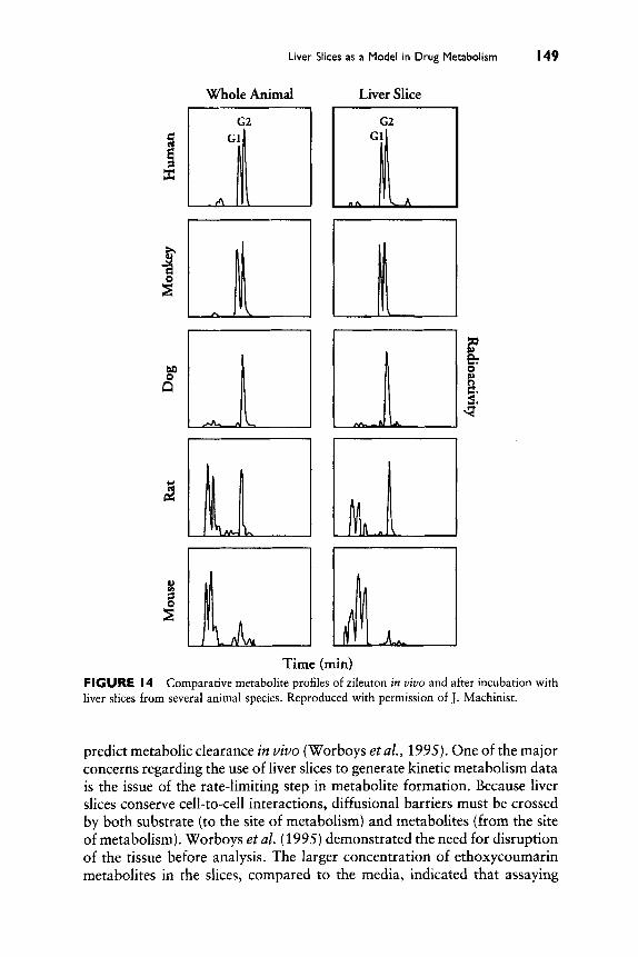
Liver Slices as a Model in Drug Metabolism I49
Whole Animal Liver Slice
3: ‘I%
Time (min) FIGURE 14 liver slices from several animal species. Reproduced with permission of J. Machinist.
Comparative metabolite profiles of zileuton in vivo and after incubation with
predict metabolic clearance in vivo (Worboys et al., 1995). One of the major concerns regarding the use of liver slices to generate kinetic metabolism data is the issue of the rate-limiting step in metabolite formation. Because liver slices conserve cell-to-cell interactions, diffusional barriers must be crossed by both substrate (to the site of metabolism) and metabolites (from the site of metabolism). Worboys et al. (1995) demonstrated the need for disruption of the tissue before analysis. The larger concentration of ethoxycoumarin metabolites in the slices, compared to the media, indicated that assaying

150 James L. Ferrero and Klaus Brendel
only the media would lead to an underestimation in the rates of metabolism. Dogterom (1993) found that the rate of tolbutamide metabolism by rat liver slices was highly dependent on slice thickness. Although the rate of metabolism (expressed per milligram of liver protein) was greatest in thin slices and slowest in thick slices, the absolute rate of metabolism (expressed per slice) was remarkably constant. Ekins et al. (1995) demonstrated that the rate of testosterone hydroxylation and ethoxycoumarin metabolism was significantly slower in liver slices than in isolated hepatocytes. To test the hypothesis that the slower rate of metabolism in liver slices was due to slower diffusion of the compound into the tissue, 2-(5'chloro-2'-phosphoryl- oxyphenyl)-6-chloro-4-(3H)quinazolinone (CPPCQ) was incubated with liver slices and freshly isolated hepatocytes. CPPCQ is converted to a fluo- rescent product by alkaline phosphatase. Fluoresence histochemistry using CPPCQ demonstrated rapid uptake and product formation within minutes in hepatocytes, whereas in slices the product was confined to the outer two to three layers after 1 hr. These results suggested that the rate of xenobiotic uptake and metabolism in liver slices may reside primarily in the outer cell layers.
The penetration of most xenobiotic substances into tissues is predomi- nantly governed by passive diffusion. A major determinant of this process is the degree of lipophilicity of a compound. We studied the liver slice metabolism of two agents of similar chemical structure and lipophilicity but which demonstrated substantial differences in bioavailability when adminis- tered to animals. Table I depicts the structure, lipophilicity (logP), and bioavailability of A-82891 and A-80426 in rats. Although these compounds are structurally almost identical, demonstrating similar log P values and physicakhemical properties, they exhibited dramatic differences in bioavail- ability. In viuo metabolism studies in rats indicated that the low bioavailabil- ity of A-82891 was due to extensive first-pass metabolism (see metabolic scheme, Fig. 4) compared to A-80426. The metabolic pathways for each compound were, not surprisingly, similar. When we studied the metabolism of each compound in rat liver slices, there were substantial differences in the rates of metabolism (Table I). A-82891 was metabolized approximately eightfold faster than A-80426 in liver slice preparations from the same animal. These differences in rates of metabolism agreed well with the differ- ences in bioavailability and suggested that the low bioavailability of A- 82891 was due to rapid first-pass liver metabolism. A-82891 appeared to be a good substrate for hepatic metabolism compared to A-80426. Given the similar structure and lipophilicity of each compound, the diffusion and tissue penetration of each should be similar and, therefore, the differences in rates of metabolism were likely due to differences in enzymatic (metabolic) turnover and not in rates of drug or metabolite diffusion into or out of the slices.

Liver Slices as a Model in Drug Metabolism 151
TABLE I Comparison of the Lipophilicity, Bioavailability (Rat), and Rates of Liver Slice Metabolism (Rat) of Abbott-8289 I and Abbott-80426
H3C, 0
H3C, 0
Abbott - 82891 Abbott - 80426 ~ ~
4.6 Lipophilicity (log P) 5.2 3-5 % Bioavailability (rat) 50-55% 0.176 Rate of liver slice metabolism (hi ') 0.022
'Position of tritium label.
Morboys et al. (1995) described the metabolism kinetics of ethoxycou- marin (a high clearance compound in vivo) and tolbutamide (a low clearance compound in vivo) by rat liver slices. The 0-deethylation of ethoxycoumarin by rat liver slices demonstrated biphasic kinetics best described by a modified two-site Michaelis-Menten equation, whereas tolbutamide hydroxylation displayed monophasic single-site kinetics. These differences in kinetic behav- ior were comparable to those reported in hepatic microsomes, freshly iso- lated hepatocytes, and in vivo. It should be kept in mind that in situations where the hepatic clearance of a drug is blood flow dependent or renal clearance is high, a prediction of the bioavailability in humans from liver slice data is not feasible. Further studies are needed comparing liver slice kinetic data with in vivo clearance and kinetic data from other hepatic in vitro systems before the reliability of liver slices as a model to predict in vivo pharmacokinetics can be fully assessed.
IV. Prediction of Metabolic Drug-Drug Interactions
If a compound is cleared from the body primarily by metabolism, then coadministration of another compound that inhibits its metabolism will usually result in increases in parent drug concentrations and changes in

152 James L. Ferrero and Klaus Brendel
pharmacokinetic behavior. This can occur by alterations in first-pass metab- olism, drug clearance, or both. Another type of metabolic drug interaction can occur from the induction of drug-metabolizing enzymes (during multiple dosing), resulting in an increase in the metabolism of an agent. This discus- sion focuses on the former type of drug interaction.
The dependability of liver slices to accurately predict liver metabolism- based drug interactions has currently not been established. The majority of clinical metabolic drug interactions reported to date have involved the CYP isozymes (Cuccinel et af., 1965; Perucca, 1987; Perrot et af., 1989; Bailey et a/., 1991; Nation et al., 1990). Liver microsomes, which contain these enzymes, are the technique of choice for studying CYP-mediated drug inter- actions. Alternatively, precision-cut liver slices may represent another useful tool in studying hepatically mediated drug interactions.
As has been shown by other authors in this volume, the use of liver microsomes with CYP-selective probe substrates and inhibitors is a powerful tool in elucidating the mechanisms and CYP isozymes involved in drug interactions and in predicting potential oxidative drug interactions. Liver slices have features that make it attractive as a model to assess several aspects of metabolic drug interactions not addressed by liver microsomes. The pres- ence of virtually all the enzymes involved in drug metabolism allows liver slices to study CYP- and non-CYP-mediated drug interactions. Numerous drug interactions with phase I1 substrates have been reported (Kornhauser et al., 1989; De Miranda et al., 1989). If a compound has more than one site and/or route of metabolism, inhibition of a single site or pathway could result in metabolic switching, i.e., where a relatively minor metabolic path- way becomes a major pathway in the presence of a selective inhibitor (Brmen and Gram, 1989). Liver slices may be useful in this area. Currently, there are few reports examining drug interactions in vitro with liver slices (Ro- driques et a/., 1995). We describe here some of our initial studies to begin to evaluate the utility of liver slices in predicting metabolic drug interactions. For these validation studies, we chose compounds that had previously dem- onstrated a metabolically based clinical pharmacokinetic drug interaction.
A. Single Substratellnhibitor Concentration Studies
Theophylline is a potent bronchodilator commonly prescribed for use in asthma. The drug has a narrow therapeutic window (approximately 5- 20 pg/ml), and decreases in metabolic clearance can result in increases in plasma concentrations. When theophylline is administered in the presence of zileuton, there is a decrease in clearance and an increase in the plasma area under the curve for theophylline (Fig. 15) (Granneman et al., 1995). This has been shown to be mediated by zileuton inhibition of CYPlA2 activity (Machinist et al., 1995), the primary CYP isozyme involved in theophylline metabolism in humans (Campbell et al., 1987; Sarkar et al.,

Liver Slices as a Model in Drug Metabolism 153
0.1 0 6 12 18 24 30 36 42 48
Hours After Dosing FIGURE I 5 Mean plasma concentrations of theophylline in humans after oral administra- tion of theophylline with (0) and without zileuton (0). Reproduced with permission from Machinist et al. (1995).
1992). We incubated carbon-14 ring-labeled theophylline with human liver slices in the presence and absence of zileuton, quinidine (a CYP2D6-selective inhibitor, as negative control), and a-naphthoflavone (ANF, a CYPlA2- selective inhibitor, as positive control). ANF and zileuton significantly inhib- ited the metabolism of theophylline by human liver slices, whereas quinidine had a minimal effect (Fig. 16). The effect of zileuton on theophylline metabo- lism by human liver slices reflected the inhibition observed in vivo. It could be argued, however, that the inhibitory effect of zileuton on theophylline metabolism by liver slices may be due to a toxicity of zileuton on liver slices rather than selective CYPlA2 inhibition. Although this issue would not arise using liver microsomes, toxicity of liver slices or hepatocytes is a possibility in drug interaction studies, as the cellular viability of these preparations is critical to metabolic activity. With liver slices and hepatocytes, however, one can monitor both metabolism and viability in the same incubation. This has been a major advantage of these preparations for metabolic toxicity studies (Sipes et al., 1987). In the early stages of cellular damage, several liver enzymes are released, including SGOT (Grisham, 1979; Boyd, 1982). Therefore, we analyzed the liver slice incubations containing theophylline and the compounds described earlier for leakage of SGOT into the media (Fig. 17). Neither theophylline alone nor in combination with zileuton, ANF, or quinidine caused significant leakage of SGOT into the media,

154 James L. Ferrero and Klaus Brendel
3000 ' .d 3 d
0 n
" Control ANF Quinidine Zileuton
Effect of a-napthoflavone (ANF), quinidine, and zileuton on the rate of theoph- FIGURE I6 ylline metabolism by human liver slices.
whereas our positive control (a known hepatotoxin) caused substantial leak- age of the enzyme. These results suggest that the inhibition of theophylline metabolism by zileuton in liver slices was due to selective inhibition of CYPlAP and not cellular toxicity.
Terfenadine is a nonsedating H,-receptor antagonist widely prescribed for histamine-mediated allergic conditions (Simonson, 1991). The drug un- dergoes extensive (>99%) first-pass metabolism, primarily to the pharmaco- logically active carboxylic acid metabolite (M-3) (Fig. 18) (Jurima-Romet etal., 1994). CYP3A is the major isozyme family involved in the metabolism of terfenadine in humans (Yun et a/., 1993). Coadministration of several drugs known to be CYP3A substrates or inhibitors, e.g., erythromycin and
280 ' 240 -
200
160 - SGOT
Activity 120- (IUIL)
" Control ANF Quinidine Zdeuton +++
FIGURE I7 SGOT into the liver slice incubation media (12-hr incubation).
Effect of a-napthoflavone (ANF), quinidine, and zileuton on the leakage of

Liver Slices as a Model in Drug Metabolism I 5 5
- MI/M2? -
Terfenadine
/ Ho&N&COOH ' I OH
w M3 FIGURE I8 Proposed metabolic pathways of terfenadine in humans.
ketoconazole, can significantly elevate terfenadine plasma concentrations. This can lead to an increased risk of cardiotoxicity (torsade de pointes) due to altered cardiac repolarization (QT prolongation), resulting in ventricular arrythmias (Honig et al., 1993). Although liver microsomes have been an accurate model in predicting potential drug interactions with terfenadine, the effect of ketoconazole on the metabolism of terfenadine by several differ- ent in vitro liver preparations was compared. Figure 19 depicts representative metabolite profiles after incubation of radiolabeled terfenadine with human liver slices, human liver microsomes, and pure CYP3A fusion protein.' The major metabolite generated by the CYP3A fusion protein and human liver slices was the carboxylic acid metabolite, whereas liver microsomes formed primarily M-4 (the alcoholic precursor to M-3) and relatively small amounts of M-3. This indicated that the liver slice and CYP3A4 fusion protein prepa- rations readily metabolized terfenadine via oxidative coupling, similar to the situation in vivo. When ketoconazole (5 pM) was included in the incuba- tions, the metabolism of terfenadine by all three preparations was dramati- cally inhibited (Fig. 20). These results indicated that, in addition to human liver microsomes and recombinant CYP3A fusion protein, human liver slices may have the potential to study liver CYP3A-mediated drug interactions with terfenadine. Future studies that involve the metabolism of other CYP3A
Reconstituted purified recombinant fusion protein containing CYP3A4 rF4SO[mHUM3A/ mRatORIL1 linked to rat NADPH-P45O reductase. This preparation was kindly provided by Dr. R. Estabrook (University of Texas Southwestern Medical Center, Dallas, TX).

I56 James L. Ferrero and Klaus Brendel
H-3 CPM
H-3 CPM
H-3 CPM 171 138
I M3 umtll Liver ssue Slices
0 0 5 10 IS 20 25 30 35 40 45 50 SS 60
Time (Mitu) FlGURE I 9 HPLC metabolite profiles after incubation of [JH]terfenadine with human liver microsomes, human liver slices, or CYP3A4 fusion protein rF450[mHUM3A44/mRatOR]Ll. Reproduced with permission from Rodrigues et at. (1995).
substrates with liver and intestineal slices may shed information on the relative contribution of liver and gut CYP3A in the overall metabolism/ clearance of compounds.
B. Kinetics of Liver Stice Metabolism
Compared to liver microsomes and hepatocytes, few studies have exarn- ined the kinetic aspects of drug metabolism by liver slices (Houston, 1994). We earlier described encouraging results by Worboys et af. (1995) demon- strating Michaelis-Menten kinetic parameters for the metabolism of tolbuta- mide and ethoxycoumarin by rat liver slices. Differences in the intrinsic

Liver Slices as a Model in Drug Metabolism I57
Human Liver Microsomes Human Liver Tissue Slices
l 2 O 7
" M1 M3 M4
Metabolite
1201r-----
" M1 M3 M4
Metabolite
CYP3A4 Fusion Protein
100
8 80
f 60
' 40 :e
20
n M1 M2 M3 M4
Metabolite FIGURE 20 Effect of ketoconzole on the metabolism of [3H]terfenadine by human liver microsomes, CYP3A4 fusion protein rF450[mHUM3A4/mRatOR]Ll or human liver slices. Reproduced with permission from Rodrigues et al. (1995).
clearance (Clint) values for the two substrates were comparable to those previously reported in liver microsomes, hepatocytes, and in vivo. Although the majority of clinical drug interactions involve CYP enzymes, metabolic drug interactions with phase I1 enzyme systems have also been reported (Kornhauser et al., 1989; De Miranda et al., 1989). For many compounds that undergo glucuronidation (phase 11), incubation conditions with hepatic microsomes must often be optimized with the appropriate concentration

I58 James L Ferrero and Klaus Brendel
and type of activator (detergent) in order to obtain acceptable metabolic activity. This is generally not required for phase I (oxidative) reactions with liver microsomes. With liver slices, however, all enzyme cofactors are self- sufficient and no additional activators or cofactors are required. For these reasons, liver slices may have advantages over microsomal preparations in studying phase I1 metabolism. Zidovudine (AZT) is a substituted thymidine derivative that is frequently prescribed for individuals infected with the human immunodeficiency virus (HIV). AZT is well absorbed and rapidly metabolized (Fig. 21), which accounts for its relatively short plasma half- life of about 1 hr in humans (Collins and Unadkat, 1989). Several reports indicate that coadministration of AZT with either probenecid or valproate significantly increases the plasma area under the curve of AZT (Lertora et al., 1994; Kornhauser et al., 1989). Studies with liver microsomes indicate that the basis of the pharmacokinetic interactions with probenecid and valproate was due to competitive inhibition of glucuronidation (Macleod et a[., 1992; Resetar et al., 1991; Agrawal et al., 1992). We initially studied the kinetics of AZT glucuronidation by human liver slices at concentrarions ranging from 10 p M to 5 mM. Figure 22 shows that the glucuronidation of AZT by human liver slices appeared to obey single enzyme Michaelis- Menten kinetics. The apparent K , value of about 0.6 mM was comparable to the values reported for liver microsomes (0.4-5 m M ) (Resetar and Spec- tor, 1989; Cretton et al., 1990; Sim and Back, 1990; Rajaonarison et al.,
0 Uridine Diphosphoglucuronyl-
transferase b
AZT
COOH
OH N j
GAZT
,CH3
Reductase
COOH
NH 2 OH NH2
AMT GAMT FIGURE 21 Proposed metabolic pathways of zidovudine (AZT) in humans.

Liver Slices as a Model in Drug Metabolism I59
K, = 0.646+0.068 (mM)
Vmax = 8.729.29 (nmoleslmintg)
0 1000 2000 3000 4000 5000
[AZT] $4 FIGURE 22 slices (n = two human livers, incubations were performed in duplicate).
Michaelis-Menten curve for zidovudine (AZT) glucuronidation in human liver
1991). When we studied the effect of varying concentrations of probenecid and valproate on the rate of liver slice metabolism of AZT (Fig. 23), we observed concentration-dependent inhibition with apparent K j values of
K;(mM) leppl 0 500 1000 1500 2000 2500
Concentration ($4) FIGURE 23 human liver slices (n = two human livers, incubations were performed in duplicate).
Inhibition of zidovudine (AZT) metabolism by probenecid and valproate in

I60 James L. Ferrero and Klaus Brendel
0.34 and 0.39 mM for probenecid and valproate, respectively. These values compared favorably to those previously determined using human liver micro- somes (0.2-1.0 m M ) (Resetar et al., 1989). We incorporated these experi- mentally determined parameters for AZT metabolism by human liver slices into the equation derived by Segal(l993) relating in vitro kinetic parameters to in viuo pharmacokinetic parameters (Table 11). Coadministration of AZT with probenecid and valproate, respectively, resulted in a 57 and 80% increase in plasma AUC (Lertora et al., 1994; Kornhauser et al., 1989). The predicted degree of inhibition derived from human liver slice data was 67 and 64% for probenecid and valproate, respectively. Although more validation work needs to be conducted, these results are encouraging regard- ing the potential use of liver slices to obtain kinetic information and drug inhibition data which may be used to predict clinical drug interactions mediated by liver metabolism.
C. Metabolic Switching
Many compounds are metabolized by more than one pathway and enzyme. Selective inhibition of a particular pathway or enzyme could result in a shift in the major metabolites formed. This is known as metabolic switching or shunting. An example of this is illustrated by the tricyclic antidepressant, imipramine. The metabolism of imipramine is well docu- mented (Fig. 24) (Sutfin et al., 1984). Although the in vivo metabolism of imipramine is complex (the compound undergoes aromatic 2- and 10- hydroxylation, N-demethylation, N-oxidation, 0-glucuronidation, and N- glucuronidation, as well as secondary and tertiary metabolism by these pathways), the predominant primary pathways are 2-hydroxylation and N-demethylation (Sallee and Pollock, 1990). The 2-hydroxylation of imipra- mine is mediated by the high-affinity polymorphic CYP2D6, whereas N- demethylation (to form the pharmacologically active desipramine metabo-
TABLE II Comparison of the Inhibition of Zidovudine Metabolism by Probenecid and Valproate in Vivo and in Human Liver Slices
Treatment Plasma AUC % increase in plasma AUC % inhibition in vitro"
Probenecid Control 659 57 Probenecid 1032
Control 650 80 Valproate 1170
Valproate
67
64
AZT, apparent K , = 0.65 mM; probenecid, apparent I 4 K , ( 1 + [SIKJ + "1'
% Inhibition =
K , = 0.34 mM; and valproate, apparent K , = 0.39 rnM.

Liver Slices as a Model in Drug Metabolism 161
Imipramine N-glucuronide
4 glucuronidation
Imipramine Desipramine
2-hydroxylation (CYPZDG)
demethylation - (CYPSA)
2-hydroxyimipramine 2-hydroxydesipramine
glucuronidation 1 2-hydroxydesipramine
glucuronide
1 glucuronidation
2-hydroxyimipramine glucuronide
FIGURE 24 Proposed metabolic pathways of imipramine in humans.
lite) is mediated by the low-affinity CYP3A (Lemoine et al., 1993). The CYP2D6 isozyme demonstrates a bimodal distribution in the population and, hence, the metabolism of imipramine is manifested as poor (PM) and extensive metabolizers (EMS), with PMs representing a small minority (-7% of Caucasians) (Brersen et al., 1986). Although PMs demonstrate relatively small changes in imipramine pharmacokinetics compared to EMS, there are dramatic differences between EMS and PMs in desipramine pharmacokinet- ics (Gram et al., 1983). The plasma clearance of desipramine in PMs is 5- to 10-fold lower than in EMS, whereas the steady-state plasma concentra- tions are dramatically higher (Brersen and Gram, 1989). This increase in the plasma half-life of desipramine in PMs, and accumulation in the plasma, is due primarily to the relative inability of desipramine to be further metabo- lized by CYP2D6-mediated 2-hydroxylation. Quinidine is a potent and selective CYP2D6 inhibitor (Brersen and Gram, 1989). Hence, the adminis- tration of quinidine to EM individuals results in a shift in metabolism from predominantly 2-hydroxylation to N-demethylation (Brersen and Gram, 1989). Pharmacokinetically and metabolically, the effect of quinidine is

I62 James L. Ferrero and Klaus Brendel
generally regarded as one of changing EMS to PMs, i.e., poor metabolizers of imipramine (deficient in CYP2D6) or extensive metabolizers given quinidine (CYP2D6 is inhibited) both manifest the pharmacokinetic behavior de- scribed earlier. In order to determine the ability of liver slices to reflect this quinidine-mediated shift in the in vivo metabolism of imipramine from 2- hydroxylation to N-demethylation, we incubated radiolabeled imipramine with human liver slices in the presence and absence of quinidine. The in vitro concentrations of imipramine and quinidine, 2 and 5 p M , respectively, were within the therapeutic plasma concentration range for each compound (Brprsen and Gram, 1989).
The pattern of metabolites generated after incubating imipramine with human liver slices in the presence and absence of quinidine is depicted in Fig. 25. Human liver slices generated numerous metabolites, resulting from both oxidative and conjugative metabolism, as well as the coupling of these reactions. Major metabolites were the 2-hydroxylated and 2- hydroxylated/N-desmethylated derivatives of imipramine and their respec-
ContYoZ
Time (min) FIGURE 25 Effect of quinidine on the metabolism of imipramine by human liver slices. Tentative metabolite identification: ( 1 ) Imipramine, (2) desipramine, (3) imipramine N-glucu- ronide, (4) 2-hydroxyimipramine, (5) 2-hydroxydesipramine, (6) 2-hydroxyimipramine glucu- ronide, and (7) 2-hydroxydesipramine glucuronide.

Liver Slices as a Model in Drug Metabolism I63
tive 0-glucuronide conjugates. The quaternary N-glucuronide of imipramine was also formed, whereas minor amounts of desipramine were generated. In vivo metabolism results indicate that after administration of imiprp ine to humans, the 0-glucuronides of 2-hydroxyimipramine and 2-hydroxydesi- pramine are the major metabolites excreted in the urine, whereas the respec- tive deconjugated derivatives of these metaboli9s are found in lower amounts (Crammer et al., 1969). Our resultsdemonstrate that human liver slices generated metabolites of imipramine representative of those seen in vivo. The addition of 5 p M quinidine to the incubation caused a dra- matic shift in metabolite pattern (Fig. 25). Although small amounts of 2-hydroxydesipramine and the 0-glucuronides of 2-hydroxyimipramine and 2-hydroxydesipramine were formed, the major metabolite was imipra- mine N-glucuronide. The formation of desipramine (although still a rela- tively minor metabolite) increased four to sevenfold in the presence of quini- dine, whereas the overall rate of 2-hydroxylation decreased dramatically compared to control incubations. The effect of quinidine on the metabolism of imipramine by human liver slices accurately reflected the decrease in 2-hydroxylation and the increase in N-demethylation observed clinically. However, a clearly surprising result was the fact that quinidine shifted the major metabolic pathway of imipramine from 2-hydroxylation to predomi- nantly N-glucuronidation. This shift to N-glucuronidation as the major pathway was demonstrated in liver slices obtained from several different human donors, although the magnitude of the shift was less in individuals who demonstrated low basal amounts of N-glucuronide formation in the absence of quinidine.
The N-glucuronide has been shown to be a relatively minor metabolite in the urine of individuals following administration of imipramine (Crammer et al., 1969, Sutfin et al., 1984). A major route of excretion of quarternary ammonium compounds, including quarternary ammonium-linked N- glucuronides, is biliary (Lehman et al., 1983). If liver slices accurately re- flect the metabolic response of the liver in vivo during coadministration of imipramine and quinidine to EMS, or in PMs given imipramine, then these results suggest that N-glucuronidation may be an important metabolic pathway in these individuals. This may not be apparent from plasma and urinary metabolite analysis following imipramine administration because the N-glucuronide of imipramine may be secreted in bile and undergo hydro- lysis and enterohepatic recirculation. The low-affinity CYP3A-mediated N- demethylation activity may continue slowly, resulting in the gradual accumu- lation of desipramine during daily dosing (due to slow 2-hydroxylation). Quinidine has been shown to have only minimal effects on CYP3A-mediated metabolism at the concentrations used in the present study (Guengerich, 1995). In science, we often learn the most when we are surprised by the result. These results suggest some interesting further studies that may verify our results, or perhaps we will be forced to invent another hypothesis.

I64 James L. Ferrero and Klaus Brendel
V. Conclusions
Precision-cut liver slices are a reliable in vitro system to determine the metabolites most likely generated by the liver in vivo. Because the cytoarchi- tecture of the liver remains intact, all enzymes and cofactors for metabolism are present. This is demonstrated by the ability of liver slices to couple metabolic reactions, even between different enzyme systems, similar to the situation in vtuo. This wide spectrum of metabolic capability also allows this system to be useful in studying metabolite-mediated liver toxicity. Al- though the appearance of metabolites generated by in vitro liver preparations often reflects the in vivo profile, metabolites formed by the liver in vivo can be significantly affected by physiological factors such as renal clearance and biliary secretion. In addition, extrahepatic metabolism can contribute significantly to the overall metabolic picture in vivo. Although the in viuo clearance of several compounds has been reflected in the relative rates of liver slice metabolism, it should be kept in mind that in situations where the hepatic clearance of a drug is blood flow dependent or renal clearance is high, a prediction of the bioavailability or clearance from liver slice data is not feasible. These factors can make it difficult to assess the relative contributions of the different metabolic pathways and mechanisms involved in the metabolism and overall clearance of a compound from liver slice data alone. Although our results with imipramine suggest an interesting hypothesis regarding the effect of quinidine on the metabolism of imipra- mine, more importantly, it illustrates the utility of liver slices to generate metabolite profiles involving several different pathways and enzymes, and how these profiles may be differentially affected by other compounds. It is for us to evaluate and validate the relevance of this information for each drug to the in vivo situation.
The areas of metabolism kinetics and metabolic drug-drug interactions are only beginning to be evaluated using liver slices. This system may offer advantages to hepatic microsomes because essentially all metabolic pathways are functional. This may allow a more accurate evaluation of the relative contribution ( f m ) of the different metabolic pathways. The integration of kinetic information for each metabolic pathway with the relative contribu- tion of each pathway ( f m ) represents potentially a powerful tool that may enable us to more accurately predict drug metabolism, pharmacokinetics, and metabolic drug interactions.
Acknowledgments
The authors thank the following individuals at Abbott Laboratories: Drs. A. D. Rodrigues and J. Machinist for their contributions, Drs. K. C. Marsh, G. R. Granneman, D. Ouellet, and W. Awni for providing pharrnacokinetic data; and M. Amann and P. Johnson for their technical

Liver Slices as a Model in Drug Metabolism 165
support. The authors also thank Drs. M. Shet and R. Estabrook (University of Texas Southwest- ern Medical Center, Dallas, TX) for their productive collaborations.
References
Agrawal, K. C., Dipiazza, N. J., and Agrawal, S. K. (1992). Inhibition of human hepatic glucuronyltransferase (GT) activity to alter zidovudine (AZT) metabolism. Proc. Am. Assoc. Cancer Res. 33, 400.
Azri, S., Gandolfi, A. J., and Brendel, K. (1990). Precision cut liver slices: An in vitro system for profiling potential hepatotoxicants. In Vitro Toxicol. 3 , 309-320.
Bailey, D. G., Spence, J. D., Munzo, C., and Arnold, J. M. 0. (1991). Interaction of citrus juicve with felodipine and nifedipine. Lancet 337, 268-269.
Barr, J., Weir, A. J., Brendel, K., and Sipes, I. G. (1991a). Liver slices in dynamic organ culture. I. An alternative in vitro technique for the study of rat drug metabolism. Xenobiotica
Barr, J., Weir, A. J., Brendel, K., and Sipes, I. G. (1991b). Liver slices in dynamic organ culture. 11. An in vitro cellular technique for the study of integrated drug metabolism using human tissue. Xenobiotica 21, 341-350.
Bienvenu, T., Pons, G., Rey, E., Thiroux, G., and Olive, G. (1993). Caffeine metabolism in liver slices during postnatal development in rats. Drug Metab. Dispos. 21, 178-180.
Boyd, J. W. (1982). The mechanisms relating to increases in plasma enzymes and isoenzymes in diseases of animals. Vet. Clin. Pathol. 12, 9-24.
Brendel, K., Gandolfi, A. J., Krumdieck, C. L., and Smith, P. F. (1987). Tissue slicing and culturing revisited. Trends Pharm. Sci. 8 , 11-15.
Bresen, K., and Gram, L. F. (1989). Quinidine inhibits the 2-hydroxylation of imipramine and desipramine but not the demethylation of imipramine. Eur. J. Clin. Phamacol.
Bresen, K., Otton, S. V., and Gram, L. F. (1986). hipramine demethylation and hydroxylation: Impact of the sparteine oxidation phenotype. Clin. Pharmacol. Ther. 40, 543-549.
Campbell, M. E., Grant, D. M., Tadanobu, I., and Kalow, W. (1987). Biotransformation of caffeine, paraxanthine, theophylline, and theobromine by polycyclic aromatic hydrocar- bons-inducible cytochrome(s) P-450 in human liver microsomes. Drug Metab. Dispos.
Carlile, D., Dickens, M., Houston, J. B. (1993). Comparison of drug metabolising capacity in hepatic microsomes and freshly isolated hepatocytesof rat. in “Proceedings of the Second Annual UKAPS Conference” (J. Hadgraft, I. W. Kellaway, and G. D. Parr, eds.), p. 47. STS Publishing, Cardiff, UK.
Clouser, T. E. (1977). Tissue bath improvements for the Oxford vibratome. Stain Technol.
Collins, J. M., and Unadkat, J. D. (1989). Clinical pharmacokinetics of zidovudine. Clin. Pharmacokinet. 17, 1-9.
Copeland, K. R., Yatscoff, R. W., and McKenna, R. M. (1990). Activity of cyclosporin metabo- lites compared and characterized by mass spectroscopy and nuclear magnetic resonance. Clin. Chem. ( Winston-Salem, N.C.) 32, 229-229.
Crammer, J. L., Scott, B., and Rolfe, B. (1969). Metabolism of 14C-imipramine: 11. Urinary metabolites in man. Psychopharmacologia 15, 207-225.
Cretton, E. M., Waterhous, D. V., Beva, R., and Sommadossi, J. (1990). Glucuronidation of 3’-azido-3’-deoxythymidine by rat and human liver microsomes. Drug Metab. Dispos.
21, 331-339.
37, 155-160.
15,237-249.
52,319-322.
18, 369-372.

I66 James L. Ferrero and Klaus Brendel
Cuccinel, S. A., Conney, A. H., Sansur, M., and Burns, J. J. (1965). Drug interaction in Man. lowering effect of phenobarbital on plasma levels of bishydroxycoumarin (dicoumarlo) and diphenylhydantoin (dilantin). Clin. Phannacol. Ther. 6, 420-426.
De Miranda, P., Good, S . S . , Yarchoan, R., Thomas, R. V., Blum, M. R., Myers, C. E., and Broder, S. (1989). Alteration of zidovudine pharmacokinetics by probenecid in patients with AIDS or AIDS-related complex. Clin. Pharmacol. Ther. 46, 494-500.
Dogterom, P. (1993). Development of a simple incubation system for metabolism studies with precision-cut liver slices. Drug Metab. Dispos. 21, 699-704.
Dogterom, P., and Rothuizen, J. (1993). A species comparison of tolbutamide metabolism in precision-cut liver slices from rats and dogs. Drug Metab. Dispos. 21, 705-709.
Ekins, S. (1996). Past, present, and future applications of precision-cut liver slices for in vitro xenobiotic metabolism. Drug. Metab. Rev. 28(4), 591-623.
Ekins, S., Murray, G. I., Burke, M. D., Marchant, N. C., and Hawkesworth, G. M. (1995). Quantitative differences in xenobiotic metabolism by precision-cut liver slices. Int. ISSX Meet, 4th, Seattle, WA, 1995, Abstr. No. 419.
Fentiman, I., Taylor-Papadimitriou, J., and Stoker, M. (1976). Selective contact-dependent cell communication. Nature (London) 264, 760-764.
Fernandes, P. B., Bailey, R., Sevanson, R., Hanson, C. W., McDonald, E., Ramer, N., Hardy, D., Shipkowitz, N., Bower, R. R., and Gade, E. (1986). In vitro and in vivo evaluation of A-56268(TE-031): A new macrolide. Antimicrob. Agents Chemother. 30, 865-873.
Ferrero, J. L., Bopp, B. A., Marsh, K. C., Quigley, S. C., Johnson, M. J., Anderson, D. J., Lamm, J. E., Tolman, K. C., Sanders, S . W., Cavenaugh, J. H., and Sonders, R. C. (1990). Metabolism and disposition of clarithromycin in man. Drug Metab. Dispos. 18,441-446.
Fisher, R. L., Shaughnessy, R. P., Jenkins, P. M., Austin, M. L., Roth, G . L., Gandolfi, A. J., and Brendel, K. (1995). Dynamic organ culture is superior to muftiwell plate culture for maintaining precision-cut tissues: Optimization of tissue slice culture. Part 1. Toxicol. Methods 5 , 99-1 13 .
Gorodischer, R., Yaari, A., Margalith, M., Warszawski, D., and Ben-Zvi, 2. (1986). Change in theophylline metabolism during postnatal development in rat liver slices. Biochem. Pbarmacol. 35 , 3077-3081.
Gram, L. F., Bjerre, M., Kragh-Serensen, P., Kvinesdal, B., Molin, J., Pedersen, 0. L., and Reisby, N. (1983). Imipramine metabolites in blood of patients during therapy and after overdose. Clin. Pharmacol. Ther. 33, 335442.
Granneman, G. R., Braekman, R. A., Locke, C. S., Cavanaugh, J. H., Dube, L. M., and Awni, W. M. (1995). Effect of zileuton on theophylline pharmacokinetics. Clin. Pharmacokinet.
Grisham, J. W. (1979). Use of hepatic cell cultures to detect and evaluate the mechanisms of action of toxic chemicals. Znt. Rev. Exp. Pathol. 20, 123-210.
Guengerich, F. P. (1995). Cytichromes P450 of human liver. Classification and activity profiles of the major enzymes. In “Advances in Drug Metabolism in Man” (G. M. Pacifici and G. N. Fracchia, eds.), pp. 181-231. European Commission, Brussels, Belgium.
Gunawardhana, L., Barr, J., Weir, A. J., Brendel, K., and Sipes, I. G. (1991). The N-acetylation of sulfamethazine and p-aminobenzoic acid by human liver slices in dynamic organ culture. Drug Metub. Dispos. 19, 648-654.
Harris, J. L., Rahman, A., Kim, B.-R., Guengerich, F. P., and Collins, J. M. (1994). Metabolism of taxol by human hepatic microsomes and liver slices: Participation of cytochrome P- 450 3A4 and an unknown P450 enzyme. Cancer Res. 54,4026-4035.
Hilliiker, K. S., and Roth, R. A. (1980). Prediction of mescaline clearance by rabbit lung and liver from enzyme kinetic data. Biochem. Pharmacol. 29, 253-255.
Hodinka, R. L., lack-wait, K., and Gilligan, P. H. (1987). Comparative in vitro activity of A- 56268(TE-031), a new macrolide antibiotic. Eur. /. Clin. Microbiof. 6 , 103-108.
Honig, P. K., Wortham, D. C., Zumani, K., Connor, D. P., Mullin, J. C., and Cantilena, L. R. (1993). Terfenadine-ketoconazole interaction. JAMA 269, 1513-1518.
29,77-83.

Liver Slices as a Model in Drug Metabolism I67
Houston, J. B. (1994). Utility of in uitro drug metabolism data in predicting in uruo metabolic clearance. Biochem. Pharmacol. 47, 1469-1479.
Jurima-Romet, M., Crawford, K., Cyr, T., and Inaba, T. (1994). Terfenadine metabolism in human liver. Drug Metub. Dispos. 22, 849-857.
Kornhauser, D. M., Petty, B. G., Hendrix, C. W., Woods, A. S., Nerhood, L. J., Bartlett, J. G., and Leitrnan, P. S. (1989). Probenecid and zidovudine metabolism. Lancet 2, 473-475.
Krumdieck, C. L., Dos Santos, J. E., Ho, K. J. (1980). A new instrument for the rapid preparation of tissue slices. Anal. Biochem. 104, 118-123.
Lake, B. G., Beamand, J. A., Japenga, A. C., Renwick, A., Davies, S., and Price, R. J. (1993). Induction of cytochrome P-450-dependent enzyme activities in cultured rat liver slices. Food Chem. Toxicol. 31, 377-386.
Lehman, J. P., Fenselau, C., and Depaulo, J. R. (1983). Quarternary ammonium-linked gluc- uronides of amitriptyline, imipramine and chlorpromazine. Drug Metab. Dispos. 11,
Lemoine, A., Gautier, J. C., Azoulay, D., Kiffel, L., Belloc, C., Guengerich, F. P., Maurel, P., Beaune, P., and Leroux, J. P. (1993). Major pathway of imipraminemetabolism is catalyzed by cytochromes P-450 1A2 and P-450 3A4 in human liver. Mol. Pharmacol. 43,827-832.
Lertora, J. L., Rege, A. B., Greenspan, D. L., Akula, S., George, W. J., Hyslop, N. E., and Agrawal, K. C. (1994). Pharmacokinetic interaction between zidovudine and valproic acid in patients with immunodeficiency virus. Clin. Phamacol. Ther. 56, 272-278.
Machinist, J. M., Mayer, M. D., Shet, M. S., F ~ & L + t d R o d r i g u e s , A. D. (1995). Identification of the human liver cytochrome P450 enzymes involved in the metabolism of zileuton (ABT-077) and its N-dehydroxylated metabolite, Abbott-66193. Drug Metab.
Macleod, R., Eagling, V. A., Sim, S. M., and Back, D. J. (1992). In vitro inhibition studies of the glucuronidation of 3’-azido-3-deoxythymidine catalysed by human liver UDP- glucuronosyl transferase. Biochem. Pharmacol. 43, 382-386.
Maurer, G., and Lemaire, M. (1986). Biotransfromation and distribution in blood of cyclosporin and its metabolites. Transplant. Proc. 18, 25-34.
Maurer, G., Loosli, E., Schreier, E., and Keller, B. (1984). Disposition of cyclosporin in several animal species and man. I. Structural elucidation of its metabolites. Drug Metab. Dispos.
McNutt, N. S., Hershberg, R. A., and Weinstein, R. S. (1971). Further observations on the occurrence of nexus in benign and malignant cervical epithe1ium.J. Cell Biol. 51,805-825.
Nation, R. L., Evans, A. M., and Milne, R. W. (1990). Pharmacokinetic drug interactions with phenytoin. Clin. Pharmacokinet. 18, 37-60.
Perrot, N., Nalpas, B., Yang, C. S., and Beaune, P. H. (1989). Modulation of cytochrome P450 isoforms in liver by ethanol and drug intake. Eur. J . Clin. Invest. 19, 549-555.
Perucca, E. (1987). Clinical implications of microsornal enzyme induction by antiepileptic drugs. Pharmacol. Ther. 33, 139-144.
Pitts, J. D., and Burk, R. R. (1976). Specificity of junctional communication between animal cells. Nature (London) 264, 762-764.
Rajaonarison, J. F., Lacarelle, B., de Sousa, G., Catalin, J., and Rahmani, R. (1991). In vitro glucuronidation of 3 ’-azido-3’-deoxythymidine by human liver: Role of UDP-glucuronyl- transferase 2 form. Drug Metab. Dispos. 19(4), 809-825.
Rane, A., Wilkinson, G. R., and Shand, D. G. (1977). Prediction of hepatic extraction ratio from in uitro measurement of intrinsic clearance.]. Pharmacol. Exp. Ther. 200,420-424.
Reed, G. B., and Grisham, J. W. (1975). Insulin and hydrocortosone effects on viability and glycogen stores of postnatal rat liver organ culture. Lab. Invest. 33, 298-304.
Resetar, A., and Spector, T. (1989). Glucuronidation of 3’-azido-3’-deoxythymidine, human and rat enzyme specificity. Biochem. Pharmacol. 38, 1389-1393.
221-225.
D ~ s ~ o s . 23, 1163-1174.
12, 120-126.
I68 James L. Ferrero and Klaus Brendel
Resetar, A., Minick, D., and Spector,T. (1991). Glucuronidation of 3‘-azido-3’-deoxythymidine catalyzed by human liver glucuronyltransferase-Significance of nucleoside hydropho- bicity and inhibition by xenobiotics. Biochem. Pharmacol. 42(3), 559-568.
Rodrigues, A. D., Mulford, D. J., Lee, R. D., Surber, B. W., Kukulka, M. J., Ferrero, J. L., Thomas, S. B., Shet, M. S., and Estabrook, R. W. (1 995). In vitro metabolism of terfenadine by a purified recombinant fusion protein containing cytochrome P4503A and NADPH- P450 reductase: Comparison to human liver microsomes and precision-cut liver tissue slices. Drug Metah. Dispos. 23, 765-775.
Rowland, M., Benet, L.. Z., and Graham, G. G. (1973). Clearance concepts in pharmacokinetics. J. Biophurmacokinet. Biopharm. 1, 123-136.
Sallee, F. R., and Poilock, B. G. (1990). Clinical pharmacokinetics of imipramine and desipra- mine. Clin. Pharmacokinet. 18, 346-364.
Sarkar, M. A., Hunt, C., Guzelian, P. S., and Kames, H. T. (1992). Chracterization of human liver cytochromes P-450 involved in theophylline metabolism. Drug Metab. Dispos.
Saylers, K. L., Barr, J., and Sipes, I. G. (1994). In vitro metabolism of theophylline in rat and
Segal, 1. H. (1993). “Enzyme Kinetics: Behavior and Analysis of Rapid Equilibrium and Steady-
Sirn, S. M., and Back, D. la. (1990). Glucuronidation of zidovudine (azidothymidine) by human
Sirnonson, L. (1991). What are pharmacists dispensing most often? Pharm. Times, April,
Sipes, I. G., Fisher, R. L., Smith, P. F., Stine, E. R., Gandolfi, A. J., and Brendel, K. (1987). A dynamic liver culture system: A tool for studying chemical biotransformation and toxicity. Arch. Toxicol., Suppl. 11, 20-33.
Smith, B. R., and Bend, J. R. (1980). Prediction of pulmonary benz(a)opyrene 4,s oxide clearance: A pharmacokinetic analysis of epoxide-metabolizing enzymes in rabbit lung. J . Pbarmacol. Exp. Ther. 214,478-482.
Smith, P. F., Krack, G., McKee, R., Johnson, D. G., Gandolfi, A. J., Hruby, V., Krurndieck, C. L., and Brendel, K. (1986). Maintenance of adult rat liver slices in dynamic organ culture. In Vitro 22, 706-712.
Smith, P. F., Fisher, R., &UcKee, R., Gandolfi, A. J., Krumdieck, C. I.., and Brendel, K. (1989). Precision cut liver slices: A new in vitro tool in toxicology. In “In Vitro Toxicology: Model Systems and Methods,” pp. 93-130. Telford Press, Caldwell, NJ.
Steams, R. A., Miller, K. R., Doss, G. A., Chakravarty, P. K., Gatto, G. J., and Chiu, S. H. (1992). The metabolism of Dup 753, a nonpeptide angiotensin I1 receptor antagonist, by rat, monkey, and human liver slices. Drug Metab. Dispos. 20, 281-287.
Steensma, A., Beamand, J. A., Walters, D. G., Price, R. J., and Lake, B. G. (1994). Metabolism of couniarin and 7-ethoxycoumarin by rat, mouse, guinea pig, cynomofgus monkey and human precision-cut liver slices. Xenobiotica 24, 893-907.
Sutfin, T. A., De Vane, C. L., and Jusko, W. J. (1984). The analysis and disposition of imipramine and its active metabolites in man. Psychopharmacology 82, 310-317.
Sweeney, G. D. (1983). Functional heterogeneity among liver cells: Implications for drug toxicity and metabolism. In “Drug Metabolism and Distribution,” pp. 42-48. Elsevier Biomedical Press, New York.
Vickers, A. E. M. (1997). Liver slices: An in vitro tool to predict biotransformation and to support risk assessment. In Vitro Toxicol 10, 71-80.
Vickers, A. E. M., Fischer, V., Connors, S., Fisher, R. L., Baldeck, J.-P., and Maurer, G., and Brendel, K. (1992). Cyclosporin A metabolism in human liver, kidney, and intestine slices. Comparison to rat and dog slices and human cell lines. Drug Metab. Dispos. 20,802-809.
Vickers, A. E. M., Ferrero, J. L., Fischer, R. L., and Brendel, K. (1995). Xenobiotic metabolism in precision-cut dynamic cultured human liver slices. In “Advances in Drug Metabolism
20, 31-37.
human liver tissue. Xenobiotica 24, 389-399.
State Enzyme Systems,” pp. 106-226. Wiley, New York.
liver microsomes. Rr. J . Clin. Pharmacol. 30, 321-322.
pp. 57-70.

Liver Slices as a Model in Drug Metabolism I69
in Man” (G. M. Pacifici and G. N. Fracchia, eds.), pp. 683-753. European Commission, Brussels, Belgium.
Wiersma, D. A., and Roth, R. A. (1980). Clearance of 5-hydroxytryptamine by rat lung and liver: Importance of relative perfusion and intrinsic clearance. J. Phamacol. Exp. Ther.
Wiersma, D. A., and Roth, R. A. (1983). The prediction of benzo{a}pyrene clearance by rat liver and lung from enzyme kinetic data. Mol. Phamacol. 24, 300-308.
Wishnies, S. M., Parrish, A. R., Sipes, I. G., Gandolfi, A. J., Putnam, C. W., Krumdieck, C. L., and Brendel, K. (1991). Biotransformation activity in vitrified human liver slices. Cryobiology 28, 216-226.
Worboys, P. D., Bradbury, A., and Houston, J. B. (1995). Kinetics of drug metabolism in rat liver slices. Rates of oxidation of ethoxycoumarin and tolbultamide, examples of high and low clearance compounds. Drug Metab. Dispos. 23, 393-397.
Yun, C.-H., Okerholm, R. A., and Guengerich, F. P. (1993). Oxidation of the antihistamine drug terfenadine in human liver microsomes. Drug Metab. Dispos. 21, 403-407.
212, 97-102.

Charles L. Crespi and Bruce W. Penman GENTEST Corporation
Woburn, Massachusetts 0 I80 I
Use of cDNA-Expressed Human Cytochrome P450 Enzymes to Study Potential Drug-Drug Interactions
1. Summary
Complementary DNA (cDNA)-expressed human cytochrome P450 en- zymes provide a reproducible, consistent source of single enzymes for many types of studies. The use of single enzymes systems, relative to multienzyme systems, has distinct advantages and disadvantages depending on the specific application. cDNA-expressed materials have advantages in the analysis of cytochrome P450 form-selective metabolism of a drug or drug candidate. This analysis can be accomplished by direct incubation of the drug with microsomes prepared from cells expressing a single cytochrome P450 form coupled with analysis of either metabolite formation or loss of parent com- pound. This approach allows the unambiguous assignment of specific bio- transformations to specific enzymes. However, extending these data to the
Advances in Pharmacology, Volume 43 Copyright 0 1997 by Academic Press. All rights of reproduction in any form reserved 1054-3589/97 $25.00 171

I72 Charles L. Crespi and Bruce W. Penman
balance of enzymes present in human liver microsomes can be problematic. New approaches for relating rates of metabolism for cDNA-expressed en- zymes to human liver microsomes metabolism are being developed (Crespi, 1995).
In addition, cDNA-expressed enzymes can be used to study the cyto- chrome P450 form-selective inhibition by drugs or drug candidates. This analysis is accomplished through the study of the inhibition of the metabo- lism of a model substrate by the drug or drug candidate. Through these analyses, apparent K , values can be obtained and compared to K , values for known, clinically significant inhibitors of the same enzyme. For this application, cDNA-expressed, single enzyme systems have distinct advan- tages because of greater flexibility in the choice of model substrates and the lack of competing pathways of metabolism. Specific data for the use of cl3NA-expressed CY P2C9, CYP2D6, and CYP3A4 are presented.
II. Introduction
The cytochrome P450 system is the principal enzyme system for the metabolism of lipophillic xenobiotics, including drugs, carcinogens, and environmental pollutants. Cytochrome P450 is a heme-containing, membrane-bound, multienzyme system that is present in many tissues in zwo but is present at the highest level in liver (Gonzalez, 1989; Conzalez and Gelboin, 1994). A coenzyme, cytochrome P450 NADPH oxidoreductase (OR), is essential for P450 catalytic function, and cytochrome bc can stimu- late catalytic activities of some enzymes. The levels of these two coenzymes affect the rate of substrate metabolism per unit enzyme (Schenkman et ul., 1994).
In human liver, it is estimated that there are 15 to 20 different xenobiotic- metabolizing cytochrome P450 forms (Gonzalez, 1989). A standard nomen- clature, based on relatedness of the amino acid sequences, has been developed (Nelson et al., 1996). This nomenclature is used in this chapter. Certain P450 forms are known to be polymorphic in humans (Mahgoub et al., 1977; Wrighton et al., 1993; Shimada et al., 1986) and some are regulated in response to exposure to environmental agents (Song et al., 1986; Spatzeneg- ger and Jaeger, 1995; Watkins et al., 1987). Competition for metabolism by a particular cytochrome P450 form is a principal mechanism of some clinically significant drug-drug interactions (Spatzenegger and Jaeger, 1995; First et al., 1991). Complementary DNA encoding nearly every major human P450 form has been isolated and sequenced (Gonzales, 1989).
Identification of the human enzymes involved in the metabolism is be- coming an increasingly important aspect of drug development. Such identifi- cations should consider two processes involving the new drug: metabolism and inhibition. The identification of the enzymes involved in metabolism
Use of cDNA-Expressed Cytochrome P450 Enzymes I73
of the new drug allows prediction, based on knowledge of the ability of coadministered drugs to inhibit the same enzymes, of which coadministered drugs may inhibit the metabolism of the new drug. This information can also be used to predict individual variability based on known metabolic polymorphisms. The identification of the enzymes most sensitive to inhibi- tion by the new drug allows prediction, based on the knowledge of which coadministered drugs metabolized by the same enzymes may be subject to inhibition of metabolism by the new drug. Although there is considerable overlap in cytochrome P450 form specific inhibition and metabolism, these two processes are distinct and there are striking examples of differences between the enzyme inhibited by a drug and that which is principally respon- sible for its metabolism. For example, quinidine is a potent inhibitor of CYP2D6 (Dayer et al., 1987) but is metabolized by CYP3A4 (Guengerich et al., 1986).
Obtaining information for a series of drug candidates early in the drug discovery phase can assist in the choice, among the alternatives, of the best candidate( s) to take into development. In addition, this information can focus in vivo studies to specific areas of concern.
Two principal approaches have been developed to study form specificity in cytochrome P450-mediated metabolism that use either human tissue frac- tions or cloned/expressed enzymes. These two approaches are complemen- tary and both are often used to establish form specificity. With human tissue-derived enzymes, the cytochrome P450s are present as a mixture and chemical inhibitors, immunochemical inhibitors, and/or correlation analyses with marker activities must be used to obtain information on which enzymes are performing specific biotransformations. However, given that individual human cytochrome P450s are not present in equimolar amounts (Shimada et al., 1994), the presence of a “natural” balance of enzymes facilitates establishment of which, among several active enzymes, is principally respon- sible for metabolism. The use of human tissues is described in other chapters in this volume. It should be noted that this “natural” balance of P450s is synonymous with “average.” The implications of applying findings based on such an average to a population that contains null (or essentially null) polymorphisms is well appreciated. The levels of particular P450s in individ- ual human liver microsome samples vary substantially even when the null phenotype is excluded. For example, the range of microsomal CYP3A4 content and catalytic activity was found to be -10-fold (Shimada et al., 1994). We have seen similar variability. The variation in relative activities (ratios) can be even larger. The extent to which such a “natural” or “average” balance of P450 content is applicable to all within the “normal” range is unknown. However, it is, at present, the best available approach.
In contrast, with cDNA-expressed enzymes, only a single active cyto- chrome P450 is present. Therefore, chemical inhibitors, immunochemical inhibitors, and correlation analyses are not needed to assign specific biotrans-

174 Charles L. Crespi and Bruce W. Penman
formations to specific enzymes. These assignments can be performed by direct incubation of the drug or drug candidate with a panel of individual enzymes. This approach is inherently more sensitive because one is measuring the de nouo formation of a metabolite and not a reduction in the amount of a metabolite. However, because individual enzymes are being incubated, the balance of enzymes, present in vivo, is lost. Therefore, assignment of one enzyme to a principal role can be complicated. Methods for data analysis and interpretation are being developed to aid in this assignment (Crespi, 1995).
Cytochrome P450 cDNAs have been heterologously expressed in a vari- ety of systems, including bacteria (Waterman, 1993), yeast (Peyronneau et al., 1992), mammalian cells (Doehmer et al., 1988), and insect cells (Buters et al., 1994). There appears to be no one “ideal” heterologous expression system. Methods for cytochrome P450 expression have been discussed exten- sively in Gonzalez and Korzekwa (1995). Our laboratory has expressed many human cytochrome P450 enzymes in human lymphoblastoid cells. Our results with this system are the focus of this chapter.
111. Methods
A. Materials
Microsomes prepared from human lymphoblastoid cells, bufuralol, 1 ’- hydroxybufuralol, and 4’-hydroxydiclofenac were obtained from GENTEST Corporation. 6P-Hydroxytestosterone was obtained from Steraloids (Wil- ton, NH). All solvents were HPLC grade and were obtained form J. T. Baker. All other chemicals were obtained from Sigma Chemical Company (St. Louis, MO). Pooled human liver microsomes were obtained from Hu- man Biologics Inc. (Phoenix, AZ).
B. Metabolite Formation
Cytochrome P450 form-selective metabolism was performed with mi- crosomes prepared from human lymphoblastoid cells containing individual, cDNA-expressed, cytochrome P450 enzymes. The standard initial incuba- tion conditions were 1-2 mg/ml microsomal protein and a NADPH- generating system (1.3 mM NADP’, 3.3 mM glucose-6-phosphate, 0.4 U/ml glucose-6-phosphate dehydrogenase, 3.3 mM magnesium chloride). Protein concentrations may be adjusted based on the degree of metabolism observed. The buffer was 0.1 M potassium phosphate (pH 7.4) for all enzymes except CYP2C9 and CYP2A6, which were incubated in 0.1 M Tris, pH 7.5 (phos- phate buffers have been found to substantially inhibit these two enzymes). Specific substrate concentrations varied depending on the goals of the specific

Use of cDNA-Expressed Cytochrome P450 Enzymes I75
experiment. These microsomes exhibited linear metabolite formation for 1.5 to 2 hr and monotonic metabolite formation for over 3 hr.
C. Inhibition Studies
The inhibition studies were conducted in two parts. First, an ICso was determined using a single substrate concentration and multiple inhibitor concentrations (half-log separation in concentrations). The protein concen- trations were chosen such that the total amount of metabolism of the sub- strate was less than 10% under all conditions. Second, a Ki was determined using three substrate concentrations and four, linearly spaced, inhibitor concentrations. The apparent Ki was calculated using Dixon plots. The apparent K, was calculated by nonlinear kinetics. The specifics of the individ- ual assays are contained below.
D. Diclofenac 4’-Hydroxylation (CYPZC9)
The specific protocol for diclofenac 4’-hydroxylation was adapted from the method of Leeman et al. (1993). A 0.25-ml reaction mixture contain- ing 0.025 mg/ml protein, 1.3 mM NADP+, 3.3 mM glucose-6-phosphate, 0.4 U/ml glucose-6-phosphate dehydrogenase, 3.3 mM magnesium chloride, and 6 p M diclofenac for the ICSO and 3, 6, and 20 p M diclofenac for the K, in 0.1 MTris (pH 7.5) was incubated at 37°C for 30 min. After incubation, 50 p1 of 94% acetonitrile, 6% acetic acid was added and the mixture was centrifuged at 12,000 g to pellet the protein. A portion of the supernatant was injected into a 250 X 4.6-mm, Spm, C18 HPLC column and separated at 50°C with a mobile phase of methanol/acetonitrile/water with perchloric acid. The absorbance of the product was measured at 280 nm. The response was quantitated by comparing to a standard curve of product 4‘-hydroxydi- clofenac.
E. Bufuralol I ‘4iydroxylation (CYPZD6)
The specific protocol for bufuralol 1 ‘-hydroxylation was adapted from the method of Kronbach et al. (1987). A 0.25-ml reaction mixture containing 0.1 mg/ml protein, 1.3 mM NADP+, 3.3 mM glucose-6-phosphate, 0.4 U/ ml glucose-6-phosphate dehydrogenase, 3.3 mM magnesium chloride, and 10 p M ( 2 ) bufuralol for the ICso and 10,20, and 40 p M (2)bufuralol for the K, in 100 mM potassium phosphate (pH 7.4) was incubated at 37°C for 30 min. After incubation, 25 pl of 70% perchloric acid was added and the mixture was centrifuged at 12,000 g to pellet the protein. A portion of the supernatant was injected into a C18 HPLC column and separated at 50°C with a mobile phase of 30% acetonitrile with perchloric acid. The fluorescence of the product was measured in the flow cell of a spectrofluor-

I76 Charles L. Crespi and Bruce W. Penman
ometer with excitation at 252 nm and emission at 302 nm. The response was quantitated by comparing to a standard curve of product 1 '-hydroxybu- furalol.
F. Testosterone 6pHydroxylation (CYP3A4)
The specific protocol for testosterone 6P-hydroxylation was adapted from the method of Waxman et al. (1983). A 0.25-ml reaction mixture containing 0.2 mg/ml protein, 1.3 mM NADP+, 3.3 mM glucose-6- phosphate, 0.4 U/ml glucose-6-phosphate dehydrogenase, 3.3mM magne- sium chloride, and 120 p M testosterone for the ICso and 40, 80, and 200 p M testosterone for the K , in 100 mM potassium phosphate (pH 7.4) was incubated at 37°C for 30 min. The reaction was stopped by the addition of 0.125 ml acetonitrile and was centrifuged (10,000 g) for 3 min. A portion of the supernatant was injected into a 4.6 X 250-mm, 5-pm C18 HPLC column and separated at 50°C with a mobile phase methanouwater at a flow rate of 1 mllmin. The product was detected by absorbance at 254 nm and quantitated by comparing to the absorbance of a standard curve for 6P-hydroxytestosterone.
IV. Results and Discussion
A. cDNA Expression Systems
There are two common approaches to cDNA expression: transient ex- pression and stable expression. Transient expression systems are usually based on a viral vector; the host cells are infected with cDNA-bearing virus and the cDNA-derived protein is produced. At some point a maximum level of cDNA-derived protein expression is obtained and the protein is harvested for use in incubations. Viral vectors often have cytopathic effects on the host cells which usually precludes analysis of induced toxicity to the host cell. Stable expression systems can be based on integrating or episomal vectors. With both stable expression approaches, homogeneous, clonally derived populations of cells stably expressing the cDNA are identified and characterized.
We have found the stable expression approach to yield very reproducible levels of cytochrome P450 catalytic activity. Table I contains the levels of CYP3A4-catalyzed testosterone 60-hydroxylase activity for 20 sequential lots of microsomes (produced over the course of a year). The overall range in activities was less than 1.6-fold. In contrast, we and others (Shimada et al., 1994) have observed a -10-fold range in CYPA3A4 catalytic activity among a comparable number of human liver microsome samples. We have begun using a transient expression system (baculovirus). Variability among

Use of cDNA-Expressed Cytochrome P450 Enzymes I77
TABLE I Activity and P450 Contents of Sequential Lots of EDNA-Expressed CYP3A4O
Testosterone 6P-hydroxylase Cytochrome P450 Lot activity (nmol/(rng min)] content (pmolhg)
A B C D E F G H I J K L M N 0 P
R S T
Q
2.2 2.0 2.8 2.4 1.9 2.0 2.2 1.8 2.4 2.2 2.1 2.4 2.5 2.2 2.0 2.2 2.1 1.9 1.8 2.3
50 65 51 48 67 46 45 45 52 45 56 59 67 75 67 67 56 49 56 48
a Levels of testosterone 6P-hydroxylase activity (measured using 200 pM testosterone) and spectral cytochrome P450 content (measured according to Omura and Sato, 1964) for sequential lots of CYP3A4 enzyme with OR cDNA coexpression (GENTEST Cat. No. M107r).
independent preparations with this system appears to be greater than with the stable expression system but less than that encountered with human liver samples.
The episomal system that we have used successfully in human lymph- oblasts is based on the OriP sequences derived from the Epstein-Bar virus (Sugden et al., 1985; Yates et al., 1985). With the episomal system, the vector is not integrated into the cellular genome and selection for the vector is necessary for long-term stability. Vectors with independent methods for selection and different copy numbers are available which allow the introduc- tion of multiple cDNAs expressed at different levels (Crespi et al., 1990, 1991a). The episomal system offers the advantage that expression levels and stability of expression from the vector are not influenced by the integra- tion site as they are with integrating vectors. Therefore, very little variability is usually observed among clonal isolates from bulk-transfected cells and the need to screen many clones for expression level and stability of expression is usually avoided (Crespi et al., 1993).
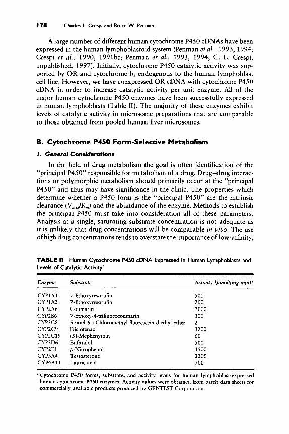
178 Charles L. Crespi and Bruce W. Penman
A large number of different human cytochrome P450 cDNAs have been expressed in the human lymphoblastoid system (Penman et al., 1993,1994; Crespi et al., 1990, 1991bc; Penman et al., 1993, 1994; C. L. Crespi, unpublished, 1997). Initially, cytochrome P450 catalytic activity was sup- ported by OR and cytochrome b, endogenous to the human lymphoblast cell line. However, we have coexpressed OR cDNA with cytochrome P450 cDNA in order to increase catalytic activity per unit enzyme. All of the major human cytochrome P450 enzymes have been successfully expressed in human lymphoblasts (Table 11). The majority of these enzymes exhibit levels of catalytic activity in microsome preparations that are comparable to those obtained from pooled human liver microsomes.
8. Cytochrome P450 Form-Selective Metabolism
I . General Considerations
In the field of drug metabolism the goal is often identification of the “principal P450” responsible for metabolism of a drug. Drug-drug interac- tions or polymorphic metabolism should primarily occur at the “principal P450” and thus may have significance in the clinic. The properties which determine whether a P450 form is the “principal P450” are the intrinsic clearance (V,,,,&,,) and the abundance of the enzyme. Methods to establish the principal P450 must take into consideration all of these parameters. Analysis at a single, saturating substrate concentration is not adequate as it is unlikely that drug concentrations will be comparable in vivo. The use of high drug concentrations tends to overstate the importance of low-affinity,
TABLE II Human Cytochrome P450 cDNA Expressed in Human Lymphoblasts and Levels of Catalytic Activity“
Enzyme
CYPlAl CYP1.42 CYP2A6 CYP2B6 CYP2C8 CYP2C9 CYP2Cl9 CYP2D6 CYPZEl CYP3A4 CYP4A11
Substrate
7-Ethoxyresorufin 7-Ethoxyresorufin Coumarin 7-Ethox y -4-trifluorocoumarin 5-(and 6-)-Chloromethyl fluorescein diethyl ether Diclofenac (S )-Mephenytoin Bufuralol p-Nitrophenol Testosterone Lauric acid
Activity [pmolJ(mg min)]
500 200 3000 300 2 3200 60 500 1500 2200 700
a Cytochrome P450 forms, substrate, and activity levels for human lymphoblast-expressed human cytochrome P450 enzymes. Activity values were obtained from batch data sheets for commercially available products produced by GENTEST Corporation.

Use of cDNA-Expressed Cytochrome P450 Enzymes I79
high-capacity enzymes. On some occasions, the enzyme specificity is such that substrate concentration is not a major consideration. For example, diclofenac, coumarin, and testosterone are substantially enzyme selective. If limited resources dictate the use of a single substrate concentration, the use of a low concentration, less than 5 pM, is well advised.
When multiple enzymes are involved in metabolism, the rates of metabo- lism can be expressed as nanomoles of metabolite per nanomole of P450 (spectrophotometric quantitation of P450 content is possible for the human lymphoblast system and many other systems). However, human liver does not contain equirnolar concentrations of the different P450 forms (Fig. l), and a rigorous quantitation of the levels of all the different P450 forms has not been performed, particularly for the CYP2C subfamily. Moreover, the levels of OR differ among the different cDNA-expressed preparations (Fig. 2) and also differ from that found in human liver. As a consequence, enzyme turnover numbers vary substantially among systems (Fig. 3). Therefore, there is no firm basis for extrapolation from cDNA-expressed to human liver based on a nanomole P450 basis.
We have proposed that a comparison of the activity levels for cyto- chrome P45O-selective substrates can provide relative activity factors (RAFs) for extrapolating an individual cDNA-expressed enzyme to the balance of human liver (Crespi, 1995). The RAF is the ratio of the activity of the enzyme (per unit protein) in human liver microsomes to the activity in the cDNA expression system. The accurate calculation of RAF requires that the reference substratekonditions be enzyme selective in human liver rnicro- somes (i.e., measure the activity of the subject enzyme only). Unlike normal- ization per unit P450, the RAF approach is not sensitive to differences in absolute turnover between the cDNA-expressed enzyme and human liver
iA2 2A6 286 X 206 2E 3A
FIGURE I Abundance (expressed in pmol/mg microsomal protein) of different human cytochrome P450 forms as reported by Shimada et al. (1994). Values are the means of 60 livers, and variation among individual specimens is substantial, as would be expected. All CYP2C forms are expressed as a single term and are not reported as the relative amount of the individual forms (CYP2C8, CYP2C9, CYP2C18, CYP2C19). Much higher levels of CYP2B6 have been detected among a panel of 17 human liver specimens (Code et al., 1997).

I80 Charles L. Crespi and Bruce W. Penman
Construction Cyt. C Reductase Level*
Control Cells 16 OR alone 1500 OR + rat CYPZD1 1200 OR + rat CYP2El 550 OR + rat CYP2Al 130 OR + 2 human CYP2El 80 OR + human CYP3A4 250
Levels after full induction with dexamethasone.
FIGURE 2 Levels of NADPH-cytochrome P450 oxidoreductase (OR) expression when different cytochrome P450 cDNA are coexpressed with OR cDNA in the same vector. The vectors all have the same promoters for the cytochrome P450 cDNA (Herpes simplex virus thymidine kinase) and for OR cDNA (mouse mammary tumor virus long terminal repeat), which is induced by exposure to dexamethasone, and are based on the pRedHyHo vector reported in Crespi et af. (1995). The OR expression level was measured as cytochrome c reductase activity (Phillips and Langdon, 1962).
Expressed at nmole/(mg min).
or among different cDNA-expressed enzymes. The RAF value is specific to the enzyme and expression system, and the accurate application of this approach requires a high degree of reproducibility among independent prep- arations of the same enzyme. This approach does assume that relative rates of metabolism are maintained for cDNA-expressed enzymes relative to hu- man liver microsomes. Clearly, a representative HLM sample must be used;
Yeast+OR+bB
Yeast-Native 0.04
E. coliCSH 5.5
E. Coli-Native 0.2
Lyrnp.+OR 1-44
Lyrnphoblast -15
0 10 20 30 40 50
Turnover Number (per minute)
FlGURE 3 Differences in testosterone 6/3-hydroxylation (expressed as turnover number per unit enzyme) for CYP3A4 expressed in different heterologous systems. Yeast data were obtained from Peyronneau et uf. (1992), Esherichiu cofi data were obtained from Gillam et af. (1993), vaccinia virus data were obtained from Buters et af. (1994), and human lymphoblast data, without OR, were obtained from Crespi et al. (1991). We have expressed CYP3A4 with OR and cytochrome b5 using a baculovirus expression system and obtained turnover numbers of greater than 100 min-' (C. Crespi and V. P. Miller, unpublished observation).

Use of cDNA-Expressed Cytochrome P450 Enzymes 181
we have used a panel of 17 human liver specimens to establish RAF values for cDNA-expressed enzymes.
The intrinsic clearances for each of the active, cDNA-expressed enzymes (determined by kinetic analyses or estimated by analysis of rates of metabo- lism at physiological concentrations) can be multiplied by the measured RAFs to calculate the “contribution” of that enzyme. The enzyme with the highest contribution should be the “principal P450.” RAFs can also be applied to V,,, data if it is anticipated that all enzymes will be saturated in vivo. However, if some enzymes are saturated and others are not at a given concentration, RAFs may be applied only to rates of metabolism at that concentration.
The RAF approach has been applied to previously published data (hu- man liver microsomes and cDNA-expressed enzymes) for the metabolism and bioactivation of the tobacco smoke carcinogen N N K (Crespi, 1995). This retrospective analysis successfully reconciled disparate data obtained independently with human liver microsomes and cell lines expressing individ- ual cDNAs. A more comprehensive evaluation of this approach is currently being performed.
2. Diclofenac Metabolism
Diclofenac is a nonsteroidal, anti-inflammatory drug. Leeman et al., (1993) reported that cytochrome P450TB (TB for tolbutamide, CYP2C) was the principal enzyme catalyzing the 4’-hydroxylation of this substrate in human liver. The evidence included the observation of a single K , in human liver microsomes, inhibition by tolbutamide, phenytoin, and ( t )-warfarin (known CYP2C substrates), and competitive inhibition by sulfaphenazole, a CYP2C-selective inhibitor. The presence of the CYP2D6 polymorphism did not affect the rate of diclofenac metabolism. These properties are consis- tent with metabolism by CYP2C9.
We have extended these studies by analyzing diclofenac 4’-hydroxyla- tion using a panel of cDNA-expressed human cytochrome P450s. The results, expressed as nanomoles of 4’-hydroxydiclofenac per incubation, are shown in Table 111. The specific conditions were 20 pmol of cDNA-expressed cytochrome P450 enzymes (based on reduced carbon monoxide differ- ence spectra) incubated with 150 p M diclofenac for 30 min with a NADPH-generating system. The final incubation volume was 0.5 ml. 4’- Hydroxydiclofenac production was measured as described in Section 111. Diclofenac 4’-hydroxylation was found to be highly CYP2C9 selective, with only a very minor amount of 4’-hydroxylation by CYP2C19. If data are interpreted on a per nanomole P450 present in the incubation, CYP2C9 accounts for -99% of the metabolism by an equimolar amount of cyto- chromes P450. However, P450 forms are not present in equimolar amounts. Therefore, the actual percentage of the metabolism catalyzed by CYP2C9 will depend on the molar ratio of CYP2C9 and CYP2C19, the two active

I82 Charles L. Crerpi and Bruce W. Penman
TABLE 111 P450 Enzymesa
Diclofenac 4'-Hydroxylation by cDNA-Expressed Human Cytochrome
E n m e 4'-Hydroxydiclofenac (nmol in 30 min) Percentage of total
CYPlAl CYPl A2 CYP2A6 CYP2B6 CYP2C8 CYP2C9 CYP2C19 CYP2D6 CYP2El CYP3A4
Not detectable Not detectable Not detectable Not detectableb Not detectableb 43.6 0.16' Not detectable Not detectable Not detectable
0 0 0 0 0
99.6 0.4 0 0 0
~
* Incubations and analysis of metabolite formation were performed as described in the text and in Section 111. Produces an unknown metabolite that chromatographs after 4'-hydroxydiclofenac and with a total amount, based on the absorbance of 4'-hydroxydiclofenac, of less than 0.3 nmol.
enzymes. Although this ratio is not known, it appears that CYP2C9 is more abundant than CYP2C19 (Romkes et al., 1991). Therefore, any adjustment based on the ratio of CYP2C9 to CYP2C19 is unlikely to decrease the proportion of diclofeac 4'-hydroxylation attributed to CYP2C9.
The rates of diclofenac metabolism for microsomes prepared from CYP2C9 cDNA-expressing human lymphoblasts and pooled HLM are quite similar (3.2 nmol/mg min in either system). Two common CYP2C9 alleles differ in the identity of the amino acid at position 144. The Arg,,, allele is the more common allele whereas the Cys14 allele is less common (Furuya et al., 1995). Analysis of warfarin and tolbutamide metabolism with cDNA- expressed enzymes reveals that the CysI4 protein is substantially less active for warfarin metabolism, whereas both enzymes exhibit comparable rates of tolbutamide metabolism (Rettie et al., 1994). We have examined diclofenac metabolism by both these alleles and have found modest differences in rates of metabolism (turnover numbers of 52 and 24 min-' for Arg,,, and CysI4,, respectively). The apparent K , values were also nearly identical.
C. Cytochrome P450 Inhibition
Cytochrome P450 inhibition studies are readily conducted using cDNA- expressed materials. We routinely conduct analyses with three enzymes: CYP2C9, CYP2D6, and CYP3A4. We have also analyzed inhibition of CYP2A6 (coumarin 7-hydroxylase), CYPlA2 (7-ethoxyresorufin 0- deethylase), and CYP2E1 ( p-nitrophenol hydroxylase). In principle, these types of studies can be conducted with any enzyme for which a substrate is known. Appropriate design of the inhibition parameters requires determi-

Use of cDNA-Expressed Cytochrome P450 Enzymes I83
nation of the apparent K , and V,,, for the substrate and cDNA-expressed enzyme. The apparent K , value is used to choose the substrate concentrations such that they are in and around this value. The apparent V,, value is used to choose enzyme concentrations and incubation times that provide readily detectable levels of metabolism without exceeding 10% of the initial amount of substrate being metabolized.
For the following three enzymes, we have adapted published procedures in order to enhance assay throughput. We have avoided organic solvent extractions by precipitating protein by the use of acetonitrile or perchloric acid followed by centrifugation. The supernatant is directly injected for HPLC separation and analysis of metabolite formation.
1. CYPZC9
As discussed earlier, analysis of diclofenac 4'-hydroxylation human liver microsomes (Leeman et al., 1993) and cDNA-expressed enzymes (this chapter) indicates that CYP2C9 is the principal enzyme responsible for the 4'-hydroxylation of diclofenac. Any contribution by CYP2C19 should be very minor. Therefore, this assay can be used in HLM and cDNA-expressed enzymes to selectively measure CYP2C9-mediated metabolism and inhibi- tion of CYP2C9. We have used this biotransformation to analyze CYP2C9 inhibition in pooled HLM and cDNA-expressed CYP2C9. The apparent K, values for diclofenac 4'-hydroxylation in HLM and cDNA-expressed CYP3A4 were quite similar (2.9 and 2.3 p M , respectively). Our measured apparent K , value in HLM is in agreement with that reported by Leeman et al. (1993). We have also analyzed CYP2C9 inhibition by prototypical CYP2C9 inhibitors. The apparent K, values are shown in Table IV. For these determinations, the procedure described in Section I11 was used.
As with the apparent K , values, there is good agreement between HLM and cDNA-expressed CYP2C9 for apparent K, values with sulfaphenazole, (+)-warfarin, and tolbutamide. Corresponding values in HLM and cDNA- expressed enzyme are within 2-fold over a -1000-fold range of apparent K , values. Our observed apparent K, value for sulfaphenazole is in good agreement with that reported by Leeman et al. (1993).
TABLE IV Apparent K, Values for CYP2C9 Inhibition"
Inhibitor Apparent K, (HLM) Apparent K, (cDNA)
Sulfaphenazole 0.11 p M 0.09 p M (?)-Warfarin 0.32 p M 0.25 pM Tolbutamide 96 pM 160 pM
Inhibition analyses for cDNA-expressed CYP2C9 and HLM CYP2C9 were conducted as described in Section 111. Apparent K, was determined by Dixon plots and the values are the mean of three determinations.

184 Charles L. Crespi and Bruce W. Penman
2. CYPZD6
Studies using human liver microsomes (Dayer et af., 1987) indicate that CYP2D6 is the principal enzyme responsible for the 1 '-hydroxylation of bufuralol at low bufuralol concentrations. Significant metabolism by CYPlA2 can occur at high bufuralol concentrations (Yamazaki etal., 1994). The difference in affinity for CYPlA2 and CYP2D6 is sufficiently large that this assay can be used in HLM and cDNA-expressed enzymes to selectively measure CYP2D6-mediated metabolism and inhibition of CYP2D6. We have used this biotransformation to analyze CYP2D6 inhibition in pooled HLM and cDNA-expressed CYP2D6. The apparent K, values for bufuralol 1 '-hydroxylation in HLM and cDNA-expressed CYP2D6 were quite similar (15 and 10 pM, respectively). We have also analyzed CYP2D6 inhibition by prototypical CYP2D6 inhibitors. The apparent K, values are shown in Table V. For these determinations, the procedure described in Section I11 was used.
As with the apparent K , values, there is good agreement between HLM and cDNA-expressed CYP2D6 for apparent K, values with quinidine, per- hexiline, dl-propranalol, and ( - )-sparteine. Corresponding values in HLM and cDNA-expressed enzyme are within 2- to 3-fold over a -10,000-fold range of apparent K , values.
3. CYP3A4
Studies using human liver microsomes (Waxman et al., 1988) and cDNA-expressed enzymes (Waxman et af., 1991) indicate that CYP3A en- zymes (CYP3A4 being the most abundant) are the principal enzymes respon- sible for the 60-hydroxylation of testosterone. Therefore this assay can be used in HLM and cDNA-expressed enzymes to selectively measure CYP3A4- mediated metabolism and inhibition of CYP3A4. We have used this bio- transformation to analyze CYP3A4 inhibition in pooled HLM and cDNA-expressed CYP3A4. The apparent K, values for testosterone 6p- hydroxylation in HLM and cDNA-expressed CYP3A4 were quite similar (74 and 70 pM, respectively). We have also analyzed CYP3A4 inhibition
TABLE V Apparent Ki Values for CYP2D6 Inhibition"
inbibitor Appurenf K , (HLM) Apparent K , JcDNA)
Quinidine 0.014 pM Perhexiline 0.14 pM dl-Propranalol 9.7 p M ( - )-Sparteine 140 pM
0.008 p M 0.19 pM 4.5 pM 59 pM
Inhibition analyses for cDNA-expressed CYP2D6 and HLM CYP2D6 were conducted as described in Section 111. Apparent K, was determined by Dixon plots and the values are the mean of three determinations.

Use of cDNA-Expressed Cytochrome P450 Enzymes I85
TABLE VI Apparent Ki Values for CYP3A4 Inhibition"
Inhibitor Apparent K , (HLM) Apparent K , (cDNA)
Ketoconazole 0.011 pM 0.027 p M ( 2 )-Miconazole 0.055 p M 0.086 p M Terfenadine 1.6 p M 1.1 pM Erythromycin 59 p M 24 p M
a Inhibition analyses for cDNA-expressed CYP3A4 and HLM CYP3A4 were conducted as described in Section 111. Apparent K, was determined by Dixon plots and the values are the mean of three determinations.
by prototypical CYP3A4 inhibitors. The apparent K, values are shown in Table VI. For these determinations, the procedure described in Section I11 was used.
As with the apparent K, values, there is good agreement between HLM and cDNA-expressed CYP3A4 for apparent K, values with ketoconazole, miconazole, terfenadine, and erythromycin. Corresponding values in HLM and cDNA-expressed enzyme are within 2- to 3-fold over a -5000-fold range of apparent K, values.
V. Conclusions
cDNA expression of human cytochrome P450 enzymes provides a repro- ducible source of material for the study of metabolite formation and enzyme inhibition. In general, high levels of expression for individual enzymes can be achieved. cDNA-expressed enzymes generally retain affinity-related prop- erties such as apparent K, and apparent Ki. However, variations in the levels of coenzymes can substantially affect V,,, (expressed per unit enzyme). Therefore, if relative rates of metabolism by cDNA-expressed enzymes are to be related to relative rates of metabolism in human tissues (e.g., human liver microsomes), these differences must be taken into account.
Acknowledgments
Cytochrome P450 cDNAs (all except CYP2A6 and CYP2C19) were obtained from the laboratory of Drs. F. J. Gonzalez and H. V. Gelboin of the National Cancer Institute. Human CYP2C19 cDNA was obtained from the laboratory of Dr. J. Goldstein of the National Institute of Environmental Health Sciences. The CYP2A6 cDNA was isolated at GENTEST.
References
Buters, J. T. M., Korzekwa, K. R., Kunze, K. L., Omata, Y., Hardwick, J. P., and Gonzalez, F. J. (1994). cDNA-directed expression of human cytochrome P450 CYP3A4 using bacu- lovirus. Drug Metab. Dispos. 22, 688-692.

I86 Charles L. Crespi and Bruce W. Penman
Code, E. L., Crespi, C. I.., Penman, B. W., Gonzalez, F. J., Chang, T. K. H., and Waxman, 1). J. 1997). Human cytochrome P450286: Interindividual hepatic expression, substrate specificity and role in procarcinogen activation. Drug Metab. Disp. (in press).
Crespi, C. L. ( 1995). Xenobiotic-metabolizing human cells as tools for pharmacological and toxicological research. Adit. Drug Res. 26, 179-235.
Crespi, C. L., Penman, B. W., Leakey, J. A. E., Arlotto, M. P., Stark, A., Turner, T., Steimel, D., Rudo, K., Davies, R. L., and Langenbach, R. L. (1990). Human cytochrome P450IIA3: cDNA sequence, role of the enzyme in the metabolic activation of promutagens, compari- son to nitrosamine activation by human cytochrome P45011E1. Carcinogenesis (London)
Crespi, C. I.., Gonzalez, F. J., Steimel, D. T., Turner, T. R., Gelboin, H. V., Penman, B. W., and Langenbach, R. (1991a). A metabolically competent cell line expressing five cDNAs encoding procarcinogen-activating enzymes: Applications to mutagenicity testing. Chem. Res. Toxicol. 4, 566-572.
Crespi, C. I.., Penman, B. W., Steimel, D. T., Gelboin, H. V., and Gonzalez, F. J. (1991b). The development of a human cell line stably expressing human CYP3A4: Role in the metabolic activation of aflaroxin B, and comparison to CYPlA2 and CYP2A3. Carcino- genesis (London), 12, 355-359.
Crespi, C. L., Gonzalez, F. J., Steimel, D. T., Turner, T. R., Gelboin, H. V., Penman, B. W., and Langenbach, R. ( 1 9 9 1 ~ ) . A tobacco smoke-derived nitrosamine, 4-(methylnitrosamino)-l- (3-pyridyl)-l -butanone, is activated by multiple human cytochrome P450s including the polymorphic cytochrome P4502D6. Carcinogenesis (London) 12, 1197-1201.
Crespi, C. L., Langenbach, R., and Penman, B. W. (1993). Human cell lines, derived from AHH-1 TK? human Iymphoblasts, genetically engineered for expression of cytochromes
Crespi, C. L., Steimel, D. T., Penman, B. W., Korzekwa, K. R., Fernandez-Salguero, P., Buters, J. T. M.. Gelboin, H. V., Gonzalez, F. J., Idle, J. R.. and Daly, A. K . (1995). Comparison of substrate metabolism by wild type CYP2D6 protein and a variant containing methio- nine, not valine, at position 374. Pbarmacogenetics 5, 234-243.
Dayer, P., Kronbach, T., Eichelbaum, M., and Mayer, U. A. (1987). Enzymatic basis of the debrisoquine/sparteine-type polymorphism of drug oxidation. Biochem. Pharmacol.
Doehmer, J., Dogra, S., Friedberg, T., Monier, S., Adesnik, M., Glatt, H., and Oesch, F. (1988). Stable expression of rat cytochrome P45OIIB1 cDNA in Chinese hamster cells (V79) and metabolic activation of aflatoxin B,. Proc. Natf. Acad. Sci. U.S.A. 85, 5769-5773.
First, M. R., Schroeder, T. J., Alexander, J. W., Stephens, G . W., Weiskittel, P., Myre, S. A,, and Pesci, A. J. (1991). Cyclosporine dose reduction by ketoconazole administration in renal transplant patients. Transplantation 5 1, 365-370.
Furuya, H., Fernandez-Salguero, P., Gregory, W., Taber, H., Steward, A,, Gonzalez, F. J., and Idle, J. R. (1995). Genetic polymorphism of CYP2C9 and its effect on warfarin mainte- nance dose requirement in patients undergoing anticoagulation therapy. Pharmacogenetics
Gillam, E. M. J., Baba, T., Kim, B.-R., Ohmori, S., and Guengerich, F. P. (1993). Expression of modified human cytochrome P450 3A4 in Escherichia coli and purification and reconsti- tution of the enzyme. Arch. Biochem. Biophys. 305, 123-131.
Gonzalez, F. J. (1989). The molecular biology of cytochrome P4SOs. Pharmacol. Rev. 40,
Gonzalez, F. J., and Gelboin, H. V. (1994). Role of human cytochromes 1’450 in the metabolic
Gonzalez, F. J., and Korzekwa, K. R. (1995). Cytochromes P450 expression systems. Annu.
8,1293-1300.
P450. T O X K O ~ O ~ Y 82, 89-104.
36, 4145-41.52.
5,389-392.
243-288.
activation of chemical carcinogens and toxins. Drug Metab. Rev. 26, 165-183.
Rev. Pharmacol. Toxrcol. 35. 369-390.

Use of cDNA-Expressed Cytochrorne P450 Enzymes I87
Guengerich, F. P., Muller-Enoch, D., and Blair, I. A. (1986). Oxidation of quinidine by human liver cytochrome P-450. Mol. Pharmacol. 30, 287-295.
Kronbach, T., Mathys, D., Gut, J., Catin, T., and Meyer, U. A. (1987). High performance liquid chromatographic assays for bufuralol 1 ’hydroxylation, debrisoquine 4-hydroxylation and dextromethorphan 0-demethylation in microsomes and purified cytochrome P-450 iso- zymes of human liver. Anal. Biochem. 162, 24-32.
Leeman, T., Transon, C., and Dayer, P. (1993). Cytochrome P45OTB (CYPZC): A major monooxygenase catalyzing diclofenac 4’-hydroxyation in human liver. Life Sci. 52,29-34.
Mahgoub, A., Idle, J, R., Dring, L. G., Lancaster, R., and Smith, R. L. (1977). Polymorphic oxidation of debrisoquine in man. Lancet 2, 584-586.
Nelson, D. R., Koyrnans, L., Kamataki, T., Stegeman, J. J., Feyereisen, R., Waxman, D. J., Waterman, M. R., Gotoh, O., Coon, M.J., Estabrook, R. W., Gunsalus, I. C., and Nebert, D. W. ( 1996). P450 superfami1y:update on new sequences, gene mapping, accession numbers and nomenclature. Pharmacogenetics 6, 1-42.
Omura, T., and Sato, R. (1964). The carbon monoxide-binding pigment of liver microsomes 11: Solubilization, purification and properties. J. Biol. Chem. 239, 2379-2385.
Penman, B. W., and Crespi, C. L. (1987). Analysis of human lyrnphoblast mutation assays by using historical negative control data bases. Envron. Mol. Mutagen. 10, 35-60.
Penman, B. W., Reece, J., Smith, T., Yang, C. S., Gelboin, H. V., Gonzales, F. J., and Crespi, C. L. (1993). Characterization of a human cell line expressing high levels of cDNA- derived CYP2D6. Pharmacogenetics 3 , 28-39.
Penman, B. W., Chen, L. P., Gelboin, H. V., Gonzalez, F. J., and Crespi, C. L. (1994). Development of a human lymphoblastoid cell line constitutively expressing human CYPlAl cDNA-substrate specificity with model substrates and promutagens. Carcino- genesis (London) 15, 1931-1937.
Peyronneau, M.-A., Renaud, J.-P., Truan, G., Urban, P., Pompon, D., and Mansuy, D. (1992). Optimization of yeast-expressed human liver cytochrome P450 3A4 catalytic activities by co-expressing NADPH-cytochrome P450 reductase and cytochrome bs. Eur. J . Bio- chem. 207, 109-116.
Phillips, A. H., and Langdon, R. G. (1962). Hepatic triphosphopyridine nucleotide cytochrome C reductase: Isolation, characterization and kinetic studies. J. Biol. Chem. 237, 2652- 2660.
Rettie, A. E., Wienkers, L. C., Gonzales, F. J., Trager, W. F., and Korzekwa, K. R. (1994). Impaired (S)-warfarin metabolism catalysed by the R144C allelic variant of CYP2C9. Pharmacogenetics 4, 39-42.
Romkes, M., Feletto, M. B., Baisdell, J. A., Raucy, J. L., and Goldstein, J. A. (1991). Cloning and expression of complementary DNAs for multiple members of the human cytochrome P450IIC subfamily. Biochemistry 30, 3247-3255.
Schenkman, J. B., Voznesensky, A. I., and Jansson, I. (1994). Influence of inonic strength on the P450 monooxygeanse reaction and the role of cytochrome bs in the process. Arch. Biochem. Biophys. 314, 234-241.
Shimada, T., Misono, K. S . , and Guengerich, F. P. (1986). Human liver microsomal cytochrome P-450 mephenytoin 4-hydroxylase, a prototype of genetic polymorphism in oxidative metabolism. J. Biol. G e m . 261, 909-921.
Shimada, T., Yamazaki, H., Mimura, M., Inui, Y., and Guengerich, F. P. (1994). Interindividual variations in human liver cytochrome P-450 enzymes involved in the oxidation of drugs, carcinogens and toxic chemicals-studies with liver microsomes of 30 Japanese and 30 Caucasians. J. Phamacol. E x p . Ther. 270, 414-423.
Song, B.-J., Gelboin, H. V., Park, S.-S., Yang, C. S., and Gonzalez, F. J. (1996). Complementary DNA and protein sequences of ethanol-inducible rat and human cytochrome P-450’s. J. Biol. Chem. 261,16689-16697.
Spatzenegger, M., and Jaeger, W. (1995). Clinical importance of hepatic cytochrome P450 in drug metabolism. Drug Metab. Rev. 27, 397-417.

I88 Charles L. Crespi and Bruce W. Penman
Sugden, B., Marsh, K., and Yates, J. (1985). A vector that replicates as a plasmid and can be efficicently selected in B-lymphoblasts transformed by Epstein-Barr virus. Mol. Cell. Biol. 5,410-413.
Waterman, M. R. (1993). Heterologous expression of cytochrome-P-4.50 in Escherichiu coli. Biochem. Soc. Trans. 21, 1081-1085.
Watkins, P. B., Wrighton, S. A., Scheutz, E. G., Molowa, D. T., and Guzelian, P. S. (1987). Identification of glucocorticoid-inducible cytochromes P450 in the intestinal mucosa of rats and man. J . Clin. Invest. 80, 1029-1036.
Waxman, D. J., KO, A., and Walsh, C. (1983). Regioselectivity and stereoselectivity of androgen hydroxylations catalyzed by cytochrome P-450 isozymes purified from phenobarbital- induced rat liver. J . Biol. Chem. 258, 11937-1 1947.
Waxman, D. J., Attisano, C., Guengerich, F. P., and Lapenson, D. (1988). Human liver microsoma1 steroid metabolism: Identification of the major microsomal steroid hormone 6 beta-hydroxylase cytochrome P-450 enzyme. Arch. Biochem. Biophys. 263,424-436.
Waxman, D. J., Lapenson, D. P., Aoyama, T., Gelboin, H. V., and Gonzalez, F. J. (1991). Steroid hormone hpdroxylase specifies of eleven cDNA-expressed human cytochrome P450 enzymes. Arch. Biochem. Biophys. 290, 160-166.
Wrighton, S. A., Steven, J. C., Becker, G. W., and VandenBranden, M. i1993). Isolation and characterization of human liver cytochrome P450 2C19: Correlation between 2C19 and S-mephenytoin 4’-hydroxylase. Arch. Biochem. Biophys. 306, 240-245.
Yamazaki, H., Guo, Z . Y., Persmark, M., Mimura, M., Inoue, K., Guengerich, F. P., and Shimada, T.(1994). (1994). Bufuralol hydroxylation by cytochrome P450 2D6 and IA2 enzymes in human liver microsomes. Mol. Pharmacol. 46, 568-577.
Yates, J. L.. Warren, N., and Sugden, B. (1985). Stable replication of plasmids derived from Epstein-Barr virus in various mammalian cells. Nature (London) 313, 812-815.

Gregory L. Kedderis Chemical Industry Institute of Toxicology
Research Triangle Park, North Carolina 27709
Pharmacokinetics of Drug Interactions
1. Introduction
Drug interactions can occur when two or more drugs are administered together or in close succession to each other. The administration of multiple drugs is a common situation in modern society. Multiple drug therapy is popular because it has been found to be beneficial in treating a variety of conditions, including infections, neurological disorders, and gastrointestinal diseases (Caranasos et al., 1985). Patients frequently undergo concurrent drug therapy for multiple conditions, creating the possibility of drug interac- tions. Additionally, exposure to substances through the diet, life-style, work- place, and environment can produce drug interactions.
Advances in Pharmucology, Volume 43 Copyright 0 1997 by Academic Press. All rights of reproduction in any form reserved. 1054.3589197 $25.00 I89

I90 Gregory L. Kedderis
This chapter deals with the pharmacokinetics of drug interactions. Inter- acting drugs can affect all aspects of pharmacokinetics, including absorption, distribution, metabolism, and excretion through a variety of mechanisms (Labaune, 1989; Rowland and Tozer, 1995). Various nutritional and disease states can affect the magnitude of these interactions. Drug interactions affect- ing pharmacokinetics can have pharmacodynamic consequences, such as diminished or exaggerated efficacy or increased toxicity. These adverse ef- fects are dose dependent. Drug interactions can also influenced by genetics and disease states. The pharmacodynamic aspects of drug interactions will be covered in more detail in other chapters of this book.
II. Drug Interactions Affecting Absorption
Drug interactions can affect drug absorption by affecting the dissolution of the drug in the stomach, by influencing gastric emptying or intestinal blood flow, or by inhibition of active transport processes (Labaune, 1989). Drug dissolution in the stomach can be modulated by gastric pH changes or by complexation or chelation of the drug.
A. Changes in pH
The degree of absorption of an ionizable drug from the stomach or duodenum depends on the p K , of the drug and the pH of the environment (Labaune, 1989). In general, alkalinizing agents such as bicarbonate salts decrease the absorption of weak acids, having a p K , between approximately 2.5 and 7.5. Examples of weak acids affected by alkalinizing agents include nonsteroidal anti-inflammatory drugs, vitamin K antagonists, and orally active penicillins. Conversely, acidifying agents such as citric or tartaric acids affect the absorption of weak bases having a p K , between approximately 5 and 11. Examples of weak bases include propoxyphene and reserpine. The gastric absorption of very weak bases having a p K , less than 5 , such as caffeine ( p K , 0.81, will usually be independent of gastric pH.
A good example of a drug interaction due to increasing gastric pH is the interaction between the antibiotic tetracycline and sodium bicarbonate. Coadministration of these drugs decreases the absorption of tetracycline. A simiiar interaction is observed between tetracycline and cimetidine. Cimeti- dine is a potent Hl-receptor antagonist that inhibits gastric acid secretion, thus raising gastric pH. These types of drug interactions can lead to the therapeutic failure of the antibiotic.
Drugs that are hydrolyzed in the stomach are also sensitive to changes in gastric pH. Hydrolysis occurs more slowly at higher pH values. Therefore, increasing gastric pH facilitates the absorption of drugs such as penicillin.

Pharmacokinetics of Drug Interactions 191
B. Complexation and Chelation
Some drugs can interact to form complexes that are poorly absorbed (Labaune, 1989). Tetracyclines can form complexes with calcium, magne- sium, iron, or aluminum ions that are present in many antacids. This leads to a decreased absorption of the antibiotic. The cholesterol-lowering drug cholestyramine can form poorly absorbed complexes not only with choles- terol but also with drugs containing carboxylic acids or hydroxyl groups. Thus cholestyramine can reduce the absorption of warfarin, nonsteroidal anti-inflammatory drugs, and sulfonamides. Kaolin, a component of drugs used to treat diarrhea, can complex certain drugs such as digoxin.
C. Effects on Gastric Emptying
Gastric emptying controls the length of time that a drug remains in the stomach. Compared to the duodenal mucosa, the gastric mucosa is not a major site for drug absorption (Labaune, 1989). Therefore the longer a drug remains in the stomach, the slower it is absorbed. Increasing the rate of gastric emptying increases the rate of drug absorption. The increased rate of drug absorption can result in adverse or toxic effects due to exaggerated tissue concentrations of the drug (Fig. 1). A variety of drugs are known to affect the rate of gastric emptying (Table I). Additionally, the presence of food in the stomach can affect the absorption of many drugs (Table 11). Food can reduce or delay the absorption of some drugs, while not affecting or increasing the absorption of other drugs.
Several drug interactions affecting the absorption of acetaminophen can serve as illustrations of drug interactions due to effects on gastric emptying.
Effects
I
Time FIGURE I Effect of absorption rate on the tissue concentration of drug 1. The lower curve shows the oral absorption kinetics of drug 1. Coadministration of drug 2 increases the rate of gastric emptying, increasing the absorption rate of drug 1 such that toxic tissue concentrations are achieved.

I92 Gregory L. Kedderis
TABLE I Effect of Drugs on Gastric Emptying"
Increase Decrease
Metoclopramide Anticholinergics: Atropine, tricyclic antidepressants Reserpine Central analgesics: Morphine Anticholinesterases Isoniazid Sodium Bicarbonate Chloroquine
Phenytoin Aluminum hydroxide Magnesium hydroxide
-I Adapted from Nirnmo (1976).
The ulcer drug propantheline decreases the rate of acetaminophen absorp- tion but has no effect on the extent of absorption. The heartburn drug metoclopramide increases the rate of absorption of acetaminophen. The central analgesics pethidine and diamorphine delay the absorption of acet- aminophen, but the overall extent of absorption is unchanged.
I). Effects on Intestinal Blood Flow
Intestinal blood flow can be the rate-limiting step in the absorption of some lipophilic drugs. Intestinal blood flow can be modulated by vasodila- tors and vasoconstrictors and hypothetically affect the absorption of lipo- philic drugs. However there is no experimental evidence for this type of drug interaction (Labaune, 1989).
E. Inhibition of Active Transport
Another possible drug interaction affecting absorption can occur through inhibition of the active uptake of a drug. Although very few drugs
TABLE I t Effect of Food on Drug Absorption"
Reduced Delayed Unaffected Increased
Ampicillin Acetaminophen Chlorpropamide Carbamazepine Aspirin Aspirin Ethambutol Diazepam Captopril Cephalosporins Hydralazine Dicumarol Erhanof Sulfonamides Oxazepam Griseofulvin Penicillins Diclofenac Phenazone Metoprolol Tetracyclines Digoxin Propoxyphene Propranolol Iron Furosernide Tolbutamide Nitrofurantoin L-DOPA Indoprofen a-Tocopheryl nicotinate Warfarin Tolmesoxide
Valproate
'Adapted from Welling (1986, p. 70), with permission of the American Chemical Society.

Pharmacokinetics of Drug Interactions I93
require active uptake for absorption, one significant example of this type of drug interaction is the inhibition of folic acid uptake by phenytoin.
111. Drug Interactions Affecting Distribution
Drug interactions affecting drug distribution arise from effects on drug binding in plasma and tissues. Modulation of drug binding can be an impor- tant effect as only the free drug is distributed to tissues and has pharmacologi- cal activity. Two drugs can compete with each other for the same protein- binding site, leading to displacement of the drug with the lower affinity from the binding site. There can also be a noncompetitive or allosteric drug interaction due to a drug-induced conformational change in the binding site that leads to the displacement of the bound drug. Both these mechanisms lead to a larger circulating concentration of the unbound drug.
Several conditions must be met to favor drug displacement from binding. The drug must be highly bound (the unbound fraction, fu, must be small). The displacing drug must occupy most of the binding sites with high affinity. The concentration of the displacing drug must approach or exceed the molar concentration of the protein binding sites.
Albumin is the most important protein involved in the binding of acidic drugs in plasma and interstitial fluids. The albumin concentration in plasma is on the order of 0.6 mM (Rowland and Tozer, 1995), indicating that relatively high drug concentrations are required for drug interactions affect- ing binding. A variety of acidic drugs bind to albumin at two different sites (Table 111). Some drugs bind to both sites. Because of the high plasma concentration of albumin, only a few drugs are regarded as effective dis- placers, including salicylic acid, phenylbutazone, and some sulfonamides.
TABLE 111 Binding of Acidic Drugs to Albumin"
Site 1 Site 2
Chlorothiazide Furosemide Indomethacinb Naproxen Phenylbutazone Phenytoin Sulfadimethoxine Tolbutamideb Valproate Warfarin
Benzodiazepines Cloxacillin Dicloxacillin Glibenclamide Ibuprofen Indomethacin Naproxen Probenecid Tolazamide Tolbutamide
Adapted from Sjijholm et al. (1979). Drugs that bind to both sites.

I94 Gregory L. Kedderis
These drugs generally have high plasma concentrations and bind to albumin with high affinity.
The high concentration of albumin is in contrast to the much lower concentrations of specific transport proteins involved in hormone binding. Displacement interactions can occur between endogenous steroids and exog- enous steroid analogs at low plasma concentrations because of the very low concentrations of the specific binding proteins.
Drug interactions involving displacement from binding do not affect the unbound clearance of drugs. Usually the volume of distribution will increase for drugs with high extraction ratios, leading to an increase in the half-life. For drugs with low extraction ratios, the volume of distribution and half-life will either decrease or be unaffected.
One example of a drug interaction largely on protein binding is the interaction of the antibacterial agent sulfaphenazole with the hypoglycemic agent tolbutamide (Labaune, 1989). Coadministration of these drugs potent- iates the hypoglycemic effect of tolbutamide. Sulfaphenazole increases the unbound fraction of tolbutamide by 108% and also increases the volume of distribution and the half-life. The total clearance of tolbutamide decreases due to the inhibition of metabolism by sulfaphenazole. Another example involves the displacement of warfarin from its binding sites by trichloroacetic acid, a metabolite of chloral hydrate. This interaction increases the anticoag- ulant effect of warfarin.
JV. Drug Interactions Affecting Metabolism
The most common drug interactions are those affecting metabolism as most drugs are eliminated by metabolism. Drug metabolism generally takes place in the liver, although other organs, such as the intestines and lungs, can also be important. Drug interactions affecting metabolism are of two types: inhibition or induction. Inhibition decreases metabolism, whereas enzyme induction can increase metabolism. In general, high-extraction drugs are less affected by these interactions than low-extraction drugs. However, the lower the therapeutic index of a drug, the more serious the potential consequences of drug interactions affecting metabolism.
A. Inhibition
Inhibition of drug metabolism will result in a decrease in unbound clearance provided metabolism is the major component of clearance. Many drugs, endogenous compounds, and xenobiotics are enzyme inhibitors. There are three basic types of enzyme inhibition: competitive, noncompeti- tive, and uncompetitive.

Pharmacokinetics of Drug Interactions I95
E + S & ES L E + P + I
E l FIGURE 2 Kinetic scheme for competitive inhibition of metabolism. The inhibitor ( I ) com- petes with the substrate (S) for a common binding site on the enzyme (E). The binding constants K, and Ks describe the formation of the EI and ES complexes, respectively. Product (P) formation proceeds at rate k, from the ES complex.
Competitive inhibitors compete with substrates for the same binding site on the enzyme. Competitive inhibitors can be substrate analogs, alternative substrates, or products of the reaction. Competitive inhibition is the most common type of inhibition giving rise to a decrease in unbound clearance. Many drugs are metabolized by cytochromes P450 and can inhibit each other’s metabolism. The kinetic scheme for competitive inhibition is shown in Fig. 2. A competitive inhibitor modifies the K , of the substrate without affecting V,,,, the maximal rate of the enzyme at infinite substrate concentra- tion. The K,, or Michaelis constant, is the substrate concentration that gives one-half V,,,,,. Competitive inhibition can be overcome by increasing the concentration of the substrate (Segel, 1975).
Noncompetitive inhibitors bind to the enzyme at different sites than the substrate (Fig. 3). The noncompetitive inhibitor can bind to the enzyme or the enzyme-substrate complex, whereas the substrate can bind to the enzyme or the enzyme-inhibitor complex. The ternary complex of enzyme, inhibitor, and substrate is catalytically inactive. Pure noncompetitive inhibitors de- crease V,,, for an enzyme-catalyzed reaction, whereas K , remains un- changed. Some noncompetitive inhibitors, termed mixed-type inhibitors, affect both the V,,, and K , of an enzyme-catalyzed reaction (Segel, 1975).
/Ks L E + P E + S + - E+s I I
FIGURE 3 Kinetic scheme for noncompetitive inhibition of metabolism. The inhibitor (I) can bind to the enzyme (E) or the enzyme-substrate (ES) complex with binding constant K , at a site other than the substrate (.$)-binding site. S can bind to E or EI with binding constant Ks. Product (P) formation proceeds at rate k, from the ES complex.

I96 Gregory L. Kedderis
E + S !-, KS ES L E + P + I
E S I FIGURE 4 Kinetic scheme for uncompetitive inhibition of metabolism. The inhibitor (I) binds to the enzyme-substrate (ES) complex with binding constant K , to form an inactive ESI complcx. The other symbols are defined in the legend for Fig. 3.
Uncompetitive inhibitors bind reversibly to the enzyme-substrate com- plex to form an inactive ternary complex (Fig. 4). Uncompetitive inhibitors decrease V,,, and K , to the same extent. Unlike competitive inhibitors, the extent of inhibition by an uncompetitive inhibitor increases with increasing substrate concentration as there is a greater concentration of the enzyme- substrate complex at higher substrate concentrations. At high enough con- centrations, uncompetitive inhibitors can drive the reaction velocity to zero (Segel, 1975). Uncompetitive inhibitors are rare in nature. Considering the devastating effects that an uncompetitive inhibitor could have on cellular function, Cornish-Bowden (1986) suggested that evolution has selected against enzymes that are prone to uncompetitive inhibition. However, the design of therapeutic agents that are pathogen-specific uncompetitive inhibi- tors holds considerable promise (Westley and Westley, 1996).
Inhibition of drug metabolism leads to elevated tissue concentrations of the parent drug, which, in turn, can lead to the development of adverse effects or target organ toxicity (Table IV). The adverse effects can stem from exaggerated therapeutic effects of the drug or from side effects. Because the
TABLE IV Toxic Effects Due to Inhibition of Drug Metabolism"
Inhibitor Inhibited drug Toxic effect
Dicumarol Chlopropamide Phenytoin Tolbutamide
Phenytoin Tolbutamide
C hloramphenicol Tolbutamide Sulfaphenazole To1 butamide Sulfamethizole Phenytoin Disulfiram Phen ytoin
Pheny lhutazane Chlnrpropamtde
Hypoglycemic collapse Vertigo, anorexia Hypoglycemic collapse Hypoglycemic collapse Vertigo, brain damage Hypoglycemic collapse Hypoglycemic collapse Hypoglycemic collapse Vertigo, brain damage Brain damage
' Adapted from Kristensen (1976).
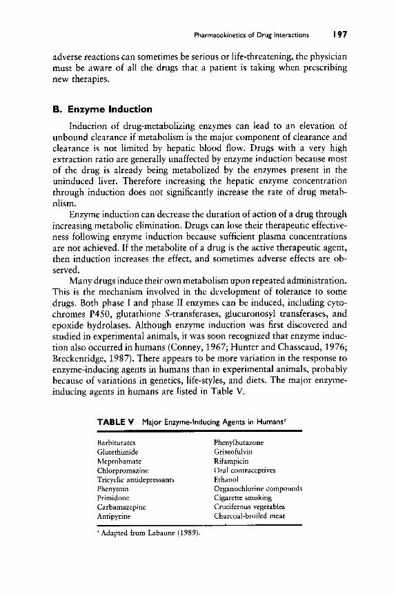
Pharmacokinetics of Drug Interactions I97
adverse reactions can sometimes be serious or life-threatening, the physician must be aware of all the drugs that a patient is taking when prescribing new therapies.
B. Enzyme Induction
Induction of drug-metabolizing enzymes can lead to an elevation of unbound clearance if metabolism is the major component of clearance and clearance is not limited by hepatic blood flow. Drugs with a very high extraction ratio are generally unaffected by enzyme induction because most of the drug is already being metabolized by the enzymes present in the uninduced liver. Therefore increasing the hepatic enzyme concentration through induction does not significantly increase the rate of drug metab- olism.
Enzyme induction can decrease the duration of action of a drug through increasing metabolic elimination. Drugs can lose their therapeutic effective- ness following enzyme induction because sufficient plasma concentrations are not achieved. If the metabolite of a drug is the active therapeutic agent, then induction increases the effect, and sometimes adverse effects are ob- served.
Many drugs induce their own metabolism upon repeated administration. This is the mechanism involved in the development of tolerance to some drugs. Both phase I and phase I1 enzymes can be induced, including cyto- chromes P450, glutathione S-transferases, glucuronosyl transferases, and epoxide hydrolases. Although enzyme induction was first discovered and studied in experimental animals, it was soon recognized that enzyme induc- tion also occurred in humans (Conney, 1967; Hunter and Chasseaud, 1976; Breckenridge, 1987). There appears to be more variation in the response to enzyme-inducing agents in humans than in experimental animals, probably because of variations in genetics, life-styles, and diets. The major enzyme- inducing agents in humans are listed in Table V.
TABLE V
Barbiturates Phenyl butazone Glutethimide Griseofulvin Meprobamate Rifampicin Chlorpromazine Oral contraceptives Tricyclic antidepressants Ethanol Phenytoin Organochlorine compounds Primidone Cigarette smoking Carbamazepine Cruciferous vegetables Antipyrine Charcoal-broiled meat
a Adapted from Labaune (1989).
Major Enzyme-Inducing Agents in Humans"

I98 Gregory L. Kedderis
In addition to increasing the biosynthesis of xenobiotic metabolizing enzymes, some inducing agents can also produce physiological changes that can affect drug metabolism (Hunter and Chasseaud, 1976). Phenobarbital and related barbiturates in particular can increase hepatic blood flow and bile flow. Phenobarbital treatment also increases liver weight in experimental animals, but this effect is not observed in humans (Hunter and Chasseaud, 1976). The induction of enzymes by drugs can cause changes in the metabo- lism of endogenous substances such as carbohydrates through the glucuronic acid pathway, bilirubin, corticosteroids, sex hormones, vitamin D, folate, vitamin K, and heme biosynthesis.
Representative drug interactions due to enzyme induction are given in Table VI. The antibiotic rifampicin decreases the half-life and plasma concentrations of tolbutamide and other drugs. A number of women who were taking oral contraceptives and were also being treated with rifampicin for tuberculosis displayed menstrual disturbances, increased estrogen excre- tion, and pregnancy (Nocke-Finck et al., 1973). These effects were subse- quently shown to be due to enzyme induction by rifampicin (Bolt et al., 1977). Phenobarbital also stimulates the metabolism of oral contraceptives, as well as other drugs (Table VI). Induction of cytochrome P450 by pheno- barbital can dramatically decrease the anticoagulant effect of coumarin derivatives. Phenytoin induction can increase the metabolism of the antiarr- thymic drug disopyramide.
C. Dietary and Environmental Factors
As noted in Table V, a number of agents encountered in the diet, Iife- style, and environment are enzyme inducers in humans. Exposure to these
TABLE VI Enzyme Induction"
Drug Interactions Due to
Inducing agent Drugs affected
Rifampicin Toibutarnide Hexobarbital Digit oxin Oral contraceptives
phenobarbital Oral contraceptives Cournarin anticoagulants Phen ytoin Antipyrine Desirniprarnine
Phenytoin Disopyrarnide Phenyl butazone Aminophenazone
Adapted from Labaune (1989).
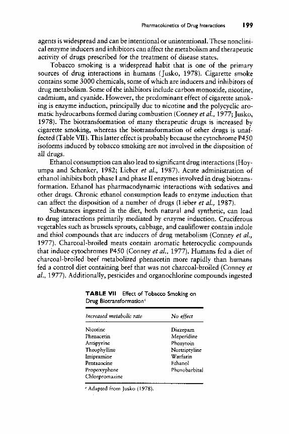
Pharmacokinetics of Drug Interactions I99
agents is widespread and can be intentional or unintentional. These nonclini- cal enzyme inducers and inhibitors can affect the metabolism and therapeutic activity of drugs prescribed for the treatment of disease states.
Tobacco smoking is a widespread habit that is one of the primary sources of drug interactions in humans (Jusko, 1978). Cigarette smoke contains some 3000 chemicals, some of which are inducers and inhibitors of drug metabolism. Some of the inhibitors include carbon monoxide, nicotine, cadmium, and cyanide. However, the predominant effect of cigarette smok- ing is enzyme induction, principally due to nicotine and the polycyclic aro- matic hydrocarbons formed during combustion (Conney et al., 1977; Jusko, 1978). The biotransformation of many therapeutic drugs is increased by cigarette smoking, whereas the biotransformation of other drugs is unaf- fected (Table VII). This latter effect is probably because the cytochrome P450 isoforms induced by tobacco smoking are not involved in the disposition of all drugs.
Ethanol consumption can also lead to significant drug interactions (Hoy- umpa and Schenker, 1982; Lieber et al., 1987). Acute administration of ethanol inhibits both phase I and phase I1 enzymes involved in drug biotrans- formation. Ethanol has pharmacodynamic interactions with sedatives and other drugs. Chronic ethanol consumption leads to enzyme induction that can affect the disposition of a number of drugs (Lieber et al., 1987).
Substances ingested in the diet, both natural and synthetic, can lead to drug interactions primarily mediated by enzyme induction. Cruciferous vegetables such as brussels sprouts, cabbage, and cauliflower contain indole and thiol compounds that are inducers of drug metabolism (Conney et al., 1977). Charcoal-broiled meats contain aromatic heterocyclic compounds that induce cytochromes P450 (Conney et al., 1977). Humans fed a diet of charcoal-broiled beef metabolized phenacetin more rapidly than humans fed a control diet containing beef that was not charcoal-broiled (Conney et al., 1977). Additionally, pesticides and organochlorine compounds ingested
TABLE VII Drug Biotransformation"
Increased metabolic rate No effect
Effect of Tobacco Smoking on
Nicotine Phenacetin Antipyrine Theophylline Imipramine Pentazocine Propoxyphene Chlorpromazine
Diazepam Meperidine Phenytoin Nortriptyline Warfarin Ethanol Phenobarbital
a Adapted from Jusko (1978).

200 Gregory L. Kedderis
in the diet or by other means of exposure can induce drug-metabolizing enzymes (Conney and Burns, 1972; Conney et al., 1977).
One dietary source of enzyme inhibitors is grapefruit juice, Bioflavinoids such as naringin in grapefruit juice can inhibit the metabolism of dihydropyr- idine calcium antagonists, cyclosporin, and caffeine (Bailey et a/., 1994). The interaction between grapefruit juice and the calcium antagonist felodip- ine can produce clinically significant increases in felodipine blood concentra- tion and decreases in diastolic blood pressure (Bailey et al., 1994).
Nutrition itself can modulate the metabolism of drugs and xenobiotics as many essential cofactors and constitutive compounds are obtained from food. A diet high in protein and low in carbohydrate has been shown to enhance the metabolic disposition of antipyrine, theophylline, and phenace- tin (Conney et al., 1977; Parke and Ioannides, 1981). Conversely, a low- protein diet can decrease the phase I clearance of many drugs (Hoyumpa and Schenker, 1982).
V. Drug Interactions Affecting Excretion
The urinary excretion of drugs involves glomerular filtration, tubular reabsorption, and tubular secretion. Drug interactions affecting excretion can occur during tubular reabsorption and secretion (Labaune, 1989). Be- cause only the unionized form of a drug is reabsorbed in the renal tubule, agents affecting urinary pH can modulate tubular reabsorption. For exam- ple, the thiazide diuretics and the carbonic anhydrase inhibitor acetazolam- ide can raise urinary pH, which facilitates the excretion of weak acids. The renal tubular secretion of drugs is often mediated by active transport systems for weak acids and weak bases. Two drugs that are transported by the same system can compete with each other. For example, probenecid inhibits the tubular secretion of the penicillins, prolonging their activity. This combina- tion therapy is also an example of a beneficial drug interaction.
VI. Drug Interactions during Liver Disease
Liver disease can have profound effects on drug metabolism (Hoyumpa and Schenker, 1982; Howden et al., 1989). During liver disease there are changes in hepatic cell mass and hepatic blood flow. Both of these effects contribute to a general decrease in drug clearance. The disposition of all drugs is not affected equally by liver disease (Table VIII). There are two major theories to explain the alterations in drug metabolism during liver disease. The sick- cell theory postulates that the reduced numbers of hepatocytes in the cirrhotic liver have an impaired metabolic capacity. The intact hepatocyte theory emphasizes decreased blood flow to the cells. Although evidence supports

Pharmacokinetics of Drug Interactions 20 I
TABLE Vlll Drug Disposition"
Effect of Liver Disease on
Significantly reduced Minimally affected
Diazepam Oxazepam Meprobamate Lorazepam Phenobarbital Chlorpromazine Glurethimide Acetaminophen Chlormethiazole Morphine Meperidine Methadone Salicylates Phenylbutazone Antipyrine Aminopyrine
a Adapted from Hoyumpa and Schenker (1982), with permission of Annual Reviews, Inc.
both theories, neither seems to adequately explain the changes in drug metabolism observed during liver disease (Howden et al., 1989).
VII. Genetic Factors in Drug Interactions
Individuals with particular genotypes are more prone to develop adverse reactions due to drug interactions (Vesell, 1974; Kiipfer and Preisig, 1983). Metabolic polymorphisms have been described for a variety of enzymes, some of which are involved in drug metabolism. Polymorphisms have been characterized in phase I enzymes, including cytochromes P450, flavin- containing monooxygenases, other oxidases and dehydrogenases, esterases, and epoxide hydrolases (Daly et al., 1993). Polymorphisms described in phase I1 enzymes include glucuronosyl transferases, sulfotransferases, acetyl- transferases, and methyltransferases (Daly et al., 1993). These polymor- phisms can lead to drug interactions and toxicity in the affected individuals that are not seen in the general population. Diphenylhydantoin toxicity is increased in slow acetylators of isoniazid compared to rapid acetylators (Vesell, 1974). Some drug interactions arise from polymorphisms in enzymes that are not directly involved in drug metabolism. For example, hemolysis is seen in response to a number of drugs in people with a glucose-6-phosphate dehydrogenase deficiency. Warfarin resistance is seen in individuals with abnormally tight binding of vitamin K (Vesell, 1974).
VIII. Beneficial Drug Interactions
Not all drug interactions result in adverse effects. Some of the enzyme- inducing and inhibiting effects of drugs can be used to therapeutic advantage.

202 Gregory L. Kedderis
For example, phenobarbital has been used to treat both unconjugated and neonatal hyperbilirubinemia, as this drug induces glucuronosyl transferase (Hunter and Chasseaud, 1976). Multiple drug therapy that takes advantage of synergistic or inhibitory effects of drug combinations is used to treat many conditions (Caranasos et al., 1985). The antimicrobial combination therapy of trimethoprim and sulfamethoxazole produces a synergistic inhibi- tion of bacterial folate synthesis. Penicillins and aminoglycosides are used in combination to produce synergistic bacteriostatic effects. Probenecid is administered with penicillin to reduce renal loss of the antibiotic. Carbidopa is coadministered with levodopa to inhibit dopa decarboxy-lase in peripheral tissues, allowing more of the drug to enter the brain. Additional examples of beneficial drug interactions include diuretic combinations to prevent pot- assium loss and antacid combinations to reduce constipation (Caranasos et al., 1985). Thus understanding the mechanisms underlying drug interactions is useful, not only in preventing drug toxicity, but also in devising safer, more effective therapies for diseases.
References
Bailey, D. G., Arnold, J. M. O., and Spence, J. D. (1994). Grapefruit pice and drugs: How significant is the interaction? Clin. Pharmacokinet. 26, 91-98.
Bolt, H. M., Bolt, M., and Kappus, H. (1977). Interaction of rifampicin treatment with pharma- cokinetics and metabolism of ethinyloestradiol in man. Acta Endocrinol. (Copenhagen)
Breckenridge, A. (1987). Enzyme induction in humans. Clinical aspects-an overview. Pharma- col. Ther. 33, 95-99.
Caranasos, G. J., Stewart, R. B., and Cluff, L. E. (1985). Clinically desirable drug interactions. Annu. Reii. Phamiacol. Tooxicol. 25, 67-95.
Connev, A. H. ( 1967). Pharmacological implications of microsomal enzyme induction. Pharma- col. Rev, 19, 317-366.
Conneg, A. H., and Burns, J. J. (1972). Metabolic interactions among environmental chemicals and drugs. Science 178, 576-586.
Conney, A. H., Pantuck, E. J., Hsiao, K.-C., Kuntzman, R., Alvares, A. P., and Kappas, A. (1977). Regulation of drug metabolism in man by environmental chemicals and diet. Fed. Proc. Fed. Am. Soc. Exp. Biol. 36, 1647-1652.
Cornish-Bowden, A. ( 1 986). Why is uncompetitive inhibition so rare? A possible explanation, with implications for the design of drugs and pesticides. FEBS Lett. 203, 3-6.
Daly, A. K. , Cholerton, S., Gregory, W., and Idle, J. R. (1993). Metabolic polymorphisms. Phamacol. Ther. 57,129-160.
Howden, C. W., Birnie, G. G., and Brodie, M. J. (1989). Drug metabolism in liver disease. Pharmacol. Ther. 40, 439-474.
Hoyumpa, A. M., Jr., and Schenker, S. (1982). Major drug interactions: Effect of liver disease, alcohol, and malnutrition. Aiinu. Rev. Med. 33, 113-149.
Hunter, J., and Chasseaud, L. F. (1976). Clinical aspects of microsonial enzyme induction. Prog. Drug Metab. 1, 129-191.
Jusko, W. J. (1978). Role of tobacco smoking in pharmacokinetics. 1. Pharmacokinet. Bio- phann. 6,7-39.
85, 189-197.

Pharmacokinetics of Drug Interactions 203
Kristensen, M. B. (1976). Drug interactions and clinical pharmacokinetics. Clin. Pharmacoki- net. 1, 351-372.
Kiipfer, A., and Preisig, R. (1983). Inherited defects of hepatic drug metabolism. Semin. Liver Dis. 3, 341-354.
Labaune, J. P. (1989). “Handbook of Pharmacokinetics: Toxicity Assessment of Chemicals,” pp. 569-600. Wiley, New York.
Lieber, C. S., Baraona, E., Leo, M. A., and Garro, A. (1987). Metabolism and metabolic effects of ethanol, including interaction with drugs, carcinogens and nutrition. Mutat. Res. 186, 201-233.
Nimmo, W. S. (1976). Drugs, diseases and altered gastric emptying. Clin. Pharmacokinet.
Nocke-Finck, L., Breuer, H., and Reimers, D. (1973). Wirkung von rifampicin auf den menstrua- tionszyklus und die ostrogenausscheidung bei einnahme oraler kontrazeptiva. Dtsch. Med. Wochenscbr. 98, 1521-1523.
Parke, D. V., and Ioannides, C. (1981). The role of nutrition in toxicology. Annu. Rev. Nutr.
Rowland, M., and Tozer, T. N. (1995). “Clinical Pharmacokinetics: Concepts and Applica- tions,” 3rd ed., pp. 267-284. Williams & Wilkins, Baltimore.
Segel, I. H. (1975). “Enzyme Kinetics: Behavior and Analysis of Rapid Equilibrium and Steady- State Enzyme Systems,” pp. 100-226. Wiley, New York.
Sjoholm, I., Ekman, B., Kober, A., Ljungstedt-Pihlman, I., Seiving, B., and Sjodin, T. (1979). Binding of drugs to human serum albumin: XI. The specificity of three binding sites as studied with albumin immobilized in microparticles. Mol. Pharmacol. 16, 767-777.
Vesell, E. S. (1974). The role of genetic factors in drug interactions. In “Drug Interactions” (P. L. Morselli, S. Garattini, and S. N. Cohen, eds.), pp. 181-198. Raven Press, New York.
Welling, P. G. (1986). “Pharmacokinetics: Processes and Mathamatics,” p. 70. American Chemical Society, Washington, DC.
Westley, A. M., and Westley, J. (1996). Enzyme inhibition in open systems: Superiority of uncompetitive agents. J. Biol. Chem. 271, 5347-5352.
1,189-203.
1,207-234.

Thomas N. Thompson Department of Drug Metabolism
North American Pharmacokinetics Hoechst Marion Roussel, Inc. Kansas City, Missouri 64 I 37
Experimental Models for Evaluating Enzyme Induction Potential of New Drug Candidates in Animals and Humans and a Strategy for Their Use
1. PurposelScope
The purpose of this chapter is twofold: (1) to provide a survey of various experimental models available for the evaluation of enzyme induction in animals and humans and (2) to propose a practical, stepwise strategy for evaluating the enzyme induction potential for drugs in development.
The experimental models presented here are described in order of in- creasing complexity. The main focus will be on models that have had wide application thus far. However, many new models that are currently being developed may have future applications in evaluating enzyme induction potential. Next, a strategy to evaluate the enzyme induction potential of drug candidates will be outlined. This strategy uses a combination of new and established techniques to evaluate data in a stepwise manner that is appropriate to the drug’s current stage of development. The mechanism of enzyme induction will not be addressed at all in this chapter and its signifi-
Advances b r Phamacoo(ogy, Volume 43 Copyright 0 1997 by Academic Press. All rights of reproduction in any form reserved. 1054-3589/97 $25.00 205

206 Thomas N. Thompson
cance to pharmaceutical development and a rationale for evaluating enzyme induction potential will be presented only in brief. The reader is directed to several other outstanding review articles that describe in detail the various classes of inducers (Conney, 1967; Okey, 1990; Snyder and Remmer, 1979), the molecular mechanism (Barry and Feely, 1990; Waxman and Azaroff, 1992; Okey, 1990; Okey et al., 1986; Bresnick et al., 1984; Gonzalez et al., 1993), and toxicological and clinical relevance of this phenomenon in more detail (Conney, 1967; Okey et al., 1986; Okey, 1990; Parke et al., 1991; Perucca, 1987; Park and Breckenridge, 198 1; Breckenridge, 1987; McInnes and Brodie, 1988; Gibaldi, 1991).
II. Introduction
Enzyme induction is a biological response to repeated exposure of a drug or other xenobiotic (Nebert, 1979). Specifically, enzyme induction is an increase in the amount of functional enzyme protein as a result of repeated exposure to the xenobiotic. The net result of enzyme induction is an increase in the capacity to metabolize the “offending” xenobiotic or that of other endogenous or exogenous drugskhemicals (Barry and Feely, 1990; Po- land, 1982).
When the term enzyme induction is used in the context of drug metabo- lisrdpharmacokinetics studies, it is most frequently taken to mean induction of hepatic microsomal enzymes, especially one or more of the isozymes of cytochromes P450 (CUP). It should be noted, however, that other non-CYP microsomal enzymes such as epoxide hydrolase (Batt et al., 1992; Thompson et al., 1982; Siest et al., 1988; Waxman and Azaroff, 1992), glucuronosyl transferase (Batt et al., 1992; Thompson et al., 1982; Siest et al., 1988; Waxman and Azaroff, 1992; Bock et al., 1990), carboxylester- ase (Heymann, 1980; Hosokawa et al., 1994), and even nonmicrosomal en- zymes such as glutathione transferase (Batt et al., 1992; Thompson et al., 1982; Waxman an Azaroff, 1992; Bock et al., 1990), N-acetyltransferase (Thompson et al., 1982), sulfotransferase (Thompson et al., 1982) y-gluta- myitransferase (Batt et al., 1992; Siest et al., 1988), aldehyde dehydrogenase (Waxman and Azaroff, 1992; Dunn et a/., 1988), and quinone reductase (Williams et al., 1986) can also be induced. Furthermore, enzyme induction is not limited to liver enzymes. Induction of enzymes in many other nonhepatic issues has been reported (Batt et al., 1992; Siest et al., 1988; Waxman and Azaroff, 1992; Gonney, 1967; Okey, 1990), including adrenal (Waxman and Azaroff, 1992; Conney, 1967), aorta endothelial cells (Stegeman et al., 1995), brain (Bhagwat et al., intestine (Waxman and Azaroff, 1992; Conney, 1967; Hietanen et al., 1880; Buchthal et al., 1995), kidney (Conney, 1967), leukocytes (Batt et al., 1992; Siest et al., 1988; Mahnke et al., 1996), lung (Conney, 1967; Stanley et al., 1992), placenta (Siest et al., 1988; Okey,

Evaluating Enzyme Induction Potential 207
1990), skin (Conney, 1967), testes (Conney, 1967), thymus (Conney, 1967) and thyroid (Conney, 1967).
As in other areas of pharmacokinetics and pharmacodynamics, there are pronounced species differences in the occurrence of enzyme induction. For historical and practical reasons, enzyme induction has been most often studied in rodents, especially rats and mice (Siest et al., 1988; Bock et al., 1990, Conney, 1967; Soucek and Gut, 1992). However, induction has been reported in many other mammalian species as well (Conney, 1967), including dogs (McKillop, 1985; Lan et al., 1983; Thompson et al., 1990), rabbits (Benedetti and Dostert, 1994; Gleizes et al., 1991), nonhuman primates (Lan et al., 1983; Bullock et al., 1995; Jones et al., 1992), and humans (Barry and Feely, 1990; Batt et al., 1992; Siest et al., 1988; Waxman and Azaroff, 1992; Okey, 1990; Okey et al., 1986), and even nonmammalian species, including birds (Okey, 1990), fish (Okey, 1990), invertebrates (Wax- man and Azaroff, 1992; Okey, 1990), bacteria, fungi, and yeast (Okey, 1990). Significant differences often exist between species in the potency of inducers and the magnitude of their effect (Lan et al., 1983; Thompson et al., 1990; Benedetti and Dostert, 1994; Kocarek et al., 1995).
Significantly, although literally hundreds of chemicals have been re- ported to be inducers in one or more animal species (Okey, 1990; Snyder and Remmer, 1979), far fewer ( - = G O ) have been demonstrated to be inducers in humans (Barry and Feely, 1990; Batt et al., 1992; Siest et al., 1988; Okey, 1990; Perucca, 1987; Park and Breckenridge, 1981; Parkinson, 1996), although this may be due as much to limited techniques for studying induc- tion in humans as it is to the lack of inducers. Even so, not all inducers of human enzymes have been shown to have a clinically relevant effect, perhaps as few as half that list (Perucca, 1987; Park and Breckenridge, 1981; McInnes and Brodie, 1988; Goldberg, 1980). Nonetheless, if a potential drug candi- date is an enzyme inducer in animals or humans, this may have an impact on its development, as described in Section 111.
111. Rationale for Evaluating Enzyme Induction Potential during Drug Development
A. Preclinical Significance
1. Efficacy
Enzyme induction in animals may have significance in the overall evalua- tion of pharmacology data. For example, in certain cases, a chronically administered drug induces the very enzymes that lead to its own increased metabolism (autoinduction). This leads to lower blood levels of the parent drug and increased levels of the metabolite(s) that may contribute to the

208 Thomas N. Thompson
development of tolerance in repeat dose pharmacology studies (Conney, 1967).
2. Sufety
Enzyme induction can also influence the outcome of toxicology studies. For example, if a drug is an autoinducer, during the course of the toxicology study the drug may alter its own clearance, leading to lower systemic expo- sure at the end of the study than during the initial phase. This lower systemic exposure of animal to the test drug must be taken into account when inter- preting the toxicology results (Takacs, 1995). Other effects associated with enzyme induction could also potentially influence the outcome of toxicology studies, although unequivocal correlations have not yet been established in many cases. For example, the effect of enzyme induction on levels of endoge- nous substances such as lipids, eicosonoids, and steroids has not been fully studied and may have implications for the outcome or interpretation of toxicology studies (Barry and Feely, 1990; Conney, 1967). Furthermore, it has been proposed that enzyme induction, which occurs relatively early (2-13 weeks) into a toxicology study, is associated with the later (i-e., 1 or 2 years) development of liver, thyroid, or other tumors (Paolini et ai., 1992; Griffin et al., 1995; Grasso et al., 1991; Shaw and Jones, 1994; Barter and Klaassen, 1994; Hill et a/., 1989; Ionanides and Parke, 1987, 1993). It should be pointed out that it is still unclear whether this is a causal or coincidental relationship (Grasso et al., 1991).
Enzyme induction by a xenobiotic has also been known to potentiate the toxicity of a second agent by inducing the formation of reactive intermediates {Okey, 1990; Paolini et al., 1992; Griffin et al., 1995; Ioannides and Parke, 1987). Moreover, induction is also thought to play a contributing role in carcinogenesis, both by activating procarcinogens (Ioannides and Parke, 1987,1993,) or by serving as a promoter (Ioannides and Parke, 1987,1993; Lubet et al., 1989; Rice et al., 1994). Again, it should be pointed out that an unequivocal causal relationship has not been established between induction and promotion of carcinogenesis (Rice et al., 1994).
B. Clinical Significance
1. Eficacy
If manifested in humans, enzyme induction may also have a significant effect on a potential drug’s efficacy profile. As was the case with animals, there are a limited number of cases where a drug induces its own metabolism (autoinduction) in a clinical setting. If the parent drug itself is the principal active species, this would diminish the expected effect. In contrast, if an active metabolite contributes more to the overall effect, there can actually be an increase in efficacy caused by autoinduction. Among the drugs reported to be autoinducers are rifamycin (Benedetti and Dostert, 1994), carbamazep-

Evaluating Enzyme Induction Potential 209
ine (Kudriakova et al., 1992; Bernus et af., 1994), ifosfamide (Boddy et al., 1995), and teniloxazine (Orlando et al., 1995).
The same principles just listed also apply in the presence of concomitant medications, and thus induction of drug-metabolizing enzymes can be a contributing factor in drug interactions. In this case, induction can alter the effect of concommitant medications by increasing the rate of their metabo- lism and hence their clearance. Examples of clinically significant drug interac- tions due to enzyme induction are numerous and have been well documented (Barry and Feely, 1990; Okey, 1990; Okey et al., 1986; Perucca, 1987; Park and Breckenridge, 198 1; McInnes and Brodie, 1988; Goldberg, 1980; Gibaldi, 1991; Grange et al., 1994; Venkatesan, 1992; Schrenzel et al., 1993; Fazio, 1991). In most of these cases, induction resulted in lower efficacy of the concomitant medication. In principle, induction could also result in increased efficacy if the concomitant medication exerted its effect via an active metabolite, although examples are few (Conney, 1967). Because of the potential ramifications of this, induction studies have a significant place as a class of drug metabolism studies that help define the potential for a drug interaction.
2. Safety
There are also many examples of how enzyme induction has led to adverse drug reactions in the clinical setting. An adverse drug reaction can be direct (i.e., caused by the inducing drug alone), most commonly as a result of affecting levels of endogenous substances (Barry and Feely, 1990; Conney, 1967; Perucca, 1987; Bammel et af., 1992; Isojarvi et af., 1994; Alderman and Hill, 1994). As was mentioned earlier for animals, it is not fully understood what the long-term implications are for subtle alterations in the levels of endogenous substances. In fact, there is reason to believe that enzyme induction in some cases may have a beneficial effect on levels of endogenous substances (Luoma, 1988; Franceschini et af., 1995).
As was true in animal studies, enzyme induction has also been known to cause adverse drug interactions in humans by inducing the formation of reactive intermediates (Okey, 1990; Ioannides and Parke, 1987). Moreover, induction is also thought to play a contributing role in human carcinogenesis by enhancing the activation of procarcinogens (Okey et al., 1986; Ioannides and Parke, 1987, 1993; Remmer, 1987; Kawajiri and Fujii-Kuriyama, 1991; Gonzalez and Gelboin, 1994). Again, it should be pointed out that an unequivocal causal relationship has not been established between induction and enhancement of carcinogenesis. In fact, the significance of enzyme induc- tion as a contributing factor to human carcinogenesis is currently being vigorously debated in the literature (Diaz et al., 1990; Farrell and Murray, 1990; Parkinson and Hurwitz, 1991). Furthermore, evidence also shows that induction may actually have a chemoprotective effect against carcinogenesis (Remmer, 1987; McLean, 1988; Talalay et al., 1995).

2 10 Thomas N. Thompson
IV. Models Available for the Evaluation of Enzyme Induction in Animals and Humans
A. Evaluation of Toxicology Data from Multiple Dose Studies
As part of the typical evaluation of data, toxicology departments track organ weight changes in their multiple dose studies. The first of these is the range-finding study, although any subsequent multiple dose study (such as the toxicokinetic study) is also suitable. An increase in the liver-to-body- weight ratio during one of these studies may be a sign of enzyme induction (1301and, 1982; Thompson et al., 1982; Conney, 1967; Snyder and Remmer, 1979; Howard et al., 1991). An increase of 10% or more is considered significant, although it should be noted that the magnitude of liver weight increase can depend on the inducer (Conney, 1967) or may be due to reasons other than induction.
In addition to tracking organ weight changes, there is a histological evaluation of tissue from each organ. If there is electron microscopic evidence of hepatocellular hypertrophy (caused by an increase in the smooth endoplas- mic reticulum), this is also a sign of enzyme induction (Conney, 1967; Snyder and Remmer, 1979; Howard et al., 1991).
8. Western Blot Assay for Induction Screening
There are examples of enzyme induction where there is little or no change, or perhaps only a very subtle change in liver weights (Poland, 1982), so it is always prudent to have a second line of evidence for lack of induction. The Western blot technique is a convenient second line screen (Laemmli, 1970; Arlotto et al., 1989; Ryan et al., 1979, 1980; Towbin et al., 1979; Thomas et af., 1984). Livers are obtained from rats a t all dose levels from any study in which the drug has been administered at various levels for more than 4 consecutive days. Most frequently, this would be any of the multiple-dose toxicology trials that are conducted during the course of drug development. Liver microsomes are prepared from the vehicle control (no drug) and high-dose groups and are screened for the presence of specific inducible isozymes using commercially available protein antibodies. If a particular specific isozyme is present in the microsomal protein, a visible protein-antibody complex is formed, the intensity of which is related to concentration. As described, this is only a qualitative observation, using vehicle control microsomes as the basis for comparison. However, Western blot analysis can be quantitative by use of densitometry or other means of measuring the intensity of the signal (Diaz et al., 1990; Guengerich et al., 1982; Shimada et a\., 1994).
Although the Western blot technique is a proven technique and provides very useful and well-accepted data, it is labor intensive and does not lend

Evaluating Enzyme Induction Potential 2 I I
itself well to high throughput. Furthermore, although this technique can be quantitative, this only exacerbates the previously described concerns. One can compensate for these limitations by selective screening and pooling of samples. However, other techniques are under investigation that may lend themselves to more facile quantitation and high throughput assessment of induction. One such techniques is an HPLC approach used to separate protein “fingerprint” bands (Iversen and Franklin, 1985). Another poten- tially promising technique is an ELISA assay for different CYP isozymes, as exemplified by Turner et al. (1994).
C. Ex Vivo Assays for Measuring Enzyme Induction in Rat Liver
This approach provides more direct evidence of induction than measur- ing liver weight increases or positive Western blot data. An example of the ex vivo approach might be as follows. Liver microsomes are prepared from all animals at all dose levels and the following assays are conducted:
1. Protein content of the microsomes is determined by the Bio-Rad/ Bradford method (Bradford, 1976) or by the method of Lowry (Lowry et at., 1951), and the amount of microsomal protein per gram of liver is calculated.
2. The cytochrome P450 assay (Omura and Sato, 1964) is performed to calculate the amount of total (i.e., all isozymes) P450 per gram of liver.
3. An aminopyrine-N-demethylase (APND) assay is conducted (Mazel, 1971; Nash, 1953). This colorometric assay is a nonspecific assay that detects induction of the multiple CYP isozymes: lA2,2A2,2El, 3A1 (Soucek and Gut, 1992; Imaoka et al., 1988; Poland and Kappas, 1971), and 2B1 (Soucek and Gut, 1992; Poland and Kappas, 1971).
If there is evidence of induction from these experiments, at least two more assays should be run:
4. An ethoxyresorufin-0-deethylase (EROD) assay is run. This is a fluorometric assay that is quite specific for induction of the CYPlAl and 2 isozymes (Burke and Mayer, 1974; Pohl and Fouts, 1980).
5. A pentoxyresorufin-0-deethylase assay is run. This is also a fluoro- metric assay related to the EROD assay, except that it is specific for CYP2B isozymes (Lubet et al., 1985).
This is a minimal testing scheme and includes isozyme-specific as- says only for the 1A and 2B classes of CYP isozymes. Addition of the p - nitrophenol hydroxylation assay for CYP2E1 (Koop, 1986), erythromycin- N-demethylase assay (Jacolot et al., 1991) for CYP3A, and the lauric acid hydroxylation assay for CYP4A1/3 (Romano et al., 1988; Swinney et al., 1991) would round out the list of the major classes of CYP isozymes induc-

2 I 2 Thomas N. Thompson
ible in rodents. Many other assays for both CYP and non-CYP inducible enzymes could be conducted, depending on the potency of' induction, timing for when information is needed, development stage of test compound, and level of concern over potential adverse effects and/or drug interaction po- tential.
With respect to other assays, most numerous are other assays for CYP isozymes. Nedelcheva and Guts (1994) have provided detailed tables com- plete with literature references that summarize the specificity of given sub- strates for the various CYP isozymes in both rats and humans. This allows the reader to select an assay with full knowledge of the isozyme specificity it provides. A subset of CYP assays is worth specific mention here because they permit the detection of multiple CYP isozymes either in a single assay or in a family of closely related assays. This list includes the testosterone hydroxylation assay (Dutton and Parkinson, 1989; Sonderfan et al., 1987), where the position of hydroxylation can give information on the CYP iso- zyme induction profile (Ryan and Levin, 1990). Another similar assay is the warfarin metabolite assay (Bush et a!., 1983), again where the position of metabolism gives information on the CYP profile of induction (Porter et al., 1981). A slightly different approach is provided by alkoxycoumarin (Fry et al., 1992) and alkoxyresorufin (Burke et al., 1994) assays, where a single product is measured (7-hydroxycoumarin or 7-hydroxyresorufin) but where selection of a particular alkyl ether as the substrate gives information on the isozyme induction profile.
There are a multitude of assays for non-CYP isozymes as well, Induction of glucuronosyl transferase was probed with multiple substrates ( Watkins et al., 1982), including naphthol (Bock et al., 1978), morphine (Bock et al., 1978), p-nitrophenol (Bock et al., 1973), chloramphenicol (Young and Lietman, 1978), bilirubin (VanRoy and Heirwegh, 1968), testosterone (Rao et al., 1976), valproic acid (Watkins and Klaassen, 1982), digitoxigenin monodigitoxoside (Castle, 1980), and phenolphthalein (Winsnes, 1969). Epoxide hydrolase induction was measured (Thompson et al., 1982) by the styrene oxide assay (Oesch et al., 1971). trans-Stilbene oxide hydrolysis can also be used to measure the induction of epoxide hydrolase (Waechter et al., 1988). Induction of glutathione-S-transferase activity was evaluated (Thompson et al., 1982) using 1-chloro-2,4-dinitrobenzene, 1,2-dichloro- 4-nitrobenzene, and sulfobromophthalein as substrates (Habig et al., 1974). Induction of N-acetyltransferase activity has been probed (Thompson et al., 1982) using p-aminobenzoic acid (Hearse and Weber, 1973; Glowinski et al., 1978), isoniazid (Weber, 1971), P-naphthylamine (Glowinski et al., 1978), and 2-aminofluorine (Glowinski et al., 1978) as substrates. Induction of sulfo- transferase was measured (Thompson et al., 1982) using dehydroepiandrost- erone (Sekura and Jakoby, 1979; Sekura et al., 1979), estrone (Adams and Poulos, 19671, sodium taurolithocholate (Chen et al., 1977), and 2-naphthol (Sekura and Jakoby, 1979) as substrates. Induction of carboxylesterases has

Evaluating Enzyme Induction Potential 2 13
been evaluated (Hosokawa et al., 1994) using p-nitrophenol acetate, isocar- boxazid, acetanilide, and butanilicaine (Hosokawa et al., 1987,1990).
D. Human Hepatocyte Model
The human hepatocyte model represents a comparatively recent and promising development in methodology for evaluating enzyme induction (Kocarek et al., 1995; Kostrubsky et al., 1995; Kreamer et al., 1986; Li et al., 1992, 1995a; Maekubo et al., 1982; Morel et al., 1990; Pichard et al., 1990). This technique offers the ability to evaluate enzyme induction in humans in a way that can be achieved early in preclinical development. In this model, human hepatocytes are isolated from one to three donors and are cultered in the presence of the putative inducer at three different concen- trations. After the prescribed induction period (usually 48 hr), the putative inducer drug is removed by a change of media. Selected enzyme activities are then assayed by incubating the induced hepatocytes in the presence of model substrates known to be metabolized by specific isozymes. For exam- ple, 1-hydroxylation of midazolam is a marker for CYP3A4,O-deethylation of 7-ethoxyresorufin is a marker for CYPlA2, and 4-hydroxylation of tolbu- tamide is a marker for CYP 2C8/9 (Parkinson, 1996). A more complete list of substrates for other CYP isozymes can be found in Rodrigues (1994). There are fewer examples of conjugation reactions validated in this model. Glucuronidation of 3’-azido-3’-deoxythimidine (AZT) (Nicolas et al., 1995) is the best example of a conjugation reaction catalyzed by human hepato- cytes. It is imperative that comparisons are made to relevant vehicle (nega- tive) control and positive control groups. Rifampicin can serve as a positive control for the induction of CYPs 2C9, 2C19, and 3A4 (Parkinson, 1996) whereas omeprazole can be used as a positive control for the induction of CYPlA2 (Diaz et al., 1990).
Although the human hepatocyte model is valuable, it is still a compara- tively new technique and there are some limitations that need to be consid- ered. First, as with any technique, the results obtained vary from laboratory to laboratory, depending on the exact conditions used. Culturing hepatocytes seems to be particularly sensitive to technique, as pointed out in a review calling for the standardization of techniques (Skett and Bayliss, 1996). Sec- ond, the number of known inducers, as well as the full complement of enzyme assays that have been used for human in vitro studies, has not yet been validated in the human hepatocyte model. A corollary to this statement is that while there is substantial experimental evidence validating induction results in animal hepatocytes to the corresponding in vivo induction results, such data are largely unavailable in humans because of obvious ethical considerations. A study done on induction by omeprazole provides a rare opportunity to directly compare induction in human hepatocytes with induc- tion in humans in vivo (Diaz et al., 1990).

2 14 Thomas N. Thompson
It should be pointed out that while these limitations may exist today, the obvious utility of the human hepatocyte model and enthusiasm for its use will undoubtedly drive further research to address these issues. Also, although the hepatocyte model is currently the best-studied in vitro model for human induction, other models such as liver slices (Lake et al., 1993) may also prove useful in the future.
E. Evaluation of Enzyme Induction in Humans in Vivo
I . Measurement of Endogenous Urinary Markers
This IS perhaps the simplest in uivo approach because it is a noninvasive procedure and can be added as an extra step to any clinical protocol that has a multiple dose regimen. In these studies, marker substances such as D-
glucaric acid (Hunter et al., 1971; Aarts, 1964, 1971; Hoyle et al., 1992; Sandstad et al., 1993; Rost et al., 1994) or 6P-hydroxy cortisol and cortisol (Ohnhaus and Park, 1979; Inoue et al., 1994; Durand et af., 1986; Ged et al., 1989; Bienvenu et al., 1991; Rost et al., 1994; Fleishaker et al., 1995) are present in minute amounts in the urine of uninduced subjects. After multiple drug treatment with an enzyme inducer, levels of these substances and/or the ratio of metabolite to parent tend to rise. D-Glucaric acid is the older of the two models and actually is directly indicative of induction of the glucuronic acid pathway (Hunter etal., 1971; Aart, 1964; 1971). Insofar as CYP isozymes are often coindufed with UDP-glucuronosyl transferase, an increase in D-glucuronic acid can also be taken as indirect evidence for CYP induction as well (Hoyle et al., 1992; Sandstad et al., 1993; Rost et al., 1994).
In contrast, increases in 6/3-hydroxycortisol levels are directly indicative of CYP induction (Ohnhaus and Park, 1979; Inoue et al., 1994). In fact, increases in the ratio of b~-hydroxycortisol to cortisol in urine are now known to be indicative of CYP3A4 induction (Durand et al., 1986; Ged et al., 1989; Bienvenu et al., 1991; Rost et al., 1994; Fleishaker et al., 1995).
Although these are the simplest of the in vim studies to run, there are issues of which to be aware. First, these assays do not provide a complete profile of induction. The D-glucaric acid assay is more directly connected with induction of glucuronic acid conjugation and oniy indirectly indicative of CYP induction. The 6~-hydroxycortisol/cortisol ratio has been well corre- lated to the induction of CYP3A4, but its relation to other CYP isozymes is less clear. Second, because constitutive levels of these endogenous markers are quite low and tend to vary widely among individuals, statistically signifi- cant changes are sometimes hard to achieve. The best results are achieved when urinary levels from drug-treated subjects are compared to predose levels, with each patient serving as hidher own control.
2. lnteraction with Marker Substrates
When marker drugs of known specificity are administered to patients already receiving repeat doses of the potential inducer drug, often a pharma-

Evaluating Enzyme Induction Potential 2 15
cokinetic drug interaction can be measured from plasma (or urine) levels of the marker drug. This approach is potentially much more precise than tracking endogenous urinary markers because a wealth of information is currently available about the specificity of certain substrates for human enzymes (Rodrigues, 1994; Brockmoller and Roots, 1994).
A typical protocol might be designed as follows. As was the case earlier for endogenous markers, due to high intersubject variability, each subject serves as his or her own predose control. The marker substrate is first administered alone before the test drug is given. Baseline levels of a character- istic metabolite or metabolites of the marker substrate are measured. Next, the test drug is administered alone for up to as much as 2 weeks in order to initiate induction. After a suitable number of doses, the marker drug is administered again, this time concomitantly with the test drug. After exten- sive plasma sampling, various pharmacokinetic parameters are measured. A significant change in selected pharmacokinetic parameters in the treatment group relative to the predose controls is interpreted as a sign of induction. Depending on the marker substrate employed, the information can either be a general index of induction of multiple CYP isozymes or can indicate induction of specific isozymes. The following is a list of marker substrates that have been typically used. Antipyrine (Barry and Feely, 1990; Engel et al., 1996) has been used as a nonspecific marker of induction. In contrast, caffeine is a specific marker for CYPlA1/2 (Watkins, 1990; Kalow and Tang, 1991; Rost et af., 1994), coumarin for CYP2A6 (Van Iersel et al., 1994), tolbutamide for CYP2C8/9 (Tassaneeyakul et al., 1992), omeprazole (Balian et af., 1995) or mephenytoin (Balian et af., 1995; Partovian et al., 1995) for CYP2C19, chlorzoxazone for CYP2E1 (Batt et al., 1992; Watkins, 1990; Peter et af., 1990; Kharasch et al., 1993), and midazolam for CYP3A4/ 5 (Thummel et af., 1994; Backman et af., 1996).
The COz breath tests represent a slight variation in approach to the studies just described. In this case, the marker drugs that are administered have N- or 0-methyl substituents that are labeled with either 13C or I4C; when metabolized, the label can be measured in expired air as labeled COz rather than urine or plasma (Barry and Feely, 1990; Baker et af., 1983; Lane and Parashos, 1986). When this measurement is made after treatment with a potential inducer drug, this test can be either a general or a specific index of induction, depending on the substrate. Common substrates used for breath tests include aminopyrine and antipyrine (Baker et al., 1983; Lane and Parashos, 1986) as nonspecific markers, caffeine (Watkins, 1990; Baker et af., 1983; Kotake et af., 1982; Rost et al., 1992; 1994) as a marker of CYPlA2 induction, or erythromycin as a marker of CYP3A4/S induction (Watkins, 1990; Watkins et af., 1990, 1992; Lown et al., 1995).
Although quite useful, interaction with marker drugs has its own limita- tions. For example, this approach is more involved than that described earlier for endogenous markers because a second drug substance must be administered along with the test drug. This tends to make approval of such
2 16 Thomas N. Thompson
studies by institutional review boards somewhat more complicated. Also, of necessity, these must be stand-alone protocols (as opposed to measuring a urinary marker) so they represent additional development time and expense. Finally, they require substantial resources to collect and analyze multiple plasma samples and to perform the detailed pharmacokinetic analysis of the resulting data. A variation of the approach just described, which requires only a single plasma sample, may offer an alternative (Bachmann and Jaure- gui, 1993). As a second alternative to measuring changes in pharmacokinetic parameters, measuring increased concentrations of the marker metabolite in plasma, either directly or as an increase in its ratio to parent drug levels, could also be used as an index of induction, although intersubject variability would have to be controlled.
F. Other Techniques
Although the techniques presented earlier are among the most commonly used experimental models, other techniques have also been used to measure enzyme induction in animals and humans. Many of these unique approaches have been summarized by Brockmoller and Roots (1 994).
In some cases, these other techniques deal with modification of a pre- viously used analytical method. An example of one such approach involved the use of 'H or I3C NMR spectroscopy to detect changes in D-glucaric acid levels instead of the usual HPLC measurement (Hoyle et al., 1992). In other cases, a completely different approach to quantifying induction has been utilized. One such new approach involves detection and isolation of circulat- ing antiliver microsomal antibodies from dihydralizine-treated patients. When these antibodies were found to react specifically with CYP1A2, this was interpreted as evidence for induction of this CYP isozyme (Bourdi et al., 1992).
Another example of an alternate approach to measuring induction is exemplified by the analysis of messenger RNA (mRNA) coding for inducible enzyme proteins. This approach is particularly significant because it has the potential for providing a much more sensitive index of induction than any of techniques described previously. In a clinical setting, this is important because it would allow the detection of induction in tissues much more accessible than the liver (such as blood lymphocytes), which in turn allows a more sensitive and convenient assay of induction. In a nonclinical setting, if applied to analysis of liver microsomes or hepatocytes in place of the assays described earlier it may allow differentiation of compounds that cause negligible induction from those that cause weak or intermediate induction. Alternately, because of its sensitivity, this technique can be used to detect induction in other tissues not previously able to be studied. Reports of the use of mRNA measurement are now common. For example, levels of the mRNA for CYPlA1/2 have been probed by Northern blot analysis (Diaz

Evaluating Enzyme Induction Potential 2 17
et al., 1990), slot-blot analysis (Dragnev et al., 1995; Nims et al., 1992), or reverse-transcription polymerase chain reaction (Omiecinski et al., 1990).
V. A Strategy for Evaluation of Enzyme Induction Potential of Drugs in Development
Figure 1 depicts a flow diagram of a strategy for evaluating the enzyme induction potential of drugs in development. This strategy is multitiered and gets progressively more detailed as the drug advances through development.
The first level of evaluation is to carefully review all data from multiple- dose toxicology studies (typically in rats first, and later in dogs and/or any other species available) for signs of enzyme induction. Enzyme induction might manifest itself as a increase in the liver-to-body weight ratio (usually 10% or more). Alternately, histopathological examination of liver tissue from the animals in the toxicology study might reveal hepatocellular hyper- trophy. Both of these observations are considered evidence of enzyme induc- tion. It should be noted that while studies in rats usually provide the first and most readily available indication of induction, there have been cases where rats were a nonresponsive species (Benedetti and Dostert, 1994), so care must be taken to continuously monitor data from multiple dose studies in all subsequent species tested. In this regard, the reader is reminded that evidence of induction can also be inferred from other multiple dose animal studies that are not necessarily toxicology studies. For example, if tolerance for the observed pharmacological effect develops after multiple admistration, this can be a sign of autoinduction and would merit additional studies to define whether induction is observed.
The second level of evaluation for induction will vary, depending on whether the first level showed signs of induction. As shown in Fig. 1, if there is no evidence of induction from toxicology studies, a simple induction screen is conducted in order to provide direct experimental evidence for lack of induction. For this purpose, one could utilize the Western blot technique for analyzing liver microsomal protein from the control and high- dose groups. If there is evidence of induction by this technique, the other intermediate dose groups should also be examined. Lack of any evidence of enzyme induction by this technique is considered a stopping point for evaluation of induction, at least until such a time as studies in other species suggest liver weight increases.
A second course of action is taken if the descriptive evaluation of data at level 1 reveals some evidence of induction or, alternately, if evidence of induction arises from the Western blot screen. In either case, an ex vivo assessment of enzyme induction is undertaken. For this assessment, livers are obtained from rats at all dose levels in a multiple dose toxicology study and microsomes are prepared as described earlier. Cytochrome P450 concen-

2 18 Thomas N. Thompson
Strategy for Enzyme induction I
2
L E v E L
1
Evaluate existing Tox data for single species
NO
Induction Sereen 'I YES
Uver ex vivo studieb
YES
t L Human hepatocyie study E v E NO L
3
V
I
-^ L"
L E v E L
4
Human in vivo induction studies
NO
plan other drug
Use data for part
FIGURE I Flow chart for enzyme induction studies.
tration and microsomal protein yield are determined, followed by assaying selected CY P enzyme activities. This provides direct, quantitative evidence of the degree of induction of each dose group relative to a vehicle (negative) control group. Typically, these values would also be compared to those from a positive control treatment group such as phenobarbital treatment at 75 mg/kg/day for 4-7 days. Evidence of potent induction in rats or evidence

Evaluating Enzyme Induction Potential 2 19
of liver weight increases in other species will usually warrant evaluation of induction by other toxicology species such as dog or monkey. The screening paradigm for these species proceeds as described earlier for the rat. If, after this second level of evaluation, there is little or no significant degree of induction in animals, no further action is taken, as indicated in Fig. 1.
However, if the ex vivo studies indicate induction, a level 3 evaluation is undertaken. In this level, it is important to establish for the first time whether the drug is an enzyme inducer in humans. Because of cost, timing, and regulatory requirements, the evaluation of enzyme induction in humans is initially conducted in vitro using a human hepatocyte induction model.
Finally, evidence of enzyme induction in the human hepatocyte model may warrant moving to a level 4 evaluation of induction. In this level, enzyme induction is evaluated in vivo in humans. The simplest evaluation of induction in humans in vivo is to measure certain endogenous marker substances in the urine of patients in any multiple dose clinical trial. Elevation of these levels after multiple dosing relative to predose levels is an indication of induction.
Alternately, if additional or more specific enzyme induction data are required, marker substrates specific for various CYP isozymes can be coad- ministered along with the test drug. Increased levels of metabolites of these marker substrates in the urine after multiple dosing relative to levels found prior to dosing with the test drug indicate induction of a specific CYP isozyme. Along this same principle, breath tests have been developed whereby increased levels of 13C or I4C measured in the breath of subjects after multiple administration of the test drug are an indication of induction. The decision for this level of evaluation would be made only after there is strong evidence from other studies or if the compound class warrants it, such as might be the case for an anticonvulsant.
VI. Summary
Experimental models that have application for evaluating enzyme induc- tion potential have been described in order of increasing complexity. The main focus was on models that have had wide application thus far. However, many new models are currently being developed that may have future appli- cations in evaluating enzyme induction potential. A strategy to evaluate the enzyme induction potential of drug candidates was outlined. This scheme uses a combination of new and established techniques to evaluate data in a stepwise manner that is appropriate to the drug’s current stage of devel- opment.
Acknowledgments
The author acknowledges the contributions of Lisa Connor, Larry Cheng, Julie Geary, John Ling, and Kelly Reith, who have first-hand experience with many of the models presented

220 Thomas N. Thompson
here. It was by discussions with these scientists that the strategy proposed for evaluating enzyme induction potential was derived. Also, the help of Kathy Paul, Sheila Carter, and Marlea Fritts in the preparation of this manuscript is greatly appreciated.
References
Aarts, E. M. (1964). Evidence for the function of D-glucaric acid as an indicator for drug induced enhanced metabolism through the glucuronic acid pathway in man. Biochem. Pharmacol. 14, 359-363.
Aarts, E. M. (1971). 0-glucaric-acid excretion as a test for hepatic enzyme induction. Lancet 1(704), 859.
Adams, J. B., and Poulos, A. (1967). Enzymatic synthesis of steroid sulphates. 111. Isolation and properties of estrogen sulphotransferase of bovine adrenal glands. Biochim. Biophys.
Alderman, C. P., and Hill, C. I-. (1994). Abnormal bone mineral metabolism after long-term anticonvulsant treatment. Ann. Phannacofher. 28, 47-48.
Arlotto, M. P., Greenway, D. J., and Parkinson, A. (1989). Purification of two isozymes of rat liver microsomal cytochrome P450 with testosterone 7;ga-hydroxylase activity. Arch. Biochem. Biophys. 270, 44 1-457.
Bachmann, K. A., and Jauregui, 1.. (1993). Use of single sample clearance estimates of cyto- chrome P450 substrates to characterize human hepatic CYP status in vivo. Xenobiotica
Backman, J. T., OlkkoIa, K. T., and Neuvonen, P. J. (1996). Rifampin drastically reduces plasma concentrations and effects of oral midazolam. Clin. Pbannacol. Ther. 59, 7-13.
Baker, A. L., Kotake, A. N., and Schoeller, D. A. (1983). Clinical utility of breath tests for the assessment of hepatic function. Semirz. Liver Dis. 3(4), 318-329.
Balian, J. D., Sukhova, N., Harris, J. W., Hewett, J., Pickle, L., Goldstein, J. A., Woosley, R. L., and Flockhart, D. A. (1995). The hydroxylation of omeprazole correlates with S - mephenytoin metabolism: A population study. Clin. Pharmacol. Tber. 56(6), 662-669.
Bammel, A., van der JMee, K., Ohnhaus, E. E., and Kirch, W. (1992). Divergent effects of different enzyme-inducing agents on endogenous and exogenous testosterone. Eur. /. Clin. Pharrnacol. 42, 641-644.
Barry, M., and Freely, J. (1990). Enzyme induction and inhibition. Pharmacol. Ther. 48,71-94. Barter, R. A., and Klaassen, C. D. (1994). Reduction of thyroid hormone levels and alteration
of thyroid function by four representative UDP-glucuronosyltransferase inducers in rats. Toxicol. Appl. Pharmacol. 128, 9- 17.
Batt, A. M., Siest, G., Magdalou, J., and Galteau, M. M. (1992). Enzyme induction by drugs and toxins. Clin. Chim. Acta 209, 109-121.
Benedetti, M. S., and Dostert, P. (1994). Induction and autoinduction properties of rifamycin derivatives: A review of animal and human studies. Environ. Health Perspect. 102(9),
Bernus, I., Dickinson, R. G., Hooper, W. D., and Eadie, M. J. (1 994). Early stage autoinduction of carbamazepine metabolism in humans. Eur. I . Clin. Pharmacol. 47, 355-360.
Bhagwat, S. V., Boyd, hf. R., and Ravindranath, V. (1995). Brain mitochondria1 cytochromes P450: Xenobiotic metabolism, presence of multiple forms and their selective inducibility. Arch. Biochem. Biophys. 320( I ) , 73-83.
Bienvenu, T., Rey, E., Pons, G., DAthis, P., and Olive, G. (1991). A simple non-invasive procedure for the investigation of cytochrome P-450 IlIA dependent enzymes in humans. lnt. I. Clin. Pharrnacol. Ther. Toxicol. 29, 441-445.
Act6 146, 493-508.
23(3), 307-315.
101-105.

Evaluating Enzyme Induction Potential 22 I
Bock, K. W., Frohling, W., Remmer, H., and Rexer, B. (1973). Effects of phenobarbital and 3-methylcholanthrene on substrate specificity of rat liver microsomal UDP-glucuronyltran- sferase. Biochim. Biophys. Acta 327, 46-56.
Bock, K. W., Brunner, G., Hoensch, H., Huber, E., and Josting, D. (1978). Determination of microsomal UDP-glucuronyl-transferase in needle-biopsy specimens of human liver. Eur. J. Clin. Pharmacol. 14, 367-373.
Bock, K. W., Lipp, H. P., and Bock-Hennig, B. S. (1990). Induction of drug-metabolizing enzymes by xenobiotics. Xenobiotica 20, 1101-111 1.
Boddy, A. V., Cole, M., Pearson, A. D. J., Idle, J. R. (1995). The kinetics of the auto-induction of ifosfamide metabolism during continuous infusion. Cancer Chemother. Pharmacol.
Bourdi, M., Gautier, J. C., Mircheva, J., Larrey, D., Guillouzo, A,, Andre, C., Belloc, C., and Beaune, P. H. (1 992). Anti-liver microsomes autoantibodies and dihydralazine-induced hepatitis: Specificity of autoantibodies and inductive capacity of the drug. Mol. Phamacol.
Bradford, M. M. (1976). A rapid and sensitive method for the quantitation of microgram
36,53-60.
42,280-285.
quantities of protein utilizing the principle of protein-dye binding. Anal. Biocheml 72, 248-254.
Breckenridge, A. (1987). Enzyme induction in humans-clinical aspects-an overview. Pharma- col. Ther. 33, 95-99.
Bresnick, E., Foldes, R., and Hines, R. N. (1984). Induction of cytochrome P450 by xenobiotics. Pharmacol. Rev. 36(2), 43s-S1S.
Brockmoller, J., and Roots, I. (1994). Assessment of liver metabolic function-clinical implica- tions. Clin. Phamacokinet. 27(3), 216-248.
Buchthal, J., Grund, K. E., Buchmann, A., Schrenk, D., Beaune, P., and Bock, K. W. (1995). Induction of cytochrome P4501A by smoking or omeprazole in comparison with UDP- glucuronosyltransferase in biopsies of human duodenal mucosa. Eur. J. Clin. Pharmacol.
Bullock, P., Pearce, R., Draper, A., Podval, J., Bracken, W., Veltman, J., Thomas, P., and Parkinson, A. (1995). Induction of liver microsomal cytochrome P450 in cynomolgus monkeys. Drug Metab. Dispos. 23(7), 736-748.
Burke, M. D., and Mayer, R. T. (1974). Ethoxyresorufin: Direct fluorometric assay of a microsomal 0-dealkylation which is preferentially inducible by 3-methylcholanthrene. Drug Metab. Dispos. 2, 583-588.
Burke, M. D., Thompson, S., Weaver, R. J., Wolf, C. R., and Mayer, R. T. (1994). Cytochrome P450 specificities of alkoxyresorufin o-dealkylation in human and rat liver. Biochem. Pharmacol. 48(5), 923-936.
Bush, E. D., Low, L. K., and Trager, W. F. (1983). A sensitive and specific stable isotope assy for warfarin and its metabolites. Biomed. Mass Spectrom. 10(7), 395-398.
Castle, M. C. (1980). Glucuronidation of digitalis glycosides by rat liver microsomes: Stimula- tion by spironolactone and pregnenolone-16;ga-carbonitrile. Biochem. Pharmacol. 29,
Chen, L. J., Bolt, R. J., and Admirand, W. H. (1977). Enzymatic sulfation of bile salts. Partial purification and characterization of an enzyme from rat liver that catalyzes the sulfation of bile salts. Biochim. Biophys. Actu 480, 219-227.
Conney, A. H. (1967). Pharmacological implications of microsomal enzyme induction. Phama- col. Rev. 19(3), 317-366.
Diaz, D., Fabre, I., Daujat, M., Saint Aubert, B., Bories, P., Michel, H., and Maurel, P. (1990). Omeprazole is an aryl hydrocarbon-like inducer of human hepatic cytochrome P450. Gastroenterology 99, 737-747.
Dragnev, K. H., Nims, R. W., Fox, S. D., Lindahl, R., and Lubet, R. A. (1995). Relative potencies of induction of hepatic drug-metabolizing enzyme genes by individual PCB congeners. Toxicol. Appl. Pharmacol. 132(2), 334-342.
47,431-435.
1497-1502.

222 Thomas N. Thompson
Dunn, T. J., Lindahl, R., and Pitot, H. C. (1988). Differential gene expression in response to 2,3,7,8-tetrachlorodibenzo-p-dioxin (TCDD). Noncoordinate regulation of a TCDD- induced aldehyde dehydrogenase and cytochrome P-45Oc in the rat. J . Biol. Chem.
Durand, D., Hampden, C., Boobis, A. R., Park, B. K., and Davies, D. S. (1986). Induction of mixed function oxidase activity in man by rifapentine (MDL 473), a long-acting rifamycin derivative. Br. J . Clin. Pharmacol. 21, 1-7.
Dutton, D. R., and Parkinson, A. (1989). Reduction of 7-alkoxyresorufins by NADPH- cytochrome P450 reductase and its differential effects on their 0-dealkylation by rat liver microsomal cytochrome P450. Arch. Biochem. Biophys. 268, 617-629.
Engel, G., Hofmann, U., Heidemann, H., Cosme, J., and Eichelbaum, hl. (1996). Antipyrine as a probe for human oxidative drug metabolism: Identification of the cytochrome P450 enzymes catalyzing 4-hydroxyantipyrine, 3-hydroxymethylantipyrine, and norantipyrine formation. Clin. Pharmacol. Ther. 59, 61 3-623.
Farrell, G. C., and Murray, M. ( 1990). Human cytochrome P450 Isoforms-their genetic heterogeneity and induction by omeprazole. Gastroenterology 99, 885-889.
Fazio, A. ( 199 1 ). Oral contraceptive drug interactions: Important considerations (see comments) (review). South. Med. J. 84(8), 997-1002.
Fleishaker, J. C., Pearson, L. K., and Peters, G. R. (1995). Phenytoin causes a rapid increase in 6;gb-hydroxycortisol urinary excretion in humans-a putative measure of CYP3A induction. J . P h a m . Sci. 84(3), 292-294.
Franceschini, G., Werba, J. P., D’Acquarica, A. L., Gianfranceshi, G., Michelagnoli, S., and Sirtori, C . R. (1995). Microsomal enzyme inducers raise plasma high-density lipoprotein cholesterol levels in healthy control subjects but not in patients with primary hypoalphali- poproteinemia. Clin. Pharmacol. Ther. 57,434-440.
Fry, J. R., Garle, AM. J., and Lal, K. (1992). Differentiation of cytochrome P-450 inducers on the basis of 7-alkoxycoumarin 0-dealkylase activities. Xenobioticu 22(2), 21 1-215.
Ged, C., Rouillon, J. M., Pichard, L., Combalbert, J., Bressot, N., Bories, P., Michel, H., Beaune, P., and Maurel, P. (1989). The increase in urinary excretion of 6;gb-hydroxycortisol as a marker of human hepatic cytochrome P45OIIIA induction. BY. J. Clin. Phamacol.
Gibaldi, M. ( 1991). Induction of drug-metabolizing enzymes. Perspect. Clin. Pharm. 9(6),
Gleizes, C., Eeckhoutte, C., Pineau, T., Alvinerie, M., and Galtier, P. (1991). Inducing effect of oxfendazole on cytochrome P450IA2 in rabbit liver. Consequences on cytochrome P450 dependent monooxygenases. Biochem. Pharmacol. 41(12), 1813-1820.
Glowinski, I. B., Radtke, H. E., and Weber, W. W. (1978). Genetic variation in N-acetylation of carcinogenic arylamines by human and rabbit liver. Mol. Phamacol. 14, 940-949.
Goldberg, D. M. (1980). The expanding role of microsomal enzyme induction, and its implica- tions for clinical chemistry. Clin. Chem. 26(6), 691-699.
Gonzalez, F. J., and Gelboin, H. V. (1994). Role of human cytochromes 1’450 in the metabolic activation of chemical carcinogens and toxins. Drug Metab. Rev. 26( 1&2), 165-183.
Gonzalez, F. J., Liu, and S. Y., Yano, M. (1993). Regulation of cytochrome P450 genes: Molecular mechanisms. Pharmacogenetics 3 , 51 -57.
Grange, J. M., Winstanley, P. A., and Davies, P. D. 0. (1994). Clinically significant drug interactions with antituberculosis agents. Drug Saf. 11(4), 242-2.51.
Grasso, P., Sharratt, M., and Cohen, A. J, (1991). Role of persistent, non-genotoxic tissue damage in rodent cancer and relevance to humans. Annu. Rev. Pharmacol. Toxicol.
Griffin, R. J., Burka, L.T., a.nd Cunningham, M. L. (1995). Activity of hepatic drug metabolizing enzymes following oxazepam-dosed feed treatment in B6C3F1 mice. Toxicol. Lett. 76,
263(22), 10878-10886.
28, 373-387.
41-46.
31,253-287.
251-256.

Evaluating Enzyme Induction Potential 223
Guengerich, F. P., Wang, P., and Davidson, N. K. (1982). Estimation of isozymes of microsomal cytochrome P-450 in rats, rabbits, and humans using immunochemical staining coupled with sodium dodecyl sulfate-polyacrylamide gel electrophoresis. Biochemistry 21, 1698- 1706.
Habig, W. H., Pabst, M. J., and Jakoby, W. B. (1974). Glutathione transferases: The first step in mercapturic acid formation. J . Biol. Chem. 249, 7130-7139.
Hearse, D. J., and Weber, W. W. (1973). Multiple N-acetyltransferases and drug metabolism, tissue distribution, characterization and significance of mammalian N-acetyltransferase. Biochem. J. 132, 519-526.
Heymann, E. (1980). Carboxylesterases and amidases. In “Enzymatic Basis of Detoxication” (W. B. Jakoby, ed.) Academic Press; Vol. 2, pp. 291-323, New York.
Hietanen, E., Laitinen, M., and Koivusaari, U. (1980). Effect of administration route of 3- methylcholanthrene on the inducibility of intestinal drug-metabolizing enzymes. Enzyme
Hill, R. N., Erdreich, L. S., Paynter, 0. E., Roberts, P. A., Rosenthal, S. L., and Wilkinson, C. F. (1989). Thyroid follicular cell carcinogenesis. Fundam. Appl. Toxicol. 12(4),
Hosokawa, M., Maki, T., and Satoh, T. (1987). Multiplicity and regulation of hepatic micro- soma1 carboxylesterases in rats. Mol. Pharmacol. 31, 579-584.
Hosokawa, M., Maki, T., and Satoh, T. (1990). Characterization of molecular species of liver microsomal carboxylesterases of several animal species and humans. Arch. Biochem. Biophys. 277, 219-227.
Hosokawa, M., Hirata, K., Nakata, F., Suga, T., and Satoh, T. (1994). Species differences in the induction of hepatic microsomal carboxylesterases caused by dietary exposure to di(2- ethlhexyl)phthalate, a peroxisome proliferator. Drug Metab. Dispos. 22(6), 889-894.
Howard, M. O., Schwartz, L. W., Newton, J. F., Qualls, C. W., Yodis, L. A. P., and Ventre, J. R. (1991). Comparative biochemical and morphometric changes associated with induc- tion of the hepatic mixed function oxidase system in the rat. Toxicol. Pathol. 19(2),
Hoyle, V. R., Gilbert, P. J., Troke, J. A., Vose, C. W., and Nicholson, J. K. (1992). Studies on the biochemical effects of the aldose reductase inhibitor 2,7-difluorospirofluorene- 9,5’-imidazolidine-2’,4’-dione (A1 1576, HOE 843). Detection of D-glucaric and D- glucuronic acid excretion by high resolution 1H and 13C NMR spectroscopy. Biochem. Pharmacol. 44(2), 231-241.
Hunter, J., Maxwell, J. D., Carrella, M., Stewart, D. A., and Williams, R. (1971). Urinary D- glucaric-acid excretion as a test for hepatic enzyme induction in man. Lancet 1(699),
Imaoka, S., Inoue, K., and Funae, Y. (1988). Aminopyrine metabolism by multiple forms of cytochrome P-450 from rat liver microsomes: Simultaneous quantitation of four aminopy- rine metabolites by high-performance liquid chromatography. Arch. Biochem. Biophys. 265(1), 159-170.
Inoue, S., Inokuma, M., Harada, T., Shibutani, Y., Yoshitake, T., Charles, B., Ishida, J., and Yamaguchi, M. (1994). Simultaneous high performance liquid chromatographic determi- nation of 6P-hydroxycortisol and cortisol in urine with fluorescence detection and its application for estimating hepatic drug-metabolizing enzyme induction. J. Chromatogr. B: Biomed. Appl. 661, 15-23.
Ioannides, C., and Parke, D. V. (1987). Commentary: The cytochromes P-448-a unique family of enzymes involved in chemical toxicity and carcinogenesis. Biochem. Phamacol.
Ioannides, C., and Parke, D. V. (1993). Induction of cytochrome P4501 as an indicator of potential chemical carcinogenesis. Drug Metab. Rev. 25(4), 485-501.
Isojarvi, J. I. T., Pakarinen, A. J., Rautio, A., Pelkonen, O., and Myllyl~, V. V. (1994). Liver enzyme induction and serum lipid levels after replacement of carbamazepine with oxcarbazepine. Epilepsia 35(6), 1217-1220.
25, 153-157.
629-697.
115-122.
572-575.
36(24), 4197-4207.
224 Thomas N. Thompson
Iversen, P. L., and Franklin, M. R. (1985). Microsomal cytochrome P-450 “handprints”: Five fractions from anion-exchange high-pressure liquid chromatography provide a rapid preliminary screen for selectivity in the induction and destruction of rat hepatic cytochrome P-450 subpopulations. Toxicol. Appl. Pharmacol. 78, 1-9.
Jacolot, F., Simon, I., Dreano, Y., Beaune, P., Riche, C., and Berthou, F. ( 1 991). Identification of the cytochrome P-150 IIIA family as the enzymes involved in the N-demethylation of tamoxifen in human liver microsomes. Biochem. Pharmacol. 41, 1911-1919.
Jones, C. R., Cuengerich, F. P., Rice, J. M., and Lubet, R. A. (1992). Induction of various cytochromes CYPZB, CYP2C and CYP3A by phenobarbitone in non-human primates. Pharmacogenetics 2, 160-172.
Kalow, W., and Tang, B. K. (1991). Caffeine as a metabolic probe: Exploration of the enzyme- inducing effect of cigarette smoking. Clin. Pharmacol. Ther. 49, 44-48.
Kawajiri, K., and Fujii-Kurijama, Y. (1991). P450 and human cancer. Jpn. I . Cancer Res.
Kharasch, E. D., Thummel, K. E., Mhyre, J., and Lillibridge, J. H. (1993). Single-dose disulfiram inhibition of chlorzoxazone metabolism: A clinical probe for P450 2E1. Clin. Pharmacol. Ther. 53(6), 643-650.
Kocarek, T. A., Schuetz, E. G., Strom, S. C., Fisher, R. A., and Guzelian, P. S. (1995). Comparative analysis of cytochrome P4503A induction in primary cultures of rat, rabbit, and human hepatocytes. Drug Metab. Dispos. 23(3), 415-421.
Koop, D. R. ( 1 986). Hydroxylation of para-nitrophenol by rabbit ethanol inducible cytochrome P-450 isozyme 3A. Mol. Pharmacol. 29, 399-404.
Kosrrubsky, V. E., Strom, S . C., Wood, S. G., Wrighton, S . A., Sinclair, P. R., and Sinclair, J. F. (1995). Ethanol and isopenranol increase CYP3A and CYPZE in primary cultures of human hepatocytes. Arch. Biochem. Biophys. 322, 516-520.
Kotake, A. N., Schoeller, D. A., Lambert, G. H., Baker, A. L., Schaffer, D. D., and Josephs, H. (1982). The caffeine C 0 2 breath test: Dose response and route of N-demethylation in smokers and nonsmokers. Clin. Phamacol. Tber. 32(2), 261-269.
Kreamer, 8. L., Staecker, J. L., Sawada, N., Sattler, G. L., Hsia, M. T. S . , and Pitot, H. C. (1986). Use of a low-speed iso-density Percoll centrifugation method to increase the viability of isolated hepatocyte preparations. In Vitro 22, 201.
Kudriakova, T. B., Sirota, L. A., Rozova, G. I., and Gorkov, V. A. ( 1 992). Autoinduction and steady-state pharmacokinetics of carbamazepine and its major metabolites. Br. I . Clin. Pharmacol. 33, 61 1-615.
Laemmli, U. K. (1970). Cleavage of structural proteins during the assembly of the head of bacteriophage T4. Nature (London) 227, 680-685.
Lake, B. G., Beamand, J. A., Japenga, A. C., Renwick, A., Davies, S., and Price, R. J. (1993). Induction of cytochrome P-450-dependent enzyme activities in cultured rat liver slices. Food Chem. Toxicol. 31(5), 377-386.
Lan, S. J., Weinstein, S. H., Keim, G. R., and Migdalof, B. H. (1983). Induction of hepatic drug-metabolizing enzymes in rats, dogs, and monkeys after repeated administration of an anti-inflammatory hexahydroindazole. Xenobiotica 13(6), 329-335.
Lane, E. A., and Parashos, 1. (1986). Drug pharmacokinetics and the carbon dioxide breath test. J. Phamacokinet. Biopham. 14( l), 29-49.
L.i, A. P., Roque, M. A., Beck, D. J., and Kaminski, D. L. (1992). Isolation and culturing of hepatocytes from human liver. 1. Tissue Cult. Methods 14, 139-146.
Li, A. P., Rasmussen, A,, Xu, L., and Kaminiski, D. L. (199Sa). Rifampincin induction of lidocaine metabolism in cultured human hepatocytes. 1. Phamacol. Exp. Ther. 274,
Li, A. P., Kaminski, D. t., and Rasmussen, A. ( 1 995b). Substrates of human hepatic cyrochrome P450 3A4. Toxicology 104, 1-8.
Lown, K. S . , Thummel, K. E., Benedict, P. E., Shen, D. D., Turgeon, D. K., Berent, S . , and Watkins, P. B. (1995). Pharmacokinetics and drug disposition. The erythromycin breath test predicts the clearance of midazolam. Clin. Pbamacol. Ther. 57, 16-24.
82, 1325-1335.
673-677.
Evaluating Enzyme Induction Potential 225
Lowry, 0. H., Rosebrough, J., Farr, A. L., and Randall, R. J. (1951). Protein measurement with the Folin phenol reagent. J. Biol. Chem. 193, 265-275.
Lubet, R. A., Mayer, R. T., Cameron, J. W., Nims, R. W., Burke, M. D., Wolff, T., and Guengerich, F. P. (1985). Dealkylation of pentoxyresorufin: A rapid and sensitive assay for measuring induction of cytochrome(s) P-450 by phenobarbital and other xenobiotics in the rat. Arch. Biochem. Biophys. 238(1), 43-48.
Lubet, R. A., Nims, R. W., Ward, J. M., Rice, J. M., and Diwan, B. A. (1989). Induction of cytochrome P45Ob and its relationship to liver tumor promotion. J. Am. Coll. Toxicol.
Luoma, P. V. (1988). Microsomal enzyme induction, lipoproteins and atherosclerosis. Pharma- col. Toxicol. 62, 243-249.
Maekubo, H., Ozaki, S . , Mitmaker, B., and Kalant, N. (1982). Preparation of human hepato- cytes from primary culture. In Vitro 18,483-491.
Mahnke, A., Roos, P. H., Hanstein, W. G., and Chabot, G. G. (1996). In vivo induction of cytochrome P450 CYP3A expression in rat leukocytes using various inducers. Biochem. Pharmacol. 51, 1579-1582.
Mazel, P. (1971). Experiments illustrating drug metabolism in vitro. In “Fundamentals of Drug Metabolism and Disposition” B. N. LaDu, H. G. Mandel, and E. L. Way, (eds.), pp. 546-550, Williams & Wilkins, Baltimore.
McInnes, G. T., and Brodie, M. J. (1988). Drug interactions that matter. A critical reappraisal. Drugs 36, 83-110.
McKillop, D. (1985). Effects of phenobarbitone and p-naphthoflavone on hepatic microsomal drug metabolising enzymes of the male beagle dog. Biochem. Pharmacol. 34( 17), 3137- 3142.
McLean, A. E. M. (1988). Is enzyme induction good for you? A problem of epidemiology and toxicology. Hum. Toxicol. 7, 419-422.
Morel, F., Beaune, P. H., Ratanasavanh, D., Flinois, J. P., and Yang, C. S . (1990). Expression of cytochrome P-450 enzymes in cultured human hepatocytes. Eur. J. Biochem.
Nash, T. (1953). The colorimetric estimation of formaldehyde by means of the Hantzsch reaction. J. Biol. Chem. 5 5 , 416-421.
Nebert, D. W. (1979). Multiple forms of inducible drug-metabolizing enzymes: A reasonable mechanism by which any organism can cope with adversity. Mol. Cell. Biochem.
Nedelcheva, V., and Gut, I. (1994). P450 in the rat and man: Methods of investigation, substrate specificities and relevance to cancer. Xenobiotica 24( 12), 1151-1175.
Nicolas, F., De Sousa, G., Thomas, P., Placidi, M., Lorenzon, G., and Rahmani, R. (1995). Comparative metabolism of 3’-azido-3’-deoxythymidine in cultured hepatocytes from rats, dogs, monkeys, and humans. Drug Metab. Dispos. 23(3), 308-313.
Nims, R. W., Beebe, L. E., Dragnev, K. H., Thomas, P. E., Fox, S . D., Issaq, H. J., Jones, C. R., and Lubet, R. A. (1992). Induction of hepatic CYPlA in male F344/NCr rats by dietary exposure to Aroclor 1254: Examination of immunochemical, RNA, catalytic, and pharmacokinetic endpoints. Environ. Res. 59(2), 447-466.
Oesch, F., Jerina, D. M., and Daly, J. (1971). A radiometric assay for hepatic epoxide hydrase activity with {7-’H}styrene oxide. Biochim. Biophys. Acta 227, 685-691.
Ohnhaus, E. E., and Park, B. K. (1979). Measurement of urinary 6-;gb-hydroxycortisoI excre- tion as an in vivo parameter in the clinical assessment of the microsomal enzyme-inducing capacity of antipyrine, phenobarbitone and rifampicin. Eur.J. Pharmacol. 15(2), 139-145.
Okey, A. B. (1990). Enzyme induction in the cytochrome P-450 system. Pharmacol. Ther.
Okey, A. B., Roberts, E. A., Harper, P. A., and Denison, M. S . (1986). Induction of drug-
8(2), 259-268.
191(2), 437-444.
27( l), 27-46.
45,241-298.
metabolising enzymes: Mechanisms and consequences. Clin. Biochem. 19, 132-141.

226 Thomas N. Thompson
Omiecinski, C. J., Redlich, C. A,, and Costa, P. (1990). induction and developmental expression of cytochrome P450IA1 messenger RNA in rat and human tissues: Detection by the polymerase chain reaction. Cancer Res. 50, 4315-4321.
Omura, T., and Sato, R. (1964). The carbon monoxide-binding pigment of liver microsomes. J . Biol. Chem. 239(7), 2370-2378.
Orlando, R., Benvenuti, C., Mazzo, IM., and Palatini, P. (1995). The pharmacokinetics of reniloxazine in healthy subjects and patients with hepatic cirrhosis. BY. J. Clin. Pharmacol.
Paolini, M., Biagi, G . l.., Bauer, C., and Cantelli-Forti, G. (1992). O n the nature of non- genotoxic carcinogens. A unified theory including NGCs, co-carcinogens and promotors. Mutat. Res. 281(4); 245-246.
Park, B. K., and Breckenridge, A. M. (1981). Clinical implications of cnzyme induction and enzyme inhibition. Clin. Pharmacokinet. 6, 1-24.
Parke, D. V., Ioannides, C., and Lewis, D. F. V. (1991). The role of the cytochromes P450 in the deroxication and activation of drugs and other chemicals. Can. I. Physiol. Pharmacol. 69,537-549.
Parkinson, A. ( 1 946). Biotransformation of xenobiotics. I n “Casarett & Doull’s Toxicology, The Basic Science of Poisons” C. D. Klaasen, (ed.), 5th ed., pp. 113-156. McCraw-Hill, New York.
Parkinson, A., and Hurwitz, A. (1991). Omeprazole and the induction of human cytochrome P-450: A response to concerns about potential adverse effects. Gastroenterology
Partovian, C., Jacqz-Aigrain, E., Keundjian, A., Jaillon, P., and Funck-Brentano, C. ( 1995). Comparison of chloroguanide and mephenytoin for the in vivo assessment of genetically determined CYP2C19 activity in humans. Clin. Pharmacol. Ther. 58(3), 257-263.
Perucca, E. [ 1987). Clinical implications of hepatic microsomal enzyme induction by antiepilep- tic drugs. Phurmacol. Ther. 33, 139-144.
Peter, R., Bocker, R., Beaune, P. H., iwasaki, M., Guengerich, F. P., and Yang, C Hydroxylation ofchlorzoxazone as a specific probe for human liver cytochrome P-45OIIE1. Chem. Res. Toxicol. 3(6), 566-573, published erratum appears in Chem. Res. Toxicol. 4(3), 389 (1991).
Pichard, L., Fabre, I., Fabre, G., Domergue, J., Saint Aubert, B., Mourad, G., and Maurel, P. (1990). Cyclosporin A drug interactions. Screening for inducers and inhibitors of cyto- chrome P-450 (cyclosporin A oxidase) in primary cultures of human hepatocytes and in liver microsomes. Drzrg Metab. Dispos. 18(5), 595-606.
Pohl, R., and Fouts, J. R. (1980). A rapid method for assaying the metabolism of 7-ethoxyresoru- fin by microsomal subcellular fractions. Anal. Biochem. 107, 150-1 55.
Poland A. (1982). Induction of the drug-metabolizing enzymes (review). IARC Sci. Pub/.
Poland, A., and Kappas, A. (1971). The metabolism of aminopyrine in chick embryo hepatic cell culture: Effects of competitive substrates and carbon monoxide. Mol. Pharmacol.
Porter, W. R., Wheeler, C., and Trager, W. F. (1981). Changes in the metabolic profiles of R- and S-warfarin and R- and S-phenprocoumon as a probe to categorize the effect of inducing agents on microsomal hydroxylases. Biochern. Pharrnacol. 30(22), 3099-3104.
Rao, G. S., Haueter, G., Rao, M. L., and Breuer, H. (1976). An improved assay for steroid glucuronyltransferase in rat liver microsomes. Anal. Biochem. 74, 35-40.
Remmer, H. (1987). Induction and its influence on human cancer. Pharmacol. Ther. 33,89-94. Rice, J. M., Diwan, B. A., Hu, H., Ward, J. iM., Nims, R. W., and Lubet, R. A. (1994).
Enhancement of hepatocarcinogenesis and induction of specific cytochrome P450- dependent monooxygenase activities by the barbiturates allobarbital, aprobarbital, pento- barbital, secobarbital and 5-phenyl- and 5-ethylbarbituric acids. Curcinogenesis (London)
39(4), 445-448.
100(4), 1157-1 164.
39, 351-364.
7[6), 697-705.
15(2), 395-402.

Evaluating Enzyme Induction Potential 227
Rodrigues, A. D. (1994). Use of in vitro human metabolism studies in drug development. An industrial perspective. Biochem. Pharmacol, 48( 12), 2147-2156.
Romano, M. C., Strauh, K. M., Yodis, Y. A. D., Eckardt, R. D., and Newton, J. F. (1988). Determination of microsomal lauric acid hydroxylase activity by HPLC with flow-through radiochemical quantitation. And. Biochem. 170, 83-93.
Rost, K., Brosicke, H., Scheffler, M., and Roots, I. (1992). Dose dependent induction of CYPlA2 activity by omeprazole (Antra ). lnt. J. Clin. Pharmacol. Ther. Toxicol. 30(11), 542-543.
Rost, K. L. Brosicke, H., Heinemeyer, G., and Roots, I. (1994). Specific and dose-dependent enzyme induction by omeprazole in human beings. Hepatology 20, 1204-1212.
Ryan, D. E., and Levin, W. (1990). Purification and characterization of hepatic microsomal cytochrome P-450. Pharmacol. Ther. 45, 153-239.
Ryan, D. E., Thomas, P. E., Korzeniowski, D., and Levin, W. (1979). Separation and character- ization of highly purified forms of liver microsomal cytochrome P-450 from rats treated with polychlorinated biphenyls, phenobarbital, and 3-methylchoianthrene. J. Biol. Chem.
Ryan, D. E., Thomas, P. E., and Levin, W. (1980). Hepatic microsomal cytochrome P-450 form rats treated with isosafrole. J. Biol. Chem. 255, 7941-7955.
Sandstad, O., Osnes, T., Skar, V., and Osnes, M. (1993). Urinary D-glucaric acid, a marker substance for microsomal enzyme induction. Methodological aspects, responses to alcohol and findings in workers exposed to toluene. Scand. J. Clin. Lab. Invest. 53, 327-333.
Schrenzel, J., Dayer, P., Leemann, T., Weidekamm, E., Portmann, R., and Lew, D. P. (1993). Influence of rifampin on fleroxacin pharmacokinetics. Antimicrob. Agents Chemother.
Sekura, R. D., and Jakoby, W. B. (1979). Phenol sulfotransferases. J. Biol. Chem. 254, 5658- 5663.
Sekura, R. D., Marcus, C. J., Lyon, E. S. and Jakoby, W. B. (1979). Assay of sulfotransferases. Anal. Biochem. 95, 82-86.
Shaw, I. C., and Jones, H. B. (1994). Mechanisms of non-genotoxic carcinogenesis. Trends Pharmacol. Sci. 15, 89-93.
Shimada, T., Yamazaki, H., Mimura, M., Inui, Y., and Guengerich, F. P. (1994). Interindividual variations in human liver cytochrome P-450 enzymes involved in the oxidation of drugs, carcinogens and toxic chemicals: Studies with liver microsomes of 30 Japanese and 30 Caucasians. J. Pharmacol. Exp. Ther. 270(1), 414-423.
Siest, G., Batt, A. M., Fournel-Gigleux, S., Galteau, M. M., Wellman-Bednawska, M., Minn, A., and Costesec, A. A. (1988). Induction of plasma and tissue enzymes by drugs: Signifi- cance in toxicological studies. Xenobiotica 18(Suppl. l), 21-34.
Skett, P., and Bayliss, M. (1996). Time for a consistent approach to preparing and culturing hepatocytes? Xenobiotica 26(1), 1-7.
Snyder, R., and Remmer, H. (1979). Classes of hepatic microsomal mixed function oxidase inducers. Pharmacol. Ther. 7, 203-244.
Sonderfan, A. J., Arlotto, M. P., Dutton, D. R., McMillen, S. M., and Parkinson, A. (1987). Regulation of testosterone hydroxylation by rat liver microsomal cytochrome P-450. Arch. Biochem. Biophys. 255, 27-41.
Soucek, P., and Gut, I. (1992). Cytochromes P-450 in rats: Structures, functions, properties and relevant human forms (review). Xenobiotica 22( l), 83-103.
Stanley, L. A,, Carmichael, J., and Wolf, C. R. (1992). Cytochrome P-450 induction in human lung tumor-derived cell lines. Characterization and effect of inflammatory mediators. Eur. J . Biochem. 208,521-529.
Stegeman, J. J., Hahn, M. E., Weishrod, R., Woodin, B. R., Joy, J. S., Najibi, S., and Cohen, R. A. (1995). Induction of cytochrome P4501A1 by aryl hydrocarbon receptor agonists in porcine aorta endothelial cells in culture and cytochrome P45OlA1 activity in intact cells. Mol. Pharmacol. 47, 296-306.
254, 1365-1374.
37(10), 2132-2138.

228 Thomas N. Thompson
Swtnney, D. C., Webb, A. S., and Freedman, R. (1991). A novel rat hepatic clofibrate-inducible cytochrome P450 that is not a lauric acid hydroxylase. Biochem. Pharmacol. 42(12),
Takacs, A. R. ( 1995). Ancillary approaches to toxicokinetic evaluations. Toxicol. Pathol.
Talalay, P., Fahey, J. W., Holtzclaw, W. D., Prestera, T., and Zhang, Y. (1995). Chemoprotec- tion against cancer by Phase 2 enzyme induction. Toxicol. Lett. 82/83, 173-179.
Tassaneeyakul, W., Veronese, M. E., Birkett, D. J., Doecke, C. J., McManus, M. E., Sansom, L. N., and Miners, J. 0. (1992). Co-regulation of phenytoin and tolhutamide metabolism in humans. Br. 1. Clin. Pharmacof. 34(6), 494-498.
Thomas, P. E., Reik, L. M., Ryan, D. E., and Levin, W. (1984). Characterization of nine monoclonal antibodies against rat hepatic cytochrome P-45Oc. /. B i d . Chem. 259,3890- 3899.
Thompson, T. N., Watkins, J. B., Gregus, Z., and Klaassen, C. D. (1982). Effect of microsomal enzyme inducers on the soluble enzymes of hepatic phase I1 biotransformation. Toxicof.
Thompson, T. N., G a r y , J. G., Unwin, S. E., Dunn, J. J., Brown, P. W., Osborne, D. R., and Hwang. K. K. (1990). Evaluation of ML 1012 as an hepatic enzyme inducer in rats, dogs, and monkeys. Abstr., North Am. lnternational Society for the Study of Xenobiotics (ISSX) Meet., 3rd, San Diego, Ca, 1990, Abstr. No. 185.
Thummel, K. E., Shen, D. D., Podall, T. D., Kunze, K. L., Trager, W. F., Hartwell, P. S., Raisys, V. A., Marsh, C. L., McVicor, J. P., Barr, D. M., Perkins, J. D., and Carithers, R. L., Jr. (1994). Use of midazolam as a human cytochrome P450 3A probe: I. In vitro- in vivo correlations in liver transplant patients.]. Pharmacol. Exp. Ther. 271( l) , 549-556.
Towbin, H., Staehelin, T., and Gordon, J. (1979). Electrophoretic transfer of proteins from polyacrylamide gels to nitrocellulose sheets: Procedure and some applications. Proc. Natl. Acad. Sci. U.S.A. 76, 4350-4354.
Turner, J. E., Leighton, J. H., Cunningham, M., Cox, S. L., Hogan, L. B., and O’Sullivan, ,M. J. (1994). The development of enzyme linked immunoadsorbent assays (ELISA’s) for cytochrome P450’s. Abstr. North Am. ISSX Meet., 6th, Raleigh, NC, 1994, Abstr. No. 180.
Van Iersel, M. L.. P. S., Henderson, C. I., Walters, D. G., Price, R. J., Wolf, C. R., and Lake, 8. G. (1994). Metabolism of (3- “C] coumarin by human liver microsomes. Xenobiotica,
VanRoy, F. P., and Heirwegh, K. P. M. (1968). Determination of bilirubin glucuronide and assay of glucuronyltransferase with bilirubin as acceptor. Biochem. 1. 107, 507-518.
Venkatesan, K. ( 1992). Pharmacokinetic drug interactions with rifampicin. Clin. Pharmacoki- net. 22(1), 47-65.
Waechter, F., Bieri, F., Staubli, W., and Bentley, P. (1984). Induction ot cytosolic and micro- soma! epoxide hydrolases by the hypolipidaemic compound nafenopin in the mouse liver. Biochem. Pharmacol. 33( l ) , 31-34.
Waechter, F., Bentley, P., Bieri, F., Muakkassah-Kelly, S., Staubli, W., and Villermain, M. ( 1988). Organ distribution of epoxide hydrolases in cytosolic and microsomal fractions of normal and nafenopin-treated male DBN2 mice. Biochem. Pharmacol. 37(20), 3897- 3903.
Watkins, J. B., and Klaassen, C. D. (1982). Effect of inducers and inhibitors of glucuronidation on the biliary excretion and choleretic action of valproic acid in the rat. /. Pharmacol. E x p . Ther. 220, 305-310.
Watkins, J. B., Gregus, Z., Thompson, T. N., and Klaassen, C. D. (1982). Induction studies on the functional heterogeneity of rat liver UDP-glucuronosyltransferases. Toxicol. Appl. Phamacol. 64,439-446.
Watkins, P. B. ( I 990). Role of cytochromes P450 in drug metabolism and hepatoxicity. Semin. Liver Dis. 10(4), 235-250.
2341-2349.
23(2), 179-186.
Appl. P b a ~ a c o t . 66,400-408.
24(8), 795-803.

Evaluating Enzyme Induction Potential 229
Watkins, P. B., Hamilton, R. A., Annesley, T. M., Ellis, C. N., Kolars, J. C., and Voorhees, J. J. (1990). The erythromycin breath test as a predictor of cyclosporine blood levels. Clin. Pharmacol. Ther. 48, 120-129.
Watkins, P. B., Turgeon, D. K., Saenger, P., Lown, K. S., Kolars, J. C., Hamilton, T., Fishman, K., Guzelian, P. S. and Voorhees, J. J. (1992). Comparison of urinary 6-P-cortisol and the erythromycin breath test as measures of hepatic P45OIIIA (CYP3A) activity. Clin. Pharmacol. Ther. 52, 265-273.
Waxman, D. J., and Azaroff, L. (1992). Phenobarbital induction of cytochrome P-450 gene expression. Biochem. J . 281, 577-592.
Weber, W. W. (1971). N-acetyltransferase (mammalian liver). In “Methods in Enzymology” (H. Tabor, and C. W. Tabor eds.), Vol. 17B, pp. 805-811. Academic Press, New York.
Williams, J. B., Lu, A. Y., Cameron, R. G., and Pickett, C. B. (1986). Rat liver NAD(P)H: quinone reductase. Construction of a quinone reductase cDNA clone and regulation of quinone reductase mRNA by 3-methylcholanthrene and in persistent hepatocyte nodules induced by chemical carcinogens. J. Biol. Chem. 261(12), 5524-5528.
Winsnes, A. (1969). Studies on the activation in vitro of glucuronyltransferase. Biochim. Biophys. Acta 191, 279-291.
Young, W. S., and Lietman, P. S. (1978). Chloramphenicol glucuronyltransferase: Assay, ontogeny, and inducibility. J . Pharmacol. Exp. Ther. 204, 203-211.

John Dikran Balian Atiqur Rahman
Office of Clinical Pharmacology and Biopharmaceutics Center for Drug Evaluation and Research
U. S. Food and Drug Administration Rockville, Maryland 20857
Metabolic Drug-Drug Interactions: Perspective from FDA Medical and Clinical Pharmacology Reviewers
1. Case Report
The U.S. Food and Drug Administration (FDA) frequently receives Med- watch case reports as part of its spontaneous reporting system (SRS), a program designed to conduct postmarketing surveillance. One such worri- some case report involves a 29-year-old female who was found dead one afternoon after returning from work. The patient had exhibited no abnormal signs or symptoms prior to the incident. Her medical history was significant for schizophrenia, obsessive compulsive disorder, and mild mental retarda- tion. She had been on thioridazine (200-mg daily dose) maintenance therapy for many years. Two months prior to the incident, fluvoxamine therapy was initiated. Two weeks prior to the patient’s death, the daily dose of fluvoxamine was increased from 150 to 200 mg. The autopsy revealed a thioridazine plasma level of 3.6 mdliter, only slightly higher than the thera-
Advances in Pharmacology, Volume 43 Copyright 0 1997 by Academlc Press. All rights of reproduction in any form reserved 1054-3589/97 $25.00 23 I

232 john Dikran Balian and Atiqur Rahman
peutic range (0.1-2.6 mdliter), whereas the concentration in the liver was 64.89 mdkg (three- to fivefold higher than the usual finding). The fluvoxa- mine plasma concentration was therapeutic (0.54 mdliter).
The physician filing this report concluded that this was a case of drug- drug interaction leading to sudden death. Because of this incident, the medi- cal authorities in charge of the halfway houses in the state seriously consid- ered banning the use of fluvoxamine. Authorities in the neighboring state were also so alarmed that they wrote to the FDA asking for guidance.
With this case, there were no conclusive observations to implicate drug- drug interaction. What guidance should the FDA give to the medical prac- titioner, the pharmaceutical industry, and concerned patients in a situation like this? This is a case involving an old drug that is not extensively studied, and there is no incentive for its sponsor to conduct costly studies; however, this is juxtaposed against a new drug from a class of agents that is widely used and studied both in vivo and in vitro.
A case described by Gascon and Dayer (1991) is more revealing than the one just discussed. It involves a 61-year-old male who was admitted to the coronary care unit with an arrhythmia and pretreated with amiodarone. The patient also received erythromycin 4 &day for suspected legionnaire’s disease. Intravenous midazolam (total dosage 300 mg over 14 hr) was used for sedation during electrical cardioversion. Despite the expected half-life of 1.5-2.0 hr, the rnidazolam-induced sleep lasted for 6 days. Administration of flumazenil, on two occasions, produced a brief awakening. With this case, there were observations implicating drug-drug interaction.
These cases are examples of multitude of reports that the agency receives. Clinically significant drug-drug interactions involving the cytochrome P450 enzymes are frequently reported and the potential for more to occur is sub- stantial.
Of course sleep is not as worrisome as Torsades or sudden death, but the goal is to avoid all surprises. Clinically significant drug-drug interac- tions may be avoidable by the thorough study and analysis of drug metab- olism in the drug development phase. The current state of knowledge of in vitro technologies and in vitro-in vivo correlations provides for such assess- ments.
II . Assessment of in Vitro Drug Metabolism
In recent years, the use of in vitro methods for assessing drug metabolism has become a significant contributing factor in addressing metabolic drug- drug interactions. Availability of precision tissue slices, subcellular fractions (microsomal, S9 supernatant, cytosolic), cultured or freshly isolated whole cells, purified enzymes, and cDNA-derived enzymes have expanded the range and use of in vitro systems to study drug metabolism and metabolism-related
Metabolic Drug-Drug Interactions 233
phenomena (Yun et al., 1993; Harris et al., 1994; Rahman et al., 1994). Use of these techniques collectively can provide reliable understanding of biotransformation of an unknown compound and in turn provide knowledge in predicting potential drug-drug interactions.
A number of institutions have invested in developing human liver banks to study mostly drug metabolism, to explain retrospectively drug-associated toxicity, or to predict potential drug-drug interactions and prospectively design rational clinical trials.
A family of enzymes involved predominantly in the biotransformation of the majority of drugs are collectively termed “the cytochrome P450.” The first step in establishing whether cytochrome P450 enzymes are involved with the formation of an unknown metabolite in human hepatic microsomal system is to conduct two definitive studies: (1) formation of the unknown metabolite in the presence and absence of NADPH and (2) inhibition of the unknown metabolite formation by carbon monoxide. These experiments establish the involvement of cytochrome P450 in biotransformation of the drug. Identifying enzymes associated with biotransformation pathways can be accomplished using the following techniques.
A. Chemical Inhibition Studies
Specific substrates and inhibitors have been associated with various enzymatic pathways representing a number of subfamilies of cytochrome P450. Chemical inhibition studies are performed with human hepatic micro- some using various selective substrates or inhibitors of human cytochrome P450s. After establishing a specific pathway with the formation of an un- known metabolite, the inhibition experiment should be repeated with a number of compounds (substrates and inhibitors) associated with the specific P450 subfamily or isoenzyme. Inhibition constant values ( K , ) for different inhibitors and types of inhibition (competitive, uncompetitive, and noncom- petitive) are determined to assess the potential for in vivo drug-drug interac- tions. Chemical inhibition studies usually identify the subfamily of P450 enzymes that may be associated with the formation of the unknown metab- olite.
6. Correlation Studies
Correlations between the formation of a metabolite of interest and the metabolite formation of markers representing several distinct cytochrome P450 enzymes in a number of liver samples provide another way of identify- ing a subfamily or an isoenzyme associated with the biotransformation of a new compound. Currently, test kits containing microsomal fractions from human livers can be obtained commercially, with or without prior phenotyp- ing. These liver microsomal fractions are characterized for their activity in

234 John Dikran Balian and Atiqur Rahman
the formation of known metabolites of various marker substrates for specific cytochrome P4.50 pathways.
After determining the rate of formation of the unknown metabolite in microsomal fractions provided in the test kit, correlation values are obtained and a specific pathway(s) either associated or not associated with formation of the unknown metabolite is identified.
Another approach is to correlate the amount of specific P450s in a number of human livers with the rate of formation of the unknown metabo- lite by microsomal fractions prepared from those livers. In this approach, the enzyme content in each liver sample is determined immunochemically using the appropriate antibody. The rate of formation of the unknown metabolite is measured in the microsomal preparations, and the relationship between the specific P450 enzyme content and the rate of metabolite forma- tion is determined. The correlation studies usually identify a subfamily or specific isoenzyme associated with the formation of the unknown metabolite.
C. lmmunochemical Inhibition Studies
Inhibition of formation of the unknown metabolite by P450 form- selective IgG preparations in liver microsome can identify the subfamily of P450, which may be associated with the biotransformation of the unknown compound. Preparation of the cytochrome P450 selective antibody is the most critical step in conducting this type of experiment. Strong inhibition of metabolite formation by a specific antibody is usually indicative of the pathway involved in the formation of the unknown metabolite. If a specific pathway is identified with the formation of the unknown metabolite, repeat- ing the experiment with positive (inhibition of known metabolite formation) and negative (absence of inhibition by preimmune serum) controls is highly recommended.
D. Metabolism by cDNA-Expressed P450s
Expression vectors such as vaccinia virus constructed with human P450 cDNAs are transfected in Escherichia coli or transformed cell lines (human B-lymphoblast, Hep G,) with low P450 enzyme activity. Formation of the unknown metabolite by the cell lysate or microsomal preparations contain- ing cDNA-expressed human P450s establishes the specific P450 enzyme associated with the biotransformation. The experiment should include a positive control where formation of a known metabolite of a marker sub- strate by the expressed P450 enzyme is determined and a negative control where no enzyme activity should be present in the preparation containing cells transfected with expression vectors without the cDNA construct.
E. P450 induction Studies
Several laboratories have reported on the application of primary human hepatocytes in the evaluation of drug interactions via enzyme induction (see

Metabolic Drug-Drug interactions 235
chapter by A. P. Li). The results obtained so far show that drugs that are P450 inducers in human in vivo, such as rifampin, phenobarbital, and dexamethasone, are also potent inducers in cultured hepatocytes. This exper- imental system may be a promising system for the evaluation of drug-drug interactions via the induction mechanism.
111. In Vitro-in Vivo Correlation
The major contribution of the knowledge of drug metabolism will be in designing clinical studies to evaluate potential drug-drug interactions. In vitro assessment of drug metabolism, in addition to identifying the enzyme, can measure the enzyme kinetics (K,, V,,,,,, Ki, etc.). The estimated parameter values can be used to determine the in vitro intrinsic clearance of the drug. This in turn can be extrapolated to predict in vivo clearance (Houston, 1994). In a similar manner, knowing the K, or ICso (concentration inhibiting product formation by 50%) values for a class of drugs associated with the enzymatic pathway involved in formation of the unknown metabolite, we can predict whether it is essential to conduct a clinical trial to evaluate potential drug-drug interactions. The therapeutic concentrations of the com- peting drugs, as well as the K , and K, values estimated using the in vitro system, will play a significant role in deciding and designing a rational protocol for the clinical study.
In vivo and in vitro studies conducted by Greenblatt et al. (1992), von Moltke et al. (1995), Olkkola et al. (1994), Wrighton and King (1994), and many others have alerted us to the pharmacokinetic and pharmacodynamic interactions of the benzodiazepines triazolam, midazolam, and alprazolam with the P450 3A enzyme.
A closer examination of two of these studies is instructive: An in vivo study done by Greenblatt et al. (1992) showed that the coadministration of alprazolam and fluoxetine prolonged the half-life of alprazolam (20 vs 17 hr), reduced clearance (48 vs 61 mumin), and increased AUC (356 vs 282 ng/ml/hr). The same group then wanted to reproduce this observation with an in vitro study and make a prediction from in vitro to in vivo. Von Moltke et al.s (1994a) in vitro study predicted that alprazolam clearance will be reduced by 29% at typical fluoxetine and norfluoxetine plasma levels. This in vitro observation reproduces the in vivo findings of Greenblatt et al. (1992).
IV. Conclusions
To make the review process more comprehensive and the labeling more informative, ideally a reviewer would have easy access to the necessary information, such as the identity of enzymes involved in the metabolism of

236 John Dikran Balian and Atiqur Rahman
a compound. However, just as important is the knowledge of complementary data which should include ( i ) information of inhibitory pathways, e.g., omeprazole is a substrate (Balian et al., 1995) and inhibitor of CYP 2C19 (Anderson et al., 1990a,b), whereas quinidine is a substrate of CYP 3A4 (Guengerich et al., 1986) but an inhibitor of CYP 2D6 (von Moltke et al., 1994b); (ii) information of induction phenomenon to avoid potential therapeutic failure or the formation of active and toxic metabolites; and (iii) presentation of negative data (stating that a drug is not a substrate or inhibitor of a certain pathway). Also, attention has to be paid to what effect the new compound will have on other compounds on the market and, conversely, what effect the older compounds will have on the new com- pound.
In uitro metabolic studies cannot model all possible drug-drug interac- tions. It is not possible to review and design definitive experiments on all drugs already on the market for labeling revisions. Do we choose a few select drugs from each class with similar metabolic pathways and then extrapolate across classes? Should the studies be performed in uivo or in uitro? The one thing that should be done is to be proactive and study drug metabolism, inhibition, and induction characteristics of all new compounds while in the drug development phase. Knowing the pathway involved in biotransformation, we can predict a class of drugs that may have a similar interaction with the unknown drug. For example, if we know from in uitro studies that CYP3A4 is responsible for the formation of the unknown metabolite, then we can assume that inhibition of the metabolite formation will be similar by ketoconazole and intraconazole or by erythromycin and troleandomycin.
The successful prediction model of Greenblatt et al. (1992) is encourag- ing, but a retrospective study is not as scientifically convincing as a prospec- tive study. Well-designed prospective studies are needed to determine if predictions and extrapolations are possible. Once clear methods are estab- lished that define the circumstances in which extrapolation is reliable, specific standards and guidelines can be drafted, and it is hoped that the labeling of a drug can be made more accurate and more brief than it currently is. The goal, as stated in the current code of Federal Regulations (1995), is “to prevent clinically significant drug-drug and drug-food interactions.”
With our current state of knowledge, would in vitro testing have pre- dicted certain adverse events? In the case of the terfenadine-ketoconazole interaction (Honig et af., 1991), the answer is yes; with the midazolam- ketoconazole interaction (Olkkola et al., 1994), the answer is probably; and with the midazolam-erythromycin interaction (Olkkola et al., 1993), the answer is more complicated and it is probably no. The high K , values observed with erythromycin are deceiving ( Wrighton and King, 1994) and may not predict the in uivo interactions.

Metabolic Drug-Drug Interactions 237
If existing theories and testing methods are not sufficient to predict the action of some of the compounds, are actual in vivo studies a necessity in all cases? The challenge to the scientific community is finding the right model to avoid costly in vivo studies, especially following unwanted clinical sur- prises.
Acknowledgments
The authors thank Jerry M. Collins, Ph.D. (Director of Clinical Pharmacology-USFDA) for his advice and assistance. This chapter was written by the authors in their private capacity. No official support or endorsement by the U. S. Food and Drug Administration is intended or should be inferred. This article is partly a reproduction of the oral presentation given at the “The First International Symposium on Drug-Drug Interactions” at St. Louis, Missouri, in November 1995.
References
Anderson, T., Cederberg, C., Edvardsson, G., Heggelund, A., and Lundborg, P. (1990a). Effect of omeprazole treatment on diazepam plasma levels in slow versus normal rapid metabolizers of omeprazole. Clin. Pharmacol. Ther. 47, 79-85.
Anderson, T., Andren, K., Cederberg, C., Lagerstrom, P.-O., Lundborg, P., and Skanberg, I. (1 990b). Pharmacokinetics and bioavailability of omeprazole after single and repeated oral administration in healthy subjects. Br. I. Clin. Pharmacol. 29, 557-563.
Balian, J. D., Sukhova, N., Harris, J. W., Hewett, J., Pickle, L., Goldstein, J. A., Woosley, R. L., and Flockhart, D. A. (1995). The hydroxylation of omeprazole correlates with S- mephenytoin metabolism: A population study. Clin. Pharmacol. Ther. 57, 662-669.
Code of Federal Regulations (1995). 21(201.57 (d) and (f-4-i)):21-22. Gascon, M. P., and Dayer, P. (1991). In vitro forecasting of drugs which may interfere with
the biotransformation of midazolam. Eur. J. Clin. Pharmacol. 41, 573-578. Greenblatt, D. J., Preskorn, S. H., Cotreau, M. M., Holst, W. D., and Harmatz, J. S. (1992).
Fluoxetine impairs clearance of alprazolam but not of clonazepam. Clin. Pharmacol. Ther. 52, 479-486.
Guengerich, F. P., Muller-Enoch, D., and Blair, I. A. (1986). Oxidation of quinidine by liver cytochrome P-450. Mol. Pharmacol. 30, 287-295.
Harris, J. W., Rahman, A., Kim, B. R., Guengerich, F. P., and Collins, J. M. (1994). Metabolism of tax01 by human hepatic microsomes and liver slices: Participation of cytochrome P450 3A4 and an unknown P450 enzyme. Cancer Res. 54,4026-4035.
Honig, P. K., Wortham, D. C., Zamani, K., Conner, D. P., Mullin, J. C., and Cantilena, L. R. (1991). Terfenadine-ketoconazole interaction: Pharmacokinetic and electrocardiographic consequences. JAMA, 1. Am. Med. Assoc. 269, 1513-1518.
Houston, J. B. (1994). Utility of in vitro drug metabolism data in predicting in vivo metabolic clearance. Biochem. Pharmacol. 47(9), 1469-1479.
Olkkola, K. T., Aranko, K., Luurla, H., Hiller, A., Saarnivaara, L., Himberg, J. J., and Neuvo- nen, P. J. (1993). A potentially hazardous interaction between erythromycin and midazo- lam. Clin. Pharmacol. Ther. 53,298-305.
Olkkola, K. T., Backman, J. T., and Neuvonen, P. J. (1994). Midazolam should be avoided in patients receiving the systemic antimycotics ketoconazole or itraconazole. Clin. Pharma- col. Ther. 55, 481-485.

238 John Dikran Balian and Atiqur Rahman
Rahman, A., Korzekwa, K. R., Grogan, J., Gonzalez, F. J., and Harris, J. W. (1994). Selective biotransformation of taxol to 6;ga-hydroxytaxol by human cytochrome P450 2C8. Cancer Res. 54, 5543-5546.
von Moltke, L. L., Greenblatt, D. J., Cotreau-Bibbo, M. M., Harmatz, J. S., and Shader, K. 1. (19Y4a). inhibitors of alprazolam metabolism in vitro: Effect o f serotonin-reuptake- inhibitor antidepressants, ketoconazole and quinidine. BY. J . Clin. Psychopharmacol.
von Moltke, L. L., Greenblatt, D. J., Cotreau-Bibbo, M. M., Duan, S. X., Harmatz, J. S., and Shader, R. I. (1994b). Inhibition of desipramine hydroxylation in vitro by serotonin- reuptake-inhibitor antidepressants, and by quinidine and ketoconazole: A model system to predict drug interactions in vivo. J . Pharmacol. Exp. Ther. 268(3), 1278-1283.
von Moltke, L. L., Greenblatt, D. J., Court, M. H., Duan, S. X., Harmatz, J. S., and Shader, R. I. (1995). Inhibition of alprazolam and desipramine hydroxylation in vitro by paroxetine and fluvoxamine: Comparison with other selective serotonin reuptake inhibitor antidepres- sants. J. Clzn. Psychopharmacol. 15, 125-131.
Wrighton, S. A., and Ring, B. J. (1994). Inhibition of human CYP3A catalyzed 1’-hydroxy midazolam formation by ketoconazole, nifedipine, erythromycin, cimetidine, and nizati- dine. Pharma. Res. l l ( 6 ) , 921-924.
Yun, C. H., Okerholm, R. A., and Guengerich, F. P. (1993). Oxidation of the antihistaminic drug terfenadine in human liver microsornes. Drug Metab. Dispos. 21(3), 403-409.
38, 23-31.

Malle Jurima-Romet Bureau of Drug Research, Drugs Directorate
Banting Research Center Department of National Health, Canada
Ottawa, Ontario, Canada K I A OL2
Drug Interactions: Perspectives of the Canadian Drugs Directorate
1. Introduction
Multiple drug therapy is widely used for the treatment of many common medical disorders. Among some patients, such as the elderly and immuno- compromised patients, several different concurrently existing ailments neces- sitate the concomitant use of multiple drugs, sometimes for prolonged peri- ods of time. The concurrent use of more than one drug can significantly alter the therapeutic effect of each drug and the adverse reaction profile. The risk of drug-drug interactions increases with the number of drugs administered. With increased development of new drugs, increased use of multiple drug therapy, an aging population, and a consumer movement toward greater “self care,” drug interactions have become an important concern for pharmaceutical manufacturers, health care professionals, gov- ernment regulatory agencies, and consumers.
Advances in Pharmacology, Volume 43 Copyright 6 1997 Governmenr of Canada 1054-3589/97 $25.00 239

240 Malle Jurima-Romet
A. Categories of Drug Interactions
Drug interactions are generally classified either as pharmacokinetic or pharmacodynamic. A third category of interactions, seldom described in the drug interaction literature, is pharmaceutical, referring to drug interactions in vitro, in the absence of a biological component, as for example, a precipita- tion of two coadministered drugs in infusion tubing. Pharmacodynamic interactions between drugs with similar actions may be additive or syner- gistic, whereas combinations of drugs with opposing actions are often antag- onistic. When pharmacodynamic mechanisms for each individual drug are understood, such interactions are usually predictable. For example, severe central nervous system depression can be expected when two or more drugs from the barbiturate, tranquilizer, or narcotic analgesic categories are coad- ministered. However, when drugs have effects at multiple receptor sites, prediction of pharmacodynamic interactions is more difficult. Pharmacoki- netic drug interactions often are not predictable and may not be recognized until toxicity or lack of therapeutic efficacy has been observed. Certain categories of drugs that are associated with higher incidences of adverse drug interactions are listed in Table I.
6. Pharmacokinetic Drug Interactions
Pharmacokinetic interactions most frequently involve changes in drug clearance, by affecting biotransformation or excretion, but can also involve
TABLE I Clinically Important Drug Interactions
Antacids Histamine H2 receptor antagonists (cimetidine) Antihyperlipidemics (cholestyramine) Anticoagulants (warfarin) Antihyperglycemic agents (tolbutamide) Antimicrobials (macrolides) Anticonvulsants (phenobarbital) Antifungals (azoles) Antihistamines (terfenadine, astemizole) Antidepressants (MAOls, SSRIs) Antiarrhythmics (quinidine) Theoph yllines Alcohol Oral contraceptives NSAIDs Antituberculosis agents (rifampin) Cancer chemotherapeutic agents Cyclosporine Grapefruit juice
Categories of DrugdAgents with

Drug Interactions 24 I
alterations in absorption or distribution. Many drug interactions in the gastrointestinal tract are due to chemical reactions (complex formation) and physical antagonism (adsorption). For example, adsorption of toxins or overdosed drug on activated charcoal or kaolin-pectin (Kaopectate) can be considered a useful drug interaction. However, Kaopectate, used as an antidiarrheal, can interfere with the absorption of other drugs, such as certain antibiotics and neuroleptics (Cadwallader, 1983). Complexation of tetracycline or quinolone antibiotics may occur when these drugs are admin- istered with aluminum, calcium or magnesium antacids, or ferrous sulfate. Drug interactions involving complexation can also occur when ion-exchange resins used as cholesterol-lowering agents, e.g., cholestyramine, are adminis- tered with other drugs. Drug interactions affecting drug absorption may also be caused by changes in gastrointestinal pH, mucosal integrity, motility, or blood flow.
Pharmacokinetic drug interactions affecting drug distribution may occur when one drug displaces another drug from protein-binding sites in plasma, resulting in high, possibly toxic, levels of circulating unbound drug. This type of interaction will be clinically relevant only if the extent of protein binding is very high, 90% or greater, and the displaced drug has a narrow therapeutic window, small volume of distribution, and rapid onset of action. Sulfonamides, salicylates, and phenylbutazone derivatives are strongly protein-bound drugs that displace many moderately bound drugs. Serious bleeding episodes and hypoglycemic reactions have been associated with the displacement of warfarin and tolbutamide, respectively, by strongly bound drugs. However, other mechanisms, inhibition of metabolism in particular, have been suggested to be the major ones responsible for the effects observed clinically. Drug interactions by displacement from plasma protein-binding sites may have been formerly overemphasized (Rolan, 1994).
Pharmacokinetic interactions altering excretion may occur when changes in urinary pH by a drug increases or decreases the renal clearance of weakly acidic or basic drugs. For example, carbonic anhydrase inhibitor diuretics or magnesium hydroxide antacids can increase the urinary pH and reduce the elimination rate of basic drugs such as amphetamine or codeine. Pharmacokinetic interactions alter drug elimination by affecting hepatic clearance more often than renal clearance. Biotransformation-mediated mechanisms can involve enzyme inhibition or enzyme induction: The ability of many drugs to inhibit drug-metabolizing enzymes can result in higher blood levels and prolonged half-life of other drugs. Examples of well-known cytochrome P450 (CYP) enzyme-inhibiting drugs include ketoconazole, tro- leandomycin, cimetidine, and quinidine. The ability of many drugs to in- crease the biosynthesis of drug-metabolizing enzymes in the liver can result in the increased metabolism of other drugs. Examples of enzyme-inducing drugs include rifampin, erythromycin, dexamethasone, phenobarbital, phe- nytoin, carbemazepine, clofibrate, and ethanol.

242 Malle Jurima-Romet
II. Current Regulatory Approach
The regulatory framework in Canada does not impose stringent require- ments on pharmaceutical manufacturers for drug interaction studies. Over- all, the approach is one of flexibility, allowing the pharmaceutical developer to conduct the most appropriate interaction studies for each unique chemical entity and drug formulation.
A. Drug Interactions as Mentioned in Guidelines
The Food and Drugs Act and the Narcotic Control Act are the two pieces of legislation that govern the availability and use of drugs in Canada. Existing legislation and guidelines do not address in any detail the require- ments for, or the design of, drug interaction studies. However, several guide- lines indicate that there is a requirement to describe interactions. To be able to perform clinical trials, the manufacturer of a new drug must file an investigational new drug (IND) submission with the Drugs Directorate. This submission requests permission to distribute the drug to responsible clinical investigators who are named in the submission. Information that is offered in an IND submission must include all data pertaining to testing on humans and animals, and rn vitro, that has been performed up to the time of submis- sion. “The Drugs Directorate Guidelines for the Preparation of IND Submis- sions” (Health and Welfare Canada, 1991) state that preclinical pharmacol- ogy studies should provide information concerning the routes and rates of metabolism, the occurrence of active metabolites, and possible enzyme induction or inhibition. Pharmaceutical manufacturers are advised to include reports of drug interaction studies under clinical studies. However, in the future, we can expect to see more data on drug interactions derived from in vitro and preclinical studies.
A second set of guidelines which specifically address drug interactions is the “Drugs Directorate Guidelines for Product Monographs” (Health and Welfare Canada, 1989). A product monograph is intended to provide the necessary information for the safe and effective use of a new drug and also to serve as a standard against which all promotion and advertising of the drug can be compared. The “Guidelines for Product Monographs” specify that information on drug interactions should be detailed in the precautions section. The section on pharmacology should include information on metab- olites and their concentrations, rate of metabolism, and evidence of enzyme induction or saturation. Factors that influence the pharmacodynamic or pharmacokinetic profiles, including drug interactions, should be described in the product monograph. There have been instances in the past where the Health Protection Branch has requested a pharmaceutical manufacturer to make changes to a product monograph, after a drug had been on the market and interactions were noted.

Drug Interactions 243
The Canadian Drugs Directorate has adopted, as of 1994, the “ICH’ Harmonized Tripartitie Guideline in Support of Special Populations: Geriat- rics” (Health Canada, 1994). This guideline was developed by the an ICH Expert Working Group in consultation with regulatory parties, including Canada’s Drugs Directorate. The international guideline was adopted to ensure that new drugs entering the Canadian market have undergone appro- priate safety and efficacy testing in the elderly, a group that constitutes a growing segment of our society and is especially vulnerable to adverse drug reactions. The guideline recognizes that concomitant drug therapy and the consequent risk of drug interactions is common in this population. In situa- tions where the therapeutic range of the drug or likely concomitant drugs is narrow, and the likelihood of the concomitant therapy is high, specific drug-drug interaction studies are advised. The required studies must be determined on a case-by-case basis; however, four general recommendations are made in the guideline: (1) Digoxin and oral anticoagulant interaction studies; (2) for drugs that undergo extensive liver metabolism, studies to determine the effects of enzyme inducers and inhibitors; (3) for drugs metab- olized by CYP enzymes, studies to determine the effects of known inhibitors, such as quinidine (CYP2D6) or ketoconazole and macrolide antibiotics (CYP3A4); and (4) interaction studies with other drugs that are likely to be used with the test drug, unless important interactions have been ruled out by a pharmacokinetic screen.
6. Role of Postmarketing Surveillance in Monitoring Drug-Drug Interactions
The major source of information alerting the Drugs Directorate to seri- ous drug interactions has traditionally been postmarketing surveillance. The postmarketing pharmaceutical surveillance program monitors drug products primarily through Adverse Drug Reaction (ADR) reports. ADR case reports are provided by health practitioners, on a voluntary basis, to the pharmaceu- tical manufacturer, regional reporting centers, or the Health Protection Branch directly. The ADR form provided to physicians and pharmacists in Canada requires information on age, gender, ethnic origin, and condition of the patient prior to the reaction, allergies or previous adverse reactions, and concomitant drugs. Regional ADR centers have been created since 1994 and are located across the country, in drug information centers. ADR reports are also sent directly to the National Reporting Center by pharmaceutical manufacturers and severe, previously unreported reactions are entered into a computerized database. Canada’s Postmarketing Pharmaceutical Surveil- lance Program is linked to the World Health Organization (WHO) Center
International Conference on Harmonisation of Technical Requirements for the Registration of Pharmaceuticals for Human Use.

244 Malle Jurima-Rornet
for International Drug Monitoring in Uppsala, Sweden. The WHO database encompasses information on drug safety from approximately 40 countries worldwide. A national advisory committee, the Canadian Adverse Drug Reaction Advisory Committee (CADRAC), has been formed to work with the ADR Monitoring Division of the Drugs Directorate, to assess the implica- tions of reported adverse events and to suggest further action where appro- priate.
111. New Initiatives
A. Adverse Drug Reaction Reporting
As of June 1994, an amendment was made to the Food and Drug Regulations (Canada Gazette, 1994), requiring manufacturers of all drugs marketed in Canada to submit ADR reports. Previously, ADR reports were required for new drugs only. This enhanced portmarket surveillance will ailow the acceleration of drugs out of new drug status, while ensuring that safety issues, including adverse drug-drug interactions, are brought quickly to the attention of the Drugs Directorate.
B. Product Reassessment
The Canadian Drugs Directorate is moving toward a system of continu- ous product assessment. This refers to a systematic monitoring, evaluation, and decision-making with respect to the safety, quality, and efficacy of drug products after they have received marketing approval. Under the proposed product reassessment program, periodic safety update reports by the manu- facturer would be required. The guidelines for these reports are being adopted from recommendations of the Council for International Organiza- tions of Medical Science (CIOMS), which state specifically that update re- ports should contain information on drug interactions. The information submitted to regulatory agencies would be based on spontaneous and clinical trial reports, as well as any postmarketing surveillance studies. The required frequency of submission of periodic safety update reports would depend on the class of license issued for a specific drug product, with drugs in the highest risk category requiring submission of safety reports every 6 months.
C. Impact of Information Technology
Increasingly, pharmacists, physicians, provincial ministries of health, and, in some cases, the professional colleges are being linked electronically. In part, these computer networks are being developed to facilitate payments; however, the networks can be used, and are being used for other purposes,

Drug Interactions 245
such as identifying drug-drug interactions. For example, if a pharmacist has access to information about a customer’s current prescriptions, he or she can be alerted to potentially adverse drug interactions. The Pharmanet system in British Columbia is an example of a well-developed provincial network, linking all pharmacies in the province electronically.
IV. TrendsKoncerns
A. Nonprescription Drugs
As our society is becoming more informed about medical topics, including use of pharmaceuticals, people are becoming more actively involved in their own health care (Newton et al., 1994). The trend toward greater “self- care” is increasing the use of nonprescription drugs, which places greater responsibility on pharmacists to recognize that nonprescription drugs inter- act with other drugs, to be aware of new warnings on product labels, and to advise consumers of potential problems.
Categories of nonprescription drugs for which drug-drug interactions are a particular concern are antacids, decongestants, and antihistamines. Antacids can interfere with the bioavailability of coadministered drugs, as has already been mentioned. Sympathomimetic amines, such as phenyleph- rine, present in many over-the-counter cold remedies, are a source of adverse drug-drug interaction with monoamine oxidase inhibitors (MAOIs), with the potential to produce hypertensive episodes. Certain nonsedating antihis- tamines, available as nonprescription drugs in Canada, are associated with serious cardiac arrhythmias in susceptible individuals taking concomitant medications, as will be described in greater detail.
Obviously, any drug product being considered for over-the-counter mar- keting should have a low toxicity profile, which includes the absence of significant interaction problems. In Canada, both federal and provincial law have jurisdiction over the sale of pharmaceuticals. Provincial law may be more restrictive, but not less restrictive, than federal law. For instance, some drugs are nonprescription drugs at the federal level but prescription drugs at the provincial level. All prescription drugs, however, have the same status provincially as they do federally. The National Drug Scheduling Advisory Committee (NDSAC) advises Canada’s provincial pharmacy statutory orga- nizations, and has developed a process to harmonize drug schedules across Canada. The NDSAC model is based on four drug categories: Schedule I, drugs that require a prescription; schedule 11, drugs that are available from the pharmacist and without a prescription; schedule 111, drugs that are available without a prescription from the self-selection area of a pharmacy; and unscheduled, drugs that may be sold from any retail outlet. One of the criteria for the inclusion of drugs in schedule I or I1 is if serious interactions

246 Malle Jurima-Romet
of the drug are known to occur, which require special monitoring or interven- tion by a health care professional.
B. Herbals and Alternative Medicines
Alternative medicines, including homeopathics and herbal medicines, are gaining in popularity by consumers. Information on potential drug interactions between these and conventional medicines is scarce. A 1994 Health Canada commissioned studyZ to examine the federal government’s role in the dissemination of information about drugs, based on interviews with health care professionals and representatives of advocacy organizations, identified as one information “gap,” the lack of information on herbals and their potential for interaction with prescription and nonprescription medica- tions.
C. Biologicals
Another area for which there is very little information concerning drug interactions is biologicals. Interactions of different vaccines in vaccine combi- nations and interactions of immunomodulatory agents, such as cytokines, with ‘‘chemical’’ drugs can be considered drug-drug interactions. As more biological drugs are developed, this is an issue of increasing concern that both pre- and postmarketing research should address.
V. Regulatory Research in Drugs Directorate Laboratories: Case of Terfenadine-Drug Interactions
Terfenadine was developed as the prototype for a new group of antihista- mines that are nonsedating and is available in some countries, including Canada and the United Kingdom, as a nonprescription drug. Cardiotoxic effects associated with terfenadine overdose were first reported in 1989 (Davies et al., 1989). These were characterized by prolonged QT intervals and widened, notched T waves. In 1990, a case of torsade de pointes ventricu- lar arrhythmia, a rare but potentially fatal arrhythmia, was described in a patient who was taking terfenadine with the antifungal agent ketoconazole (Monahan et al., 1990). Subsequently, there were several more reports of serious ventricular arrhythmias associated with terfenadine, some at normal doses with concomitant use of ketoconazole or erythromycin (Matthews et al., 1991; MacConnell & Stanners, 1991; Woosley et a!., 1993). Another nonsedating antihistamine, astemizole, was also reported to cause prolonged
Panacea Consulting: “Federal Government Role in Dissemination of Government Informa- tion,” July 1994.

Drug Interactions 247
QT intervals and ventricular dysrhythmias after overdose (Craft, 1986; Simons et al., 1988; Sakemi and Van Natta, 1993). The seriousness of the adverse cardiac effects prompted a World Health Organization alert, and labeling changes were recommended by the HPB in Canada and the FDA in the United States (Food and Drug Administration, 1991, 1992). In Can- ada, the HPB also transferred both terfenadine and astemizole from “over- the-counter” to “behind-the-counter” in pharmacies and issued a “Dear Doctor/Pharmacist” letter to physicians and pharmacists warning of adverse cardiac effects and advising of contraindications for terfenadine in patients taking ketoconazole or erythromycin or having severe liver disease.
Terfenadine is rapidly and extensively (>99%) metabolized during the first pass by hepatic CYP. It had been suggested that azole antifungals and macrolide antibiotics may inhibit the metabolism of terfenadine, thereby reducing its clearance and leading to higher blood levels with associated toxicity (Monahan et af., 1990). In the Bureau of Drug Research, we tested this hypothesis by developing an in vitro system using human liver micro- somes. This system was applied to determine the ability of ketoconazole, erythromycin, and other drugs implicated in clinical adverse interactions to inhibit terfenadine metabolism (Jurima-Romet et al., 1994). The system also allowed the testing of drugs which, at the time, were not associated with clinical interactions.
Terfenadine is metabolized into two major metabolites (Fig. l), which are eliminated by urinary excretion: a carboxylic acid derivative that is responsible for most of the antihistaminic activity and a N-desalkylated piperidine derivative, azacyclonol, which has no antihistaminic activity (Gar- teiz et al., 1982). After normal therapeutic doses, unmetabolized parent terfenadine is undetectable in plasma. Accumulation of unchanged terfena- dine in plasma has been observed in cases of adverse cardiac events after concomitant terfenadine and azole antifungals or macrolide antibiotics (Mo- nahan et d., 1990; Crane and Shih, 1993; Pohjola-Sintonen et al., 1993) and in clinical pharmacokinetic studies of these interactions in healthy subjects (Honig et a/., 1992, 1993a, b). The cardiotoxicity of terfenadine has been demonstrated in isolated myocytes, whereas the acid metabolite does not have this effect (Woosley et af., 1993; Rampe et af., 1993). CYP3A4 has been identified as the major enzyme catalyzing the formations of both the alcohol metabolite, which is the precursor for the acid metabolite (Fig. l), and of azacyclonol (Yun et al., 1993; Ling et af., 1995; Rodrigues et af., 1995). In our in vitro incubations of terfenadine with human liver micro- somes in the presence of NADPH, two metabolites were formed: azacyclonol and the alcohol metabolite (Fig. 2). Their formation followed Michaelis- Menten kinetics: For alcohol metabolite formation, the mean -+ SD (N = 3 different human livers) for apparent K , and V,, values were 60 ? 8 p M and 29 5 12 pmollmidmg protein, respectively; for azacyclo- no1 formation, the apparent K , and V,,, values were 27 ? 5 p M and

248 Malle Jurima-Romet
fi \r
V
t IVJ Azaq clonol
FIGURE I Terfenadine metabolic pathways.
47 _f 26 pmol/min/mg protein, respectively. Azole antifungals (ketoconazole, itraconazole, fluconazole) and macrolide antibiotics (erythromycin, ciari- thromycin, troleandomycin) were potent inhibitors of terfenadine metabo- lism in the in vitro system (Fig. 3). Other well-recognized substrates of CYP3A4 (cyclosporine, midazolam, naringenin) also were potent inhibitors, whereas representative substrates of other CYP families (sparteine: CYP2D6; diclofenac: CYP2C9; caffeine: CYPlA2) inhibited only very slightly (Fig. 3). Dixon and Cornish-Bowden plots of the test compounds’ effects on rates of formation of alcohol metabolite and azacyclonol indicated that inhibition was competitive in all cases, indicating binding to the same meta- bolising enzyme. Apparent K, values were estimated from Dixon plots (Table 11). For the CYP3A substrates and inhibitors tested, K, values (10-22 p M ) were near or below the K , values for metabolite formations, indicating that they were potent inhibitors in the in vitro system.
These in vitro inhibition studies using human liver microsomes provided an explanation at a biochemical level for the adverse interactions of terfena- dine with azole antifingals and macrolide antibiotics and also served to identify other potential inhibitors of terfenadine metabolism, which need to be evaluated in clinical studies. In human volunteers, a pharmacokinetic

Drug Interactions 249
t Terfenadine
0 10 20 30 40
Elution Time (min) FIGURE 2 HPLC fluorescence chromatogram of terfenadine and its metabolites formed during incubation with human liver microsomes. I, azacyclonol; 111, alcohol metabolite. From Jurima-Romet et al. (1994).
interaction of terfenadine with concomitant grapefruit juice has been re- ported (Benton et al., 1994; Honig et al., 1995). The potent inhibition of in vitro terfenadine metabolism by naringenin, the aglycone of a bioflavonoid present in grapefruit juice at high levels, possibly may be related to the in vivo observations. Drugs that fail to inhibit in the in vitro studies are not expected to interact in vivo by interfering with the metabolism of terfenadine. This can be as important a finding as identifying potential inhibitors. For example, the failure of sparteine to inhibit terfenadine biotransformation by human liver microsomes indicates that individuals with deficient CYP2D6, so-called “poor metabolisers,” are not at increased risk of terfena- dine-related cardiac toxicity, as CYP2D6 does not appear to be involved to any appreciable degree in terfenadine metabolism.
VI. General Comments on in Vitro Studies
The clinical relevance of competitive inhibition demonstrated in vitro using any human-based system (liver microsomes, liver slices or hepatocytes, or genetically engineered cell lines expressing human drug-metabolizing en- zymes) depends on the therapeutic index of the interacting drugs, the role of the target CYP (or other drug-metabolizing enzyme) in the overall elimina-

250 Malle Jurima-Romet
60 -
40 -
20 -
100
80
60
40
20
0 0 0 5 1-0 1.5 2-0 2.5 3
Log [Inhibitor1 (pM)
-o- Keioconrzole + Troleandomycin --*- Fluconizole + Niringenin ---kc-- Erythromycin - + - Sparteina
80 -I - L c c 0 0
M
F 0
0 05 1.0 1.5 2.0 2.5 3
Log [Inhibitor] (pM1
-c1- Itraconazole + Cyclosporin --F- Clarithromycin -Q- Caffeine ---&.*- Midrzolam -+- Diclofenac
FtGURE 3 microsomes. From Jurima-Romet et al. ( 1994).
Effects of various drugs on the metabolism of terfenadine by human liver
tion of the interacting drugs, and on the concentrations achieved in the liver (or other site of metabolism). Some in vitro systems, particularly liver microsomes, may fail to model sequential metabolism occurring in viva Usually, in correlations of in vitro results to in vivo pharmacokinetics, therapeutic plasma concentrations of the inhibiting drugs are compared to the apparent K, values determined in vitro. Mathematical models can be theoretically used to predict clinically important interactions. For example, a model based on in vitro inhibition of terfenadine metabolism by ketocona-

Drug Interactions 25 I
TABLE II Liver Microsomes“
Inhibition of Terfenadine Metabolism by Various Drugs in Human
Test drug Related P4.50 Azacyclonal K , (pM) Alcohol metabolite K, (pM)
Fluconazole Itraconazole Ketoconazole Clarithromycin Erythromycin Troleandomycin Cyclosporin A Mid a z o 1 am Naringenin Caffeine Diclofenac Spar t e i n e
3A? 3A? 3A 3A? 3A 3A 3A 3A 3A 1A2 2C9 2D6
6 4 3 4 6 5 NI 11 NI NI NI 187
11 11 10 10 13 10 19 NI 22 168 186 NI
~ ~~ ~~ ~
a Adapted from Jurima-Romet et al. (1994). No inhibition.
zole and the usual clinical range of plasma unbound ketoconazole concentra- tions retroactively “predicted” that coadministration of terfenadine and ketoconazole would increase plasma terfenadine levels from less than 2 ng/ ml to more than 10 ng/ml (von Moltke et al., 1994). (The model was described after the terfenadine-ketoconazole interaction had been well docu- mented in clinical reports.) An important limitation to predictive models of pharmacokinetic interactions is that the partitioning between plasma and liver is not known for many drugs. For drugs with extensive liver partition- ing, plasma drug concentrations underestimate the concentrations in the liver. Another factor that should be considered in mathematical predictive models is plasma protein binding. For highly protein-bound drugs, total (free plus bound) plasma concentrations would overestimate liver intracellu- lar levels.
VII. General Comments on Clinical Studies
For most drugs, it is not practical to evaluate all theoretically possible interactions. Therefore, a selection has to be made of drugs to be tested in clinical interaction studies. Several criteria should be considered for selection, including severity of the disease, subject population (patients or healthy volunteers), drugs that are frequently coadministered, drugs identified from in vitro or animal studies as interacting, if the drug of interest undergoes high first-pass metabolism, if the drug of interest is metabolized predominantly by one enzyme, if metabolism of the target drug is subject to genetic polymor- phism (e.g., CYP2D6, CYP2C19), if interactions are known to occur for

252 Malle Jurima-Romet
chemicalfy or functionally related drugs, or if pharmacological mechanisms of action would suggest the potential for pharmacodynamic interactions. Traditionally, clinical drug interaction studies are carried ou t using pairwise drug combinations. However, in clinical practice, more than two drugs may be involved, for example, in HIVIAIDS therapies, where multiple drug regimens are used. Interactions in such multiple drug regimens can sometimes be easier to investigate using in vitro systems. During clinical trials and once a drug has received market approval, ADR reports that suggest interactions may be an indication for conducting specific studies to evaluate drug interac- tions.
Vlil. Conclusion
The issue of drug-drug interactions is addressed in several places in Canada’s drug regulatory system, including specific guidelines and the post- marketing surveillance program. The system is flexible and adaptable to meet changing approaches to the preclinical investigation of drug interac- tions. Studies of drug metabolism in vitro, together with evolving knowledge of the molecular pharmacology of human drug-metabolizing enzymes, are being increasingly applied to identify and predict pharmacokinetic drug interactions. As the pharmaceutical industry and regulatory agencies acquire increased experience in conducting and evaluating in vitro studies, the useful- ness and limitations of such studies will be better defined.
References
Benton, R., Honig, P., Zamani, K., Hewett, J., Cantilena, L. R., and Woosley, R. L. (1994). Grapefruit juice alters terfenadine pharmacokinetics resulting in prolongation of QT,. Clin. Pharmacol. Ther. 55, 146.
Cadwallader, D. E. (1983). “Biopharmaceutics and Drug Interactions,” 3rd ed., pp. 110-117. Raven Press, New York.
Canada Gazette (1994). Food and Drug Regulations-Amendment (Schedule No. 844), pp. 31 64-3169. Queen’s Printer, Ottawa, Canada.
Craft, T. M. (1986). Torsade de pointes after astemizole overdose. Br. Med. J. 298, 660. Crane, J. K., and Shih, H.-T. (1993). Syncope and cardiac arrhythmia due to an interaction
between itraconazole and terfenadine. Am. /. Med. 95, 445-446. Davies, A. J., Harindra, V., McEwan, A., and Ghose, R. R. (1989). Cardiotoxic effect with
convulsions in terfenadine overdose. Br. Med. J. 298, 325. Food and Drug Administration (1991). Labeling change to reflect drug interaction between
terfenadine and ketoconazole. FDA Med. Bull. 7, 4-5. Food and Drug Administration (1992). Warnings issued on nonsedating antihistamines terfena-
dine and astemizole. JAMA, 1. Am. Med. Assoc. 268, 705. Garceiz, D. A., Hook, R. H., Walker, B. J., and Okerholm, R. A. (1982). Pharmacokinetics
and biotransformation studies of terfenadine in man. Arzneim-Forscb. 32, 11 85-1 190.

Drug Interactions 253
Health Canada (1994). “ICH Harmonised Tripartite Guidelines: Studies in Support of Special Populations: Geriatrics,” adopted by the Drugs Directorate, Health Protection Branch, Health Canada, Ottawa, Canada.
Health and Welfare Canada (1 989). “Drugs Directorate Guidelines: Product Monographs.” Drugs Directorate, Health Protection Branch, Health and Welfare Canada, Ottawa, Canada.
Health and Welfare Canada ( 1991). “Drugs Directorate Guidelines: Preparation of Investiga- tional New Drug Submissions.” Drugs Directorate, Health Protection Branch, Health and Welfare Canada, Ottawa, Canada.
Honig, P. K., Woosley, R. L., Zamani, K., Conner, D. P., and Cantilena, L. R., Jr. (1992). Changes in the pharmacokinetic and electrocardiographic pharmacodynamics of terfena- dine with concomitant administration of erythromycin. Clin. Phamacol. Ther. 52,
Honig, P. K., Wortham, D. C., Hull, R., Zamani, K., Smith, J. E., and Cantilena, L. R. (1993a). Itraconazole affects single-dose terfenadine pharmacokinetics and cardiac repolarization pharmacodynamics. J . Clin. Phamacol. 33, 1201-1206.
Honig, P. K., Wortham, D. C., Zamani, K., Conner, D. P., Mullin, J. C., and Cantilena, L. R., Jr. (1993b). Terfenadine-ketoconazole interaction: Pharmacokinetic and electrocar- diographic consequences. JAMA, J. Am. Med. Assoc. 269, 1513-1518.
Honig, P., Wortham, D., Lazarev, A., and Cantilena, L. R. (1995). Pharmacokinetics and cardiac effects of terfenadine in poor metabolizers receiving concomitant grapefruit juice. Clin. Phamacol. Ther. 57, 185.
Jurima-Romet, M., Crawford, K., Cyr, T., and Inaba, T. (1994). Terfenadine metabolism in human liver: In vitro inhibition by macrolide antibiotics and azole antifungals. Drug Metab. Dispos. 22, 849-857.
Ling, K.-H., Leeson, G. A., Burmaster, S. D., Hook, R. H., Reith, K., and Cheng, L. K. (1995). Metabolism of terfenadine associated with CYP3A(4) activity in human hepatic microsomes. Drug Metab. Dispos. 23, 631-636.
MacConnell, T. J., and Stanners, A. J. (1991). Torsades de pointes complicating treatment with terfenadine. Br. Med. J . 302, 1469.
Matthews, D. R., McNutt, B., Okerholm, R., Flicker, M., and McBride, G. (1991). Torsades de pointes occurring in association with terfenadine use. JAMA, J. Am. Med. Assoc.
Monahan, B. P., Ferguson, C. L., Killeavy, E. S., Lloyd, B. K., Troy, J., and Cantilena, L. R., Jr. (1990). Torsades de pointes occurring in association with terfenadine use. JAMA, J. Am. Med. Assoc. 264,2788-2790.
Newton, G. D., Pray, S., and Popovich, N. G. (1994). New OTCs: A selected review. Am. Pharm. [N.S.] 34,31-40.
Pohjola-Sintonen, S., Viitasalo, M., Toivonen, L., and Neuvonen, P. (1 993). Itraconazole prevents terfenadine metabolism and increases risk of torsades de pointes ventricular tachycardia. Eur. J. Clin. Phamacol. 45, 191-193.
Rampe, D., Wible, B., Brown, A. M., and Dage, R. C. (1993). Effects of terfenadine and its metabolites on a delayed rectifier K+ channel cloned from human heart. Mol. Phamacol.
Rodrigues, A. D., Mulford, D. J., Lee, R. D., Surber, B. W., Kukulka, M. J., Ferrero, J. L., Thomas, S. B., Shet, M. S., and Estabrook, R. W. (1995). In uitro metabolism of terfenadine by a purified recombinant fusion protein containing cytochrome P4503A4 and NADPH- P450 reductase. Drug Metab. Dispos. 23, 765-775.
Rolan, P. E. (1994). Plasma portein binding displacement interactions-why are they still regarded as clinically important? Br. J. Clin. Phamacol. 37, 125-128.
Sakemi, H., and VanNatta, B. (1993). Torsade de pointes induced by astemizole in a patient with prolongation of the QT interval. Am. Heart J . 125, 1436-1438.
231-238.
266,2375-2376.
44, 1240-1245.

254 Malle Jurima-Romet
Simons, F. E. R., Kesselman, M. S., Giddins, N. G., Pelech, A. N., and Simons, K. J. (1988). Astemizole-induced torsade de pointes. Lancet 2, 624.
von Moltke, L. L., Greenblatt, D. J., Duan, S. X., Harmatz, J. S., and Shader, R. I. (1994). In vitro prediction of the terfenadine-ketoconazole pharmacokinetic interaction. J . Clin. Pharmacol. 34, 1222-1227.
Woosley, R. L., Chen, Y., Freiman, J. P., and Gillis, R. A. (1993). Mechansim of the cardiotoxic actions of terfenadine. JAMA, J . Am. Med. Assoc. 269, 1532-1536.
Yun, C.-H., Okerholm, A., and Guengerich, F. P. (1993). Oxidation of the antihistamine drug terfenadine in human liver microsomes. Role of cytochrome P-450 3A(4) in N-dealkylation and C-hydroxylation. Drug Metab. Dtspos. 21, 403-409.
Frank J. Gonzalez National Cancer Institute
Bethesda, Maryland 20892
Overview of Experimental Approaches for Study of Drug Metabolism and Drug-Drug Interactions
1. Drug Metabolism and Drug Interactions
The understanding of metabolism is absolutely central to drug develop- ment and proper and effective clinical use. In most cases, when a drug is administered to humans, it is usually metabolized to a derivative that is therapeutically inactive. However, some compounds are developed as a pro- drug that requires metabolism to an active form. Generally, drug metabolites are more rapidly excreted than the parental compounds; in the absence of metabolism, the drug would remain in the body for long periods of time. Thus, metabolism could impact both the pharmacokinetics and the pharma- codynamics of a drug. Depending on the therapeutic index of a drug, differ- ences in rates of metabolism could potentially result in differences in drug response and potential toxicities, e.g., cardiovascular drugs demonstrating low therapeutic indexes that are substrates for the polymorphic CYP2D6 (Buchert and Woosley, 1992). Many anticancer drugs by nature and design
Advances in Pharmacology, Volume 43 Copyright Q 1997 by Academic Press. All rights of reproduction in any form reserved 1054-3~~97 $25.00 255

256 Frank 1. Gonzalez
are cellular toxins that have a narrow therapeutic index. Dosing regimens are calculated from body mass without taking into account the interindividual differences in activation or inactivation of the drug. Different rates of metab- olism could therefore alter the efficacy of a drug. Indeed, it has been demon- strated that when these agents are administered at doses that invariably result in some degree of systemic toxicities, they are the most effective at tumor cell kifling (Evans, 1994). Thus coadministration of other drugs, such as an antinausea medication, could alter the metabolism of the anticancer drug if a drug-drug interaction occurs, leading to increased toxicities. In cases where drugs have wide therapeutic indexes due to low toxicities, metabolism would only affect the clearance of the drug from the body and not the toxicity or efficacy. Therefore, drug-drug interactions would not be significant.
Interindividual differences in rates of drug metabolism are usually due to different levels of drug-metabolizing enzymes. Deficiencies in drug metab- olism have been well documented and, in most cases, are due to genetic polymorphisms. Differences in rates of metabolism could also be due to drug interactions that can cause induction of metabolism or, more commonly, inhibition of metabolism. Using a variety of complementary experimental approaches, complications due to polymorphic gene expression and drug- drug interactions can be accurately predicted. This is acheived through the identification of the human enzymes responsible for metabolism of a particular drug.
II. Drug-Metabolizing Enzymes
A large number of drug-metabolizing enzymes exist in mammals. Most are found in the liver, the principal organ resposible for the metabolism and clearance of foreign chemicals. These enzymes are also found at lower levels in other tissues such as the gastrointestinal (GI) tract, lung, and kidney. Although expression of drug metabolism in the GI tract could affect drug absorbtion (Watkins, 1992; Gomez et al., 1995), most efforts have focused on characterizing the expression of drug metabolism in the liver.
Among the most important enzymes of drug metabolism are the flavin- containing monooxygenases (FMO) and the cytochromes P450 (P450). These are usually called the phase I or functionalizing enzymes that are among the first to oxidize drugs. FMO are a small superfamily of enzymes that carry out oxidation of nucleophilic phosphorous, nitrogen, and sulfur groups present in a many structurally diverse exogenous chemicals. A single family containing five subfamilies has been described (Lawton et al., 1993; Phillips et al., 1995). These enzymes are expressed in liver, lung, and kidney.

Drug Metabolism and Drug Interactions 257
A low frequency genetic defect in FMO 1Al was described that results in a condition called “fish odor syndrome” that has no known impact on drug metabolism (Phillips et al., 1995). The levels of expression of the FMOs are not significantly affected by induction. In humans, large interindividual differences in their expression in liver are not extensive and may be of little relevance to drug therapy.
P450s have the largest impact on drug therapy. They exist as a superfam- ily of heme-containing monooxygenases, and although a large number of P450s have been described, the P450s involved in drug metabolism are found in families 1, 2, 3, and 4 (Nelson et al., 1996). Family 2 is the most complex, having a number of P450s that are important in drug metabolism (Gonzalez, 1993).
P450s are responsible for the metabolic inactivation of most therapeuti- cally used drugs. Although they can have broad and overlapping substrate specificity toward certain chemicals, there is a high degree of substrate selectivity that is apparent at low substrate concentrations. A highly P450- selective substrate has a low K, and higher V,,, for the enzyme as compared to less discriminate substrates. Thus, for most drugs, when the levels of a certain form of P450 are low, other P450s will not significantly affect rates of metabolism. Any agents or pathophysiologic conditions that alter P450 levels or interfere with P450 metabolism could significantly impact the safety and effectiveness of therapy.
Other superfamilies of enzymes that metabolize drugs include carboxyl- esterases, UDP-glucuronosyltransferases, glutathione transferases, sulfotran- sferases, methyltransferases, and N-acetytransferases. Most of these classes of enzymes are present in multiple forms and, in some cases, related forms have overlapping substrate specificities. Polymorphisms in some these en- zymes also exist but only a few are known to have an impact on drug therapy. For example, the thiopurine methyltransferse deficiency results in toxicities during the treatment of leukemia with thiopurine drugs in cancer (Krynetski et al., 1996) and the N-acetyltransferase 1 deficiency causes toxicity upon administration of sulfonamide antibiotics (Weber, 1990). However, in general, the impact of phase I1 transferases on drug metabolism is secondary to the FMOs and P450s. It is generally accepted that understand- ing the role of P450s in metabolism of a drug will greatly aid in avoiding toxicities and maximizing therapeutic efficacy.
111. Species Differences
Animals have been widely studied for their metabolism of drugs. Wil- liams was among the first to recognize that marked species differences exist in the way animals metabolize drugs, but that “preclinical pharmacological

258 Frank J. Gonzalez
and safety evaluation of new chemicals is of necessity carried out using the lower animals rather than man” (Williams, 1971; Smith and Williams, 1974). The molecular basis for species differences is now beginning to emerge (Gonzalez and Nebert, 1990). Not suprisingly, P450s differ markedly among species, whereas the FMOs appear to be more well conserved, albeit species differences have been described for some of these enzymes. The various transferases also exhibit a high degree of species differences in their activities (Smith and Williams, 1974).
Among species, even in rats and mice which diverged about 17 million years ago, the P450s differ in their complexities, catalytic activities, and regulation. For example, in the CYP2D subfamily, humans have a single active member CYP2D6 and two pseudogenes whereas rats and mice have at least five genes and no known pseudogenes (Gonzalez and Nebert, 1990). The substrate selective activities associated with CYP2D6 are found with the rat CYPZDl gene product but none of the mouse genes encode these activities. In addition, some of the mouse CYP2D genes are expressed in a sex-dependent manner, in contrast to their rat counterparts that are pro- duced in both males and females. In humans, CYP2D6 is expressed in the liver and kidney of both sexes.
Among the drug-metabolizing P450s, the CYPlA family, CYP2E1, and the CYP4 P450s are the most well conserved with respect to their substrate specificities. However, these P450s show little activity toward drugs and are mostly active against carcinogens, toxins, and, in the case of the CYP4A P450s, fatty acids. The other five CYP2 subfamilies are quite species specific in their activities and regulation. The CYP3A P450s in rats, mice, and humans share some substrate specificities but are regulated differently. CYP3A4 is among the most abundant hepatic P450s in humans, whereas the CYP3A P450s in rats and mice are constitutively expressed at low levels. In rats, CYP3A2 is an adult male-specific form (Gonzalez et al., 1986). In essence, rodents are poor models to use to predict the metabolism of drugs by humans. Primates may be more useful in this regard (Smith et al., 1974), and data are beginning to accumulate on P450 expression in the monkey Macucu fasicularis (Komori et al., 1992; Edwards et al., 1994; Bullock et a/., 1995).
Species difference also occur in the expression of some transferases but these may not have a significant impact on drug metabolism. More important may be the relationships of these polymorphisms to toxicology and cancer susceptibility. For example, a form of GST was found that affects the sensitiv- ity to hepatocarcinogenesis induced by aflatoxin B1 (AFB,) (Eaton et al., 1995). Mice, which are resistant to this carcinogen, constitutively express the GST that rapidly inactivates the 8,9-epoxide of AFB,, whereas rats, a sensitive species, do not express this GST at levels that are functionally significant. However, if GST is induced by treatment with oltipraz or other related 1,2-dithiole-3-thiones, rats become resistant to AFBl (Kensler et al.,

Drug Metabolism and Drug Interactions 259
1992). It is noteworthy that humans are more similar to rats with respect to GST expression, and clinical trials are currently underway to determine whether oltipraz could be an effective chemopreventive agent in regions of the world where the two main environmental risk factors for liver cancer, AFBl and hepatitis B virus, are present (Scholl et al., 1995).
It is apparent that rodent tissues are not suitable for a comprehensive analysis of metabolism of a drug when the object is to predict metabolism in humans. Rodents could be used as a prescreen to identify compounds that might be metabolically activated to electrophilic metabolites that could cause toxicities. Indeed, the most well-conserved P450s noted earlier are those that are involved in the activation of promutagens and procarcinogens. Thus, it is likely that a chemical that is activated by rodent P450s will also be activated by human P450s. Rodent systems are probably most appropriate to use for screening compounds that are P450 inducers. The aryl hydrocar- bon receptor-mediated and phenobarbital inductions are well conserved, but differences exist in response to peroxisome proliferators (Lake, 1995). Humans are resistant to the effects of peroxisome proliferators in induction of the CYP4A P450s. Differences are also found between induction of CYPlA2 in humans and rodents. The latter species do not respond to induction by the drug omeprazole, which induces CYPlA2 in humans (Diaz et al., 1990; Daujat et al., 1992). Of interest is the finding that omeprazole is only significantly induced in individuals that lack expression of CYP2C19, the principal P450 responsible for metabolism (Rost et al., 1992a,b).
IV. Methods for Studying Drug Metabolism for Prediction of Drug Interactions
Two general areas of study are used to determine how a drug is metabo- lized and what enzymes are involved in the metabolic transformations. The first is the use of purified human enzymes, human tissues, and antibodies. These studies are limited by the availabilities of the reagents and the variabil- ity in purity of enzymes and quality of tissues. However, a number of commercial organizations now provide human hepatocytes, liver slices, mi- crosomes, and antibodies. The second and complementary approach to study human drug metabolism is through the use of recombinant enzymes (Gonzalez and Korzekwa, 1995).
The usual goal of determining how a compound is metabolized is to first identify the primary metabolites produced by human enzyme prepara- tions and the specific P450 forms that will carry out the metabolism in vivo. To identify the metabolites, whole liver cell or microsomal preparations can be used in conjunction with the appropriate methods for separation and structure determinations. For identification of the primary P450s responsible for producing the metabolites, purified native or recombinant P450s and

260 Frank j. Gonzalez
human liver microsomes are used. Care must be taken to assure that full kinetic studies are done to identify those P450s with the highest rates of metabolism in order to predict the P450s that will be involved in the reactions in vivo under the expected hepatic substrate concentrations. The following sections briefly outline the various approaches to drug metabolism, including their advantages and disadvantages.
A. Whole Cell Systems
I. Hepatocyte Cultures
Hepatocyte cultures would be the ideal system for the study of drug metabolism as whole cells would contain the full complement of enzymes, including the phase I and phase I1 classes. It would be a simple matter to add the substrate to the culture medium and determine the nature of the metabolites and whether metabolism resulted in toxicity and cell death. Unfortunately, rodent primary hepatocyte cultures have been found to be unstable with respect to the expression of differentiated functions of the liver, including the expression of P450s (Schuetz et al., 1988; Zangar et al., 1995). These cultures are prepared by collagenase perfusion fol- lowed by plating on a substratum that maximizes expression of hepatocyte- differentiated functions. One such matrix, matrigel, a reconstituted basement membrane prepared by salt extraction of the Engelbreth-Holm-Swarm sarcoma, maintains induction of P450s by phenobarbital and 3-methylchol- anthrene (Schuetz et al., 1988). Extracellular matrix overlays and dexameth- asone treatment have also been found to preserve the induction of P450 genes in hepatocyte cultures (Sidhu and Omiecinski, 1995). Under the best conditions, however, P450 expression is frequently lost. For example, CYP2El and its mRNA are rapidly diminished after a day in cuture on either matrigel or vitrogen (Zangar etal., 1995). The loss of P450 expression could be due to a transcriptional mechanism as a result of loss of hepatocyte- enriched transcription factors such as the CEBPs and HNF-1 (Padgham et al., 1993) which are known to control expression of the CYP2DS (Lee et al., 1994) and CYP2El genes (Gonzalez et al., 1991; Liu and Gonzalez, 1995), respectively. Another factor, designated DBP, that is not expressed when hepatocytes are placed in culture (Wuarin et al., 1992) controls tran- scription of certain CYP2C P450 genes (Yano et al., 1992). Although pri- mary rat hepatocyte cutures may not be ideal for studying drug metabolism, they could be used for examining the ability of a drug to induce P450 levels. Most known P450 inducers such as the dioxins, polycyclic aromatic hydrocarbons, dexamethasone, and phenobarbital will induce expression in cultured hepatocytes.
Human hepatocyte cultures have been used sucessfully in a number of studies (Grant et al., 1987; Schuetz et al., 1988, 1993; Loretz et al., 1989; Morel et al., 1990; Guillouzo et al., 1993; Koebe et al., 1994; Li et al.,
Drug Metabolism and Drug Interactions 26 I
1995; Kocarek et af., 1995; Muntane-Relat et af., 1995). To prepare human hepatocytes, pieces of liver are shaken with collagenase until the hepatocytes are dispersed or, if a blood vessel is available, the liver piece is perfused. Culture conditions are similar to those used for rat hepatocytes and about lo8 to lo9 cells can be obtained from 50 g of liver. Similar to rat hepatocytes, human hepatocyes are also unstable when placed in culture. For example, a loss of CYP3A P450s has been noted when human hepatocytes are cultured but levels could be restored by treatment with inducers such as rifampin (Schuetz et al., 1988, 1993; Li et af., 1995; Kocarek et al., 1995). CYPlA2 is also unstable when hepatocytes are cultured (Muntane-Relat et af., 1995), but can be readily induced (Morel et af., 1990; Merrill et al., 1995). A major disadvantage for the use and further development of human hepatocyte cultures in drug metabolism studies is lack of tissue availability to most laboratories, as the livers must be used immediately after resection. However, efforts have been made to cryopreserve isolated hepatocyes that may result in more convenient use and distribution (Loretz et af., 1989; Coundouris et af., 1993). In fact, it appears that human cells may be more stable than rat hepatocytes after cryopreservation (Coundouris et al., 1990, 1993).
In general, hepatocyte culures can be a part of the arsenal to use in drug metabolism and induction studies but, as a general rule, the cultures should be used soon after culturing, usually within 1 day. Although induction could be demonstrated, it is difficult to assess in most cases because, constitu- tive expression is lost after 24 to 72 hr in culture and these levels are only restored by treatment with the inducer.
2. Immortalized Cell Lines
Immortalized cell lines derived from human liver that express the full complement of drug-metabolizing enzymes would be convenient for use in drug development. However, to date, a cell line has not been found that is not substantially dedifferentiated as compared to human liver hepatocytes. HepG2 cells, derived from a human hepatoma, have been studied and do not significantly express P450s, but they can be used to examine compounds that may induce levels of CYPlAl (Lipp et al., 1992).
3. Liver Slices
Liver slices have been used in drug metabolism studies with some degree of success (Vickers, 1994; Prueksaritanont et al., 1994; Jamis-Dow et al., 1995; Rodrigues et al., 1995). The preparation of slices is less time- consuming and costly than hepatocyte cultures but the same disadvantages associated with primary cultures exist, including loss of expression of indi- vidual P450 forms, albeit some expression could be partially induced (or restored) by treatment of the cultures with the inducers arochlor and peroxi- some proliferators (Lake et al., 1996).

262 Frank J. Gonzalez
B. Transgenic Mice
I . Gene Replacement
Transgenic mice could potentially be used to express human P450s. The main problem with this approach is the presence of endogenous mouse P450s. However, through the gene knockout approach, mice were generated that lack expression of P450s CYPlA2 (Pineau et a/., 1995) and CYP2E1 (Lee et al., 1996). These animals develop normally and show no ill effects due to P450 loss and can be used as recipients for the introduction of human P450 genes. Human P450 expression could be introduced by placing the P450 cDNAs under control of a promoter and polyadenylation signal. This has been accomplished with the human fetal CYP3A7 gene (Kamataki et a/., 1995). A human P450 gene with its native regulatory elements could also be introduced into the mouse germline. Using mice that are P450 null as recipient, human P450s can be expressed in a zero endogenous P450 background. The mice obtained could be used for pharmacokinetic studies and for in vivo determination of drug metabolism.
2. Xenogenic Hepatocytes
A mouse model was developed that is able to accept liver cells from another mammalian species (Rhim et al., 1994,1995). This procedure relies on the proliferation potential of hepatocytes that is observed on partial hepatectomy or liver injury. Transgenic expression of the urokinase under control of the albumin promoter results in liver toxicity and death of the cells expressing the transgene (Rhim et al., 1995). It was found that a few cells within the liver spontaneously delete the urokinase transgene and start to proliferate, eventually replacing the sick cells and completely reconstitut- ing the whole liver. Introduction of rat hepatocytes into these mice, which were first made immunotolerant, resulted in animals with a liver that was regenerated with rat hepatocytes (Rhim et al., 1994). Potentially, human hepatocytes could be introduced into the mice, resulting in a reconstituted human liver that can be used to study drug metabolism in vivo. The obvious disadvantage of this proceedure is that each mouse must be prepared from freshly isolated human hepatocytes, which are not readily available to most laboratories. Mice reconstituted with human liver cells might prove of some utility in in vivo drug metabolism studies.
C. In Vitro Systems
1. Purified P45Os
Human P450s have been purified from liver tissues (Guengerich, 1989, 1994). In some cases, when the P450 is abundant such as CYP2A4, it can be purified at a high yield and in a high state of purity. However, most human P450s are expressed at levels that are too low to achieve a satisfactory

Drug Metabolism and Drug interactions 263
yield and purity as assessed by SDS-polyacrylamide gel electrophoresis, a measurement of apoprotein, and carbon monoxide-reduced difference spec- tra, which measure the content of intact undenatured holoenzyme. These techniques cannot distinguish between closely related forms within the same subfamily that would comigrate during electrophoresis. For example, the CY2C family in humans contains at least four closely related P450s (Goldstein and De Morais, 1994).
To determine whether a certain P450 can carry out metabolism of a drug, it must be reconstituted in artificial lipid vesicles along with the NADPH-P450 oxidoreductase (OR) and, in some cases, cytochrome b5 (b5). The activities obtained should reflect those of the enzyme in its native envi- ronment. However, exceptions do exist, particularly with the CYP3A P450s that require the presence of charged lipids (Eberhart and Parkinson, 1991; Imaoka eta!., 1992). This method of analysis of the metabolic capabilities of individual P450 forms has largely been superceded by the use of recombinant P45Os (see later).
2. Microsomes
P450s can also be studied in uitro in their native membrane environment. Microsomes are prepared by differential centrifugation from human livers obtained from kidney donor cadavers, accident victims and, in some cases, from surgical waste. A mixture of P450s exist in microsomes, and correlative and immunochemical methods must be used to distinguish the P450 forms that predominate in a reaction. The use of microsomes and correlative procedures is straightforward and usually yields accurate results. However, in cases where the P450 involved is a member of a subfamily of closely related forms, data must be carefully interpreted and complementary proce- dures should be used.
a. lmmunochemical Inhibition Studies Antibodies prepared against individ- ual P450 forms can be used to assess activities in total human liver microsomal membranes. In one type of experiment, the antibodies are added to the micro- somes before carrying out an enzyme assay. If the P450 to which the antibody binds is involved in a catalysis of the substrate, microsomal activity will be decreased. If the P450 is not involved, no difference will be detected when compared with a control assay preparation that had been preincubated with a preimmune antibody. If two different P450s, one of which does not react with the antibody, are involved in the reaction, partial inhibition is observed. An example of this can be found in a study to determine the P450s responsible for themetabolismof imipramine (Lemoineetal., 1993). The antibody against CYPlA2 and CYP3A P450s partially inhibited imipramine N-demethylation. When the antibody was mixed and added to human liver microsomes, a near complete inhibition was observed. The accuracy of these studies depend on the specificity of the antibodies employed. In most cases, polyclonal antibodies
264 Frank 1. Gonzalez
generated against rat or human P450s have been used. Monoclonal antibodies (MAb) against recombinant human P450s have also been produced that yield immunoinhibition (Gelboin et al., 1995, 1996, 1997; Krause et a/., 1997). MAb are clearly preferable to polyclonals when performing reaction pheno- typing. However, there are a number of concerns. (1) Maximal inhibition can only be observed if the molar concentration of MAb is equivalent to that of the P450. (2) MAb never achieves 100% inhibition, which is different from chemical inhibitors. The reason for this might be due to the large size of the MAb (150 kDa for IgC and 900 kDa for IgM) which may not allow access to the epitope at the active site for every P450 molecule due to steric constraints. MAb only binds to epitopes that are exposed at the surface of the P450. Thus, substrates with a small size can still access the active site from channels that are not completely blocked by Mab. In contrast, chemical inhibitors (see later) can theoretically reach 100% inhibition through competitive or noncompeti- rive mechanisms if the inhibitor is present at a far greater concentration than the substrate. (3) Even some monoclonal antibodies cannot distinquish be- tween closely related subfamily members. Despite these shortcomings, immu- noinhibitions can be used to accurately determine those P450s responsible for metabolizing a particular drug. However, care must be taken to perform studies on human liver microsomes from different subjects in order to over- come interindividual variation in the expression of different P450 forms (Shi- mada et af., 1994).
b. form-Specific Chemical lnhibiton Another method that has been used to determine P450 catalytic specificity is form-specific chemical inhibitors or activators. For example, 7,8-benzoflavone inhibits the activities of CYPlAl and CYPlA2 P450s and stimulates the activities of the CYP3A P450s. Other inhibitors are inhibitory for specific P450 (Table I). Furafylline, a mechanism-based inhibitor that inactivates CYPlA2, can be highly specific. Competitive inhibitors such as quercetin can be used to distinguish between the different CYP2C P450s. Quinidine is a highly selective competitive inhib- itor of CYP2D6.
The accuracy of reaction phenotyping using chemical inhibitors relies on the specificities of the inhibitors used and the conditions of experimentation. Similar to P450 substrates, at high concentrations of inhibitor, multiple P450s will be inhibited. Other compounds such as cimetidine and SKF- 524A are less specific and could be considered general P450 inhibitors.
c. Immunocorrelurion Studies By correlating levels of individual P450s with rates of metabolism, the involvement of individual P450s in metabolism of a drug can be determined. For this, a collection of human liver microsome samples are analyzed for levels of different P450s by use of Western immu- noblots. Because a large degree of interindividual differences exist in levels of most P450 forms, a wide variation in microsomal P450 content is found.
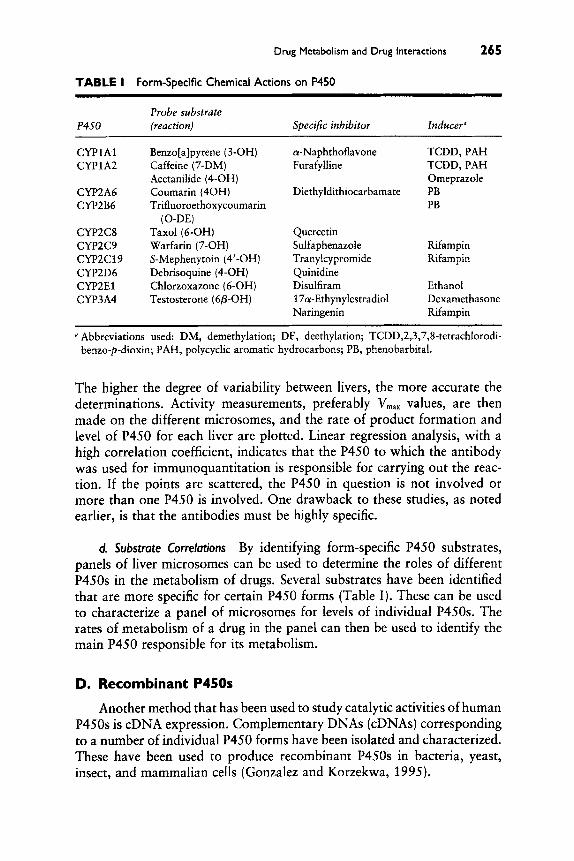
Drug Metabolism and Drug Interactions 265
TABLE I Form-Specific Chemical Actions on P450
Probe substrate P450 (reaction) Specific inhibitor Inducer"
CYPlAl CYPlA2
CYP2A6 CYP2B6
CYP2C8 CYP2C9 CYPZCl9 CYP2D6 CYP2E1 CYP3A4
Benzo[a]pyrene (3-OH) Caffeine (7-DM) Acetanilide (4-OH) Coumarin (40H) Trifluoroethoxycoumarin
Taxol (6-OH) Warfarin (7-OH) S-Mephenytoin (4'-OH) Debrisoquine (4-OH) Chlorzoxazone (6-OH) Testosterone (6P-OH)
(0-DE)
a-Naphthoflavone Furafylline
Diethyldithiocarbamate
Quercetin Sulfaphenazole Tranylcypromide Quinidine Disulfiram 17a-Ethynylestradiol Naringenin
TCDD, PAH TCDD, PAH Omeprazole PB PB
Rifampin Rifampin
Ethanol Dexamethasone Rifampin
a Abbreviations used: DM, demethylation; DE, deethylation; TCDD,2,3,7$-tetrachIorodi- benzo-p-dioxin; PAH, poIycyclic aromatic hydrocarbons; PB, phenobarbital.
The higher the degree of variability between livers, the more accurate the determinations. Activity measurements, preferably V,, values, are then made on the different microsomes, and the rate of product formation and level of P450 for each liver are plotted. Linear regression analysis, with a high correlation coefficient, indicates that the P450 to which the antibody was used for immunoquantitation is responsible for carrying out the reac- tion. If the points are scattered, the P450 in question is not involved or more than one P450 is involved. One drawback to these studies, as noted earlier, is that the antibodies must be highly specific.
d. Substrate Correlations By identifying form-specific P450 substrates, panels of liver microsomes can be used to determine the roles of different P450s in the metabolism of drugs. Several substrates have been identified that are more specific for certain P450 forms (Table I). These can be used to characterize a panel of microsomes for levels of individual P450s. The rates of metabolism of a drug in the panel can then be used to identify the main P450 responsible for its metabolism.
D. Recombinant P450s
Another method that has been used to study catalytic activities of human P450s is cDNA expression. Complementary DNAs (cDNAs) corresponding to a number of individual P450 forms have been isolated and characterized. These have been used to produce recombinant P450s in bacteria, yeast, insect, and mammalian cells (Gonzalez and Korzekwa, 1995).

266 Frank J. Gonzalez
1. Transient Expression Systems
Transient systems are those that produce recombinant P450s with lytic viruses that kill their host cells. Thus, for every experiment to produce a batch of recombinant P450, cells must be infected and then harvested at specific times to recover active enzymes. The P450s produced can then be used immediately or stored frozen.
Human P450s were first produced using the lytic vaccinia virus. In this system, viruses engineered with human P450 cDNAs could be used to infect human HepG2 cells to produce catalytically active enzymes. HepG2 cells are ideal hosts for cDNA expression because they are derived from liver and contain ample intracellular membranes and OR and bi. Catalytic activi- ties can be measured directly from total cell lysate or membrane fractions or by in sztu metabolism where the substrate is directly added to the medium during the lytic cycle (Ono et al., 1995a,b).
The baculovirus has proven to be a useful system for the production of human P450s (Grogan et al., 1995; Buters et al., 1994, 1995). Similar to vaccinia virus, the baculovirus is also lytic but is able to produce higher levels of P450. However, the host insect cells do not contain OR or b, and the P450 must be reconstituted with these proteins in order to achieve full catalytic activities. Alternatively, recombinant viruses containing OR and br can be coexpressed with the recombinant virus containing the P450 cDNA (Chen et al., 1997).
Bacteria have also been used to produce human P4.50~ (Fisher et al., 1992; Guo et al., 1994; Gillam et af., 1994; Sandhu et af., 1994; Gillam et al., 1995; Guengerich et al., 1996a,b). The bacterial system requires modification of the P450 proteins’ amino terminus in order to be efficiently expressed. Changing amino acids at the N terminus is not thought to have an impact on the substrate specificities and catalytic activities of P450s since this region of the protein is primarily required for insertion of the enzyme into the lipid bilayer of the endoplasmic reticulum. High levels of P450 can be obtained in bacteria, but similar to baculovirus, the P450 must be isolated and reconstituted with OR for use in determining catalytic activities. P450 fusion proteins in which OR was joined to the CYP3A4 were produced in a bacterial expression system that has catalytic activity in the absence of added OR (Shet et al., 1993). It is not certain, however, whether these fusion constructs will work with all human P4.50~.
2. Stable Expression Systems
Many recombinant proteins can be stably expressed in cells with the aid of selectable markers that confer resistance to antibiotics and toxins or that complement metabolic deficiencies. These sytems are more difficult to establish and optimize but are more simple to use. With the exception of yeast, they generally yield lower levels of expression than the transient expression systems.

Drug Metabolism and Drug Interactions 267
Yeast was the first system that was successfully used to express P450s (Sakaki et af., 1986). A number of human P450s have been expressed using yeast (Gonzalez and Korzekwa, 1995; Pompon et al., 1995, 1996). The most successful system for the production of P450 uses an expression vector where the recombinant protein can be induced after the yeast cells have been expanded (Pompon et al., 1995). This avoids toxicities that result from overexpression of the P450 resulting in selection of cells with lower levels of expression. OR and bs have also been coexpressed to maximize P450 catalytic activities that can be determined from membrane fractions or by incubation of cells in culture with substrates.
Mammalian cells have also been used to stably express human P450s. B-lymphoblastoid cells have proven especially useful, although the levels of expression achieved vary depending on the P450 form (Gonzalez et al., 1992; Crespi et al., 1993; Gonzalez and Korzekwa, 1995). Several P450s and even phase I1 enzymes can be coexpressed in a single cell by use of different selectable markers (Crespi et al., 1991). P450 activities can be assessed from microsome preparations or by in situ metabolism during cell growth. In addition, the lymphoblastoid cells can be used to determine the toxicity and mutagenicity of chemicals. These cells and microsome prepara- tions are available commercially.
E. Substrate-Activity Relationships
The catalytic mechanism for P450 oxidation is reasonably well under- stood (White and Coon, 1980; Guengerich and Macdonald, 1990). How- ever, a three-dimensional structure has not been determined for a mamma- lian P450 or any other eukaryote P450 that would aid in prediction of the dimensions and substrate-binding potential of the active site. Comparison of the substrate chemical structures and enzymatic products could potentially be used to predict which human P450 will metabolize a given substrate. Indeed, chemical structures, electronic properties, and points of metabolism have yielded insight into templates of drugs known to be metabolized by CYP2C9 and CYP2D6 (Smith and Jones, 1992). Almost all CYP2D6 sub- strates have an extended hydrophobic region, positively charged basic nitro- gens, and groups with the ability to accept hydrogen bonds positioned 5- 7 A from the basic nitrogen. It is noteworthy that quinidine, a potent inhibitor of CYP2D6, is structurally similar to the metabolizable substrate propranolol but, when compared to the latter, contains a ring nitrogen instead of a carbon at the oxidation site, thus accounting for the fact that it binds the active site of the enzyme but is not metabolized (Smith and Jones, 1992). Substrates and inhibitors for CYP2C9 can also be overlayed in space to fit a possible active site model. However, no similarities were found for the large and diverse substrates for CYP3A4. Kinetic studies suggest that CYP3A4 has a large active site in which a substrate can freely

268 Frank J. Gonzalez
rotate or that can even accommodate two substrates (Shou et af., 1994). Computer modeling using structure-activity relationships may have limited use in predicting potential rates and sites of drug metabolism until more is known about the structure of eukaryote P450s.
F. Reporter Gene Systems to Predict Inducers
Reporter gene systems have been used for a number of years to determine the mechanism of gene regulation. In particular, these systems were of value in delineating the cis-acting DNA control elements responsible for inducing gene expression. Regulatory elements for the aryl hydrocarbon receptor (AHR) that controls the CYPl A genes (Fujii-Kuriyama et al., 1994; Denison and Whitlock, 1995), the peroxisome proliferator-activator receptor a form (PPARa) that activates the CYP4A genes (Aldridge et al., 1995; Tugwood, 1993, and the dexamethasone response element have been characterized (Quattrochi et af., 1995; Huss et af., 1996). Reporter genes and transfection experiments were used to determine the mechanism of gene induction re- sponses in a number of systems. As shown in Fig. 1, the receptor can be introduced into a cultured cell line by transfection of a plasmid construct in which the receptor gene is under control of a constitutively expressed promoter such as the cytomegalovirus promoter/enhancer (CMV). The regu- latory element (RE) for the receptor, placed in front of a reporter gene, is cotransfected with the receptor. The most commonly used reporters are genes encoding enzymes that are not expressed in eukaryote cells. These include the chloramphenicol acetyltransferase, luciferase, and p- galactosidase. Introduction of a ligand into the transfected cell will result in activation of transcription of the reporter gene. Thus, any chemical can be tested for its ability to interact with the receptor using this system. Dose-response studies can also be carried out to determine the potency of the inducer. A reporter system using PPARa was used to determine whether certain anticancer drugs were peroxisome proliferators (Pineau et af., 1996).
CMV RE Receptor Gene
FIGURE I
RE ReporterGene
Reporter Activity 0 A reporter gene system.

Drug Metabolism and Drug Interactions 269
Reporter systems can also be developed to evaluate chemicals that will activate AHR. A receptor or cis-acting element for phenobarbital and related chemicals has not been found (Waxman and Azaroff, 1992).
V. A Case Study with Taxol Metabolism
Taxol (Paclitacel) is an antitumor drug that is active against solid tumors such as breast and ovarian cancers. It acts by interfering with the tubulin- microtubulin system. Most of the dose of taxol is found in the bile and feces as a 6-hydroxy metabolite (Fig. 2). Minor metabolites having a hydroxyl group at the meta position of phenolic ring 1 and the para position of phenolic ring 2 have also been found on incubation with human liver micro- somes (Kumar et al., 1994a). These metabolites are derived from P450- catalyzed reactions. Metabolism and in drug-drug interactions could affect toxicity or efficacy of taxol and its derivative. Therefore, efforts were made to determine which human P450 carries out taxol metabolism using some of the methods outlined earlier.
Rates of production of the 6a-hydroxy metabolite of taxol (T-6a) were found to vary over 10- to 20-fold in four studies (Cresteil et al., 1994; Harris et al., 1994; Kumar et al., 1994b; Rahman et al., 1994). There was no correlation between the production of this metabolite and the phenolic hydroxy metabolites, indicating that they are produced by different P450s (Cresteil et al., 1994). The effects of inhibitors on the production of T-6a in human liver microsomes were then examined, but the results were not clear. In one study, narigenin and quercetin, presumably selectable inhibitors of CYP3A4, inhibited the reaction whereas troleandomycin and cyclosporin, an inhibitor and a specific substrate for this P450, respectively, only inhibited hydroxylation of a phenolic ring (Harris et al., 1994). In a second study, the CYP3A4 substrates midazolam, quercetin, verapamil, testosterone, and 17a-ethynylestradiol inhibited 6a-hydroxylation (Kumar et al., 1994b). The inhibition studies were not totally conclusive, but suggested that CYP3A4 catalyzes the production of T-6a. To futher explore the role of this P450 in taxol metabolism, immunoinhibition analysis was carried out using antibody prepared against rat CYP3A1 (Kumar et al., 199413). Sixty percent of the T-6a-metabolites was inhibited by this antibody preparation In contrast, others found no inhibition of production of this metabolite by anti-CYP3A4 (Harris et al., 1994) and anti-CYP3Al sera (Cresteil et al., 1994). Instead, these antibodies inhibited production of one of the phenolic metabolites. No significant correlation was found between levels of CYP3A4 and T-6a production in a panel of human liver microsomes (Cresteil et al., 1994). Recombinant CYP3A4 produced using B lymphoblastoid cells was not able to catalyze taxol 6a-hydroxylation but instead was active toward phenolic hydroxylation (Harris et al., 1994; Kumar et al., 1994b).

270 Frank 1. Gonzalez
NH 9 \ R
CYP3A4
FIGURE 2 Structure of taxol and sites of metabolism by CYP3A4 and CYPZCS.
Evidence was more suggestive for a role of CYP2C P450 in taxol 6a- hydroxylation. The reaction was inhibited by diazepam, a substrate for the CYPZC enzymes, and a correlation was noted between levels of immuno- chemically detected CYP2C P450s and production of T-6a (Cresteil et al., 1994; Harris et al., 1994; Rahman et al., 1994). The flavenoid quercetin was inhibitory, indicating a possible involvement of CYPlA2, but other inhibitory, correlative, and cDNA expression data did not support this possibility (Harris et al., 1994; Rahman et al., 1994). Subsequent studies showed that quercetin was a competitive inhibitor of CYP2C8 (Rahman et al,, 1994). cDNA expression confirmed that CYP2C8 was highly active toward taxol 6a-hydroxylation and that CY P3A4 and CYP3A5 catalyzed the phenolic hydroxylation (Rahman et al., 1994). The results are summa- rized in Fig. 2. These data suggest that care must be taken not to coadminis- trate with taxol substrates for CYP2C8.
VI. Conclusions
One wonders whether Williams ever imagined how far along we would be in developing tools to use in predicting how drugs might be metabolized in humans. He had spent his career studying the metabolism and disposition of drugs in many species, from fruit bats to elephants, in search of an animal model that would be similar to humans. He concluded that Old World monkeys may be the most similar to humans with respect to drug conjugation reactions, but even this species metabolized many drugs differently than humans (Smith and Williams, 1974). We now know that P450s can differ between primates and humans; CYPlA2, a P450 that is constitutively ex- pressed in humans and rodents, is not expressed to any appreciable degree

Drug Metabolism and Drug Interactions 27 I
in the cynomologus monkey (Edwards et al., 1994; Bullock et al., 1995). Thus, even monkeys are not the most suitable model to use in drug devel- opment.
We now have the capability of using human tissues and cultured cells and recombinant enzymes to predict how a drug may be metabolized in clinical trials. Knowledge of the complexity of human drug-metabolizing enzymes is at its peak. By understanding the mechanism of drug oxidation polymorphisms and by using genotyping methods, it is now possible to predict those individuals that lack expression of P450s CYP2C19 and CYP2D6 (Gonzalez and Idle, 1994). Most importantly, it is now possible to determine which human P450 and transferase will be responsible for metabolizing a particular drug. The immediate impact of these studies will be in predicting drug interactions when two drugs that are coadministered are metabolized by the same P450 form.
The most conclusive studies, as shown earlier with the taxol problem, use a number of complementary approaches to identify the P450 responsible for metabolism of a drug. There are commercial organizations that provide human tissues, cryopreserved hepatocytes, cDNA-expressed recombinant enzymes, and antibodies that will greatly facilitate human drug metabolism studies. Once the mechanisms of regulation of drug-metabolizing enzymes are known, it may be possible to produce a stable hepatocyte or hepatocyte- derived cell line that contains all of the common and most important en- zymes. In uiuo modeling may now be a possiblity using transgenic or gene replacement in mice. Cultured cells and gene reporter systems may be devel- oped to rapidly screen for possible P450 inducers. These new tools will greatly facilitate preclinical drug development.
References
Aldridge, T. C., Tugwood, J. D., and Green, S . (1995). Identification and characterization of DNA elements implicated in the regulation of CYP4Al transcription. Biocbem. J.
Buchert, E., and Woosley, R. L. (1992). Clinical implications of variable antiarrhythmic drug metabolism. Pbarmacogenetics 2,2-11.
Bullock, P., Pearce, R., Draper, A., Podval, J., Bracken, W., Veltman, J., Thomas, P., and Parkinson, A. (1995). Induction of liver microsomal cytochrome P450 in cynomolgus monkeys. Drug Metab. Dispos. 23, 736-748.
Buters, J. T., Korzekwa, K. R., Kunze, K. L., Omata, Y., Hardwick, J. P., and Gonzalez, F. J. (1994). cDNA-directed expression of human cytochrome P450 CYP3A4 using bacu- lovirus. Drug Metab. Dispos. 22, 688-692.
Buters, J. T., Shou, M., Hardwick, J. P., Korzekwa, K. R., and Gonzalez, F. J. (1995). cDNA- directed expression of human cytochrome P450 CYPl A1 using baculovirus: Purification, dependency on NADPH-P4SO oxidoreductase, and reconstitution of catalytic properties without purification. Drug Metab. Dispos. 23, 696-701.
306,473-479.
272 Frank J. Gonzalez
Chen, L. P., Buters, J. T. M., Tamura, S., Hardwick, J. P., Penman, B. W., Gonzalez, F. J., and Crespi, C. L. (1997). Coexpression of CYP2A6 and NADPH-P450 oxidoreductase in the baculovirus system. Drug. Metab. Disp. 25, 399-405.
Coundouris, J. A., Grant, M. H., Engeset, J., Petrie, J. C., and Hawksworth, G. M. (1993). Cryopreservation of human adult hepatocytes for use in drug metabolism and toxicity studies. Xenobioticu 23, 1399-1409.
Coundouris, J. A., Grant, M. H., Simpson, J. G., and Hawksworth, G. M. (1990). Drug metabolism and viability studies in cryopreserved rat hepatocytes. Cryobiology 27,
Crespi, C. L., Gonzalez, F. J., Steimel, D. T., Turner, T. R., Gelboin, H. V., Penman, B. W., and Langenbach, R. (1991). A metabolically competent human cell line expressing five cDNAs encoding procarcinogen-activating enzymes: Application to mutagenicity testing. Chem Res Toxicol4, 566-572.
Crespi, C. L., Langenbach, R., and Penman, B. W. (1993). Human cell lines, derived from AHH-1 TIC+/- human lymphoblasts, genetically engineered for expression of cyto- chromes P450. Toxicology 82, 89-104.
Cresteil, T., Monsarrat, B., Alvinerie, P., Treluyer, J. M., Vieira, I., and Wright, M. (1994). Taxol metabolism by human liver microsomes: Identification of cytochrome P450 isozymes involved in its biotransformation. Cancer Res. 54, 386-392.
Dauiat, M., Peryr, B., Lesca, P., Founanier, G., Domergue, J,, and Maurel, P. (1992). Omepra- zole, an inducer of human CYPlAl and 1A2, is not a ligand for the Ah receptor. Biochem. Biophys. Res. Commun. 188, 820-825.
Denison, M. S. and Whitlock, J. P., Jr. (1995). Xenobiotic-inducible transcription of cytochrome P450 genes. J. Biol. Chem. 270, 18175-18178.
Diaz, D., Fabre, I., Daujat, M., Saint Auben, B., Bories, P., Michel, H., and Maurel, P. (1990). Omeprazole is an aryl hydrocarbon-like inducer of human hepatic cytochrome P450 Gastroenterology 99, 737-747.
Eaton, D. L., Gallagher, E. P., Bammler, T. K., and Kunze, K. L. (1995). Role of cytochrome P4501 A2 in chemical carcinogenesis: Implications for human variability in expression and enzyme activity. Pharmacogenetics 5,259-274.
Eberhart, D. C. and Parkinson, A. (1991). Cytochrome P450 IIIAl (P450p) requires cytochrome b5 and phospholipid with unsaturated fatty acids. Arch. Biochem. Biophys. 291,23 1-240.
Edwards, R. J., Murray, B. P., Murray, S., Schulz, T., Neubert, D., Gant, T. W., Thorgeirsson, S. S., Boobis, A. R., and Davies, D. S. (1994). Contribution of CYPlAl and CYPlA2 to the activation of heterocyclic amines in monkeys and human. Carcinogenesis 15,829-836.
Evans, W. E. (1994). Optimizing individual drug dosages. J. Pharm. Pharmacol. 46 (Suppl
Fisher, C . W., Caudle, D. L., Martin-Wixtrom, C., Quattrochi, L. C., Tukey, R. H., Waterman, M. R., and Estabrook, R. W. (1992). High-level expression of functional human cyto- chrome P450 1A2 in Escherichia coli. FASEB ],6, 759-764.
Fujii-Kuriyama, Y., Ema, M., Mimura, J., and Sogawa, K. (1994). Ah receptor: A novel ligand- activated transcription factor. Exp. Clin. Immunogenet. 11, 65-74.
Gelboin, H. V., Goldfarb, I., Korzekwa, K., Grogan, J., Gonzalez, F. J., and Shou, M. (1996). Inhibitory and non-inhibitory monoclonal antibodies to human cytochrome P450 2E1. Chem. Res. Toxicol. 9, 1023-1030.
Gelboin, H. V., Krause, K. W., Goldfarb, I., Buters, J. T., Yang, S. K., Gonzalez, F. J., Korzekwa, K. R., and Shou, M. (1995). Inhibitory and non-inhibitory monoclonal antibodies to human cytochrome P450 3A3/4. Biochem. Pharmacol. 50, 1841-1 850.
Gelboin, H. V., Krause, K. W., Shou, M., Gonzalez, F. J., and Yang, T. J. (1997). Inhib- itory monoclonal antibodies defice the role of cytochromes P450 in human tissue: The polymorphically-expressed CYP2D6 as a paradigm. Pharmacogenetics (in press).
Gillam, E. M., Guo, Z., and Guengerich, F. P. (1994). Expression of modified human cyto- chrome P450 2E1 in Escherichia coli, purification, and spectral and catalytic properties. Arch, Riochem. Riophys. 312, 59-56.
288-300.
l ) , 451-455.

Drug Metabolism and Drug Interactions 273
Gillam, E. M., Guo, Z., Ueng, Y. F., Yamazaki, H., Cock, I., Reilly, P. E., Hooper, W. D., and Guengerich, F. P. (1995). Expression of cytochrome P450 3A5 in Escherichia coli: Effects of 5’ modification, purification, spectral characterization, reconstitution condi- tions, and catalytic activities [published erratum appears in Arch. Biochem. Biophys. 318(2), 498, 19951. Arch. Biochem. Biophys. 317, 374-384.
Goldstein, J. A., and De Morais, S. M. (1994). Biochemistry and molecular biology of the human CYP2C subfamily. Pharmacogenetics 4, 285-299.
Gomez, D. Y., Wacher, V. J., Tomlanovich, S. J., Hebert, M. F., and Benet, L. Z. (1995). The effects of ketoconazole on the intestinal metabolism and bioavailability of cyclosporine. Clin. Pharmacol. Ther. 58, 15-19.
Gonzalez, F. J. (1993). Molecular biology of human xenobiotic-metabolizing cytochromes P450: Role of vaccinia virus cDNA expression in evaluating catalytic function. Toxicology
Gonzalez, F. J., Crespi, C. L., Czerwinski, M., and Gelboin, H. V. (1992). Analysis of human cytochrome P450 catalytic activities and expression. Tohoku J. Exp. Med. 168, 67-72.
Gonzalez, F. J., and Idle, J. R. (1994). Pharmacogenetic phenotyping and genotyping. Present status and future potential. Clin. Pharmacokinet. 26, 59-70.
Gonzalez, F. J., and Korzekwa, K.R. (1995). Cytochromes P450 expression systems. Annu. Rev. Pharmacol. Toxicol. 35, 369-390.
Gonzalez, F. J., and Nebert, D. W. (1990). Evolution of the P450 gene superfamily: Animal- plant ‘warfare’, molecular drive and human genetic differences in drug oxidation. Trends Genet. 6, 182-186.
Gonzalez, F. J,, Song, B. J., and Hardwick, J. P. (1986). Pregnenolone 16 alpha-carbonitrile- inducible P-450 gene family: Gene conversion and differential regulation. Mol. Cell. Biol.
Gonzalez, F. J., Ueno, T., Umeno, M., Song, B. J., Veech, R. L., and Gelboin, H. V. (1991). Microsomal ethanol oxidizing system: Transcriptional and posttranscriptional regulation of cytochrome P450, CYP2E1. Alcohol Alcohol Suppl. 1, 97-101.
Grant, M. H., Burke, M. D., Hawksworth, G. M., Duthie, S. J., Engeset, J., and Petrie, J. C. ( 1987). Human adult hepatocytes in primary monolayer culture: Maintenance of mixed function oxidase and conjugation pathways of drug metabolism. Biochem. Pharmacol.
Grogan, J., Shou, M., Andrusiak, E. A., Tamura, S., Buters, J. T., Gonzalez, F. J., and Korzekwa, K. R. (1995). Cytochrome P450 2A1,2E1, and 2C9 cDNA-expression by insect cells and partial purification using hydrophobic chromatography. Biochem. Pharmacol. 50, 1509- 1515.
Guengerich, F. P. (1989). Characterization of human microsomal cytochrome P-450 enzymes. Annu. Rev. Pharmacol. Toxicol. 29,241-264.
Guengerich, F. P. (1994). Catalytic selectivity of human cytochrome P450 enzymes: relevance to drug metabolism and toxicity. Toxicol. Lett. 70, 133-138.
Guengerich, F. P., Gillam, E. M., and Shimada, T. (1996a). New applications of bacterial systems to problems in toxicology. Crit. Rev. Toxicol. 26, 551-583.
Guengerich, F. P., and Macdonald, T. L. (1990). Mechanisms of cytochrome P-450 catalysis.
Guengerich, F. P., Martin, M. V., Guo, Z., and Chun, Y. J. (1996b). Purification of functional recombinant P450s from bacteria. Methods Enzyrnol. 272, 35-44.
Guillouzo, A., Morel, F., Fardel, O., and Meunier, B. (1993). Use of human hepatocyte cultures for drug metabolism studies. Toxicology 82, 209-219.
Guo, Z., Gillam, E. M., Ohmori, S., Tukey, R. H., and Guengerich, F. P. (1994). Expression of modified human cytochrome P450 1Al in Escherichia coli: Effects of 5‘ substitution, stabilization, purification, spectral characterization, and catalytic properties. Arch. Bio- chem. Biophys. 312,436-446.
82, 77-88.
6,2969-2976.
36,2311-2316.
FASEB 1. 4,2453-2459.

274 Frank J. Gonzalez
Harris, J. W., Rahman, A., Kim, B. R., Guengerich, F. P., and Collins, J. M. (1994). Metabolism of taxol by human hepatic microsomes and liver slices: Participation of cytochrome P450 3A4 and an unknown P450 enzyme. Cancer Res. 54,4026-4035.
Huss, J. M., Wang, S. I., Astrom, A., McQuiddy, P., and Kasper, C. B. (1 996). Dexamethasone responsiveness of a major glucocorticoid-inducible CYP3A gene is mediated by elements unrelated to a glucocorticoid receptor binding motif. J. Biol. Chem. 93, 28917-28923.
Imaoka, S., Imai, Y., Shimada, T., and Funae, Y. (1992). Role of phospholipids in reconstituted cytochrome P450 3A form and mechanism of their activation of catalytic activity. Bio- chemistry 31,6063-6069.
Jamis-Dow, C . A., Klecker, R. W., Katki, A. G., and Collins, J. M. (1995). Metabolism of taxol by human and rat liver in vitro: A screen for drug interactions and interspecies differences. Cancer Chemother. Pharmacol. 36, 107-1 14.
Kamataki, T., Hashimoto, H., Shimoji, M., Itoh, S., Nakayama, K., Hattori, K., Yokoi, T., Katsuki, M., and Aizawa, S. (1995). Expression of CYP3A7, a human fetus-specific cytochrome P450, in cultured cells and in the hepatocytes of p53-knockout mice. Toxicol. Lett. 82-83, 879-882.
Kensler, T. W., Groopman, J. D., Eaton, D. L., Curphey, T. J., and Roebuck, B. D. (1992). Potent inhibition of aflatoxin-induced hepatic tumorigenesis by the monofunctional en- zyme inducer 1,2-dithiole-3-thione. Carcinogenesis 13, 95-100.
Kocarek, T. A., Schuetz, E. G., Strom, S. C., Fisher, R. A., and Guzelian, P. S. (1995). Comparative analysis of cytochrome P4503A induction in primary cultures of rat, rabbit, and human hepatocytes. Drag Metab. Dispos. 23, 415-421.
Koebe, H. G., Pahernik, S., Eyer, P., and Schildberg, F. W. (1994). Collagen gel immobilization: A useful cell culture technique for long-term metabolic studies on human hepatocytes. Xenobiotica 24, 95-107.
Komori, M., Kikuchi, O., Sakuma, T., Funaki, J., Kitada, M., and Kamataki, T. (1992). Molecular cloning of monkey liver cytochrorne P-450 cDNAs: Similarity of the primary sequences to human cytochromes P-450. Biochim. Biophys. Acta 1171, 141-146.
Krause, K., Yang, T. J., Gonzalez, F. J., and Gelboin, H. V. (1997). Inhibitory monoclonal antibodies to human cytochrome P4SO 2D6. Biochem. Pharmacol. (in press).
Krynetski, E., Tai, H. L., Yates, C. R., Fessing, M., Loennechen, T., Schuetz, J. D., Relling, M. V., and Evans, W. E. (1996). Genetic polymorphism of thiopurine S-methyltransferase: Clinical importance and molecular mechanisms. Pharmacogenetics 6, 279-290.
Kumar, G. N., Oatis, J. E., Jr., Thornburg, K. R., Heldrich, F. J., Hazard, E. S., and Walle, T. (1994a). 6 alpha-Hydroxytaxol: Isolation and identification of the major metabolite of taxol in human liver microsomes. Drug Metab. Dispos. 22, 177-179.
Kumar, G. N., Walle, U. K., and Walle, T. (1994b). Cytochrome P450 3A-mediated human liver microsomal taxol6 alpha-hydroxylation. J. Pharmacol. Exp. Ther. 268,1160-1 165.
I.ake, B. G. (1 995). Mechanisms of hepatocarcinogenicity of peroxisome-proliferating drugs and chemicals. Annu. Rev. Pharmacol. Toxicol. 35,483-507.
Lake, B. G., Charzat, C., Tredger, J. M., Renwick, A. B., Beamand, J. A., and Price, R. J. (1 996). Induction of cytochrome P450 isoenzymes in cultured precision-cut rat and human liver slices. Xenobiotica 26, 297-306.
Lawton, M. P., and Philpot, R. M. (1993). Molecular genetics of the flavin-dependent monooxy- genases. Pharmacogenetics 3, 40-44.
Lee, S. S. T., Buters, J. T. M., Pineau,T., Fernandez-Salguero, P., and Gonzalez, F. J. (1996). Role of CYP2E1 in the hepatotoxicity of acetaminophen. J. Biol. Chem. 271, 12063-12067.
Lee, Y. H., Yano, M., Liu, S. Y., Matsunaga, E., Johnson, P. F., and Gonzalez, F. J. (1994). A novel cis-acting element controlling the rat CYP2D5 gene and requiring cooperativity between CEBP beta and an Spl factor. Mol. Cell. Biol. 14, 1383-1394.
Lemoine, A., Gautier, J. C., Azoulay, D., Kiffel, L., Belfoc, C., Guengerich, F. P., Maurel, P., Beaune, P., and Leroux, J. P. (1993). Major pathway of imipramine metabolism is catalyzed by cytochromes P-450 1A2 and P-450 3A4 in human liver. Mol. Pharmacol. 43,827-832.

Drug Metabolism and Drug Interactions 275
Li, A. P., Rasmussen, A., Xu, L., and Kaminski, D. L. (1995). Rifampicin induction of lidocaine metabolism in cultured human hepatocytes. J. Pharmacol. Exp. Ther. 274, 673-677.
Lipp, H. P., Schrenk, D., Wiesmuller, T., Hagenmaier, H., and Bock, K. W. (1992). Assessment of biological activities of mixtures of polychlorinated dibenzo-p-dioxins (PCDDs) and their constituents in human HepG2 cells. Arch. Toxicol. 66, 220-223.
Liu, S. Y., and Gonzalez, F. J. (1995). Role of the liver-enriched transcription factor HNF-1 alpha in expression of the CYP2El gene. DNA Cell Biol. 14, 285-293.
Loretz, L. J., Li, A. P., Flye, M. W., and Wilson, A. G. (1989). Optimization of cryopreservation procedures for rat and human hepatocytes. Xenobiotica 19, 489-498.
Merrill, J. C., Beck, D. J., Kaminski, D. A., and Li, A. P. (1995). Polybrominated biphenyl induction of cytochrome P450 mixed function oxidase activity in primary rat and human hepatocytes. Toxicology 99, 147-152.
Morel, F., Beaune, P. H., Ratanasavanh, D., Flinois, J. P., Yang, C. S., Guengerich, F. P., and Guillouzo, A. (1990). Expression of cytochrome P-450 enzymes in cultured human hepatocytes. Eur. 1. Biochem. 191, 437-444.
Muntane-Relat, J., Ourlin, J. C., Domergue, J., and Maurel, P. (1995). Differential effects of cytokines on the inducible expression of CYPlAl, CYPlA2, and CYP3A4 in human hepatocytes in primary culture. Hepatology 22, 1143-1153.
Nelson, D. R., Koymans, L., Kamataki, T., Stegeman, J. J., Feyereisen, R., Waxman, D. J., Waterman, M. R., Gotoh, O., Coon, M. J., Estabrook, R. W., Gunsalus, I. C., and Nebert, D. W. (1996). P450 suoerfamily: Update on new sequences, gene mapping, accession numbers and nomenclature. Pharmacogenetics 6, 1-42.
Ono, S., Hatanaka, T., Hotta, H., Tsutsui, M., Satoh, T., and Gonzalez, F. J. (1995a). Chlorzox- azone is metabolized by human CYPlA2 as well as by human CYP2E1. Pharmacogenetics
Ono, S., Tsutsui, M., Gonzalez, F. J., Satoh, T., Masubuchi, Y., Horie, T., Suzuki, T., and Narimatsu, S. (1995b). Oxidative metabolism of bunitrolol by complementary DNA- expressed human cytochrome P450 isozymes in a human hepatoma cell line (Hep G2) using recombinant vaccinia virus. Pharmacogenetics 5, 97-102.
Padgham, C. R., Boyle, C. C., Wang, X. J., Raleigh, S. M., Wright, M. C., and Paine, A. J. (1993). Alteration of transcription factor mRNAs during the isolation and culture of rat hepatocytes suggests the activation of a proliferative mode underlies their dedifferentiation. Biochem. Biophys. Res. Commun. 197, 599-605.
Phillips, I. R., Dolphin, C. T., Clair, P., Hadley, M. R., Hutt, A. J., McCombie, R. R., Smith, R. L., and Shephard, E. A. (1995). The molecular biology of the flavin-containing monooxygenases of man. Chem. Biol. Interact. 96, 17-32.
Pineau, T., Fernandez-Salguero, P., Lee, S. S., McPhail, T., Ward, J. M., and Gonzalez, F. J. (1995). Neonatal lethality associated with respiratory distress in mice lacking cytochrome P450 1A2. Proc. Natl. Acad. Sci. USA 92,5134-5138.
Pineau, T., Sher, T., Hudgens, W. R., Chen, L. C., Liu, L., Gonzalez, F. J., and Samid, D. (1996). Activation of peroxisome proliferator-activated receptor by the nontoxic anticancer agent phenylacetate and its analogs correlates with their cytostatic effect on human tumor cells. Biochem. Pharmacol. 52, 659-667.
Pompon, D., Louerat, B., Bronine, A., and Urban, P. (1996). Yeast expression of animal and plant P450s in optimized redox environments. Methods Enzymol. 272, 51-64.
Pompon, D., Perret, A., Bellamine, A., Laine, R., Gautier, J. C., andurban, P. (1995). Genetically engineered yeast cells and their applications. Toxicol. Lett. 82-83, 815-822.
Prueksaritanont, T., Dwyer, L. M., Balani, S. K., and Theoharides, A. D. (1994). In vitro metabolism of L-696,229, an HIV-1 reverse transcriptase inhibitor in rats and humans: Hepatic and extrahepatic metabolism and identification of enzymes involved in the hepatic metabolism. Drug Metab. Dzspos. 22, 281-288.
Quattrochi, L. C., Mills, A. S., Barwick, J. L., Yockey, C. B., and Guzelian, P. S. (1995). A novel cis-acting element in a liver cytochrorne P450 3A gene confers synergistic induction by glucocorticoids plus antiglucocorticoids. J. Biol. Chem. 270,28917-28923.
5, 143-150.

276 Frank J. Gonzalez
Rahman, A., Korzekwa, K. R., Grogan, J., Gonzalez, F. J., and Harris, J. W. (1994). Selective biotransformation of taxol to 6 alpha-hydroxytaxol by human cytochrome P450 2C8. Cancer Res, 54, 5543-5546.
Rhim, J. A., Sandgren, E. P., Degen, J. L., Palmiter, R. D., and Brinster, R. L. (1994). Replace- ment of diseased mouse liver by hepatic cell transplantation. Science 263, 1149-1152.
Rhim, J. A,, Sandgren, E. P., Palmiter, R. D., and Brinster, R. L. (1995). Complete reconstitution of mouse liver with xenogeneic hepatocytes. Proc. Natl. Acad. Sci. USA 92,4942-4946.
Rodrigues, A. D., Mulford, D. J., Lee, R. D., Surber, B. W., Kukulka, M. J., Ferrero, J. L., Thomas, S. B., Shet, M. S., and Estabrook, R. W. (1995). In vitro metabolism of terfenadine by a purified recombinant fusion protein containing cytochrome P4503A4 and NADPH- P450 reductase: Comparison to human liver microsomes and precision-cut liver tissue slices. Drug Metab. Dispos. 23, 765-775.
Rost, K. L., Brosicke, H., Brockmoller, J., Scheffler, M., Helge, H., and Roots, 1. (1992a). Increase of cytochrome P450IA2 activity by omeprazole: Evidence by the 13C-[N-3- methyl]-caffeine breath test in poor and extensive metabolizers of S-mephenytoin. Clin. Phannacol. Ther. 52 , 170-180.
Rost, K. L., Brosicke, H., Scheffler, M., and Roots, I. (199213). Dose dependent induction of CYPlA2 activity by omeprazole (Antra). Int. J. Clin. Phamacol. Ther. Toxicol. 30,
Sakaki, T., Oeda, K., Yabusaki, Y., and Ohkawa, H. (1986). Monooxygenase activity of Succharomyces cerevisiue cells transformed with expression plasmids carrying rat cyto- chrome P- 45OMC cDNA. J. Biochem. (Tokyo) 99, 741-749.
Sandhu, P., Guo, Z., Baba, T., Martin, M. V., Tukey, R. H., and Guengerich, F. P. (1994). Expression of modified human cytochrome P450 1A2 in Escherichia coli: Stabilization, purification, spectral characterization, and catalytic activities of the enzyme. Arch. Bio- chem. Biophys. 309, 168-177.
Scholl, P., Musser, S. M., Kensler, T. W., and Groopman, J. D. (1995). Molecular biomarkers for aflatoxins and their application to human liver cancer. Phamacogenetics 5 Spec No,
Schuetz, E. G., Li, D., Omiecinski, C. J., Muller-Eberhard, U., Kleinman, H. K., Elswick, B., and Guzelian, P. S. (1988). Regulation of gene expression in adult rat hepatocytes cultured on a basement membrane matrix. J. Cell. Physiol. 134, 309-323.
Schuetz, E. G., Schuetz, J. D., Strom, S. C., Thompson, M. T., Fisher, R.A ., Molowa, D. T., Li, D., and Guzelian, P. S. (1993). Regulation of human liver cytochromes P-450 in family 3 A in primary and continuous culture of human hepatocytes. Hepatology 18,1254-1262.
Shet, M. S., Fisher, C. W., Holmans, P. L., and Estabrook, R. W. (1993). Human cytochrome P450 3A4: Enzymatic properties of a purified recombinant fusion protein containing NADPH-P450 reductase. Proc. Natl. Acud. Sci. USA 90, 1174841752.
Shimada,T, Yamazaki, H., Mimura, M., Inui, Y., and Guengerich, F. P. (1994). Interindividual variations in human liver cytochrome P-450 enzymes involved in the oxidation of drugs, carcinogens and toxic chemicals: studies with liver microsomes of 30 Japanese and 30 Caucasians. J . Pharmucol. Exp. Ther. 270, 414-423.
Shou, M., Grogan, J., Mancewicz, J. A., Krausz, K. W., Gonzalez, F. J., Gelboin, H. V., and Korzekwa, K. R. (1994). Activation of CYP3A4: Evidence for the simultaneous binding of two substrates in a cytochrome P450 active site. Biochemistry 33, 6450-6455.
Sidhu, J. S., and Omiecinski, C. J. (1995). Modulation of xenobiotic-inducible cytochrome P450 gene expression by dexamethasone in primary rat hepatocytes. Pharmacogenetics
Smith, D. A., and Jones, B. C. (1992). Speculations on the substrate structure-activity relation- ship (SSAR) of cytochrome P450 enzymes. Biochem. Phamacol. 44,2089-2098.
Smith, R. L., and Williams, R. T. (1974). Comparative metabolism of drugs in man and monkeys. J. Med. Primatol. 3, 138-152.
542-543.
S171 -S 176.
5, 24-36.

Drug Metabolism and Drug Interactions 277
Tugwood, J. D. (1995). Rodent responses to peroxisome proliferators: A receptor-mediated
Vickers, A. E. (1994). Use of human organ slices to evaluate the biotransformation and drug-
Watkins, P. B. (1992). Drug metabolism by cytochromes P450 in the liver and small bowel.
Waxman, D. J., and Azaroff, L. (1992). Phenobarbital induction of cytochrome P-450 gene
Weber, W. W. (1990). Acetylation. Birth Defects 26, 43-65. White, R. E., and Coon, M. J. (1980). Oxygen activation by cytochrome P-450. Annu. Rev.
Biochem. 49,315-356. Williams, R. T. (1971). Some recent studies on species variations in drug metabolism. Chem.
Biol. Interact. 3 , 311-312. Wuarin, J., Falvey, E., Lavery, D., Talbot, D., Schmidt, E., Ossipow, V., Fonjallaz, P., and
Schibler, U. (1992). The role of the transcriptional activator protein DBP in circadian liver gene expression. J. Cell. Sci. Suppl. 16, 123-127.
Yano, M., Falvey, E., and Gonzalez, F. J. (1992). Role of the liver-enriched transcription factor DBP in expression of the cytochrome P450 CYP2C6 gene. Mol. Cell. Biol. 12,2847-2854.
Zangar, R. C., Woodcroft, K. J., Kocarek, T. A., andNovak, R. F. (1995). Xenobiotic-enhanced expression of cytochrome P450 2E1 and 2B1/2B2 in primary cultured rat hepatocytes. Drug Metab. Dzspos. 23, 681-687.
phenomenon? Arch. Toxicol. Suppl. 17, 125-133.
induced side effects of pharmaceuticals. Cell. Biol. Toxicol. 10, 407-414.
Gastroenterol. Clin. North Am. 21, 511-526.
expression. Biochem. J . 281, 577-592.

Index
ABT-538, see Ritonavir Acetaminophen, adsorption, effect of food
in stomach, 191-192 N- Acetyltransferase, drug-metabolizing
enzyme system in liver, 65 Acquired immune deficiency syndrome, use
of AZT and protease inhibitors, 81 Aflatoxin B,, oxidation by cytochrome P450
3A4, effect of cx-naphthoflavone, 15 AIDS, see Acquired immune deficiency
syndrome Alcohol, chemical-chemical interaction
modulating substance, 59 Alternative medicines, drug interactions,
Canadian Drugs Directorate perspectives, 246
Albumin, drug binding, 193 Aluminum
antacid, complex formation with tetracycline
effect on adsorption, 191 and quinolone antibiotics, 241
Ampicillin, adsorption, effect of food in stomach, 19 1 - 192
Antacids complex formation with tetracycline
effect on absorption, 191 and quinolone antibiotics, 241
drugs, concerns, 245-246 Antihistamines, interaction with other
interaction with other nonprescription
nonprescription drugs, concerns, 245-246
Antipyrine, adsorption, effect of food in
Aspirin, adsorption, effect of food in
Autoinducers, rifamycin, carbamazepine,
stomach, 19 1 - 192
stomach, 191-192
ifosfamide, and teniloxazine, effect on toxicology studies, 208
AZT, see Zidovudine
Barbiturates, induction of cytochrome P450,
Benzodiazepine, binding to albumin, 193 Biologicals, interactions with chemical
Biotransformation, in liver, predisposition
Bromotrichloromethane
24-25
drugs, concerns, 246
for injury, 39-41
all-trans-retinol induced liver injury, effect of
gadolinium chloride, and methylpalmitate, 56-57
toxicodynamic potentiation, 54-58 Bufuralol 1’-hydroxylation
analysis with cDNA-expressed cytochrome P450 enzymes in study of potential drug-drug interactions, 175-1 76
microsome preparation from, 174
Caffeine, inhibition of terfenadine in liver microsomes, 248
279

280 index
Calcium antacid, complex formation with
tetracycline effect on adsorption, 191 and quinolone antibiotics, 241
Canadian Drugs Directorate, drug interactions perspectives, 239-254
Cancer, drug-drug interactions in treating, clinical significance, 1
Captopril, adsorption, effect of food in stomach, 191-192
Carbamazepine adsorption, effect of food in stomach,
autoinducer, effect on toxicology studies,
enzyme-inducing properties, 241
191-192
208-209
Carbon tetrachloride, interaction with 1,2- dichlorobenzene, 42-46
Carcinogens, chemical, oxidation by cytochrome P450 inducers, 9
Cardiotoxicity, see Terfenadine, cardiotoxity
Cardiovascular disease, drug-drug interactions in treating, clinical significance, 1
stomach, 19 1 - 192
target for, 37-63
Cephalosporin, adsorption, effect of food in
Chemical-chemical interactions, liver as
Chiorothiazide, binding to albumin, 193 Chlorpropamide, adsorption, effect of food
Cimetidine in stomach, 191-192
cytochrome P450 enzyme inhibitors, 241 interactions, 16
liver microsomes, 248 Clarithromycin, inhibition of terfenadine in
Clofibrate, enzyme-inducing properties, 241 Cloxacillin, binding to albumin, 193 Complexations, formation with antibiotic
Cryopreservation, hepatocytes, use of
Cyclosporin, 9, 22-23 cytochrome P450
efficacy, 9
administration with antacids, 191, 241
dimethyl sulfoxide, 107
induction, effect on therapeutic
interactions with, 22-23 inhibition of terfenadine in liver
microsomes, 248
and ketoconazole, proactive
levels, effect of erythromycin, 22 management, 67
CYP, see Cytochrome P450 Cytochrome P450
cDNA-expressed human enzymes, use in analysis of potential drug-drug interactions, 17 1 - 188
conclusions, 185 introduction, 172-174 methods, 174-176
bufuralol 1 '-hydroxylation, 175-176 diclofenac 4'-hydroxylation, 175 inhibition studies, 175 materials, 174 metabolite formation, 174-175 testosterone 6P-hydroxylation, 176
cDNA expression systems: transient
inhibition, 182-185
results and discussion, 176-185
and stable, 176-178
CYP3A4, 184-185 CYP2C9, 183 CYP2D6, 183-184
selective metabolism, 178-1 82 diclofenac metabolism, 181-182 general considerations, 178-18 1
summary, 171-172 complementary DNA expressed,
reproducible source for metabolite formation analysis, 185
drug-drug interactions, role, 3-5, 7-26 effects of
cimetidine, 16 cyclosporin, 22-23 erythromycin, 22 17cr-ethnylestradiol, 18-20 grapefruit juice, 21-22 omeprazole, 23-24
terfenadine, 20-21 P450 2D6, 16-18
introduction, 7-8 mechanisms attributable to, 11-15
induction, 11-12 inhibition, 12-15 stimulation, 15
other issues, 24-26 barbiturate and peroxisome
proliferation inducers, 24-25 in vitro and in uiuo comparisons,
25-26 potential consequences, 8-10

Index 28 I
summary and conclusions, 26 use of information, 10-11
in drug-drug interactions and drug metabolism, experimental approaches for study, overview, 265-267
liver, 65 drug-metabolizing enzyme system in
effect on 1-nitronaphthalene, 50 induction
in hepatocytes, 5 with phenobarbital, 50 potential, evaluation, 5 in rat liver, measurement with ex vivo
studies, in vitro metabolic drug-drug assays, 21 1
interactions, FDA perspectives, 234-235
inhibition by ABT-538, 77-82 potential, evaluation, 5
inhibitors: ketoconazole, troleandomycin, cimetidine, and quinidine, 241
isoenzymes, specificity, definition, 4 oxidation of Seldane component
terfenadine, 20 principal system for metabolism of
lipophilic xenobiotics, 172 rapamycin metabolization, 23 ritonavir inhibition, in vitro-in vivo
correlations, 88-92
Decongestants, interaction with other nonprescription drugs, concerns, 245-246
Dexamethasone cytochrome P450 induction in
enzyme-inducing properties, 241 Dextromethorphan, metabolization by
hepatocytes, 5
cytochrome P450 2D6, exhibition of wide therapeutic index, 69
stomach, 191-192 Diazepam, adsorption, effect of food in
1,2-Dichlorobenzene induced liver injury, effect of carbon
tetrachloride-containing drinking water, 45
interaction with carbon tetrachloride, 42-46 1,3-dichlorobenzene, 51-54
dichlorobenzene, 5 1-54 1,3-DichIorobenzene, interaction with 1,2-
Diclofenac adsorption, effect of food in stomach,
inhibition of terfenadine in liver 191 -1 92
microsomes, 248 Diclofenac 4'-hydroxylation
analysis with cDNA-expressed cytochrome P450 enzymes in study of potential drug-drug interactions, 175
Dicloxacillin, binding to albumin, 193 Dicumarol, adsorption, effect of food in
stomach, 191-1 92 Diet, drugs in, effect on enzyme induction,
198 Digoxin, adsorption, effect of food in
stomach, 191 - 192 Dihydropyrimidine dehydrogenase,
inhibition by sorivudine- fluoropyrimidine, 2
cryopreservation, 107
by, in analysis of potential drug-drug interactions, 171-1 88
stomach, 192
Dimethyl sulfoxide, use for hepatocyte
cDNA, cytochrome P450 enzymes expressed
L-DOPA, adsorption, effect of food in
Drug candidates in animals and humans, enzyme induction
potential, evaluation models, 205-229
enzyme induction in humans in vivo, 210-217
endogenous urinary markers measurement, 214
ex vivo assays for measuring enzyme induction in rat liver, 211-213
human hepatocyte model, 213-214 interaction with substrates, 214-216 other techniques: and 'H,
toxicology data from multiple dose
Western blot assay for induction
2 16-21 7
studies, 210
screening, 210-21 1
clinical significance, 208-209 evaluation rationale, 207-209
efficacy, 208-209 safety, 208-209
efficacy, 207-208 safety, 208
preclinical significance, 207-208

282 Index
Drug candidates (continued) introduction, 206-207 purposeJscope, 205-206 strategy, 2 17-21 9 summary, 219
Drug development, use of in uitro human drug-drug interaction, 68-69
Drug discoveries, use of in uitro human drug-drug interaction, 68-69
Drug-drug interactions Canadian Drugs Directorate perspectives,
239-254 clinical studies, general comments,
conclusions, 252 initiatives, 244-245
25 1-252
adverse drug reaction reporting, 244 information technology impact,
product reassessment, 244
catagories, 240 pharmacokinetics, 240-241
regulatory approach, 242-244 guidlines, 242-243 monitoring, role of postmarketing
surveillance, 243-244 terfenadine, case research, 246-249 trendstconcerns, 245-246
244-245
introduction, 239-241
biologicals, 246 herbals and alternative medicines, 246 nonprescription drugs, 245-246
in uitro studies, general comments, 249-251
cytochrome P450 role, 7-26 effects of
cimetidine, 16 cyclosporin, 22-23 erythromycin, 22 17a-ethnylestradiol, 18-20 grapefruit juice, 21-22 omeprazole, 23-24 P450 2D6, 16-18
introduction, 7-8 mechanisms attributable to, 11-15
induction, 11-12 inhibition, 12-15 stimulation, 15
other issues, 24-26 barbiturate and peroxisome inducers,
24-25
potential consequences, 8-10 summary and conclusions, 26 use of information about, 10-1 1
and drug metabolism. experimental approaches for study, overview, 255-277
analytical methods for drug interaction predictions, 259-269
conclusion, 270-271 recombinant P450s, 265-267
stable expression systems,
transient expression systems, 266 266-267
reporter gene systems, application,
substrate-activity relationships,
taxol metabolism, case study,
transgenic mice, 262
268-269
267-268
269-270
gene replacement, 262 xenogenic hepatocytes, 262
in vifro systems, 262-265 microsomes, 263-265
form-specific chemical inhibitors, 264
immunochemical inhibition studies, 263-264
immunocorrelation studies,
substrate correlations, 265 264-265
purified P450s, 262-263 whole cell systems, 260-261
hepatocyte cultures, 260-261 immortalized cell lines, 261 liver slices, 26 1
drug-metabolizing enzymes, 256-257 species differences, 257-259
liver as evaluation organ, 104-105 metabolic
FDA perspectives, 231-238 case reports, 233 -232 conclusions, 235-237 in uitro drug metabolism, assessment,
chemical inhibition studies, 233 correlation studies, 233-234 immunochemical studies, 234 metabolism by cDNA-expressed
P450 induction studies,
232-235
P450s, 234
in uitro-in uiuo comparisons, 25-26 234-235

Index 283
in uitro-in vivo correlation, 235
conclusions, 164 kinetics, 156-160 metabolic switching, 160-163 single substratehnhibitor
in liver slice models, predictions
concentration studies, 152-156 pharmacokinetic, 189-203
absorption, effects, 190 changes in pH, 190 complexation and chelation, 191 effect on
gastric emptying, 191-192 intestinal blood flow, 192
inhibition of active transport, 192-193
beneficial effects, 201-202 clinical significance, 1-3
oral contraceptive failures, 2-3 sorivudine-fluoropyrimidine
terfenadine cardiotoxicity, 2 conclusions and future directions,
cytochrome P450 role, 3-5 distribution, effects, 193-194 drug displacement from protein-binding
sites in plasma, 241 excretion, effects, 200, 241 genetic factors, 201 introduction, 189-190 liver disease, effects, 200-201 mechanism, 3 metabolism, effects, 194-200
lethality, 2
5-6
dietary and environmental factors,
enzyme induction, 197-198 inhibition, 194-197
predictions regarding potential, 4 CYP
198-200
induction potential, evaluation, 5 inhibition potential, evaluation, 5 isoenzyme specificity of
metabolism, definition, 4-5 primary hepatocyte cultures as in vitro
xeniobiotic metabolism, 3-4 evaluation model, 103-130
potential, analysis with cDNA-expressed human cytochrome P450 enzymes, 171-188
conclusions, 185 introduction, 172-174
methods, 172-174 materials, 174 metabolic formation, 174-175
results and discussion inhibition, 182-185
CYP2C9, 183 CYP3A4, 184-185
CYP2D6,183-184 selective metabolism, 178-182
diclofenac metabolism, 181-182 general considerations, 178-181
summary, 171-172
models, 70-71 new drug entity
in vitro human
as inhibitor, 75-82 as substrate, 71-75
studies, 69-82 use in drug discoveries and
development, 68-69 Drug metabolism
and drug-drug interactions, experimental approaches for study, overview, 255-277
analytical methods for drug interaction predictions, 259-269
conclusion, 270-271 recombinant P450s, 265-267
stable expression systems,
transient expression systems, 266 266-267
reporter gene systems, application,
substrate-activity relationships,
taxol metabolism, case study,
transgenic mice, 262
268-269
267-268
269-270
gene replacement, 262 xenogenic hepatocytes, 262
in vitro systems, 262-265 microsomes, 263-265
form-specific chemical inhibitors, 264
immunochemical inhibition studies, 263-264
immunocorrelation studies,
substrate correlations, 265 264-265
purified P~SOS, 262-263 whole cell systems, 260-261
hepatocyte cultures, 260-261
284 Index
Drug metabolism (continued) immortalized cell lines, 261 liver slices, 261
drug-metabolizing enzymes, 256-257 species differences, 257-259
Drug metabolism model liver slices, 131-169
characteristics, comparison with in
coupled metabolism, 138-143 metabolic profiles: qualitative and
quantitative aspects, 138 species comparison, 147-148 stereoselective metabolism, 143-147
introduction, 131-132 history, 132-134 preparation and drug incubation,
134-137
VIVO, 138-148
metabolic drug-drug interaction predictions, 151-164
conclusions, 164 kinetics, 156-1 60 metabolic switching, 160-163 single substratelinhibitor
concentration studies, 152-156 metabolic drug stability:
pharmacokinetic comparisons, 148-151
Drug-metabolizing enzyme systems effect of drugs exhibiting narrow
therapeutic index or steep dose- response profile, 66
in human liver, 65-68 in vitro-in vivo correlation, 83-94
Drugs, nonprescription, interaction with antacids, decongestants, and antihistamines, concerns, 245-246
Elderly population, drug interaction guidelines, 243
Endogenous compounds, cytochrome P450 transformation, effect of drugs, 10
Endothelial cells, role in progression of liver disease, 104-105
Environment, effect on enzyme induction, 198
Enzymc inducers effect on xenobiotic properties, 197-198 rifampin, erythromycin, dexamethasone,
phenobarbital, phenytoin, carbamezapine, clofibrate, and ethanol, 241
Enzyme induction potential, drug candidates in animals and
humans, 205-229
ex vivo assays for measuring enzyme induction in rat liver, 211-213
human hepatocyte model, 213-214 ocher techniques, 216-217 toxicology data from multiple dose
in vivo analysis
evaluation models, 210-217
studies, 210
endogenous urinary markers measurement, 2 14
interaction with substrates,
Western blot assay for induction 214-216
screening, 2 10-2 1 1
clinical significance, 208-209 evaluation rationale, 207-209
efficacy, 208-209 safety, 209
efficacy, 207-208 safety, 208
introduction, 206-207 purposelscope, 205-206 strategy, 217-219 summary, 21 9
mechanisms attributable to cytochrome P450, 15
8,9-Epoxidation, stimulation induced by u-
naphthoflavone, 15 Erythromycin
preclinical significance, 207-208
Enzyme stimulation, drug interaction
effect on cylcosporin Levels, 22 enzyme-inducing properties, 24 1 interactions with cytochrome P450, 22 terfenadine
cardiotoxicity association, 2 inhibition in liver microsomes, 248
stomach, 191-1 92 Ethambutol, adsorption, effect of food in
Ethanol, see Ethyl alcohol Ethoxycoumarin, control for assessment of
Ethyl alcohol phase I and I1 metabolism, 136
adsorption, effect of food in stomach,
enzyme-inducing properties, 241 17a-Ethynylestradiol
cytochrome P450
efficacy, 9
191-192
induction Level, effect on therapeutic
interactions with, 18-20
Index 285
FDA, see United States Food and Drug
Fluconazole, inhibition of terfenadine in
Fluoropyrimidine-sorivudine lethality, effect
Furosemide
Administration
liver microsomes, 248
of drug-drug interactions, 2
adsorption, effect of food in stomach,
binding to albumin, 193 191-192
Gadolinium chloride, and methylpalmitate, effect on all-trans-retinol-potentiated bromotrichloromethane-induced liver injury, 56-57
Glibenclamide, see Glyburide o-Ghcaric acid, changes detected by "C
and 'H, for analysis of enzyme induction models, 216
Glyburide, binding to albumin, 193 Grapefruit juice, interactions with
cytochrome P450,21-22 Griseofulvin
adsorption, effect of food in stomach,
induced oral contraceptive failure, 2-3 191-192
Hepatocytes, see Primary hepatocytes Herbal medicines, drug interactions,
Canadian Drugs Directorate perspectives, 246
Hydralazine, adsorption, effect of food in stomach, 191- 192
1 '-Hydroxybufuralol, microsome preparation from, 174
4'-Hydroxydiclofenac, microsome preparation from, 174
3a-Hydroxylation, aflatoxin B, oxidation inhibition by a-naphthoflavone, 15
6P-Hydroxytestosterone, microsome preparation from, 174
Ibuprofen, binding to albumin, 193 Ifosfamide, autoinducer, effect on
Immune cells, see specific type Indomethacin, binding to albumin, 193 Indoprofen, adsorption, effect of food in
toxicology studies, 208-209
stomach, 191-192
Induction, see also Enzyme induction
effect on drug oxidation rate,
in vitro metabolic drug-drug
cytochrome P450
11-12
interactions, FDA perspectives, 234-235
metabolic clearance, mechanism of pharmacokinetic drug-drug interactions, 103
Inhibition effect on enzyme activity: competitive,
noncompetitive, and uncompetitive, 12-13
metabolic clearance, mechanism of pharmacokinetic drug-drug interactions, 103
Inhibition index, in vitro-in vivo correlations, 85-88
In vitro-in vivo correlations for drug metabolism enzymes, 83-94 inhibition index, 85-88 ritonavir, 88-94
CYP3A inhibition, 88-92 CYP2D6 inhibition, 92-94
and drug metabolism, experimental approaches for study, overview,
In vitro systems, in drug-drug interactions
262-265 Iron
adsorption, effect of food in stomach,
antacid, complex formation with 191-1 92
tetracycline effect on adsorption, 191 and quinolone antibiotics, 191
inhibition of terfenadine in liver
terfenadine cardiotoxicity association, 2
Itraconazole
microsomes, 248
Kaolin-pectin, pharmacokinetic drug interactions, Canadian Drugs Directorate perspectives, 241
Ketoconazole and cyclosporin, proactive
management, 67 cytochrome P450 enzyme inhibitors,
241 inhibition of terfenadine in liver
microsomes, 248 terfenadine cardiotoxicity association, 2

Kupffer cells potential drug-drug interactions, effect of vitamin A, 55 role in progression of liver disease,
174 inhibition of terfenadine in, 248
104- 10.5 1 -nitronaphthalene-induced injury, effect
P450 enzyme location for drug of phenobarbital pretreatment, 50
Lipocytes, role in progression of liver
Liver disease, 104-105
hiotransformation in. predisposition for
chemical-chemical interactions in, 37-63
dichlorobenzene interactions,
injury, 39-41
carbon tetrachloride and 1,2-
42-46 conclusions, 58-60 1,2-dichlorobenzene and 1,3-
dichlorobenzene interactions,
phenobarbital and 1 -nitronaphthalene
all-trans-retinol and
51-54
interactions, 46-51
bromotrichloromethane interactions, 54-58
chemically induced toxicity target, 39-41 1,2-dichlorobenzcne-induccd injury, effect
of carbon tetrachloride-containing drinking water, 45
drug-drug interaction evaluation organ,
drug-metabolizing enzyme systems, 65-68 microsomes
104-1 05
application in metabolism-based drug-drug interactions, 65-101
conclusions, 94-95 utility in drug discovery and
development, 68-69 in vttro-in vivo correlations, 83-94
ABT-538 correlations, 88-94 CYP3A inhibition, 88-92 CYP2D6 inhibition, 92-94
inhibition index, 85-88
models, 70-71 new drug entity as inhibitor,
75-82 ABT-538 analysis, 77-82
new drug entity as substrates,
in titro studies, 69-82
71-75 zileuton use, 74-75
cDNA-expressed cytochrome P450 enzymes from, use in analysis of
oxidation, 7 tissue, chemically induced injury, 37-39 toxicodynamics, effect, 41-60 toxicokinetics, effect. 41-60
drug metabolism model, 131-169 Liver slices
characteristics, comparison with in
coupled metabolism, 138-143 metabolic profiles: qualitative and
quantitative aspects, 138 species comparison, 147-148 stereoselective metabolism, 143-147
vivo, 138-148
conclusions, 164 introduction, 131 - 132
history, 132-134 preparation and drug incubation,
134- 137 metabolic drug-drug interaction
predictions, 1.5 1-164 conclusions, 164 kinetics, 156-160 metabolic switching, 160-163 single substratehnhibitor
concentration studies, 152-156 metabolic drug stability:
pharmacokinetic comparisons, 148-151
Lymphoblastoid cells, microsome preparation from, 174
Macrophages, effect of vitamin A, 55 Magnesium
antacid, complex formation with tetracycline
effect on adsorption, 191 and quinolone antibiotics, 241
Metabolic clearance induction, mechanism of pharmacokinetic
inhibition, mechanism of pharmacokinetic drug-drug interactions, 103
drug-drug interactions, 103 Metabolic drug stability, liver slice models,
pharmacokinetic comparisons, 148-1 5 1

Index 287
Metabolic switching, drug-drug interactions
Metabolism, see also Drug metabolism in liver slices, 160-164
model drug-drug interactions: FDA perspectives,
231-238 case reports, 231-232 conclusions, 235-237 in vitro drug metabolism, assessment,
chemical inhibition studies, 233 correlation studies, 233-234 immunochemical studies, 234 metabolism by cDNA-expressed
P450 induction studies, 234-235
232-235
P450s, 234
in vitro-in vivo correlation, 235 lipophilic xenobiotic, induction by
in vitro, human data for regulatory cytochrome P450, 172
agencies, 66 Metabolization, effect of cytochrome P450
2D6 on dextromethorphan, exhibition of wide therapeutic index, 69
induction in hepatocytes, 5
effect on all-trans-retinol-potentiated bromotrichloromethane-induced liver injury, 56-57
stomach, 191-192
3-Methylcholanthrene, cytochrome P450
Methylpalmitate, and gadolinium chloride,
Metoprolol, adsorption, effect of food in
Microsomes liver
application in metabolism-based drug-drug interactions, 65-101
conclusions, 94-95 introduction, 65-68 utility in drug discovery and
development, 68-69 in vitro-in vivo correlations,
83-94 ABT-538 correlations, 88-94
CYP3A inhibition, 88-92 CYP2D6 inhibition, 92-94
inhibition index, 85-88
models, 70-71 new drug entity as inhibitor,
ABT-538 analysis, 77-82
in vitro studies, 69-82
75-82
new drug entity as substrates, 71-75
zileuton use, 74-75 cDNA-expressed cytochrome P450
enzymes from, use in analysis of potential drug-drug interactions, 174
inhibition of terfenadine in, 248 preparation from
bufuralol, 174 1 '-hydroxybufuralol, 174 4'-hydroxydiclofenac, 174 6P-hydroxytestosterone, 174 lymphoblastoid cells, 174
Midazolam, inhibition of terfenadine in liver microsomes, 248
Multiple dose studies, toxicology data from, for enzyme induction potential studies, 210
a-Naphthoflavone effect on
8,9-epoxidation, 15 3a-hydroxylation of aflatoxin B,, 15 rate of theophylline metabolism by
human liver slices, 154 Naproxen, binding to albumin, 193 Naringenin, inhibition of terfenadine in
Neutrophils, effect of vitamin A, 55 New drug entity
liver microsomes, 248
in drug-drug interactions as inhibitor, 75-82 as substrate, 71-75
potential, 66-82
stomach, 191 - 192
drug-metabolizing enzyme interaction
Nitrofurantoin, adsorption, effect of food in
1-Nitronaphthalene effect of cytochrome P450, 50 induced liver injury, effect of
interaction with phenobarbital, 46-51
"C, detection of changes in o-glucaric acid, for analysis of enzyme induction models, 216
'H, detection of changes in D-glucaric acid, for analysis of enzyme induction models, 216
phenobarbital pretreatment, 50
Nuclear magnetic resonance

288 index
Occupational exposure, chemical-chemical
Omeprazole interaction modulating substance, 59
induction of cytochromes P450 1A1 and
interactions with cytochrome P450, 1A2,23
23-24 Oral contraceptives, failure, effect of
drug-drug Interactions, 2-3 Oxazepam, adsorption, effect of food in
stomach, 191 - 192
Parenchymal cells, xenobiotic metabolism site, 104-105
Penicillin, adsorption, effect of food in stomach, 191-192
Peroxisome, proliferation, induction of cytochrome P450, 25-26
pH, gastric, effect of tetracycline interaction with sodium bicarbonate, 190
Pharmacodynamics, drug interactions, Canadian Drugs Directorate perspectives, 240
Pharmacokinerrcs drug-drug interactions, 189-203
changes in pH, 190-191 complexation and chelation, 191 effects on
gastric emptying, 19 1 - 192 intestinal blood flow, 192
inhibition of active transport,
absorption, effects, 190
192-193 beneficial effects, 201 -202 Canadian Drugs Directorate
perspectives, 240 clinical significance. 1-3
oral contraceptive failures, 2-3 sorivudine-fluorop yrimidine
terfenadine cardiotoxicity, 2 conclusions and future directions,
cytochrorne P450 role, 3-5 distribution, effects, 193-194 evaluation with hepatocyres, 1 1 1-124 excretion, effects, 200 generic factors, 201 introduction, 189-190 liver disease, effects, 200-201 mechanism, 3 metabolism, effects, 194-200
lethality, 2
5-6
dietary and environmental factors,
enzyme induction, 197-198 inhibition, 194-197
predictions regarding potential, 4 CYP
198-200
induction potential, evaluation, S inhibition potential, evaluation, 5 isoenzyme specificity of
xeniobiotic metabolism, 3-4 metabolism, definition, 4-5
metabolic drug stability in liver slice models, 148-151
Phenazone, see Antipyrine Phenobarbital
cytochrome P450 hepatic induction, 5 enzyme-inducing properties, 241 interaction with 1-nitronaphthalene,
effect on hepatocytes, 46-51 Phenylbutazone, binding to albumin,
Phen ytoin 193
binding to albumin, 193 cytochrome P450 induction in
hepatocytes, 5 enzyme-inducing properties, 241
in intact hepatocytes Plasma membrane
active excretion of xenobiotics,
active transport of xenobiotics, 108-110
109- 110 Primary hepatocytes, see also Parenchymal
cells cytochrome P450 induction in, S experimental mode1 for in v r ~ o evalution
of pharmacokinetic drug-drug interactions, 103-130
application in pharmacokinetic drug-drug interactions, 11 1-124
CYP induction, 113-124 CYP450 inhibition, 112-113
conclusions and future directions,
intact cell properties, 107-1 10
levels, 107-108
evaluation via
124-127
enzymes and cofactors, physiological
plasma membrane, intact, 108-110 active excretion of xenobiotics,
109-110
Index 289
active transport of xenobiotics,
permeability, 108-109
uninterrupted, 108
109
xenobiotic metabolism, complete and
introduction, 103- 104 isolation, culturing, and
cryopreservation of hepatocytes, 105-1 07
liver as evaluative organ, 104-105 validity as drug metabolism system: in
vitro-in vivo correlation, 110-1 11 Probenecid, binding to albumin, 193 Product monograph, description of factors
affecting pharmacodynamic and pharmacokinetic profiles, 242
Propanolol, adsorption, effect of food in stomach, 191-192
Propoxyphene, adsorption, effect of food in stomach, 191 - 192
Quinidine cytochrome P450 enzyme inhibitors, 241 effect on rate of theophylline metabolism
by human liver slices, 154 Quinolone, complex formation with
aluminum, calcium, magnesium antacids, and ferrous sulfate, 241
Rapamycin, immunosuppressant, metabolization by cytochrome P450 3A4,23
interactions and drug metabolism, experimental approaches for study, overview, 268-269
Reporter gene systems, in drug-drug
all-trans-Retinol, see Vitamin A Rifampicin, see Rifampin Rifampin
cytochrome P450 induction in
effect on cylcosporin levels, 22 enzyme-inducing properties, 241 induced oral contraceptive failure, 2-3
hepatocytes, 5
Rifamycin, autoinducer, effect on toxicology studies, 208
Ritonavir CYP inhibition, 77-82 in vitro-in vivo correlations
CYP3A inhibition, 88-92 CYP2D6 inhibition, 92-94
Seldane, terfenadine component of, oxidation by P450 3A4, 20
Smoke, tobacco, chemical-chemical interaction modulating substance, 59
Sodium bicarbonate, interaction with tetracycline, effect on gastric pH, 190
Sorivudine-fluoropyrimidine lethality, effect of drug-drug interactions, 2
Sparteine, inhibition of terfenadine in liver microsomes, 248
Stimulation
Substrate-activity relationships enzyme, see Enzyme stimulation
in drug-drug interactions and drug metabolism, experimental approaches for study, overview, 267-268
Sulfadimethoxine, binding to albumin, 193 Sulfonamide, adsorption, effect of food in
stomach, 191-192
Tacrolimus, immunosuppressant, metabolization by cytochrome P450 3A4,23
interactions and drug metabolism, experimental approaches for study, overview, 269-270
Teniloxazine, autoinducer, effect on toxicology studies, 208-209
Terfenadine
Taxol metabolism, in drug-drug
cardiotoxicity, effect of drug-drug interactions, 2
component of antihistamine formulation Seldane, oxidation by P450 3A4, 20
-drug interactions, Canadian Drugs Directorate perspectives, 246-249
inhibition in liver microsomes, 248 interactions with cytochrome P450,
Testosterone 6P-hydroxylation, analysis with cDNA-expressed cytochrome P450 enzymes in study of potential drug-drug interactions, 176
20-21
Tetracycline adsorption, effect of food in stomach,
complex formation with aluminum, 191-192
calcium, magnesium, and iron antacids, 191, 241
effect on gastric pH, 190 interaction with sodium bicarbonate,

290 index
Theophylline, metabolism by human liver slices, effect of a-naphthoflavone, 154
Tissue injury, chemically induced, stages in progression, 37-39
a-Tocopheryl nicotinate, adsorption, effect of food in stomach, 191-192
Tol butamide adsorption, effect of food in stomach,
binding to albumin, 193
stomach, 191-192
191-192
'Tolmesoxide, adsorption, effect of food in
Tolzamide, binding to albumin, 193 Toxicodynamics, effects on liver, 41-60 Toxicokinetics, effects on liver, 41-60 Toxicology studies, effect of enzyme
induction, 208 Transgenic mice, in drug-drug interactions
and drug metabolism, experimental approaches for study, overview, 262
Troleandomycin
24 1
microsomes, 248
cytochrome P450 enzyme inhibitors,
inhibition of terfenadine in liver
UDP-glucuronosyltransferase, drug- metabolizing enzyme system in liver, 65
h i r e d States Food and Drug Administration, metabolic drug-drug interactions, 23 1-238
Valproate adsorption, effect of food in stomach,
binding to albumin, 193 191-192
Viral diseases, drug-drug interactions in treating, clinical significance, 1
Vitamin A bromotrichloromethane
induced liver injury, effect of gadolinium chloride, and methylpalmitate, 56-57
interaction with, 54-58 effect on immune cells, 55
Warfarin adsorption, effect of food in stomach,
binding to albumin, 193 191-192
Water, potable, carbon tetrachloride- containing, effect on 1,2- dichlorobenzene-induced liver injury, 45
interactions and drug metabolism, experimental approaches for study, overview, 260-261
Whole cell systems, in drug-drug
Xenobiotics active excretion from hepatocytes,
active transport in hepatocytes, 109-1 10 induction of enzymes, 208 lipophilic, metabolism by cytochrome
meta holism
109-110
P450, 172
in parenchymal cells, 104-105 possible application of cultured
hepatocytes in, 106
Zidovudine, and protease inhibitors, use in
Zileuton treatmenr of AIDS, 8 1
effect on rate of theophylline metabolism
in vivo metabolic profile, use of human by human liver slices, 154
liver microsomes, 74-75

Contents of Previous Volumes
Volume 32 Signal Sorting by G-Protein-Linked Receptors Graeme Milligan
Regulation of Phospholipase A2 Enzymes: Selective Inhibitors and Their Pharmacological Potential Keith B. Glaser
Platelet Activating Factor Antagonists James B. Summers and Daniel H. Albert
Pharmacological Management of Acute and Chronic Bronchial Asthma Michael K. Gould and Thomas A. Raffin
Anti-Human Immunodeficiency Virus Immunoconjugates Seth H. Pincus and Vladimir V. Tolstikov
Recent Advances in the Treatment of Human Immunodeficiency Virus Infections with Interferons and Other Biological Response Modifiers Orjan Strannegard
Advances in Cancer Gene Therapy Wei-Wei Zhang, Toshiyoshi Fujiwara, Elizabeth A. Grimm, and Jack A. Roth
Melanoma and Melanocytes; Pigmentation, Tumor Progression, and the Immune Response to Cancer Setaluri Vijayasaradhi and Alan N. Houghton
29 I

292 Contents of Previous Volumes
High-Density Lipoprotein Cholesterol, Plasma Triglyceride, and Coronary Heart Disease: Pathophysiology and Management Wolfgang Patsch and Antonio M. Gotto, Jr.
Neurotransmitter-like Actions of I-DOPA Yoshimi Misu, Hiroshi Ueda, and Yoshio Goshima
New Approaches to the Drug Treatment of Schizophrenia Gavin P. Reynolds and Carole Czudek
Membrane Trafficking in Nerve Terminals Flavia Valtorta and Fabio Benfenati
Volume 33 Endothelin Receptor Antagonism Terry J. Opgenorth
The Ryanodine Receptor Family of Intracellular Calcium Release Channels Vincenzo Sorrentino
Design and Pharmacology of Peptide Mimetics Graham J. Moore, Julian R. Smith, Barry W. Baylis, and John M. Matsoukas
Alternative Approaches for the Application of Ribozymes as Gene Therapies for Retroviral Infections Thomas B. Campbell and Bruce A. Sullenger
Inducible Cyclooxygenase and Nitric Oxide Synthase Kenneth K. Wu
Regulation of Airway Wall Remodeling: Prospects for the Development of Novel Antiasthma Drugs Alastair G. Stewart, Paul R. Tomlinson, and John W. Wilson
Advances in Selective Immunosuppression Lucian0 Adorini, Jean-Charles Guery, and Sylvie Trembleau
Monoclonal Antibody Therapy of Leukemia and Lymphoma Joseph G. Jurcic, Philip C. Caron, and David A. Scheinberg
4-Hydroxyphenylretinamide in the Chemoprevention of Cancer Harmesh R. Naik, Gregory Kalemkerian, and Kenneth J. Pienta

Contents of Previous Volumes 293
Immunoconjugates and Immunotoxins for Therapy of Carcinomas lngegerd Hellstrom, Karl Erik Hellstrom, Clay B. Siegall, and Pamela A. Trail
Discovery and in Vitro Development of AIDS Antiviral Drugs as Biopharmaceuticals William G. Rice and John P. Bader
Volume 34
Chemistry of Nitric Oxide: Biologically Relevant Aspects Jon M. Fukuto
Reactions between Nitric Oxide, Superoxide, and Peroxynitrite: Footprints of Peroxynitrite in Vivo John P. Crow and Joseph S. Beckman
Oxygen Radical-Nitric Oxide Reactions in Vascular Diseases Bruce A. Freeman, Roger White, Hector Gutierrez, Andres Paler-Martinez, Margaret Tarpey, and Homero Rubbo
Nitric Oxide Synthases: Gene Structure and Regulation Yang Wang and Philip A. Marsden
Transcription of the Human Neuronal Nitric Oxide Synthase Gene in the Central Nervous System Is Mediated by Multiple Promoters Anthony P. Young, Ferid Murad, Harald Vaessin, Jinling Xie, and Terrie K. Rife
Regulation of the Expression of the Inducible Isoform of Nitric Oxide Synthase Csaba Szabo and Christoph Thiemermann
Regulation and Function of Inducible Nitric Oxide Synthase during Sepsis and Acute Inflammation James W. Wong and Timothy R. Billiar
Expression and Expressional Control of Nitric Oxide Synthases in Various Cell Types Ulrich Forstermann, Hartmut Kleinert, lngolf Gath, Petra Schwarr, Ellen I. Closs, and Nae J. Dun
Control and Consequences of Endothelial Nitric Oxide Formation Ingrid Fleming and Rudi Busse

294 Camems of Previous Volumes
Control of Electron Transfer in Neuronal Nitric Oxide Synthase by Calmodulin, Substrate, Substrate Analogs, and Nitric Oxide Dennis J. Stuehr, Husam M. Abu-Soud. Denis L. Rousseau. Paul L. Feldman, and Jianling Wang
Negative Modulation of Nitric Oxide Synthase by Nitric Oxide and Nitroso Compounds Jeanette M. Griscavage, Adrian J. Hobbs, and Louis J. lgnarro
Regulation of Nitric Oxide Synthase: Role of Oxygen Radicals and Cations in Nitric Oxide Formation Chandra K. Mittal and Chander S. Mehta
Why Tetrahydrobiopterin? Bernd Mayer and Ernst R. Werner
Nitric Oxide and cGMP Signaling Lee J. McDonald and Ferid Murad
Nitric Oxide and Intracellular Heme Young-Myeong Kim, Hector A. Bergcqia, Claudia Muller, Bruce R. Pitt, W. David Watkins, and Jack R. Lancaster, Jr.
High-Level Expression of Biologically Active Soluble Guanylate Cyclase Using the Baculovirus System Is Strongly Heme-Dependent Wolfgang A. Buechler, Sujay Singh, Janet Aktas, Stefan Muller, Ferid Murad, and Rupert Gerzer
cGMP Signaling through CAMP- and cGMP-Dependent Protein Kinases Thomas M. Lincoln, Padmini Komalavilas, Nancy J. Boerth, Lee Ann MacMillan-Crow, and Trudy L. Cornwell
Physiological and Toxicological Actions of Nitric Oxide in the Central Nervous System Valina L. Dawson and Ted M. Dawson
S-Nitrosothiols: Chemistry, Biochemistry, and Biological Actions Gilbert R. Upchurch, Jr., George N. Welch, and Joseph Loscalzo
Glyceraldehyde-3-Phosphate Dehydrogenase: A Target for Nitric Oxide Signaling Bernhard Brune and Eduardo G. Lapetina
Nitric Oxide Donors: Biochemical Pharmacology and Therapeutics John Anthony Bauer, Brian P. Booth, and Ho-Leung Fung

Contents of Previous Volumes 295
Nitric Oxide Donors: A Continuing Opportunity in Drug Design Stephen R. Hanson, Thomas C. Hutsell, Larry K. Keefer, Daniel L. Mooradian, and Daniel J. Smith
Nitric Oxide and Peripheral Adrenergic Neuromodulation Roberto Levi, Kwan Ha Park, Michiaki Imamura, Nahid Seyedi, and Harry M. Lander
A Study on Tumor Necrosis Factor, Tumor Necrosis Factor Receptors, and Nitric Oxide in Human Fetal Glial Cultures Barbara A. St. Pierre, Douglas A. Granger, Joyce L. Wong, and Jean E. Merrill
Inhaled Nitric Oxide, Clinical Rationale and Applications Claes G. Frostell and Warren M. Zapol
Inhaled Nitric Oxide Therapy of Pulmonary Hypertension and Respiratory Failure in Premature and Term Neonates Steven H. Abman and John P. Kinsella
Clinical Applications of Inhaled Nitric Oxide in Children with Pulmonary Hypertension David L. Wessel and Ian Adatia
Volume 35
Interactions between Drugs and Nutrients C. Tschanz, W. Wayne Stargel, and J. A. Thomas
Induction of Cyclo-Oxygenase and Nitric Oxide Synthase in Inflammation Ian Appleton, Annette Tomlinson, and Derek A. Willoughby
Current and Future Therapeutic Approaches to Hyperlipidemia John A. Farmer and Antonio M. Gotto, Jr.
In Vivo Pharmacological Effects of Ciclosporin and Some Analogues Jean F. Borel, Gotz Baumann, Ian Chapman, Peter Donatsch, Alfred Fahr, Edgar A. Mueller, and Jean-Marie Vigouret
Mono-ADP-ribosylation: A Reversible Posttranslational Modification of Proteins Ian J. Okazaki and Joel Moss

296 Contents of Previous Volumes
Activation of Programmed (Apoptotic) Cell Death for the Treatment of Prostate Cancer Samuel R. Denmeade and John T. lsaacs
Reversal of Atherosclerosis with Therapy: Update of Coronary Angiographic Trials Howard N. Hodis
Unnatural Nucleotide Sequences in Biopharmaceutics Lawrence A. Loeb
Pharmacology of the Neurotransmitter Release Enhancer Linopirdine (DuP 996), and Insights into Its Mechanism of Action Simon P. Aiken, Robert Zaczek, and Barry S. Brown
Volume 36
Regulation of Somatostatin Gene Transcription by CAMP M. Montminy, P. Brindle, 1. Arias, K. Ferreri, and R. Armstrong
Dissection of Protein Kinase Cascades That Mediate Cellular Response to Cytokines and Cellular Stress Philip Cohen
Cyclic Nucleotide Phosphodiesterases: Gene Complexity, Regulation by Phosphorylation, and Physiological Implications Fiona Burns, Allan Z. Zhao, and Joseph A. Beavo
Structural Analysis of the MAP Kinase ERK2 and Studies of MAP Kinase Regulatory Pathways Melanie H. Cobb, Shuichan Xu, Mangeng Cheng, Doug Ebert, David Robbins, Elizabeth Goldsmith, and Megan Robinson
Novel Protein Phosphatases That May Participate in Cell Signaling Patricia T. W. Cohen, Mao Xiang Chen. and Christopher G. Armstrong
Protein Tyrosine Phosphatases and the Control of Cellular Signaling Responses N. K. Tonks
Roles of the MAP Kinase Cascade in Vertebrates Tetsuo Moriguchi, Yukiko Cotoh, and Eisuke Nishida

Contents of Previous Volumes 297
Signal Transductions of SH2/SH3: Ash/Grb-2 Downstream Signaling Tadaorni Takenawa. Kenji Miura, Hiroaki Miki, and Kazutada Watanabe
Sphingolipid-Dependent Protein Kinases Sen-itiroh Hakomori
G Protein-Coupled Receptor Kinase: Phosphorylation of Muscarinic Receptors and Facilitation of Receptor Sequestration Tatsuya Haga, Kazuko Haga, Kimihiko Karneyarna, and Hirofurni Tsuga
Molecular and Cellular Pharmacology of a Calcium/Calmodulin- Dependent Protein Kinase I1 (CaM Kinase 11) Inhibitor, KN-62, and Proposal of CaM Kinase Phosphorylation Cascades Hiroyoshi Hidaka and Hisayuki Yokokura
Intrasteric Regulation of Calmodulin-Dependent Protein Kinases B. E. Kemp, J. A. Barden, B. Kobe, C. House, and M. W. Parker
Structure, Regulation, and Function of CalciumKalmodulin- Dependent Protein Kinase I Marina R. Picciotto, Kent L. Nastiuk, and Angus C. Nairn
Gene Expression and CREB Phosphorylation Induced by CAMP and Ca2+ in Neuronal Cells Masatoshi Hagiwara, Atsushi Shimornura, Kazuhiko Yoshida, and Junko lmaki
Volume 37
Opioid and Nonopioid Cardiovascular Effects of Dynorphins Michel Durnont and Simon Lemaire
Cytokines in Neuronal Development G. Miller Jonakait
Heterogeneity and Functional Properties of Subtypes of Sodium-Dependent Glutamate Transporters in the Mammalian Central Nervous System Michael B. Robinson and Lisa A. Dowd
Development and Therapeutic Potential of Calpain Inhibitors Kevin K. W. Wang and Po-wai Yuen
298 Contents of Previous Volumes
The Pharmacology of ( - )-Nicotine and Novel Cholinergic Channel Modulators Jorge D. Brioni, Michael W. Decker, James P. Sullivan, and Stephen P. Arneric
Cryptococcosis Judith A. Aberg and William G. Powderly
Antimalarial Activity of Artemisinin (Qinghaosu) and Related Trioxanes: Mechanism( s) of Action Jared N. Cumming, Poonsakdi Ploypradith, and Gary H. Posner
The Role of Endothelin in the Pathogenesis of Atherosclerosis Mark C. Kowala
The Pharmacology and Molecular Biology of Large-Conductance Calcium-Activated (BK) Potassium Channels Valentin K. Gribkoff, John E. Starrett, Jr., and Steven I. Dworeuky
Update on Invasive Candidiasis Libsen J. Rodriguez, john H. Rex, and Elias J. Anaissie
Volume 38
Antioxidants: The Basics-What They Are and How to Evaluate Them Barry Halliwell
Metabolism of Vitamin C in Health and Disease Ann M. Bode
Regulation of Human Plasma Vitamin E Maret G. Traber
Glutathione and Glutathione Delivery Compounds Mary E. Anderson
a-Lipoic Acid: A Metabolic Antioxidant and Potential Redox Modulator of Transcription Lester Packer, Sashwati Roy, and Chandan K. Sen
Antioxidant Actions of Melatonin Russel J. Reiter
Antioxidative and Metal-Chelating Effects of Polyamines Erik Lavaas

Contents of Previous Volumes 299
Antioxidant and Chelating Properties of Flavonoids Ludmila G. Korkina and lgor B. Afanas’ev
Potential Use of Iron Chelators against Oxidative Damage Jean-Baptiste Galey
N-Acetylcysteine: Pharmacological Considerations and Experimental and Clinical Applications Ian A. Cotgreave
Ebselen as a Glutathione Peroxidase Mimic and as a Scavenger of Peroxynitrite Helmut Sies and Hiroshi Masumoto
Salen-Manganese Complexes: Combined Superoxide Dismutase/ Catalase Mimics with Broad Pharmacological Efficacy Susan R. Doctrow, Karl Huffman, Catherine B. Marcus, Wael Musleh, Annadora Bruce, Michel Baudry, and Bernard Malfroy
Antioxidant Drug Targeting Anthony C. Allison
Antioxidant-Inducible Genes Thomas Primiano, Thomas R. Sutter, and Thomas W. Kensler
Redox Signaling and the Control of Cell Growth and Death Garth Powis, John R. Gasdaska, and Amanda Baker
Protective Action of Nitrone-Based Free Radical Traps against Oxidative Damage to the Central Nervous System Robert A. Floyd
Reactive Oxygen Species and Their Contribution to Pathology in Down Syndrome Judy B. de Haan, Ernst J. Wolvetang, Francesca Cristiano, Rocco lannello, Cecile Bladier, Michael J. Kelner, and lsmail Kola
Antioxidants, Redox-Regulated Transcription Factors, and Inflammation Paul G. Winyard and David R. Blake
Relationships among Oxidation of Low-Density Lipoprotein, Antioxidant Protection, and Atherosclerosis Hermann Esterbauer, Reinhold Schmidt, and Marianne Hayn
Adult Respiratory Distress Syndrome: A Radical Perspective Samuel Louie, Barry Halliwell, and Carroll Edward Cross

300 Contents of Previous Volumes
Oxidative Stress in Cystic Fibrosis: Does It Occur and Does It Matter? Albert van der Vliet, Jason P. Eiserich, Gregory P. Marelich, Barry Halliwell, and Carroll E. Cross
Oxidative Stress and Antioxidant Function in Relation to Risk for Cataract Allen Taylor and Thomas Nowell
The Macular Pigment: A Possible Role in Protection from Age-Related Macular Degeneration John T. Landrum, Richard A. Bone, and Mark D. Kilburn
Neurological Disease David P. R. Muller
Role of Cysteine and Glutathione in HIV Infection and Cancer Cachexia: Therapeutic Intervention with N-Acetylcysteine Wulf Droge, Andrea Gross, Volker Hack, Ralf Kinscherf, Michael Schykowski, Michael Bockstette. Sabine Mihm, and Dagmar Galter
Role of Oxidative Stress and Antioxidant Therapy in Alcoholic and Nonalcoholic Liver Diseases Charles S. Lieber
Antioxidant Therapy for the Prevention of Type I Diabetes Birgit Heller, Volker Burkart, Eberhard Lampeter, and Hubert Kolb
Photoaging of the Connective Tissue of Skin: Its Prevention and Therapy Karin Scharffetter-Kochanek
Antioxidant Nutrients and Cancer Incidence and Mortality: An Epidemiologic Perspective Susan T. Mayne
Volume 39
Colorectal Cancer and Nonsteroidal Anti-inflammatory Drugs Walter E. Smalley and Raymond N. DuBois
Mouse Mammary Tumor Virus and the Immune System Susan R. Ross

Contents of Previous Volumes 30 I
Sodium Channels and Therapy of Central Nervous System Diseases Charles P. Taylor and Lakshmi S. Narasimhan
Anti-adhesion Therapy Carol J. Cornejo, Robert K. Winn, and John M. Harlan
Use of Azoles for Systemic Antifungal Therapy Carol A. Kauffman and Peggy L. Carver
Pharmacology of Neuronal Nicotinic Acetylcholine Receptor Subtypes Lorna M. Colquhoun and James W. Patrick
Structure and Function of Leukocyte Chemoattractant Richard D. Ye and Francois Boulay
Pharmacologic Approaches to Reperfusion Injury James T. Willerson
Restenosis: Is There a Pharmacologic Fix in the Pipeline? Joan A. Keiser and Andrew C. G. Uprichard
Role of Adenosine as a Modulator of Synaptic Activity in the Central Nervous System James M. Brundege and Thomas V. Dunwiddie
Combination Vaccines Ronald W. Ellis and Kenneth R. Brown
Pharmacology of Potassium Channels Maria L. Garcia, Markus Hanner, Hans-Gunther Knaus, Robert Koch, William Schmalhofer, Robert S. Slaughter, and Gregory J. Kaczorowski
Volume 40
Advances in Understanding the Pharmacological Properties of Antisense Oligonucleotides Stanley T. Crooke
Targeted Tumor Cytotoxicity Mediated by Intracellular Single- Chain Anti-oncogene Antibodies David T. Curie1

302 Contenrs of Previous Volumes
In Vivo Gene Therapy with Adeno-Associated Virus Vectors for Cystic Fibrosis Terence R. Flotte and Barrie J. Carter
Engineering Herpes Simplex Virus Vectors for Human Gene Therapy Joseph C. Glorioso, William F. Goins, Martin C. Schmidt, Tom Oligino, Dave Krisky, Peggy Marconi, James D. Cavalcoli. Ramesh Ramakrishnan, P. Luigi Pofiani, and David j. Fink
Human Adenovirus Vectors for Gene Transfer into Mammalian Cells Mary M. Hitt, Christina L. Addison, and Frank L. Graham
Anti-oncogene Ribozymes for Cancer Gene Therapy Akira Irie, Hiroshi Kijima, Tsukasa Ohkawa. David Y. Bouffard, Toshiya Suzuki, Lisa D. Curcio, Per Sonne Holm. Alex Sassani, and Kevin J. Scanlon
Cytokine Gene Transduction in the Immunotherapy of Cancer Giorgio Parmiani, Mario P. Colombo. Cecilia Melani, and Flavio Arienti
Gene Therapy Approaches to Enhance Antitumor Immunity Daniel L. Shawler, Habib Fakhrai. Charles Van Beveren, Dan Mercoa, Daniel P. Gold, Richard M. Barrholomew, lvor Royston, and Robert E. Sobol
Modified Steroid Receptors and Steroid-Induci ble Promoters as Genetic Switches for Gene Therapy John H. White
Strategies for Approaching Retinoblastoma Tumor Suppressor Gene Therapy Hong-Ji Xu
Immunoliposomes for Cancer Treatment fohn W. Park, Keelung Hong, Dmitri B. Kirpotin, Demetrios Papahadjopoulos, and Christopher C. Benz
Antisense Inhibition of Virus Infection R. E. Kilkuskie and A. K. Field
Volume 41
Apoptosis: An Overview of the Process and Its Relevance in Disease Stephanie Johnson Webb, David J. Harrison, and Andrew H. Wyllie
Contents of Previous Volumes 303
Genetics of Apoptosis Serge Desnoyers and Michael 0. Hengartner
Methods Utilized in the Study of Apoptosis Peter W. Mesner and Scott H. Kaufrnann
In Vitro Systems for the Study of Apoptosis Atsushi Takahashi and William C. Earnshaw
The Fas Pathway in Apoptosis Christine M. Eischen and Paul J. Leibson
Ceramide: A Novel Lipid Mediator of Apoptosis Miriam J. Srnyth, Lina M. Obeid, and Yusuf A. Hannun
Control of Apoptosis by Proteases Nancy A. Thornberry, Antony Rosen, and Donald W. Nicholson
Death and Dying in the Immune System David S. Ucker
Control of Apoptosis by Cytokines W. Stratford May, Jr.
Glucocorticoid-Induced Apoptosis Clark W. Distelhorst
Apoptosis in AIDS Andrew D. Badley, David Dockrell, and Carlos V. Paya
Virus-Induced Apoptosis J. Marie Hardwick
Apoptosis in Neurodegenerative Diseases lkuo Nishirnoto, Takashi Okarnoto, Ugo Giambarella, and Takeshi lwatsubo
Apoptosis in the Mammalian Kidney: Incidence, Effectors, and Molecular Control in Normal Development and Disease States Ralph E. Buttyan and Glenda Gobe
Apoptosis in the Heart Sarnuil R. Urnansky and L. David Tomei
Apoptosis and the Gastrointestinal System Florencia Que and Gregory J. Gores

304 Contents of Previous Volumes
Role of p53 in Apoptosis Christine E. Canman and Michael B. Kastan
Chemotherapy-Induced Apoptosis Peter W. Mesner, Jr., 1. lmawati Budihardjo, and Scott H. Kaufmann
Bcl-2 Family Proteins: Strategies for Overcoming Chemoresistance in Cancer John C. Reed
Role of Bcr-Abl Kinase in Resistance to Apoptosis Afshin Samali, Adrienne M. Gorman, and Thomas G. Cotter
Apoptosis in Hormone-Responsive Malignancies Samuel R. Denmeade, Diane E. McCloskey, Ingrid B. J. K. Joseph, Hillary A. Hahm, John T. lsaacs. and Nancy E. Davidson
Volume 42
Catecholamine: Bridging Basic Science Edited by David S. Goldstein, Graeme Eisenhofer, and Richard McCarty
Part A. Catecholamine Synthesis and Release
Part B. Catecholamine Reuptake and Storage
Part C. Catecholamine Metabolism
Part D. Catecholamine Receptors and Signal Transduction
Part E. Catecholamine in the Periphery
Part F. Catecholamine in the Central Nervous System
Part G. Novel Catecholaminergic Systems
Part H. Development and Plasticity
Part 1. Drug Abuse and Alcoholism