introduction to biochemical pharmacology and drug...
TRANSCRIPT

Chapter 17
Introduction toBiochemical Pharmacology and Drug Discovery
Gabriel Magoma
Additional information is available at the end of the chapter
http://dx.doi.org/10.5772/52014
1. Introduction
This chapter introduces biochemical pharmacology and highlights drug absorption and drugtransformation reactions and a general introduction to pharmacology, drug discovery andclinical trials for new drug candidates. It also introduces the concept of individualization ofdrug therapies. After studying this chapter, one is expected to demonstrate understanding thefollowing: (i) Linkage between the various pharmacological processes (ii) Routes of drugadministration, (iii) Mechanisms of drug absorption (iv) The kinetics of drug disposition andconcepts, such as volume of distribution, initial dose and half-life, (v) The biotransformationand excretion of drugs.(vi) The role of biochemical knowledge in the discovery and develop‐ment of candidate drug compounds into useful drugs (vii) Basic design of clinical trials of newdrugs and the drug approval process.(viii) The linkage between genetic variations and varieddrug responses in different individuals (ix) The various adverse drug reactions in differentpatients (x) How different dosage regimens are calculated with respect to the prevailing healthstatus of individuals and how adjustments are carried out in old patients or geriatrics.
2. Pharmacology
Pharmacology is the science that deals with drugs, their properties, actions and fate in thebody. It embraces the sciences of pharmaceutics (preparation of drugs), therapeutics (treat‐ment of diseases by use of drugs) and toxicosis or adverse side-effects that arise from thetherapeutic interventions. Pharmacology can be divided into the following processes:-
i. The pharmaceutical process of drugs; deals with chemical synthesis, formulation anddistribution of drugs.
© 2013 Magoma; licensee InTech. This is an open access article distributed under the terms of the CreativeCommons Attribution License (http://creativecommons.org/licenses/by/3.0), which permits unrestricted use,distribution, and reproduction in any medium, provided the original work is properly cited.
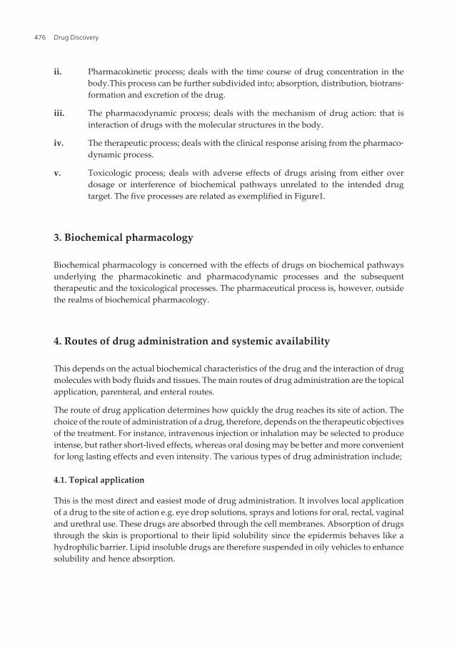
ii. Pharmacokinetic process; deals with the time course of drug concentration in thebody.This process can be further subdivided into; absorption, distribution, biotrans‐formation and excretion of the drug.
iii. The pharmacodynamic process; deals with the mechanism of drug action: that isinteraction of drugs with the molecular structures in the body.
iv. The therapeutic process; deals with the clinical response arising from the pharmaco‐dynamic process.
v. Toxicologic process; deals with adverse effects of drugs arising from either overdosage or interference of biochemical pathways unrelated to the intended drugtarget. The five processes are related as exemplified in Figure1.
3. Biochemical pharmacology
Biochemical pharmacology is concerned with the effects of drugs on biochemical pathwaysunderlying the pharmacokinetic and pharmacodynamic processes and the subsequenttherapeutic and the toxicological processes. The pharmaceutical process is, however, outsidethe realms of biochemical pharmacology.
4. Routes of drug administration and systemic availability
This depends on the actual biochemical characteristics of the drug and the interaction of drugmolecules with body fluids and tissues. The main routes of drug administration are the topicalapplication, parenteral, and enteral routes.
The route of drug application determines how quickly the drug reaches its site of action. Thechoice of the route of administration of a drug, therefore, depends on the therapeutic objectivesof the treatment. For instance, intravenous injection or inhalation may be selected to produceintense, but rather short-lived effects, whereas oral dosing may be better and more convenientfor long lasting effects and even intensity. The various types of drug administration include;
4.1. Topical application
This is the most direct and easiest mode of drug administration. It involves local applicationof a drug to the site of action e.g. eye drop solutions, sprays and lotions for oral, rectal, vaginaland urethral use. These drugs are absorbed through the cell membranes. Absorption of drugsthrough the skin is proportional to their lipid solubility since the epidermis behaves like ahydrophilic barrier. Lipid insoluble drugs are therefore suspended in oily vehicles to enhancesolubility and hence absorption.
Drug Discovery476

Parenteral Topical Oral
*
Pharmaceutical doseform of a drug
Pharmacokinetic, drugentry into systemic circulation
Pharmacodynamic, mechanism of drug action at target site
Therapeutic, clinical response
Toxicologic,adverse drugeffects
* * *
*
*
*
Figure 1. Relationships between the five pharmacological processes starting with the entry of the drug and endingwith the clinical response and/or the toxic effects
4.2. Oral administration
The drugs administered orally are absorbed at different sites along the gastrointestinal tract(GIT):
4.2.1. Oral mucosal or sublingual
Drug absorption is generally rapid because of the rich vascular supply to the mucosa and the ab‐sence of a stratum corneum. Drugs delivered using this route are not exposed to gastric and in‐testinal digestive juices and are not subjected to immediate passage through the liver. Thereforethere is no prior biotransformation or first-effect before the drugs enter the systemic circulation.
4.2.2. Stomach and intestine
Absorption depends on different factors such as pH, gastric emptying, intestinal motility andsolubility of solid drugs. The rapidity with which a drug reaches the small intestine is enhancedwhen a drug is taken with water and when the stomach is relatively empty. However drugsabsorbed in the stomach and intestine are subjected to first-pass effect.
Introduction to Biochemical Pharmacology and Drug Discoveryhttp://dx.doi.org/10.5772/52014
477

4.3. Rectal administration
This is the preferred route when the oral route is unsuitable because of nausea or if thedrugs have objectionable taste or odor. This route also protects susceptible drugs fromthe biotransformation reactions in the liver. However, absorption by this route is often ir‐regular and incomplete. Formulations such as suppositories or enemas are applied viarectal route.
4.4. Parenteral administration
This mode of administration is also known as injection. It is generally more rapid and enablesmore accurate dose selection and predictable absorption. Parenteral routes include;
4.4.1. Subcutaneous injection
This mode of administration is mainly used for non-irritating drugs. It provides even and slowsabsorption producing sustained drug effects. Vasoconstrictor agents such as epinephrine canbe added to the drug solution to decrease the rate of absorption.
Large volumes of drugs may, however, be painful because of tissue distention.
4.4.2. Intramuscular injection
This method of drug delivery ensures rapid absorption of the drug in aqueous solutions. Slowand even absorption is possible when drugs are suspended in oily vehicles.
4.4.3. Intravenous administration
This route ensures rapid delivery of the desired blood concentration of the drug to be obtainedaccurately and immediately and is the preferred route of delivery in emergency situations.Irritating drugs are delivered intravenously because the veins have low sensitivity to pain.This mode of delivery is also preferred for drug such as the barbiturates and phenytoin, anti-seizure drugs which dissolve only in rather strong alkaline solution and therefore need theblood to buffer the pH of the drug solution for better solubility. Drugs such as ethylene di-amine tetra acetic acid (EDTA) for treatment of heavy-metal poisoning are given by intrave‐nous injection or through an infusion because they are poorly absorbed in the gut. The otheradvantage of this mode of delivery is the avoidance of the hepatic and pulmonary first-passeffect. Generally, the properties of the drug may determine the route that must be used forreasonable efficacy.
5. Mechanisms of drug absorption across membranes
In order for drugs to elicit their pharmacological effects, they have to cross the biologicalmembranes into systemic circulation and reach the site of action. Therefore an insight into thestructure and function of the membrane leads to a better understanding of drug absorption.
Drug Discovery478
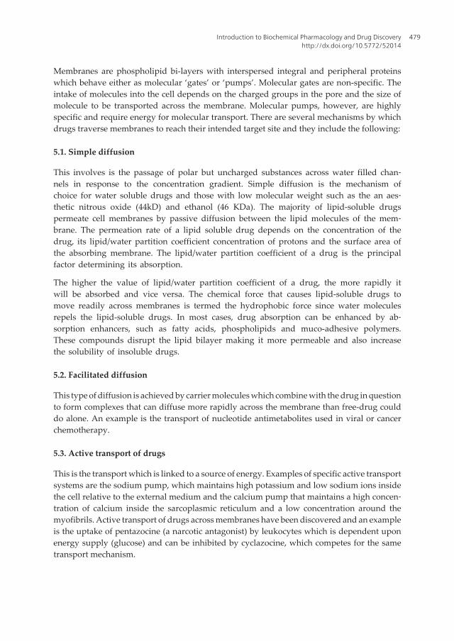
Membranes are phospholipid bi-layers with interspersed integral and peripheral proteinswhich behave either as molecular ‘gates’ or ‘pumps’. Molecular gates are non-specific. Theintake of molecules into the cell depends on the charged groups in the pore and the size ofmolecule to be transported across the membrane. Molecular pumps, however, are highlyspecific and require energy for molecular transport. There are several mechanisms by whichdrugs traverse membranes to reach their intended target site and they include the following:
5.1. Simple diffusion
This involves is the passage of polar but uncharged substances across water filled chan‐nels in response to the concentration gradient. Simple diffusion is the mechanism ofchoice for water soluble drugs and those with low molecular weight such as the an aes‐thetic nitrous oxide (44kD) and ethanol (46 KDa). The majority of lipid-soluble drugspermeate cell membranes by passive diffusion between the lipid molecules of the mem‐brane. The permeation rate of a lipid soluble drug depends on the concentration of thedrug, its lipid/water partition coefficient concentration of protons and the surface area ofthe absorbing membrane. The lipid/water partition coefficient of a drug is the principalfactor determining its absorption.
The higher the value of lipid/water partition coefficient of a drug, the more rapidly itwill be absorbed and vice versa. The chemical force that causes lipid-soluble drugs tomove readily across membranes is termed the hydrophobic force since water moleculesrepels the lipid-soluble drugs. In most cases, drug absorption can be enhanced by ab‐sorption enhancers, such as fatty acids, phospholipids and muco-adhesive polymers.These compounds disrupt the lipid bilayer making it more permeable and also increasethe solubility of insoluble drugs.
5.2. Facilitated diffusion
This type of diffusion is achieved by carrier molecules which combine with the drug in questionto form complexes that can diffuse more rapidly across the membrane than free-drug coulddo alone. An example is the transport of nucleotide antimetabolites used in viral or cancerchemotherapy.
5.3. Active transport of drugs
This is the transport which is linked to a source of energy. Examples of specific active transportsystems are the sodium pump, which maintains high potassium and low sodium ions insidethe cell relative to the external medium and the calcium pump that maintains a high concen‐tration of calcium inside the sarcoplasmic reticulum and a low concentration around themyofibrils. Active transport of drugs across membranes have been discovered and an exampleis the uptake of pentazocine (a narcotic antagonist) by leukocytes which is dependent uponenergy supply (glucose) and can be inhibited by cyclazocine, which competes for the sametransport mechanism.
Introduction to Biochemical Pharmacology and Drug Discoveryhttp://dx.doi.org/10.5772/52014
479

5.4. Pinocytosis and phagocytosis of drugs
Proteins, bacterial toxins and drugs with high molecular weights, (1000 KDa or more) en‐ter cells by means of pinocytosis and endocytosis. These substances finally enter the lyso‐somal system.
6. Factors affecting absorption of drugs
6.1. Surface area
For any substance that can penetrate the GIT in measurable amounts, the small intestinerepresents the greatest area of absorption. For instance, ethanol can be absorbed by thestomach, but it is absorbed eight times faster from the small intestine because of the largesurface area provided by the villi. The rate at which the stomach empties its contents into thesmall intestine also markedly affects the overall rate at which drugs reach general circulationafter oral administration. For this reason many agents are administered on an empty stomachwith sufficient water to ensure their rapid passage to the small intestine.
6.2. Tissue pH
Drugs can be classified either as organic amines or organic acids and therefore their absorptionis markedly affected by pH. Tertiary amines are not charged at high pH and have a high lipid/water partition coefficient and hence readily penetrate membranes.
At low pH, the tertiary amine is protonated and has low lipid/water partition coefficient thuslower rate of permeation.
NRR
R H+ N
R
R
R
Small intestinesHigh pH(unprotonated form)Higher absorption.
+ H+
StomachLow pH(protonated form)Lower absorption
In case of an organic acid, the same general principle applies. The unprotonated organic acidat low pH permeates the tissues more readily as compared to the charged form of the drugat high pH.
Low pH High pHR-COOH ⇌ R – COO-+ H+
Stomach Intestine
Drug Discovery480

Therefore organic acids such as barbiturates and acetyl salicylic acid (aspirin) have a higherabsorption rate in the stomach.
The degree of ionization of the drug when in the GIT or other body fluids is the main deter‐minant of the amount of the drug found in an uncharged form and this depends upon therelation between pH of the fluid and the pKa of the drug:
Acidic drugs:
RCOOH⇌Ka
RCOO-+ H+
Basic drugs:
R –+NH3⇌Ka
RNH2+ H+
If the pH of the fluid is low, the ionization of acidic drugs is less while the ionization of basicdrugs will be high.When the pKa of a drug is equal to the pH of the surrounding fluid, thereis 50% ionization.
7. Types of tissue barriers to drugs
Most of these barriers are typically the same systems that animals use for defense againstinvasion by foreign agents. These barriers include the skin, the GIT membranes, blood-brainbarrier and placenta.
7.1. Skin
The superficial layer of the skin, stratum corneum is particularly impermeable to most drugs.The skin permeability for the drugs is enhanced by using a co-solvent system such as ethanol/water which increases drug partition into the skin. The lipid domains of the buccal and nasalmucosa also restrict drug entry and the drugs which permeate are able to do so through passivediffusion using the hydrophilic trans-cellular spaces and direct permeation through themembrane.
7.2. Tight junctions
The gap junctions between cells in different cell types within a tissue can form channels forthe passage of drugs between epithelial, endothelial, and mesothelial cells of the same tissue.These channels comprise of a group of proteins known as connexin. Cells in different tissuesare however connected by tight junctions and these can impair transport between cells indifferent tissues. The tight junctions are dynamic structures, which normally regulate thetrafficking of nutrients, medium sized compounds between cells, and form a regulated barrierin spaces between cells. There is need therefore to use drug absorption enhancers such as bilesalts and long chain acyl-carnitines which act as Ca2+chelatorsand disrupt the tight junctionsthereby improving transport across the junctions. Tight junctions are shown in Figure 2.
Introduction to Biochemical Pharmacology and Drug Discoveryhttp://dx.doi.org/10.5772/52014
481

Tight junction
cells
Figure 2. Arrangement of epithelial cells with tight junctions
7.3. Cerebrospinal fluid barrier (CSF)
Epithelial cells which are in contact with the brain ventricular spaces form a barrier to the movement of drugs. These epithelial cells
are connected by occluding zonulae (blood- brain barrier) as shown in Figure 3. The zonulae severely restrict the passage of most
molecules between the bloodstream and the parenchyma of the central nervous system. Drug entry across this barrier is through
either passive diffusion or carrier mediated transport.Only the lipid soluble drugs cross into the CSF from blood.
Figure 3. Epithelial cells with tight junctions as part of the blood brain barrier
Epithelial cells that separate the CSF from the brain are connected with tight junctions and are characterized by marked scarcity of
pinocytic vesicles. However, the epithelial cells that lines the brain are not connected by occluding zonulae and therefore, there is
unrestricted passage of drug molecules from CSF to the brain. Drugs like penicillin which are not much lipid-soluble and required
in high concentrations for the treatment of brain abscesses are administered through intrathecal injections directly into the CFS.
7.4. Placental barrier
The placental membrane limits the amount of maternal blood following through the placenta to the foetus and passive diffusion is
the main mechanism of drug entry from the maternal blood to the foetus. The shortest time required for equilibration of a drug
between mother and foetus is about ten minutes and this delay is useful as it can allow a mother to be anaesthetized during final
stages of labour.
8. Systemic availability of drugs
A drug will reach systemic arterial circulation only if it is absorbed from the GIT and if it escapes metabolism in the gut, liver, and
lungs. When the concentration of the drug in plasma is measured at specified time intervals, it is possible to construct
concentration versus time graph and hence be able to determine the extent of drug availabilityas shown in Figure 4.
The availability depends on both the extent of absorption and the extent of presystemic metabolism and comprises three aspects;
Peak concentration (Cmax), Time taken to reach the peak (Tmax) and area under the curve (AUC)as shown in Figure 4. The Cmax and
Tmax are measures of the rate of availability while AUC is a measure of the extent of availability (i.e. proportion of the administered
Figure 2. Arrangement of epithelial cells with tight junctions
7.3. Cerebrospinal fluid barrier (CSF)
Epithelial cells which are in contact with the brain ventricular spaces form a barrier to themovement of drugs. These epithelial cells are connected by occluding zonulae (blood- brainbarrier) as shown in Figure 3. The zonulae severely restrict the passage of most moleculesbetween the bloodstream and the parenchyma of the central nervous system. Drug entry acrossthis barrier is through either passive diffusion or carrier mediated transport. Only the lipidsoluble drugs cross into the CSF from blood.
Figure 3. Epithelial cells with tight junctions as part of the blood brain barrier
Epithelial cells that separate the CSF from the brain are connected with tight junctions and arecharacterized by marked scarcity of pinocytic vesicles. However, the epithelial cells that linesthe brain are not connected by occluding zonulae and therefore, there is unrestricted passageof drug molecules from CSF to the brain. Drugs like penicillin which are not much lipid-solubleand required in high concentrations for the treatment of brain abscesses are administeredthrough intrathecal injections directly into the CFS.
Drug Discovery482

7.4. Placental barrier
The placental membrane limits the amount of maternal blood following through the placentato the foetus and passive diffusion is the main mechanism of drug entry from the maternalblood to the foetus. The shortest time required for equilibration of a drug between mother andfoetus is about ten minutes and this delay is useful as it can allow a mother to be anaesthetizedduring final stages of labour.
8. Systemic availability of drugs
A drug will reach systemic arterial circulation only if it is absorbed from the GIT and if itescapes metabolism in the gut, liver, and lungs. When the concentration of the drug in plasmais measured at specified time intervals, it is possible to construct concentration versus timegraph and hence be able to determine the extent of drug availability as shown in Figure 4.
The availability depends on both the extent of absorption and the extent of presystemic metabo‐lism and comprises three aspects; Peak concentration (Cmax), Time taken to reach the peak (Tmax)and area under the curve (AUC)as shown in Figure 4. The Cmax and Tmax are measures of the rateof availability while AUC is a measure of the extent of availability (i.e. proportion of the admin‐istered drug which reaches systemic circulation intact). For the three curves shown for the for‐mulations a, b, and c; the AUC is the same, but the rate of availability is different in each case; a,has the lowest rate of availability followed by b, while c has the highest rate of availability.
oo
oo o
o
abc
Increased risk of toxicity
Minimum effective concentration
[Pla
sma]
Time (minutes)Tmaxc Tmaxb Tmaxa
Cmaxa
Cmaxb
Cmaxc
Figure 4. Plasma concentration versus time curves for different drug formulations
The speed at which a particular drug is needed to reach the site of action will determine the typeof formulation to use. Drugs with the same relative bioavailability and can be used to treat thesame condition using either the same routes or dosages are known as bioequivalent drugs.
Introduction to Biochemical Pharmacology and Drug Discoveryhttp://dx.doi.org/10.5772/52014
483

9. Dosage and effect
A particular dose of an administered drug is subject to the biochemical processes in the bodyas shown in Figure 5. The desired effect of a drug is proportional to the concentration of thedrug at its site of action which is described by the following kinetic parameters: (i) The apparentvolume of distribution (Vd) which is the volume of the hydrophilic and hydrophobic spacesin the body that the drug is distributed in. It is obtained by dividing the injected dose (Do) bythe initial concentration (Co) in blood plasma. Drugs that bind to tissues extensively exhibitlow concentrations in the plasma and therefore, have higher a Vd compared to those that aremainly bound by blood plasma proteins. An average 70kg person has a total body watervolume of ~ 50L of which ~ 10L occupy extra-cellular space.
Locus of action
ReceptorBound drug
Freedrug
Tissue deposits
Bounddrug
Freedrug
Plasma
Free drug
Metabolites
Biotransformation
ExcretionAbsorption
Figure 5. Drug disposition routes from absorption to excretion
The apparent volume of distribution cannot tell us where in the body the drug really is. The(Ctox) is the maximum drug concentration beyond which there would be toxic effects in thebody, while the (Cther) is the plasma concentration of a drug that would achieve a therapeuticeffect or effective clinical response. The steady state concentration (Css) is that concentrationthat should be maintained between any two drug administration intervals. These pharmaco‐kinetic data are important in that they characterize the fate of drugs in the body and arerequired by pharmacologists to calculate doses and frequencies of drug administration.However, in some clinical responses, the intensity of pharmacological action correlates betterwith the concentration of free drug in plasma, while in other responses there is no directrelationship between drug concentration and clinical response. The main variations of the drugresponse effects include;
i. Drugs which combine with their receptors as quickly as they dissociate from them;for this category of drugs, the pharmacological effect increases or reduces in tandemwith the plasma drug concentration.
Drug Discovery484

ii. Drugs which do not readily dissociate from their receptors. In this case the pharma‐cological effect persists despite the falling plasma concentration.
iii. Drugs which combine with receptors and irrespective of their rates of association/dissociation sets in motion a cascade of events which runs on despite falling plasmaconcentrations.
10. Kinetics of drug disposition process
Drug disposition process most of the times follows the 1st order kinetics in which dispositionis proportional to the concentration of the drug at any given time. Therefore, the concentrationof a drug in plasma will decrease at a rate that is proportional at all times to the concentrationitself. Therefore;
-d Cdt = K C
∫ -d CC
= ∫Kdt
=In[C]ct
co
Kt
In[C]t In[C]o- = Kt or InC InCo= - Kt
eIn [C]t
[C]oe-Kt
[Ct] [Co]
=
= e-Kt
A more convenient form of this equation is obtained by taking log10
Since
ln x = 2.303log10 x (1)
It follows that;
2.303log10 C = 2.30.3log10 C0 - Kt
and log10 C=log10 C0 - Kt2.303
A linear relationship is obtained when the logarithm of concentrations (log10C) is plottedagainst (t), times of observation (Figure 6).
Introduction to Biochemical Pharmacology and Drug Discoveryhttp://dx.doi.org/10.5772/52014
485

y-intercept = log10 Co
slope = -K2.303
log10C
Time (Hrs)
Figure 6. Logarithmic time course of drug concentration
Half-life (t1/2) of a drug: this is the time period during which the concentration decreases toone- half of its previous value. T 1/2 can be evaluated from the elimination rate constant.
When t = t1/2, Ct = C0
2
therefore,
C0
2 =C0e-K t1/2
12 = e -K t1/2 but, Kt1/2 = loge 2
t1/2 = loge2K = 0.693
K
11. Drug biotransformation reactions
Drugs and other foreign substances (xenobiotics) undergo series of biotransformation reac‐tions in the body. The biotransformation reactions act as first line defense strategy against thesexenobiotics. It is armed with a battery of enzymes which convert the lipid-soluble xenobioticsinto more water-soluble metabolites to allow more efficient excretion of the drugs in a limitedvolume of water in urine or bile.
The enzymes involved in the biotransformation of endogenous chemicals are the same onesthat are used in the biotransformation of xenobiotics. There is, therefore, a close relationshipbetween drug biotransformation and fundamental homeostatic processes.
The drug biotransformation reaction may result in the following potential effects with respectto pharmacological activity:
11.1. Activation
An inactive precursor may be converted into a pharmacologically active drug. For instance,the nucleoside analogue used as an anti-HIV drug, have to undergo in vivo phosphorylation
Drug Discovery486

to form the active triphosphates which functions to inhibit the enzyme reverse transcriptase,while L– dopa (inactive), which is used in the treatment of parkinsons disease, is convertedinto dopamine (active) in the basal ganglia. Futamide, a drug used in the treatment of prostatecancer, undergoes hydroxylation at the alkyl side chain to form hydroxyflutamide, a metab‐olite that is more active and has a longer duration of action compared to the parent drug.
11.2. Maintenance of activity
An active drug is converted into another form which is also active, for instance diazepam, asedative hypnotic, is metabolized to an equally active metabolite, oxazepam.
11.3. Inactivation
An active drug is converted to inactive products, for example, pentobarbital is hydroxylatedto forminactive metabolites.
11.4. Phase I reactions
These include oxidation, reduction and hydrolytic reactions and such reactions generallyintroduce or unmask a functional group (hydroxyl, amine, sulfhydryl etc) that make the drugmore polar.
11.5. Phase II reactions
Consist of synthetic/conjugation reactions in which an endogenous substance such as glucur‐onic acid or glutathione combines with the functional group derived from phase I reactions toproduce a highly polar drug conjugate. All tissues have some ability to carry out drugbiotransformation reactions but the most important organs of biotransformation include; theliver, GIT, lungs, skin, and kidneys in that order and most phase II reactions result in a decreasein the pharmacological activity of the drug. The fact that the GIT and liver are the major sitesof drug biotransformation means that drugs which are administered orally will be extensivelybio-transformed before they eventually reach systemic circulation. This first-pass effect canseverely limit the oral bio- availability of some drugs. In addition, intestinal micro-organismsare capable of catalyzing drug biotransformation reactions e.g. a glucuronide conjugate of adrug may be excreted through the intestine via the bile where gut bacteria may convert theconjugate back into free drug. The free drug is then reabsorbed and re-enters the liver via theportal vein where the conjugation process is repeated. This leads to a phenomenon known asentero-hepaticcirculation.
At sub-cellular level, enzymes of drug biotransformation are located in the endoplasmicreticulum, mitochondria, cytosol and lysosome. The major site of drug biotransformationwithin the hepatocytes and other cells is the membrane of the smooth endoplasmic reticulum.The smooth endoplasmic reticulum constitutes the microsome fraction during differentialcentrifugation of whole blood. The microsome fraction can be used to carry out many drugbiotransformation reactions in vitro.
Introduction to Biochemical Pharmacology and Drug Discoveryhttp://dx.doi.org/10.5772/52014
487
11.6. Mechanisms of phase I reactions
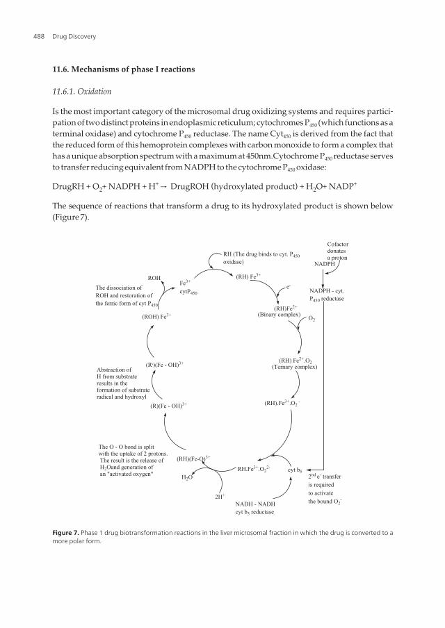
11.6.1. Oxidation
Is the most important category of the microsomal drug oxidizing systems and requires partici‐pation of two distinct proteins in endoplasmic reticulum; cytochromes P450 (which functions as aterminal oxidase) and cytochrome P450 reductase. The name Cyt450 is derived from the fact thatthe reduced form of this hemoprotein complexes with carbon monoxide to form a complex thathas a unique absorption spectrum with a maximum at 450nm.Cytochrome P450 reductase servesto transfer reducing equivalent from NADPH to the cytochrome P450 oxidase:
DrugRH + O2+ NADPH + H+ → DrugROH (hydroxylated product) + H2O+ NADP+
The sequence of reactions that transform a drug to its hydroxylated product is shown below(Figure 7).
NADPH
(RH) Fe3+ROH
(RH)Fe2+
(ROH) Fe3+ O2
(RH) Fe2+.O2
RH.Fe3+.O22-
H2O
2H+
Fe3+
cytP450
(R.)(Fe - OH)3+
(Binary complex)
RH (The drug binds to cyt. P450oxidase)
NADPH - cyt.P450 reductase
e-
(Ternary complex)Abstraction ofH from substrateresults in theformation of substrate radical and hydroxyl
The O - O bond is split with the uptake of 2 protons. The result is the release of H2Oand generation of an "activated oxygen"
Cofactor donatesa proton
(RH).Fe3+.O2 -
(RH)(Fe-O)3+
(R)(Fe - OH)3+
cyt b52nd e- transferis requiredto activatethe bound O2
-NADH - NADHcyt b5 reductase
The dissociation of ROH and restoration of the ferric form of cyt P450
Figure 7. Phase 1 drug biotransformation reactions in the liver microsomal fraction in which the drug is converted to amore polar form.
Drug Discovery488

The phospholipids of the endoplasmic reticulum are required for substrate binding, electrontransfer, and facilitating the interaction between CytP450 and its reductase. However, cyto‐chrome P450 does not catalyze all Oxidation reactions. The microsomal flavin– containingmonooxygenases (FMOs) catalyze NADPH – dependent oxygenation of nucleophilic phos‐phorous, nitrogen and sulfur atoms. These atoms are present in a wide variety of xenobioticsincluding the carbamate containing pesticides and therapeutic agents such as phenothiazines,ephedrine and N-methylamphetamine. Another important drug–oxidizing system is theprostaglandin synthetase–dependent co-oxidation.
Prostaglandin G2 reducedprostaglandin synthetase
prostaglandin H2
Many xenobiotics including phenytoin can be co-oxidized along with the above reduc‐tion reaction. This pathway is of considerable toxicological importance as it often leadsto generation of toxic reactive metabolites. Other enzymes that catalyze oxidation ofxenobiotic include alcohol dehydrogenase, aldehyde dehydrogenase, xanthine oxidaseand monoamine oxidase.
11.6.2. Reduction
Some drugs with azo-linkages (RN=NR, e.g. prontosil) and nitrogen groups (RNO2, such aschloramphenicol) are transformed by reductive pathways. The Cyt P450 and NADPH–cytP450reductase enzymes that catalyze oxidation reactions are also involved in reductionreactions for drugs containing quinine moieties. These transformation results in the formationof semiquinone free radicals illustrated in Figure 8. The free radicals that are generated causeoxidative stress, lipid peroxidation, DNA damage, and hence cytotoxicity. These effects areparticularly responsible for the antitumor property of a drug like doxorubicin.
11.6.3. Hydrolysis
Drugs containing ester functions (R1COOR2) such as procaine are hydrolyzed by a variety ofnon-specific esterases in liver, and plasma while drugs with amide bonds are hydrolyzed byamidases in the liver. The polypeptide drugs such as insulin and growth hormones arehydrolyzed by peptidases in the plasma and erythrocytes. The metabolites resulting fromhydrolysis reactions are subjected to phase II biotransformation reactions before excretion inthe bile or urine.
11.7. Mechanisms of phase II reactions
The phase II reactions generally involve coupling of drug/drug metabolite with an endogenoussubstance to enhance their removal from the body. They require participation of specifictransferase enzymes and high energy activated endogenous substances.
Introduction to Biochemical Pharmacology and Drug Discoveryhttp://dx.doi.org/10.5772/52014
489
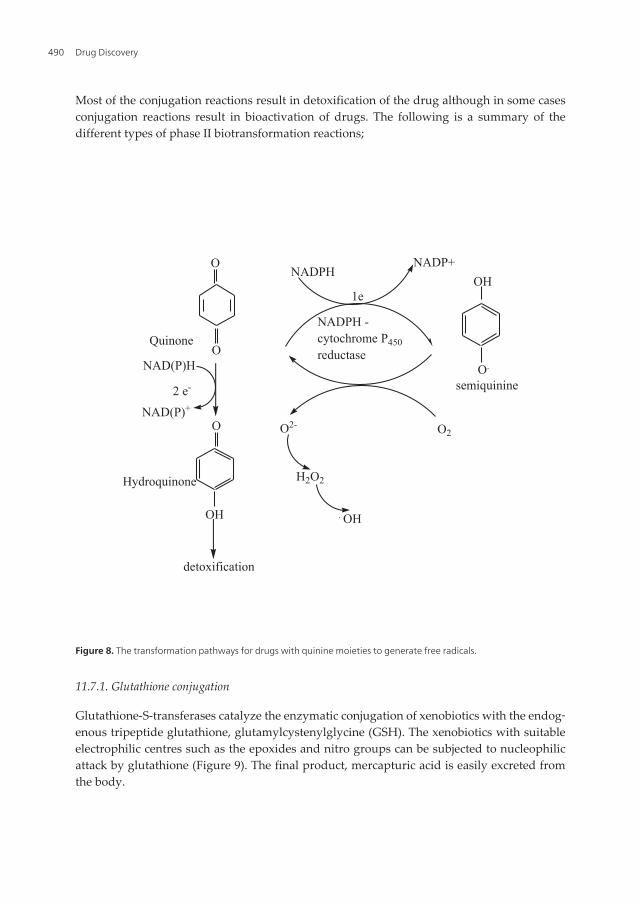
Most of the conjugation reactions result in detoxification of the drug although in some casesconjugation reactions result in bioactivation of drugs. The following is a summary of thedifferent types of phase II biotransformation reactions;
NADP+NADPH
OOH
ONAD(P)H O.
O O2O2-
H2O2
OH . OH
2 e-
Hydroquinone
1e
NADPH -cytochrome P450reductase
detoxification
semiquinine
NAD(P)+
Quinone
Figure 8. The transformation pathways for drugs with quinine moieties to generate free radicals.
11.7.1. Glutathione conjugation
Glutathione-S-transferases catalyze the enzymatic conjugation of xenobiotics with the endog‐enous tripeptide glutathione, glutamylcystenylglycine (GSH). The xenobiotics with suitableelectrophilic centres such as the epoxides and nitro groups can be subjected to nucleophilicattack by glutathione (Figure 9). The final product, mercapturic acid is easily excreted fromthe body.
Drug Discovery490

Glu - Cys - Gly + drug XGlutathione - S - transferase
Glu - Cys - Gly
SX
Glutamyl transpeptidase
Glutamylaminoacid
Cys - Gly
Cysteinylglycinase
Gly
Cys
Acetyl CoA
Acetyltransferase
N- Acetyl - Cys - SX(Mercapturic acid)
SX
CoA
H2O
SH SXAmino acid
Figure 9. Glutathione conjugation reactions for a drug with a suitable nucleophilic centre leads to the formation ofmercapturic acid which is easily excreted from the body
11.7.2. Glucuronidation
This is the conjugation of a drug or xenobiotic with glucuronic acid. Many functional groupsare subject to glucuronidation. The benzoyl group in morphine, (an analgesic) and the aminegroup in meprobamate (a sedative) can undergo glucuronidation. A drug with a benzoyl groupcan undergo glucoronidation by a transferase as shown below:
OH
HOOC
OHUDPGAtransferase
+Benzoylglucuronide
+ UDP
Benzoyl group
O
H
OH HGlucuronyl
C
C
O
O O
R
R
Introduction to Biochemical Pharmacology and Drug Discoveryhttp://dx.doi.org/10.5772/52014
491

11.7.3. Epoxide hydration
A number of aromatic compounds are transformed by phase I reactions to form epoxideintermediates. The epoxides are reactive electrophilic species that can bind covalently toproteins and nucleic acids to bring about toxic effects. These epoxides are detoxified via thenucleophilic attack of water molecule on one of the electron deficient carbon atoms of theoxizane ring as shown below:
R2R1
CHCH
Drug substrate - epoxide
+ H H+
epoxidehydrolase
R1
CH
R2
OH
CH
OH
OH
O
The glucuronide conjugates can be excreted via the bile or urine.
11.7.4. Acetylation
Acetylation is achieved by cytosolic enzymes known as N-acetyl transferases whichcatalyze transfer of acetate from acetyl co-enzyme A to primary aromatic amine or hy‐drazides (figure 10)
Acetate + CoA CH3
C
CoAAcetyl CoA
SO2 NH2
SulfanilamideN- ac
etyl
transf
erase
CH3
H
N
N- acetylsulfanilamide
O
C
O
S
H2N SO2 NH2
SH
Figure 10. Acetylation reations leading to the formation of N– Acetylsulfanilamide, the final metabolite of the antimi‐crobial agent sulfanilamide which is secreted from the body.
Drug Discovery492

11.7.5. Methylation
Most of the methyl transferases are cytosolic enzymes. They utilize S-adenosyl methionine(SAM) as the methyl donor. The final metabolite, thiopurine, has antineoplastic properties andis used as an anticancer agent (Figure 11)
Figure 11. Methylation reactions leading to the formation of methylthiopurine
12. Adverse drug reactions associated with drug biotransformationreactions
Many adverse drug reactions can be traced to an improper balance between bioactiva‐tion and detoxification reactions. For example, when the analgesic acetaminophen is giv‐en at normal therapeutic doses, it undergoes glucuronidation and sulfation reactions thatterminate the action of the drug and hasten its elimination. However, some of the drugis bioactivated via Cyt P450 to form N-acetylbenzoquinimine, a reactive intermediate thatcan be detoxified by conjugation with glutathione (GSH). When excessive doses of thedrug are given, glucuronidation and sulfation reactions become saturated and more acet‐aminophen is bioactivated via Cyt P450. This imbalance leads to high concentrations of N-acetylbenzoqunonine which cannot be sufficiently eliminated by the limitedconcentrations of gluthathione. This metabolite binds covalently to cellular protein thiolsand initiates hepatotoxicity leading to hepatic necrosis.
Introduction to Biochemical Pharmacology and Drug Discoveryhttp://dx.doi.org/10.5772/52014
493

12.1. Revision exercise 1
1. Discuss the absorption of ά-D- Ribose-5-phosphate, given that the two ionizable hydroxylgroups of the monophosphate ester ribose have pKa values of 1.2. and 6.6. The fullyprotonated form of ά-D-ribose 5-phosphate has the following structure;
H
P O
O
H
CH2
OH
HO O
OH OHOH
H
2. Using specific examples of drugs, justify their various routes of administration.
12.2. Revision exercise 2
If the concentration of a drug in plasma decreases at a rate that is proportional to its initialconcentration, give an expression that describes this relationship and hence show that [Ct] =[Co].e-kt
12.3. Worked examples
Problem 1: Describe how you can determine the partition coefficient of a labeled drug
Solution: The partition coefficient of a drug is its differential distribution between thehydrophobic and hydrophilic phases. The distribution of the drug between these two phasescan be determined by allowing equilibration of a radioactively labeled drug between aqueousbuffer containing the drug and a cell membrane preparation obtained by homogenization andfractionation of a tissue sample. The ratio of drug concentration in the membrane to theconcentration in the aqueous phase gives the partition coefficient.
Problem 2: Describe how you can demonstrate transport across membranes
Solution: Erythrocyte ‘ghosts’ or self sealing micelles formed when the erythrocytes releasecytoplasmic contents upon exposure into a hypotonic solution can be used to study the uptakeor release of labeled molecules across erythrocytes. When the ghosts are prepared in 14Cglucose, it will be possible to monitor the rate of release or uptake of the labeled 14C glucosefrom the membranes into the aqueous environment. These ‘ghosts’ can also be used to studythe uptake of various molecules at various concentrations under different conditions oftemperatures and the presence of inhibitors for specific molecular uptake.
Problem 3: A 120mg per kg dose of a drug was injected intravenously and its concentration(mg/L) monitored regularly over time. When log 10 C was plotted vs time (h) a linear responsewas obtained with a slope of – 0.08 and an extrapolated y-intercept of 1.3. Calculate thefollowing pharmacokinetic parameters;
Drug Discovery494

i. elimination rate constant (k)
ii. initial concentration of the drug in blood plasma (Co)
iii. volume of distribution (Vd)
iv. half-life of drug elimination (t ½).
Solution:
i. Slope = - k/2.303, therefore, k = -2.303x -0.08 = 0.184
ii. Co = Antilog of 1.3 = 10 1.3 = 20 mg/L
iii. Volume of distribution (Vd) = Do/Co = 120/20 = 6 L
iv. The half-life of elimination t1/2 = 0.693/k = 0.693/0.184 = 3.7 hours.
Once the apparent volume of distribution is known for a particular drug the amount of drugthat must be given to achieve a desired concentration can be determined from
Do = Co.Vd
Problem 4: You have been given the following data based on a 65 kg patient; t1/2 of drug X =4.5hrs, Vd = 0.56L/kg, Cmin the = 5mg/L, C tox = 20mg/L and Css = 10mg/L; calculate:-
i. Drug clearance from the body
ii. Average rate of drug intake (Dosing rate)
iii. Maintenance dose
iv. Maintenance interval
v. Initial loading dose
vi. Loading dose at steady state concentration
Solution
Since t1/2 = 0.693/ k; k= 0.693/ t1/2 i.e. 0.693/4.5=0.154, and V= 0.56 x 65 = 36.4.
i. Total drug clearance = kV = 0.154 x 36.4 = 5.6L/h = 93.3ml/min.
ii. Average rate of drug intake = rate elimination constant
= kxVxCss
= 0.154 x 36.4 x 10
= 56 mg/h
iii. Maintenance dose
= (Ctox - Cther). V
= (20 – 5) x 36.4 = 546 mg
Introduction to Biochemical Pharmacology and Drug Discoveryhttp://dx.doi.org/10.5772/52014
495

iv. Maintenance interval = maintenance dose/ rate of elimination
= 546/56
= 9.75 hrs
For a practical loading schedule, the maintenance interval should be lowered to say 8.0 hrsand the maintenance dose reduced proportionately: = 546 x 8/9.75 ~ 437 mg.
v. The initial loading dose
= Ctox.V
= 36.4 x 20
= 728 mg
vi. The loading dose at steady state
= Css x V
= 36.4 x 10 = 364 mg
Practical problem 1
The analytical method of assaying paracetamol relies on the introduction of a nitro group intothe molecule after the removal of plasma proteins through precipitation. The resultantnitrophenol compound which is formed has a deep yellow colour in an alkaline medium andabsorbs at 430nm Figure 12.
CH3 C
O
NH
HNO2
OHNO2
O-
OH-
CH3 C NH
OH
CH3 C NH
NO2
O O
Figure 12. Formation of a chromogenic nitro compound from an analgesic Acetaminophen
i. Describe how you would construct the standard curve for determination of parace‐tamol concentration.
K=ln X1X
t1-t2
2. Design an experiment that would enable you to determine the t1/2 of paracetamol
Drug Discovery496

Practical problem 2
Liver damage can be induced by 20% w/v carbon tetrachloride. Given 10mg/ml pentobarbitoneand 10mg/ml Phenolbarbitone, design an experiment that demonstrates that the duration ofaction of short acting barbiturates are dependent on the integrity of the liver.
13. Drug discovery and preclinical trials
The development of new drugs over the past 30 years has revolutionalized the practice ofmedicine and has for instance seen the increased use of new anti-hypertensives and drugs thatreduce cholesterol synthesis or dissolve blood clots which led to a 50% reduction in the numberof deaths from cardio-vascular diseases and stroke among other diseases.
13.1. Conventional approaches to drug discovery
These are the classical approaches to drug discovery that do not initially involve detailedscientific study they include the following;
Traditional knowledge approach
This is the discovery of drugs based on traditional medical knowledge. The best example isthe documented analgesic effects of extracts from opium poppy that led to the isolation ofmorphine from the plant and the subsequent synthesis of related analgesics.
Discovery through serendipity
This is the accidental discovery of novel drugs based on the ingenuity of a scientist investi‐gating a problem initially unrelated to the observed phenomenon; examples of such discov‐eries include the observation by Alexander Flemmings that penicilliummould could inhibitthe growth of bacteria. This finding led to the discovery of antibiotics.
Discovery of therapeutic usefulness of a side effect e.g. clonidine originally used as a nasaldecongestant was found to have antihypertensive properties while, the hypoglycemic effectsof sulphonamides used in the treatment of typhoid fever led to the development of structurallyrelated sulphonylureas as oral hypoglycemic drugs.
Discovery from effects of endogenous agents in test animals
An example of discovery arising from studies of endogenous agents in test animals is theanticoagulant action of the venom from the Malayan viper that led to the identification of theanticoagulant ancrod.
Modern approaches to drug discovery
These are those approaches that form a basis for the rational design of drugs and include thefollowing;
Bioprospecting
Introduction to Biochemical Pharmacology and Drug Discoveryhttp://dx.doi.org/10.5772/52014
497
This is the screening of a large number of natural products, chemical entities, large libraries ofpeptides, nucleic acids and other organic molecules for biological activity. This approach maylead to identification and development of new drug molecules.
Metabolomics
This is the profiling of natural products of related plant species screening using either liquidor gas chromatography mass spectrometry to determine active metabolites that may be presentin novel crude herbal medical preparations.
In silico screening
This is the most advanced technique for drug discovery. It entails virtual screening or dockingof compounds on the 3-D- structure of a known receptor based on homologies of the test drugmolecules with a known test parent drug. In silico screening can form a basis for the modifi‐cation of a known drug molecule to determine possible therapeutic applications and may leadto the development of putative drugs against new targets.
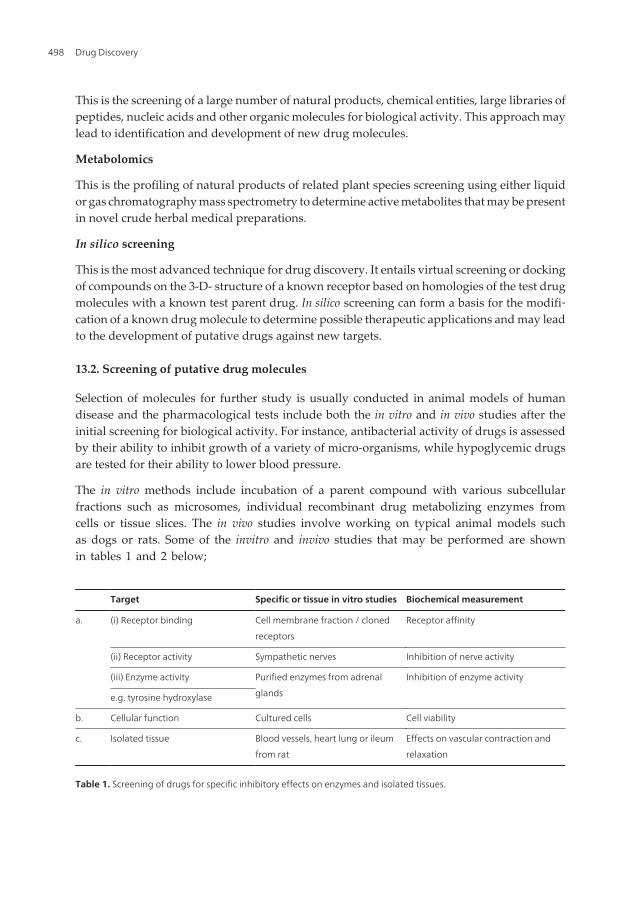
13.2. Screening of putative drug molecules
Selection of molecules for further study is usually conducted in animal models of humandisease and the pharmacological tests include both the in vitro and in vivo studies after theinitial screening for biological activity. For instance, antibacterial activity of drugs is assessedby their ability to inhibit growth of a variety of micro-organisms, while hypoglycemic drugsare tested for their ability to lower blood pressure.
The in vitro methods include incubation of a parent compound with various subcellularfractions such as microsomes, individual recombinant drug metabolizing enzymes fromcells or tissue slices. The in vivo studies involve working on typical animal models suchas dogs or rats. Some of the invitro and invivo studies that may be performed are shownin tables 1 and 2 below;
Target Specific or tissue in vitro studies Biochemical measurement
a. (i) Receptor binding Cell membrane fraction / cloned
receptors
Receptor affinity
(ii) Receptor activity Sympathetic nerves Inhibition of nerve activity
(iii) Enzyme activity Purified enzymes from adrenal
glands
Inhibition of enzyme activity
e.g. tyrosine hydroxylase
b. Cellular function Cultured cells Cell viability
c. Isolated tissue Blood vessels, heart lung or ileum
from rat
Effects on vascular contraction and
relaxation
Table 1. Screening of drugs for specific inhibitory effects on enzymes and isolated tissues.
Drug Discovery498

Disease model Animal model Route of administration Physiological
measurements
a. Blood pressure Hypertensive rat
(conscious)
Parenteral Systolic/diastolic
b. Cardiac effects Dog (conscious)
Dog (anesthetized)
Oral Electrocardiography
(cardiac output)Parenteral
c. CNS Mouse, rat Parenteral Degree of sedation
d. Respiratory
effects
Dog/guinea pig Parenteral Respiratory rate and
amplitude
e. GIT effects Rat Oral GIT motility and secretions
Table 2. Putative animal models used in studying effects of drugs
If an agent possesses useful activity it would be further studied for possible adverse effects onother major organs. These studies might suggest the need for further chemical modification toachieve desirable pharmacokinetic/pharmacodynamic properties.
13.3. Preclinical trials
The data from animal studies form a basis for the calculation of the initial or starting doses tobe used in the subsequent clinical studies. The human equivalent dose calculations for themaximum recommended dose are normally based on either the body surface area or bodyweight. The candidate drugs that survive initial screening and profiling must be carefullyevaluated for potential risks before and during clinical testing. The main types of evaluationneeded from safety and toxicity studies include:-
Acute toxicity
This involves looking at the effects of large single doses of therapeutic agent. Acute toxicitystudies are usually performed in animal models such as mice and rats. These studies enableinvestigators to correlate any observed effects with the systemic level of the drug.
Sub-acute toxicity
This is similar to acute toxicity but measures the effects of multiple doses based on expectedduration of clinical usage. It entails haematological, histology and electron microscope studiesto identify organs which might be affected by toxicity. It usually lasts between one to threemonths. This enables the selection of putative compounds for subsequent studies.
Chronic toxicity testing
These studies are required when the drug is intended to be used in humans for prolongedperiods. The goals of this investigation are mostly similar to those of sub-acute toxicity.
Introduction to Biochemical Pharmacology and Drug Discoveryhttp://dx.doi.org/10.5772/52014
499

The reproductive performance
These are measurements intended to determine the effects of the drug agents on; matingbehaviour, reproduction, parturition, progeny birth defects, and postnatal development.
Carcinogenicity studies
These studies are required to determine the effects of prolonged usage of the drug underinvestigation. They involve hematological and histological autopsy analysis.
Mutagenicity studies
These studies look at the genetic stability and mutations of bacterial or mammalian cells inculture. These studies are at the academic research level and are intended to provide data forfuture research.
Investigative toxicology
The main purpose of toxicology is to discover the pathways that are involved in toxic action.It includes studies on mechanisms of toxic action of drugs which may lead to the developmentof safer drugs.
14. Evaluation of new drugs and drug approval process
Toxicity testing is time consuming and expensive and may require two to five years to collectand analyze data before the drug can be considered ready for testing in humans.
Large numbers of animals are needed to obtain valid preclinical data.
Extrapolation of toxicity data from animals to humans may not be completely reliable.
The safety or efficacy of a drug must be thoroughly understood before the drug is ad‐ministered to any group of individuals. Therefore regulations governing the develop‐ment of new drugs have evolved to assure safety and efficacy of new medications. Theclinical trials during drug development and post marketing experience form the scientificbasis of patient response to a drug.
Once a drug is judged ready to be studied in humans, a notice of clinical investigationalexemption for a new drug (IND) must be filled with the government body concerned with theregulation and registration of drugs. The IND includes manufacturing information, all datafrom animal studies, clinical plans and protocols and the names and credentials of physicianswho will conduct the clinical trials.
14.1. Phase I clinical trials
The main goal in phase I is to determine whether test animals and humans show signifi‐cant different responses to the drug and to establish limits of the safe clinical dosagerange. The measurements carried out in phase I include, the rate of absorption, t1/2 and
Drug Discovery500

metabolism of the candidate drug compound. The effects of the drug as a function ofdosage are established in a small number 25 – 50 of healthy volunteers. When the drugis expected to have significant toxicity, as often the case with cancer and AIDS therapy,volunteer patients with the disease are used instead of the healthy volunteers. The re‐quirements of clinical trials include the following:
i. Homogenous populations of patients must be selected.
ii. Appropriate controls for the investigation must be included.
iii. Meaningful and sensitive indices for drug effects must be used i.e. well defined end-points such as survival or pain relief should be used rather than surrogate orintermediate markers e.g. levels of enzymes involved in the process of survival/painrelief.
iv. The experimental observations must be converted to data and then into validconclusions.
v. The accuracy of diagnosis and severity of the disease must be comparable betweenthe groups being contrasted.
vi. The dosages of the drugs must be chosen and individualized in a manner that allowsrelative efficacy to be compared at equivalent toxicities.
vii. Compliance with experimental regimens should be assessed before subjects areassigned to experimental or control groups. Non-compliance may cause falseestimates of the true potential benefits or toxicity of a particular treatment.
viii. Ethical considerations. These may be the major determinants of the types of controlsthat can be used e.g. for therapeutic trials that involve life threatening diseases forwhich there is already in-effective therapy, the use of a placebo is consideredunethical. In such cases, new treatments must be compared with standard therapies.
14.2. Study design of phase I trials
For clinical trials to have validity they must be based on a sound statistical basis. Some of theof the criteria that must be met include;
Randomization
Randomization is a design which ensures that there is no bias in allocation of treatments amongthe different groups. The purpose of randomization is to minimize the possibility that anobserved treatment effect is due to inherent differences between groups. Randomizationeliminates bias by avoiding recruiting patients who have a particular characteristic to onegroup and not the other e.g. only women/men and smokers/alcoholics. Randomization shouldnot be carried out until immediately before treatment. The delay allows a patient to havesecond thoughts about taking part or the investigator to have to re-consider about admittingpatients to the study. Simple methods of randomization can be designed using published tablesof random numbers, where treatments are in a form of a square in which each treatment is
Introduction to Biochemical Pharmacology and Drug Discoveryhttp://dx.doi.org/10.5772/52014
501

contained only once in each row and column and the order of treatment is different in eachgroup (Table 3).The presence of other diseases or risk factors should be taken into considerationi.e. need for careful selection and assignment of patients to each of the study groups.
Group Treatment
1 A B C D
2 C D A B
3 D C B A
4 B A D C
Table 3. A random number table array for assignment of various treatment regimes to various groups of patients inclinical trials
This approach eliminates systematic variation between groups since the patients are allocatedat random order to group 1, 2, 3, or 4. A code list should be drawn up so that the maininvestigators may be kept blind to the treatment an individual is receiving but also so that itis possible to know the treatment by breaking the code. Coding should be such that whenbroken, it does not yield information about the treatments other patients are getting. Thetreatment information about all the patients should be left to one person preferably thepharmacist or trial co-ordinator.
Blindness
Blinding is a design which does not allow the investigator to know what treatment the patientis receiving. The purpose of blinding is to eliminate bias in reporting the outcome of thetreatment since if an investigator knows what treatment the patient is taking, he/she may insome way influence a measurement or an outcome thus shifting the outcome in one directionor another consciously or unconsciously. The ideal trial should be double blind where neitherthe investigator nor the patient knows what treatment the patient is taking. Placebos ordummies are used in order to achieve blindness. The placebos should match the activetreatment as closely as possible in terms of size, shape, color, texture, weight, taste and smellwith the active formulation.
Number of testing centers
Clinical trial should be carried out in a defined centre so as to minimize variations in the popula‐tion and in the investigators techniques. This also avoids problems of data collection, communi‐cation and follows up. Multi-centre trials may become necessary when studying a rare diseasehence scarcity of patients or when the effect being investigated is small one e.g. when one islooking out for an interaction effect between a major condition and a minor condition.
Clinical trials designed to evaluate efficacy of new drugs should always be prospective i.e. thecharacteristics of the population to be studied should be identified before the study begins.For example, if one randomized all patients with heart failure to treatment with either digoxinor a new drug X and then studied the outcome over six months that would be a prospective
Drug Discovery502

trial. In case of control study the outcome is first identified and then comparisons are maderetrospectively between the characteristics of patients who did or did not have the outcome.Such a study for instance has shown that oral anticoagulants can reduce incidences of re-infarction in patients who have already had a myocardial infarction. The case control studiesmay be carried out some time, after the introduction of a drug therapy in order to get someidea of its place in the overall management of the disease since the results of a case controlstudy may prompt formal prospective trials in order to confirm original findings.
14.3. Molecular markers in drug development
In vitro predictive efficacy and toxico-genomics should be carried out after phase 1 clinicaltrials in order to validate the results of the phase 1 clinical trials. This is achieved by usinganimal cell lines in which gene expression profiling and patterns of protein production areused to identity candidate biomarkers for the disease. The utilization of markers that areassociated with the disease or those that indicate a known response to a therapeutic interven‐tion or reflect a clinical outcome may yield information on efficacy or toxicity of a test drug.An example of a biological readout that has traditionally been used to determine efficacyduring the treatment of diabetics is the determination of glucose in the urine of a diabeticpatient. Reliable and specific biomarkers that act as predictors of efficacy or long-term toxicityare useful because they reduce the time, size and cost of clinical trials.
14.4. Phase II clinical trials
These are studies that recruit willing and informed patients and are designed to assess longterm safety, refine pharmacokinetic data, determine optimal dose. The purpose of phase IIstudies is to determine efficacy. Typically, phase II trials require 100-150 subjects and take 9-12months. An assessment of no effect or no worthwhile effect of a given drug demonstrates thatit is futile to proceed with further clinical testing of the drug. It is therefore important tominimize type I errors or false negatives in the study design in order to minimize the risk ofdiscontinuing a potentially effective drug. The data from well designed phase I and phase IItrials are therefore critical in planning the subsequent trials. The phase III trials are large trialsintended to determine whether a treatment is effective and to establish safety data.
Phase II clinical trials include inert placebos as negative controls and older active drugs aspositive controls alongside the investigative compound. These studies are done in specialclinical centers such as University Hospitals. A broader range of toxicities may be detected atthis phase.
14.5. Phase III clinical trials
The drug is evaluated in a much larger number of patients (thousands) to further establishsafety and efficacy. Phase III trials are performed in settings similar to those anticipated forthe ultimate use of the drug. After successful phase III trials, the next step is the applicationfor review of the new drug to seek approval to use the drug for clinical management of thedisease condition.
Introduction to Biochemical Pharmacology and Drug Discoveryhttp://dx.doi.org/10.5772/52014
503

14.6. Phase IV clinical trials
This phase is concerned with post-marketing surveillance and the main goal is to as‐sess adverse reactions, patterns of drug utilization, discovery of additional indications.The interrelationships between the various studies in drug development are illustratedin Fig 13 below;
Phase II clinical trials include inert placebos as negative controls and older active drugs as positive controls alongside the investigative compound. These studies are done in special clinical centers such as University Hospitals. A broader range of toxicities may be detected at this phase. Phase IIIclinical trials The drug is evaluated in a much larger number of patients (thousands) to further establish safety and efficacy. Phase III trials are performed in settings similar to those anticipated for the ultimate use of the drug. After successful phase III trials, the next step is the application for review of the new drug to seek approval to use the drug for clinical management of the disease condition. Phase IV clinical trials This phase is concerned withpost-marketing surveillance and the main goal is to assess adverse reactions, patterns of drug utilization, discovery of additional indications.The interrelationships between the various studies in drug development are illustrated in Fig 1.13 below; Figure 1.13Illustration of the key steps in the development of a drug from a putative drug candidate extract
Crude drug preparation
Biochemical profiling of crude drug product
In vivo/in vitro therapeutic profile for test and control model
In vivo toxicity profiles for test and control
model
Phase IV Clinical trials
Phase I Clinical trials
Phase II Clinical trials
Phase III Clinical trials
Figure 13. Illustration of the key steps in the development of a drug from a putative drug candidate extract
Drug Discovery504

14.7. Pharmacogenomics and drug development
The personalized medication which takes into account the genetic make-up of individu‐als is known as pharmacogenomics. The pharmacogenomic differences that determine in‐dividualized therapy include genetic polymorphisms of drug transporters, drugreceptors, and drug metabolizing enzymes. For example, genetic variation in Cyt P450 en‐zymes that are largely responsible for drug metabolism shows that different individualsrespond differently to drug efficacy or toxicity. Genetic variants in the drug target, thedisease pathway, genes or drug metabolizing enzymes could all be used as predictors ofdrug efficacy or toxicity. For example, drug monitoring using perpherazine, a Cyt P450
substrate, shows that there are three main categories of individuals; the efficient metabo‐lizers obtained from the heterozygotes, the poor metabolizers from the homozygotes andthe ultra-rapid metabolizers which carry two or more active genes in the same chromo‐some, a phenomenon known as gene duplication.
The information obtained from pharmacogenetic studies can be used to design newdrugs that take the persons’ genetic profile into consideration. The most common type ofgenetic variation are single nucleotide polymorphisms, therefore, a high resolution of sin‐gle nucleotide map may expedite the identification of genes for various diseases. Themolecular profiles of patients identified in phase I and II clinical trials as likely non-res‐ponders to the putative drug under investigation might present an opportunity to ini‐tiate new discovery programs for other pharmaceutical compounds.
14.8. Individualized drug therapy
Clinical usage of drugs requires a basic understanding of the pharmacokinetic and pharma‐codynamic drug processes and an appreciation that a relationship does exist between thepharmacological effect or toxic response to a drug and the concentration of the drug. Theinterpatient and intrapatient variation in disposition of a drug must be taken into account inchoosing a drug regimen.
A drug dosage regimen therefore is a recipe for the administration of a drug so as to pro‐duce a desired therapeutic effect with minimum toxic effects.
The regimen is described in terms of the following:
i. Dose of the drug to be used and the formulation.
ii. Frequency with which it is administered.
iii. Route of drug administration.
The factors that determine the relationship between the prescribed drug dosage and drugeffect operate at three levels; prescription level, drug administration level and at the physio‐logical level of patient (Figure 14).
Introduction to Biochemical Pharmacology and Drug Discoveryhttp://dx.doi.org/10.5772/52014
505
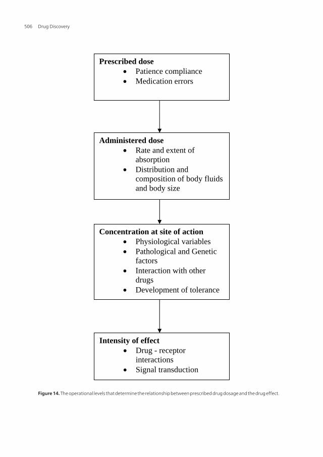
Figure 1.14: The operational levels that determine the relationship between prescribed drug dosage and the drug effect. Drugs that are excreted primarily unchanged by the kidneys tend to have low variation among patients with similar renal function than do drugs which are inactivated by metabolism. For the extensively metabolized drugs, those with high metabolic clearance and large first pass elimination have marked difference in bioavailability, whereas those with low biotransformation tend to have largest variation in elimination rates among individuals.
Prescribed dose Patience compliance Medication errors
Administered dose Rate and extent of
absorption Distribution and
composition of body fluids and body size
Concentration at site of action Physiological variables Pathological and Genetic
factors Interaction with other
drugs Development of tolerance
Intensity of effect Drug - receptor
interactions Signal transduction
Figure 14. The operational levels that determine the relationship between prescribed drug dosage and the drug effect.
Drug Discovery506

Drugs that are excreted primarily unchanged by the kidneys tend to have low variation amongpatients with similar renal function than do drugs which are inactivated by metabolism. Forthe extensively metabolized drugs, those with high metabolic clearance and large first passelimination have marked difference in bioavailability, whereas those with low biotransfor‐mation tend to have largest variation in elimination rates among individuals.
14.9. Determination of drug dosage
The simplest way of determining a drug dosage regimen is to base it on the publishedrecommended dosage. These are derived from the pharmacokinetics studies of the drugand the general procedure in using the published recommendations is to start at the low‐er end of the recommended dosage range and monitor the therapeutic effect. If the de‐sired effect does not occur, the dosage can be increased gradually until one reaches theupper limit of the range. In certain conditions it may be necessary a sufficiently highdose for the drug to accumulate in the body to a satisfactory degree. This dose is knownas the loading dose and is equal to the volume of distribution multiplied by the targetconcentration in the plasma. The reason for giving a loading dose is to circumvent thesometimes unacceptable time lag preceding the steady state levels. Once the correct load‐ing dose is given, a steady-state concentration can be achieved rapidly and then main‐tained by giving a smaller maintenance dose.
Adjustment of dosage in individual patients is often as a result of the modification ofpharmacokinetic parameters of which the three most important include; the bioavailabili‐ty or the fraction of a drug that is absorbed into systemic circulation, its clearance andthe volume of distribution.
For drugs with a high toxicity to therapeutic ratio, the loading dose can be given as a singledose and for drugs with a low toxicity: therapeutic ratio and a long half-life, the loading dosecan be divided into several portions and given at intervals long enough to allow detection ofadverse effects, but short enough to ensure that the loading dose is a true loading dose i.e.relatively little amounts of the drug is eliminated from the body during the period of loading.
14.10. Systemic drug availability
The extent of availability of a drug after oral administration is expressed as a percentage of thedose. The fractional availability (F) varies from 0 to 1. The extent of availability is moreimportant parameter to measure rather than the rate of availability.
A true decrease in bioavailability could be due to several reasons including, a poorly ad‐ministered dosage form that fails to disintegrate or dissolve in the GIT, interaction withother drugs in the GIT, metabolism of the drug in the GIT and/or first pass hepatic me‐tabolism or biliary excretion.
Hepatic disease may in particular cause high availability because the metabolic capacitydecreases or development of vascular shunts in the liver. Significantly high availabilityrequires dosage adjustment by a factor of two, while significantly low in availability requiresdosage adjustment by a factor of half.
Introduction to Biochemical Pharmacology and Drug Discoveryhttp://dx.doi.org/10.5772/52014
507

14.11. Maintenance dose
In most clinical situations drugs are administered in such a way as to maintain a steadyconcentration i.e. just enough drug is given in each dose to replace the drug eliminated sincethe preceding dose. Therefore, clearance is the most important pharmacokinetic term to beconsidered in defining a rational steady-state drug dosage regimen.
The rate of elimination = Cl x Tc
Where, Cl is the rate of clearance and Tc is the target concentration of the drug.
The Dosing rate = Rate of elimination = Cl x Tc.
Therefore, if the target concentration is known, the prevailing clearance in that patient willdetermine the dosing rate and if the drug is given by a route that has a bio availability of lessthan 100%, then the dosing rate above can be modified using the formula:
Dosing rate oral = Dosing rateFractional availability
If intermitted doses are given, then maintenance dose = Dosing rate x Dosing interval.
14.12. Alteration of maintenance dose
The maintenance dose is usually altered when the clearance of the drug changes. For example,during renal impairment, the clearance of drugs which are predominantly cleared by thekidney is greatly reduced and therefore, the desired steady state concentration can only beachieved either through altering the dose or altering the dosing interval. Therefore, when adrug is cleared almost completely via kidneys, the dosage interval should be changed inproportion to renal clearance as follows:
The % eliminated in dosing interval should be proportional to creatinine clearance by apublished constant to yield the percentage excreted in one dosage interval.
The quantities required for this adjustment are:
i. Fraction of normal function remaining, and the
ii. Fraction of drug usually excreted unchanged in urine.
The fraction of normal function remaining is equal to the ratio of patient’s creatinine clearanceto a normal value (120 ml/min/70kg).
The following equation is for adjustment of renal clearance
rfpt= 1 – fenl(1 – rfxpt)
Where;
rfpt = the adjusted total clearance of the patient,
fenl = fraction of drug excreted unchanged in normal individuals,
rfxpt = fraction of renal clearance of the normal individual.
Drug Discovery508

Clearance should also be adjusted for the size of the patient and for convenience, the publishedvalues are normalized to the metabolic rate = weight 0.75.
When clearance is low, t1/2 is similarly high and when the volume of distribution is high,the t1/2 is also high. Therefore, by using parameters for the individual patient, the dosingrate = Tc x Cl/F where, Tc = target concentration, Cl = clearance and F = fractional availa‐bility of the drug.
If a drug is relatively non toxic then the maximum loading strategy can be employed so thatthe dosing interval is much longer than t1/2.. For example t½, of penicillin is less than one hourbut it is usually given in very large doses every six to twelve hours since it is non-toxic. Thenormal steady-state theophylline concentration can be determined using the equation:
Css, max =F.dose/Vss
1 – exp(-KT)
Css, min =F.dose/Vss. (exp-KT)
1 – exp(-KT)
Where, Css, max and Css min are the maximum and minimum steady state concentrations,
T = dosage interval and K =0.693
t½
14.13. Drug dosage adjustment in old patients (geriatrics)
Drug absorption in the elderly is slightly different from the normal patients and thereforeadjustment of the dosage should be taken into consideration during drug therapy. The rate oftransdermal drug absorption may be diminished in elderly because of reduced tissue bloodperfusion. Compounds that permeate the intestinal epithelium by carrier mediated transportmechanisms may be absorbed at lower rates in the elderly.
14.14. Drug distribution
In geriatrics the ‘body mass’ declines with age and the total body water content falls by between10 – 15%. The volume of distribution of hydrophilic drugs will therefore decrease while plasmaconcentration will increase and the likelihood of toxic drug effects will also increase. Whengeriatric patients use diuretics, the extracellular space reduces even further leading to a higherlikelihood of drug toxicity. The total body fat in the elderly increases by 12 – 18%, therefore,for hydrophobic drugs, the higher volume of distribution implies an increase in half life ofdistribution and the time needed to reach steady-state serum concentration. Therefore, forgeriatrics a once or twice daily drug administration is optimal. This can be achieved thoughdelayed release or fixed drug combinations.
14.15. Patient compliance and rational use of drugs
Drug treatment of any kind is often compromised by lack of full compliance by the patient.The common errors of compliance to a regimen by a patient include; omission in taking the
Introduction to Biochemical Pharmacology and Drug Discoveryhttp://dx.doi.org/10.5772/52014
509
drug, wrong timing of dosages, premature termination of therapy or using additionalmedications. In order to improve patient compliance, the patient should be made to under‐stand the nature and prognosis of the illness and what to expect from the medication bydetailing both the acceptable and undesirable unwanted effects as well as signs of efficacy thatmay help enforce compliances.
Patients frequently discontinue taking a medication such as septrin because they have not beentold the necessity of continuing with the drug after the acute symptoms have subsided.
The effectiveness of physician-patient communication is inversely related to the error rate inthe taking of drugs. A physician might prescribe a drug to be taken three times a day withmeals for a patient who either eats only twice a day or sleeps all day and works at night.Therefore, an exploration of the patients eating, sleeping and working habits is necessarybefore a prescription is given.
The educational level of a patient may also require that the prescription is carefully wordedand oral instructions given in the primary language of the patient since when such patientstake three or more medications they are less likely to use them properly. It is thereforeimportant to provide identifying symbols for each medication e.g. “Heart pill” or “sugarpill”and to reduce the doses into once or twice daily regimens.
14.16. Adverse drug reactions
Pharmacological formulations are potentially harmful to the individuals taking the drugs.There is need to ascertain the safety of new drugs before allowing them to be marketed.The following figures highlight the magnitude of the problem: ~ 10 – 20% of hospital‐ized patients suffer adverse drug reactions, while 0.3 – 5.0% inpatients admissions and ~0.3% deaths in hospital are due to adverse drug reactions. Adverse drug reactions can beclassified into two main categories: They may be dose related or non dose related witheach being short-term or long-term.
14.17. Dose related adverse reactions
Adverse drug reactions can occur because of the changes in the systemic availability of aformulation. For instance, the change of excipients in phenytoin capsules from CaSO4 tolactose leads to high availability and hence adverse drug reactions. Sometimes, adversedrug reactions can occur due to the presence of contaminants like bacteria if quality con‐trol breaks down. Out-of-date formulations may also cause adverse drug reactions be‐cause of degradation products arising from the drug e.g. outdated tetracycline may causeFaconis Syndrome (a type of rickets) because of the transformation product,epiandrote‐tracycline. Dose related adverse reactions may also arise from pharmacokinetic variationsin the individuals taking the drug. Pharmacokinetic variations may also arise due to hep‐atic disease like advanced cirrhosis which lowers the clearance of drugs such as pheny‐toin and morphine. The pharmacological variations could be environmental such as dietor smoking, while others are genetic.
Drug Discovery510

14.18. Non-dose related adverse drug reactions
These include immunologic reactions and are related to the surface proteins present on β-humans lymphocytes (HLA antigens) which are important in the function of T-lymphocytes.The association of HLA antigens with foreign antigens stimulates T-lymphocytes. Some ofthese antigens expressed by major histocompatibility complex (MHC) genes have beenassociated with an increased risk of adverse drugs e.g. nephrotoxicity from penicillamine isincreased in patients with HLA types B8 and DR 3 while patients with HLA- DR, 7 are protectedagainst adverse drug effects.
14.19. Types of drug allergies
Drug allergies can be classified into five categories:
Type I
This anaphylaxis or immediate hypersensitivity reactions; the body reacts within five to thirtyminutes. The IgE molecules fixed to mast cells and basophil leucocytes release histamine andother pharmacological mediators such as kinins. Drugs likely to cause are anaphylactic shockinclude;-penicillins, streptomycin, local anaesthetics etc.
Type II
In these reactions, the circulating antibody of 1gG, 1gM or 1gA interacts with the drugcombined with a cell membrane protein to form a hapten-protein/antigen-Ab complex. Thisleads to the activation of the complement leading to cell lysis of phagocytic attack of the cellwith the complex. Drugs such as the cephalosporins, penicillins, quinine and transfusion ofimproperly matched blood can yield this type of reactions.
Type III
In this type of allergy, the immune complex reactions initiate an inflammatory response dueto the combination of the excess drug- protein complex with the IgG in circulation. The complexthus formed is deposited in the tissues and causes activation of the complement and damageof capillary endothelium. This type of reaction is manifested mostly as fever, arthritis, and/orenlarged lymph nodes. Penicillins, sulphonamides and streptomycin may elicit type III allergicreactions.
Type IV
This is the cell-mediated or delayed hypersensitivity reactions in which the T - lymphocytesare sensitized by a hapten to form protein-antigenic complex such that when the lymphocytescome into contact with the antigen, an inflammatory response ensues. Type IV reactions areexemplified by contact dermatitis caused by local anaesthetic areas, antihistamine areas,topical antibiotics and antifungal drugs.
Type V
These are pseudo allergic reactions, that resemble allergic reactions clinically, but for whichno immunological basis can be found, e.g. asthma and skin rashes caused by aspirin. Admin‐
Introduction to Biochemical Pharmacology and Drug Discoveryhttp://dx.doi.org/10.5772/52014
511

istration of ampicillin or amoxicillin causes a skin rash which resembles the one caused bypenicillin hypersensitivity. The ampicillin-caused rash can be distinguished from penicillinhypersensitivity on the basis of two features; it has a later onset, typically ten to fourteen days,compared to penicillin sensitivity which comes between seven to ten days. Furthermore, thesensitivity does not recur following re-exposure to ampicillin and is not as serious as the onecaused by penicillin.
14.20. Clinical evaluation of adverse drug reactions
The two basic approaches for clinical evaluation of adverse drug reactions include the cohortstudies (or follow-up studies) of patients taking the drug and the case control studies whichrecord the incidences of adverse drug effects retrospectively.
Cohort or prospective studies
In cohort studies, drugs are identified and incidences of adverse effects recorded. The weak‐nesses of these studies include; the relatively small number of patients likely to be recruited,and lack of suitable control groups to assess the background incidence of any apparent adversereaction noted.
Case control or retrospective studies
The approach here is to start with the incidence of adverse reaction(s) and then look for thedrug and the individuals with symptoms which could be due to an adverse drug reaction.These individuals are screened to see if they had taken the drug. The prevalence of drug takingin the group is then compared with the prevalence in a reference population which did nottake the drug. This approach is excellent for validation and assessment of adverse drug effects,but it may not detect new adverse effects. Furthermore, it requires a very large number ofpatients and is very expensive to undertake hence difficult to justify and organize for everynew product.
14.21. Worked examples
Problem 5
Drug clearance must always be adjusted for alterations of renal function using the formula:rfpt = 1 - fenl (1 - rfxpt),where fenl = fraction of the drug excreted unchanged in normal individuals,rfpt = adjustment factor for total clearance in patient, rfxpt = patients’ clearance as a fraction ofnormal clearance and Clnl= normal clearance.
Given that an asthmatic patient has a creatinine clearance of 40mlmin-1 70kg-1 and that thefraction of terbutaline excreted unchanged, fenl= 0.56, the normal clearance,Clnl = 3.4ml/min/kg, calculate the clearance of the drug in the patient.
Solution
The patient has depressed renal function: rfxpt = (40 ml/min)/ (120 ml/min) = 0.33
rfpt= 1 - fenl(1-rfxpt)
Drug Discovery512

inline formula rfpt= 1 - 0.56 (1 - 0.33) = 0.62
inline formula
Clpt= Clnl.rfpt
= 3.4ml.min-1kg-1x 0.62 = 2.1ml.min-1kg-1
Problem 6
Given the following characteristics of drug A; t½ = 8h, given at a dosage of 450mg every 12h,has Vss = 0.5 L/kg, effective concentration is 12mg/L and that
Cssmin = F x dose / V ss(ex p −kt)
1 – ex p (−KT )
Css, max =F x dose/Vss
1 – exp(-kT)
Determine the Cssmin and Cssmax for a 60 kg patientif F = 1 and expKT = 0.35. Explain howchanging of the dosage interval to 6 hours would affect Cssmin.
Solution
The term exp(-KT) is the fraction of the last dose that remains in the body at the end of a dosinginterval and is equal to 0.35 and Css min = Css max.exp(-kt). Therefore,
Cssmin =450/30 x 0.35
1- 0.35 =15/0.65 x 0.35 ~ 8.0 mg/L while Cssmax =15/0.65=23 mg/L
The predicted minimum of 8.0mg/L is below the effective concentration to achieve efficacy.Therefore the dosing interval should be reduced. A reduction of the interval to, say, six hours,increases denominator and therefore causes an increase of Cssmin. Since t1/2 = 0.693/K, K = 0.086i.e. 1 - e-KT = 1 - 2.71-0.086 x 6 = 1 - 0.596 = 0.4.
The new Css minimum becomes; = 450/30 x 0.4 = 15 mg/L which is within the required thera‐peutic concentration.
Review exercise
1. Write an essay on statistical considerations that guide clinical evaluation of new drugagents
2. Write an essay on bioprospecting for new antimicrobial agents.
3. Drug clearance must always be adjusted for alterations of renal function using theformula: rfpt = 1- fenl (1- rfxpt), explain what each term in the above equation represents.Consequently, calculate the clearance of acetaminophen (panadol) in a 70 kg patient withdepressed renal function given the following; normal clearance = 350 ml min -170 kg; fenl
= 0.56. The patient creatinine clearance = 80 mlmin-1 70 kg. Normal creatinine clearance =120 ml min-1 70 kg. What effect would the impaired renal function have on dosing interval?
Introduction to Biochemical Pharmacology and Drug Discoveryhttp://dx.doi.org/10.5772/52014
513

4. Given that fenl for drug X = 0.42, and the normal creatinine clearance is 120 ml/min/70 kg,calculate the clearance rate of the drug by a patient with creatinine clearance of 75 ml/min/70 kg.
5. Write an essay on adverse drug reactions associated with the use of macrolide antimicro‐bial agents.
Author details
Gabriel Magoma
Department of Biochemistry, Jomo Kenyatta University of Agriculture and Technology. Nai‐robi, Kenya
References
[1] Christoph & F., Heike Brotz-Oesterhelt (2005). Functional genomics in antibacterialdrug discovery. Today Targets DD. 10
[2] David, A. L. and Michael P. W. (2004). Molecular biomarkers in drug development.Research Focus DDT. 9, 22
[3] Di Bernardo, A.B. and Cudkowicz, M.E. (2006). Translating preclinical insights intoeffective human trials in ALS. Biochemicaet Biophysica Acta1762, 1139 -1149.
[4] Harvey A, L. (2007). Natural products as screening source. Current Opinions in ChemicalBiology.11, 480 - 484.
[5] Jaennefer M. F., Paul, A. L. (2000) Emerging novel antifungal agents. Therapeutic focus.DD. 5, 1 January 2000
[6] Iris, R. (2008). PK/PD Modelling and simulations: utility in drug development. DrugDiscovery Today.348, 1 - 6.
[7] Klaus, T. (2003).When drug therapy gets old: pharmacokinetics and pharmacodynam‐ics in the elderly. Experimental Gerontology38 843-853.
[8] Kieran, F. R., Eric S. and Philip H. W. (2001).Modelling and simulation in clinical drugdevelopment. Research Focus DD Vol. 6, 15
[9] Lindpaintner, K. (2002). The Impact of pharmacogenetics and pharmagenomics ondrug discovery. Naure Review on Drug Discovery.1, 463 - 469.
[10] Sundbergh, M. I. (2001). Genetic susceptibility to adverse effects of drugs and environ‐mental toxicants. The role of the CYP family of enzymes Mutants Researh 482. 11-19
Drug Discovery514

[11] Timothy, D. R., Steven R.G., Herve, T., Brian A.D. (2001). Finding drug targets inmicrobial genomes. Drug Discovery Today6, (17): 887-892.
[12] Tim, R. C. and Marc L. (2007). Pharmacogenitics of antiretroviral drugs for the treat‐ment of HIV-infected patients: An update. Infection, Genetics and Evolution 7, 333-342
[13] Turnheim, K. (2003). Why drug therapy gets old Pharmacokinetics and pharmacody‐namics in the eldery. Experimental Gerontology 38 843-853.
[14] Paul, B. (2003). Toxicokinetics in preclinical evaluation. Research Focus DD Vol. 8
[15] Ulrich, R., Friend, S. H. (2000). Toxicogenomics and drug discovery: Will new technol‐ogies help us produce better drugs? Nat. Rev. Drug Discov. 1, 84 – 88.
[16] Werbovetz, K. A. (2000). Target based drug discovery for Malaria, Leishmania andTrypanosomiasis. Current medicinal Chemistry 7 (8) 835 – 860.
[17] Aungst B. (2000). Intestinal permeation enhancers. Journal of Pharmacological Sciences,89(4), 429-442.
[18] Salama, N.N. Eddington N.D. Fasano, A. (2006).Tight junction modulation and itsrelationship with drug delivery. Advanced Drug Delivery Reviews.58: 15-28.
[19] Guengerich F. P. (2001). Common and uncommon cytochrome P450 reactions related tometabolism and chemical toxicity. Chemical Research and Toxicology.14: 611 - 650.
[20] Blanka R., (1998). Receptor-mediated targeted drug or toxin delivery. Advanced DrugDelivery Reviews 29: 273-289.
[21] John R. C. (2005). Some distinctions between flavin-containing and cytochromeP450monooxygenases.Biochemical and Biophysical Research Communications 338: 599-604.
[22] Xiaoling L. William K. C. (1999). Transport, metabolism and elimination mechanismsof anti-HIV agents. Advanced Drug Delivery Reviews29: 273-289 39, 81-103.
[23] Brown, M.J., Bennet, P.N., Lawrence, D.R. (1997). Clinical pharmacology (8th Edition)Wesley, Longman China Ltd.
[24] Hardman J.G., Gilman A.G., Limbird L. E.(1996). The pharmacological basis of therapeu‐tics (9thEdition) McGraw-Hill.
[25] Katzung B. G.,(2004). Basic and clinical pharmacology (9th Edition) Mcgraw Hill Inc.
Introduction to Biochemical Pharmacology and Drug Discoveryhttp://dx.doi.org/10.5772/52014
515
