recalcitrant corneal ulcer - ophthalmicresearch.in corneal.pdf · as per who, corneal blindness is...
TRANSCRIPT
Current Indian Eye Research 45
Review ArticleReview ArticleReview ArticleReview ArticleReview Article
Recalcitrant corneal ulcerAnubha Rathi1, Namrata Sharma1, Neelima Aron1, Rajesh Sinha1, Tushar Agarwal1
1Dr R P Center of Ophthalmic Sciences, All India Institute of Medical Sciences, New Delhi, India.Corresponding Author : Prof. Namrata Sharma, E-mail: [email protected] on : 08/10/2016, Revision accepted on : 18/11/2016Conflict of Interest : None, Financial Disclosure : None© Current Indian Eye Research.
A corneal ulcer is defined as loss of surface cornealepithelium along with infiltration into the surrounding
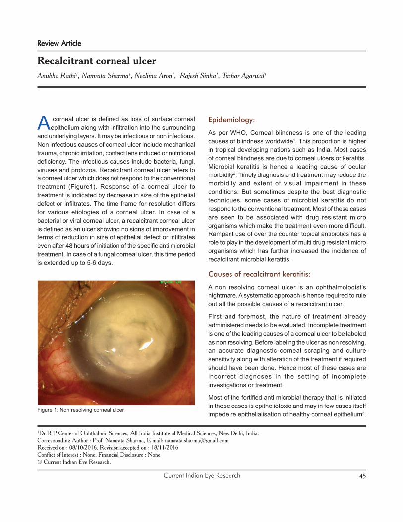
and underlying layers. It may be infectious or non infectious.Non infectious causes of corneal ulcer include mechanicaltrauma, chronic irritation, contact lens induced or nutritionaldeficiency. The infectious causes include bacteria, fungi,viruses and protozoa. Recalcitrant corneal ulcer refers toa corneal ulcer which does not respond to the conventionaltreatment (Figure1). Response of a corneal ulcer totreatment is indicated by decrease in size of the epithelialdefect or infiltrates. The time frame for resolution differsfor various etiologies of a corneal ulcer. In case of abacterial or viral corneal ulcer, a recalcitrant corneal ulceris defined as an ulcer showing no signs of improvement interms of reduction in size of epithelial defect or infiltrateseven after 48 hours of initiation of the specific anti microbialtreatment. In case of a fungal corneal ulcer, this time periodis extended up to 5-6 days.
Epidemiology:
As per WHO, Corneal blindness is one of the leadingcauses of blindness worldwide1. This proportion is higherin tropical developing nations such as India. Most casesof corneal blindness are due to corneal ulcers or keratitis.Microbial keratitis is hence a leading cause of ocularmorbidity2. Timely diagnosis and treatment may reduce themorbidity and extent of visual impairment in theseconditions. But sometimes despite the best diagnostictechniques, some cases of microbial keratitis do notrespond to the conventional treatment. Most of these casesare seen to be associated with drug resistant microorganisms which make the treatment even more difficult.Rampant use of over the counter topical antibiotics has arole to play in the development of multi drug resistant microorganisms which has further increased the incidence ofrecalcitrant microbial keratitis.
Causes of recalcitrant keratitis:
A non resolving corneal ulcer is an ophthalmologist’snightmare. A systematic approach is hence required to ruleout all the possible causes of a recalcitrant ulcer.
First and foremost, the nature of treatment alreadyadministered needs to be evaluated. Incomplete treatmentis one of the leading causes of a corneal ulcer to be labeledas non resolving. Before labeling the ulcer as non resolving,an accurate diagnostic corneal scraping and culturesensitivity along with alteration of the treatment if requiredshould have been done. Hence most of these cases areincorrect diagnoses in the setting of incompleteinvestigations or treatment.
Most of the fortified anti microbial therapy that is initiatedin these cases is epitheliotoxic and may in few cases itselfimpede re epithelialisation of healthy corneal epithelium3.
Figure 1: Non resolving corneal ulcer
Current Indian Eye Research46
Even in the best possible situation with accurate and timelycorneal scraping and microbiological examination alongwith timely initiation of anti microbial treatment, some ulcersdo not show any improvement. With increasing incidenceof drug resistant micro organisms, such ulcers are on therise. Microorganisms develop resistance to the antimicrobial treatment by genetic mutation and inductiveexpression of latent chromosomal genes with exchangeof genetic material via transformation, bacteriophagetransduction or plasmid conjugation4.
In certain cases, the size and extent of the ulcer is alreadylarge reaching upto the limbus with impending perforationat the time of presentation to the ophthalmologist. In suchcases, standard anti microbial treatment may not work.They may require surgical intervention. Atypical microorganisms may also cause corneal ulcers not respondingto the routine treatment.
Adnexal abnormalities such as eye lid malposition mayalso affect the healing process of a corneal ulcer5. Coexisting Thyroid eye disease with axial proptosis andlagophthalmos may impair the normal healing process ofthe ulcer despite suitable antimicrobial treatment6.
Patients with systemic diseases such as Diabetes mellitusare known to have delayed healing and may develop arecalcitrant corneal ulcer.
Other immune compromised conditions such as HIV-AIDS,patients on immune suppressive treatment, anti cancerdrugs etc are also prone to develop recalcitrant cornealulcers7.
Non infective recalcitrant corneal ulcers may be associatedwith mechanical trauma to the cornea in the form of papillaeor follicles in upper lid palpebral conjunctiva as seen intrachoma.
The possible causes of a recalcitrant corneal ulcer aresummarized in Table 1.
Clinical Presentation:
A case of a non healing or recalcitrant ulcer usuallypresents with an acute red eye with complaints of redness,pain, photophobia and vision loss. There may be a historyof trauma with vegetative material or a febrile viral illness.Such history may be a pointer towards the probable etiologyof the ulcer and may guide the treatment.
A. History and Symptoms:
The presence of pain is due to the irritation to the sensorynerve fibres present in the superficial layers of the cornea.The nature of pain is also an indicator of the probablecause. Bacterial keratitis is usually characterized bysignificant pain. Fungal keratitis is usually not associatedwith severe pain. Acanthamoeaba keratitis is characterizedby severe pain owing to radial keratoneuritis. This pain isusually out of proportion to the size and extent of the ulcer.Herpetic dendritic ulcers are usually associated withminimal pain. In cases of recalcitrant corneal ulcers, amixed picture may be present thereby complicating thediagnosis based on history. A sudden relief in pain in suchcases may indicate a perforated corneal ulcer. Bacterialkeratitis usually has a sudden onset with rapid progressionwhereas fungal and acanthamoeba keratitis typically havea chronic course.
B. Predisposing Factors:
For timely initiation of empirical anti microbial treatmentbefore the microbiological evidence becomes available, itis important to look for the specific predisposing factorswhich would point towards a specific etiology. Cornealtrauma by a vegetative matter usually leads to mycoticulcer. Prolonged contact lens wear especially overnight orextended wear soft contact lenses may cause cornealulcers especially due to infection by the Pseudomonasspecies8. Exposure to dirty swimming pool water or pond
Rathi A et al: Recalcitrant corneal ulcerVolume 3, Issue 2, December 2016
Table 1. Causes of a recalcitrant corneal ulcer
1. Incomplete treatment
2. Epiltheliotoxic drugs
3. Inaccurate diagnosis/ scraping
4. Drug resistant micro organisms
5. Atypical micro organisms
6. Advanced severe corneal ulcers
7. Ulcers with impending perforation or alreadyperforated
8. Posterior segment involvement with endophthalmitisor Intra ocular retained Foreign body
9. Immuno compromised state
10. Diabetes mellitus
11. Adnexal abnormalities such as lid malposition
12. Co existing thyroid eye disease
Current Indian Eye Research 47
water and contaminated contact lens solutions has beenseen to be associated with Acanthamoeba keratitis.Atypical causes such as Pneumococcus are seen to beassociated with co existing dacryocystitis.
In case of recalcitrant corneal ulcer, the medications thatthe patient has been prescribed earlier for the ulcer needevaluation. Topical corticosteroids may worsen a case ofprobable mycotic keratitis rapidly9. The use of non steroidalanti inflammatory agents has been associated withworsening of corneal ulcers and in rare cases may causecorneal melt as well10.
History of previous ocular surgery including cataract andrefractive surgery is important. Post LASIK corneal ulcersmay be due to atypical mycobacteria11.
C. Ocular Examination:
Visual Acuity
A central or large corneal ulcer may significantly impairthe visual acuity of the patient. In advanced severe ulcerswith posterior segment involvement, there may be aninaccurate projection of rays (PR) as well.
Adnexa
It is important to rule out eyelid malpositions,lagophthalmos, proptosis, blepharitis, dacryocystitis whichmay impede the healing process of the corneal ulcer. Lidor lacrimal system abnormalities need to be addressedalong with the ulcer.
Conjunctiva
Gonococcal, Pneumococcal and Haemophilus cornealulcers are rare but are characterized by severe conjunctivalinjection. A greenish purulent discharge may indicate thatPseudomonas keratitis is the likely cause.
Cornea
In order to diagnose a case of recalcitrant keratitis,sequential corneal examinations need to be done forlocation, size and depth of the corneal ulcer. A schematicdiagram should be drawn at each visit.
o Location:
The ulcer may be central, paracentral or peripheral. Isolatedperipheral ulcers are seen with Herpes simplex virus andMycobacterial tuberculosis. There may be multiple islandsof ulcer. Each ulcer needs to be schematically drawn forthe sake of future record to look for any response totreatment.
o Shape:
Dry looking ulcers with feathery margins and stellate lesionsare usually mycotic. Dendritic ulcers are characteristic ofHerpes simplex keratitis. Ring ulcer is indicative ofAcanthamoeba keratitis. Bacterial ulcers are usually wetlooking with punched out appearance.
o Margins of the ulcer:
In case of peripheral ulcerative keratitis, overhanging edgesare characteristically present in case of Moorens ulcer.
o Size:
The most important indicator of response to treatment incase of a corneal ulcer is the size of both the epithelialdefect and the surrounding infiltrates. Using the slit lampbiomicroscope with in-built micrometer, an accuratedetermination of the size of ulcer in all dimensions ispossible.
An accurate measurement of the size of the corneal ulcerneeds to be done and recorded.
The epithelial defect is measured after staining it withFlourescein stain or Rose Bengal stain. Rose Bengal stainis specific particularly for viral herpetic dendritic keratitis.
The size, density and depth of infiltration should berecorded in detail and used as reference in follow up toevaluate response of the corneal ulcer to treatment.
o Corneal sensations :
Corneal sensations can be checked with the help of cottonwisp or with Cochet-Bonnet aesthesiometer. They areparticularly decreased in case of viral keratitis.
Anterior Chamber
A hypopyon may be associated with any corneal ulcer.Mycotic keratitis is usually associated with a fixed hypopyonthat does not change position despite making the pateintlie supine for at least 10 minutes.
Posterior Segment
Posterior segment needs to be assessed for the presenceof any retained intraocular foreign body or co existingendophthalmitis which may be the predisposing factorsfor recalcitrant keratitis.
Documentation:
Documentation of location, size, shape, vascularisation ofa corneal ulcer is very important. It becomes even more
Rathi A et al: Recalcitrant corneal ulcerVolume 3, Issue 2, December 2016
Current Indian Eye Research48
important in case of a recalcitrant corneal ulcer. It can bedone either by colour clinical photographs or by schematicdiagram.
Clinical Photography (CP)
CP of the index eye needs to be taken at regular intervals.Both diffuse and slit sections need to be recorded.Measurements should be done using in built micrometer.
Schematic Diagram
In many ways, the age old schematic corneal drawingsare superior to clinical photography. A lot of details whichmay not get highlighted can be duly pointed out effectivelyusing a well drawn corneal diagram. But the limitation istime, effort and skill. A schematic diagram carriessignificance only when it abides by the standard colourcoding. This becomes even more important when morethan ophthalmologist examines the patient.
Black colour is meant for limbus, scars, foreign bodies,deposits, guttae, sutures, tissue adhesive and lipidkeratopathy. Blue colour denotes edema and bullae.Stromal edema is depicted by shading while epithelialedema is drawn using small circles. Wavy blue lines denotethe Descemet’s membrane folds. Brown colour is for pupil,iris and melanin or iron pigmentation. Red colour is usedto depict vascularisation. Straight red lines indicate stromalvessels while wavy lines are for sub epithelial vessels.Dotted lines depict ghost vessels. Superficialvascularisation begins outside the corneal circle whereasdeep vessels begin at the margin of the circle. Orangecolour is used to indicate collection of white blood cellssuch as stromal infiltrates, Keratitic precipitates andhypopyon. Green colour denotes flourescein staining of
the epithelial defect or dendrites. The colour coding ofthe cornea is summarized in Table 2.
Investigations:
Both ocular and systemic investigations hold importancein case of a recalcitrant keratitis. Systemic investigationsto rule out conditions associated with delayed healingshould be done at the earliest. This includes a battery ofinvestigations including fasting and post prandial bloodsugar, complete hemogram, urine analysis, renal functiontest and liver function test. In suspected cases, furtherimmunological work up including HIV ELISA, ESR, ANA,ANCA, RF may be required.
Among the ocular investigations, measurement of IOP andultrasonography are ddont to rule out secondary glaucomaand evaluate the posterior segment respectively. Theprimary ocular investigation that holds special diagnosticimportance is Corneal Scraping. In a case of recalcitrantkeratitis, the previous treatment needs to be withdrawn atleast for 24-48 hours and fresh corneal scraping takenusing standard techniques.
If there is any conjunctival discharge or lacrimal sacdischarge or bandage contact lens, they should beseparately sent for microbiological testing.
Corneal scraping is ideally done under topical anaesthesiausing 0.5 percent paracaine drops. It is routinely done ona slit lamp biomicroscope. In case of small children anduncooperative patients, a general anaesthesia may beneeded and scraping is then done under operatingmicroscope. A corneal scraping sample is ideally obtainedusing Kimura’s spatula. Other instruments that may be usedfor this purpose include Bard Parker blade number 57,surgical blade number 15 and 26 gauge needle. It isimportant to be cautious and not cause any perforationwhile scraping especially with a needle. Before cornealscraping, all the debris and drug deposits need to beswabbed away first. Then the sample is taken from theleading edges and base of the ulcer. It is important to brushthe instrument in a single direction. In ideal circumstanceswith good and timely microbiology assistance at hand, thescraping sample should be plated onto the culture plate ofchoice straight away. This ‘eye to the plate’ techniqueincreases the productivity of corneal scraping anddecreases the chance of sample drying off or inadequatesample. Along with culture, other microbiologicalinvestigations that need to be done include Gram staining,Giemsa staining, Ziehl Nelson staining, KOH wet mount.Other stains and culture may be done in cases with strong
Rathi A et al: Recalcitrant corneal ulcerVolume 3, Issue 2, December 2016
Table 2. Schematic colour coding of cornea
Color
Black Scar, degeneration, deposits, foreign body,tissue adhesive, limbus, lipid keratopathy
Blue Edema, bullae, Descemet folds
Brown Iris, iron or melanin pigmentation
Orange Keratitic precipitates, hypopyon, stromalinfiltrates
Green Florescein stained epithelial defect,dendrites, vitreous
Red Vascularisation, hemorrhage
Current Indian Eye Research 49
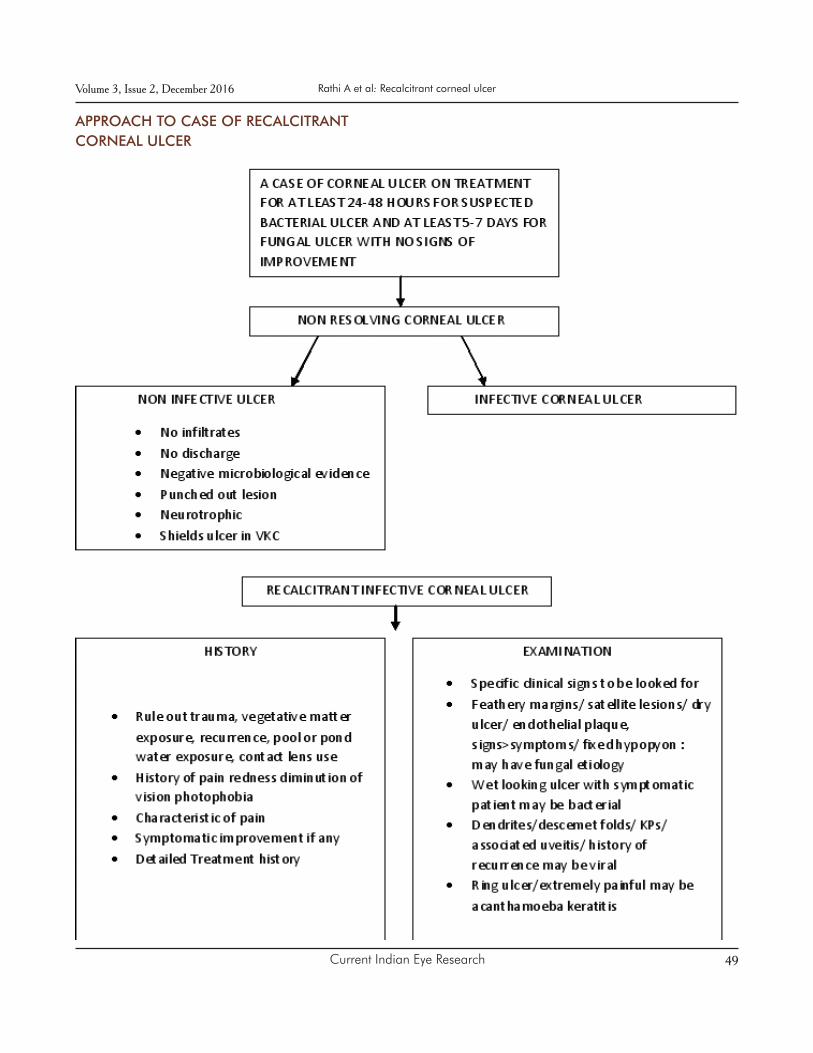
APPROACH TO CASE OF RECALCITRANTCORNEAL ULCER
Rathi A et al: Recalcitrant corneal ulcerVolume 3, Issue 2, December 2016

Current Indian Eye Research50
suspicion of atypical organisms.
It is important to have an adequate sample and accuratestaining and fixing skills to be able to diagnose thecausative micro organism and start anti microbial therapyaccordingly. This step is the usual limiting factor in caseslabeled as recalcitrant keratitis. Hence it is advisable tostop all treatment in such cases for 24-48 hours and repeatthe scraping process accurately.
The routine smears and stains have their own limitations.Gram stain is able to identify the causative organism in 75percent cases while 10 percent KOH wet mount has asensitivity of 92% and specificity of 96%12-14.
Other stains that can be used in suspected cases includeCalcoflour white for Acanthamoeba and Ziehl nelson formycobacteria and nocardia.
Corneal scraping is inoculated onto culture plate in orderto culture the micro organism. These plates aretemperature sensitive. Hence an accurate diagnosisrequires good microbilogical laboratory assistance andfacilities. Commonly used culture media include Blood agarplate, Chocolate agar plate and Sabouraud’s dextrose agar.Other culture media that can be used if a particularorganism is suspected include Thioglycollate broth foranaerobes, Lowenstein Jensen medium for mycobacteria,Brain heart infusion for fungi and E.coli enriched nonnutrient agar for Acanthamoeba.
A negative culture report may indicate partial antibiotictreatment, inadequate sample, inaccurate culturetechniques etc. Such cases go on to become recalcitrantdue to lack of directed anti microbial therapy15.
Once the organism has grown on the culture plate, anantibiotic sensitivity testing is done and accordingly thetreatment may be altered or started in these cases.
Other investigations such as Confocal microscopy andserological testing including PCR may also aid in thediagnosis.
A corneal biopsy or a suture biopsy may be done in deeperulcers where superficial corneal scraping fails to revealany micro organism16. Corneal biopsy may be done usinga crescent blade or Bard parker blade. Suture biopsy isusually taken using a 10-0 monofilament nylon suture of avicryl suture.
An anterior chamber paracentesis may be taken and sentfor microbiological investigation in cases of hypopyoncorneal ulcer17.
Management of A Case of Recalcitrant Keratitis
A case of recalcitrant keratitis requires a systematic stepwise approach. The initial steps of history taking andexamination are same as any fresh case of corneal ulcer.Once the basic history taking and clinical examination isover, the first step is to stop all the treatment the patient isbeing given for at least 24-48 hours. One of the mostcommon causes of negative culture report is previousantibiotic treatment. The 24-48 hour period is to wash offthe effect of antibiotics. Once the wash off period is over,steps similar to that in a fresh case of corneal ulcer arerepeated. An adequate corneal scraping sample is takenas described above and immediately sent formicrobiological investigations. The smears and stainsshould be done immediately without delay to increase theprobability of growth. Also the plating on the culture platesshould be direct. Once the scraping is done, the empiricaltreatment based on history or examination may be started.This treatment may be altered according to the smear orculture report once that is obtained.
In case of a negative culture after repeat scraping as well,other methods of sample collection such as corneal biopsyof suture biopsy may be tried especially for deeper nonresponding ulcers. Confocal microscopy also has a role insuch cases.
Empirical therapy for a case of corneal ulcer usuallyincludes broad spectrum antibiotics such as fortifiedcefazolin 5 percent and fortified tobramycin 1.3 percentalong with a cycloplegic for cyclitic pain relief. Large ulcerswith impending perforation or with sclera involvement orposterior segment involvement in the form ofendophthalmitis may require systemic antibiotics as well.
Once the causative micro organism is identified after themicrobiological investigations, the treatment is accordinglyaltered.
Bacterial keratitisBacterial keratitisBacterial keratitisBacterial keratitisBacterial keratitis
For small peripheral bacterial corneal ulcers, monotherapywith a topical flouroquinolone may be sufficient. With theadvent of flouroquinolone resistant micro organismsespecially Moxifloxacin resistant Staphylococcus aureus,topical vancomycin 5 percent has found favour in suchpatients. For larger ulcers, fortified therapy may be neededaccording to culture sensitivity. Most bacteria are sensitiveto the empiric therapy of cefazolin-tobramycin itself and itusually continues then. The therapy in the initial period isintensive with hourly application.
Rathi A et al: Recalcitrant corneal ulcerVolume 3, Issue 2, December 2016
Current Indian Eye Research 51
Fungal keratitisFungal keratitisFungal keratitisFungal keratitisFungal keratitis
For suspected filamentous fungal infections, 5 percentnatamycin is the initial treatment of choice. Topicalvoriconazole 1 percent may be used in adjunct to topicalnatamycin for filamentary fungal keratitis and has beenshown to be effective in the MUTT trial19. Topical 0.15percent amphotericin B may be used in case of suspectedyeast infection. Oral antifungals may be given in case oflarge ulcers with sclera involvement or associatedendophthalmitis or with impending perforation.
Acanthamoeba keratitisAcanthamoeba keratitisAcanthamoeba keratitisAcanthamoeba keratitisAcanthamoeba keratitis
Cysticidal therapy includes biguanides such as PHMB0.02% and propamidine 0.1% or brolene. Other agentsinclude chlorhexidine 0.02% may be used in place ofPHMB. Topical steroids may be added in case ofprogressive vascularisation in such cases.
Viral keratitisViral keratitisViral keratitisViral keratitisViral keratitis
The treatment in case of a viral keratitis depends on thetype of keratitis. In case of a purely epithelial dendritickeratitis, antivirals alone are the treatment of choice andsteroids should be avoided. A topical steroid in adjunctionwith oral antivirals is the treatment of choice for stromalkeratitis and endothelitis. The balance between topicalsteroids and antivirals is needed in case of stromal orendothelial keratitis along with epithelial involvement.
Non resolving corneal ulcer which fails to respond to theabove standard protocols is a therapeutic challenge andan ophthalmologist’s dilemma. Several newer treatmentregimens have been tried especially for non respondingcorneal ulcers.
Collagen cross linking in case of recalcitrantCollagen cross linking in case of recalcitrantCollagen cross linking in case of recalcitrantCollagen cross linking in case of recalcitrantCollagen cross linking in case of recalcitrantcorneal ulcercorneal ulcercorneal ulcercorneal ulcercorneal ulcer
Several studies have evaluated the role of collagen crosslinking in a case of non healing corneal ulcer. Some ofthese studies were in vitro and showed the beneficialbactericidal effect of 365 nm UVA photo activated riboflavin.Tsugita et al showed that UVA-riboflavin combinationdeactivates RNA in tobacco mosaic virus20. Collagencrosslinking using 365 nm UVA and riboflavin solution hasbeen shown to have a role in preventing corneal meltcaused by Gram-negative bacteria (92%) followed byGram-positive bacteria (84%), acanthamoeba (71%) andfungi (61%)21-27. Shetty R et al reported that CXL is aneffective procedure in treating non-resolving microbialkeratitis with superficial stromal involvement28. They further
revealed that the prognosis was better in case of bacterialkeratitis as compared to fungal keratitis which was deeper.Hence CXL may be used as an adjunct to the standardtherapy. CXL in these cases also gives early symptomaticrelief to the patients by decreasing corneal sensations andhence reducing the pain.
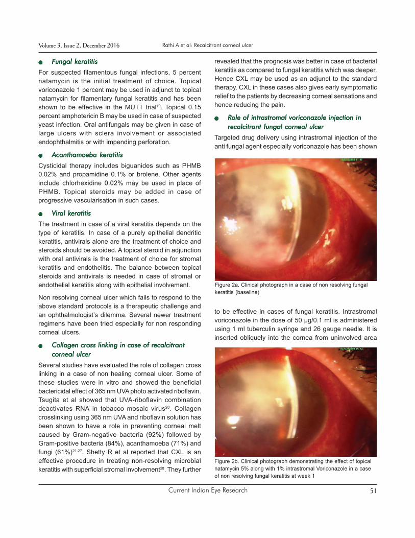
Role of intrastromal voriconazole injection inRole of intrastromal voriconazole injection inRole of intrastromal voriconazole injection inRole of intrastromal voriconazole injection inRole of intrastromal voriconazole injection inrecalcitrant fungal corneal ulcerrecalcitrant fungal corneal ulcerrecalcitrant fungal corneal ulcerrecalcitrant fungal corneal ulcerrecalcitrant fungal corneal ulcer
Targeted drug delivery using intrastromal injection of theanti fungal agent especially voriconazole has been shown
to be effective in cases of fungal keratitis. Intrastromalvoriconazole in the dose of 50 µg/0.1 ml is administeredusing 1 ml tuberculin syringe and 26 gauge needle. It isinserted obliquely into the cornea from uninvolved area
Rathi A et al: Recalcitrant corneal ulcerVolume 3, Issue 2, December 2016
Figure 2b. Clinical photograph demonstrating the effect of topicalnatamycin 5% along with 1% intrastromal Voriconazole in a caseof non resolving fungal keratitis at week 1
Figure 2a. Clinical photograph in a case of non resolving fungalkeratitis (baseline)

Current Indian Eye Research52
and a barrage is made all around the involved or ulceratedarea. These injections may be repeated 72 hours later. Ina study published in 2011, Sharma N et al injectedintrastromal voriconazole in 12 eyes with recalcitrant fungalkeratitis29. Of these, 10 eyes healed with scar formation(Figure 2 a,b,c) while two eyes required therapeuticpenetrating keratoplasty. In another study by Sharma N etal, forty cases of fungal corneal ulcer not responding tostandard topical natamycin 5 percent drops for 2 weekswere randomised into treatment with either hourly topicalvoriconazole or at least three intrastromal voriconazoleinjections30. There was no statistical difference in meanduration to healing. The incidence of perforation andposterior synechiae were similar while pain wassignificantly more frequent in the intrastromal group.
Role of Intracameral and Intrastromal voricona-Role of Intracameral and Intrastromal voricona-Role of Intracameral and Intrastromal voricona-Role of Intracameral and Intrastromal voricona-Role of Intracameral and Intrastromal voricona-zole in recalcitrant fungal keratitis with hypopyonzole in recalcitrant fungal keratitis with hypopyonzole in recalcitrant fungal keratitis with hypopyonzole in recalcitrant fungal keratitis with hypopyonzole in recalcitrant fungal keratitis with hypopyon
Previous studies have shown that the experimental use ofintracameral voriconazole in humans shows no toxic effectswhen the aqueous concentration is in the safe range of10ìg/mL - 1.5mg/mL. There may be a reduction in cornealendothelial cells, trabecular meshwork cells, and retinalpigment epithelial cells in dose more than 1.5mg/ml31. Inthe safe range, intracameral voriconzole can be used inadjunct with intrastromal voriconazole in recalcitrant caseswith hypopyon. (Figure 3a and 3b)
Other novel methods of antifungal drug deliveryOther novel methods of antifungal drug deliveryOther novel methods of antifungal drug deliveryOther novel methods of antifungal drug deliveryOther novel methods of antifungal drug deliveryin deeper recalcitrant ulcersin deeper recalcitrant ulcersin deeper recalcitrant ulcersin deeper recalcitrant ulcersin deeper recalcitrant ulcers
Pallikaris et al in 2015 published a report where theysuccessfully used Femtosecond laser to create a cornealpocket for direct instillation of antifungal drug deeper intothe cornea32.
Natamatrix, a tiny dissolvable matrix which can be insertedinto stroma is another novel technique being describedfor targetd drug delivery.
Role of therapeutic penetrating keratoplastyRole of therapeutic penetrating keratoplastyRole of therapeutic penetrating keratoplastyRole of therapeutic penetrating keratoplastyRole of therapeutic penetrating keratoplasty(TPK)(TPK)(TPK)(TPK)(TPK)
Several studies have shown that Therapeutic PK has arole in the management of severe and refractory keratitiswith a high success in restoring anatomical integrity. Buton the other hand graft re infection rates are high especiallyin case of fungal and acanthamoeba keratitis33.
Rathi A et al: Recalcitrant corneal ulcerVolume 3, Issue 2, December 2016
Figure 3a. Pre operative clinical photograph of fungal keratitis withfungal ball in the anterior chamber
Figure 3b. Post operative day 14 clinical photograph of intra-cameral along with intrastromal voriconazole
Figure 2c. Clinical photograph demonstrating the effect of topicalnatamycin 5% along with 1% intrastromal Voriconazole in a caseof non resolving fungal keratitis at week 4

Current Indian Eye Research 53
Conclusion:
Recalcitrant microbial keratitis is a challenge to everyophthalmologist. Its management involves a step by stepapproach. Timely diagnosis and intervention may save theulcer from perforation. Newer modalities such as Collagencross linking, intrastromal and intracameral drug injectionsand other methods of targeted drug delivery may be tried.
References:
1. Whitcher JP, Srinivasan M, Upadhyay MP. Cornealblindness: a global perspective. Bull World HealthOrgan 2001; 79: 214–21.
2. Burd EM, Ogawa GSH, Hyndiuk RA. Bacterial keratitisand conjunctivitis. In: Smolin G, Thoft RA, eds. Thecornea scientific foundations and clinical practice. 3rdedn. Boston: Little, Brown, & Co, 1994:115–67.
3. Gokhale NS. Medical management approach toinfectious keratitis. Indian J Ophthalmol. 2008; 56:215-20.
4. BertinoJ S. Impact of antibiotic resistance in themanagement of ocular infections: the role of current andfuture antibiotics. Clin Ophthalmol 2009; 3: 507–21.
5. Keay L, Edwards K, Naduvilath T, et al. Microbialkeratitis predisposing factors and morbidity.Ophthalmology 2006; 113: 109–16.
6. Maheshwari R, Weis E. Thyroid associatedorbitopathy. Ind J Ophthalmol 2012; 6: 87-93.
7. Aristimuño B, Nirankari VS, Hemady RK, RodriguesMM. Spontaneous ulcerative keratitis inimmunocompromised patients. Am J Ophthalmol.1993; 115: 202–8.
8. Yildiz Eh et al. Trends in contact lens-related cornealulcers at a tertiary referral center. Cornea 2012 Oct;31: 1097-102.
9. D. Yorston, A. Foster. Traditional eye medicines andcorneal ulceration in Tanzania. J Trop MedHyg 1994; 97: 211–4.
10. Guidera AC, Luchs JI, Udell IJ. Keratitis, ulcerationand perforation associated with topical nonsteroidalanti-inflammatory drugs. Ophthalmology 2001;108:936-44.
11. Solomon A, Karp CL, Miller D, Dubovy SR, HuangAJ, Culbertson WW. Mycobacterium interface keratitisafter laser in situ keratomileus. Ophthalmology2001; 108: 2201–8.
12. Vajpayee RB, Angra SK, Sandramouli S, Honavar SG,Chhabra VK. Laboratory diagnosis of keratomycosis:comparative evaluation of direct microscopy andculture results. Ann Ophthalmol 1993; 25: 68–71.
Rathi A et al: Recalcitrant corneal ulcerVolume 3, Issue 2, December 2016
13. Bharathi MJ, Ramakrishnan R, Meenakshi R, MittalS, Shivakumar C, Srinivasan M. Microbiologicaldiagnosis of infective keratitis: comparative evaluationof direct microscopy and culture results. Br JOphthalmol2006; 90: 1271-6.
14. Gopinathan U, Sharma S, Garg P, Rao GN. Reviewof epidemiological features, microbiological diagnosisand treatment outcome of microbial keratitis:Experience of over a decade. IndiJ Ophthalmol2009;57:273-9.
15. Liesegang TJ, Forster RK. Spectrum of microbialkeratitis in South Florida. Am J Ophthalmol 1980; 90:38–47.
16. Alexandrakis G, Haimovici R, Miller D, et al. Cornealbiopsy in the management of progressive microbialkeratitis. Am J Ophthalmol 2000;129: 571–6.
17. Sridhar MS, Sharma S, Gopinathan U, Rao GN.Anterior chamber tap: diagnostic and therapeuticindications in the management of ocular infections.Cornea 2002; 21: 718–22.
18. Presterl E, Mueller-Uri P, Grisold A, Georgopoulos A,Graninger W. Ciprofloxacin- and methicillin-resistantstaphylococcus aureus susceptible to moxifloxacin,levofloxacin, teicoplanin, vancomycin and linezolid.Eur J Clin Microbiol Infect Dis 2001; 20: 486–9.
19. Prajna NV, Krishnan T, Mascarenhas J, et al. TheMycotic Ulcer Treatment Trial: A Randomized TrialComparing Natamycin vs Voriconazole. JAMAophthalmol 2013;131:422-9.
20. Tsugita A, Okada Y, Uchara K. Photosensitizedinactivation of ribonucleic acids in the presence ofriboflavin. Biochim Biophys Acta 1965;103:360–3.
21. Price MO, Tenkman LR, Schrier A, Fairchild KM, TrokelSL, Price FW Jr. Photoactivated riboflavin treatmentof infectious keratitis using collagen cross-linkingtechnology. J Refract Surg 2012; 28: 706–13.
22. El-Danasoury AM, Hashem AN. Corneal collagencrosslinking in the treatment of infectious keratitis. ClinOphthalmol 2011; 5: 1277–80.
23. Morén H, Malmsjö M, Mortensen J, et al. Riboflavin andultraviolet a collagen crosslinking of the cornea for thetreatment of keratitis. Cornea 2010; 29: 102–4.
24. Iseli HP, Thiel MA, Hafezi F, et al. Ultraviolet A/riboflavincorneal cross-linking for infectious keratitis associatedwith corneal melts. Cornea 2008; 27: 590–4.
25. Makdoumi K, Mortensen J, Crafoord S. Infectiouskeratitis treated with corneal crosslinking. Cornea2010; 29: 1353–8.
26. Khan YA, Kashiwabuchi RT, Martins SA, et al.Riboflavin and ultraviolet light a therapy as an adjuvanttreatment for medically refractive Acanthamoeba

Current Indian Eye Research54
Cite this article as:Rathi A, Sharma N, Aron N, Sinha R, Agarwal T. Recalcitrant corneal ulcer. Current Indian Eye Research 2016; 3:45-54.
keratitis: report of 3 cases. Ophthalmology 2011;118:324–31.
27. Martins SA, Combs JC, Noguera G, et al. Antimicrobialefficacy of riboflavin/UVA combination (365 nm) in vitrofor bacterial and fungal isolates: a potential newtreatment for infectious keratitis. Invest OphthalmolVis Sci 2008; 49: 3402–8.
28. Shetty R, Nagaraja H, Jayadev C, Shivanna Y, KugarT. Collagen crosslinking in the management ofadvanced non-resolving microbial keratitis.Br JOphthalmol 2014; 98:1033-5.
29. Sharma N, Agarwal P, Sinha R, Titiyal JS, Velpandian T,Vajpayee RB. Evaluation of intrastromal voriconazoleinjection in recalcitrant deep fungal keratitis: caseseries.Br J Ophthalmol 2011; 95: 1735–7.
30. Sharma N1, Chacko J, Velpandian T, et al.
Comparative Evaluation of Topical versus IntrastromalVoriconazole as an Adjunct to Natamycin inRecalcitrant Fungal Keratitis. Ophthalmology 2013;120: 677– 81.
31. Haddad RS, El-Mollayess GM. Combination ofIntracameral and Intrastromal Voriconazole in theTreatment of Recalcitrant Acremonium Fungal Keratitis.Middle East Afr J Ophthalmol 2012;19:265-8.
32. Pallikaris IG, Kymionis GD, Plaka AD, Binder PS,Kontadakis GA, Tsoulnaras KI. Femtosecond laser-assisted Intra-corneal drug delivery. Semin ophthalmol2015;30:457-61.
33. Sharma N, Jain M, Sehra SV, Maharana P, AgarwalT, Satpathy G, Vajpayee RB. Outcomes of therapeuticpenetrating keratoplasty from a tertiary eye care centrein northern India. Cornea 2014; 33: 114–8.
Rathi A et al: Recalcitrant corneal ulcerVolume 3, Issue 2, December 2016