reactive stroma in human prostate cancer: induction of ... · reactive stroma in prostate cancer...
TRANSCRIPT
Reactive Stroma in Human Prostate Cancer: Induction ofMyofibroblast Phenotype and ExtracellularMatrix Remodeling1
Jennifer A. Tuxhorn, Gustavo E. Ayala,Megan J. Smith, Vincent C. Smith,Truong D. Dang, and David R. Rowley2
Departments of Molecular and Cellular Biology [J. A. T., T. D. D.,D. R. R.] and Pathology [G. E. A., M. J. S., V. C. S.] Baylor Collegeof Medicine, Houston, Texas 77030
ABSTRACTPurpose: Generation of a reactive stroma environment
occurs in many human cancers and is likely to promotetumorigenesis. However, reactive stroma in human prostatecancer has not been defined. We examined stromal cellphenotype and expression of extracellular matrix compo-nents in an effort to define the reactive stroma environ-ment and to determine its ontogeny during prostate cancerprogression.
Experimental Design: Normal prostate, prostatic intra-epithelial neoplasia (PIN), and prostate cancer were exam-ined by immunohistochemistry. Tissue samples includedradical prostatectomy specimens, frozen biopsy specimens,and a prostate cancer tissue microarray. A human prostatestromal cell line was used to determine whether transform-ing growth factor �1 (TGF-�1) regulates reactive stroma.
Results: Compared with normal prostate tissue, reac-tive stroma in Gleason 3 prostate cancer showed increasedvimentin staining and decreased calponin staining (P <0.001). Double-label immunohistochemistry revealed thatreactive stromal cells were vimentin and smooth muscle�-actin positive, indicating the myofibroblast phenotype. Inaddition, reactive stroma cells exhibited elevated collagen Isynthesis and expression of tenascin and fibroblast activa-tion protein. Increased vimentin expression and collagen Isynthesis were first observed in activated periacinar fibro-blasts adjacent to PIN. Similar to previous observations inprostate cancer, TGF-�1-staining intensity was elevated inPIN. In vitro, TGF-�1 stimulated human prostatic fibro-blasts to switch to the myofibroblast phenotype and to ex-press tenascin.
Conclusions: The stromal microenvironment in humanprostate cancer is altered compared with normal stroma andexhibits features of a wound repair stroma. Reactive stromais composed of myofibroblasts and fibroblasts stimulated toexpress extracellular matrix components. Reactive stromaappears to be initiated during PIN and evolve with cancerprogression to effectively displace the normal fibromuscularstroma. These studies and others suggest that TGF-�1 is acandidate regulator of reactive stroma during prostate can-cer progression.
INTRODUCTIONActivation of the host stromal microenvironment is pre-
dicted to be a critical step in adenocarcinoma growth andprogression (1–5). Several human cancers have been shown toinduce a stromal reaction or desmoplasia as a component ofcarcinoma progression. However, the specific mechanisms ofstromal cell activation are not known, and the extent to whichstroma regulates the biology of tumorigenesis is not fully un-derstood. In cancers where a stromal reaction has been ob-served, it seems that the response is similar, if not identical, toa generic wound repair response (6). In wound repair, stromalcells exhibit elevated production of ECM3 components, growthfactors, and matrix remodeling enzymes to create a growth-promoting microenvironment (7). Similarly, reactive stroma incancer would be predicted to enhance tumor progression bystimulating angiogenesis and by promoting cancer cell survival,proliferation, and invasion (5, 8–12). Indeed, two recent studieshave shown that prostate stromal cells stimulate the develop-ment and rate of human prostate tumorigenesis in mouse xe-nograft models (13, 14). Accordingly, it is important to identifyand characterize reactive stroma in cancer to establish keyregulators of reactive stroma and to delineate the specific mech-anisms through which reactive stroma affects carcinomaprogression.
An initial step in understanding mechanisms of the stromalreaction in tumor progression is to fully define the reactivestroma phenotype and its formation. Although poorly under-stood in most cancers, reactive stroma has been described inbreast and colon carcinoma (9–12). In these cancers, reactivestroma is a mix of fibroblasts, myofibroblasts, endothelial cells,and immune cells. Although all of these cells potentially affecttumorigenesis, myofibroblasts are of particular interest. Myofi-Received 11/1/01; revised 5/13/02; accepted 6/3/02.
The costs of publication of this article were defrayed in part by thepayment of page charges. This article must therefore be hereby markedadvertisement in accordance with 18 U.S.C. Section 1734 solely toindicate this fact.1 This work was supported by NIH Grants RO1-CA58093, RO1-DK45909, SPORE CA58204, and UO1-CA84296.2 To whom requests for reprints should be addressed, at Department ofMolecular and Cellular Biology, Baylor College of Medicine Houston,TX 77030. Phone: (713) 798-6220; Fax: (713) 790-1275; E-mail:[email protected].
3 The abbreviations used are: ECM, extracellular matrix; FAP, fibroblastactivation protein; MMP, matrix metalloproteinase; TGF, transforminggrowth factor; PIN, prostatic intraepithelial neoplasia; SPORE, special-ized program of research excellence; sm �-actin, smooth muscle �-actin; DAPI, 4�,6 diamidino-2-phenylindole; CAF, carcinoma-associ-ated fibroblast.
2912 Vol. 8, 2912–2923, September 2002 Clinical Cancer Research
Research. on March 3, 2020. © 2002 American Association for Cancerclincancerres.aacrjournals.org Downloaded from
broblasts are activated stromal cells typically found at sites ofpathologic tissue remodeling (15). In wound repair, myofibro-blasts are derived from granulation tissue fibroblasts (15). Incancer, it has been shown that carcinoma cells have the capacityto induce normal fibroblasts to the reactive myofibroblast phe-notype (16, 17). Myofibroblasts in reactive stroma synthesizeECM components such as collagen I, collagen III, fibronectinisoforms, tenascin, and versican (18–22). In addition, myofi-broblasts express proteases, including urokinase plasminogenactivator, FAP, and MMPs (23–25). Production of these com-ponents results in ECM remodeling that could stimulate cancercell growth and migration. Moreover, myofibroblasts have beenreported to secrete growth factors that promote angiogenesis(26, 27). Therefore, myofibroblasts appear to be a key cell typeinvolved in creation of the tumor-promoting reactive stromaenvironment.
The inductive mechanisms involved in the development ofreactive stroma in cancer are poorly understood. Predictably,experimental evidence suggests that TGF-�1, a key mediator ofthe stromal response in wound repair (28), is likely to play animportant role (5, 11, 29). In vitro studies have shown thatTGF-�1 stimulates phenotypic switching of fibroblasts to myo-fibroblasts (16) and regulates expression of ECM components(28). In addition, s.c. injection of TGF-�1 was sufficient toinduce a stromal reaction characterized by differentiation ofstromal cells to myofibroblasts, enhanced collagen production,and stimulated angiogenesis (30, 31). Elevated levels ofTGF-�1 have also been shown to enhance tumor growth in vivo(32–35). Because TGF-� is overexpressed in many human can-cers, including prostate cancer (36–38), it seems likely thatTGF-� promotes formation of reactive stroma.
We have suggested previously that reactive stroma in hu-man prostate cancer is likely to function as a mediator oftumorigenesis (5, 39). However, a detailed analysis of whethera stromal reaction occurs as a component of prostate cancerprogression has not been reported. Furthermore, the putativereactive stroma in prostate cancer may differ from that describedin breast and colon cancer because of fundamental differences inthe normal stroma of these tissues. The purpose of this studywas to determine whether reactive stroma is a component ofprostate carcinoma, to specifically define cell and matrix alter-ations in this reactive stroma, to characterize the ontogeny ofsuch a reactive stroma relative to the development of prostatecancer, and to identify potential regulators. We show here that awound repair type of reactive stroma is associated with prostatecancer and was composed of myofibroblasts and fibroblastsrather than normal prostate smooth muscle, which appeared tobe displaced by the reactive stroma. Our results show thatreactive stroma exhibits fundamental alterations in ECM andexpression of markers commonly associated with wound repairstroma. Moreover, we show that genesis of the reactive stromaphenotype occurs in precancerous PIN lesions and may beassociated with elevated TGF-�1 expression by PIN epithelialcells. In addition, TGF-�1 induced a human prostate stromalcell line to switch to the myofibroblast phenotype. This studyrepresents the first report to identify and specifically definereactive stroma in human prostate cancer and sets the stage foradditional assessing of the specific mechanisms and role of thestromal reaction in prostate tumorigenesis.
MATERIALS AND METHODSTissues. Radical prostatectomy specimens from 20 pa-
tients were obtained from the Baylor College of MedicineProstate SPORE tissue bank located at Methodist Hospital.Specimens were processed as described previously (40). Briefly,the tissues were cut into 5-mm thick slices, fixed in 10% neutralbuffered formalin, and embedded in paraffin as whole mounts.Whole mount thin sections (5 �m) were stained with H&E andevaluated for histological differentiation. Tissues were selectedby a pathologist (G. E. A.) based on the presence of normalprostate, PIN, and Gleason 3 prostate cancer. For initial exam-ination of stromal components, whole mount thin sections werestained with Masson’s Trichrome following the standard proce-dure (Sigma Diagnostics, St. Louis, MO). For immunostaining,the whole mounts were cut in half, and thin sections weremounted on standard microscope slides.
The prostate cancer tissue microarrays were assembledfrom specimens in the Baylor SPORE tissue bank (n � 89Gleason 3 and n � 17 Gleason 4 cancers). For each specimen,the largest and/or highest Gleason cancer focus was identifiedand mapped on the whole mount sections. Accordingly, 2-mmcores were punched out of the tissue slices and transferred to arecipient block. Samples from normal prostate tissue (n � 64)and PIN lesions (n � 27) were also included in the array. Thetissue microarrays were built using a manual tissue arrayer(Beecher Instruments, Silver Spring, MD). Internal controlswere placed at a pre-established pattern throughout the blocks toassess adequacy of staining.
Prostate cancer tissues for frozen sections were obtainedthrough the Baylor SPORE and Methodist Hospital. Tissuecores harvested in the operating room were frozen immediatelyin liquid nitrogen, embedded in Tissue-Tek Optimal CuttingTemperature compound (Sakura, Torrance, CA), and sectioned.
Immunohistochemistry. The following primary anti-bodies were used: sm �-actin (mouse monoclonal 1A4), calpo-nin (mouse monoclonal hCP), and tenascin (mouse monoclonalBC-24) were purchased from Sigma Diagnostics; vimentin (goatpolyclonal AB1620) and procollagen I (rat monoclonalMAB1912) were purchased from Chemicon (Temecula, CA);vimentin (mouse monoclonal VIM 3B4; Boehringer Mannheim,Indianapolis, IN); FAP (mouse monoclonal 3D11, kindly pro-vided by Dr. John Park, Boehringer Ingelheim); and TGF-�1(rabbit polyclonal sc-146; Santa Cruz Biotechnology, SantaCruz, CA). Specificity of each primary antibody has been eval-uated and published previously. The following secondary anti-bodies were used: biotin-conjugated Universal Secondary Anti-body was used for mouse monoclonals (Research Genetics,Huntsville, AL); biotin-conjugated goat antirat IgG (554014;BD PharMingen, San Diego, CA); and Texas Red-conjugateddonkey antigoat IgG and fluorescein-conjugated donkey anti-mouse IgG (705-075-147 and 715-095-150; Jackson Immu-noResearch, West Grove, PA). No significant staining wasobserved if sections were incubated with secondary antibodyonly.
All immunostaining was performed with the MicroProbeStaining System (FisherBiotech, Pittsburgh, PA), which usescapillary gap technology. Standard slides mounted with tissuewere paired with BioTek Solutions POP130 capillary gap plus
2913Clinical Cancer Research
Research. on March 3, 2020. © 2002 American Association for Cancerclincancerres.aacrjournals.org Downloaded from

slides (Ventana Medical Systems, Tucson, AZ). Reagents for-mulated for use with capillary action systems were purchasedfrom Research Genetics and used according to the manufactur-er’s protocol.
Paraffin sections were deparaffinized using Auto Dewaxerand cleared with Auto Alcohol. Brigati’s Iodine and Auto Prepwere used to improve tissue antigenicity. After washing withUniversal Buffer, antigen retrieval was performed if required(VIM 3B4, incubated in pepsin solution for 3 min at 50°C;procollagen, incubated in PBS � 1% trypsin for 20 min at roomtemperature). Tissues were washed in Universal Buffer, incu-bated in Protein Blocker, and washed again. Antibodies werediluted in Primary Antibody Diluent and used at the followingconditions: sm �-actin 1:200, VIM 3B4 1:50, and calponin1:2,000 for 8 min at 50°C; and pro-collagen 1:500 for 1 h at37°C. Tissues were washed with Universal Buffer and incubatedin secondary antibody: Universal Secondary undiluted for 4 minat 50°C; and antirat IgG 1:100 for 45 min at 37°C. Tissues werewashed in Universal Buffer, treated with Auto Blocker tosquelch endogenous peroxidase activity, and washed again. Fordetection, sections were incubated in streptavidin horseradishperoxidase, washed in Universal Buffer, and then incubated instable 3,3�-diaminobenzidine twice at 3 min each at 50°C.Tissues were counterstained with Auto Hematoxylin for 30 s.
For double-label immunofluorescent staining, sectionswere deparaffinized and treated as described above, excepttissues were blocked in Universal Buffer � 5% donkey serum(Sigma Diagnostics) for 15 min at room temperature. Vimentinstaining was done first using the goat polyclonal antibody di-luted 1:100 for 1 h at 37°C. The antigoat IgG secondary wasused at 1:200 for 45 min at 37°C. Then, sections were incubatedwith sm �-actin, and the antimouse IgG secondary was appliedas above. After staining, tissues were dehydrated and mountedwith coverslips using VectaShield with DAPI (Vector Labora-tories, Burlingame, CA).
Frozen sections were fixed in acetone for 10 min at 4°C,then washed in PBS and Universal Buffer. Tissues were incu-bated in Protein Blocker at 37°C and then washed in UniversalBuffer. Primary antibodies were used at the following dilutions:procollagen 1:20,000; tenascin 1:4,000; and FAP 1:10 for 1 h at37°C. Secondary antibodies were diluted as before and used for45 min at 37°C. For detection, Ready-To-Use VectaStain EliteAvidin: Biotinylated enzyme complex reagent (Vector Labora-tories) was used according to manufacturer protocol, and stable3,3�-diaminobenzidine was used twice at 3 min each at 37°C.Tissues were counterstained with Auto Hematoxylin.
Histological and Statistical Analysis. To evaluate thelevel of vimentin, sm �-actin, and calponin expression, thepercentage of positive-staining stromal cells and the stainingintensity were graded on a scale of 0–3. Staining percentage:0 � 0% positive cells; 1 � 1–33% positive cells; 2 � 34–66%positive cells; and 3 � 67–100% positive cells. Staining inten-sity: 0 � no staining; 1 � staining obvious only at �400; 2 �staining obvious at �100 but not �40; and 3 � staining obviousat �40. For each sample, the staining percentage and stainingintensity scores were multiplied to give the staining index.Staining index: 0 � zero; 1–2 � low; 3–4 � moderate; and6–9 � high. For each marker, Fisher’s exact test was used tocompare zero/low staining to moderate/high staining in normal
and Gleason 3 stroma (19). P � 0.05 was considered statisti-cally significant. Analysis was performed with GraphPad Prismfor Macintosh v3.0 (GraphPad Software, San Diego, CA).
To examine colocalization of fluorescent staining, separatevimentin (red), sm �-actin (green), and DAPI (blue) imageswere captured using a Spot Real Time color digital camera(Diagnostic Instruments, Sterling Heights, MI) attached to aNikon Eclipse TE300 microscope equipped for epifluorescence(Nikon, Melville, NY). The images were merged using SpotReal Time Software v3.2.6 for Mac OS. The percentage ofyellow stromal cells (resulting from colocalization of red andgreen staining) was graded on a scale of 0–3. Fisher’s exact testwas used to compare the proportion of grade 3 cases in Gleason3 and Gleason 4 cancer. Statistical analysis was performed asdescribed above.
Cell Culture. To establish the HPS-TZ1A human pros-tate stromal cell line, a fresh tissue core from the transition zonewas obtained through the Baylor SPORE at Methodist Hospital.The core was cut into discs and placed in a 6-well tissue platecontaining Bfs media [DMEM (Life Technologies, Inc., Rock-ville, MD) supplemented with 5% fetal bovine serum (Hyclone,Logan, UT), 5% Nu Serum (Collaborative Research, Bedford,MA), 0.5 �g/ml testosterone, 5 �g/ml insulin, 100 units/mlpenicillin, and 100 �g/ml streptomycin (Sigma Diagnostics)].The explants were incubated at 37°C with 5% CO2, and mediawere changed every 48 h or as necessary. Stromal cells migratedout of the explant and attached to the tissue culture dish. Afterthe cells reached confluence, the explant was removed, and thecells were passaged. The resulting stromal cell line showedvimentin expression in 100% of cells, but cytokeratin expressionwas not observed with AE1/AE3 pooled monoclonal antibody(Boehringer Mannheim). Cultures at passage 6–14 were usedfor experiments.
To examine the effect of TGF-�1, HPS-TZ1A cells weretrypsinized, diluted to 1.5 � 104 cells/ml in Bfs media, andseeded onto coverslips in 6-well plates (2 ml/well). The cellswere allowed to attach overnight, then they were washed withHBSS (Life Technologies, Inc.) and switched to experimentalconditions. MCDB 110 supplemented with insulin, transferrin,and sodium selenite (Sigma Diagnostics) was used for basalmedia (M0). Cells were incubated in M0, M0 � 25 pM (0.625ng/ml) TGF-�1 (R&D Systems, Minneapolis, MN), or M0 � 25pM TGF-�1 � 150 ng/ml TGF-�1 neutralizing antibody (AF-101-NA; R&D Systems) for 72 h. Coverslips were fixed withcold 4% paraformaldehyde according to standard technique andstored at 4°C in PBS. This experiment was repeated three timeswith multiple coverslips of each condition.
For immunocytochemistry, standard staining procedureswere followed. Briefly, coverslips were incubated in PBS �0.1% Triton X-100 for 5 min, then washed with PBS. Coverslipswere blocked with PBS � 1% donkey serum for 30 min at roomtemperature. Primary antibodies were diluted in PBS � 1%BSA and applied to coverslips for 1 h at 37°C. A goat polyclonalantibody against vimentin (sc-7557; Santa Cruz Biotechnology)was used at 1:50; sm �-actin and tenascin antibodies werediluted as described above. Coverslips were washed with PBS,then incubated in fluorescent-labeled secondary antibodies di-luted 1:200 for 45 min at 37°C. Coverslips were washed in PBS,dehydrated, and mounted using VectaShield with DAPI.
2914 Reactive Stroma in Human Prostate Cancer
Research. on March 3, 2020. © 2002 American Association for Cancerclincancerres.aacrjournals.org Downloaded from
RESULTSHistology of Prostate Stroma. For initial examination
of prostate stroma, radical prostatectomy specimens containingnormal tissue and moderately differentiated prostate cancer(Gleason 3) were stained with Masson’s Trichrome. This tech-nique differentially stains stromal components and, hence, isuseful in distinguishing prostate smooth muscle cells from col-lagen fibers and other stromal cell types. Analysis of normalprostate stroma showed a mixture of red-staining smooth musclecells and blue-staining collagen fibers and fibroblasts (Fig. 1A).In contrast, the stroma in moderately differentiated prostatecancer stained predominantly blue with very little red staining(Fig. 1B). Although isolated smooth muscle cells were presentin some cases, this blue staining pattern was consistently asso-ciated with regions of cancer and suggested a loss of typicalsmooth muscle cells plus elevated collagen in prostate cancerstroma. These observations suggest that the stromal microenvi-ronment in cancer is altered and led us to further examinespecific reactive stroma markers in human prostate cancer.
Identification of Stromal Cell Phenotypes in ProstateCancer. To better define reactive stroma in human prostatecancer, stromal cell phenotype was assessed by immunohisto-chemistry. Fibroblast, myofibroblast, and smooth muscle cellphenotypes were classified according to the immunostainingprofiles of stromal cell differentiation markers. Expression ofvimentin (mesenchymal cell intermediate filament) without ad-ditional smooth muscle markers indicated the fibroblast pheno-type. Coexpression of sm �-actin microfilaments and microfila-ment-associated calponin (early and late stage smooth musclemarkers, respectively; Ref. 41) identified prostate smooth mus-cle cells. The myofibroblast phenotype is considered to beintermediate between fibroblasts and smooth muscle (15).Hence, myofibroblasts were distinguished by coexpression ofvimentin and sm �-actin without expression of calponin (15).
Immunostaining was initially performed on radical prosta-tectomy specimens from 20 patients. Stroma from normal pros-tate gland showed high levels of sm �-actin and calponinexpression in prostate smooth muscle cells (Fig. 1, E and G),which was consistent with the red staining pattern observed withMasson’s Trichrome (Fig. 1A). Vimentin staining was observedin interstitial fibroblasts and in vascular smooth muscle but notin prostate smooth muscle (Fig. 1C). Fibroblasts were mixedamong the prostate smooth muscle cells typical of the fibromus-cular stroma reported in human prostate. Of interest, a singlelayer of periacinar fibroblasts was differentially observed im-mediately peripheral and adjacent to epithelial acini (Fig. 1C,arrow). This layer may be discontinuous because periacinarfibroblasts were not detected around all acini. Alternatively,visualization of these cells may depend on the plane of section.As suggested by the initial trichrome stain, there was a funda-mental change in expression of stromal cell differentiationmarkers throughout prostate cancer reactive stroma. Stroma inGleason 3 cancer nodules exhibited a dramatic increase invimentin staining (Fig. 1D) that directly correlated with bluestaining regions observed with Masson’s Trichrome (Fig. 1B).Similarly, expression of sm �-actin (Fig. 1F) was observed inthe stroma in Gleason 3 cancer. However, these regions of
cancer-associated stroma were negative or exhibited low stain-ing intensity for calponin (Fig. 1H).
To confirm these observations, tissue microarrays contain-ing normal prostate tissue (n � 64) and moderately differenti-ated prostate cancer (n � 89) were stained for vimentin, sm�-actin, and calponin. Subsequently, each tissue sample wasevaluated for the percentage of positive-staining stromal cellsand for the staining intensity as described in “Materials andMethods.” These parameters were graded on a 0–3 scale andthen multiplied to yield the staining index. The stroma fromnormal regions of radical prostatectomy specimens (n � 20)was also evaluated for the staining index of each marker. Quan-tifications of the results are shown in Table 1. Vimentin expres-sion in normal prostate stroma was typically low (staining index,1–2). In contrast, 57% of Gleason 3 cancer cases had moderateto high vimentin staining in the reactive stroma (staining index,3–4 and 6–9). Analysis with Fisher’s exact test revealed that theincrease in vimentin staining was statistically significant (P �0.001). The level of sm �-actin staining in Gleason 3 reactivestroma relative to normal stroma was slightly reduced; however,the staining index remained moderate to high in 71% of Gleason3 patients. Although calponin expression was moderate to highin normal prostate stroma, 78% of Gleason 3 cancers showedzero or low calponin staining in the reactive stroma. The de-crease in calponin expression was also significant (P � 0.001).Consistent with Masson’s Trichrome staining and qualitativeanalysis of marker expression in radical prostatectomy speci-mens, the tissue array data suggest that human prostate cancerreactive stroma is typified by the myofibroblast and fibroblastphenotypes with a nearly complete reduction in differentiatedsmooth muscle.
To determine whether reactive stroma in cancer was com-posed of a homogeneous population of myofibroblasts or amixed myofibroblast/fibroblast population, double-label immu-nohistochemistry for vimentin and sm �-actin was evaluated inthe radical prostatectomy specimens. Normal prostate stromawas composed of sm �-actin-positive smooth muscle cells(green) and vimentin-positive interstitial and periacinar fibro-blasts (red; Fig. 2A). Colocalization (yellow) was only observedin vascular smooth muscle of blood vessel walls. Analysis ofmoderately differentiated cancer (Gleason 3) in these specimensshowed colocalization of sm �-actin and vimentin in a majorityof reactive stromal cells (Fig. 2, B–D). These data indicate thatthe myofibroblast phenotype predominates in prostate cancerreactive stroma, whereas the remaining cells are fibroblasts.
To quantify the proportion of myofibroblasts in reactivestroma, double-label immunohistochemistry was performed ona prostate cancer tissue microarray. Gleason 3 (n � 36) andGleason 4 (n � 17) cancers were graded for the percentage ofreactive stromal cells coexpressing vimentin and sm �-actin. Asshown in Table 2, colocalization was observed in all casesexamined. The majority of Gleason 3 cancers showed coexpres-sion in 50% of reactive stromal cells. Interestingly, an evenhigher percentage of myofibroblasts was observed in the reac-tive stroma of poorly differentiated Gleason 4 cancers. WithFisher’s exact test, the increased proportion of myofibroblasts inGleason 4 cancers as compared with Gleason 3 cancers wasstatistically significant (P � 0.03). Taken together, these datashow that while normal prostate gland stroma is primarily
2915Clinical Cancer Research
Research. on March 3, 2020. © 2002 American Association for Cancerclincancerres.aacrjournals.org Downloaded from

Fig. 1 Stromal cell phenotype in normal prostate tissue and moderately differentiated prostate cancer. Staining with Masson’s Trichrome identifiedsmooth muscle cells (red) and collagen fibers (blue) in normal prostate stroma (A), whereas reactive stroma in Gleason 3 prostate cancer showed aloss of smooth muscle cells (B). Vimentin immunostaining in normal prostate stroma was low (C), but vimentin-positive fibroblasts were distributedthroughout the stroma and adjacent to epithelial acini (arrows). Vimentin was also expressed in blood vessel walls (asterisks). sm �-actin (E) andcalponin (G) staining was high in the smooth muscle cells of normal prostate stroma. Reactive stroma in human prostate cancer showed elevatedvimentin staining (D), whereas sm �-actin staining was maintained (F) and calponin staining was reduced (H). The expression pattern of these markerssuggests that reactive stroma is composed of fibroblasts and myofibroblasts rather than smooth muscle cells. �400.
2916 Reactive Stroma in Human Prostate Cancer
Research. on March 3, 2020. © 2002 American Association for Cancerclincancerres.aacrjournals.org Downloaded from

composed of smooth muscle cells, reactive stroma in humanprostate cancer is enriched with myofibroblasts and fibroblasts.
Expression of ECM Components in Prostate Cancer.Myofibroblasts synthesize ECM components and remodelingenzymes, thus identification of myofibroblasts in human pros-tate cancer reactive stroma suggests that the ECM may bealtered. The strong blue staining observed with Masson’sTrichrome (Fig. 1B) indicated a possible increase in collagenfibers, therefore, we examined collagen type I synthesis byimmunohistochemistry. The antibody used specifically recog-nizes the NH2 terminus of procollagen I peptides in cells syn-thesizing collagen type I but not mature extracellular collagenfibers in the tissue. Although procollagen I staining was notdetected in the stroma of normal prostate tissue (Fig. 3A), it wasobserved in the reactive stroma of 16 of 20 patients with
moderately differentiated prostate cancer (Fig. 3B). We alsoexamined frozen tissue sections from seven additional cases ofGleason 3 prostate cancer. Intense procollagen I staining wasobserved in the reactive stroma of all patients. These data
Table 1 Immunohistochemistry staining index for markers of stromal cell differentiation
Marker
Normal prostate stroma (n � 84) Gleason 3 cancer reactive stroma (n � 89)
PaZero Low Moderate High Zero Low Moderate High
Vimentin 0 74 9 1 2 36 34 17 �0.001�-Actin 0 0 4 80 1 25 30 33 �0.001Calponin 0 2 26 56 14 55 14 6 �0.001a Fisher exact test comparing normal stroma to Gleason 3 reactive stroma (zero/low staining versus moderate/high staining).
Fig. 2 Double-label fluorescent immunohistochemistry identifies myofibroblasts in human prostate cancer reactive stroma. A, merged imageshowing expression of vimentin (red) and sm �-actin (green) in normal prostate stroma. Colocalization (yellow) was only observed in blood vesselwalls. Nuclei are stained with DAPI (blue). B, elevated expression of vimentin in reactive stromal cells. C, expression of sm �-actin in reactive stromalcells. D, the images shown in B and C were merged as described in “Materials and Methods.” Coexpression (yellow) of vimentin and sm �-actin inprostate cancer reactive stromal cells indicates the myofibroblast phenotype. �400.
Table 2 Percentage of myofibroblasts in reactive stroma
% positive cells Gleason 3 Gleason 4 Pa
0 (0% cells) 0 01 (1–33%) 7 32 (34–66%) 20 43 (67–100%) 9 10 0.03
a Fisher exact test comparing the proportion of Gleason 3 andGleason 4 cases with 67–100% myofibroblasts.
2917Clinical Cancer Research
Research. on March 3, 2020. © 2002 American Association for Cancerclincancerres.aacrjournals.org Downloaded from

suggest that collagen type I synthesis is associated with reactivestroma in cancer but is essentially quiescent in normal prostategland stroma.
Studies of human cancers have identified several additionalECM proteins and remodeling enzymes that are produced byreactive stromal cells. We examined the reactive stroma asso-ciated with moderately differentiated prostate cancer for expres-sion of two of these factors. Tenascin is an ECM glycoproteinthat modulates cell adhesion and migration. It is typically ex-pressed during development and at sites of tissue remodeling(42). Previous reports revealed that tenascin expression wasweak in normal tissues, whereas prostate carcinoma showedstrong staining in the peritumoral stroma (43, 44). Our resultsconfirmed these observations; stromal expression of tenascinwas identified in 7 of 7 Gleason 3 prostate cancers examined. Asshown in Fig. 3C, tenascin was present throughout the reactivestroma.
FAP was originally identified as a cell surface glycoproteinexpressed by reactive stromal cells in colorectal, breast, andother human carcinomas (45). FAP is also expressed in fibro-blasts during wound repair but is not observed in normal stroma.Recent studies have shown that FAP exhibits protease activityand may contribute to ECM remodeling (24). Accordingly, wewanted to determine whether the reactive stromal cells in humanprostate cancer also express this factor. FAP was observed in 7of 7 cases examined, with the most intense staining in stromal
cells adjacent to carcinoma cells (Fig. 3D). To our knowledge,this is the first study to examine FAP expression in humanprostate cancer stroma. Production of ECM components andproteases by reactive stromal cells indicates that the ECM inhuman prostate cancer is being actively remodeled and furthersuggests a modified stromal microenvironment.
Reactive Stroma in PIN. The reactive stroma microen-vironment is predicted to promote cancer progression, particu-larly at the early stages of tumorigenesis. Because PIN is con-sidered to be the precursor to prostate cancer (46), we examinedPIN lesions for early signs of reactive stroma. PIN was identi-fied in 17 of 20 radical prostatectomy specimens. Elevatedvimentin expression was observed in the periacinar fibroblastsimmediately adjacent to PIN foci (Fig. 4A). Additionally, theperiacinar fibroblast layer appeared to be thickened in severalareas. To confirm these observations, the vimentin stainingindex was determined for PIN lesions on the prostate tissuemicroarray (n � 27). Compared with the low staining levelobserved in normal prostate tissue, vimentin staining was in-creased to moderate/high levels in 48% of PIN cases (P �0.001; Fisher’s exact test). Double-label immunohistochemistrywith vimentin and sm �-actin antibodies also revealed an in-crease in fibroblasts and identified some of the cells as myofi-broblasts (Fig. 4B). These data suggest a local activation ofperiacinar fibroblasts in response to PIN.
Similar to reactive stroma in Gleason 3 prostate cancer,
Fig. 3 Immunostaining for markers of ECM remodeling. Synthesis of procollagen I was observed in reactive stoma cells in moderately differentiatedhuman prostate cancer (B) but not in normal prostate stroma (A). Tenascin was detected throughout the reactive stroma of Gleason 3 prostate cancer(C). FAP was expressed by reactive stromal cells adjacent to prostate carcinoma cells (D). �400.
2918 Reactive Stroma in Human Prostate Cancer
Research. on March 3, 2020. © 2002 American Association for Cancerclincancerres.aacrjournals.org Downloaded from

Masson’s Trichrome showed intense blue staining of the stromaimmediately surrounding and within PIN lesions (Fig. 4C). Thisobservation indicates that the ECM in PIN stroma may beremodeled. Similar to the vimentin staining pattern, procollagenI was detected in the periacinar fibroblasts and myofibroblastsimmediately adjacent to PIN sites (Fig. 4D). These results areconsistent with previous studies that have shown tenascin ex-
pression in the ECM immediately surrounding high-grade PIN(43, 44). Taken together, these data suggest that stromal cellactivation and ECM remodeling are induced at the PIN stage.Moreover, the increase in periacinar fibroblasts/myofibroblastsand elevated collagen synthesis appeared to separate the PINcells from the smooth muscle cells in the normal prostatestroma. This separation can be visualized with double-label
Fig. 4 Reactive stroma in PIN. A, elevated vimentin immunostaining was observed in stromal cells immediately adjacent to PIN. B, double-labelfluorescent immunostaining of vimentin and sm �-actin was used to identify stromal cell phenotypes in PIN. Vimentin-positive fibroblasts (red) andmyofibroblasts (yellow) coexpressing vimentin and sm �-actin were observed adjacent to PIN lesions. In some areas, these reactive stromal cellsappear to separate PIN cells from sm �-actin-positive smooth muscle cells (green) in the normal prostate stroma. C, collagen fibers in reactive stromaadjacent to PIN lesions were stained blue with Masson’s Trichrome (arrows) and appear to displace the prostate smooth muscle cells (red) from thePIN cells. D, immunohistochemistry with a procollagen I antibody detected collagen I synthesis in the stroma immediately adjacent to PIN foci butnot in the normal prostate stroma. E, TGF-�1 expression was elevated in PIN cells (arrows) compared with normal prostate epithelium (arrowheads).�400.
2919Clinical Cancer Research
Research. on March 3, 2020. © 2002 American Association for Cancerclincancerres.aacrjournals.org Downloaded from
immunohistochemistry and Masson’s Trichrome (Fig. 4, B andC, arrows). Therefore, reactive stroma appears to be induced atthe earliest stages of tumorigenesis and may evolve with pros-tate cancer progression to effectively displace the normal fibro-muscular stroma.
TGF-�1 Induction of the Myofibroblast Phenotype.Elevated expression of TGF-�1 has been reported in prostatecancer and was associated with disease progression (38, 47–49).TGF-�1 is thought to act directly on the stroma because TGF-�receptors are down-regulated by carcinoma cells but maintainedin stromal cells (48, 50–52). Furthermore, TGF-�1 regulates thestromal response in wound repair (28). Accordingly, TGF-�1 isconsidered to be a candidate regulator for the genesis of reactivestroma at PIN sites. In support of this concept, a preliminaryimmunohistochemistry study showed that TGF-�1 staining waselevated in high grade PIN lesions in 4 of 5 cases examined(Fig. 4E).
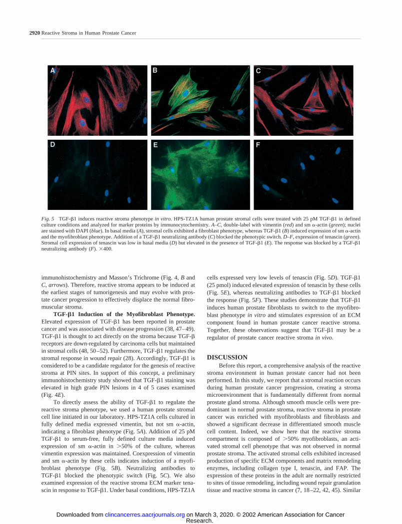
To directly assess the ability of TGF-�1 to regulate thereactive stroma phenotype, we used a human prostate stromalcell line initiated in our laboratory. HPS-TZ1A cells cultured infully defined media expressed vimentin, but not sm �-actin,indicating a fibroblast phenotype (Fig. 5A). Addition of 25 pMTGF-�1 to serum-free, fully defined culture media inducedexpression of sm �-actin in 50% of the culture, whereasvimentin expression was maintained. Coexpression of vimentinand sm �-actin by these cells indicates induction of a myofi-broblast phenotype (Fig. 5B). Neutralizing antibodies toTGF-�1 blocked the phenotypic switch (Fig. 5C). We alsoexamined expression of the reactive stroma ECM marker tena-scin in response to TGF-�1. Under basal conditions, HPS-TZ1A
cells expressed very low levels of tenascin (Fig. 5D). TGF-�1(25 pmol) induced elevated expression of tenascin by these cells(Fig. 5E), whereas neutralizing antibodies to TGF-�1 blockedthe response (Fig. 5F). These studies demonstrate that TGF-�1induces human prostate fibroblasts to switch to the myofibro-blast phenotype in vitro and stimulates expression of an ECMcomponent found in human prostate cancer reactive stroma.Together, these observations suggest that TGF-�1 may be aregulator of prostate cancer reactive stroma in vivo.
DISCUSSIONBefore this report, a comprehensive analysis of the reactive
stroma environment in human prostate cancer had not beenperformed. In this study, we report that a stromal reaction occursduring human prostate cancer progression, creating a stromamicroenvironment that is fundamentally different from normalprostate gland stroma. Although smooth muscle cells were pre-dominant in normal prostate stroma, reactive stroma in prostatecancer was enriched with myofibroblasts and fibroblasts andshowed a significant decrease in differentiated smooth musclecell content. Indeed, we show here that the reactive stromacompartment is composed of 50% myofibroblasts, an acti-vated stromal cell phenotype that was not observed in normalprostate stroma. The activated stromal cells exhibited increasedproduction of specific ECM components and matrix remodelingenzymes, including collagen type I, tenascin, and FAP. Theexpression of these proteins in the adult are normally restrictedto sites of tissue remodeling, including wound repair granulationtissue and reactive stroma in cancer (7, 18–22, 42, 45). Similar
Fig. 5 TGF-�1 induces reactive stroma phenotype in vitro. HPS-TZ1A human prostate stromal cells were treated with 25 pM TGF-�1 in definedculture conditions and analyzed for marker proteins by immunocytochemistry. A–C, double-label with vimentin (red) and sm �-actin (green); nucleiare stained with DAPI (blue). In basal media (A), stromal cells exhibited a fibroblast phenotype, whereas TGF-�1 (B) induced expression of sm �-actinand the myofibroblast phenotype. Addition of a TGF-�1 neutralizing antibody (C) blocked the phenotypic switch. D–F, expression of tenascin (green).Stromal cell expression of tenascin was low in basal media (D) but elevated in the presence of TGF-�1 (E). The response was blocked by a TGF-�1neutralizing antibody (F). �400.
2920 Reactive Stroma in Human Prostate Cancer
Research. on March 3, 2020. © 2002 American Association for Cancerclincancerres.aacrjournals.org Downloaded from

to the results presented here, previous studies have pointed toalterations in the ECM of prostate cancer. These studies haveshown elevated expression of versican (53), hyaluronic acid(54), tenascin (43, 44), MMP-2, and MMP-9 (55) in prostatecancer stroma, which is consistent with our classification of thisstroma as reactive.
Alterations in stromal cell phenotype and increased ECMsynthesis were initiated early in prostate cancer progression atthe precancerous PIN stage. Moreover, our observations suggestthat the normal prostate fibromuscular stroma is displaced bythe developing reactive stroma. Alternatively, it has been sug-gested that abnormal epithelial-smooth muscle signaling duringprostate carcinogenesis may lead to smooth muscle dedifferen-tiation (56). However, our data suggest that myofibroblasts inprostate cancer evolved from a periacinar ring of fibroblastsimmediately adjacent to PIN foci, not from normal prostatedifferentiated smooth muscle. Elevated vimentin staining andcollagen I synthesis in stromal cells immediately adjacent toPIN foci suggest that PIN epithelial cells induce a local stromalreaction. In addition, previous studies have reported an increasein tenascin expression in the ECM adjacent to PIN (43, 44).These data suggest that local activation of stromal cells imme-diately adjacent to PIN is the origin of reactive stroma. Our dataalso suggest that reactive stroma progresses with the develop-ment of prostate cancer. The proportion of myofibroblasts inreactive stroma appears to increase from PIN to moderatelydifferentiated to poorly differentiated carcinoma. Furthermore,we suggest the development of this reactive stroma in PIN actsto displace the normal prostate smooth muscle as PINprogresses to Gleason 3 and 4 cancer. These data suggest thatrather than invading into the normal stromal compartment, pre-carcinoma PIN cells induce a reactive stroma, which coevolveswith the carcinoma cells. Together, the carcinoma cells and thereactive stroma displace the normal tissue to comprise thecancer foci, which ultimately forms the tumor.
The regulatory factors that direct a stromal reaction inhuman prostate cancer are not known. TGF-�1 emerges as astrong candidate because of its key regulatory role in woundrepair and stromal cell biology (5, 28). Elevated expression ofTGF-�1 has been reported in prostate carcinoma cells (38, 47,48). Moreover, TGF-�1 action in prostate cancer appears to bedirected toward the stromal compartment. Several previousstudies have shown that TGF-� receptors are typically ex-pressed in reactive stromal cells and are down-regulated inprostate carcinoma cells (48, 50–52). In this study, we haveshown that TGF-�1 is overexpressed in PIN epithelial cells andis capable of inducing HPS-TZ1A human prostate fibroblasts todifferentiate to the myofibroblast phenotype and express tenas-cin in vitro. Similarly, TGF-�1 induction of the myofibroblastphenotype has been observed in mammary gland fibroblasts(16). Furthermore, it is well established that TGF-� regulatesexpression of several ECM components and regulatory pro-teases/protease inhibitors, including factors expressed specifi-cally at sites of tissue remodeling (28). Previous studies havedemonstrated elevated FAP expression in response to TGF-�1(57). Additionally, it has been reported that TGF-�1 increasescollagen type I synthesis (58) and versican expression (59) inhuman prostate stromal cells. Together, these studies suggestthat TGF-�1 is capable of regulating many aspects of periacinar
fibroblast activation. In support of this hypothesis, s.c. injectionof TGF-�1 was shown to be sufficient to induce a stromalreaction characterized by myofibroblast differentiation, collagenproduction, and angiogenesis (30, 31). Therefore, TGF-�1 is alikely candidate for the regulation of the stromal reaction in PINand development of reactive stroma during prostate cancerprogression.
Once reactive stroma has formed, a central questionemerges as to how reactive stroma may affect the rate ofprostate cancer tumorigenesis. It is clear that stromal-epithelialinteractions are important to prostate gland development andmaintenance of the adult phenotype (56). Accordingly, replace-ment of normal stroma with a reactive stroma is likely to altersuch stromal-epithelial interactions and affect cancer progres-sion. Work by Olumi et al. (13) has shown that prostate CAFspromoted tumorigenesis of a SV40 immortalized prostate epi-thelial cell line (TAg-HPE, derived from a benign prostatichyperplasia) when cografted in the renal capsule. TAg-HPEcells alone were nontumorigenic, and cografts with normalprostatic stromal cells, termed fibroblasts in this report, did notaffect TAg-HPE growth and malignant characteristics. Tag-HPEcell proliferation was increased, and apoptosis was decreasedwhen these cells were cocultured with CAFs, compared withcoculture with normal fibroblasts. Because CAFs were isolatedfrom regions of frank carcinoma in radical prostatectomy spec-imens, it is likely that these cells were derived from the fibro-blasts and myofibroblasts of reactive stroma. Additionally, ourown studies have shown that the incidence and rate of LNCaPtumorigenesis in nude mice depends on the source of humanprostate stromal cells coinoculated with the cancer cells (14).These studies went on to show that the effect of reactive stromaon LNCaP tumorigenesis was due, in part, to initiation of rapidangiogenesis in early tumor development, relative to controltumors lacking human prostate stromal cells.
The altered ECM makeup of reactive stroma is likely to bekey in the overall effects of reactive stroma on angiogenesis andtumorigenesis. During wound repair, fibroblasts and myofibro-blasts create a matrix that stimulates angiogenesis and promotesepithelial cell growth and migration (7, 42). In support of thesefindings, a recent study has shown that an inhibitor of collagentype I synthesis and ECM deposition (halofuginone) suppressedtumor progression and angiogenesis in both transplantable andchemically induced mouse bladder carcinomas (60). In halofugi-none-treated animals, collagen �1(I) gene expression was de-creased in the tumor stromal compartment, which resulted in a60–70% reduction in tumor volume and a significant decreasein microvessel density (60). Interestingly, collagen type I hasbeen shown to direct the migration and assembly of endothelialcells into new blood vessels (61). These data, in combinationwith data presented here, suggest that collagen type I expressionis likely to be a key feature of reactive stroma in tumorigenesis.Together, these studies point to the central role reactive stromaplays in stimulating tumorigenesis of prostate cancer cells andsuggest that possible mechanisms include altered proliferationand apoptotic rates in carcinoma cells, as well as increasedangiogenesis.
This study and others indicate that a reactive stroma, dis-tinct from normal stroma, occurs in adenocarcinoma progres-sion. Data reported here represent an initial step in defining
2921Clinical Cancer Research
Research. on March 3, 2020. © 2002 American Association for Cancerclincancerres.aacrjournals.org Downloaded from

reactive stroma in prostate cancer progression and in identifyingpotential regulators of this response. It is possible that specificmarkers of reactive stroma can be used to better predict the rateof cancer progression or the possibility of recurrence. Futurestudies will be directed toward identifying specific markers ofreactive stroma, which may fulfill the criteria of a differentialprognostic indicator. In addition, because reactive stroma biol-ogy is likely to affect cancer progression, it may be possible totarget specific components of reactive stroma in novel therapeu-tic approaches to prostate cancer.
REFERENCES1. Liotta, L. A., and Kohn, E. C. The microenvironment of the tumour-host interface. Nature (Lond.), 411: 375–379, 2001.
2. Matrisian, L. M., Cunha, G. R., and Mohla, S. Epithelial-stromalinteractions and tumor progression: meeting summary and future direc-tions. Cancer Res., 61: 3844–3846, 2001.
3. Park, C. C., Bissell, M. J., and Barcellos-Hoff, M. H. The influenceof the microenvironment on the malignant phenotype. Mol. Med. Today,6: 324–329, 2000.4. Hanahan, D., and Weinberg, R. A. The hallmarks of cancer. Cell,100: 57–70, 2000.5. Tuxhorn, J. A., Ayala, G. E., and Rowley, D. R. Reactive stroma inprostate cancer progression. J. Urol., 166: 2472–2483, 2001.6. Dvorak, H. F. Tumors: wounds that do not heal. Similarities betweentumor stroma generation and wound healing. N. Engl. J. Med., 315:1650–1659, 1986.7. Clark, R. A. F. Wound repair. In: R. A. F. Clark (ed.) The MolecularCell Biology of Wound Repair, pp. 3–50. New York: Plenum Press,1996.8. Iozzo, R. V. Tumor stroma as a regulator of neoplastic behavior. LabInvestig., 73: 157–160, 1995.9. Ronnov-Jessen, L., Petersen, O. W., and Bissell, M. J. Cellularchanges involved in conversion of normal to malignant breast: impor-tance of the stromal reaction. Physiol. Rev., 76: 69–125, 1996.10. Noel, A., and Foidart, J. M. The role of stroma in breast carcinomagrowth in vivo. J. Mammary Gland Biol. Neoplasia, 3: 215–225, 1998.11. Gregoire, M., and Lieubeau, B. The role of fibroblasts in tumorbehavior. Cancer Metastasis Rev., 14: 339–350, 1995.12. Martin, M., Pujuguet, P., and Martin, F. Role of stromal myofibro-blasts infiltrating colon cancer in tumor invasion. Pathol. Res. Pract.,192: 712–717, 1996.13. Olumi, A. F., Grossfeld, G. D., Hayward, S. W., Carroll, P. R.,Tlsty, T. D., and Cunha, G. R. Carcinoma-associated fibroblasts directtumor progression of initiated human prostatic epithelium. Cancer Res.,59: 5002–5011, 1999.14. Tuxhorn, J. A., McAlhany, S. J., Dang, T. D., Ayala, G., andRowley, D. R. Stromal cells promote angiogenesis and growth of humanprostate tumors in a differential reactive stroma (DRS) xenograft model.Cancer Res., 62: 3298–3307, 2002.15. Sappino, A. P., Schurch, W., and Gabbiani, G. Differentiationrepertoire of fibroblastic cells: expression of cytoskeletal proteins asmarker of phenotypic modulations. Lab Investig., 63: 144–161, 1990.16. Ronnov-Jessen, L., and Petersen, O. W. Induction of �-smoothmuscle actin by transforming growth factor �1 in quiescent humanbreast gland fibroblasts. Implications for myofibroblast generation inbreast neoplasia. Lab Investig., 68: 696–707, 1993.17. Ronnov-Jessen, L., Petersen, O. W., Koteliansky, V. E., and Bissell,M. J. The origin of the myofibroblasts in breast cancer. Recapitulationof tumor environment in culture unravels diversity and implicates con-verted fibroblasts and recruited smooth muscle cells. J. Clin. Investig.,95: 859–873, 1995.18. Lagace, R., Grimaud, J. A., Schurch, W., and Seemayer, T. A.Myofibroblastic stromal reaction in carcinoma of the breast: variations
of collagenous matrix and structural glycoproteins. Virchows Arch. APathol. Anat. Histopathol., 408: 49–59, 1985.
19. Brown, L. F., Guidi, A. J., Schnitt, S. J., Van De Water, L.,Iruela-Arispe, M. L., Yeo, T. K., Tognazzi, K., and Dvorak, H. F.Vascular stroma formation in carcinoma in situ, invasive carcinoma, andmetastatic carcinoma of the breast. Clin Cancer Res., 5: 1041–1056,1999.20. Mackie, E. J., Chiquet-Ehrismann, R., Pearson, C. A., Inaguma, Y.,Taya, K., Kawarada, Y., and Sakakura, T. Tenascin is a stromal markerfor epithelial malignancy in the mammary gland. Proc. Natl. Acad. Sci.USA, 84: 4621–4625, 1987.21. Hauptmann, S., Zardi, L., Siri, A., Carnemolla, B., Borsi, L., Cas-tellucci, M., Klosterhalfen, B., Hartung, P., Weis, J., Stocker, G., et al.Extracellular matrix proteins in colorectal carcinomas. Expression oftenascin and fibronectin isoforms. Lab Investig., 73: 172–182, 1995.22. Hanamura, N., Yoshida, T., Matsumoto, E., Kawarada, Y., andSakakura, T. Expression of fibronectin and tenascin-C mRNA by myo-fibroblasts, vascular cells, and epithelial cells in human colon adenomasand carcinomas. Int. J. Cancer, 73: 10–15, 1997.23. Nielsen, B. S., Sehested, M., Timshel, S., Pyke, C., and Dano, K.Messenger RNA for urokinase plasminogen activator is expressed inmyofibroblasts adjacent to cancer cells in human breast cancer. LabInvestig., 74: 168–177, 1996.24. Park, J. E., Lenter, M. C., Zimmermann, R. N., Garin-Chesa, P.,Old, L. J., and Rettig, W. J. Fibroblast activation protein, a dualspecificity serine protease expressed in reactive human tumor stromalfibroblasts. J. Biol. Chem., 274: 36505–36512, 1999.25. DeClerck, Y. A. Interactions between tumour cells and stromal cellsand proteolytic modification of the extracellular matrix by metallopro-teinases in cancer. Eur. J. Cancer, 36: 1258–1268, 2000.26. Frazier, K. S., and Grotendorst, G. R. Expression of connectivetissue growth factor mRNA in the fibrous stroma of mammary tumors.Int. J. Biochem. Cell Biol., 29: 153–161, 1997.27. Shimo, T., Nakanishi, T., Nishida, T., Asano, M., Kanyama, M.,Kuboki, T., Tamatani, T., Tezuka, K., Takemura, M., Matsumura, T.,and Takigawa, M. Connective tissue growth factor induces the prolif-eration, migration, and tube formation of vascular endothelial cells invitro, and angiogenesis in vivo. J. Biochem. (Tokyo), 126: 137–145,1999.28. Roberts, A. B., and Sporn, M. B. Transforming growth factor �. In:R. A. F. Clark (ed.) The Molecular Cell Biology of Wound Repair, pp.275–308. New York: Plenum Press, 1996.29. Sieweke, M. H., and Bissell, M. J. The tumor-promoting effect ofwounding: a possible role for TGF-�-induced stromal alterations. Crit.Rev. Oncog., 5: 297–311, 1994.30. Desmouliere, A., Geinoz, A., Gabbiani, F., and Gabbiani, G. Trans-forming growth factor �1 induces �-smooth muscle actin expression ingranulation tissue myofibroblasts and in quiescent and growing culturedfibroblasts. J. Cell Biol., 122: 103–111, 1993.31. Roberts, A. B., Sporn, M. B., Assoian, R. K., Smith, J. M., Roche,N. S., Wakefield, L. M., Heine, U. I., Liotta, L. A., Falanga, V., Kehrl,J. H. et al. Transforming growth factor type �: rapid induction offibrosis and angiogenesis in vivo and stimulation of collagen formationin vitro. Proc. Natl. Acad. Sci. USA, 83: 4167–4171, 1986.32. Sieweke, M. H., Thompson, N. L., Sporn, M. B., and Bissell, M. J.Mediation of wound-related Rous sarcoma virus tumorigenesis byTGF-�. Science (Wash. DC), 248: 1656–1660, 1990.33. Lieubeau, B., Garrigue, L., Barbieux, I., Meflah, K., and Gregoire,M. The role of transforming growth factor �1 in the fibroblastic reactionassociated with rat colorectal tumor development. Cancer Res., 54:6526–6532, 1994.34. Stearns, M. E., Garcia, F. U., Fudge, K., Rhim, J., and Wang, M.Role of interleukin 10 and transforming growth factor �1 in the angio-genesis and metastasis of human prostate primary tumor lines fromorthotopic implants in severe combined immunodeficiency mice. Clin.Cancer Res., 5: 711–720, 1999.35. Lohr, M., Schmidt, C., Ringel, J., Kluth, M., Muller, P., Nizze, H.,and Jesnowski, R. Transforming growth factor �1 induces desmoplasia
2922 Reactive Stroma in Human Prostate Cancer
Research. on March 3, 2020. © 2002 American Association for Cancerclincancerres.aacrjournals.org Downloaded from
in an experimental model of human pancreatic carcinoma. Cancer Res.,61: 550–555, 2001.
36. Barrett-Lee, P., Travers, M., Luqmani, Y., and Coombes, R. C.Transcripts for transforming growth factors in human breast cancer:clinical correlates. Br. J. Cancer, 61: 612–617, 1990.
37. Coffey, R. J., Jr., Shipley, G. D., and Moses, H. L. Production oftransforming growth factors by human colon cancer lines. Cancer Res.,46: 1164–1169, 1986.
38. Eastham, J. A., Truong, L. D., Rogers, E., Kattan, M., Flanders,K. C., Scardino, P. T., and Thompson, T. C. Transforming growth factor�1: comparative immunohistochemical localization in human primaryand metastatic prostate cancer. Lab Investig., 73: 628–635, 1995.
39. Rowley, D. R. What might a stromal response mean to prostatecancer progression? Cancer Metastasis Rev., 17: 411–419, 1998.40. Wheeler, T. M., and Lebovitz, R. M. Fresh tissue harvest forresearch from prostatectomy specimens. Prostate, 25: 274–279, 1994.41. Owens, G. K. Regulation of differentiation of vascular smoothmuscle cells. Physiol. Rev., 75: 487–517, 1995.42. Yamada, K. M., and Clark, R. A. F. Provisional matrix. In: R. A. F.Clark (ed.), The Molecular Cell Biology of Wound Repair, pp. 51–93.New York: Plenum Press, 1996.43. Ibrahim, S. N., Lightner, V. A., Ventimiglia, J. B., Ibrahim, G. K.,Walther, P. J., Bigner, D. D., and Humphrey, P. A. Tenascin expressionin prostatic hyperplasia, intraepithelial neoplasia, and carcinoma. Hum.Pathol., 24: 982–989, 1993.44. Xue, Y., Smedts, F., Latijnhouwers, M. A., Ruijter, E. T., Aalders,T. W., de la Rosette, J. J., Debruyne, F. M., and Schalken, J. A.Tenascin-C expression in prostatic intraepithelial neoplasia (PIN): amarker of progression? Anticancer Res., 18: 2679–2684, 1998.45. Garin-Chesa, P., Old, L. J., and Rettig, W. J. Cell surface glyco-protein of reactive stromal fibroblasts as a potential antibody target inhuman epithelial cancers. Proc. Natl. Acad. Sci. USA, 87: 7235–7239,1990.46. Montironi, R., Mazzucchelli, R., Algaba, F., and Lopez-Beltran, A.Morphological identification of the patterns of prostatic intraepithelialneoplasia and their importance. J Clin Pathol., 53: 655–665, 2000.47. Steiner, M. S., and Barrack, E. R. Transforming growth factor �1overproduction in prostate cancer: effects on growth in vivo and in vitro.Mol. Endocrinol., 6: 15–25, 1992.48. Gerdes, M. J., Larsen, M., McBride, L., Dang, T. D., Lu, B., andRowley, D. R. Localization of transforming growth factor �1 and typeII receptor in developing normal human prostate and carcinoma tissues.J. Histochem. Cytochem., 46: 379–388, 1998.49. Wikstrom, P., Stattin, P., Franck-Lissbrant, I., Damber, J. E., andBergh, A. Transforming growth factor �1 is associated with angiogen-esis, metastasis, and poor clinical outcome in prostate cancer. Prostate,37: 19–29, 1998.
50. Kim, I. Y., Ahn, H. J., Zelner, D. J., Shaw, J. W., Lang, S., Kato,M., Oefelein, M. G., Miyazono, K., Nemeth, J. A., Kozlowski, J. M.,and Lee, C. Loss of expression of transforming growth factor � type Iand type II receptors correlates with tumor grade in human prostatecancer tissues. Clin. Cancer Res., 2: 1255–1261, 1996.51. Williams, R. H., Stapleton, A. M., Yang, G., Truong, L. D., Rogers,E., Timme, T. L., Wheeler, T. M., Scardino, P. T., and Thompson, T. C.Reduced levels of transforming growth factor � receptor type II inhuman prostate cancer: an immunohistochemical study. Clin. CancerRes., 2: 635–640, 1996.52. Guo, Y., Jacobs, S. C., and Kyprianou, N. Down-regulation ofprotein and mRNA expression for transforming growth factor � (TGF-�1) type I and type II receptors in human prostate cancer. Int. J. Cancer,71: 573–579, 1997.53. Ricciardelli, C., Mayne, K., Sykes, P. J., Raymond, W. A., McCaul,K., Marshall, V. R., and Horsfall, D. J. Elevated levels of versican butnot decorin predict disease progression in early-stage prostate cancer.Clin. Cancer Res., 4: 963–971, 1998.54. Lokeshwar, V. B., Rubinowicz, D., Schroeder, G. L., Forgacs, E.,Minna, J. D., Block, N. L., Nadji, M., and Lokeshwar, B. L. Stromal andepithelial expression of tumor markers hyaluronic acid and HYAL1hyaluronidase in prostate cancer. J. Biol. Chem., 276: 11922–11932,2001.55. Wood, M., Fudge, K., Mohler, J. L., Frost, A. R., Garcia, F., Wang,M., and Stearns, M. E. In situ hybridization studies of metalloprotein-ases 2 and 9 and TIMP-1 and TIMP-2 expression in human prostatecancer. Clin. Exp. Metastasis, 15: 246–258, 1997.56. Hayward, S. W., Cunha, G. R., and Dahiya, R. Normal developmentand carcinogenesis of the prostate. A unifying hypothesis. Ann. N. Y.Acad Sci., 784: 50–62, 1996.57. Rettig, W. J., Su, S. L., Fortunato, S. R., Scanlan, M. J., Raj, B. K.,Garin-Chesa, P., Healey, J. H., and Old, L. J. Fibroblast activationprotein: purification, epitope mapping, and induction by growth factors.Int. J. Cancer, 58: 385–392, 1994.58. Fukabori, Y., Nakano, K., Ohyama, A., and Yamanaka, H. Stimu-lative effect of transforming growth factor � on collagen synthesis byhuman prostatic stromal cells in vitro. Int. J. Urol., 4: 597–602, 1997.59. Sakko, A. J., Ricciardelli, C., Mayne, K., Tilley, W. D., Lebaron,R. G., and Horsfall, D. J. Versican accumulation in human prostaticfibroblast cultures is enhanced by prostate cancer cell-derived trans-forming growth factor �1. Cancer Res., 61: 926–930, 2001.60. Elkin, M., Ariel, I., Miao, H. Q., Nagler, A., Pines, M., de-Groot,N., Hochberg, A., and Vlodavsky, I. Inhibition of bladder carcinomaangiogenesis, stromal support, and tumor growth by halofuginone. Can-cer Res., 59: 4111–4118, 1999.61. Jackson, C. J., and Jenkins, K. L. Type I collagen fibrils promoterapid vascular tube formation upon contact with the apical side ofcultured endothelium. Exp. Cell Res., 192: 319–323, 1991.
2923Clinical Cancer Research
Research. on March 3, 2020. © 2002 American Association for Cancerclincancerres.aacrjournals.org Downloaded from
2002;8:2912-2923. Clin Cancer Res Jennifer A. Tuxhorn, Gustavo E. Ayala, Megan J. Smith, et al. Myofibroblast Phenotype and Extracellular Matrix RemodelingReactive Stroma in Human Prostate Cancer: Induction of
Updated version
http://clincancerres.aacrjournals.org/content/8/9/2912
Access the most recent version of this article at:
Cited articles
http://clincancerres.aacrjournals.org/content/8/9/2912.full#ref-list-1
This article cites 56 articles, 21 of which you can access for free at:
Citing articles
http://clincancerres.aacrjournals.org/content/8/9/2912.full#related-urls
This article has been cited by 73 HighWire-hosted articles. Access the articles at:
E-mail alerts related to this article or journal.Sign up to receive free email-alerts
Subscriptions
Reprints and
To order reprints of this article or to subscribe to the journal, contact the AACR Publications
Permissions
Rightslink site. Click on "Request Permissions" which will take you to the Copyright Clearance Center's (CCC)
.http://clincancerres.aacrjournals.org/content/8/9/2912To request permission to re-use all or part of this article, use this link
Research. on March 3, 2020. © 2002 American Association for Cancerclincancerres.aacrjournals.org Downloaded from