reactions in biological systems: thereduction of ferricytochrome · pdf file ·...
TRANSCRIPT
Proc. Nat. Acad. Sci. USAVol. 71, No. 4, pp. 1408-1412, April 1974
Electron Transfer Reactions in Biological Systems: The Reduction ofFerricytochrome c by Chromous Ions[Cr(III)-ferrocytochrome c complex/crosslinkage]
CAROL J. GRIMES, DENNIS PISZKIEWICZ, AND EVERLY B. FLEISCHER
Departments of Chemistry and Biological Chemistry, University of California at Irvine, Irvine, Calif. 92664
Communicated by Harry B. Gray, January 10, 1974
ABSTRACT Chromous ion reacts with ferricytochromec to yield a one-to-one Cr(III)-ferrocytochrome c complex.This material, when hydrolyzed by trypsin and subjectedto chromatographic procedures, yielded two fragmentscontaining chromium. The amino-acid compositions andchemical characteristics of each of these fragments indi-cated that the chromium had crosslinked two segments ofpolypeptide chain; these were residues 40-53-Cr(III)-resi-dues 61-72 and residues 40-53-Cr(III)-residues 61-73.Examination of a model of the ferricytochrome c moleculeindicated that only two residues of the crosslinked pep-tides were sufficiently close to allow crosslinking to takeplace. These residues were tyrosine 67 and asparagine 52.Enzymatic hydrolysis of one of those fragments by amino-peptidase M supported this identification. The position ofthe chromic ion implies what is the path of electron trans-fer from the chromous ion to the ferric ion in this chemicalreduction of cytochrome c, and suggests a possible path ofelectron transfer in biological oxidation-reduction reac-tions.
The various components of the mitochondrial electron trans-port system have been characterized, their order of reactivityhas been ascertained, and some of their molecular propertieshave been defined (1-3). The general outlines of the electrontransport chain are understood, but the detailed mechanismby which electron transfer (or atom transfer) takes placebetween the various components of the respiratory chain isnot well understood. We report the results of studies of sys-tems that will lead to some insight into the details of electrontransfer in cytochrome c.
Cytochrome c is a well-understood component of the elec-tron transport chain (4). The structures of both the oxidizedand reduced cytochrome c have been determined (5, 6).Kowalsky studied the reduction of ferricytochrome c withchromous ions and demonstrated that the inert Cr(III)product was tightly bound to the cytochrome c moiety (7);several groups have studied the kinetics of this reaction (8, 9).The chemistry of the chromic ion is characterized by its sub-stitutional inertness (10); thus, the product of this reactionwhich has Cr(III) bound to cytochrome c should be relativelystable and amenable to chemical studies of its structure. Thispaper reports the location of the chromic ion in the chromic-cytochrome c complex, and considers the implications re-garding the mechanism of electron transfer in cytochrome c.
MATERIALS AND METHODS
Materials. Horse-heart cytochrome c (type III) was ob-tained from Sigma Chemical and was used without furtherpurification. Trypsin treated with L-(tosylamido-2-phenyl)
1408
ethyl chloromethyl ketone was obtained from WorthingtonBiochemical Co. Aminopeptidase M was from Henley and Co.of New York. Sephadex G-50 superfine and G-25 superfinewere from Pharmacia. Dowex 50-X2 resin (AG50W-X2) andDowex-1-chloride (AG1-X8) were supplied by Bio-Rad,Richmond, Calif. ICN, Irvine, Calif., was the source of the51Cr(III) used to generate 51Cr(II). The 51Cr(III) was obtainedin a solution of 0.5 N HCl, and was diluted with H20 1000-foldbefore use. All other chemicals were of the highest qualityavailable. Nitrogen gas from Matheson was passed throughscrubbing towers of chromous solution before use in reactionvessels.
Preparation of Cr(III)-Cytochrome c. Chromous solutionsfor' reduction of the protein were prepared by reducing adeoxygenated solution of about 10 mM Cr(CI04)3 in 0.1 mMHC104 with Zn(Hg), under an atmosphere of nitrogen. When-ever radioactive labeling was desired, up to 250 uCi of51Cr(III) in solution was added before generation of Cr(II).The procedure for labeling the cytochrome c with chromic
ion follows that of Kowalsky (7) in essence, but differs indetails. To a solution of cytochrome c (20-40 mg/ml of H20,50-200 mg total) was added 20 mole % K3Fe(CN)6 to ensurethat all the protein was in the oxidized, or ferric, form. Thissolution was passed through a column of Dowex-1-chloride(1 X 10 cm) (7), and the visible spectrum checked for thepresence of only the ferric form. The pH of the solution wasadjusted to 4.5, and the volume adjusted to 25 ml. An aliquotwas then analyzed for protein concentration, using the magni-tude of the absorbance at 528 nm [e = 11200 (11)].The concentration of the chromous ion in the reducing solu-
tion was determined by the change in absorbance of a deoxy-genated permanganate solution with and without addedchromous ion [Xm,. = 545 nm, e = 2340 (9) ]. A stoichiometricamount of chromous ion was added to the deoxygenatedprotein solution, which was maintained under nitrogen at 00.The chromous solution must be added slowly, with agitationof the protein solution, in order to effect a 1: 1 complex ofCr(III) with cytochrome c. The labeled protein was passedthrough a Dowex-50 column to remove any chromium notbound to the protein; excess Cr(III) can be completely sepa-rated from ferricytochrome c by either ion exchange or di-alysis procedures. All experiments reported here were donewith labeled Cr(III)-cytochrome c having a chromium/protein ratio of 0.8-1.0. This ratio of chromium binding is pH-dependent, with the maximum ratio of 1.0 occurring at a pHof 4.0. Thus, the binding ratio of 1.0, which was previously
Pa(Reduction of Ferricytochrome c 1409
noted (7) to be attainable only in the presence of phosphate,has now been demonstrated to be a pH effect. (At pH = 6.0the binding ratio is about 0.5.)
Analyses. The protein concentration was estimated by theabsorbance at 410 nm [isosbestic point, E = 106.1 X 10³ (11)].Chromium was determined by the diphenylcarbazide methodafter oxidation of the chromium to chromium (VI) withammonium persulfate and silver nitrate (12). The proteinsolution was decomposed by addition of two drops of concen-trated HNO3, followed immediately by oxidation withpersulfate.
Hydrolysis by Trypsin. Labeled protein was hydrolyzed bytrypsin at room temperature (220) immediately after denatur-ation of the cytochrome c with trichloroacetic acid (5%).After the protein had been suspended in water at a concen-tration of 2-4 mg/ml, the pH was adjusted to 8.0, and 2% byweight of trypsin was added. The pH was maintained at 8.0by addition of 0.05 N NaOH. When the rate of consumptionof base neared zero, another 2% by weight of trypsin wasadded, and the hydrolysis was continued until no more basewas consumed (3-4 hr total). Glacial acetic acid was added tolower the pH to below 3, and the sample was evaporated todryness.
Column Chromatography. Columns of both Sephadex G-50superfine (2.5 X 140 cm) and G-25 superfine (2.5 X 140 cm)were used in separation of the tryptic peptides. The eluantwas 0.1 M acetic acid, and ambient temperatures were either220 or 4°. Polypeptide fractions were located by the ninhydrinmethod after alkaline hydrolysis (13). Other methods used insequencing are given elsewhere (14, 14a).
Amino-Acid Analysis. Samples were hydrolyzed in evacu-ated glass tubes at 1100 for 24 hr with 6 N HCl containing 1drop of 1% phenol in water. Analyses were performed with aBeckman model 121 automatic amino-acid analyzer.
Physical Measurements. Visible-UV spectra were collectedwith a Beckman Acta III Spectrophotometer. Radioactivityof samples was measured with a Tracerlab Versamatic IIscaler with a NaCl well-type gamma counter. Activationanalysis was performed at the UCI TRIGA Reactor complex;the standard chromium solution was prepared from 99.99%pure chromium metal.
Sedimentation Velocity Experiments. Samples in water weredialyzed exhaustively against 1 mM Tris-0.1 M KC1. Theconcentrations of samples to be compared were adjusted to beequal. Sedimentation velocity measurements were made witha Beckman model E Ultracentrifuge at 56,000 rpm and 200.
RESULTS
Cr(lIl)-Cytochrome c. Ferricytochrome c, when reduced bychromous ion, consistently gave products with Cr: proteinratios of 0.8-1.0. Kowalsky (7) has reported that he was un-able to achieve 1:1 adducts of Cr(III) to cytochrome c withoutthe introduction of phosphate ions into the solution. In ourexperiments the presence of additional anions was not neces-sary. We attribute this to our rigorous attempts to excludeoxygen from our reaction mixture which could oxidize Cr(II)before reaction with cytochrome c and to proper control of thepH. We have taken samples of the Cr(III)-ferricytochrome c
without loss of chromium. Thus, many cycles of oxidation-reduction can be carried out on the chromium-labeled proteinwithout loss of the chromium.
Chromium-Protein Bond. In order to establish the strengthand covalency of the Cr(III)-protein bond, an aliquot of theradioactively labeled Cr(III)-cytochrome c was first de-natured with 6 M guanidine HCl. The protein solution wasadjusted to pH 8, and then dialyzed against 10 mM phosphatebuffer (pH = 7). After dialysis the contents of the dialysis bagand the external buffer were counted. The ratio of cpm/mg ofprotein remained unchanged by dialysis; the radioactivity ofthe external buffer remained at background level. Therefore,Cr(III) must be covalently bonded to cytochrome c.
Integrity of the Iron-Methionine 80 Sulfur Bond. The 695-nmband in the visible spectrum of ferricytochrome c is geterallyattributed (31) to an iron-sulfur charge transfer process, thesulfur being the sixth position ligand contributed by methio-nine 80. To determine whether or not the Cr binds directly tothe iron, displacing this sulfur, some of the Cr(III)-cyto-chrome c was reoxidized with K3Fe(CN)6 and the 695-nm bandwas examined. The band was present and had suffered noreduction in extinction coefficient. This experiment indicatesno large change in conformation of the protein has taken placeduring the formation of the Cr(III)-ferricytochrome c.
Identification of the Labeled Peptide. The Cr(III)-cyto-chrome c product was hydrolyzed with trypsin, and theresultant mixture of peptides was fractionated by the columnchromatographic methods described below.An initial attempt to separate the peptides on Dowex-50
with a pH gradient by a standard method (14) was made. Thechromium came through the column at the void volume,unassociated with any polypeptides, and a normal pattern ofpeptides followed (15). We concluded that the Cr-proteinbond is decomposed by the pyridine-acetate buffers used toestablish the gradient. From this experiment a pure peptidecomposed of residues 61-72 (see Fig. 2) was isolated and usedin a study described below: Glu, 4.4(4); Thr, 0.79(1); Leu,2.2(2); Met, 0.94(1); Tyr, 0.96(1); Asp, 1.0(1); Pro, 0.98(1);Lys, 1.0(1).
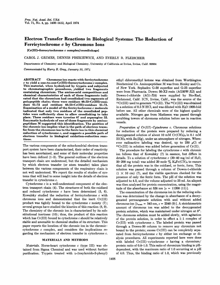
In the second attempt at fractionation, the tryptic peptideswere passed through a column of Sephadex G-50 superfine(2.5 X 140 cm) at 220. The peptide-containing fractionseluted from the column were identified by reaction with nin-hydrin after alkaline hydrolysis, and the fractions containingthe radioactive 5mCr coincided with peptide-containingregions (Fig. la). Electrophoresis of the pooled, radioactivefractions at pH 1.9 yielded four peptides, the least mobile ofwhich contained the radioactive label. Amino-acid analysis(Table 1) showed that this material, designated Fragment I,was composed of an equimolar mixture of residues 40-53 and61-72 (see Fig. 2).In the final fractionation experiment the tryptic peptides
were separated on a column of Sephadex G-25 superfine (2.5 X140 cm) at 4°. The elution pattern showed multiple radio-active peaks, probably due to incomplete hydrolysis (Fig. lb).This material was pooled into three radioactive fractions andsubjected to preparative paper electrophoresis at pH 1.9. Theonly radioactively labeled peptide isolated was from themajor radioactive fraction. This material had the amino-acidcomposition of residues 40-53 and 61-73 (see Fragment TH,
and oxidized it with ferricyanide and re-reduced the complex
Proc. Nat. Acad. Sci. USA 71 (1974)
Table 1); it was identical in composition with the crosslinked
1410 Biochemistry: grimes et al.
I-Lt)
0zm0C,)m
C.)
_.M . I ., . . . . _
20 40 60 80 20 40 60 80TUBE NUMBER
FIG. 1. Peptide maps. Elution profiles of tryptic peptidesfrom: (a) Sephadex G-50 Superfine (2.5 X 140 cm) and (b)Sephadex G-25 Superfine. Ten ml per tube were collected. Ab-sorbance, 570 nm ( ); cpm (---); background level (-.-).Solid bars represent pooled fractions.
material described above (residues 40-53 and 61-72) with anadditional lysyl residue. On the basis of the specificity oftrypsin and the known amino-acid sequence of cytochrome c,this residue must be at position 73 (Fig. 2). Activation analysisfor Cr in Fragment II indicated a Cr: crosslinked peptideratio of 1.3.
Evidence for Crosslinking of these peptides rather than anequimolar mixture of them comes from several experiments.
(1) Electrophoresis of Fragment I at both pH 1.9 and 4.7resulted in a chromatogram containing only one spot. At pH1.9 it ran with'aspartic acid, while at pH 4.7 it ran slightlyfarther toward the anode than glutamic acidj. A pure sampleof the unlabeled peptide composed of residues 61-72 ran at thesame position as the labeled peptide at pH 1.9, but ran behindglutamic acid at pH 4.7. Thus, Fragment I could not be com-posed of equimolar quantities of residues 40-53 and 61-72.
(2) In our earlier experiments any Cr-peptide productsfrom tryptic hydrolysis were decomposed to Cr plus peptidesin a pyridine-acetate buffer. Fragment I was incubated in 2Mpyridine-acetate buffer for 18 hr at 400. Electrophoresis ofthis material at pH 4.7 showed multiple components. One ofthese components migrated to about the same position aspeptide 61-72 and had the same amino-acid compositionwhen examined by electrophoresis at pH 1.9 after acidhydrolysis. A second component, which stayed near theorigin, had the composition of residues 40-53 when examinedby electrophoresis at pH 1.9.
TABLE 1. Amino-acid compositions of labeled polypeptides
Residues40-53 +
Fragment I Fragment II 61-72(73)
Aspartic acid 3.2 3.1 3Threonine 3.2 3.0 4SerineGlutamic acid 4.8 4.6 5Proline 1.8 1.8 2Glycine 2.6 2.3 2Alanine 2.1 2.2 2Valine 0.46Methionine 0.82 0.73 1Isoleucine 0.32Leucine 1.9 1.9 2Tyrosine 1.8 1.8 2Phenylalanine 1.0 1.0 1HistidineLysine 2.0 3.0 2(3)Arginmie
Yield = 17.3% Yield = 4.0%
Proof of Inter- Compared to Intramolecular Crosslinking.The question of any intermolecular bonding of cytochrome cmolecules compared to the proposed intramolecular crosslinkwas settled by the following experiment. Samples of both thelabeled and unlabeled protein were subjected to ultracen-trifugation for sedimentation velocity studies. Qualitatively,only one peak was seen for each of the samples. Quantita-tively, the sedimentation constant 520, u was 1.66 for cyto-chrome c and 1.55 for Cr(III)-cytochrome c. The value of820, ,0 is 1.83 (17) for monomeric equine cytochrome c, and is2.64 for the dimer of equine cytochrome c (16). These resultsshow that the labeled protein is a monomer rather than adimer or oligomer, as would be the case for intermolecularcrosslinks.
Identification of the Bonded Amino-Acid Residues. Havingdiscovered the region of the amino-acid sequence to which thechromium atom was bound, we wished to find out exactlywhich amino-acid residues were involved in the crosslink.Inspection of a model of the oxidized form of cytochrome c[built from coordinates supplied by Dickerson (17)] showsthat in the absence of any gross conformational changes in theprotein there are a limited number of possibilities for theamino-acid residues involved in a crosslink. The closest dis-tance of approach between the two chains occurs betweentyrosine 67 and asparagine 52, both of which are suitable
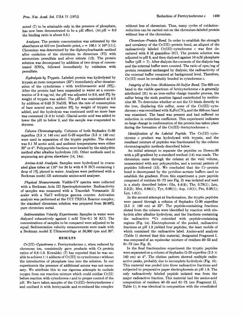
Acetyl-Gly-Asp-Val-Glu-Lys-Gly-Lys-L~ys-Ileu-Phe-Val-GluN H2-Lys-Cys-Ala-GluNH2Cys-His-Thr-Val-Glu-Lys-Gly-Gly-Lys-10 20
L- Heme-His-Lys-Thr-Gly-Pro-AspNH2r1eu-His;-Gly-Leu-Phe-Gly-Arg-Lys-Thr-Gly-GluN H2Ala-Pro-Gly-Phe-Thr-Tyr-Thr-Asp-Ala-30- 40 50
AspNH2-Lys-As NH2-Lys-Gly-Ileu-Thr-Tyr-,Lys-Glu-Glu-Thr-Leu-Met-Glu-Tyr-Leu-Glu-AspNH2-Pro-Lys-Lys-Tyr-Ileu-Pro-60 70
Gly-Thr-Lys-Met-Ileu-Phe-Ala-Gly-Ileu-Lys-Lys-Lys-Thr-Glu-A~rg-Glu-Asp-Leu-Ileu-Ala-Tyr-Leu-Lys-Lys-Ala-Thr-AspNH2-80 90 100
GluCOOH104
FIG. 2. Amino-acid sequence of horse heart cytochrome c (see ref. 23).
Proc. Nat. Acad. Sci. USA 71 (1974)
Proc. Nat. Acad. Sci. USA 71 (1974)
ligands for binding chromium. The distance between these tworesidues is about 4.9 A, and the path between them is unob-structed by other amino-acid residues. The dimensions of thiscavity are certainly adequate for the inclusion of chromium,and the distance between the two residues is admirably suitedfor a ligand-Cr-ligand interaction. Only one other crosslinkappears possible, i.e., tyrosine 67-Cr-tyrosine 48, althoughthis would necessitate a certain degree of strain and rearrange-ment to accommodate the bond. All other amino-acid side-chain ligand combinations (Table 2) are too far from eachother to act as ligands in a crosslink to chromic ion withoutlarge structural changes in the protein.
Hydrolysis by Aminopeptidase M. An aliquot of Fragment Iwas hydrolyzed with aminopeptidase M. Under normal cir-cumstances this exopeptidase could be expected to digest bothpeptides 40-53 and 61-72 completely, except for the alanine43-proline 44 bond, which is resistant to cleavage by thisenzyme (30). The results of amino-acid analysis are: Thr +Asn, 3.4; Gly, 1.4; Glu, 1.7; Ala, 0.99; Pro, 0.19; Phe, 0.87;Tyr, 1.3; Asp, 1.1; Lys, 1.1; Leu, 0.96. These results can beinterpreted as follows. Residues 61-64 are present, and resi-dues 40-53 with two exceptions: alanine 43- proline 44(enzymic exclusion), and one threonine or asparagine. Sincethe analysis was done in sodium citrate buffers at 550 (18),asparagine was not separable from threonine. There are atotal of three threonines and one asparagine in residues 40-53,and one threonine in 61-64, giving a total of five residues.The amino-acid analysis shows a maximum of four residuesat the threonine-asparagine position. Thus, the results ofaminopeptidase M hydrolysis of the crosslinked peptideagrees with the concept of a crosslink by Cr(III) of asparagine52 and tyrosine 67. The fact that amino-peptidase M releasedthe Cr(III)-bound asparagine 52 but stopped at the residuepreceding Cr(III)-bound tyrosine 67 is not understood.
In our initial separation experiment, the Cr(III) crosslinkedamino-acid bonds were found to be unstable under conditionswhere peptide bonds are stable, e.g., in pyridine-acetatebuffers. Furthermore, the instability of this material was sug-gested by the relatively low yields of the labeled fragmentsfinally isolated (Table 1). Thus, approaches to identification ofthe crosslinked residues by techniques such as the Edmandegradation did not seem promising, and further chemicalcharacterization of the crosslinked material was not at-tempted.
DISCUSSIONChromous ion reduces ferricytochrome c with the resultantformation of a one-to-one complex of chromic ion and ferro-
\ t
_j-- 7
Reduction of Ferricytochrome c 1411
TABLE 2. Various distances in theCr(III)-cytochrome c complex
Cr(III)-Fe(III) 6.8 A Tyr 67-Thr 47 13.8 ACr(III)-Met 80S 6.1 A Tyr 67-Thr 49 7.7 ACr(III)-pyrrole C .5.4 A Tyr 67-Asp 50 14.7 ACr(III)-Tyr 67 2.4 A Asp 70-Asp 52 16.0 ATyr 67-Asn 52 4.9 A Glu 66-Asp 52 15.5 ATyr 67-Tyr 48 8.0 A Met 65-Tyr 48 19.7 ATyr 67-Thr 40 10.8 A Thr 4-Asp 62 12.4 ATyr 67-Asp 41 11.7 A
Distances from possible sidechain ligands (i.e., 0, N, S). Fromref. 17.
cytochrome c. A chemical analysis of the product and stericconsiderations dictated by the three-dimensional structure offerricytochrome c (12) indicates that chromium crosslinkstyrosine 67 and asparagine 52. Although we indicate thechromium is crosslinked in the same position in the isolatedpeptide fragment, in the chromium-ferrocytochrome c com-plex, and, most important to our argument concerning elec-tron-transfer, in the chromous-ferricytochrome c complex,we cannot definitely prove that the chromous reduction takesplace without any movement of the chromium ion after re-duction. Our interpretation of the results is based on theknown chemistry of the chromic ion plus our control experi-ments; we feel our interpretation is most reasonable.An examination of the three-dimensional structure of
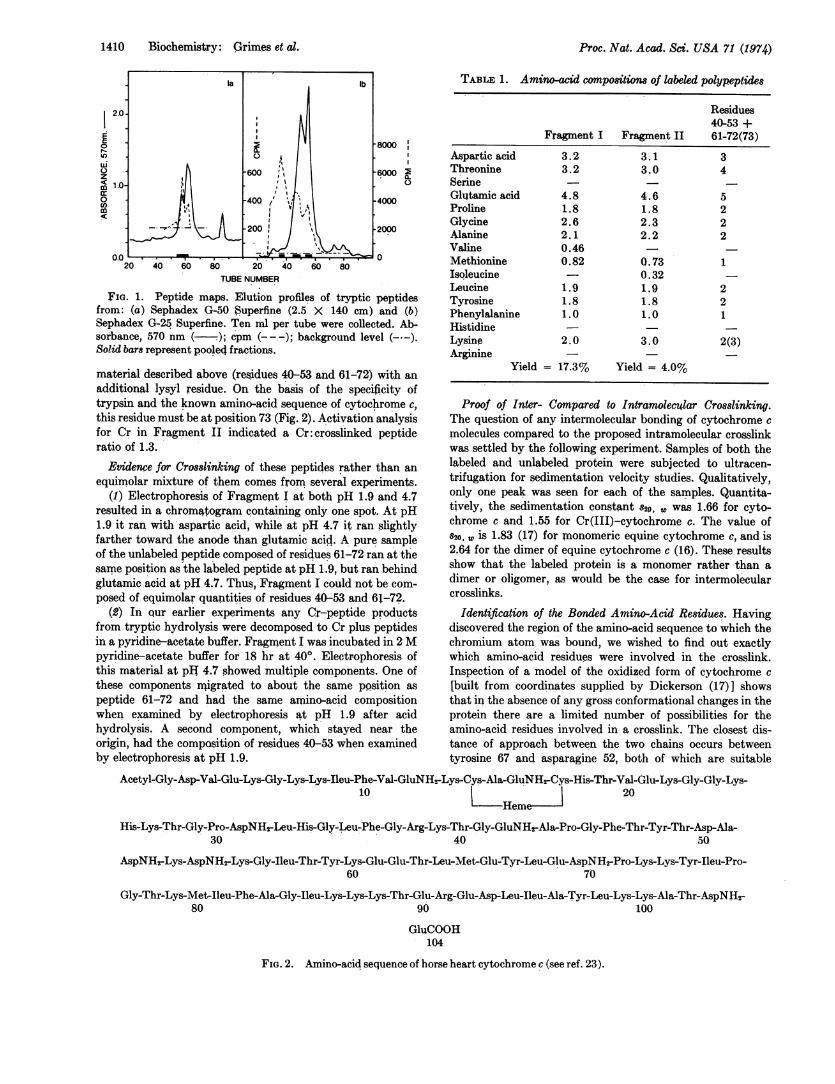
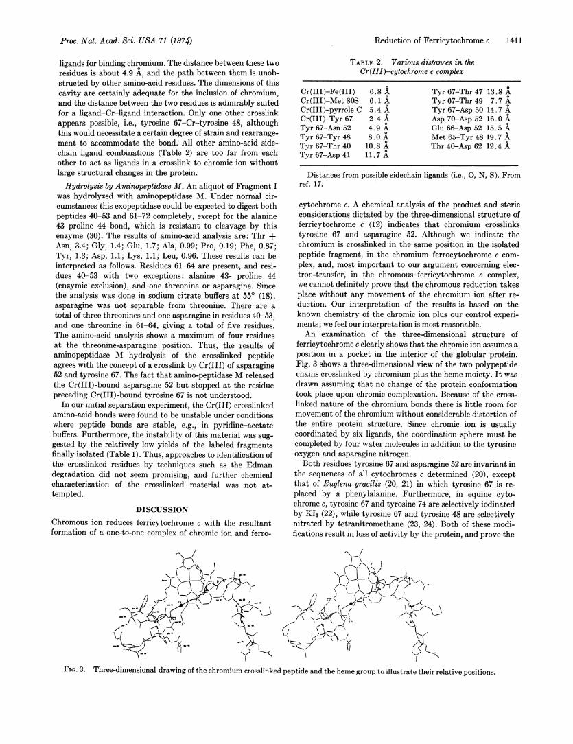
ferricytochrome c clearly shows that the chromic ion assumes aposition in a pocket in the interior of the globular protein.Fig. 3 shows a three-dimensional view of the two polypeptidechains crosslinked by chromium plus the heme moiety. It wasdrawn assuming that no change of the protein conformationtook place upon chromic complexation. Because of the cross-linked nature of the chromium bonds there is little room formovement of the chromium without considerable distortion ofthe entire protein structure. Since chromic ion is usuallycoordinated by six ligands, the coordination sphere must becompleted by four water molecules in addition to the tyrosineoxygen and asparagine nitrogen.Both residues tyrosine 67 and asparagine 52 are invariant in
the sequences of all cytochromes c determined (20), exceptthat of Euglena gracilis (20, 21) in which tyrosine 67 is re-placed by a phenylalanine. Furthermore, in equine cyto-chrome c, tyrosine 67 and tyrosine 74 are selectively iodinatedby K13 (22), while tyrosine 67 and tyrosine 48 are selectivelynitrated by tetranitromethane (23, 24). Both of these modi-fications result in loss of activity by the protein, and prove the
IN
FIG. 3. Three-dimensional drawing of the chromium crosslinked peptide and the heme group to illustrate their relative positions.
1412 Biochemistry: Grimes et al.
accessibility of the tyrosine 67 in native cytochrome c tofairly large molecules.We propose a mechanism for electron transfer in the re-
duction of cytochrome c by chromous ion which is suggestedby the position of tyrosine 67 in the tertiary structure of theprotein. The phenyl ring of the tyrosine is nearly parallel tothe plane of the heme in the oxidized form of cytochrome c(Fig. 3), and about 4-A distant. The chromous ion would firstcoordinate to the tyrosine oxygen, then transfer an electron tothe heme via the overlapping r clouds of the tyrosine and por-phyrin, and then through the conjugated system of theporphyrin to the iron.
This model study shows the possibility of certain pathwaysfor the reduction of cytochrome c that have been speculative.For example, on the basis of model studies, Winfield (27, 28)proposed a mechanism that would involve the participation ofa phenylalanine or tyrosine. Since these residues can be in-duced to form free radicals-and, consequently carry anelectron-one of these residues in proximity to the hemecould act as an intermediary between the oxidizing agent andthe metal ion. An exact mechanism for this interaction (i.e.,transfer of the electron through overlapping ir clouds) was notproposed.
Dickerson and his coworkers (25, 26) have recognized thepossible significance of tyrosine 67 in the electron transfer re-action on the basis of its position in the tertiary structure ofthe protein. They have speculated that in the in vivo reductionof cytochrome c, the electron may be transferred from otherresidues to tyrosine 67.
In conclusion, this study indicates the participation oftyrosine 67 in the reduction of ferricytochrome c by chromousion and suggests the participation of this residue as part of thereduction mechanism in vivo.
This work was supported by a grant from the National ScienceFoundation (GP-30660).
1. The Molecular Basis of Electron Transport (1972) eds.Schultz, J. & Cameron, B. F. (Academic Press, New York).
2. Electron and Coupled Energy Transfer in Biological Systems(1971) eds. King, T. E. & Klingenberg, M. (Marcel Dekker,Inc., New York), Vol. I, Parts A and B.
3. Probes of Structure and Function of Macromolecules and Mem-branes (1971) eds. Chance, B., Yonetani, T. & Mildvan, A.(Academic Press, New York), Vol. II.
4. Margoliash, E. & Schejter, A. (1966) Advan. Protein Chem.21, 113-286.
5. Dickerson, R. E., Takano, T., Eisenberg, D., Kallai, O.,Samson, L., Cooper, A. & Margoliash, E. (1971) J. Biol.Chem. 246, 1511-1535.
6. Dickerson, R. E., Takano, T. & Kallai, 0. B. (1971) CokdSpring Harbor Symp.Quant. Biol. 36, 397-404.
7. Kowalsky, A. (1969) J. Biol. Chem. 244, 6619-6625.8. Yandell, J. K., Fay, D. P. & Sutin, N. (1973) J. Amer. Chem.
Soc. 95, 1131-1137.9. Dawson, J. W., Gray, H. B., Holwerda, R. A. & Westhead,
E. W. (1972) Proc. Nat. Acad. Sci. USA 69, 30-33.10. Taube, H. (1970) Electron Transfer Reactions of Complex Ions
(Academic Press, New York).11. Margoliash, E. & Frohwirt, N. (1959) Biochemistry 71, 570-
572.12. Sandell, E. B. (1959) in Colorimetric Determination of Traces
of Metals (Interscience Publishers, New York), p. 392.13. Hirs, C. H. W., Moore, S. & Stein, W. H. (1956) J. Biol.
Chem. 219, 623-630.14. Landon. M., Melamed, M. D. & Smith, E. L. (1971) J. Biol.
Chem. 246, 2360-2373.14a. Landon, M., Piszkiewicz, D. & Smith, E. L. (1971) J. Biol.
Chem. 246, 2374-2399.15. McDowall, M. A. & Smith, E. L. (1965) J. Biol. Chem. 240,
4635-4647.16. Margoliash, E. & Lustgarten, J. (1962) J. Biol. Chem. 237,
3397-3405.17. Dickerson, R. E., private communication.18. Spackman, D. H., Stein, W. H. & Moore, S. (1958), Anal.
Chem. 30, 1190-1198.19. Dickerson, R. E. (1972) Sci. Amer. 226, 58-72.20. Pettigrew, G. W. (1973) Nature 241, 531-533.21. Lin, D. K., Niece, R. L. & Fitch, W. M. (1973) Nature 241,
533-535.22. McGowan, E. B. & Stellwagen, E. (1970) Biochemistry 9,
3047-3052.23. Skov, K., Hofmann, T. & Williams, G. R.(1969) Can. J. of
Biochem. 47, 750-752.24. Schejter, A. & Sokolovsky, M. (1969) FEBS Lett. 4, 269-
272.25. Dickerson, R. E., Takano, T., Kallai, 0. B. & Samson, L.
(1972) in Structure and Function of Oxidation Reduction En-zymes, eds. Akeson, A. & Ehrenberg, A. (Pergamon Press,New York), pp. 69-83.
26. Dickerson, R. E., Takano, T. & Kallai, 0. B. (1972) "ChainFolding, Reduction Mechanism, and Redox Potential inCytochrome c," in Fifth Jerusalem Symposium, eds. Pullman,B. & Bergmann, E. D. (Israel Academy of Sciences andHumanities, Academic Press, New York).
27. Winfield, M. E. (1964) Oxidases and Related Redox Systems,eds. King, T. E., Mason, H. S. & Morrison, M. (Wiley, NewYork).
28. Winfield, M. E. (1965) J. Mol. Biol. 12, 600-604.29. Smith, L., Davies, H. C., Reichlin, M. & Margoliash, E.
(1973)J. Biol. Chem. 248, 237- 243.30. DeLange, R. J. & Smith, E. L. (1971) in The Enzymes, ed.
Boyer, P. D., (Academic Press, New York), 3rd ed., Vol.III, pp. 81-118.
31. Eaton, W. A. & Hochstrasser, R. M. (1967) J. Chem. Phys.46, 2533-2536.
32. Margoliash, E., Smith, E. L., Kreil, G. & Tuppy, H. (1961)Nature 192, 1125-1127.
Proc. Nat. Acad. Sci. USA 71 (1974)