rapid detection of botulinum neurotoxins—a review · toxins 2019, 11, 418 2 of 28 clostridium...
TRANSCRIPT

PR
IFY
SG
OL
BA
NG
OR
/ B
AN
GO
R U
NIV
ER
SIT
Y
Rapid Detection of Botulinum Neurotoxins—A Review
Hobbs, Robert J.; Thomas, Carol A.; Halliwell, Jennifer; Gwenin, Christopher D.
Toxins
DOI:10.3390/toxins11070418
Published: 17/07/2019
Publisher's PDF, also known as Version of record
Cyswllt i'r cyhoeddiad / Link to publication
Dyfyniad o'r fersiwn a gyhoeddwyd / Citation for published version (APA):Hobbs, R. J., Thomas, C. A., Halliwell, J., & Gwenin, C. D. (2019). Rapid Detection of BotulinumNeurotoxins—A Review. Toxins, 11(7), 418. https://doi.org/10.3390/toxins11070418
Hawliau Cyffredinol / General rightsCopyright and moral rights for the publications made accessible in the public portal are retained by the authors and/orother copyright owners and it is a condition of accessing publications that users recognise and abide by the legalrequirements associated with these rights.
• Users may download and print one copy of any publication from the public portal for the purpose of privatestudy or research. • You may not further distribute the material or use it for any profit-making activity or commercial gain • You may freely distribute the URL identifying the publication in the public portal ?
Take down policyIf you believe that this document breaches copyright please contact us providing details, and we will remove access tothe work immediately and investigate your claim.
01. Jul. 2020

toxins
Review
Rapid Detection of Botulinum Neurotoxins—A Review
Robert J. Hobbs , Carol A. Thomas, Jennifer Halliwell and Christopher D. Gwenin *
Applied Research in Chemistry and Health (ARCH) Research Group, School of Natural Sciences, BangorUniversity, Bangor, Gwynedd, Wales LL57 2UW, UK* Correspondence: [email protected]; Tel.: +44-1248-383741
Received: 25 June 2019; Accepted: 15 July 2019; Published: 17 July 2019�����������������
Abstract: A toxin is a poisonous substance produced within living cells or organisms. One of themost potent groups of toxins currently known are the Botulinum Neurotoxins (BoNTs). These are sodeadly that as little as 62 ng could kill an average human; to put this into context that is approximately200,000 × less than the weight of a grain of sand. The extreme toxicity of BoNTs leads to the need formethods of determining their concentration at very low levels of sensitivity. Currently the mousebioassay is the most widely used detection method monitoring the activity of the toxin; however,this assay is not only lengthy, it also has both cost and ethical issues due to the use of live animals.This review focuses on detection methods both existing and emerging that remove the need for theuse of animals and will look at three areas; speed of detection, sensitivity of detection and finally cost.The assays will have wide reaching interest, ranging from the pharmaceutical/clinical industry forproduction quality management or as a point of care sensor in suspected cases of botulism, the foodindustry as a quality control measure, to the military, detecting BoNT that has been potentially usedas a bio warfare agent.
Keywords: Botulinum Neurotoxin; Botulism; Rapid Detection; Sensitivity; PoC
Key Contribution: The review provides not only a comparison of detection methods for BotulinumNeurotoxin that eradicate the use of animals; but also provides comment on the detection methodswhich are able to determine toxicity of the BoNT. A comparison is compiled between key areas suchas sensitivity, cost and speed of detection.
1. Introduction
Botulinum neurotoxins (BoNTs) are one of the most potent toxins known to man. With a medianlethal dose (LD50) of 1–5 ng/kg [1], they are even more toxic than Sarin, 420 mg/kg [2], Ricin, 1.5 mg/kg [3],and Novichok, 220 ng/kg [4]. BoNTs are formed as proproteins with a single polypeptide chain ofaround 150 kDa by bacteria that originate from the Clostridium genus that are gram-positive, sporeforming and are anaerobic, meaning that they occur in an environment that is free of oxygen. Theseare summarised in Table 1 [5].
Table 1. Bacteria-producing serotypes of botulinum neurotoxin.
Bacteria Group Serotype Human Botulism
C. BotulinumI A,B,F YesII E,B,F YesIII C,D No
C. Argentinense IV G NoC. Baratii V F Yes
C. Butyricum VI E Yes
Toxins 2019, 11, 418; doi:10.3390/toxins11070418 www.mdpi.com/journal/toxins
Toxins 2019, 11, 418 2 of 28
Clostridium Botulinum can grow in the soil, spoiled food, in injuries that have broken the skin, orthe human bowels, and can easily be grown in the laboratory [6]. Due to the high potency and lethalityof BoNTs, their use as biological weapons is an ever-present possibility, and as such, they form oneof the six category A agents. This is the list of agents that pose the highest risk for bioterrorism, ascompiled by the US Centres for Disease Control and prevention (CDC) [7].
BoNTs also have important therapeutic and healing uses, with the toxins being utilised in treatingconditions including, but not limited to: cervical dystonia, strabismus, blepharospasms and multiplesclerosis [6]. The use of botulinum neurotoxin serotype A (BoNT/A) is widely known for its applicationin the commercially available product Botox®in the cosmetics and beauty industry [8].
The toxin is activated through proteolytic cleavage into two parts; the first being a light chainpeptide fragment (50 kDa), which is the zinc containing catalytic portion, and a heavy chain fragmentwhich is roughly double the size (100 kDa), which comprises the receptor binding and centraltranslocation domain. The two chains are linked primarily with a disulphide bond but also multipleinteractions which are non-covalent in nature [9]. BoNTs can induce flaccid paralysis by attackingthe SNARE (soluble N-ethylmaleimide-sensitivity (NSF) attachment protein receptor) proteins withinneurons. BoNT internalisation begins with the toxin binding to two receptors situated on a neuron cellmembrane, gangliosides and protein receptors, through its heavy chain [10]. The BoNT undergoesuptake into the neuron via endocytosis, where a change in pH within the endosome causes the toxinto change conformation allowing the light chain to exit into the intracellular fluid (ICF) [11]. Oncewithin the ICF the toxin cleaves the proteins of the SNARE complex. This complex is comprised ofsynaptobrevin, SNAP-25 and syntaxin.
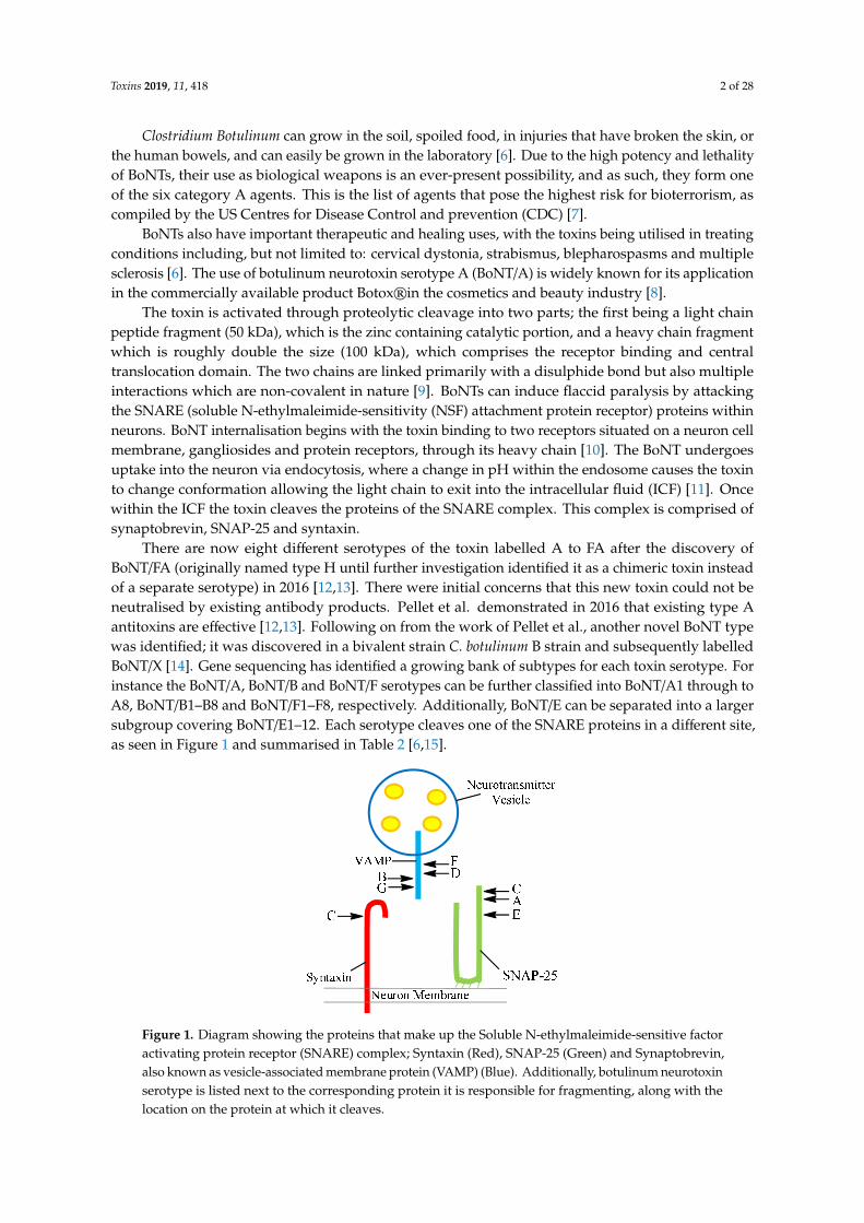
There are now eight different serotypes of the toxin labelled A to FA after the discovery ofBoNT/FA (originally named type H until further investigation identified it as a chimeric toxin insteadof a separate serotype) in 2016 [12,13]. There were initial concerns that this new toxin could not beneutralised by existing antibody products. Pellet et al. demonstrated in 2016 that existing type Aantitoxins are effective [12,13]. Following on from the work of Pellet et al., another novel BoNT typewas identified; it was discovered in a bivalent strain C. botulinum B strain and subsequently labelledBoNT/X [14]. Gene sequencing has identified a growing bank of subtypes for each toxin serotype. Forinstance the BoNT/A, BoNT/B and BoNT/F serotypes can be further classified into BoNT/A1 through toA8, BoNT/B1–B8 and BoNT/F1–F8, respectively. Additionally, BoNT/E can be separated into a largersubgroup covering BoNT/E1–12. Each serotype cleaves one of the SNARE proteins in a different site,as seen in Figure 1 and summarised in Table 2 [6,15].
Toxins 2019, 11, x FOR PEER REVIEW 2 of 29
C. Butyricum VI E Yes Clostridium Botulinum can grow in the soil, spoiled food, in injuries that have broken the skin, or
the human bowels, and can easily be grown in the laboratory [6]. Due to the high potency and lethality of BoNTs, their use as biological weapons is an ever-present possibility, and as such, they form one of the six category A agents. This is the list of agents that pose the highest risk for bioterrorism, as compiled by the US Centres for Disease Control and prevention (CDC) [7].
BoNTs also have important therapeutic and healing uses, with the toxins being utilised in treating conditions including, but not limited to: cervical dystonia, strabismus, blepharospasms and multiple sclerosis [6]. The use of botulinum neurotoxin serotype A (BoNT/A) is widely known for its application in the commercially available product Botox® in the cosmetics and beauty industry [8].
The toxin is activated through proteolytic cleavage into two parts; the first being a light chain peptide fragment (50 kDa), which is the zinc containing catalytic portion, and a heavy chain fragment which is roughly double the size (100 kDa), which comprises the receptor binding and central translocation domain. The two chains are linked primarily with a disulphide bond but also multiple interactions which are non-covalent in nature [9]. BoNTs can induce flaccid paralysis by attacking the SNARE (soluble N-ethylmaleimide-sensitivity (NSF) attachment protein receptor) proteins within neurons. BoNT internalisation begins with the toxin binding to two receptors situated on a neuron cell membrane, gangliosides and protein receptors, through its heavy chain [10]. The BoNT undergoes uptake into the neuron via endocytosis, where a change in pH within the endosome causes the toxin to change conformation allowing the light chain to exit into the intracellular fluid (ICF) [11]. Once within the ICF the toxin cleaves the proteins of the SNARE complex. This complex is comprised of synaptobrevin, SNAP-25 and syntaxin.
There are now eight different serotypes of the toxin labelled A to FA after the discovery of BoNT/FA (originally named type H until further investigation identified it as a chimeric toxin instead of a separate serotype) in 2016 [12,13]. There were initial concerns that this new toxin could not be neutralised by existing antibody products. Pellet et al. demonstrated in 2016 that existing type A antitoxins are effective [12,13]. Following on from the work of Pellet et al., another novel BoNT type was identified; it was discovered in a bivalent strain C. botulinum B strain and subsequently labelled BoNT/X [14]. Gene sequencing has identified a growing bank of subtypes for each toxin serotype. For instance the BoNT/A, BoNT/B and BoNT/F serotypes can be further classified into BoNT/A1 through to A8, BoNT/B1–B8 and BoNT/F1–F8, respectively. Additionally, BoNT/E can be separated into a larger subgroup covering BoNT/E1–12. Each serotype cleaves one of the SNARE proteins in a different site, as seen in Figure 1 and summarised in Table 2 [6,15].
Figure 1. Diagram showing the proteins that make up the Soluble N-ethylmaleimide-sensitive factor activating protein receptor (SNARE) complex; Syntaxin (Red), SNAP-25 (Green) and Synaptobrevin, also known as vesicle-associated membrane protein (VAMP) (Blue). Additionally, botulinum neurotoxin serotype is listed next to the corresponding protein it is responsible for fragmenting, along with the location on the protein at which it cleaves.
Figure 1. Diagram showing the proteins that make up the Soluble N-ethylmaleimide-sensitive factoractivating protein receptor (SNARE) complex; Syntaxin (Red), SNAP-25 (Green) and Synaptobrevin,also known as vesicle-associated membrane protein (VAMP) (Blue). Additionally, botulinum neurotoxinserotype is listed next to the corresponding protein it is responsible for fragmenting, along with thelocation on the protein at which it cleaves.

Toxins 2019, 11, 418 3 of 28
Table 2. The target substrate and cleavage site of each serotype of the toxin.
Serotype SNARE Protein Cleavage Site
A SNAP-25 197–198B Synaptobrevin (VAMP) 76–77
C SNAP-25Syntaxin
198–199253–254
D Synaptobrevin (VAMP) 59–60E SNAP-25 180–181F Synaptobrevin (VAMP) 58–59G Synaptobrevin (VAMP) 81–82FA Synaptobrevin (VAMP) 54–55
The core SNARE complex consists of four α-helices; these come from the three proteins,synaptobrevin, syntaxin and SNAP-25 (which contributes two, abbreviated as Sn1 and Sn2) [16].The toxin then moves to the SNARE proteins where it selectively cleaves one of these proteins atdifferent scissile bonds (a covalent chemical bond that can be broken by an enzyme) depending onthe toxin’s serotype. SNAP-25 is cleaved predominately by three serotypes A, C and E at residues197–198, 198–199 and 180–181 respectively. Syntaxin is fragmented by serotype C between residues253–254. Synaptobrevin (VAMP) undergoes cleavage via by serotypes B, D, F and G between aminoacids 76–77, 59–60, 58–59 and 81–82. Importantly, once a molecule of toxin has cleaved, one moleculeof the target protein is released, and the active site is regenerated to cleave further molecules of thetarget protein [17].
Infection by the toxin causes the disease botulism, which first presents with blurred vision, oraldehydration, difficulty speaking and laboured swallowing quickly followed by flaccid paralysis [18].The most prevalent forms of the disease present as food-borne botulism, infant botulism and alsowound botulism [19]. Treatment is through the administration of botulinum antitoxin and mechanicalventilation and should be provided as soon as possible as the antitoxin can only neutralise toxinmolecules that are yet to be immobilised onto nerve endings [20].
2. Botulism: Types and Medical Application
2.1. Human Botulism
In humans, the disease is predominately caused by the toxin serotypes A, B, E and F and, asmentioned above, normally manifests in one of three naturally occurring forms: either food-borne,wound or infant botulism [18,21]. The BoNT undergoes uptake into the body via two main routes,the gastrointestinal tract or through membranes comprised mainly of mucous, such as those in theeye or respiratory tract. Once uptake is complete and the BoNT is internalised, it then proceeds toassimilate into the blood and lymphatic system. Upon completion the toxin then transverse to nerveendings, where it ultimately accumulates and impedes the release of neurotransmitters. Patientsusually present with the medical difficulties described above and remain the same regardless of theserotype that is the root cause. This is a fairly rapid process with occurrences of flaccid paralysispresent in as little as 12–48 h post toxin exposure [6]. Recovery can take anywhere from a matter ofweeks to extended periods covering many months [22]. The recovery timescale is primarily dependenton the total concentration of BoNT ingested but the serotype involved in intoxication can also playa role, for example BoNT/A is generally observed to have potency levels higher than BoNT/B andBoNT/E [22].
2.2. Foodborne Botulism
Foodborne botulism is the form of human intoxication that is primarily observed and this happenswhen the Clostridium botulinum undergoes a period of growth leading to the production of toxins infood before it is consumed [22]. The bacterium is a gram-positive anaerobic bacterium; this means that

Toxins 2019, 11, 418 4 of 28
for it to undergo growth, it must be in an environment that lacks oxygen. The growth of the bacteriaand the subsequent formation of BoNT primarily occurs in products that have a reduced oxygencontent, but also in those that have certain combinations of packing and storing temperatures as wellas specific preservative factors [23]. Foods commonly involved are those that are prepared in the homeand can include products like dried or salted meats, tinned produce and fermented fish [22]. Typically,these occurrences of intoxication are sporadic and limited to a household, with symptoms typicallyappearing within 12–72 h after toxin ingestion [22]. Occasionally, commercially prepared foods areinvolved [22]. The bacterium C. botulinum favours non-acidic environments (pH > 4.6), this means thatBoNT does not get produced in foods that are acidic in nature [24]. A product with a low pH will alsonot have the ability to degrade any BoNT that may have formed prior to the onset of acidic conditions,e.g., earlier in the production process [24]. To hinder and ultimately avert bacterium growth andBoNT production, not only is pH manipulated, but various arrangements of parameters such as saltconcentration and storage conditions are also utilised. [24]. Case fatality of foodborne botulism indeveloped countries is 5–10% [22]. In cases where there is an outbreak of BoNT, produce samplesmust be collected instantly, correctly, adequately stored and sent for analysis to quickly ascertain theorigin of the contamination. This will assist in the prevention of any subsequent issues and in theremediation cause to bring closure to the outbreak and stop any ongoing issues from arising.
2.3. Infant Botulism
Infant botulism is defined as an illness that manifests in children who are less than one year old,with the majority of cases reported involving infants that are less than six months old. While themechanism is similar to that of foodborne incidents of the disease, the primary difference is that it isthe ingestion of BoNT in produce responsible for foodborne botulism whereas in cases classified asinfant botulism it is ingestion of C. botulinum spores, which in turn undergo growth to produce thebacteria which then release BoNT when accumulating in the digestive system of the young child [25].Generally, in those aged over 6 months, this production of the bacterium is inhibited by the body’snormal immune responses, which have had a chance to progress in older children and adults [25].Symptoms of botulism in infants differs from those generally experience with the illness with theseincluding; decreased neck muscle control, myasthenia, a cry that is different in tone or pitch to a child’snormal response, irregular or hard bowel movements and even appetite decrease [25]. While there area multitude of potential causes of infection, one that is frequently linked to several infant botulismcases is that of honey that has been tainted with the spores [25]. The result of this link has seen adviceprovided to both parents and guardians to avoid children under one year of age from consuminghoney and honey containing products.
2.4. Wound Botulism
The frequency with which wound botulism is observed is very sporadic and it presents whenthe bacterium spores become exposed and embed into a break in the skin, they must penetrate deepenough in the wound in order to undergo growth in anaerobic conditions [26]. Although peoplesuffering from wound botulism will suffer in the same manner as those with foodborne botulism,the resulting symptoms normally take an increased period of time to manifest, due to the slowerprogression of the spores and bacterium, typically the onset of symptoms can take as long as 10–14 daysto present. Wound botulism is commonly coupled with occurrences of complex infections and canoften be attributed to more than a single BoNT serotype [26]. One of the more common causes for thismanifestation of the botulism disease is the injection of drugs and substance misuse, frequently fromcontaminated drugs such as black tar heroin [26].
2.5. Inhalation Botulism
The route of exposure required for inhalation botulism to manifest is not a normal method ofexposure and is generally linked with either unintentional release event, or perhaps more alarmingly,
Toxins 2019, 11, 418 5 of 28
via intentional release as a result of a bioterrorism event resulting in the discharge of toxins as asuspension of fine particulates [23]. The only reported cases of inhalation botulism were recorded asa result of the exposure of three laboratory technicians to the toxin in a research facility in Germanyin 1962. This was as a result of unintentional exposure that occurred during an autopsy of someanimal test subjects in the research facility; these animals had been prior exposed to BoNT serotype A,this exposure resulted in the technicians exhibiting botulism symptoms [27]. The incubation periodgenerally ranges between 24 and 36 h, and up to 5–6 days post exposure. Clinical symptoms are similarto those seen with foodborne botulism except for the absence of digestive system-based symptoms [28].The LD50 via the inhalation route for humans has been estimated at 700 µg to 900 mg assuming abodyweight of 70 kg [7]. Laboratory confirmation of inhalation botulism is difficult because the toxinis not typically distinguishable in the serum or stool whereas it is in foodborne cases [28].
2.6. Other Types of Intoxication
Ingestion of the pre-formed toxin from a water source could occur, but standard water treatmentprocesses such as boiling and disinfection with 0.1% chlorine containing bleach solution, such assodium hypochlorite, would normally result in the destruction of the toxin [29]. The likelihood of toxinexposure proceeding via this route is considered to be minimal [29]. Where the origin of the botulismexposure in adult cases is not attributed to either foodborne or wound exposure it can prove difficult toidentify and remains undetermined, it is likely to have occurred where antibiotic therapy or a surgicalprocedure has altered the normal intestinal flora [23]. Iatrogenic botulism is another manifestation ofthe disease and is typically reported as a result of the use of BoNT in medical or cosmetic products,e.g., Botox® [30,31].
2.7. Botulism in Animals
Serotypes C and D are more commonly responsible for disease in most animals. In Europe,botulism has become a disease of interest due to its emergence in the commercial chicken sector inboth egg and meat production [32]. Birds are typically exposed to BoNT through the ingestion ofinvertebrates, after which BoNT levels in the blood reach levels that induce the signs of paralysis [32]In Brazil the disease is widespread in cattle [33]. Dogs can become affected through eating rotten foodor infected carcases [34]. Horses, however, are most commonly affected by serotype B, which in adultsis acquired through the ingestion of preformed toxins in the feed (in a similar way to human foodbornebotulism) [35]. The disease course is related to the overall toxin exposure and frequently results in fatalconsequences without prompt treatment with specific antitoxin [35].
2.8. Medical Applications of Botulinum Neurotoxin
BoNT works via suppression of transmitter release from endings of motor neurons, resulting inflaccid paralysis [36]. Injected BoNT inhibits release of the neurotransmitter acetylcholine, thereforepreventing contraction of muscle cells [8,37]. BoNT/A is currently used to treat over 20 different medicalconditions, including: Blepharospasm (spasm of the eyelids), cervical dystonia (neck and shoulderspasm), chronic migraine, excessive sweating, strabismus (eye muscle disorders), post-stroke upperlimb spasticity, urinary incontinence and hemi facial spasm [8]. Perhaps the most well-known use isas Botox®injections for cosmetic purposes, such as the treatment of glabellar lines (frown lines) andcrow’s feet. In fact, around 50% of all medical production of BoNT is currently utilised for aestheticmedical products [36]. Injections are generally well tolerated with few side effects. Around 1% ofpeople develop antibodies that make subsequent treatments ineffective [38]. Due to the fact that thetoxin is a biological product which intrinsically leads to variability between production batches, eachset has to be tested for safety, potency and stability before it can be used on humans [39]. The testingmust be conducted at multiple steps in the manufacturing timeline, this is a mandatory requirement tobe able to meet regulatory protocols, leading to marketing consent being approved [40].

Toxins 2019, 11, 418 6 of 28
3. Methods of Detection
The quick onset of the disease, the extreme toxicity of BoNTs and the absence of treatments toreverse paralysis [6] means that a quick BoNT detection method is required that is both sensitive andspecific. It also needs to be as wide reaching as possible and be compatible with food and environmentalsamples. The ability to reliably detect BoNT has applications outside of clinical diagnostics [6,41,42].As previously stated, BoNT represents a significant bioterrorism threat, and if an attack event wereto happen, then a detection method that combines speed, sensitivity, ease of use and the ability tobe used in various potentially harsh environments would be required to evaluate the magnitude ofany contamination [7]. The growing medical use of BoNT means that its definitive detection in theproduction process and research laboratories is of equal importance [6]. This review will providean unbiased comparison of our own methods, along with other promising detection methods andcompare them against the current gold standard, the mouse bioassay. Most developments towardsanimal replacement methods have been focused on the potency testing of pharmaceutical BoNTpreparations. This work has led to two FDA-approved methodologies; both are cell-based potencyassays, firstly as a result of the work in 2012 by Fernández-Salas et al. [43,44] and secondly by the MerzPharma Group in 2015 [43,45]. There have also been other cell-based assays (CBA) achieving levels ofapproval with Allergan (Canada, USA and Swiss approval 2011, EU approval 2013) and Ipsen (EU andSwiss approval 2018) providing CBA detection methods [46]. The ability to quantify active levels oftoxin, like the MBA can, is of high importance, predominantly for pharmaceutical purposes. This willbe identified in this review, along with comments on the detection methods ability to detect eithermultiple serotypes or within complex sample matrices, which are characteristics more prevalent withdiagnostics and food testing for BoNT. Finally, the review will compare the methods against threecriteria; speed of detection, sensitivity of detection and finally cost all of which are more importantin food and diagnostic testing. Despite the advances made, it is estimated that in the EU around400,000 animals per year are still being subjected to batch testing [46]. With this number contributingto a worldwide figure of approximately 600,000, of which 70,000 animals are tested in the UK alone,highlighting the need to further explore replacement detection methods [42].
3.1. Mouse Bioassay
The mouse bioassay (MBA) remains the most widely used test to confirm levels of active BoNTs [23].The test works through injections within or through the peritoneum of samples that are suspectedto contain the toxin. The mice then undergo observation for signs and symptoms that the diseaseis present, which include pilo-erection, wasp-waists, hind limb paralysis, dyspnoea and ultimatelydeath by respiratory paralysis [47]. This typically occurs within 48 h post injection [6]. It is essentialto determine the amount of BoNT in a sample, and this is done by quantifying both the maximumsample dilution that results in fatality in the mice, and also the minimum dilution that does not resultin mouse death. This part of testing may require repeating if the dilution which does not kill is notfound first time [48]. The serotype of the toxin is determined through administration of serotypespecific antitoxins prior to injection with the sample. The mice are observed for signs of botulismfor a further time period of 48 h to confirm that the specific antitoxin for a serotype in the sample isprotective. Therefore, at least 4–6 days are needed to carry out this assay. This assay is considered to bevery sensitive, detecting down to 10 pg/mL, and it is also able to detect functionally active toxin, whichis in direct contrast with a vast number of other immunological methods [6,39,41]. It does, however,have several disadvantages, including the cost and ethical issues of live animal research, as well as theamount of time taken to conduct the assay [49,50].
3.2. Enzyme-Linked Immunosorbent Assay
Enzyme-linked immunosorbent assay (ELISA) is a biochemical procedure that utilises antibodyconjugated enzymes to detect the presence of a specific antigen [51]. ELISA has wide-ranging

Toxins 2019, 11, 418 7 of 28
applications and is regularly used to diagnose a variety of diseases in medicine and as a quality controlcheck in many industries, including to test for cross-contamination in food production [52,53]. Thereare three common set ups: direct, indirect and sandwich (capture), as shown in Figure 2.
Toxins 2019, 11, x FOR PEER REVIEW 7 of 29
control check in many industries, including to test for cross-contamination in food production [52,53]. There are three common set ups: direct, indirect and sandwich (capture), as shown in Figure 2.
Figure 2. Image of common ELISA set ups: Direct, Indirect and Sandwich. In both direct and indirect ELISA, antigens (Ag) are bound to the microtiter plate first, an antibody specific to the antigen is then introduced. In direct assays this primary antibody (blue) has been modified with an enzyme (red star) such as HRP, which when exposed to a substrate produces a measurable colour change. In indirect ELISA, this enzyme is bound to a secondary antibody (green) that has been modified with an enzyme to facilitate colour change; this secondary antibody binds to the primary antibody. In sandwich ELISA, the surface is treated with a capture antibody (red) specific to a desired antigen before the rest of the assay proceeds in the same manner as the indirect assay.
The different methods vary with respect to how the antigen is bound and the number of antibodies used. For direct and indirect ELISA, the sample to be tested is first applied to the microtiter plate, allowing any antigens present to bind before blocking solution, commonly bovine serum albumin (BSA) or casein, is added in order to prevent any non-specific binding of antibodies [54]. Next, the primary antibody that is specific to the antigen is added and allowed to bind. In the case of direct ELISA, this antibody is conjugated to an enzyme which on addition of its substrate produces a measurable colour change signal [55]. With indirect ELISA, a secondary antibody which contains the enzyme conjugate is used to produce the response. This antibody binds to the Fc portion of the primary antibody so does not need to be specific for the antigen [56]. These antibodies are readily commercially available and remove the need to produce antibodies specific for the antigen with the enzyme conjugate, which is a time-consuming and costly process. In sandwich ELISA, a capture antibody (c-Ab) is used that specifically binds the antigen before the surface is blocked and the antigen, primary antibody and secondary antibody conjugate layers are built up as in indirect ELISA [57]. This allows for a more sensitive result, as there are two specific antibodies, and as such, this is the most commonly used for BoNT ELISAs. Using serotype-specific antibodies allows for the determination of which serotypes make up a toxin sample, making it possible to administer the correct antitoxin quickly which is essential for successful treatment of botulism [58]. The detection limit for ELISA ranges from 2 pg/mL to 2 ng/mL, with typical assay lengths of 5–6 hours [6,41].
The detection of BoNT complexes in immunological assays is hampered by the presence of neurotoxin-associated proteins (NAPs). It is this NAP complex that blocks antigenic sites of the toxin, making them unavailable for binding to antibodies, meaning many assays are developed using highly purified BoNT samples, which is not advantageous for use in diagnostic and food-testing [59]. This has led to several groups focusing on development, characterisation and screening of new antibodies that recognise free epitopes on the toxin overcoming the problem of NAPs [60–65].
3.3. Immuno Chromatography Assays—Lateral Flow/Column Flow
Figure 2. Image of common ELISA set ups: Direct, Indirect and Sandwich. In both direct and indirectELISA, antigens (Ag) are bound to the microtiter plate first, an antibody specific to the antigen is thenintroduced. In direct assays this primary antibody (blue) has been modified with an enzyme (red star)such as HRP, which when exposed to a substrate produces a measurable colour change. In indirectELISA, this enzyme is bound to a secondary antibody (green) that has been modified with an enzymeto facilitate colour change; this secondary antibody binds to the primary antibody. In sandwich ELISA,the surface is treated with a capture antibody (red) specific to a desired antigen before the rest of theassay proceeds in the same manner as the indirect assay.
The different methods vary with respect to how the antigen is bound and the number of antibodiesused. For direct and indirect ELISA, the sample to be tested is first applied to the microtiter plate,allowing any antigens present to bind before blocking solution, commonly bovine serum albumin (BSA)or casein, is added in order to prevent any non-specific binding of antibodies [54]. Next, the primaryantibody that is specific to the antigen is added and allowed to bind. In the case of direct ELISA, thisantibody is conjugated to an enzyme which on addition of its substrate produces a measurable colourchange signal [55]. With indirect ELISA, a secondary antibody which contains the enzyme conjugateis used to produce the response. This antibody binds to the Fc portion of the primary antibody sodoes not need to be specific for the antigen [56]. These antibodies are readily commercially availableand remove the need to produce antibodies specific for the antigen with the enzyme conjugate, whichis a time-consuming and costly process. In sandwich ELISA, a capture antibody (c-Ab) is used thatspecifically binds the antigen before the surface is blocked and the antigen, primary antibody andsecondary antibody conjugate layers are built up as in indirect ELISA [57]. This allows for a moresensitive result, as there are two specific antibodies, and as such, this is the most commonly used forBoNT ELISAs. Using serotype-specific antibodies allows for the determination of which serotypesmake up a toxin sample, making it possible to administer the correct antitoxin quickly which isessential for successful treatment of botulism [58]. The detection limit for ELISA ranges from 2 pg/mLto 2 ng/mL, with typical assay lengths of 5–6 h [6,41].
The detection of BoNT complexes in immunological assays is hampered by the presence ofneurotoxin-associated proteins (NAPs). It is this NAP complex that blocks antigenic sites of the toxin,making them unavailable for binding to antibodies, meaning many assays are developed using highlypurified BoNT samples, which is not advantageous for use in diagnostic and food-testing [59]. Thishas led to several groups focusing on development, characterisation and screening of new antibodiesthat recognise free epitopes on the toxin overcoming the problem of NAPs [60–65].

Toxins 2019, 11, 418 8 of 28
3.3. Immuno Chromatography Assays—Lateral Flow/Column Flow
Lateral flow assays (LFAs) are hand-held assays, most well known for their use as pregnancytests. For the detection of BoNT, these assays utilise antibodies conjugated to colloidal gold to yield acolour response in the presence of the toxin [66]. The sample is added on a sample pad where anytoxin present attaches to antibodies on the colloidal gold. They then migrate up the nitrocellulose stripto the capture antibodies where, if positive, they bind, producing a colour response. Excess conjugatethen travels further to the control antibodies, where it binds and produces the second colour responseshowing that the sample has migrated correctly, as shown in Figure 3.
Toxins 2019, 11, x FOR PEER REVIEW 8 of 29
Lateral flow assays (LFAs) are hand-held assays, most well known for their use as pregnancy tests. For the detection of BoNT, these assays utilise antibodies conjugated to colloidal gold to yield a colour response in the presence of the toxin [66]. The sample is added on a sample pad where any toxin present attaches to antibodies on the colloidal gold. They then migrate up the nitrocellulose strip to the capture antibodies where, if positive, they bind, producing a colour response. Excess conjugate then travels further to the control antibodies, where it binds and produces the second colour response showing that the sample has migrated correctly, as shown in Figure 3.
Figure 3. Diagram of lateral flow assay and examples of positive and negative results [67].
LFAs have several advantages when compared to other detection methods: they are very low in cost, self-contained, require no sophisticated equipment or expert analysis and have a very rapid analysis response time of around 15 minutes. The combination of advantages makes them a prime candidate for field use. Nonetheless, LFAs do have poor levels of sensitivity compared with other detection assays such as ELISA and similar immunological techniques. Normal detection limit ranges when using a detection antibody conjugated with gold nanoparticles vary between 5 and 50 ng/mL [68]. Increased sensitivity has been seen when the reporter is replaced with horseradish peroxide (HRP) or silver enhancement. The latter increases the sensitivity to 50 pg/mL [67]. The other main drawback is that due to the use of antibodies, as in the ELISA method platform, the LFAs are unable to differentiate between active and denatured toxin [66,69]. A key requirement of MBA replacements in relation to their usage in the pharmaceutical production sectors is the ability to quantify active toxin levels, so the inability to do this presents a significant hurdle. In 2017 Liu et al. achieved an increase in sensitivity for a LFA system, their system was capable of detecting as low as 20pg/mL for BoNT/A using a very small sample size (1 µL) [70]. To achieve the improvement, Liu et al. reconfigured gold nanoparticle-based lateral flow strips with specific substrate peptides that integrate endopeptidase activity to the assay [70]. It is proposed that the technology could be extended to other BoNT serotypes by designing and encompassing more specific substrate peptides that correspond with the additional serotypes. This would be required for the method to progress for uptake into the diagnostic and food testing sectors [70]. There are, however, two major drawbacks for this methodology, which are that the speed of analysis for the improved system decreased to around 12 hours and there was also an increase in cost due to the integration of the endopeptidase activity assay [70].
3.4. Immuno-PCR/Liposome-PCR
Immuno-polymerase chain reaction (Immuno-PCR) is an ELISA-type immunological test that uses polymerase chain reaction (PCR) to increase amplification of the ELISA signal. The detection methodology relies on forming complexes of antigen and antibody, in the case of Immuno-PCR, its differentiating feature is that it utilises the formation of an antigen-antibody complex, which is then bound to known DNA molecules instead of the normally used enzyme format. Once the binding has occurred to form the complex, the amplification of the DNA fragments which are bound to the BoNT unique antibody is easily implemented using traditional or real-time quantitative PCR (qPCR) [6,41].
Positive Negative
Flow D
irection
Sample
Pad
Nitrocellulose
Absorbent
Pad
Figure 3. Diagram of lateral flow assay and examples of positive and negative results [67].
LFAs have several advantages when compared to other detection methods: they are very lowin cost, self-contained, require no sophisticated equipment or expert analysis and have a very rapidanalysis response time of around 15 minutes. The combination of advantages makes them a primecandidate for field use. Nonetheless, LFAs do have poor levels of sensitivity compared with otherdetection assays such as ELISA and similar immunological techniques. Normal detection limit rangeswhen using a detection antibody conjugated with gold nanoparticles vary between 5 and 50 ng/mL [68].Increased sensitivity has been seen when the reporter is replaced with horseradish peroxide (HRP) orsilver enhancement. The latter increases the sensitivity to 50 pg/mL [67]. The other main drawback isthat due to the use of antibodies, as in the ELISA method platform, the LFAs are unable to differentiatebetween active and denatured toxin [66,69]. A key requirement of MBA replacements in relationto their usage in the pharmaceutical production sectors is the ability to quantify active toxin levels,so the inability to do this presents a significant hurdle. In 2017 Liu et al. achieved an increase insensitivity for a LFA system, their system was capable of detecting as low as 20pg/mL for BoNT/Ausing a very small sample size (1 µL) [70]. To achieve the improvement, Liu et al. reconfigured goldnanoparticle-based lateral flow strips with specific substrate peptides that integrate endopeptidaseactivity to the assay [70]. It is proposed that the technology could be extended to other BoNT serotypesby designing and encompassing more specific substrate peptides that correspond with the additionalserotypes. This would be required for the method to progress for uptake into the diagnostic and foodtesting sectors [70]. There are, however, two major drawbacks for this methodology, which are that thespeed of analysis for the improved system decreased to around 12 h and there was also an increase incost due to the integration of the endopeptidase activity assay [70].
3.4. Immuno-PCR/Liposome-PCR
Immuno-polymerase chain reaction (Immuno-PCR) is an ELISA-type immunological test thatuses polymerase chain reaction (PCR) to increase amplification of the ELISA signal. The detectionmethodology relies on forming complexes of antigen and antibody, in the case of Immuno-PCR, itsdifferentiating feature is that it utilises the formation of an antigen-antibody complex, which is thenbound to known DNA molecules instead of the normally used enzyme format. Once the binding has

Toxins 2019, 11, 418 9 of 28
occurred to form the complex, the amplification of the DNA fragments which are bound to the BoNTunique antibody is easily implemented using traditional or real-time quantitative PCR (qPCR) [6,41].Immuno-PCR has been found to detect botulinum neurotoxin serotype A to sensitivity levels similarto those associated with the MBA. It possesses the ability to determine active toxin levels, which is amajor boost to its potential as an MBA replacement in the pharmaceutical production sector [71]. Theuse of streptavidin as a bridging molecule to link biotinylated modified DNA tags and antibodies hasseen a reported sensitivity of 1 pg/mL in relation to serotype A [72].
In the liposome-PCR test, around 60 copies of reported DNA are encased within a liposome. It hasalso had its outer surface labelled with specific molecule, for BoNT binding this molecule is typicallytrisialoganglioside (GT1b) [73,74]. The modified surface of these laden lipid-based vesicles are able toconjugate with a complex made up of a capture antibody and the toxin of interest, this is then followedby disruption to the vesicles, and qPCR of released DNA. Using this method BoNT/A was detected inpurified water at levels down to 20 pg/mL [73], causing this assay to have a sensitivity detection levelaround 100,000 times lower than the mouse bioassay. One of the main drawbacks of this particulardetection method is that it has not been used and validated in other environments such as clinicalor food samples, which would prevent it from having extensive application in the diagnostic andfood-testing sectors. Both Immuno-PCR and L-PCR assays take around 9 h to run [35].
3.5. Enzyme-Linked Immunosorbent Assay on a Chip (EOC)
The combination of two detection methods is the basis of cross flow immunochromatography.The amalgamation of ELISA, lateral flow assay and utilisation of biosensor automation led to theprogression of an EOC system for the detection of botulinum neurotoxin serotype A [75,76]. The basicdesign of the EOC system for BoNT/A detection is made up of a top polycarbonate moulded plate withtwo traversing channels recessed into the underneath, along with a horizontal flow adsorption pad.An immuno-strip containing two serotype A-specific heavy chain antibodies is then contained betweenthe top and bottom polycarbonate casings using a UV sensitive adhesive [75]. The two antibodies wereused to fulfil two specific roles in the EOC device; one was immobilised onto the membrane to be usedas a capture antibody, the second was conjugated with HRP and utilised as a detection antibody [75].Upon applying a sample containing the desired BoNT/A analyte, the EOC device produced a coloursignal that was linearly correlative to the concentration of BoNT/A available [75]. The strength of anobserved colour signal was able to be quantified using a detector system equipped with a high-qualitydigital imaging. The EOC system was able to analyse BoNT/A with an LoD of 2 ng/mL with an analysistime of less than 30 minutes [76].
3.6. BiosensorsBiosensor technologies embody a wide and diverse range of BoNT detection methods, usually,
the base platforms used consist of: surface plasmon resonance (SPR), refractometer, fluorescenceand chemical luminescence. Typically, evanescence wave technology is used for fluorescence-basedbiosensors. Molecules that are labelled with fluorophores and bound to a surface become excitedupon exposure to evanescent fields, resulting in the production of a signal. Often the assay used is animmunological sandwich assay, consisting of two antibodies for immobilisation and quantificationalong with the analyte of interest, all of which subsequently locate on the sensor surface. Utilising thissystem, the sensitivities of sensors tested ranged from as little as 150 pg/mL [77] to 200 ng/mL [78] forbotulinum neurotoxin serotypes E and B, respectively. These results were generally obtained fromanalyte samples in simplified buffer systems, which would limit wide-scale adoption in the diagnosticand food testing fields. However, it has been reported that serotype A detection can achieve an LoD ofapproximately 50 ng/mL even in more complex matrices such as food samples [79].
The use of aptamers, which can be defined as oligonucleotide fragments that can achieveprotein-specific binding, has advantages over the use of antibodies, including an easier screeningmethod, increased stability, and their sustainable usage [80]. This technique combines an electrochemical

Toxins 2019, 11, 418 10 of 28
approach integrating enzymatic amplification with an aptamer probe, typically around 70–80 bp inlength. This will then undergo a structural change to its standard conformation, this is facilitated by thebinding of toxin present in the sample [80]. The aptamer was bi-labelled with both biotin and fluorescein.Post-binding to toxin the aptamers structural change introduces a conformational opening allowingthe fluorescent reporter tag in which lead to the generation of an electrical response. Consequently,only a specific toxin can generate an amplified current [80]. The LoD for this aptamer-based systemwas quantified as 40 pg/mL (BoNT/A) which is similar to sensitivities seen in traditional ELISA, butis unable to determine levels of active toxin which is barrier in the way of potential application inpharmaceutical production testing [80].
Generally, biosensor-based platforms take >20 minutes to complete, with multiple analytesdetectable simultaneously, depending on the sensor design, this would be of particular interest indevelopment of the system for its use in the diagnostic and food testing sectors. The speed with whichresults can be obtained make biosensors one of the most rapid available platforms around. The desiredrapid analysis, however, comes at a cost, as is seen with LFA devices, leading to limited analyticalsensitivity. However, a review of recent progress in the field of biosensors for their use in detectingtoxins concluded that there have been great advancements based on transducer parameters as wellas bio-recognition elements [76]. It was felt that the progressive systems could make the transitionfrom laboratory to commercial applications within the next few years [76]. Evidence of increasedsensitivity is demonstrated by the recently developed Newton Photonics SPR biosensor, which has aLoD quantified at 6.76 pg/mL (BoNT/A light chain), allowing for active toxin quantification, whichis advantageous for its adoption in the pharmaceutical production sector. The SPR method has adetection time of less than 20 minutes [81].
3.7. Fluorescent Resonance Energy Transfer Assay (FRET)An observable variation in fluorescence of a substrate upon fragmentation is commonly utilised
in the detection of enzyme endopeptidase activity. In FRET assays, Figure 4, an oligopeptide whichmimics a natural substrate is used which carries two tags either side of the cleavage site. One is knownas the fluorescent quencher, the other is the fluorescent donor [82]. FRET is detected only when thetwo tags are close to each other, allowing transfer of fluorescent energy from donor to quencher.
Toxins 2019, 11, x FOR PEER REVIEW 10 of 29
this is facilitated by the binding of toxin present in the sample [80]. The aptamer was bi-labelled with both biotin and fluorescein. Post-binding to toxin the aptamers structural change introduces a conformational opening allowing the fluorescent reporter tag in which lead to the generation of an electrical response. Consequently, only a specific toxin can generate an amplified current [80]. The LoD for this aptamer-based system was quantified as 40 pg/mL (BoNT/A) which is similar to sensitivities seen in traditional ELISA ,but is unable to determine levels of active toxin which is barrier in the way of potential application in pharmaceutical production testing [80].
Generally, biosensor-based platforms take >20 minutes to complete, with multiple analytes detectable simultaneously, depending on the sensor design, this would be of particular interest in development of the system for its use in the diagnostic and food testing sectors. The speed with which results can be obtained make biosensors one of the most rapid available platforms around. The desired rapid analysis, however, comes at a cost, as is seen with LFA devices, leading to limited analytical sensitivity. However, a review of recent progress in the field of biosensors for their use in detecting toxins concluded that there have been great advancements based on transducer parameters as well as bio-recognition elements [76]. It was felt that the progressive systems could make the transition from laboratory to commercial applications within the next few years [76]. Evidence of increased sensitivity is demonstrated by the recently developed Newton Photonics SPR biosensor, which has a LoD quantified at 6.76 pg/mL (BoNT/A light chain), allowing for active toxin quantification, which is advantageous for its adoption in the pharmaceutical production sector. The SPR method has a detection time of less than 20 minutes [81].
3.7. Fluorescent Resonance Energy Transfer Assay (FRET)
An observable variation in fluorescence of a substrate upon fragmentation is commonly utilised in the detection of enzyme endopeptidase activity. In FRET assays, Figure 4, an oligopeptide which mimics a natural substrate is used which carries two tags either side of the cleavage site. One is known as the fluorescent quencher, the other is the fluorescent donor [82]. FRET is detected only when the two tags are close to each other, allowing transfer of fluorescent energy from donor to quencher.
When the substrate is fragmented after it undergoes cleaving by the toxin, the tags are separated, leading to the energy transfer being inhibited [41]. The decrease in FRET is linearly correlative to the toxin concentration. The LoD of FRET-based platforms is dependent upon the substrate incorporated into the assay but is normally within the region of 40 ng–60 pg/mL [6].
Figure 4. Schematic explaining the action of the FRET assay. Typically, an acceptor chromophore (pink) is linked to a fluorophore donor (yellow) via a peptide; this allows the transfer of energy resulting in FRET being detected. Upon exposure to botulinum neurotoxin, the peptide linker is
FRET
BoNT
Peptide
100 Angstroms
10-100 Angstroms Fluorophore Donor
Acceptor Chromophore
Fluorescence
Excitation
Excitation
Figure 4. Schematic explaining the action of the FRET assay. Typically, an acceptor chromophore (pink)is linked to a fluorophore donor (yellow) via a peptide; this allows the transfer of energy resultingin FRET being detected. Upon exposure to botulinum neurotoxin, the peptide linker is cleaved andfragmented allowing the chromophore and fluorophore to dissociate. This inhibits the transfer ofenergy in the system.

Toxins 2019, 11, 418 11 of 28
When the substrate is fragmented after it undergoes cleaving by the toxin, the tags are separated,leading to the energy transfer being inhibited [41]. The decrease in FRET is linearly correlative to thetoxin concentration. The LoD of FRET-based platforms is dependent upon the substrate incorporatedinto the assay but is normally within the region of 40 ng–60 pg/mL [6].
The average speed of analysis via FRET is around 3 h [6,82,83], although Guo et al. reported animproved analysis time of 2 h, although this was with BoNT/B [82]. Combining the assay with animmunoseparation step enhances the sensitivity to around 1 fg/mL, approximately 100,000 times moresensitive than the MBA [83]. In this assay, the toxin is first captured at the end of the immunoseparationstep, whereby the toxin is isolated using toxin specific antibodies bound to microbeads. These beadsare then re-suspended in endopeptidase reaction buffer, which also contains synthetic FRET substrateto initiate cleaving of the toxin, allowing for determination of active toxin levels, which is required forMBA replacement in the pharmaceutical production field [83].
3.8. Flow Cytometry
Flow cytometry equipment can be used to analyse and quantify toxins using fluorescenceimmunoassays [6,41,84]. A multiplexed assay to test both BoNT/A and B alongside ricin, abrin andStaphylococcus enterotoxin B in a diverse range of food matrices utilising the application of fluorescentmagnetic beads, detected toxin levels of 21 and 73 pg/mL, respectively [85]. The assay was enhancedby using an automatic fluidic system to handle the sample. Beads alongside capture antibody arecontained in a flow chamber, where the toxin is first captured from the test matrix, it then undergoeswashing and subsequently capture via antibodies before, finally, FC investigation. The sensitivity limitwas determined as 50 pg/mL in relation to the heavy chain fragment of BoNT/A, allowing quantificationof active toxin levels, with an analysis time of 4 h [86]. This characteristic of the methodology is a keycomponent of a replacement methods ability to be used in the pharmaceutical sector.
Flow cytometry assays have several advantages over ELISA. They are easier to automate, andtherefore it is easier to detect multiple toxins and/or botulinum serotypes in a single sample, whichis key to the widespread adoption of a detection technique in the diagnostic and food testing fields.Other advantages of bead immobilisation of toxin compared to on a microtiter plate, includingimproved capture kinetics as well as improved analyte concentration [85,86]. This detection methodhas been tested in other environments and has been shown to be appropriate for BoNT detection in anextensive variety of produce matrices [85]. The principle drawback of flow cytometry is that it requiresinstrumentation that is significantly more expensive than that required for ELISA or the MBA [6,86].
3.9. Fluorescence Endopeptidase Assay
Fluorescence endopeptidase assays have been researched against several BoNT serotypes. Inone particular example, peptides that were labelled with a fluorescent tag were formulated to beserotype specific and immobilised on a solid substrate. The BoNT would initiate a specific cleavage,mediating the release of a fragment of the tagged peptide into the solution surrounding the substrate.Spatial separation allowed recognition of the different serotypes A, B, E and F to a LoD of 2 ng/mL, theability for a BoNT detection method to recognise multiple serotypes is of particular interest in both thediagnostic and food testing sectors [87]. This assay has been further developed into a semi-automatedmicrofluidic format, utilising the same cleavage of a fluorescence-tagged peptide [88]. The maindifference is that a toxin sample is added to the microfluidic device through an input entry point,allowing for an increased chance of successful cleavage of the fluorescent tagged peptide fragment fromthe solid substrate. The tagged fragments then pass through the device via a microchannel to the exitpoint, which aides in the evaporation of the solution, allowing the concentration of tagged fragmentspresent to be increased. These modifications allowed the semi-automated device format to increasefluorescent signal amplification by 300%. The first generation of this device utilised silica beads as thesolid substrate, but this allowed for an increased variation due to poor consistency related to beadloading, this ultimately affected the overall sensitivity of the device [89]. The issue was addressed by

Toxins 2019, 11, 418 12 of 28
changing the solid substrate setup to that of a gold surface on which the fluorescence-tagged peptidecould form a self-assembled monolayer [88]. The increase in sensitivity allowed BoNT/A to be detecteddown to 3 pg/mL with active toxin distinguishable, which is relevant for pharmaceutical adoption of amethodology. This was, however, for a purified sample, not a concern for pharma usage; nonetheless,when investigated in a more complex matrix, another key requirement of the diagnostic and foodtesting sectors, the LoD was drastically lowered to around 500 ng/mL with an analysis time of 3 h [88].The decrease in sensitivity suggests the major drawback for this type of device moving forward is itsinability to be utilised across a wide range of sample matrices, which would limit the usage uptake of aprospective commercial device and dramatically reduce its potential market.
3.10. Centrifugal Microfluidic Technology
The centrifugal microfluidic immunoassay platform offered by SpinDx is suitable for bothbench and field detection of BoNTs [90]. The sample is mixed with a detection solution which iscomprised of i) capture beads that have been surface functionalised with BoNT specific antibodies,ii) fluorescence-tagged detection antibodies, these become immobilised to the functionalised beadsupon exposure to the equivalent antigen. This is incubated at room temperature, although a morerecent modification to the device allows for changes to temperature due to the incorporation of aheating element. The mixture is then applied on a preloaded density medium, which is located withina channel present in the disc-based system. When this is centrifuged, the micro-particles diffuse acrossthe density medium, resulting in a pellet at the extremity of the furrow. Fluorescence is measured,allowing quantification of the level of BoNT present, with the latest modification allowing the abilityfor the system to distinguish active toxin [90]. The whole assay takes less than 30 minutes and is assimple as loading a sample (which can be of a wide range of complex matrices) onto the disc and thenplacing it in the reader and commencing the testing. It also requires no additional sample preparationand can use a sample size of 2 µL. The complex sample matrices and minimal sample preparationlend themselves very well to the adoption of this methodology for diagnostic and food testing. Thisis, however, countered by the technology needing to be laboratory-based. This incurs high costs andrequires trained personnel to interpret the results; this is also coupled with high equipment costs. Indirect evaluation with the MBA, SpinDx was found to have a sensitivity of 90 ng/mL, 100-fold moresensitive than the MBA with the aforementioned active toxin detection a key component of MBAreplacement in the pharma sector [90].
3.11. Electrochemiluminescence Immunoassay
Electrochemiluminescence (ECL) immunological assays depend on the similar reactionsthat are prevalent in the bulk of immunoassays. The main differing factor is the use of aelectrochemiluminescence tag couple to the antibody responsible for generating signal, this modificationallows the emission of light in the presence of a voltage [91]. ECL platforms are usually performed onc-Ab functionalised magnetic beads. Once binding of the toxin and reporter antibody has taken place,the beads undergo magnetic direction terminating in the vicinity of an electrode, which is where theECL process proceeds [6,92]. Increases in sensitivity can come from multiple origins: (i) improvedluminescent signal:noise ratio; (ii) bead functionalisation facilitates heavier antibody packing; (iii)improved kinetics of Ab-Ag interactions as a result of free flowing substrate; (iv) amplification of theassay results due to the quantity of beads that are held by the magnetic field facilitating an enhancedsample volume interacting with the beads [6,92].
In an early iteration of an ECL assay BoNT/A was found to have a LoD of 5 pg/mL and able todetermine levels of active toxin which is of interest for method development in the pharmaceuticalsector [92]. However, the sensitivity increase when evaluated against ELISA is minimal, as well as thediversity of matrices tested being more limited for ECL; this presents a disadvantage moving forwardwith this assay in diagnostic and food testing. The instrumentation required for ECL analysis is morespecialist and more difficult to use for other purposes [92,93].

Toxins 2019, 11, 418 13 of 28
3.12. Cyclic Voltammetry and Electrochemical Impedance Spectroscopy
Electrochemical techniques are gaining traction for their use in sensor platform development.The main advantage being that they allow for a high sensitivity to be achieved in relation to surfaceinteractions and changes [94]. Some of the areas in which these methodologies are being utilisedinclude sensors for various biomarkers [95–97] and toxins [98], along with pathogens [99,100]; this isnot only due to the high levels of sensitivity, but also because these techniques yield rapid detectiontimes compared to the mouse bio-assay [101]. Both cyclic voltammetry (CV) and electrochemicalimpedance spectroscopy (EIS) have been utilised in the development of BoNT sensors [94,101]. Briefly,in CV a charge can be measured that relates to the number of species on a working electrode thatare able to be oxidised, as well as the number of electrons involved in the oxidation [102]. In termsof a detection method, it is the variations in charge observed when there are modifications to theself-assembled monolayers formed on a working electrode surface that are of interest (Figure 5), asupon addition of an analyte, a binding event would see a change in the charge generated.
Toxins 2019, 11, x FOR PEER REVIEW 13 of 29
Electrochemical techniques are gaining traction for their use in sensor platform development. The main advantage being that they allow for a high sensitivity to be achieved in relation to surface interactions and changes [94]. Some of the areas in which these methodologies are being utilised include sensors for various biomarkers [95–97] and toxins [98], along with pathogens [99,100]; this is not only due to the high levels of sensitivity, but also because these techniques yield rapid detection times compared to the mouse bio-assay [101]. Both cyclic voltammetry (CV) and electrochemical impedance spectroscopy (EIS) have been utilised in the development of BoNT sensors [94,101]. Briefly, in CV a charge can be measured that relates to the number of species on a working electrode that are able to be oxidised, as well as the number of electrons involved in the oxidation [102]. In terms of a detection method, it is the variations in charge observed when there are modifications to the self-assembled monolayers formed on a working electrode surface that are of interest (Figure 5), as upon addition of an analyte, a binding event would see a change in the charge generated.
Figure 5. Schematic showing how botulinum neurotoxin is detected by measuring the decrease in anodic peak charge observed when BoNT cleaves a section of the self-assembled SNAP-25 monolayer.
When current moves through a circuit, consisting of a series of capacitors, inductors and resistors, the measure of the complex resistance that arises is called impedance. Electrochemical impedance spectroscopy uses redox probes including the ferri/ferrocyanide couple [Fe(CN)63−/4−] or the hexaammineruthenium III/II couple [Ru(NH3)63+/2+] and consists of measuring the ability of the ions in these redox couples to be both oxidised and reduced around the working electrode [103]. If a working electrode such as an Au (111) slide is not modified and the surface not blocked in any way, then the redox couples can easily undergo the redox reactions. If the surface of the electrode has been modified, for example, with a protein monolayer, then the ions in the redox couple are impeded from undergoing the oxidation and reduction reactions. This leads to an upturn in the charge transfer resistance of the circuit (Rct) (Figure 6) [104].
SNAP-25
BoNT
Charge
Charge
BoNT Cleave Site
Au (111)
Au (111)
Figure 5. Schematic showing how botulinum neurotoxin is detected by measuring the decrease inanodic peak charge observed when BoNT cleaves a section of the self-assembled SNAP-25 monolayer.
When current moves through a circuit, consisting of a series of capacitors, inductors andresistors, the measure of the complex resistance that arises is called impedance. Electrochemicalimpedance spectroscopy uses redox probes including the ferri/ferrocyanide couple [Fe(CN)6
3−/4−]or the hexaammineruthenium III/II couple [Ru(NH3)6
3+/2+] and consists of measuring the ability ofthe ions in these redox couples to be both oxidised and reduced around the working electrode [103].If a working electrode such as an Au (111) slide is not modified and the surface not blocked in anyway, then the redox couples can easily undergo the redox reactions. If the surface of the electrode hasbeen modified, for example, with a protein monolayer, then the ions in the redox couple are impededfrom undergoing the oxidation and reduction reactions. This leads to an upturn in the charge transferresistance of the circuit (Rct) (Figure 6) [104].

Toxins 2019, 11, 418 14 of 28Toxins 2019, 11, x FOR PEER REVIEW 14 of 29
Figure 6. Schematic showing how whole and cleaved SNAP-25 differs in blocking the reduction of the redox probe, with the whole SNAP-25 providing a greater blocking ability of the redox probe due to its larger size.
For BoNT detection, CV was used, exploiting the interaction between toxin and the SNAP-25 protein at the surface of an Au (111) electrode. The SNAP-25 protein lends itself to this setup, as it has four cysteine residues which form Au-S bonds to the surface of the gold electrode creating a self-assembled monolayer [94]. Botulinum neurotoxin is able to cleave 9 amino acids from the immobilised SNAP-25 facilitates a significant change at the surface which leads to a decrease in the anodic peak charge observed (Figure 5) [94]. The difference in charge before and after incubation with the BoNT sample is then calculated with a correlation between concentration of toxin and drop in charge observed, the higher the concentration of BoNT the larger the decrease in charge. The CV assays were able to quantify active BoNT as low as 250 pg/mL, and although this level of sensitivity is less than that observed in the MBA (10 pg/mL), it is on a similar level to the ELISA methods used for BoNT detection (~200 pg/mL) [94]. Active toxin detection is advantageous for the development of the method to be used in the pharmaceutical sector [94]. The main advantage of this assay is the speed of the assay, with the BoNT sample needing just 10 mins incubation with the SNAP-25 modified electrode and the measurement taking another 1–2 mins. This is significantly quicker than the MBA, and a lot of other BoNT detection methods [94]. It also has a relatively low cost, especially compared to the MBA, with each CV test costing ~£20, with the bulk of the cost coming from the gold electrode, which could be decreased if the method moved forward commercially.
An electrochemical impedance spectroscopy detection method was developed utilising the ferri/ferrocyanide redox probe to measure the impedance that occurred at the surface of a SNAP-25 and VAMP modified gold electrode [101]. Both proteins were used and tested due to their ability to detect different serotypes of BoNT, (Table 2). Upon incubation with the toxin the proteins immobilised on the electrode surface would become cleaved leading to the loss of a small fragment, ~9 amino acids (Figure 6). This leads to a measurable decrease in the Rct, allowing for the quantification of the concentration of BoNT present [101].
The EIS detection method was able to distinguish the presence of active BoNT to a sensitivity level of 25 fg/mL with slight variation between serotypes [101]. The ability to distinguish active toxin is a key component of MBA replacement assays, as it is the primary objective required in the pharmaceutical industry. The multiple toxin type testing is extremely advantageous for adoption in the diagnostic and food testing fields, although further study into a wider range of sample matrices would be required. As with the CV detection method the EIS method is not as sensitive as the MBA; however, the assay is still considerably quicker, with measurements taking around 35 minutes after introduction of the toxin [101]. The cost of the EIS is around £24, which is comparable to the CV detection method, with commercial uptake of the assay leading to a further decrease in potential costs.
3.13. Immuno-Detection of Cleavage Product
Figure 6. Schematic showing how whole and cleaved SNAP-25 differs in blocking the reduction of theredox probe, with the whole SNAP-25 providing a greater blocking ability of the redox probe due to itslarger size.
For BoNT detection, CV was used, exploiting the interaction between toxin and the SNAP-25protein at the surface of an Au (111) electrode. The SNAP-25 protein lends itself to this setup, asit has four cysteine residues which form Au-S bonds to the surface of the gold electrode creatinga self-assembled monolayer [94]. Botulinum neurotoxin is able to cleave 9 amino acids from theimmobilised SNAP-25 facilitates a significant change at the surface which leads to a decrease in theanodic peak charge observed (Figure 5) [94]. The difference in charge before and after incubationwith the BoNT sample is then calculated with a correlation between concentration of toxin and dropin charge observed, the higher the concentration of BoNT the larger the decrease in charge. The CVassays were able to quantify active BoNT as low as 250 pg/mL, and although this level of sensitivity isless than that observed in the MBA (10 pg/mL), it is on a similar level to the ELISA methods used forBoNT detection (~200 pg/mL) [94]. Active toxin detection is advantageous for the development of themethod to be used in the pharmaceutical sector [94]. The main advantage of this assay is the speed ofthe assay, with the BoNT sample needing just 10 min incubation with the SNAP-25 modified electrodeand the measurement taking another 1–2 min. This is significantly quicker than the MBA, and a lot ofother BoNT detection methods [94]. It also has a relatively low cost, especially compared to the MBA,with each CV test costing ~£20, with the bulk of the cost coming from the gold electrode, which couldbe decreased if the method moved forward commercially.
An electrochemical impedance spectroscopy detection method was developed utilising theferri/ferrocyanide redox probe to measure the impedance that occurred at the surface of a SNAP-25and VAMP modified gold electrode [101]. Both proteins were used and tested due to their ability todetect different serotypes of BoNT, (Table 2). Upon incubation with the toxin the proteins immobilisedon the electrode surface would become cleaved leading to the loss of a small fragment, ~9 aminoacids (Figure 6). This leads to a measurable decrease in the Rct, allowing for the quantification of theconcentration of BoNT present [101].
The EIS detection method was able to distinguish the presence of active BoNT to a sensitivity levelof 25 fg/mL with slight variation between serotypes [101]. The ability to distinguish active toxin is a keycomponent of MBA replacement assays, as it is the primary objective required in the pharmaceuticalindustry. The multiple toxin type testing is extremely advantageous for adoption in the diagnostic andfood testing fields, although further study into a wider range of sample matrices would be required.As with the CV detection method the EIS method is not as sensitive as the MBA; however, the assayis still considerably quicker, with measurements taking around 35 minutes after introduction of thetoxin [101]. The cost of the EIS is around £24, which is comparable to the CV detection method, withcommercial uptake of the assay leading to a further decrease in potential costs.
3.13. Immuno-Detection of Cleavage Product
Immunological detection of BoNT cleavage products can be used in methodologies for thequantification of toxin and antitoxin potency. Classically, a synthesised peptide substrate, obtained

Toxins 2019, 11, 418 15 of 28
from one of the SNARE protein, is exposed to BoNT. The products that result as a consequence of thecleaving are quantified with antibodies that are specific to the fragments [6,105]. Detecting cleavageproducts in relation to botulinum neurotoxins is both specific and quantitative and is consequentlyapplicable for determining the potency of pharmaceutical used BoNTs [105]. Yadirgi et al. reported theintroduction and improvement of a simplified direct ELISA platform for detecting BoNT/A activity inneurons with have been obtained from stem cells that come from pre-implementation stage embryosin mice [105]. The resulting product is caught using a specific neoepitope antibody, raised against apeptide that is equivalent to the 190-197 amino acid sequence of SNAP-25. The type of antibody usedis selected due to its ability to only distinguish fragmented SNAP-25 and not the intact protein. Thecaptured product is then detected with a two-site binding approach facilitated by dual polyclonaldetection antibodies [105]. The limit of detection was stated as 440 ng/mL with an assay time of 6 h.The assay’s ability to determine active toxin concentrations is a major advantage in its relevance toreplace MBA use in the pharmaceutical sector, subject to further testing and validation [105]. It is,however, limited in terms of its adoption as a diagnostic of food testing methodology due to the lack ofserotypes tested and also the non-complex sample matrices used in the assay.
3.14. Endopeptidase Mass Spectrometry
Endopeptidase Mass Spectrometry (Endopep-MS) assays require the use of serotype-specificantibodies, see Figure 7 for diagrammatic overview. The antibodies are conjugated to magnetic beadsand then added to a sample (a); the beads are removed and thoroughly washed before a substratethat imitates the toxins natural target is added (b). The solution is then incubated and the resultingmixture is analysed by matrix-assisted laser desorption/ionisation time-of-flight mass spectrometry(MALDI-TOF MS) [106]. The MS detects any whole substrate and any cleaved fragments that resultfrom incubation with the active toxin (c), this is of particular interest if this methodology is to beutilised by the pharma companies for production testing [107].
Toxins 2019, 11, x FOR PEER REVIEW 15 of 29
Immunological detection of BoNT cleavage products can be used in methodologies for the quantification of toxin and antitoxin potency. Classically, a synthesised peptide substrate, obtained from one of the SNARE protein, is exposed to BoNT. The products that result as a consequence of the cleaving are quantified with antibodies that are specific to the fragments [6,105]. Detecting cleavage products in relation to botulinum neurotoxins is both specific and quantitative and is consequently applicable for determining the potency of pharmaceutical used BoNTs [105]. Yadirgi et al. reported the introduction and improvement of a simplified direct ELISA platform for detecting BoNT/A activity in neurons with have been obtained from stem cells that come from pre-implementation stage embryos in mice [105]. The resulting product is caught using a specific neoepitope antibody, raised against a peptide that is equivalent to the 190-197 amino acid sequence of SNAP-25. The type of antibody used is selected due to its ability to only distinguish fragmented SNAP-25 and not the intact protein. The captured product is then detected with a two-site binding approach facilitated by dual polyclonal detection antibodies [105]. The limit of detection was stated as 440 ng/mL with an assay time of 6 hours. The assay’s ability to determine active toxin concentrations is a major advantage in its relevance to replace MBA use in the pharmaceutical sector, subject to further testing and validation [105]. It is, however, limited in terms of its adoption as a diagnostic of food testing methodology due to the lack of serotypes tested and also the non-complex sample matrices used in the assay.
3.14. Endopeptidase Mass Spectrometry
Endopeptidase Mass Spectrometry (Endopep-MS) assays require the use of serotype-specific antibodies, see Figure 7 for diagrammatic overview. The antibodies are conjugated to magnetic beads and then added to a sample (a); the beads are removed and thoroughly washed before a substrate that imitates the toxins natural target is added (b). The solution is then incubated and the resulting mixture is analysed by matrix-assisted laser desorption/ionisation time-of-flight mass spectrometry (MALDI-TOF MS) [106]. The MS detects any whole substrate and any cleaved fragments that result from incubation with the active toxin (c), this is of particular interest if this methodology is to be utilised by the pharma companies for production testing [107].
Figure 7. An overview of the endopeptidase mass spectrometry assay, serotype specific antibodies are conjugated to magnetic beads then added to a sample (A); the beads are removed and thoroughly
Figure 7. An overview of the endopeptidase mass spectrometry assay, serotype specific antibodiesare conjugated to magnetic beads then added to a sample (A); the beads are removed and thoroughlywashed before a substrate that mimics the toxins natural target is added (B). The solution is thenincubated, and the resulting mixture is analysed by mass spectrophotometry. Both whole substrate andcleaved fragments that result from incubation with BoNT can be detected (C).

Toxins 2019, 11, 418 16 of 28
The procedure takes around 4–8 h; while this is relatively lengthy, the ability to automate some ofthe steps in the detection method allow it to be adapted to process hundreds of separate samples inthis time range. Rosen et al. have developed a multiplex platform to allow the assay to simultaneouslydetect BoNT/A, B and E [108]. This adaptation will further improve assay usability, particularly in thehuman diagnostic and food testing fields, by minimising the sample volume required and reducing theassay time [108]. The sensitivity achieved is good with a detectable range across 100 fg/mL to 1 ng/mL,depending on the sample matrix [109]. The wide range of sample matrices tested, including serum,stool, culture supernatants, and a diverse set of food samples again reinforces its potentially extensiveapplication. Diagnostics and food testing require a varied range of sample matrices to be validated; thisis not so much of a concern for the pharma sector, where simple buffers are the matrix of choice [109].There are, however, problems with this assay; mass spectrometers are very expensive, and not manylaboratories have access to one. It requires a trained technician to not only run the instrument, but alsoto interpret the data, which again narrows its wide scale adoption [108]. The length of time required torun a sample from preparation through to final analysis is also too long when compared with othertechniques, which hinders the need for a rapid diagnostic or food testing detection method due to thequick progression of botulism [108].
3.15. UV/Visible Spectroscopy—Colorimetric Assay
The basic principle behind the colorimetric assay is that there is simply a variation in colourthat happens only in the presence of an analyte. Gold nanoparticles have been used to develop adiverse portfolio of biosensor platforms and diagnostic assays [110]. The tracking of colour change inrelation to colloidal gold stems from the principle that certain wavelengths of light at the surface of themetal have the ability to excite the conduction electrons which in turn results in oscillation. It is thisprocess that is referred to as localised surface plasmon resonance (LSPR) [111]. Gold colloids typicallyexhibit UV-visible spectra with distinctive absorption peak ranges of ~515–535 nm with respectivenanoparticle diameters of 5–50 nm; this is due to their absorption of specific wavelengths of lightdue to LSPR [112]. The red colouration of the nanoparticles in colloidal gold is the result of thembeing negatively charged and its effect on the LSPR of the particles. The charge also results in theparticles repelling each other remaining colloidal in nature. Upon addition of salt, the particles beginto aggregate together; this is due to the negative surface charges being masked and the particles nolonger repelling each other [113]. Once aggregation of the particles occurs, they will begin to absorblonger wavelengths of light due to a decrease in inter-particle distance, which aids the coupling ofthe plasma modes. The increased wavelengths of light absorbed, typically in the region of 700 nmresults in the colloidal solution undergoing a bathochromic shift, with the visible result observed beinga change from red to blue [114]. This effect is able to be counterbalanced by modifying the surfaceof the AuNPs, an effective example is a covalently bonded protein layer, which bonds to the AuNPsurface. This results in the particles being protected from the effects of salt addition preventing themasking of the stabilising negative charges. This helps to maintain the distinctive red colouration ofthe colloidal gold, but also to keep the particles stabilised within the colloid [115]. For BoNT detection,an assay has been developed that utilises this principle exploiting a visible colour change that occurswhen colloidal gold is exposed to sodium chloride [116].
Figure 8 shows both the visual result of exposing gold colloid to salt both prior and after surfacemodification with a protective SNAP-25 protein layer [116]. The methodology developed utilises theprocess of BoNT cleaving SNAP-25; nine amino acids are removed from the C-terminus of the protein.This shortening of SNAP-25 decreases the protective layer present around the AuNPs in the goldcolloid which in turn increase their susceptibility to aggregation upon exposure to the sodium chloride.The assay was able to detect BoNT/A via UV-visible spectroscopy on a semi-micro cuvette scale downto a detection level of 370 fg/mL, and even went on to further develop the assay on a microplate scaleto increase the high throughput of samples, managing to retain a detection limit of 600 fg/mL [116].Both of these methods are limited by the fact that SNAP-25 is exclusively cleaved by BoNT/A, C and E,

Toxins 2019, 11, 418 17 of 28
with two of these contributing to cases of human botulism and the other to animal cases, the systemwould require further serotype testing with a different SNARE complex protein, which may result inmultiplexing difficulties, which may limit its widespread use in the diagnostic and food sectors. It hasa significant advantage with respect to its adoption in the pharmaceutical sector in that it is able todistinguish active toxin, which is a core require for MBA replacement in that area. It is also a veryrapid methodology, taking ~10 min and costing around £1.40 per test [116]. The drawback of a minordecrease in sensitivity of the microplate assay is outweighed by it lowering both the time and costrequired to 7 min and ~£0.90 per test. This detection method has real promise despite the drawbacks,and further exploration is needed to enable testing across all BoNT serotypes [116].
Toxins 2019, 11, x FOR PEER REVIEW 17 of 29
microplate scale to increase the high throughput of samples, managing to retain a detection limit of 600 fg/mL [116]. Both of these methods are limited by the fact that SNAP-25 is exclusively cleaved by BoNT/A, C and E, with two of these contributing to cases of human botulism and the other to animal cases, the system would require further serotype testing with a different SNARE complex protein, which may result in multiplexing difficulties, which may limit its widespread use in the diagnostic and food sectors. It has a significant advantage with respect to its adoption in the pharmaceutical sector in that it is able to distinguish active toxin, which is a core require for MBA replacement in that area. It is also a very rapid methodology, taking ~10 mins and costing around £1.40 per test [116]. The drawback of a minor decrease in sensitivity of the microplate assay is outweighed by it lowering both the time and cost required to 7 mins and ~£0.90 per test. This detection method has real promise despite the drawbacks, and further exploration is needed to enable testing across all BoNT serotypes [116].
Figure 8. UV-visible spectra showing un-modified (left) and modified (right) gold colloids (GC), detailing that upon addition of sodium chloride, the unmodified colloid decreases in stability and undergoes aggregation. The inset images show the visible colour shift [116].
A similar methodology was developed utilising the same basic platform: BoNT cleaved peptides leading to AuNP agglomeration, facilitating a visually observable colour shift from red to blue [117]. The assay allowed for clear visually distinguishable responses at concentrations reaching as little as 1 ng/mL with a LoD around 250 pg/mL [117]. This method utilises each peptide fragment in a ‘turn-on’ method, where instead of the BoNT breaking a peptide linkage, destabilising the AuNPs (a ‘turn-off’ process), it uses Cu2+ to form a chemical complex with the post cleavage peptide fragments. This method is more efficient at inducing particle agglomeration which leads to the detection of lower concentrations of BoNT [117]. The main advantage of this methodology is simplicity of the assays coupled with the relatively high sensitivity; however, there are increased costs compared to the previously described colorimetric assay due to the use of AuNPs, Cu2+ and modified magnetic micro-particles [117]. It also, like most assays, has not been fully tested against various BoNT serotypes and in a range of sample matrices.
3.16. SERS Detection
Surface-enhanced Ramon scattering (SERS) is a plasmonics readout technology that can be utilised in a point of care device. SERS consists of exploiting extrinsic Raman labels (ERLs) made up of gold nanoparticles that have been surface functionalised by Ramon reporter molecules (RRM),
Bare GC Bare GC +
NaCl SNAP-25
GC SNAP-25 GC +
NaCl
SNAP-25 GC + NaCl SNAP-25 GC Bare GC
Bare GC + NaCl
Abs
orba
nce
(a.u
.)
Wavelength (nm) Wavelength (nm)
Abs
orba
nce
(a.u
.)
Figure 8. UV-visible spectra showing un-modified (left) and modified (right) gold colloids (GC),detailing that upon addition of sodium chloride, the unmodified colloid decreases in stability andundergoes aggregation. The inset images show the visible colour shift [116].
A similar methodology was developed utilising the same basic platform: BoNT cleaved peptidesleading to AuNP agglomeration, facilitating a visually observable colour shift from red to blue [117].The assay allowed for clear visually distinguishable responses at concentrations reaching as littleas 1 ng/mL with a LoD around 250 pg/mL [117]. This method utilises each peptide fragment in a‘turn-on’ method, where instead of the BoNT breaking a peptide linkage, destabilising the AuNPs (a‘turn-off’ process), it uses Cu2+ to form a chemical complex with the post cleavage peptide fragments.This method is more efficient at inducing particle agglomeration which leads to the detection oflower concentrations of BoNT [117]. The main advantage of this methodology is simplicity of theassays coupled with the relatively high sensitivity; however, there are increased costs compared tothe previously described colorimetric assay due to the use of AuNPs, Cu2+ and modified magneticmicro-particles [117]. It also, like most assays, has not been fully tested against various BoNT serotypesand in a range of sample matrices.
3.16. SERS Detection
Surface-enhanced Ramon scattering (SERS) is a plasmonics readout technology that can be utilisedin a point of care device. SERS consists of exploiting extrinsic Raman labels (ERLs) made up of goldnanoparticles that have been surface functionalised by Ramon reporter molecules (RRM), antibodiesused for tracing (Abs) and a capture substrate, formed from antibodies bound to a gold surface that iscast onto a glass support [118]. The magnitude of the SERS signal quantifies the amount of antigen ina sample. There are several advantages that SERS has over ELISA and other molecular techniques.
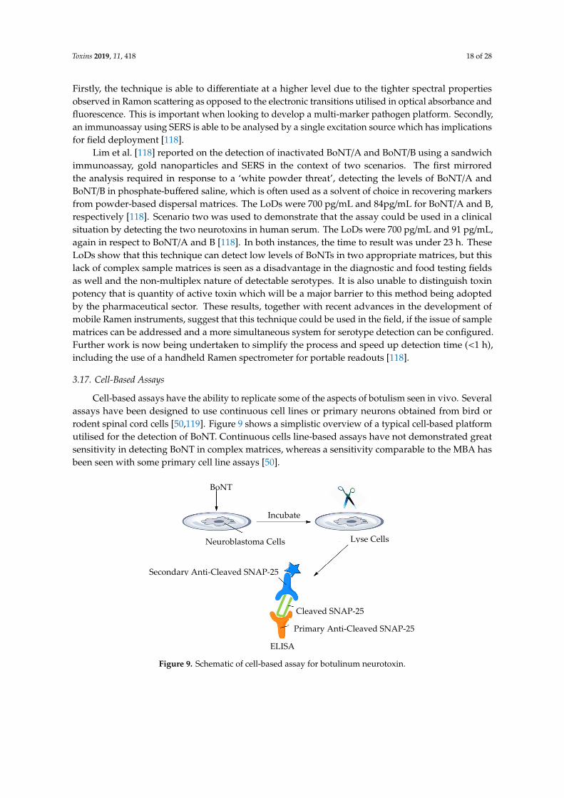
Toxins 2019, 11, 418 18 of 28
Firstly, the technique is able to differentiate at a higher level due to the tighter spectral propertiesobserved in Ramon scattering as opposed to the electronic transitions utilised in optical absorbance andfluorescence. This is important when looking to develop a multi-marker pathogen platform. Secondly,an immunoassay using SERS is able to be analysed by a single excitation source which has implicationsfor field deployment [118].
Lim et al. [118] reported on the detection of inactivated BoNT/A and BoNT/B using a sandwichimmunoassay, gold nanoparticles and SERS in the context of two scenarios. The first mirroredthe analysis required in response to a ‘white powder threat’, detecting the levels of BoNT/A andBoNT/B in phosphate-buffered saline, which is often used as a solvent of choice in recovering markersfrom powder-based dispersal matrices. The LoDs were 700 pg/mL and 84pg/mL for BoNT/A and B,respectively [118]. Scenario two was used to demonstrate that the assay could be used in a clinicalsituation by detecting the two neurotoxins in human serum. The LoDs were 700 pg/mL and 91 pg/mL,again in respect to BoNT/A and B [118]. In both instances, the time to result was under 23 h. TheseLoDs show that this technique can detect low levels of BoNTs in two appropriate matrices, but thislack of complex sample matrices is seen as a disadvantage in the diagnostic and food testing fieldsas well and the non-multiplex nature of detectable serotypes. It is also unable to distinguish toxinpotency that is quantity of active toxin which will be a major barrier to this method being adoptedby the pharmaceutical sector. These results, together with recent advances in the development ofmobile Ramen instruments, suggest that this technique could be used in the field, if the issue of samplematrices can be addressed and a more simultaneous system for serotype detection can be configured.Further work is now being undertaken to simplify the process and speed up detection time (<1 h),including the use of a handheld Ramen spectrometer for portable readouts [118].
3.17. Cell-Based Assays
Cell-based assays have the ability to replicate some of the aspects of botulism seen in vivo. Severalassays have been designed to use continuous cell lines or primary neurons obtained from bird orrodent spinal cord cells [50,119]. Figure 9 shows a simplistic overview of a typical cell-based platformutilised for the detection of BoNT. Continuous cells line-based assays have not demonstrated greatsensitivity in detecting BoNT in complex matrices, whereas a sensitivity comparable to the MBA hasbeen seen with some primary cell line assays [50].
Toxins 2019, 11, x FOR PEER REVIEW 18 of 29
antibodies used for tracing (Abs) and a capture substrate, formed from antibodies bound to a gold surface that is cast onto a glass support [118]. The magnitude of the SERS signal quantifies the amount of antigen in a sample. There are several advantages that SERS has over ELISA and other molecular techniques. Firstly, the technique is able to differentiate at a higher level due to the tighter spectral properties observed in Ramon scattering as opposed to the electronic transitions utilised in optical absorbance and fluorescence. This is important when looking to develop a multi-marker pathogen platform. Secondly, an immunoassay using SERS is able to be analysed by a single excitation source which has implications for field deployment [118].
Lim et al. [118] reported on the detection of inactivated BoNT/A and BoNT/B using a sandwich immunoassay, gold nanoparticles and SERS in the context of two scenarios. The first mirrored the analysis required in response to a ‘white powder threat’, detecting the levels of BoNT/A and BoNT/B in phosphate-buffered saline, which is often used as a solvent of choice in recovering markers from powder-based dispersal matrices. The LoDs were 700 pg/mL and 84pg/mL for BoNT/A and B, respectively [118]. Scenario two was used to demonstrate that the assay could be used in a clinical situation by detecting the two neurotoxins in human serum. The LoDs were 700 pg/mL and 91 pg/mL, again in respect to BoNT/A and B [118]. In both instances, the time to result was under 23 hours. These LoDs show that this technique can detect low levels of BoNTs in two appropriate matrices, but this lack of complex sample matrices is seen as a disadvantage in the diagnostic and food testing fields as well and the non-multiplex nature of detectable serotypes. It is also unable to distinguish toxin potency that is quantity of active toxin which will be a major barrier to this method being adopted by the pharmaceutical sector. These results, together with recent advances in the development of mobile Ramen instruments, suggest that this technique could be used in the field, if the issue of sample matrices can be addressed and a more simultaneous system for serotype detection can be configured. Further work is now being undertaken to simplify the process and speed up detection time (<1 h), including the use of a handheld Ramen spectrometer for portable readouts [118].
3.17. Cell-Based Assays
Cell-based assays have the ability to replicate some of the aspects of botulism seen in vivo. Several assays have been designed to use continuous cell lines or primary neurons obtained from bird or rodent spinal cord cells [50,119]. Figure 9 shows a simplistic overview of a typical cell-based platform utilised for the detection of BoNT. Continuous cells line-based assays have not demonstrated great sensitivity in detecting BoNT in complex matrices, whereas a sensitivity comparable to the MBA has been seen with some primary cell line assays [50].
Figure 9. Schematic of cell-based assay for botulinum neurotoxin.
BoNT
Incubate
Lyse Cells Neuroblastoma Cells
Secondary Anti-Cleaved SNAP-25
Cleaved SNAP-25 Primary Anti-Cleaved SNAP-25
ELISA Figure 9. Schematic of cell-based assay for botulinum neurotoxin.

Toxins 2019, 11, 418 19 of 28
As a model for BoNT detection, cell-based assays can be utilised in several areas of interest: cellsurface receptor binding, endocytosis internalisation, membrane translocation and SNARE cleavage,this combination makes cell-based assays a prime candidate for BoNT inhibitor screens [6,44]. For theiruse in the food or diagnostic sectors, they do have several disadvantages, speed (days for detection), itis technically demanding and requires facilities and the expertise to maintain cell lines, sensitivity is inthe range 1–10 ng/mL [6,50,120].
Recently Rust et al. proposed a SiMa cell-based assay approach where, when used in combinationwith re-engineered VAMP molecules, BoNT/B detection achieved sensitivity comparable to theMBA [39]. It combines the cell-based approach with ELISA in a one-step assay that facilitates detectionof BoNT/B via a luminescent enzymatic reaction. The limit of detection is ~3 pg/mL with an analysistime similar to that of ELISA, although the preparation of the cell reporter line prior to analysis is moretime consuming [39]. This method’s ability to distinguish active toxin is of high importance to thepharmaceutical industry, where speed and cost are less of a concern. This detection method presentsas advantageous due to the user-friendly nature of the assay and its alignment with the 3Rs concept;which describes the steps being taken to replace, refine and reduce animal research to replace the needfor animal use. The cell reporter line could also be further adapted to enable its use in real-time duringthe manufacture of botulinum vaccines [39].
4. Conclusions
There are many different detection platforms that have been developed for their ability to beutilised in the detection of botulinum neurotoxins. They all have a common goal of replacing theMBA and all of which are trying to address the 3Rs; replacement, refinement and reduction of animalresearch and use [42]. The ability for a detection method to detect active toxin levels is of particularinterest to the pharmaceutical production industry. A methodologies ability to be able to detect wherepossible multiple BoNT serotypes, as well as the samples being comprised of complex matrices isof particular interest in the diagnostic and food testing sectors. Several of the proposed detectionmethods have sensitivity levels that are lower than that of the MBA, with some able to also distinguishtoxicity of the BoNT with the ability to detect active toxin levels (highlighted in Table 3), and all of thedetection methods have a faster response times for analysis of samples. This review also pertained toexamine the costs of the detection methods, the estimated costs per test have been estimated based onthe information available from the detection methods published work.
Table 3. Summary table detailing the detection methods for BoNT. The assays that have lower limitsof detection than the mouse bioassay along with the ability to detect active toxin are highlighted ingreen. Those that are just more sensitive than the MBA, but with no distinction for active toxin, arehighlighted in blue. Finally, methods that can distinguish active toxin but have LoDs greater than theMBA are highlighted in orange.
Detection Method Sensitivity of Analysis(LoD) Speed of Analysis Estimated Cost
(Per Test)
Mouse Bioassay (MBA) [6,41] 10 pg/mL 4–6 days £320Enzyme-linked immunosorbent assay
(ELISA) [6,41] 2 pg/mL 6 h £25
Lateral Flow Assay (LFA) [67,70] 50 pg/mL20 pg/mL•
~10 min~12 h
£3£25
Immuno-PCR [72] 1 pg/mL 6–9 h £45Liposome-PCR [73] 20 ag/mL 7–9 h £25
Enzyme-Linked Immunosorbent Assay on aChip (EOC) [75] 2 ng/mL ~30 min £20
Biosensor—Fluorescence-based [77] 150 pg/mL ~10 min £20Biosensor—Aptamer-based [80] 40 pg/mL ~24 h £12
Biosensor—SPR-based [81] 6.76 pg/mL ~20 min £28Fluorescent Resonance Energy Transfer Assay
(FRET) [6,83]60 pg/mL1 fg/mLN
3 h2 h
£11£50
Flow Cytometry [86] 50 pg/mL ~4 h £40Fluorescence Endopeptidase Assay [6,88] 3 pg/mL 3 h £20

Toxins 2019, 11, 418 20 of 28
Table 3. Cont.
Detection Method Sensitivity of Analysis(LoD) Speed of Analysis Estimated Cost
(Per Test)
Centrifugal Microfluidic Technology [90] 90 fg/mL ~30 min £2Electrochemiluminescence Immunoassay
(ECL) [92] 5 pg/mL ~40 min £50
Cyclic Voltammetry (CV) [94] 250 pg/mL ~15 min £20Electrochemical Impedance Spectroscopy
(EIS) [101] 25 fg/mL ~35 min £24
Immuno detection of Cleavage Product [105] 440 fg/mL 6 h £40Endopeptidase Mass Spectrometry [109] 100 fg/mL 4–8 h £40
Colorimetric Assay [116] 370 fg/mL (cuvette)600 fg/mL (96-well plate)
~10 min~7 min
£2£1
Surface-enhanced Ramon scattering(SERS) [118] 84-700 pg/mL ~23 h £25
Cell Based Assays [6,39,50,120] 1–10 ng/mL3 pg/mL† 2–3 days £35
£45
• Combined with endopeptidase activity assay N With additional immunoseparation step, † Cell reporter linecoupled with ELISA detection.
In addition to the methods listed in Table 3, there are various other techniques that have seendevelopment for their use as BoNT detection platforms. One of these such systems is surfaceplasmon resonance, this has seen development from initial uses [121,122] to more recent andemerging advancements [81,123]. Other such techniques that were first explored several yearsago are time-resolved fluorescence assay [124] and liquid chromatography [125]; these have morerecently been explored, providing a relatively low level of detection and short detection time [40,126].The problem with these techniques as detection platforms, is that they often require expensiveinstrumentation or have simply not been tested across a wide enough set of parameters. These factorshave led to other detection methods stagnating and not progressing in advancement such as capillaryelectrophoresis [127] and even a micro-mechanical sensor able to detect synaptobrevin moleculecleavage [128].
For the detection methods summarised in this review, all have an estimated cost per test that islower than the cost of the mouse bioassay. However, these costings do not factor in instrumentationlaboratory/facilities or staff overheads. For some of the detection methods despite the relatively low-testcosts the additional expenses hinder their viability. To truly replace the mouse bioassay, detectionmethods will not only need to be able to provide similar levels of sensitivity to BoNT toxin, but do so ata lower cost while being easy to use. However, the function with which they are used will require keyparameters to be achieved. For adoption in pharmaceutical product testing, the methods will need toalso be able to distinguish and quantify active toxin levels. In contrast, for diagnostic and food testing,this is less of an issue, and instead adoption in these sectors would require the detection method tohave the ability to detect BoNT in complex samples matrices such as serum, stool, culture supernatantsand a diverse set of food samples. It would also provide a huge advantage if the assay were able toeasily detect multiple BoNT serotypes from one sample simultaneously.
Based on the three principles that we have looked at—sensitivity, speed and cost—there arefour detection methods that present as the most promising as replacements to the MBA utilising theprinciples of the 3Rs. The four platforms can be seen in Figure 10 and are located in the middle of theVenn diagram where all of the desired characteristics converge. These are the centrifugal microfluidicassay [90], colorimetric assay [116], SPR-based biosensor [81] and the EIS platform [101]. Whensummarising the advantages and disadvantages of these different methods we concluded that for thecentrifugal microfluidic assay the low cost, ease of use, the ability to use a wide base of sample matricesand the speed and sensitivity were clear advantages in the use of this detection method. The maindisadvantages in the use of this methodology is both the need to perform additional analysis to obtainthe final quantitative result, and the costs associated with both the additional analysis method and theexpected high cost of the assay device, which is still at the prototype stage [90]. With regard to the

Toxins 2019, 11, 418 21 of 28
colorimetric assay, the very low cost per test, the rapid detection time from sample to result and the highlevels of sensitivity provide considerable advantage to this detection platform. The main limitation isthat it requires lab-based equipment and analysis which limits its wider scale usage and increases thecost. This can be somewhat lessened by the fact that this methodology should be easily adaptable toa more portable field device [129]. SPR-based biosensors and the EIS platform both provide similaradvantages in their use for the detection of BoNT. Both are cheap, quick and provide greater sensitivitythan the MBA; however, they are limited again by their constraint to a laboratory-based setting due tothe need for instrumentation and result analysis. Another main drawback is their limited testing ofcomplex sample matrices [81,101]. It is hoped that these methodologies can be scaled down and bemade more portable utilising advances from other areas of biosensors and diagnostics.
Toxins 2019, 11, x FOR PEER REVIEW 21 of 29
Figure 10. A visual representation of the BoNT detection methods analysed in this review. The assays were compared using the mouse bioassay sensitivity as the baseline (10 pg/mL), an analysis time of <1 h and cost of <£30 per test. Highlighted are a method’s ability to distinguish active toxin and improved sensitivity over MBA (Green), active toxin quantification but poorer LoD than the MBA (Orange) and no active toxin quantification by still-improved sensitivity over MBA (Blue). The two detection methods that fall outside of satisfying the parameters of speed, cost and sensitivity are placed outside of the Venn diagram. Additionally, the following notation are used: (●) combined with endopeptidase activity assay, (▲) with an additional immunoseparation step, (†) cell reporter line coupled with ELISA detection, (1) have been tested in multiplex systems/with multiple serotypes and (2) complex sample matrices tested.
Based on the three principles that we have looked at—sensitivity, speed and cost—there are four detection methods that present as the most promising as replacements to the MBA utilising the principles of the 3Rs. The four platforms can be seen in figure 10 and are located in the middle of the Venn diagram where all of the desired characteristics converge. These are the centrifugal microfluidic assay [90], colorimetric assay [116], SPR-based biosensor [81] and the EIS platform [101]. When summarising the advantages and disadvantages of these different methods we concluded that for the centrifugal microfluidic assay the low cost, ease of use, the ability to use a wide base of sample matrices and the speed and sensitivity were clear advantages in the use of this detection method. The main disadvantages in the use of this methodology is both the need to perform additional analysis to obtain the final quantitative result, and the costs associated with both the additional analysis method
Figure 10. A visual representation of the BoNT detection methods analysed in this review. The assayswere compared using the mouse bioassay sensitivity as the baseline (10 pg/mL), an analysis time of <1h and cost of <£30 per test. Highlighted are a method’s ability to distinguish active toxin and improvedsensitivity over MBA (Green), active toxin quantification but poorer LoD than the MBA (Orange)and no active toxin quantification by still-improved sensitivity over MBA (Blue). The two detectionmethods that fall outside of satisfying the parameters of speed, cost and sensitivity are placed outsideof the Venn diagram. Additionally, the following notation are used: (�) combined with endopeptidaseactivity assay, (N) with an additional immunoseparation step, (†) cell reporter line coupled with ELISAdetection, (1) have been tested in multiplex systems/with multiple serotypes and (2) complex samplematrices tested.

Toxins 2019, 11, 418 22 of 28
In summary, the main goal in the field of botulinum neurotoxin detection is the replacement,refinement and reduction of animal usage regardless of the sector in which the testing is required. Thismeans a move away from the use of the mouse bioassay, the current gold standard in BoNT detection,which uses an estimated 600,000 mice each year worldwide [42]. To do this, the alternative will notonly need to present increased sensitivity, rapid sample analysis and low cost, in the pharmaceuticalproduction sector, the ability to quantify active toxin levels is paramount with speed of detectionless of a priority. In the diagnostic and food testing sectors, the ability to detect multiple serotypessimultaneously from complex sample matrices is the key requirement, while retaining the need forrapid low-cost detection. This review has looked at detection methods that are in development andidentified the most promising technologies. These will all need optimisation moving forward toovercome the remaining hurdles preventing them from wide scale multi-industry usage. The utilisationof interdisciplinary advances in the fields of chemistry, biology and engineering will hopefully facilitatea commercial alternative that conforms to the 3Rs to finally replace the MBA.
Author Contributions: Writing—original draft preparation, R.J.H.; writing—review and editing: C.A.T., J.H., andC.D.G. supervised the conception and design, and final editing of the manuscript.
Acknowledgments: The authors would like to acknowledge the Knowledge Economy Skills Scholarships (KESSI), The Welsh European Funding Office (WEFO), and The Celtic Advanced Life Sciences Network (CALIN) fortheir funding contributions.
Conflicts of Interest: The authors declare no conflict of interest.
References
1. Montecucco, C.; Molgó, J. Botulinal neurotoxins: revival of an old killer. Curr. Opin. Pharmacol. 2005, 5,274–279. [CrossRef] [PubMed]
2. Shih, T.-M.; Hulet, S.W.; McDonough, J.H. The effects of repeated low-dose sarin exposure. Toxicol. Appl.Pharmacol. 2006, 215, 119–134. [CrossRef] [PubMed]
3. Prigent, J.; Panigai, L.; Lamourette, P.; Sauvaire, D.; Devilliers, K.; Plaisance, M.; Volland, H.; Créminon, C.;Simon, S. Neutralising Antibodies against Ricin Toxin. PLoS ONE 2011, 6, e20166. [CrossRef] [PubMed]
4. Chai, P.R.; Hayes, B.D.; Erickson, T.B.; Boyer, E.W. Novichok agents: a historical, current, and toxicologicalperspective. Toxicol. Commun. 2018, 2, 45–48. [CrossRef] [PubMed]
5. Binz, T.; Rummel, A. Cell entry strategy of clostridial neurotoxins. J. Neurochem. 2009, 109, 1584–1595.[CrossRef] [PubMed]
6. Capek, P.; Dickerson, T.J. Sensing the deadliest toxin: Technologies for botulinum neurotoxin detection.Toxins (Basel) 2010, 2, 24–53.
7. Janik, E.; Ceremuga, M.; Saluk-Bijak, J.; Bijak, M. Biological Toxins as the Potential Tools for Bioterrorism. Int.J. Mol. Sci. 2019, 20, 1181. [CrossRef] [PubMed]
8. Dressler, D. Clinical applications of botulinum toxin. Curr. Opin. Microbiol. 2012, 15, 325–336. [CrossRef]9. Montal, M. Botulinum Neurotoxin: A Marvel of Protein Design. Annu. Rev. Biochem. 2010, 79, 591–617.
[CrossRef]10. Mahrhold, S.; Rummel, A.; Bigalke, H.; Davletov, B.; Binz, T. The synaptic vesicle protein 2C mediates the
uptake of botulinum neurotoxin A into phrenic nerves. FEBS Lett. 2006, 580, 2011–2014. [CrossRef]11. Willis, B.; Eubanks, L.M.; Dickerson, T.J.; Janda, K.D. The Strange Case of the Botulinum Neurotoxin: Using
Chemistry and Biology to Modulate the Most Deadly Poison. Angew. Chemie Int. Ed. 2008, 47, 8360–8379.[CrossRef] [PubMed]
12. Pellett, S.; Tepp, W.H.; Bradshaw, M.; Kalb, S.R.; Dykes, J.K.; Lin, G.; Nawrocki, E.M.; Pier, C.L.; Barr, J.R.;Maslanka, S.E.; et al. Purification and Characterization of Botulinum Neurotoxin FA from a GeneticallyModified Clostridium botulinum Strain. mSphere 2016, 1, e00100-15. [CrossRef] [PubMed]
13. Maslanka, S.E.; Lúquez, C.; Dykes, J.K.; Tepp, W.H.; Pier, C.L.; Pellett, S.; Raphael, B.H.; Kalb, S.R.; Barr, J.R.;Rao, A.; et al. A Novel Botulinum Neurotoxin, Previously Reported as Serotype H, Has a Hybrid-LikeStructure With Regions of Similarity to the Structures of Serotypes A and F and Is Neutralized With SerotypeA Antitoxin. J. Infect. Dis. 2016, 213, 379–385. [CrossRef] [PubMed]

Toxins 2019, 11, 418 23 of 28
14. Zhang, S.; Masuyer, G.; Zhang, J.; Shen, Y.; Lundin, D.; Henriksson, L.; Miyashita, S.-I.; Martínez-Carranza, M.;Dong, M.; Stenmark, P. Identification and characterization of a novel botulinum neurotoxin. Nat. Commun.2017, 8, 14130. [CrossRef] [PubMed]
15. Kalb, S.R.; Baudys, J.; Raphael, B.H.; Dykes, J.K.; Lúquez, C.; Maslanka, S.E.; Barr, J.R. FunctionalCharacterization of Botulinum Neurotoxin Serotype H as a Hybrid of Known Serotypes F and A (BoNT F/A).Anal. Chem. 2015, 87, 3911–3917. [CrossRef] [PubMed]
16. Huang, X.; Sun, S.; Wang, X.; Fan, F.; Zhou, Q.; Lu, S.; Cao, Y.; Wang, Q.-W.; Dong, M.-Q.; Yao, J.; et al.Mechanistic insights into the SNARE complex disassembly. Sci. Adv. 2019, 5, eaau8164. [CrossRef]
17. Kukreja, R.V.; Sharma, S.; Cai, S.; Singh, B.R. Role of two active site Glu residues in the molecular actionof botulinum neurotoxin endopeptidase. Biochim. Biophys. Acta - Proteins Proteomics 2007, 1774, 213–222.[CrossRef]
18. Cai, S.; Singh, B.R.; Sharma, S. Botulism Diagnostics: From Clinical Symptoms to in vitro Assays. Crit. Rev.Microbiol. 2007, 33, 109–125. [CrossRef]
19. Lindström, M.; Keto, R.; Markkula, A.; Nevas, M.; Hielm, S.; Korkeala, H. Multiplex PCR assay for detectionand identification of Clostridium botulinum types A, B, E, and F in food and fecal material. Appl. Environ.Microbiol. 2001, 67, 5694–5699. [CrossRef]
20. Sobel, J. Botulism. Clin. Infect. Dis. 2005, 41, 1167–1173. [CrossRef]21. Smith, L.A. Botulism and vaccines for its prevention. Vaccine 2009, 27, D33–D39. [CrossRef] [PubMed]22. Lindström, M.; Korkeala, H. Laboratory diagnostics of botulism. Clin. Microbiol. Rev. 2006, 19, 298–314.
[CrossRef] [PubMed]23. Thirunavukkarasu, N.; Johnson, E.; Pillai, S.; Hodge, D.; Stanker, L.; Wentz, T.; Singh, B.; Venkateswaran, K.;
McNutt, P.; Adler, M.; et al. Botulinum Neurotoxin Detection Methods for Public Health Response andSurveillance. Front. Bioeng. Biotechnol. 2018, 6, 80. [CrossRef] [PubMed]
24. Peck, M.W. Clostridium botulinum and the safety of minimally heated, chilled foods: An emerging issue?J. Appl. Microbiol. 2006, 101, 556–570. [CrossRef] [PubMed]
25. Rosow, L.K.; Strober, J.B. Infant Botulism: Review and Clinical Update. Pediatr. Neurol. 2015, 52, 487–492.[CrossRef] [PubMed]
26. Akbulut, D.; Grant, K.A.; McLauchlin, J. Improvement in laboratory diagnosis of wound botulism andtetanus among injecting illicit-drug users by use of real-time PCR assays for neurotoxin gene fragments.J. Clin. Microbiol. 2005, 43, 4342–4348. [CrossRef] [PubMed]
27. Holzer, E. Botulism caused by inhalation. Med. Klin. 1962, 57, 1735–1738.28. Dembek, Z.F.; Smith, L.A.; Rusnak, J.M. Botulism: Cause, Effects, Diagnosis, Clinical and Laboratory
Identification, and Treatment Modalities. Disaster Med. Public Health Prep. 2007, 1, 122–134. [CrossRef]29. Oie, S.; Obayashi, A.; Yamasaki, H.; Furukawa, H.; Kenri, T.; Takahashi, M.; Kawamoto, K.; Makino, S.
Disinfection methods for spores of Bacillus atrophaeus, B. anthracis, Clostridium tetani, C. botulinum and C.difficile. Biol. Pharm. Bull. 2011, 34, 1325–1329. [CrossRef]
30. Bakheit, A.M.; Ward, C.D.; McLellan, D.L. Generalised botulism-like syndrome after intramuscular injectionsof botulinum toxin type A: a report of two cases. J. Neurol. Neurosurg. Psychiatry 1997, 62, 198. [CrossRef]
31. Mezaki, T.; Kaji, R.; Kohara, N.; Kimura, J. Development of general weakness in a patient with amyotrophiclateral sclerosis after focal botulinum toxin injection. Neurology 1996, 46, 845–846. [PubMed]
32. Sato, Y.; Wigle, W.L.; Gallagher, S.; Johnson, A.L.; Sweeney, R.W.; Wakenell, P.S. Outbreak of Type C Botulismin Commercial Layer Chickens. Avian Dis. 2016, 60, 90–94. [CrossRef] [PubMed]
33. de Oliveira Júnior, C.A.; Silva, R.O.S.; Olinda, R.G.; Lobato, F.C.F.; de Oliveira Júnior, C.A.; Silva, R.O.S.;Olinda, R.G.; Lobato, F.C.F. Botulism in non-ruminants in Brazil. Ciência Rural 2016, 46, 2158–2165. [CrossRef]
34. Lamoureux, A.; Pouzot-Nevoret, C.; Escriou, C. A case of type B botulism in a pregnant bitch. J. Small Anim.Pract. 2015, 56, 348–350. [CrossRef] [PubMed]
35. Johnson, A.L.; McAdams-Gallagher, S.C.; Aceto, H. Accuracy of a Mouse Bioassay for the Diagnosis ofBotulism in Horses. J. Vet. Intern. Med. 2016, 30, 1293–1299. [CrossRef] [PubMed]
36. Pirazzini, M.; Rossetto, O.; Eleopra, R.; Montecucco, C. Botulinum Neurotoxins: Biology, Pharmacology, andToxicology. Pharmacol. Rev. 2017, 69, 200–235. [CrossRef]
37. Jankovic, J. Botulinum toxin: State of the art. Mov. Disord. 2017, 32, 1131–1138. [CrossRef]38. Dressler, D.; Hallett, M. Immunological aspects of Botox, Dysport and Myobloc/NeuroBloc. Eur. J. Neurol.
2006, 13, 11–15. [CrossRef]

Toxins 2019, 11, 418 24 of 28
39. Rust, A.; Doran, C.; Hart, R.; Binz, T.; Stickings, P.; Sesardic, D.; Peden, A.A.; Davletov, B. A Cell Line forDetection of Botulinum Neurotoxin Type B. Front. Pharmacol. 2017, 8, 796. [CrossRef]
40. Xavier, B.; Perobelli, R.; Walter, M.; da Silva, F.; Dalmora, S.; Xavier, B.; Perobelli, R.F.; Walter, M.E.; daSilva, F.S.; Dalmora, S.L. Content/Potency Assessment of Botulinum Neurotoxin Type-A by Validated LiquidChromatography Methods and Bioassays. Toxins (Basel) 2019, 11, 35. [CrossRef]
41. Singh, A.K.; Stanker, L.H.; Sharma, S.K. Botulinum neurotoxin: Where are we with detection technologies?Crit. Rev. Microbiol. 2013, 39, 43–56. [CrossRef] [PubMed]
42. Sesardic, D. Pioneering Better Science: 2013 Research Review; The National Centre for the Replacement,Refinement and Reduction of Animals in Research (NC3Rs): London, UK, 2013; pp. 84–87.
43. Stern, D.; von Berg, L.; Skiba, M.; Dorner, M.B.; Dorner, B.G. Replacing the mouse bioassay for diagnosticsand potential testing of botulinum neurotoxins - progress and challenges - Open Access - Vetline. Berl.Munch. Tierarztl. Wochenschr. 2018, 1, 1–21.
44. Fernández-Salas, E.; Wang, J.; Molina, Y.; Nelson, J.B.; Jacky, B.P.S.; Aoki, K.R. Botulinum Neurotoxin Serotypea Specific Cell-Based Potency Assay to Replace the Mouse Bioassay. PLoS ONE 2012, 7, e49516. [CrossRef][PubMed]
45. Merz Pharma GmbH, Landmark Change for Botulinum Neurotoxin: Alternative Test Method Approved in theU.S. Available online: https://www.merz.com/Ablog/news/botulinum-neurotoxin/ (accessed on 27 June 2018).
46. Taylor, K.; Gericke, C.; Alvarez, L.R. Botulinum toxin testing on animals is still a Europe-wide issue. ALTEX2019, 36, 81–90. [CrossRef] [PubMed]
47. Cheng, L.W.; Onisko, B.; Johnson, E.A.; Reader, J.R.; Griffey, S.M.; Larson, A.E.; Tepp, W.H.; Stanker, L.H.;Brandon, D.L.; Carter, J.M. Effects of purification on the bioavailability of botulinum neurotoxin type A.Toxicology 2008, 249, 123–129. [CrossRef] [PubMed]
48. Soloman, H.M.; Lilly, T., Jr. Bacteriological Analytical Manual: Chapter 17 - Clostridium Botulinum; Jackson, G.J.,Merker, R.I., Bandler, R., Eds.; 8th ed.; U.S. Food and Drug Administration, Center for Food Safety andApplied Nutrition: College Park, MD, USA, 2001.
49. Rasooly, R.; Do, P.M. Development of an in vitro activity assay as an alternative to the mouse bioassay forClostridium botulinum neurotoxin type A. Appl. Environ. Microbiol. 2008, 74, 4309–4313. [CrossRef]
50. Pellett, S.; Tepp, W.H.; Toth, S.I.; Johnson, E.A. Comparison of the primary rat spinal cord cell (RSC) assayand the mouse bioassay for botulinum neurotoxin type A potency determination. J. Pharmacol. Toxicol.Methods 2010, 61, 304–310. [CrossRef]
51. Asensio, L.; González, I.; García, T.; Martín, R. Determination of food authenticity by enzyme-linkedimmunosorbent assay (ELISA). Food Control 2008, 19, 1–8. [CrossRef]
52. Ng, A.H.C.; Uddayasankar, U.; Wheeler, A.R. Immunoassays in microfluidic systems. Anal. Bioanal. Chem.2010, 397, 991–1007. [CrossRef]
53. Krska, R.; Becalski, A.; Braekevelt, E.; Koerner, T.; Cao, X.-L.; Dabeka, R.; Godefroy, S.; Lau, B.; Moisey, J.;Rawn, D.F.K.; et al. Challenges and trends in the determination of selected chemical contaminants andallergens in food. Anal. Bioanal. Chem. 2012, 402, 139–162. [CrossRef]
54. Eteshola, E.; Leckband, D. Development and characterization of an ELISA assay in PDMS microfluidicchannels. Sensors Actuators B Chem. 2001, 72, 129–133. [CrossRef]
55. Zeng, X.; Shen, Z.; Mernaugh, R. Recombinant antibodies and their use in biosensors. Anal. Bioanal. Chem.2012, 402, 3027–3038. [CrossRef] [PubMed]
56. Burry, R.W. Controls for Immunocytochemistry. J. Histochem. Cytochem. 2011, 59, 6–12. [CrossRef] [PubMed]57. Meulenberg, E.P. Immunochemical Methods for Ochratoxin A Detection: A Review. Toxins (Basel) 2012, 4,
244–266. [CrossRef] [PubMed]58. Sharma, S.K.; Ferreira, J.L.; Eblen, B.S.; Whiting, R.C. Detection of Type A, B, E, and F Clostridium
botulinum Neurotoxins in Foods by Using an Amplified Enzyme-Linked Immunosorbent Assay withDigoxigenin-Labeled Antibodies. Appl. Environ. Microbiol. 2006, 72, 1231–1238. [CrossRef]
59. Szílagyi, M.; Rivera, V.R.; Neal, D.; Merrill, G.A.; Poli, M.A. Development of sensitive colorimetric captureelisas for Clostridium botulinum neurotoxin serotypes A and B. Toxicon 2000, 38, 381–389. [CrossRef]
60. Stanker, L.H.; Merrill, P.; Scotcher, M.C.; Cheng, L.W. Development and partial characterization of high-affinitymonoclonal antibodies for botulinum toxin type A and their use in analysis of milk by sandwich ELISA.J. Immunol. Methods 2008, 336, 1–8. [CrossRef]

Toxins 2019, 11, 418 25 of 28
61. Mazuet, C.; Dano, J.; Popoff, M.R.; Créminon, C.; Volland, H. Characterization of Botulinum NeurotoxinType A Neutralizing Monoclonal Antibodies and Influence of Their Half-Lives on Therapeutic Activity. PLoSONE 2010, 5, e12416. [CrossRef]
62. Garcia-Rodriguez, C.; Razai, A.; Geren, I.; Lou, J.; Conrad, F.; Wen, W.-H.; Farr-Jones, S.; Smith, T.; Brown, J.;Skerry, J.; et al. A Three Monoclonal Antibody Combination Potently Neutralizes Multiple BotulinumNeurotoxin Serotype E Subtypes. Toxins (Basel) 2018, 10, 105. [CrossRef]
63. Naumann, M.; Boo, L.M.; Ackerman, A.H.; Gallagher, C.J. Immunogenicity of botulinum toxins. J. NeuralTransm. 2013, 120, 275–290. [CrossRef]
64. Springer, C.J.; Niculescu-Duvaz, I. Chapter 8 - Gene-directed enzyme prodrug therapy. In AnticancerDrug Development; Kerr, B.C.B.J., Ed.; Academic Press: San Diego, CA, USA, 2002; pp. 137–155. ISBN9780120726516.
65. Benecke, R. Clinical relevance of botulinum toxin immunogenicity. BioDrugs 2012, 26, e1–e9. [CrossRef][PubMed]
66. Ching, K.H.; Lin, A.; McGarvey, J.A.; Stanker, L.H.; Hnasko, R. Rapid and selective detection of botulinumneurotoxin serotype-A and -B with a single immunochromatographic test strip. J. Immunol. Methods 2012,380, 23–29. [CrossRef] [PubMed]
67. Chiao, D.-J.; Shyu, R.-H.; Hu, C.-S.; Chiang, H.-Y.; Tang, S.-S. Colloidal gold-based immunochromatographicassay for detection of botulinum neurotoxin type B. J. Chromatogr. B 2004, 809, 37–41. [CrossRef] [PubMed]
68. Gessler, F.; Pagel-Wieder, S.; Avondet, M.-A.; Böhnel, H. Evaluation of lateral flow assays for the detection ofbotulinum neurotoxin type A and their application in laboratory diagnosis of botulism. Diagn. Microbiol.Infect. Dis. 2007, 57, 243–249. [CrossRef] [PubMed]
69. Liu, Y.Y.B.; Rigsby, P.; Sesardic, D.; Marks, J.D.; Jones, R.G.A. A functional dual-coated (FDC) microtiter platemethod to replace the botulinum toxin LD50 test. Anal. Biochem. 2012, 425, 28–35. [CrossRef]
70. Liu, J.; Gao, S.; Kang, L.; Ji, B.; Xin, W.; Kang, J.; Li, P.; Gao, J.; Wang, H.; Wang, J.; et al. An Ultrasensitive GoldNanoparticle-based Lateral Flow Test for the Detection of Active Botulinum Neurotoxin Type A. NanoscaleRes. Lett. 2017, 12, 227. [CrossRef]
71. Wu, H.C.; Huang, Y.L.; Lai, S.C.; Huang, Y.Y.; Shaio, M.F. Shaio Detection of Clostridium botulinumneurotoxin type A using immuno-PCR. Lett. Appl. Microbiol. 2001, 32, 321–325. [CrossRef]
72. Chao, H.-Y.; Wang, Y.-C.; Tang, S.-S.; Liu, H.-W. A highly sensitive immuno-polymerase chain reaction assayfor Clostridium botulinum neurotoxin type A. Toxicon 2004, 43, 27–34. [CrossRef]
73. Mason, J.T.; Xu, L.; Sheng, Z.M.; O’Leary, T.J. A liposome-PCR assay for the ultrasensitive detection ofbiological toxins. Nat. Biotechnol. 2006, 24, 555–557. [CrossRef]
74. Mason, J.T.; Xu, L.; Sheng, Z.; He, J.; O’Leary, T.J. Liposome polymerase chain reaction assay for thesub-attomolar detection of cholera toxin and botulinum neurotoxin type A. Nat. Protoc. 2006, 1, 2003–2011.[CrossRef]
75. Han, S.-M.; Cho, J.-H.; Cho, I.-H.; Paek, E.-H.; Oh, H.-B.; Kim, B.-S.; Ryu, C.; Lee, K.; Kim, Y.-K.; Paek, S.-H.Plastic enzyme-linked immunosorbent assays (ELISA)-on-a-chip biosensor for botulinum neurotoxin A.Anal. Chim. Acta 2007, 587, 1–8. [CrossRef] [PubMed]
76. Eivazzadeh-Keihan, R.; Pashazadeh-Panahi, P.; Baradaran, B.; de la Guardia, M.; Hejazi, M.; Sohrabi, H.;Mokhtarzadeh, A.; Maleki, A. Recent progress in optical and electrochemical biosensors for sensing ofClostridium botulinum neurotoxin. TrAC Trends Anal. Chem. 2018, 103, 184–197. [CrossRef]
77. Singh, B.R.; Silvia, M.A. Detection of botulinum neurotoxins using optical fiber-based biosensor. Adv. Exp.Med. Biol. 1996, 391, 499–508. [PubMed]
78. Rowe-Taitt, C.A.; Golden, J.P.; Feldstein, M.J.; Cras, J.J.; Hoffman, K.E.; Ligler, F.S. Array biosensor fordetection of biohazards. Biosens. Bioelectron. 2000, 14, 785–794. [CrossRef]
79. Sapsford, K.E.; Taitt, C.R.; Loo, N.; Ligler, F.S. Biosensor detection of botulinum toxoid A and staphylococcalenterotoxin B in food. Appl. Environ. Microbiol. 2005, 71, 5590–5592. [CrossRef] [PubMed]
80. Wei, F.; Ho, C.-M. Aptamer-based electrochemical biosensor for Botulinum neurotoxin. Anal. Bioanal. Chem.2009, 393, 1943–1948. [CrossRef] [PubMed]
81. Patel, K.; Halevi, S.; Melman, P.; Schwartz, J.; Cai, S.; Singh, B.; Patel, K.; Halevi, S.; Melman, P.; Schwartz, J.;et al. A Novel Surface Plasmon Resonance Biosensor for the Rapid Detection of Botulinum Neurotoxins.Biosensors 2017, 7, 32. [CrossRef]

Toxins 2019, 11, 418 26 of 28
82. Guo, J.; Xu, C.; Li, X.; Chen, S. A Simple, Rapid and Sensitive FRET Assay for Botulinum Neurotoxin SerotypeB Detection. PLoS ONE 2014, 9, e114124. [CrossRef]
83. Bagramyan, K.; Barash, J.R.; Arnon, S.S.; Kalkum, M. Attomolar Detection of Botulinum Toxin Type A inComplex Biological Matrices. PLoS ONE 2008, 3, e2041. [CrossRef]
84. Duracova, M.; Klimentova, J.; Fucikova, A.; Dresler, J.; Duracova, M.; Klimentova, J.; Fucikova, A.; Dresler, J.Proteomic Methods of Detection and Quantification of Protein Toxins. Toxins (Basel) 2018, 10, 99. [CrossRef]
85. Pauly, D.; Kirchner, S.; Stoermann, B.; Schreiber, T.; Kaulfuss, S.; Schade, R.; Zbinden, R.; Avondet, M.-A.;Dorner, M.B.; Dorner, B.G. Simultaneous quantification of five bacterial and plant toxins from complexmatrices using a multiplexed fluorescent magnetic suspension assay. Analyst 2009, 134, 2028. [CrossRef][PubMed]
86. Ozanich, R.M.; Bruckner-Lea, C.J.; Warner, M.G.; Miller, K.; Antolick, K.C.; Marks, J.D.; Lou, J.; Grate, J.W.Rapid Multiplexed Flow Cytometric Assay for Botulinum Neurotoxin Detection Using an Automated FluidicMicrobead-Trapping Flow Cell for Enhanced Sensitivity. Anal. Chem. 2009, 81, 5783–5793. [CrossRef][PubMed]
87. Schmidt, J.J.; Stafford, R.G.; Millard, C.B. High-Throughput Assays for Botulinum Neurotoxin ProteolyticActivity: Serotypes A, B, D, and F. Anal. Biochem. 2001, 296, 130–137. [CrossRef] [PubMed]
88. Frisk, M.L.; Tepp, W.H.; Johnson, E.A.; Beebe, D.J. Self-assembled peptide monolayers as a toxin sensingmechanism within arrayed microchannels. Anal. Chem. 2009, 81, 2760–2767. [CrossRef] [PubMed]
89. Frisk, M.L.; Berthier, E.; Tepp, W.H.; Johnson, E.A.; Beebe, D.J. Bead-based microfluidic toxin sensorintegrating evaporative signal amplification. Lab Chip 2008, 8, 1793. [CrossRef] [PubMed]
90. Koh, C.-Y.; Schaff, U.Y.; Piccini, M.E.; Stanker, L.H.; Cheng, L.W.; Ravichandran, E.; Singh, B.-R.; Sommer, G.J.;Singh, A.K. Centrifugal Microfluidic Platform for Ultrasensitive Detection of Botulinum Toxin. Anal. Chem.2015, 87, 922–928. [CrossRef]
91. Gross, E.M.; Durant, H.E.; Hipp, K.N.; Lai, R.Y. Electrochemiluminescence Detection in Paper-Based andOther Inexpensive Microfluidic Devices. ChemElectroChem 2017, 4, 1594–1603. [CrossRef]
92. Gatto-Menking, D.L.; Yu, H.; Bruno, J.G.; Goode, M.T.; Miller, M.; Zulich, A.W. Sensitive detection ofbiotoxoids and bacterial spores using an immunomagnetic electrocheminescence sensor. Biosens. Bioelectron.1995, 10, 501–507. [CrossRef]
93. Cheng, L.W.; Stanker, L.H. Detection of Botulinum Neurotoxin Serotypes A and B Using a Chemiluminescentversus Electrochemiluminescent Immunoassay in Food and Serum. J. Agric. Food Chem. 2013, 61, 755–760.[CrossRef]
94. Halliwell, J.; Savage, A.C.; Buckley, N.; Gwenin, C. Electrochemical impedance spectroscopy biosensor fordetection of active botulinum neurotoxin. Sens. Bio-Sens. Res. 2014, 2, 12–15. [CrossRef]
95. Luo, X.; Davis, J.J. Electrical biosensors and the label free detection of protein disease biomarkers. Chem. Soc.Rev. 2013, 42, 5944. [CrossRef] [PubMed]
96. Lim, J.M.; Ryu, M.Y.; Kim, J.H.; Cho, C.H.; Park, T.J.; Park, J.P. An electrochemical biosensor for detection ofthe sepsis-related biomarker procalcitonin. RSC Adv. 2017, 7, 36562–36565. [CrossRef]
97. Bertok, T.; Lorencova, L.; Chocholova, E.; Jane, E.; Vikartovska, A.; Kasak, P.; Tkac, J. ElectrochemicalImpedance Spectroscopy Based Biosensors: Mechanistic Principles, Analytical Examples and Challengestowards Commercialization for Assays of Protein Cancer Biomarkers. ChemElectroChem 2019, 6, 989–1003.[CrossRef]
98. Ye, W.; Guo, J.; Chen, S.; Yang, M. Nanoporous membrane based impedance sensors to detect the enzymaticactivity of botulinum neurotoxin A. J. Mater. Chem. B 2013, 1, 6544. [CrossRef]
99. Barreiros dos Santos, M.; Agusil, J.P.; Prieto-Simón, B.; Sporer, C.; Teixeira, V.; Samitier, J. Highly sensitivedetection of pathogen Escherichia coli O157:H7 by electrochemical impedance spectroscopy. Biosens.Bioelectron. 2013, 45, 174–180. [CrossRef] [PubMed]
100. Chiriacò, M.; Parlangeli, I.; Sirsi, F.; Poltronieri, P.; Primiceri, E.; Chiriacò, M.S.; Parlangeli, I.; Sirsi, F.;Poltronieri, P.; Primiceri, E. Impedance Sensing Platform for Detection of the Food Pathogen Listeriamonocytogenes. Electronics 2018, 7, 347. [CrossRef]
101. Savage, A.; Buckley, N.; Halliwell, J.; Gwenin, C.; Savage, A.C.; Buckley, N.; Halliwell, J.; Gwenin, C.Botulinum Neurotoxin Serotypes Detected by Electrochemical Impedance Spectroscopy. Toxins (Basel) 2015,7, 1544–1555. [CrossRef]

Toxins 2019, 11, 418 27 of 28
102. Makhotkina, O.; Kilmartin, P.A. The use of cyclic voltammetry for wine analysis: Determination ofpolyphenols and free sulfur dioxide. Anal. Chim. Acta 2010, 668, 155–165. [CrossRef]
103. Bishop, G.W.; Ahiadu, B.K.; Smith, J.L.; Patterson, J.D. Use of Redox Probes for Characterization ofLayer-by-Layer Gold Nanoparticle-Modified Screen-Printed Carbon Electrodes. J. Electrochem. Soc. 2017,164, 23–28. [CrossRef]
104. Lisdat, F.; Schäfer, D. The use of electrochemical impedance spectroscopy for biosensing. Anal. Bioanal. Chem.2008, 391, 1555–1567. [CrossRef]
105. Yadirgi, G.; Stickings, P.; Rajagopal, S.; Liu, Y.; Sesardic, D. Immuno-detection of cleaved SNAP-25 fromdifferentiated mouse embryonic stem cells provides a sensitive assay for determination of botulinum A toxinand antitoxin potency. J. Immunol. Methods 2017, 451, 90–99. [CrossRef]
106. Kalb, S.R.; Smith, T.J.; Moura, H.; Hill, K.; Lou, J.; Geren, I.N.; Garcia-Rodriguez, C.; Marks, J.D.; Smith, L.A.;Pirkle, J.L.; et al. The use of Endopep-MS to detect multiple subtypes of botulinum neurotoxins A, B, E, andF. Int. J. Mass Spectrom. 2008, 278, 101–108. [CrossRef]
107. Kalb, S.R.; Moura, H.; Boyer, A.E.; McWilliams, L.G.; Pirkle, J.L.; Barr, J.R. The use of Endopep–MS for thedetection of botulinum toxins A, B, E, and F in serum and stool samples. Anal. Biochem. 2006, 351, 84–92.[CrossRef]
108. Rosen, O.; Feldberg, L.; Yamin, T.S.; Dor, E.; Barnea, A.; Weissberg, A.; Zichel, R. Development of a multiplexEndopep-MS assay for simultaneous detection of botulinum toxins A, B and E. Sci. Rep. 2017, 7, 14859.[CrossRef]
109. Kalb, S.R.; Krilich, J.C.; Dykes, J.K.; Lúquez, C.; Maslanka, S.E.; Barr, J.R. Detection of Botulinum Toxins A, B,E, and F in Foods by Endopep-MS. J. Agric. Food Chem. 2015, 63, 1133–1141. [CrossRef]
110. Aldewachi, H.; Chalati, T.; Woodroofe, M.N.; Bricklebank, N.; Sharrack, B.; Gardiner, P. Goldnanoparticle-based colorimetric biosensors. Nanoscale 2018, 10, 18–33. [CrossRef]
111. António, M.; Nogueira, J.; Vitorino, R.; Daniel-da-Silva, A.; António, M.; Nogueira, J.; Vitorino, R.;Daniel-da-Silva, A.L. Functionalized Gold Nanoparticles for the Detection of C-Reactive Protein. Nanomaterials2018, 8, 200. [CrossRef]
112. Zuber, A.; Purdey, M.; Schartner, E.; Forbes, C.; van der Hoek, B.; Giles, D.; Abell, A.; Monro, T.;Ebendorff-Heidepriem, H. Detection of gold nanoparticles with different sizes using absorption andfluorescence based method. Sensors Actuators B Chem. 2016, 227, 117–127. [CrossRef]
113. Li, L.; Li, B.; Qi, Y.; Jin, Y. Label-free aptamer-based colorimetric detection of mercury ions in aqueous mediausing unmodified gold nanoparticles as colorimetric probe. Anal. Bioanal. Chem. 2009, 393, 2051–2057.[CrossRef]
114. Wang, G.; Sun, W. Optical Limiting of Gold Nanoparticle Aggregates Induced by Electrolytes. J. Phys. Chem.B 2006, 110, 20901–20905. [CrossRef]
115. Dominguez-Medina, S.; Blankenburg, J.; Olson, J.; Landes, C.F.; Link, S. Adsorption of a Protein Monolayervia Hydrophobic Interactions Prevents Nanoparticle Aggregation under Harsh Environmental Conditions.ACS Sustain. Chem. Eng. 2013, 1, 833–842. [CrossRef]
116. Halliwell, J.; Gwenin, C.; Halliwell, J.; Gwenin, C. A Label Free Colorimetric Assay for the Detection of ActiveBotulinum Neurotoxin Type A by SNAP-25 Conjugated Colloidal Gold. Toxins (Basel) 2013, 5, 1381–1391.[CrossRef]
117. Chen, S.; Chu, L.T.; Chen, T.-H. Colorimetric detection of active botulinum neurotoxin using Cu2+ mediatedgold nanoparticles agglomeration. Sensors Actuators B Chem. 2016, 235, 563–567. [CrossRef]
118. Lim, C.Y.; Granger, J.H.; Porter, M.D. SERS detection of Clostridium botulinum neurotoxin serotypes A andB in buffer and serum: Towards the development of a biodefense test platform. Anal. Chim. Acta X 2019,1, 100002. [CrossRef]
119. Dong, M.; Tepp, W.H.; Johnson, E.A.; Chapman, E.R. Using fluorescent sensors to detect botulinum neurotoxinactivity in vitro and in living cells. Proc. Natl. Acad. Sci. USA 2004, 101, 14701–14706. [CrossRef]
120. Hakami, R.M.; Ruthel, G.; Stahl, A.M.; Bavari, S. Gaining ground: assays for therapeutics against botulinumneurotoxin. Trends Microbiol. 2010, 18, 164–172. [CrossRef]
121. Ladd, J.; Taylor, A.D.; Homola, J.; Jiang, S. Detection of botulinum neurotoxins in buffer and honey using asurface plasmon resonance (SPR) sensor. Sens. Actuators B Chem. 2008, 130, 129–134. [CrossRef]

Toxins 2019, 11, 418 28 of 28
122. Marconi, S.; Ferracci, G.; Berthomieu, M.; Kozaki, S.; Miquelis, R.; Boucraut, J.; Seagar, M.; Lévêque, C. Aprotein chip membrane-capture assay for botulinum neurotoxin activity. Toxicol. Appl. Pharmacol. 2008, 233,439–446. [CrossRef]
123. Lévêque, C.; Ferracci, G.; Maulet, Y.; Mazuet, C.; Popoff, M.R.; Blanchard, M.-P.; Seagar, M.; El Far, O. Anoptical biosensor assay for rapid dual detection of Botulinum neurotoxins A and E. Sci. Rep. 2016, 5, 17953.[CrossRef]
124. Peruski, A.H.; Johnson, L.H.; Peruski, L.F. Rapid and sensitive detection of biological warfare agents usingtime-resolved fluorescence assays. J. Immunol. Methods 2002, 263, 35–41. [CrossRef]
125. Klaubert, B.; Vujtovic-Ockenga, N.; Wermter, R.; Schad, K.; von Meyer, L. Determination of botulinumtoxins after peptic sample pre-treatment by multidimensional nanoscale liquid chromatography andnano-electrospray ion-trap mass spectrometry. J. Chromatogr. B 2009, 877, 1084–1092. [CrossRef]
126. Wang, Y.; Liu, X.; Zhang, J.; Aili, D.; Liedberg, B. Time-resolved botulinum neurotoxin A activity monitoredusing peptide-functionalized Au nanoparticle energy transfer sensors. Chem. Sci. 2014, 5, 2651–2656.[CrossRef]
127. Wei, H.; Kang, Y.; Zhang, Z.; Cui, Z.; Zhou, Y.; Zhang, X.-E. Micellar electrokinetic chromatography and laserinduced fluorescence detection of botulinum neurotoxin type A activity using a dual-labelled substrate. Int.J. Environ. Anal. Chem. 2008, 88, 947–956. [CrossRef]
128. Liu, W.; Montana, V.; Chapman, E.R.; Mohideen, U.; Parpura, V. Botulinum toxin type B micromechanosensor.Proc. Natl. Acad. Sci. USA 2003, 100, 13621–13625. [CrossRef]
129. Luka, G.S.; Nowak, E.; Kawchuk, J.; Hoorfar, M.; Najjaran, H. Portable device for the detection of colorimetricassays. R. Soc. Open Sci. 2017, 4, 171025. [CrossRef]
© 2019 by the authors. Licensee MDPI, Basel, Switzerland. This article is an open accessarticle distributed under the terms and conditions of the Creative Commons Attribution(CC BY) license (http://creativecommons.org/licenses/by/4.0/).