rapamycin and wortmannin enhance replication of a defective encephalomyocarditis virus
TRANSCRIPT

JOURNAL OF VIROLOGY,0022-538X/98/$04.0010
July 1998, p. 5811–5819 Vol. 72, No. 7
Copyright © 1998, American Society for Microbiology. All Rights Reserved.
Rapamycin and Wortmannin Enhance Replication of aDefective Encephalomyocarditis Virus
YURI V. SVITKIN,1 HARRY HAHN,2† ANNE-CLAUDE GINGRAS,1 ANN C. PALMENBERG,2
AND NAHUM SONENBERG1*
Department of Biochemistry and McGill Cancer Center, McGill University, Montreal, Quebec, Canada H3G 1Y6,1
and Institute for Molecular Virology and Department of Animal Health and Biomedical Sciences,University of Wisconsin, Madison, Wisconsin 537062
Received 22 December 1997/Accepted 7 April 1998
Inhibitors of the phosphatidylinositol 3-kinase (PI3 kinase)–FKBP-rapamycin-associated protein (FRAP)pathway, such as rapamycin and wortmannin, induce dephosphorylation and activation of the suppressor ofcap-dependent translation, 4E-BP1. Encephalomyocarditis virus (EMCV) infection leads to activation of4E-BP1 at the time of host translation shutoff. Consistent with these data, rapamycin mildly enhances thesynthesis of viral proteins and the shutoff of host cell protein synthesis after EMCV infection. In this study, twodefective EMCV strains were generated by deleting portions of the 2A coding region of an infectious cDNAclone. These deletions dramatically decreased the efficiency of viral protein synthesis and abolished thevirus-induced shutoff of host translation after infection of BHK-21 cells. Both translation and processing of theP1-2A capsid precursor polypeptide are impaired by the deletions in 2A. The translation and yield of mutantviruses were increased significantly by the presence of rapamycin and wortmannin during infection. Thus,inhibition of the PI3 kinase-FRAP signaling pathway partly complements mutations in 2A protein and reversesa slow-virus phenotype.
The genome of picornaviruses, of which encephalomyocar-ditis virus (EMCV) is a member, is a single-stranded, positive-sense RNA of about 7,500 to 8,300 nucleotides (2). Picornavi-rus RNA is functionally monocistronic and, upon infection, istranslated into a single polyprotein that is processed to yieldstructural and nonstructural virus proteins (49). EMCV polypro-tein processing is performed solely by the 3C protease (3Cpro),except for the first cotranslational autoproteolytic cleavage atthe 2A/2B junction (20, 38).
Infection with most picornaviruses is characterized by astrong inhibition of host cell protein synthesis at a time whenvirus-specific proteins are efficiently produced (reviewed inreference 9). Enteroviruses and rhinoviruses inhibit hosttranslation, at least partially, by inactivation of eukaryotictranslation initiation factor 4F (eIF4F), which binds to the capstructure of cellular mRNAs. eIF4F is composed of threepolypeptides: eIF4E, eIF4A, and eIF4G (formerly p220). eIF4Eis the cap-binding subunit (51). eIF4A possesses RNA-depen-dent ATPase activity and, in association with eIF4B, exhibitsbidirectional RNA helicase activity (47, 48). eIF4G serves as ascaffold to bring together eIF4E, eIF4A, and eIF3 and bridgesthe mRNA and the ribosome (22). Picornavirus RNAs arenaturally uncapped and translate by a cap- and eIF4E-inde-pendent mechanism, by which the ribosomes bind to an IRES(internal ribosome entry site) (1, 2, 24). Enteroviruses andrhinoviruses disrupt eIF4F by cleavage of the eIF4G subunit by2Apro. This cleavage has been reported to be direct (18, 28) orindirect (60). eIF4G cleavage does not preclude but, rather,stimulates cap-independent initiation of viral protein synthesis,since the cap-binding subunit, eIF4E, remains associated with
the N-terminal cleavage product (5, 28). The C-terminalcleavage fragment of eIF4G interacts with eIF4A and eIF3 tosupport IRES-dependent, but not cap-dependent, translationinitiation (5, 37, 46). In contrast to enteroviruses and rhinovi-ruses, no cleavage of eIF4G occurs following infection of cellswith cardioviruses, such as EMCV (36). Also, the 2A protein ofEMCV is not similar to the enterovirus and rhinovirus 2Apro
and does not possess protease consensus motifs or detectableproteolytic activity (31). It has long been assumed that theshutoff of host cell protein synthesis after EMCV infectionresults from the ability of viral RNA to efficiently compete withcapped cellular mRNAs for some limiting component of thetranslational machinery (27, 53). Recently, it was suggestedthat EMCV causes the shutoff of host translation by dephos-phorylation and activation of a suppressor of cap-dependenttranslation, 4E-BP1 (eIF4E-binding protein 1) (14). 4E-BP1 inits underphosphorylated form binds to eIF4E and inhibits itsassociation with eIF4G (17, 32). 4E-BP1 does not inhibit cap-independent translation, such as that of picornaviruses, sincethis translation is independent of eIF4E (42). Another possiblemechanism, which is not mutually exclusive, is the dephosphor-ylation of eIF4E (25).
Phosphorylation of 4E-BP1 is decreased by rapamycin andwortmannin, which inhibit the phosphatidylinositol 3-kinase(PI3 kinase)–FKBP-rapamycin-associated protein (FRAP) sig-nal transduction pathway (3, 29, 57). PI3 kinase is activated bygrowth factors and hormones to deliver cell proliferation andsurvival signals. Upon activation, PI3 kinase phosphorylatesthe D3 position of PIs, which then act as second messengers toeffect the different functions of PI3 kinase (reviewed in refer-ence 12). Wortmannin inhibits PI3 kinase by binding irrevers-ibly to its catalytic subunit (56). The immunosuppressive drugrapamycin is a potent inhibitor of FRAP (mTOR/RAFT), amember of the phosphatidylinositol kinase-related family,which is thought to be a downstream target of PI3 kinase(reviewed in reference 7).
Rapamycin augments the shutoff of host cell protein synthe-
* Corresponding author. Mailing address: Department of Biochem-istry, McIntyre Medical Sciences Building, McGill University, 3655Drummond St., Montreal, Quebec, Canada H3G 1Y6. Phone: (514)398-7274. Fax: (514) 398-1287. E-mail: [email protected].
† Present address: Research and Instruction Biocomputer Services,University of California—Los Angeles, Los Angeles, CA 90095-1606.
5811
Dow
nloa
ded
from
http
s://j
ourn
als.
asm
.org
/jour
nal/j
vi o
n 17
Feb
ruar
y 20
22 b
y 46
.71.
33.9
5.

sis and the rate of synthesis of viral proteins after infection withpoliovirus and EMCV (4), presumably because it inhibits cap-dependent translation, and thus confers an advantage to theviral mRNA. However, the observed effect of rapamycin ismodest, probably because both EMCV and poliovirus replicaterapidly. To further explore this phenomenon, we wished tostudy the effect of rapamycin and wortmannin on the replica-tion of a debilitated EMCV strain. We used EMCV mutantsharboring deletions in the 2A coding region. These mutantswere generated originally in an effort to determine whether 2Ais required for virus replication. The deletions in 2A did notaffect virus viability but greatly reduced the growth of the virusin BHK-21 cells. Translation of the mutant virus was ineffi-cient, and the shutoff of host cell protein synthesis was signif-icantly mitigated. Translation of viral mRNA was restored toits wild-type level and the shutoff of host cell protein synthesiswas dramatically enhanced by rapamycin and wortmannin.Thus, inhibition of the PI3 kinase-FRAP pathway could be auseful tool in studying the replication of slow-growing anddefective picornaviruses.
MATERIALS AND METHODS
Materials. Rapamycin (100 mg/ml in ethanol [Calbiochem]) and wortmannin(1 mM in dimethyl sulfoxide [Sigma]) were kept in the dark at 220°C. Micro-coccal nuclease-treated rabbit reticulocyte lysate was purchased from Promega.[35S]Methionine (.1,000 Ci/mmol) of translation or cell-labeling grade was fromNew England Nuclear. Recombinant mengovirus 3Cpro was obtained from D. J.Hall. The protease was expressed in Escherichia coli and purified as describedpreviously (21).
Cells and viruses. Baby hamster kidney cells (BHK-21) cells were grown inDulbecco’s minimal essential medium (DMEM) (GIBCO) supplemented with10% fetal bovine serum. Krebs-2 ascites tumor cells were grown in BALB/c micefor 7 to 8 days. EMCV with a short poly(C) tract that was derived from theinfectious plasmid pE-C9 (19) was used as a wild-type virus. The 2A deletionmutants of EMCV, D2A and D2A*, were obtained following transfection ofKrebs-2 cells with RNA transcribed from plasmids pE-C9-D2A and pE-C9-D2A*,respectively (see below). Virus titers were determined with eight replicates byusing a 50% tissue culture infective dose (TCID50) assay on BHK-21 cells. Weused this method rather than a plaque assay to determine the virus titer, becauseno discernible plaques were detected in BHK-21 cells after infection with themutant viruses. Tenfold virus dilutions in 96-well microtiter plates were used(50). Cytopathic effects were evaluated 4 days after infection.
Plasmids. Plasmid pE-C9 was described previously (19). Plasmids pE-C9-D2Aand pE-C9-D2A* contain deletions of 174 (bases 3654 to 3827) and 360 (bases3552 to 3911) nucleotides, respectively, in the 2A coding region. To generatethese plasmids, pEss, a subclone containing the EMCV P2 coding region, wasused. pEss was created by replacing the SpeI-SacII fragment of pBS-SK1 (Strat-agene) with the corresponding fragment of EMCV. A 174-base deletion withinpEss-D2A was engineered by amplifying by PCR a fragment from bases 3829 to4386, digesting this fragment with EagI, and using it to replace the BbrPI-EagIfragment of pEss. A 360-base deletion within pEss-D2A* was engineered in atwo-step PCR. In the first step, two separate reactions were performed, ampli-fying fragments N-terminal and C-terminal to the deletion. The negative-senseprimer for the N-terminal fragment, N2, and the positive-sense primer for theC-terminal fragment, C1, are complementary to the template for 18 bases andhave 18- or 19-base noncomplementary “tails.” The two are completely comple-mentary to each other. In the second step, the products of the first step werepurified and combined in a roughly equimolar ratio to serve as a template. Thesemolecules can anneal through their short complementary sequence encoded bythe N2 and C1 primers, and extension with DNA polymerase creates a fusionproduct. The outer primers N1 and C2 were also added for further amplifica-tion. The final product was digested with Bsu36I and XcmI and used to replacethe corresponding segment of pEss. Full-length cDNA versions of the abovedeletion mutants were made by transferring the SpeI-SacII fragment of therelevant plasmids into pE-C9 (19). The full-length versions were named pE-C9-D2A and pE-C9-D2A*, accordingly.
In vitro transcription and transfection. Plasmids were linearized with SalI andtranscribed with T7 RNA polymerase (Promega) for 3 h at 37°C as recom-mended by the manufacturer. RNA integrity was examined by electrophoresis onformaldehyde-agarose gels. RNA was transfected into Krebs-2 cells by themethod of Chumakov (8). Briefly, 108 washed cells were suspended in 1 ml ofPSM buffer (0.15 M NaCl, 10 mM sodium phosphate [pH 7.3], 1 mM magnesiumacetate). DEAE-dextran (Pharmacia) solution (100 mg/ml) was added to a finalconcentration of 2.5 mg/ml. After 3 min, 10 mg of RNA in 0.5 ml of STE buffer(0.1 M NaCl, 0.01 M Tris-HCl [pH 7.5], 0.1 mM EDTA) was added to 1 ml of cellsuspension. RNA was left to adsorb under shaking for 60 min at room temper-
ature. The cells were pelleted by centrifugation and suspended in 10 ml ofEagle’s medium (GIBCO) supplemented with 0.1% glucose, 0.15% sodium bi-carbonate, and 50 U each of penicillin and streptomycin per ml. The cells weremaintained in suspension (107 cells/ml) at 37°C for 24 h. After incubation, thecells were subjected to three cycles of freezing and thawing. The virus waspassaged two more times in Krebs-2 cells, using a multiplicity of infection (MOI)of 0.1 TCID50 per cell, aliquoted, and stored at 270°C. To obtain D2A or D2A*EMCV for the purpose of viral RNA isolation, the infection was performed inthe presence of 50 ng of rapamycin per ml and 1 mM wortmannin.
Metabolic labeling. BHK-21 cells at 90 to 100% confluency in 35-mm petridishes were infected with wild-type or 2A mutant EMCV at MOI of about 10TCID50/cell in 0.5 ml of serum-free DMEM. After 30 min of adsorption at roomtemperature, the virus inoculum was removed by aspiration. The cells werewashed once with methionine-free DMEM and incubated in 1 ml of methionine-free DMEM in the absence or presence of 50 ng of rapamycin per ml, 1 mg ofwortmannin per ml, or a combination of the two drugs. The cells were labeledwith [35S]methionine (50 mCi/ml) at 37°C for 30 min for different periods. Theywere lysed by being suspended in 0.5 ml of sample buffer and heated at 95°C for8 min. Lysates from equal numbers of cells were analyzed by sodium dodecylsulfate-polyacrylamide gel electrophoresis (SDS-PAGE) (15% polyacrylamide).After electrophoresis, the gels were processed for fluorography with En3Hance(Dupont).
Isolation of EMCV RNA. EMCV RNA was extracted from virus purified by themethod of Chumakov (8) with some modifications. The crude EMCV suspension(200 ml) was supplemented with 0.01 volume of 10% Nonidet P-40 and clarifiedby low-speed centrifugation. Then 0.1 volume of protamine sulfate (10 mg/ml)was added to the suspension, and the precipitate was discarded. The virus wasfurther purified and concentrated by centrifugation at 26,000 rpm for 4 h througha 5-ml cushion containing 30% sucrose in 1 M NaCl–0.02 M Tris-HCl (pH 7.5)in an SW27 rotor. The virus was suspended in 5 ml of STMS buffer (0.1 M NaCl,50 mM Tris-HCl [pH 7.5], 14 mM b-mercaptoethanol, 1% SDS). Polyethyleneglycol 6000 as a 30% solution was added to the virus suspension to a finalconcentration of 5%. The precipitated virus was pelleted at 10,000 3 g for 10min. RNA from the purified virus preparation was extracted with phenol-chlo-roform-isoamyl alcohol (25:24:1) and purified by sucrose density gradient cen-trifugation (52, 55).
Assays. EMCV RNA was translated in a rabbit reticulocyte lysate (12.5 ml)(Promega) in the presence of [35S]methionine at 30°C as recommended by themanufacturer. Translation products were analyzed on SDS–15% polyacrylamidegels as described above. Western blot analysis of 4E-BP1 was performed asdescribed previously (14).
RESULTS
Construction and characterization of EMCV 2A deletionmutants. Two deletions were introduced into the 2A codingregion of the infectious EMCV cDNA. One deletion, D2A(deletion of 58 of the 143 amino acids), eliminated an internalone-third of the 2A coding region, while another, D2A* (de-letion of 120 amino acids), removed most of the 2A sequence(Fig. 1). Transfection of Krebs-2 cells with wild-type in vitro-transcribed RNA resulted in efficient virus production (titer ofapproximately 3 3 109 TCID50/ml) and complete cell lysiswithin 24 h. Transfection of cells with mutant RNAs generatedinfectious viruses but with a low yield (;107 TCID50/ml), andmost of the cells were alive after 24 h. Recovered mutantviruses also exhibited a small-plaque phenotype when assayedon HeLa cell monolayers (data not shown). In BHK-21 cells,the mutants also replicated more slowly and to lower titersthan did wild-type EMCV (see below).
Analysis of viral protein synthesis in vivo. To determinewhich step of EMCV replication was affected by the mutationsin 2A, protein synthesis in infected BHK-21 cells was exam-ined. Cells were pulse-labeled with [35S]methionine 4.5 hpostinfection, and extracts were analyzed by SDS-PAGE. Hostcell protein synthesis was strongly inhibited, while the synthesisof viral proteins was efficient in wild-type-EMCV-infected cells(Fig. 2A, compare lane 2 to lane 1). However, infection withD2A or D2A* viruses had little effect on host cell proteinsynthesis (compare lanes 6 and 10 to lane 1). All proteins ofmutant viruses were synthesized in much smaller amounts thanthose of wild-type virus (compare lanes 6 and 10 to lane 2). Asanticipated, no 2A protein could be detected in either D2A orD2A* EMCV-infected cells.
5812 SVITKIN ET AL. J. VIROL.
Dow
nloa
ded
from
http
s://j
ourn
als.
asm
.org
/jour
nal/j
vi o
n 17
Feb
ruar
y 20
22 b
y 46
.71.
33.9
5.

We recently demonstrated that EMCV infection of Krebs-2cells led to inhibition of phosphorylation of 4E-BP1 and thatthe decreased phosphorylation of 4E-BP1 coincided with theshutoff of host cell protein synthesis (14). Phosphorylation of4E-BP1 is also inhibited by rapamycin and wortmannin, which
block the PI3 kinase-FRAP signal transduction pathway (3, 29,57). In EMCV-infected NIH 3T3 cells, the expression of viralproteins was augmented by rapamycin (4). It was thereforepossible that rapamycin and wortmannin were able to rescuethe replication of viruses defective in the shutoff of host pro-tein synthesis. To test this, BHK-21 cells were infected withEMCV in the presence of rapamycin or wortmannin or both.With wild-type EMCV, the effects of rapamycin and wortman-nin on viral protein synthesis were minimal, probably becausethe shutoff of host translation and the induction of viral proteinsynthesis were already evident at the time of the labeling—4.5h postinfection (Fig. 2A, compare lanes 3 through 5 to lane 2).In contrast, for D2A and D2A* EMCV infections, both rapa-mycin and wortmannin strikingly stimulated the production ofviral polypeptides (D2A, compare lanes 7 and 8 to lane 6;D2A*, compare lanes 11 and 12 to lane 10). The effect of the
FIG. 1. Schematic representation of EMCV wild type (wt) and 2A deletion mutants (D2A and D2A*). aa, amino acids.
FIG. 2. Rapamycin and wortmannin enhance EMCV protein synthesis. (A)Confluent BHK-21 cells were mock infected (lane 1) or infected with wild-type(wt) (lanes 2 to 5), D2A (lanes 6 to 9), or D2A* (lanes 10 to 13) EMCV at a MOIof 10 TCID50 per cell. The cells were incubated in methionine-free medium for4.5 h and pulse-labeled for 30 min with [35S]methionine. Rapamycin (RAP) (50ng/ml) and wortmannin (WORT) (1 mM), either alone or in combination, werepresent from the beginning of infection, where indicated. After labeling, cellswere lysed in SDS-PAGE sample buffer and polypeptides were analyzed byelectrophoresis. The positions of the major EMCV proteins are shown. Thearrow indicates the position of 2A in wild-type-EMCV-infected cells. P1-D2A*and P1-D2A migrate slightly faster than P1-2A. (B) Quantification of 2B proteinsynthesis in wild-type-, D2A-, and D2A*-infected cells. The relative amount ofradioactivity in the 2B bands (from panel A) was determined with BAS-2000phosphorimager (Fuji Corp.).
VOL. 72, 1998 STIMULATION OF ENCEPHALOMYOCARDITIS VIRUS REPLICATION 5813
Dow
nloa
ded
from
http
s://j
ourn
als.
asm
.org
/jour
nal/j
vi o
n 17
Feb
ruar
y 20
22 b
y 46
.71.
33.9
5.

combination of the drugs was additive (lanes 9 and 13). Asjudged by the accumulation of the 2B polypeptide, which isgenerated by the first cleavage of the polyprotein (20, 40), thecombination of rapamycin and wortmannin stimulated proteinsynthesis of D2A and D2A* EMCV by 21- and 16-fold, respec-tively, while the stimulation with wild-type virus was less than2-fold (Fig. 2B).
A time course of virus protein synthesis shows that D2AEMCV protein synthesis was enhanced by rapamycin andwortmannin to attain wild-type levels at all times of infection(Fig. 3). Also, the drugs partially restored the ability of themutant virus to induce the shutoff of host translation. In con-trast, in wild-type-virus-infected cells, viral protein synthesiswas enhanced by rapamycin and wortmannin only early (3 h)after infection, when the shutoff of host protein synthesis andinduction of viral protein synthesis were not fully manifested(Fig. 3A).
Next, we wished to determine whether dephosphorylation of4E-BP1 correlates with the shutoff of host protein synthesisafter infection with mutant EMCV; to do this, we used West-ern analysis. Three bands of 4E-BP1 were detected in mock-infected cells (Fig. 4, lane 1). Considering earlier reports, thebottom band (a) is hypophosphorylated compared to the topand middle bands (b and g) (3, 14, 29). Mock-infected cellscontained the hyperphosphorylated form of 4E-BP1 (form g,lane 1), but infection with wild-type EMCV resulted in itsdisappearance due to its conversion to the less phosphorylatedforms (forms a and b; compare lane 2 to lane 1). In contrast,D2A EMCV infection failed to change the ratio of the 4E-BP1isoforms (compare lane 3 to lane 1). Similarly, the relativeamounts of 4E-BP1 forms were not appreciably changed fol-lowing D2A* EMCV infection (data not shown). These resultsprovide additional evidence that the phosphorylation status of4E-BP1 positively correlates with the level of host cell proteinsynthesis after EMCV infection. In a control experiment, rapa-
mycin and wortmannin at the concentrations used in virusinfections decreased 4E-BP1 phosphorylation as early as 30min after exposure of cells to the drugs (compare lanes 5 and6 to lane 4). The dephosphorylation was, however, not com-plete in either case.
Effects of rapamycin and wortmannin on virus growth. Con-sidering the above results, it was pertinent to determine wheth-er rapamycin and wortmannin could also rescue the growth ofEMCV mutants. The effects of rapamycin and wortmannin onvirus yield were determined by using a TCID50 assay. Rapa-mycin and wortmannin each had a small effect (;1.5- to 2-fold)on the yield of wild-type virus (Fig. 5A). When used together,the drugs had an additive effect and increased the virus yield bythreefold. The reason for this is not immediately clear, sincerapamycin is known to inhibit only a subset of the phosphor-ylation events inhibited by wortmannin. We do not have animmediate explanation for the additive effect of the drugs,since both are supposed to act through the same pathway. D2A
FIG. 3. Effect of rapamycin and wortmannin on the time course of protein synthesis in cells infected with wild-type and D2A EMCV. Conditions for infections wereas described in the legend to Fig. 2. Cells were mock infected or infected with D2A or wild-type EMCV for the indicated times. Rapamycin (50 ng/ml) and wortmannin(1 mM) were present where indicated. The positions of virus-specific polypeptides are indicated by bars. wt, wild type; p.i., postinfection.
FIG. 4. Phosphorylation of 4E-BP1. BHK-21 cells were mock infected orinfected for 4.5 h with wild-type or D2A EMCV. Where indicated, mock-infectedcells were incubated for 30 min in the absence or presence of rapamycin (RAP,50 ng/ml) or wortmannin (WORT, 1 mM). Heat-treated cell extracts were sub-jected to Western blotting and probed with a polyclonal anti-4E-BP1 antibody.The positions of 4E-BP1 isoforms a, b, and g are indicated. wt, wild type.
5814 SVITKIN ET AL. J. VIROL.
Dow
nloa
ded
from
http
s://j
ourn
als.
asm
.org
/jour
nal/j
vi o
n 17
Feb
ruar
y 20
22 b
y 46
.71.
33.9
5.

and D2A* EMCV yields were lower by 103- and 102-fold,respectively, than that of wild-type EMCV. Rapamycin andwortmannin increased mutant virus yield 3- to 10-fold, beingespecially effective with D2A EMCV. When the drugs wereused in combination, their stimulatory effect was additive,yielding 21- and 9-fold more virus with D2A and D2A* EMCV,respectively. Thus, rapamycin and wortmannin preferentiallystimulate the replication of mutant viruses over wild-typeEMCV. However, the mutant virus production could not befully restored by the drugs, and the titers of D2A and D2A*EMCV were considerably lower than those of the wild-typevirus under all conditions examined. It is noteworthy that toinduce a cytopathic effect, some slow mutant cardioviruses
require more infecting viral particles than wild-type viruses do(61). Thus, the titers for 2A EMCV mutants shown in Fig. 5could be underestimates.
D2A and D2A* EMCV RNAs are deficient in reinitiatingtranslation in vitro. To address the question why EMCV pro-tein synthesis is compromised by mutations in 2A, the trans-lation of wild-type and mutant EMCV RNAs in a rabbit re-ticulocyte lysate were compared. EMCV RNA is translatedefficiently in a rabbit reticulocyte lysate, and 3Cpro/3ABC-me-diated polyprotein processing takes place in this system togenerate an almost complete set of mature viral proteins (23,39, 44). No difference in translation between wild-type andD2A EMCV RNA was observed after a short (up to 20-min)incubation (Fig. 6A). However, during a longer incubation,incorporation of [35S]methionine into protein, directed bywild-type EMCV RNA, was higher (up to 1.6-fold) than thatdirected by D2A EMCV RNA (Fig. 6A). At late times (e.g.,2 h), almost all virus-specific polypeptides accumulated inhigher amounts in wild-type than in D2A EMCV RNA-pro-grammed translations (Fig. 6B). A noticeable exception wasP1-2A, which was less abundant than the P1-D2A counterpart.This probably reflects a more efficient cleavage of P1-2A thanof P1-D2A (see below). A similar pattern was observed withD2A* EMCV RNA (Fig. 6C). Thus, both deletions within the2A coding region impair viral translation in vitro.
Polyprotein processing is affected by deletions in 2A. Inaddition to viral protein synthesis, processing of the capsidprecursor polypeptide, P1-2A, is affected by the deletions in2A. P1-D2A and P1-D2A* accumulated in higher amounts thandid wild-type P1-2A (Fig. 2A, compare lanes 9 and 13 to lane5; Fig. 3, compare lane 4 to lane 6). Concomitantly, maturevirus proteins (1AB, 1D, and 1C) were generated more slowlyfor mutant EMCV than for wild-type EMCV. In the in vitrotranslation system, P1-D2A and P1-D2A* were also more sta-ble than P1-2A (Fig. 6B and C). It is possible that P1-D2A andP1-D2A* are less susceptible to proteolysis than P1-2A. Alter-natively, the failure of mutant capsid precursors to be effi-ciently processed could merely reflect low levels of 3Cpro. Todistinguish between these two possibilities, wild-type, D2A andD2A* EMCV RNAs were translated in a rabbit reticulocytelysate for a short period (25 min) in the absence or presence ofpurified 3Cpro from mengovirus. In the absence of 3Cpro, thepolypeptides L-P1-2A and 2C were synthesized, but not 3Cpro
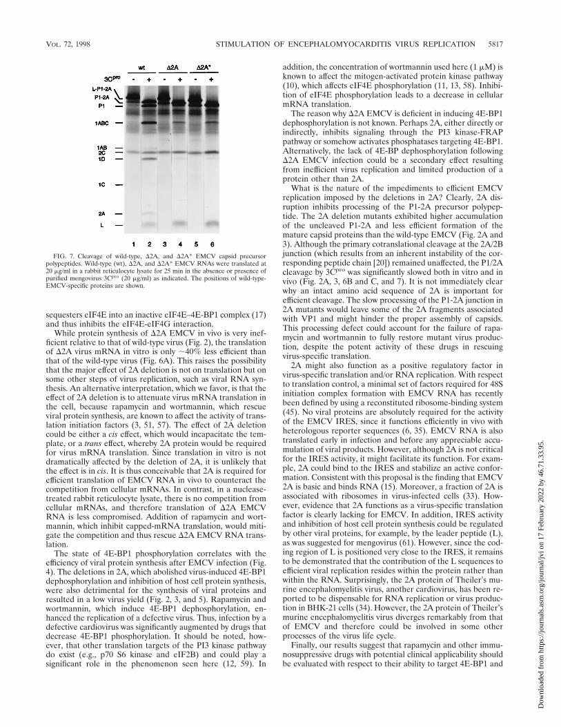
or 3ABC, which are responsible for polyprotein processing (23,39, 41, 54), or any other P3-derived polypeptides (Fig. 7). Inthe presence of exogenous 3Cpro (conditions which mimicthose existing in virus-infected cells), the largest polypeptidesynthesized was P1-2A (lane 2), which suggests that the leaderpeptide was rapidly cleaved off, apparently from the nascentpolypeptide chain. Mutations in 2A affected neither this co-translational cleavage of the leader peptide (compare lanes 2,4, and 6) nor its cleavage from the presynthesized L-P1-2Aprecursor polypeptide (data not shown). However, subsequentprocessing of P1-2A, i.e., the cleavage at the P1/2A junction,was markedly diminished by the mutations. While the wild-type P1-2A was cleaved to a significant extent by 3Cpro into P1,1ABC, 1AB, 1D, 1C, and 2A (lane 2), only a minute fraction ofP1-D2A or P1-D2A* was converted to characteristic cleavageproducts within the same period (lanes 4 and 6). Thus, P1-D2Aand P1-D2A* are less susceptible to cleavage by cardiovirus3Cpro than is wild-type P1-2A.
DISCUSSION
eIF4G serves as an adapter between mRNA and ribosomesand functions in both cap-dependent and cap-independent
FIG. 5. Rapamycin and wortmannin increase EMCV yield. BHK-21 cellswere infected with wild-type (wt) (A), D2A (B), or D2A* (C) EMCV in theabsence (control) or presence of rapamycin and/or wortmannin, as indicated.Conditions for virus infections and drug treatments were essentially as describedin the legend to Fig. 2. Virus yields (TCID50/cell) were determined 5 h postin-fection as described in Materials and Methods. The mean values of three inde-pendent titer determinations and the error bars indicating the standard deviationfrom the mean are shown.
VOL. 72, 1998 STIMULATION OF ENCEPHALOMYOCARDITIS VIRUS REPLICATION 5815
Dow
nloa
ded
from
http
s://j
ourn
als.
asm
.org
/jour
nal/j
vi o
n 17
Feb
ruar
y 20
22 b
y 46
.71.
33.9
5.

translations (22, 43, 45). Recently, a new homolog of eIF4G(termed eIF4GII) was identified which functions in a mannersimilar to the original isoform (termed eIF4GI) (16). eIF4Grecruitment to capped mRNA is facilitated by its interactionwith the cap-binding protein eIF4E. This interaction is regu-lated by a group of suppressor proteins, the 4E-BPs (30, 42).When bound to mRNA, eIF4G facilitates the binding of the43S preinitiation complex to the mRNA, most probablythrough protein-protein interactions with the ribosome-associ-ated eIF3 (26, 37). eIF4G also interacts with eIF4A, which, inconjunction with eIF4B, is thought to unwind the 59 mRNAsecondary structure.
To switch from translation of cellular mRNAs to efficientproduction of viral proteins, picornaviruses have evolved strat-egies to usurp eIF4G. Enteroviruses, rhinoviruses, and aphtho-viruses encode proteases that cleave eIF4G to generate twofragments. The C-terminal fragment retains the capacity tointeract with IRES elements, as well as with eIF3 and eIF4A,and is sufficient to promote cap-independent translation (5, 6,37, 46). However, it lacks the eIF4E-binding site and is unableto support cap-dependent translation. Cardioviruses, as exem-plified by EMCV, appear to affect eIF4G function in a differ-ent manner, namely, by inducing dephosphorylation and acti-vation of 4E-BP1 (14). The dephosphorylated form of 4E-BP1
FIG. 6. Kinetics of protein synthesis in rabbit reticulocyte lysates programmed with wild-type, D2A or D2A* EMCV RNAs. (A and B) Rabbit reticulocyte lysate(0.1 ml) was incubated with 2 mg of wild-type (wt) or D2A EMCV RNA as specified in Materials and Methods. At the indicated times, aliquots were withdrawn fromthe reaction mixtures. One part of the aliquot (1 ml) was assayed for trichloroacetic acid-insoluble radioactivity, while another (5 ml) was processed for SDS-PAGEanalysis. (A) Incorporation of [35S]methionine into trichloroacetic acid-insoluble material directed by wild-type or D2A EMCV RNAs. (B) Time course of accumulationof virus-specific polypeptides. Products of translation of wild-type and D2A EMCV RNAs are shown. (C) Rabbit reticulocyte lysate was programmed with wild-type(wt) or D2A* EMCV RNAs. Aliquots were withdrawn at the indicated times and subjected to SDS-PAGE as described for panels A and B. Products of the translationof wild-type EMCV RNA are shown.
5816 SVITKIN ET AL. J. VIROL.
Dow
nloa
ded
from
http
s://j
ourn
als.
asm
.org
/jour
nal/j
vi o
n 17
Feb
ruar
y 20
22 b
y 46
.71.
33.9
5.
sequesters eIF4E into an inactive eIF4E–4E-BP1 complex (17)and thus inhibits the eIF4E-eIF4G interaction.
While protein synthesis of D2A EMCV in vivo is very inef-ficient relative to that of wild-type virus (Fig. 2), the translationof D2A virus mRNA in vitro is only ;40% less efficient thanthat of the wild-type virus (Fig. 6A). This raises the possibilitythat the major effect of 2A deletion is not on translation but onsome other steps of virus replication, such as viral RNA syn-thesis. An alternative interpretation, which we favor, is that theeffect of 2A deletion is to attenuate virus mRNA translation inthe cell, because rapamycin and wortmannin, which rescueviral protein synthesis, are known to affect the activity of trans-lation initiation factors (3, 51, 57). The effect of 2A deletioncould be either a cis effect, which would incapacitate the tem-plate, or a trans effect, whereby 2A protein would be requiredfor virus mRNA translation. Since translation in vitro is notdramatically affected by the deletion of 2A, it is unlikely thatthe effect is in cis. It is thus conceivable that 2A is required forefficient translation of EMCV RNA in vivo to counteract thecompetition from cellular mRNAs. In contrast, in a nuclease-treated rabbit reticulocyte lysate, there is no competition fromcellular mRNAs, and therefore translation of D2A EMCVRNA is less compromised. Addition of rapamycin and wort-mannin, which inhibit capped-mRNA translation, would miti-gate the competition and thus rescue D2A EMCV RNA trans-lation.
The state of 4E-BP1 phosphorylation correlates with theefficiency of viral protein synthesis after EMCV infection (Fig.4). The deletions in 2A, which abolished virus-induced 4E-BP1dephosphorylation and inhibition of host cell protein synthesis,were also detrimental for the synthesis of viral proteins andresulted in a low virus yield (Fig. 2, 3, and 5). Rapamycin andwortmannin, which induce 4E-BP1 dephosphorylation, en-hanced the replication of a defective virus. Thus, infection by adefective cardiovirus was significantly augmented by drugs thatdecrease 4E-BP1 phosphorylation. It should be noted, how-ever, that other translation targets of the PI3 kinase pathwaydo exist (e.g., p70 S6 kinase and eIF2B) and could play asignificant role in the phenomenon seen here (12, 59). In
addition, the concentration of wortmannin used here (1 mM) isknown to affect the mitogen-activated protein kinase pathway(10), which affects eIF4E phosphorylation (11, 13, 58). Inhibi-tion of eIF4E phosphorylation leads to a decrease in cellularmRNA translation.
The reason why D2A EMCV is deficient in inducing 4E-BP1dephosphorylation is not known. Perhaps 2A, either directly orindirectly, inhibits signaling through the PI3 kinase-FRAPpathway or somehow activates phosphatases targeting 4E-BP1.Alternatively, the lack of 4E-BP dephosphorylation followingD2A EMCV infection could be a secondary effect resultingfrom inefficient virus replication and limited production of aprotein other than 2A.
What is the nature of the impediments to efficient EMCVreplication imposed by the deletions in 2A? Clearly, 2A dis-ruption inhibits processing of the P1-2A precursor polypep-tide. The 2A deletion mutants exhibited higher accumulationof the uncleaved P1-2A and less efficient formation of themature capsid proteins than the wild-type EMCV (Fig. 2A and3). Although the primary cotranslational cleavage at the 2A/2Bjunction (which results from an inherent instability of the cor-responding peptide chain [20]) remained unaffected, the P1/2Acleavage by 3Cpro was significantly slowed both in vitro and invivo (Fig. 2A, 3, 6B and C, and 7). It is not immediately clearwhy an intact amino acid sequence of 2A is important forefficient cleavage. The slow processing of the P1-2A junction in2A mutants would leave some of the 2A fragments associatedwith VP1 and might hinder the proper assembly of capsids.This processing defect could account for the failure of rapa-mycin and wortmannin to fully restore mutant virus produc-tion, despite the potent activity of these drugs in rescuingvirus-specific translation.
2A might also function as a positive regulatory factor invirus-specific translation and/or RNA replication. With respectto translation control, a minimal set of factors required for 48Sinitiation complex formation with EMCV RNA has recentlybeen defined by using a reconstituted ribosome-binding system(45). No viral proteins are absolutely required for the activityof the EMCV IRES, since it functions efficiently in vivo withheterologous reporter sequences (6, 35). EMCV RNA is alsotranslated early in infection and before any appreciable accu-mulation of viral products. However, although 2A is not criticalfor the IRES activity, it might facilitate its function. For exam-ple, 2A could bind to the IRES and stabilize an active confor-mation. Consistent with this proposal is the finding that EMCV2A is basic and binds RNA (15). Moreover, a fraction of 2A isassociated with ribosomes in virus-infected cells (33). How-ever, evidence that 2A functions as a virus-specific translationfactor is clearly lacking for EMCV. In addition, IRES activityand inhibition of host cell protein synthesis could be regulatedby other viral proteins, for example, by the leader peptide (L),as was suggested for mengovirus (61). However, since the cod-ing region of L is positioned very close to the IRES, it remainsto be demonstrated that the contribution of the L sequences toefficient viral replication resides within the protein rather thanwithin the RNA. Surprisingly, the 2A protein of Theiler’s mu-rine encephalomyelitis virus, another cardiovirus, has been re-ported to be dispensable for RNA replication or virus produc-tion in BHK-21 cells (34). However, the 2A protein of Theiler’smurine encephalomyelitis virus diverges remarkably from thatof EMCV and therefore could be involved in some otherprocesses of the virus life cycle.
Finally, our results suggest that rapamycin and other immu-nosuppressive drugs with potential clinical applicability shouldbe evaluated with respect to their ability to target 4E-BP1 and
FIG. 7. Cleavage of wild-type, D2A, and D2A* EMCV capsid precursorpolypeptides. Wild-type (wt), D2A, and D2A* EMCV RNAs were translated at20 mg/ml in a rabbit reticulocyte lysate for 25 min in the absence or presence ofpurified mengovirus 3Cpro (20 mg/ml) as indicated. The positions of wild-type-EMCV-specific proteins are shown.
VOL. 72, 1998 STIMULATION OF ENCEPHALOMYOCARDITIS VIRUS REPLICATION 5817
Dow
nloa
ded
from
http
s://j
ourn
als.
asm
.org
/jour
nal/j
vi o
n 17
Feb
ruar
y 20
22 b
y 46
.71.
33.9
5.
activate latent infections caused by viruses that use an internalribosome entry mechanism.
ACKNOWLEDGMENTS
We thank B. Raught for critical reading of the manuscript.This work was supported by grants from the National Institute of
Canada to N.S. and by National Institutes of Health grant AI-17331 toA.C.P. and training grant GM-07215 to H.H. N.S. is a Medical Re-search Council Distinguished Scientist and a Howard Hughes Inter-national Scholar. A.-C.G. is a recipient of an NSERC 67 studentship.
REFERENCES
1. Agol, V. I. 1991. The 59 untranslated region of picornaviral genomes. Adv.Virus Res. 40:103–180.
2. Belsham, G. J., and N. Sonenberg. 1996. RNA-protein interactions in reg-ulation of picornavirus RNA translational. Microbiol. Rev. 60:499–511.
3. Beretta, L., A. C. Gingras, Y. V. Svitkin, M. N. Hall, and N. Sonenberg. 1996.Rapamycin blocks the phosphorylation of 4E-BP1 and inhibits cap-depen-dent initiation of translation. EMBO J. 15:658–664.
4. Beretta, L., Y. V. Svitkin, and N. Sonenberg. 1996. Rapamycin stimulatesviral protein synthesis and augments the shutoff of host protein synthesisupon picornavirus infection. J. Virol. 70:8993–8996.
5. Borman, A. M., R. Kirchweger, E. Ziegler, R. E. Rhoads, T. Skern, and K. M.Kean. 1997. elF4G and its proteolytic cleavage products: effect on initiationof protein synthesis from capped, uncapped, and IRES-containing mRNAs.RNA 3:186–196.
6. Borman, A. M., P. Le Mercier, M. Girard, and K. M. Kean. 1997. Compar-ison of picornaviral IRES-driven internal initiation of translation in culturedcells of different origins. Nucleic Acids Res. 25:925–932.
7. Brown, E. J., and S. L. Schreiber. 1996. A signaling pathway to translationalcontrol. Cell 86:517–520.
8. Chumakov, K. M. 1980. A highly efficient method of infection with encepha-lomyocarditis virus RNA. Acta Virol. 24:225–231.
9. Ehrenfeld, E. 1996. Initiation of translation by picornavirus RNAs, p. 549–573. In J. W. B. Hershey, M. B. Mathews, and N. Sonenberg (ed.), Trans-lational control. Cold Spring Harbor Laboratory Press, Plainview, N.Y.
10. Ferby, I. M., I. Waga, M. Hoshino, K. Kume, and T. Shimizu. 1996. Wort-mannin inhibits mitogen-activated protein kinase activation by platelet-acti-vating factor through a mechanism independent of p85/p110-type phospha-tidylinositol 3-kinase. J. Biol. Chem. 271:11684–11688.
11. Flynn, A., and G. Proud. 1996. Insulin-stimulated phosphorylation of initia-tion factor 4E is mediated by the MAP kinase pathway. FEBS Lett. 389:162–166.
12. Franke, T. F., D. R. Kaplan, and L. C. Cantley. 1997. PI3K: downstreamAKTion blocks apoptosis. Cell 88:435–437.
13. Fukunaga, R., and T. Hunter. 1997. MNK1, a new MAP-kinase-activatedprotein kinase, isolated by a novel expression screening method for identi-fying protein kinase substrates. EMBO J. 16:1921–1933.
14. Gingras, A. C., Y. Svitkin, G. J. Belsham, A. Pause, and N. Sonenberg. 1996.Activation of the translational suppressor 4E-BP1 following infection withencephalomyocarditis virus and poliovirus. Proc. Natl. Acad. Sci. USA 93:5578–5583.
15. Gorbalenya, A. E., K. M. Chumakov, and V. I. Agol. 1978. RNA-bindingproperties of nonstructural polypeptide G of encephalomyocarditis virus.Virology 88:183–185.
16. Gradi, A., H. Imataka, Y. V. Svitkin, E. Rom, B. Raught, S. Morino, and N.Sonenberg. 1998. A novel functional human eukaryotic translation initiationfactor 4G. Mol. Cell. Biol. 18:334–342.
17. Haghighat, A., S. Mader, A. Pause, and N. Sonenberg. 1995. Repression ofcap-dependent translation by 4E-binding protein 1: competition with p220for binding to eukaryotic initiation factor-4E. EMBO J. 14:5701–5709.
18. Haghighat, A., Y. Svitkin, I. Novoa, E. Kuechler, T. Skern, and N. Sonen-berg. 1996. The eIF4G-eIF4E complex is the target for direct cleavage by therhinovirus 2A proteinase. J. Virol. 70:8444–8450.
19. Hahn, H., and A. C. Palmenberg. 1995. Encephalomyocarditis viruses withshort poly(C) tracts are more virulent than their mengovirus counterparts.J. Virol. 69:2697–2699.
20. Hahn, H., and A. C. Palmenberg. 1996. Mutational analysis of the encepha-lomyocarditis virus primary cleavage. J. Virol. 70:6870–6875.
21. Hall, D. J., and A. C. Palmenberg. 1996. Mengo virus 3C proteinase: recom-binant expression, intergenus substrate cleavage and localization in vivo.Virus Genes 13:99–110.
22. Hentze, M. W. 1997. eIF4G: a multipurpose ribosome adapter? Science275:500–501.
23. Jackson, R. J. 1986. A detailed kinetic analysis of the in vitro synthesis andprocessing of encephalomyocarditis virus products. Virology 149:114–127.
24. Jackson, R. J., and A. Kaminski. 1995. Internal initiation of translation ineukaryotes: the picornavirus paradigm and beyond. RNA 1:985–1000.
25. Kleijn, M., C. L. Vrins, H. O. Voorma, and A. A. Thomas. 1996. Phosphor-
ylation state of the cap-binding protein eIF4E during viral infection. Virol-ogy 217:486–494.
26. Lamphear, B. J., R. Kirchweger, T. Skern, and R. E. Rhoads. 1995. Mappingof functional domains in eukaryotic protein synthesis initiation factor 4G(eIF4G) with picornaviral proteases. Implications for cap-dependent andcap-independent translational initiation. J. Biol. Chem. 270:21975–21983.
27. Lawrence, C., and R. E. Thach. 1974. Encephalomyocarditis virus infectionof mouse plasmacytoma cells. I. Inhibition of cellular protein synthesis.J. Virol. 14:598–610.
28. Liebig, H.-D., E. Ziegler, R. Yan, K. Hartmuth, H. Klump, H. Kowalski, D.Blaas, W. Sommergruber, L. Frasel, B. Lamphear, R. Rhoads, E. Kuechler,and T. Skern. 1993. Purification of two picornaviral 2A proteinases: inter-action with eIF-4g and influence on in vitro translation. Biochemistry 32:7581–7588.
29. Lin, T. A., X. Kong, A. R. Saltiel, P. J. Blackshear, and J. C. Lawrence. 1995.Control of PHAS-I by insulin in 3T3-L1 adipocytes. Synthesis, degradation,and phosphorylation by a rapamycin-sensitive and mitogen-activated proteinkinase-independent pathway. J. Biol. Chem. 270:18531–18538.
30. Lin, T. A., X. M. Kong, T. Haystead, A. Pause, G. Belsham, N. Sonenberg,and J. C. Lawrence. 1994. PHAS-I as a link between mitogen-activatedprotein kinase and translation initiation. Science 266:653–656.
31. Lloyd, R. E., M. J. Grubman, and E. Ehrenfeld. 1988. Relationship of p220cleavage during picornavirus infection to 2A proteinase sequencing. J. Virol.62:4216–4223.
32. Mader, S., H. Lee, A. Pause, and N. Sonenberg. 1995. The translationinitiation factor eIF-4E binds to a common motif shared by the translationfactor eIF-4g and the translational repressors 4E-binding proteins. Mol.Cell. Biol. 15:4990–4997.
33. Medvedkina, O. A., I. V. Scarlat, N. O. Kalinina, and V. I. Agol. 1974.Virus-specific proteins associated with ribosomes of Krebs-II cells infectedwith encephalomyocarditis virus. FEBS Lett. 39:4–8.
34. Michiels, T., V. Dejong, R. Rodrigus, and C. Shaw-Jackson. 1997. Protein 2Ais not required for Theiler’s virus replication. J. Virol. 71:9549–9556.
35. Molla, A., S. K. Jang, A. V. Paul, Q. Reuer, and E. Wimmer. 1992. Cardio-viral internal ribosomal entry site is functional in a genetically engineereddicistronic poliovirus. Nature 356:255–257.
36. Mosenkis, J., S. Daniels-McQueen, S. Janovec, R. Duncan, J. W. Hershey,J. A. Grifo, W. C. Merrick, and R. E. Thach. 1985. Shutoff of host translationby encephalomyocarditis virus infection does not involve cleavage of theeucaryotic initiation factor 4F polypeptide that accompanies poliovirus in-fection. J. Virol. 54:643–645.
37. Ohlmann, T., M. Rau, V. M. Pain, and S. J. Morley. 1996. The C-terminaldomain of eukaryotic protein synthesis initiation factor (eIF) 4G is sufficientto support cap-independent translation in the absence of eIF4E. EMBO J.15:1371–1382.
38. Palmenberg, A. C. 1990. Proteolytic processing of picornaviral polyprotein.Annu. Rev. Microbiol. 44:603–623.
39. Palmenberg, A. C., M. A. Pallansch, and R. R. Rueckert. 1979. Proteaserequired for processing picornaviral coat protein resides in the viral replicasegene. J. Virol. 32:770–778.
40. Palmenberg, A. C., G. D. Parks, D. J. Hall, R. H. Ingraham, T. W. Seng, andP. V. Pallai. 1992. Proteolytic processing of the cardioviral P2 region: pri-mary 2A/2B cleavage in clone-derived precursors. Virology 190:754–762.
41. Parks, G. D., J. C. Baker, and A. C. Palmenberg. 1989. Proteolytic cleavageof encephalomyocarditis viral capsid region substrates by precursors to the3C enzyme. J. Virol. 63:1054–1058.
42. Pause, A., G. J. Belsham, A. C. Gingras, O. Donze, T. A. Lin, J. C. Lawrence,and N. Sonenberg. 1994. Insulin-dependent stimulation of protein synthesisby phosphorylation of a regulator of 59-cap function. Nature 371:762–767.
43. Pause, A., N. Methot, Y. V. Svitkin, W. C. Merrick, and N. Sonenberg. 1994.Dominant negative mutants of mammalian translation initiation factoreIF-4A define a critical role for eIF-4F in cap-dependent and cap-dependentinitiation of translation. EMBO J. 13:1205–1215.
44. Pelham, H. R. B. 1978. Translation of encephalomyocarditis virus RNA invitro yields an active proteolytic processing enzyme. Eur. J. Biochem. 85:457–462.
45. Pestova, T. V., C. U. T. Hellen, and I. N. Shatsky. 1996. Canonical eukaryoticinitiation factors determine initiation of translation by internal ribosomalentry. Mol. Cell. Biol. 16:6859–6869.
46. Pestova, T. V., I. N. Shatsky, and C. U. T. Hellen. 1996. Functional dissectionof eukaryotic initiation factor 4F: the 4A subunit and the central domain ofthe 4G subunit are sufficient to mediate internal entry of 43S preinitiationcomplexes. Mol. Cell. Biol. 16:6870–6878.
47. Ray, B. K., T. G. Lawson, J. C. Kramer, M. H. Cladaras, J. A. Grifo, R. D.Abramson, W. C. Merrick, and R. E. Thach. 1985. ATP-dependent unwind-ing of messenger RNA structure by eukaryotic initiation factors. J. Biol.Chem. 260:7651–7658.
48. Rozen, F., I. Edery, K. Meerovitch, T. E. Dever, W. C. Merrick, and N.Sonenberg. 1990. Bidirectional RNA helicase activity of eucaryotic transla-tion initiation factors 4A and 4F. Mol. Cell. Biol. 10:1134–1144.
49. Rueckert, R. R. 1996. Picornaviridae: the viruses and their replication, p.
5818 SVITKIN ET AL. J. VIROL.
Dow
nloa
ded
from
http
s://j
ourn
als.
asm
.org
/jour
nal/j
vi o
n 17
Feb
ruar
y 20
22 b
y 46
.71.
33.9
5.
609–654. In B. N. Fields (ed.), Fields virology, vol. 1. Lippincott-RavenPublishers, Philadelphia, Pa.
50. Schmidt, N. J. 1979. Cell culture techniques for diagnostic virology, p. 65–139. In E. H. Lennette and N. J. Schmidt (ed.), Diagnostic procedures forviral, rickettsial and chlamydial infections. American Public Health Associ-ation, Washington, D.C.
51. Sonenberg, N. 1996. mRNA 59 cap-binding protein eIF4E and control of cellgrowth, p. 245–269. In J. W. B. Hershey, M. B. Mathews, N. Sonenberg (ed.),Translational control. Cold Spring Harbor Laboratory Press, Plainview, N.Y.
52. Svitkin, Y. V., and V. I. Agol. 1978. Complete translation of encephalomyo-carditis virus RNA and faithful cleavage of virus-specific proteins in a cell-free system from Krebs-2 cells. FEBS Lett. 87:7–11.
53. Svitkin, Y. V., V. A. Ginevskaya, T. Y. Ugarova, and V. I. Agol. 1978. Acell-free model of the encephalomyocarditis virus-induced inhibition of hostcell protein synthesis. Virology 87:199–203.
54. Svitkin, Y. V., A. E. Gorbalenya, Y. A. Kazachkov, and V. I. Agol. 1979.Encephalomyocarditis virus-specific polypeptide p22 possessing a proteolyticactivity: preliminary mapping on the viral genome. FEBS Lett. 108:6–9.
55. Svitkin, Y. V., S. V. Maslova, and V. I. Agol. 1985. The genomes of attenu-ated and virulent poliovirus strains differ in their in vitro translation effi-
ciencies. Virology 147:243–252.56. Ui, M., T. Okada, K. Hazeki, and O. Hazeki. 1995. Wortmannin as a unique
probe for an intracellular signalling protein, phosphoinositide 3-kinase.Trends Biochem. Sci. 20:303–307.
57. von Manteuffel, S. R., A.-C. Gingras, X.-F. Ming, N. Sonenberg, and G.Thomas. 1996. 4E-BP1 phosphorylation is mediated by the FRAP-p70s6k
pathway and is independent of mitogen-activated protein kinase. Proc. Natl.Acad. Sci. USA 93:4076–4080.
58. Waskiewicz, A. J., A. Flynn, C. G. Proud, and J. A. Cooper. 1997. Mitogen-activated protein kinases activate the serine/threonine kinases Mnk1 andMnk2. EMBO J. 16:1909–1920.
59. Welsh, G. I., C. M. Stokes, X. Wang, H. Sakaue, W. Ogawa, M. Kasuga, andC. G. Proud. 1997. Activation of translation initiation factor eIF2B by insulinrequires phosphatidyl inositol 3-kinase. FEBS Lett. 410:418–422.
60. Wyckoff, E. E., R. E. Lloyd, and E. Ehrenfeld. 1992. Relationship of eukary-otic initiation factor 3 to poliovirus-induced p220 cleavage activity. J. Virol.66:2943–2951.
61. Zoll, J., J. M. Galama, F. J. van Kuppeveld, and W. J. Melchers. 1996.Mengovirus leader is involved in the inhibition of host cell protein synthesis.J. Virol. 70:4948–4952.
VOL. 72, 1998 STIMULATION OF ENCEPHALOMYOCARDITIS VIRUS REPLICATION 5819
Dow
nloa
ded
from
http
s://j
ourn
als.
asm
.org
/jour
nal/j
vi o
n 17
Feb
ruar
y 20
22 b
y 46
.71.
33.9
5.