radionuclide evaluation of the lower gastrointestinal...
TRANSCRIPT

C O N T I N U I N G E D U C A T I O N
Radionuclide Evaluation of the LowerGastrointestinal Tract*
Giuliano Mariani1, Ernest K.J. Pauwels1,2, Abedallatif AlSharif1, Santino Marchi3, Giuseppe Boni1, Marco Barreca4,Massimo Bellini3, Mariano Grosso1, Nicola de Bortoli3, Gloria Mumolo3, Francesco Costa3, Domenico Rubello5, andH. William Strauss6
1Regional Center of Nuclear Medicine, University of Pisa Medical School, Pisa, Italy; 2Department of Radiology, Leiden UniversityMedical Center, Leiden, The Netherlands; 3Gastroenterology Unit, Department of Internal Medicine, University of Pisa Medical School,Pisa, Italy; 4Fourth Division of General Surgery, ‘‘S. Chiara’’ University Hospital, Pisa, Italy; 5PET Center, Nuclear Medicine Service,‘‘S. Maria della Misericordia’’ Hospital, Rovigo, Italy; and 6Division of Nuclear Medicine, Memorial Sloan-Kettering Cancer Center,New York, New York
This review outlines the technical aspects and diagnostic perfor-mance parameters of nuclear medicine procedures used on pa-tients with disorders of the lower gastrointestinal tract, with theexclusion of techniques using tumor-seeking radiopharmaceuti-cals. Chronic disorders of the lower gastrointestinal tract oftenreduce the quality of life because of discomfort from constipationor diarrhea. Five classes of radionuclide procedures are usedto characterize these disorders: transit scintigraphy, searches forectopic gastric mucosa in Meckel’s diverticulum, scintigraphyof active inflammatory bowel disease, scintigraphic defecogra-phy, and scintigraphy to detect sites of gastrointestinal bleeding.Protocols for these procedures and their relative merit in patientmanagement are discussed, with special emphasis on their po-tential for semiquantitative assessment of the pathophysiologicparameter investigated. Quantitation is particularly relevant forprognostic purposes and for monitoring the efficacy of therapy.
Key Words: gastrointestinal; GI; radionuclide; transit scintigraphy
J Nucl Med 2008; 49:776–787DOI: 10.2967/jnumed.107.040113
In 2004, a review in the Journal of Nuclear Medicinesummarized nuclear medicine procedures to characterizemotor disorders of the esophagus and stomach (1). Thisreview will focus on radionuclide studies of the small andlarge bowel, excluding techniques to detect tumors.
Local logistics and the availability of different medicalspecialties dictate how diagnostic algorithms are applied inthe clinical routine when patients present with disorders of
the lower gastrointestinal tract. The approach to patients withdisorders of the lower gastrointestinal tract should include ateam of health-care experts in medical and surgical gastro-enterology, nutrition, and imaging (radiologists and nuclearphysicians).
CLINICAL BACKGROUND
In 2001, the American Gastroenterological Associationestimated the total cost of care for patients with gastrointes-tinal diseases at $41.8 billion in the United States (2). Theclinical manifestations of small- and large-bowel diseasemost frequently are constipation or diarrhea, sometimes withalternating phases between these 2 extremes. Pain can accom-pany both conditions, whereas bleeding (presenting either asan acute, clinically obvious sign or as an indolent, chronicevent) is less frequent and usually represents a warning signfor the occurrence of more severe diseases. To help the readerunderstand why this occurs, we will begin with a review ofthe anatomy and pathophysiology of the lower gastrointes-tinal tract.
ANATOMY AND PHYSIOLOGY
Small Intestine
The small intestine, a 6-m-long tube extending from thepylorus to the cecum, comprises 3 major segments: The duo-denum adds secretions from the pancreas and liver to chymepassing through the pylorus and absorbs chlorine, bicarbon-ate ions, and calcium. The jejunum absorbs chlorine andbicarbonate in particular, plus a variety of nutrients (carbo-hydrates, amino acids, and lipids). The ileum absorbs a vastvariety of nutrients.
An intrinsic neural network consisting of the myenteric(Auerbach) and the submucosal (Meissner) plexus coordi-nates motor activities, to facilitate mixing and therefore aidenzymatic digestion in the lumen. If viewed as a simple pipe,the luminal surface area of the small intestine would be abouthalf a square meter. But in reality, its absorptive surface area
Received Aug. 24, 2007; revision accepted Dec. 7, 2007.For correspondence or reprints contact: Giuliano Mariani, Regional Center
of Nuclear Medicine, University of Pisa Medical School, Via Roma 67, I-56126Pisa, Italy.
E-mail: [email protected]*NOTE: FOR CE CREDIT, YOU CAN ACCESS THIS ACTIVITY THROUGH
THE SNM WEB SITE (http://www.snm.org/ce_online) THROUGH MAY 2009.No potential conflict of interest relevant to this article was reported.COPYRIGHT ª 2008 by the Society of Nuclear Medicine, Inc.
776 THE JOURNAL OF NUCLEAR MEDICINE • Vol. 49 • No. 5 • May 2008
by on May 12, 2018. For personal use only. jnm.snmjournals.org Downloaded from

is roughly 250 square meters, the size of a tennis court,because of 3 features: mucosal folds (circular folds of theinner surface increasing surface area and also aiding to mixthe ingesta by acting as baffles), villi (multitudes of mucosalprojections that protrude into the lumen and are covered withepithelial cells), and microvilli (the luminal plasma mem-brane of absorptive epithelial cells, studded with denselypacked microvilli, or the ‘‘brush border’’).
Although goblet cells secrete lubricating mucus into theintestinal lumen, enteroendocrine cells sense the luminalenvironment and secrete peptide hormones such as chole-cystokinin, ghrelin, and gastrin into the blood. Cholecysto-kinin regulates relaxation of the lower esophageal sphincter,gastric secretion and emptying, gallbladder contraction, andintestinal and colonic motility and also plays a role in theregulation of food intake. Ghrelin, a ligand of the growthhormone secretagogue receptor, regulates energy balanceand glucose homeostasis. Gastrin primarily stimulates thesecretion of gastric acid.
The 2 most important functions of the small intestine areabsorption of nutrients and maintenance of orderly aboralmovement of chyme. Smooth muscle cells regulating motorfunction are controlled by the enteric nervous system, themotor autonomic nervous system, and spinal and vagal ex-trinsic sensory neurons. Furthermore, several hormones mod-ulate the frequency and patterns of small-intestine contraction.
Colon
The colon (a tubular muscular organ approximately 125cm long in vivo) performs 3 important functions: concen-tration of fecal effluent by absorbing water and electrolytes,storage and controlled evacuation of fecal material, anddigestion and absorption of food.
The right colon (cecum and ascending colon) plays a ma-jor role in water and electrolyte absorption and the fer-mentation of undigested sugars; the left colon (descendingcolon, sigmoid colon, and rectum) is involved in the storageand evacuation of stool.
The colonic mucosa presents a relatively smooth surfacewith numerous crypts composed predominantly of mucus-secreting goblet cells. The longitudinal muscle is organizedin 3 bands, called taeniae coli, which run from the cecum tothe rectum, where they fuse to form a uniform outer muscularlayer. Enteric nervous system neurons are organized intoganglia with interconnecting fibers, which form the submu-cosal and myenteric plexi. Local neural reflex circuits mod-ulate motility, secretion, blood flow, and probably immunefunction. Neurotransmitters such as acetylcholine, substanceP, and serotonin activate local circuits regulating musclecontractions. The major inhibitory neurotransmitter is nitricoxide. Parasympathetic nerves are predominantly excitatory,and sympathetic nerves inhibitory. Brain–gut connectionsare important both for perception of visceral stimuli and formodifying colonic function in response to central stimuli.
The contraction patterns in the right colon cause signif-icant mixing, facilitating the absorption of water, whereas
in the left colon they slow the movement of formed stool,forming a reservoir until reflexes activate contractions toadvance and evacuate stool.
CLINICAL MANIFESTATIONS OF DISORDERS
Diarrhea
Diarrhea is a common outcome of many lower gastroin-testinal disorders. It is usually considered a modification ofnormal bowel function with an increase of bowel movementsand fluidity of feces. Because stool consistency is difficult toquantify, stool weight is considered a surrogate marker ofdiarrhea. Three or more bowel movements per day are con-sidered to be abnormal, and the upper limit of stool weight isgenerally agreed to be 200 g per day in western countries.
Diarrhea can be associated with other symptoms, such asabdominal pain, fever, rectal emission of blood and muco-pus, tenesmus, malabsorption, and dehydration. Diarrheacan be classified into 4 major types: exudative, osmotic, secre-tory, and secondary to dysmotility.
The incidence of diarrheal diseases is highest in developingcountries, with an estimated 4.6–6 million deaths per year inAsia, Africa, and Latin America (3). Although deaths fromgastroenteritis are not as common in developed countries,non–food-borne gastroenteritis and other gastrointestinal in-fections are among the most prevalent digestive diseases inthe United States, and the top 2 in terms of indirect costs (4).
Irritable bowel syndrome, lactose intolerance and otherdietary factors, inflammatory bowel disease (IBD, whichincludes ulcerative colitis and Crohn’s disease), and coloncancer are the most common causes of chronic diarrheaencountered in primary clinical practice (5).
Irritable bowel syndrome affects up to 20%–25% of thepopulation in western countries, and in 47% of the patientsit alternates with constipation (6). The total direct andindirect U.S. costs incurred by the estimated 380,000–480,000 IBD patients has been estimated at around $2billion per year. Drugs account for only 10%–25% of thetotal costs, whereas surgery and hospitalization account forapproximately half (7).
Constipation
Chronic constipation is a worldwide problem. Its preva-lence is estimated to range from 2% from 28% (2), is higher inwomen than men, and increases with age (8). In the UnitedStates, constipation accounts for 2.5 million annual physi-cian visits, an overall expense of about $800 million forlaxatives (9), and an estimated annual cost of about $6.9billion for diagnostic testing (2). In addition to the directcosts, the economic impact of constipation includes reducedwork performance, premature retirement, and reduced qual-ity of life (9).
Chronic constipation can be idiopathic, secondary to drugs(especially opioids and anticholinergic agents), or associatedwith various medical conditions (2). Idiopathic chronic con-stipation can be further divided into 3 subgroups (10,11):low-transit constipation, dyssynergic defecation, and normal
EVALUATION OF LOWER GASTROINTESTINAL TRACT • Mariani et al. 777
by on May 12, 2018. For personal use only. jnm.snmjournals.org Downloaded from

transit constipation (functional constipation and irritablebowel syndrome with constipation).
Gross Gastrointestinal Bleeding
Upper and lower gastrointestinal bleeding leads to morethan 300,000 hospitalizations per year in the United States.Lower gastrointestinal bleeding is less common than uppergastrointestinal bleeding, with an estimated annual hospital-ization rate of 27 per 100,000 adults at risk (12). The rate ofacute gastrointestinal bleeding rises in patients taking aspirin(13). Lower gastrointestinal bleeding accounts for one fourthto one third of all bleeding events, is more common in menthan in women, increases in incidence with age, and has areported mortality rate of approximately 4% (12). Earlydiagnosis of patients with severe bleeding, and early inter-ventional techniques, lower mortality rates (14,15).
Colonic diverticulosis is by far the most common cause oflower gastrointestinal bleeding, with gross bleeding in almost5% of patients with this condition, either in the right or in theleft colon. Other possible causes of acute lower gastrointes-tinal bleeding include angiodysplasia and, rarely, small-bowelsources or other colonic lesions, such as colon cancer (12).
Angiodysplasia is a common cause of both acute majorlower gastrointestinal hemorrhage and slow intermittentblood loss (10%–40% of the events) (16). Angiodysplasticlesions involve primarily the cecum and right colon. Al-though angiography and spiral CT frequently identify thelesions (without, however, demonstrating active bleeding),colonoscopy has the advantage of a therapeutic potential.
Neoplastic lesions occur predominantly in the elderly andare the cause of acute lower gastrointestinal bleeding in 2%–26% of cases (17). Hemorrhoids and anal fissures are themost common causes of minor intermittent bright red bloodper rectum. Although anal fissures are often painful, bleedingfrom internal hemorrhoids is usually painless and occurs withstraining or the passage of hard stool.
Ischemic colitis, most commonly affecting the splenicflexure, descending colon, and sigmoid colon, causes up to9% of all cases of major lower gastrointestinal bleeding(12,18). Although the etiology of ischemic colitis includesthrombosis and emboli, the disorder usually develops in theabsence of vascular obstruction (19). Ischemic colitis occursmore frequently in the elderly, with conditions such as de-creased cardiac output, arrhythmia, trauma, coagulationdisorders, and use of drugs causing vasospasm (18). Acuteischemic colitis has a good prognosis and can be treated withmedical therapy. Infectious colitis often presents with bloodydiarrhea, though the degree of blood loss is rarely significant.Bleeding from IBD accounts for 2%–6% of all cases of acutelower gastrointestinal bleeding (12,18) and is usually mini-mal to moderate.
In approximately 2%–3% of the general population, adiverticulum occurs in the distal ileum. This diverticulumwas initially described by the German surgeon WilhelmFabricius Hildanus in 1598 but was recognized and describedby Meckel as an embryonic remnant nearly 2 centuries ago
(20). In addition to normal ileal mucosa, about 50% of thesediverticula contain gastric ectopic mucosa or may occasion-ally contain duodenal, colonic, or pancreatic mucosa. Thegastric mucosa is capable of acid secretion, which can resultin ulceration of adjacent ileal mucosa, leading to bleeding.Bleeding usually occurs in infancy or early childhood, al-though it may occasionally present in adults.
Intussusception, an uncommon cause of bleeding in adults,usually has a leading point, such as a polyp or a malignancy.Ileal or colonic varices (often associated with portal hy-pertension) may present with massive lower gastrointestinalbleeding, as may rectal varices.
Other rare causes of lower gastrointestinal bleeding aresolitary rectal ulcer, aortoenteric fistulae not associated withprosthetic grafts, and nonsteroidal antiinflammatory drugs.Anticoagulants seem to uncover bleeding from preexistinglesions rather than produce bleeding per se (21).
NUCLEAR MEDICINE PROCEDURES
Intestinal Transit Scintigraphy
It is often problematic to determine whether dyspepticsymptoms are caused by dysfunction of the upper or lowergastrointestinal tract. Transit studies are helpful for local-izing the potential site of disease and guiding therapy (22). Inparticular, gastrointestinal transit scintigraphy is the onlyimaging procedure that allows us to measure both total andregional transit times, defining whether dysmotility is gen-eralized or localized and quantifying its severity.
Small-bowel transit scintigraphy is useful when symptomssuch as bloating, early satiety, dyspepsia, or nausea appearsoon after the meal and are accompanied by postprandialdiarrhea. Also, symptomatic patients with prior intestinalresection should be evaluated for the possibility of an ab-normally rapid small-bowel transit, because physiologic con-trol of transit depends on the intact jejunal and ileal brakes(23,24). Small-bowel transit scintigraphy is typically per-formed in conjunction with gastric-emptying or colonictransit studies.
Colonic transit scintigraphy is used to characterize pa-tients with symptoms of altered colonic transit includingconstipation, abdominal bloating, and refractory irritablebowel syndrome. This procedure is useful for identifying thepresence and severity of transit abnormalities (includinginformation on specific colonic regions) and for assessingresponse to therapies.
Because it sometimes is helpful to evaluate motilitythroughout the entire gastrointestinal tract, whole-gut transitscintigraphy was developed. This test is useful for assessingdiffuse dysmotility involving both the upper and the lowergastrointestinal tract and frequently leads to changes indiagnosis and patient management (25).
Several techniques have been proposed. Because there isno single best protocol for performing this study, centersare encouraged to comply with what can be defined as abest-practice procedure with regard to patient preparation,
778 THE JOURNAL OF NUCLEAR MEDICINE • Vol. 49 • No. 5 • May 2008
by on May 12, 2018. For personal use only. jnm.snmjournals.org Downloaded from

radiopharmaceutical, diet (calorie, fiber, carbohydrate, fat,and protein content), protocol of acquisition, and dataelaboration. Considering the complex pathophysiology ofgastrointestinal motility, it might be advisable to adopt aprotocol that measures both gastric emptying and intestinaltransit, such as a dual-labeled solid–liquid meal.
111In-Diethylenetriaminepentaacetic acid (DTPA) is notabsorbed in the gastrointestinal tract. The 67-h physicalhalf-life of 111In allows imaging over several days. To bedelivered to the cecum as a solid, the tracer is adsorbed on asolid-phase material. Two different methods are currentlyapplied for colonic transit scintigraphy using this radio-pharmaceutical. The Mayo Clinic protocol is based on oralingestion of a single methacrylate-coated capsule contain-ing the radiolabeled agent (currently 3.7 MBq [100 mCi] of111InCl3 or 111In-DTPA adsorbed on activated charcoalparticles) (26,27). The methacrylate coating dissolves in therelatively alkaline environment of the ileum (pH 7.4), sothat the distal ileum delivers the pellets in a bolus fashion tothe cecum. Commercially available enteric capsules de-signed to deliver drugs to the region of the cecum and colonalso offer a suitable and simpler alternative.
To evaluate the upper gastrointestinal tract (gastric andsmall-bowel transit), one should perform both solid- andliquid-phase emptying studies. 99mTc-Sulfur colloid–labeledscrambled eggs and ingestion of 111In-DTPA (4.6 MBq)mixed with 300 mL of water (Temple University Hospitalprotocol) (25) can be imaged simultaneously (using multipleenergy windows) to measure gastric emptying and small-bowel transit simultaneously (28).
Imaging for gastrointestinal transit studies is performedwith large-field-of-view g-cameras equipped with medium-energy collimators (if using 111In either alone or as part of adual-tracer study), or, if the study is performed with a99mTc tracer only (typically gastric emptying with tracer-labeled scrambled eggs), a low-energy collimator can beused. Radioisotopic markers placed on the xiphoid processand iliac crests may help better define the anatomy. Gastric-emptying data, if needed, should be acquired as describedpreviously (1). All protocols for gastrointestinal transitscintigraphy are based on sequential imaging after inges-tion of the radiolabeled meals, with simultaneous imagesrecorded in both the anterior and the posterior projections(analyzed after the images are combined to produce a geo-metric mean image). The timing of acquisitions varies withthe region of the gastrointestinal tract to be evaluated; forexample, the small bowel requires more frequent imagingin the first few hours, whereas the colon requires delayedimaging up to 48 or even 72 h.
Quantification is based on region-of-interest counts ofgeometric mean images derived from the anterior and pos-terior acquisitions, correcting for radioactive decay andfor downscatter of the 111In counts in the 99mTc window inthe case of a dual-isotope meal. Various parameters for char-acterizing small-bowel and colonic transit are described laterin this article.
The small-bowel transit time can be calculated fromdeconvolution of gastric emptying and of the colonic fillingcurves (median values being 160–170 min in healthy volun-teers for either 131I-fiber or liquid 99mTc-DTPA) (29). Theorocecal transit time is defined as the time for initial arrival ofan orally administered tracer to the cecum, with the normaltime being 297 6 65 min (95% confidence interval, 154–440min) (25,30). The 10% small-bowel transit time is calculatedby subtracting the time for 10% gastric emptying from thetime to 10% colonic filling. This simple evaluation alsocorrects for delayed gastric emptying and yields averagereference values of about 280 min for solids (approximaterange, 240–320 min) and 295 min for liquids (approximaterange, 255–335 min) (30,31). The small-bowel transit indexis calculated as the percentage of small-bowel activity at theterminal ileum–cecum/ascending colon at 6 h relative to totalsmall-bowel activity (the index is .40% in healthy individ-uals) (25). This index, along with the 10% small-boweltransit time, is currently the most widely adopted parameterfor clinical purposes.
The geometric center, commonly adopted as a parameterof colonic transit, is the weighted average fraction of countsin different colonic regions, that is, (region-of-interestcounts)/(administered counts) (25,32). The Mayo Clinicand the Temple University protocols differ in the numberof regions into which they divide the colon, 4 and 6,respectively (with an additional ‘‘virtual’’ region representedby evacuated radioactivity). The weighting factor increasesfrom 1 to 5 (Mayo Clinic protocol) or from 1 to 7 (TempleUniversity protocol) in a proximal-to-distal fashion (e.g.,from cecum to rectosigmoid and to evacuated stool). There-fore, a low value for geometric center indicates that mostradioactivity is in the cecum, whereas a high value means thatactivity is in the rectosigmoid region or has been evacuated(Fig. 1).
The primary parameter of interest is the geometric centerat 24 h (reference range, 1.6–3.8). The reference values forthe geometric center according to the Mayo Clinic protocolare no more than 1.4 for 4 h and 1.7–4.0 for 24 h (26). Slowcolonic transit is defined as a geometric center lower thanthe reference value at 24 h. With the solid–liquid meal ofthe Temple University protocol, the reference values forgeometric center mean are 4.6 6 1.5 at 24 h, 6.1 6 1.0 at48 h, and 6.6 6 0.2 at 72 h.
As a practical rule, the colon should be imaged at 24 and 48h. A geometric center less than 4.1 at 48 h obviates furtherimaging, because colonic transit is obviously delayed. Whengeometric centers are in the 4.1–6.4 range at 48 h, imaging at72 h is useful for excluding functional outlet obstruction.Accelerated colonic transit, as in patients with diarrhea,results in a geometric center greater than 6.1 (at or beyond therectosigmoid tract) at 24 h. The geometric center is the bestpredictor of delayed colonic transit and has a significantimpact on patient care, often leading to changes in bothdiagnosis and treatment (25). Furthermore, this semiquanti-tative score of colonic transit can be used to compare
EVALUATION OF LOWER GASTROINTESTINAL TRACT • Mariani et al. 779
by on May 12, 2018. For personal use only. jnm.snmjournals.org Downloaded from

different patients or groups of patients, as well as serialevaluations of the same patient, for such assessments as theefficacy of prokinetic therapy or the unwanted side effects ofanalgesic drugs.
Scintigraphic transit studies have been used to charac-terize transit and motility in patients with functional bowelsyndromes, particularly in those patients who do not have asecondary form of constipation and have refractory symp-toms after a high-fiber diet and laxatives (10). The scinti-graphic study can elucidate if a patient with functionalconstipation has colonic inertia or functional rectosigmoidobstruction. Alternatively, finding a normal scintigraphic
transit pattern in a patient complaining of obstinate consti-pation may indicate that the patient has psychogenicabnormalities or a misconception of normal bowel move-ments (33).
In small-bowel transit scintigraphy, an abnormal 6-htransit index may reveal small-bowel dysmotility in patientswith functional dyspepsia and normal gastric emptying (34)or patients with a panenteric motor disorder. In addition,accelerated transit through the small bowel or colon has beenfound in patients with diarrhea-predominant irritable bowelsyndrome (35). Slow colonic transit justifies therapy withprokinetic agents or laxatives, or with biofeedback in patientswith functional rectosigmoid obstruction, or even with sub-total colectomy in severe colonic inertia. On the other hand,diffuse dysmotility is a contraindication to colectomy.
Gastrointestinal Bleeding Scintigraphy
Because gastrointestinal bleeding is typically intermittent,scintigraphy offers the advantage of continuous monitoringof patients to localize sites of gastrointestinal bleedingthroughout the entire gastrointestinal tract. In many cases,the bleeding site can be identified with sufficient confidenceto direct surgical intervention or to guide subsequent diag-nostic testing (36–38). Scintigraphy may also provide prog-nostic information because those patients who require urgentcare (surgery or transfusion) often have their bleeding siteidentified in the first hour (39). Positive findings on gastro-intestinal bleeding scintigraphy are associated with higherhospital morbidity and mortality than are negative findings(40).
The ability of gastrointestinal bleeding scintigraphy todetect bleeding at low flow rates (0.04 mL/min in an animalmodel) (41) is a distinct advantage over angiography, whichdetects bleeding only if it occurs during the procedure, witha minimum detectable blood flow rate of 0.5–1 mL/min(42). The sensitivity of 99mTc-red blood cell (RBC) imag-ing is actually linked to the volume of extravasated RBCs atthe bleeding site. A focal volume of approximately 3 mLcan readily be detected. However, if the patient has hyper-active peristalsis, the volume may be distributed over asubstantial length of bowel, reducing the sensitivity of theprocedure.
Although both 99mTc-sulfur colloid and 99mTc-labeledRBCs have been used for gastrointestinal bleeding scintig-raphy, clinical studies comparing the 2 radiopharmaceuticalsin the same patients demonstrate the clear superiority of the99mTc-RBC procedure (43,44). The Society of NuclearMedicine procedure guidelines for gastrointestinal bleedingscintigraphy recommend the use of in vitro labeled 99mTc-RBCs to minimize elution of the label from the RBCs (36).Table 1 summarizes various drug-related factors that mayaffect RBC labeling with 99mTc. Even with in vitro labeledRBCs, a small amount of activity is excreted in the urine, andit is important not to confuse this bladder activity withactivity extravasated in the rectum.
FIGURE 1. Diagrammatic representation of geometric centerapproach for estimating colonic transit time. According toTemple University protocol, colon is subdivided into 6 regionswith weighting factors increasing from 1 to 6 proximally todistally (cecum and ascending colon to rectosigmoid). Highestweighting factor (7) is assigned to radioactivity unaccounted forin images, which is therefore assumed to have been evacuatedin stools. Each black dot represents 1% (or 0.01 fraction) ofradioactivity that has reached colon. Examples of calculationare given in inserts for 2 different geometric centers, 2.50 on leftand 5.62 on right. In actual colonic transit study performed onhealthy individual, most radioactivity would be seen in trans-verse colon at 24 h (geometric center, approximately 3),progressing to descending colon at 48 h (geometric center,about 5), and being mostly evacuated at 72 h (geometric center,about 6–7). In patient with colonic inertia, radioactivity wouldnot significantly progress past hepatic flexure at 48 h or at 72 h(geometric centers, about 1.5–2 and 2–3, respectively). Inpatient with functional rectosigmoid obstruction, progression ofradioactivity would be nearly normal until 48 h after ingestion,combined however with little further progression at 72 h(geometric center, about 5–6), thus indicating obstructeddefecation.
780 THE JOURNAL OF NUCLEAR MEDICINE • Vol. 49 • No. 5 • May 2008
by on May 12, 2018. For personal use only. jnm.snmjournals.org Downloaded from
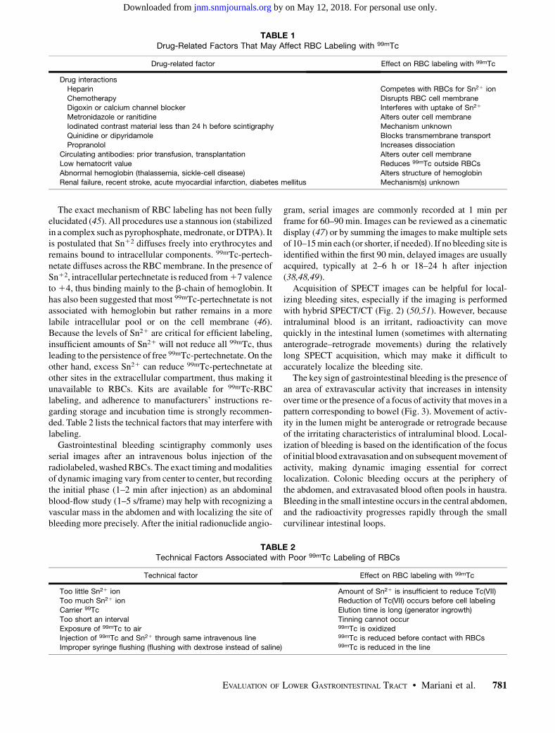
The exact mechanism of RBC labeling has not been fullyelucidated (45). All procedures use a stannous ion (stabilizedin a complex such as pyrophosphate, medronate, or DTPA). Itis postulated that Sn12 diffuses freely into erythrocytes andremains bound to intracellular components. 99mTc-pertech-netate diffuses across the RBC membrane. In the presence ofSn12, intracellular pertechnetate is reduced from 17 valenceto 14, thus binding mainly to the b-chain of hemoglobin. Ithas also been suggested that most 99mTc-pertechnetate is notassociated with hemoglobin but rather remains in a morelabile intracellular pool or on the cell membrane (46).Because the levels of Sn21 are critical for efficient labeling,insufficient amounts of Sn21 will not reduce all 99mTc, thusleading to the persistence of free 99mTc-pertechnetate. On theother hand, excess Sn21 can reduce 99mTc-pertechnetate atother sites in the extracellular compartment, thus making itunavailable to RBCs. Kits are available for 99mTc-RBClabeling, and adherence to manufacturers’ instructions re-garding storage and incubation time is strongly recommen-ded. Table 2 lists the technical factors that may interfere withlabeling.
Gastrointestinal bleeding scintigraphy commonly usesserial images after an intravenous bolus injection of theradiolabeled, washed RBCs. The exact timing and modalitiesof dynamic imaging vary from center to center, but recordingthe initial phase (1–2 min after injection) as an abdominalblood-flow study (1–5 s/frame) may help with recognizing avascular mass in the abdomen and with localizing the site ofbleeding more precisely. After the initial radionuclide angio-
gram, serial images are commonly recorded at 1 min perframe for 60–90 min. Images can be reviewed as a cinematicdisplay (47) or by summing the images to make multiple setsof 10–15 min each (or shorter, if needed). If no bleeding site isidentified within the first 90 min, delayed images are usuallyacquired, typically at 2–6 h or 18–24 h after injection(38,48,49).
Acquisition of SPECT images can be helpful for local-izing bleeding sites, especially if the imaging is performedwith hybrid SPECT/CT (Fig. 2) (50,51). However, becauseintraluminal blood is an irritant, radioactivity can movequickly in the intestinal lumen (sometimes with alternatinganterograde–retrograde movements) during the relativelylong SPECT acquisition, which may make it difficult toaccurately localize the bleeding site.
The key sign of gastrointestinal bleeding is the presence ofan area of extravascular activity that increases in intensityover time or the presence of a focus of activity that moves in apattern corresponding to bowel (Fig. 3). Movement of activ-ity in the lumen might be anterograde or retrograde becauseof the irritating characteristics of intraluminal blood. Local-ization of bleeding is based on the identification of the focusof initial blood extravasation and on subsequent movement ofactivity, making dynamic imaging essential for correctlocalization. Colonic bleeding occurs at the periphery ofthe abdomen, and extravasated blood often pools in haustra.Bleeding in the small intestine occurs in the central abdomen,and the radioactivity progresses rapidly through the smallcurvilinear intestinal loops.
TABLE 1Drug-Related Factors That May Affect RBC Labeling with 99mTc
Drug-related factor Effect on RBC labeling with 99mTc
Drug interactions
Heparin Competes with RBCs for Sn21 ion
Chemotherapy Disrupts RBC cell membraneDigoxin or calcium channel blocker Interferes with uptake of Sn21
Metronidazole or ranitidine Alters outer cell membrane
Iodinated contrast material less than 24 h before scintigraphy Mechanism unknown
Quinidine or dipyridamole Blocks transmembrane transportPropranolol Increases dissociation
Circulating antibodies: prior transfusion, transplantation Alters outer cell membrane
Low hematocrit value Reduces 99mTc outside RBCs
Abnormal hemoglobin (thalassemia, sickle-cell disease) Alters structure of hemoglobinRenal failure, recent stroke, acute myocardial infarction, diabetes mellitus Mechanism(s) unknown
TABLE 2Technical Factors Associated with Poor 99mTc Labeling of RBCs
Technical factor Effect on RBC labeling with 99mTc
Too little Sn21 ion Amount of Sn21 is insufficient to reduce Tc(VII)Too much Sn21 ion Reduction of Tc(VII) occurs before cell labeling
Carrier 99Tc Elution time is long (generator ingrowth)
Too short an interval Tinning cannot occur
Exposure of 99mTc to air 99mTc is oxidizedInjection of 99mTc and Sn21 through same intravenous line 99mTc is reduced before contact with RBCs
Improper syringe flushing (flushing with dextrose instead of saline) 99mTc is reduced in the line
EVALUATION OF LOWER GASTROINTESTINAL TRACT • Mariani et al. 781
by on May 12, 2018. For personal use only. jnm.snmjournals.org Downloaded from

Ectopic Gastric Mucosa in Meckel’s Diverticulum
Meckel’s diverticulum, a remnant caused by incompleteclosure of the omphalomesenteric duct, is located in the ileumabout 50–80 cm from the ileocecal valve. Gastric acid andpepsin produced by ectopic gastric mucosa can cause mucosaldamage and bleeding (52). Because 99mTc-pertechnetate avidlyaccumulates in gastric mucosa, it can reveal ectopic gastricmucosa in a Meckel’s diverticulum. Scintigraphy with 99mTc-pertechnetate is therefore used to localize ectopic gastricmucosa in a Meckel’s diverticulum as the potential source ofunexplained gastrointestinal bleeding (36,53). Scintigraphywith 99mTc-RBC remains instead the optimal radionuclideprocedure for visualizing active bleeding per se.
The test is the most accurate noninvasive technique foridentifying ectopic gastric mucosa in Meckel’s diverticu-lum, with high specificity and positive predictive value(close to 100%) both in children and in adults (54,55).
After intravenous injection of 99mTc-pertechnetate, serialimages of the abdomen are recorded for at least 30 min,usually at the rate of 30–60 s per frame, in the anterior view.These images may be summed to make multiple sets of 10–15 min each, to facilitate interpretation of the data. Addi-tional static images, anterior oblique projections, lateralprojections, and posterior projections are recommended atthe end of the dynamic acquisition. Similarly to bleedingscintigraphy, SPECT can greatly aid localization of the lesionin Meckel’s diverticulum (56).
A positive scan shows activity in the ectopic gastricmucosa at the same time as activity in the normal gastricmucosa (Fig. 4), although a small Meckel’s diverticulum mayseem to appear later than the stomach. Although Meckel’sdiverticulum may appear anywhere within the abdomen, it ismost frequently seen in the right lower quadrant. The activitymost often mistaken for Meckel’s diverticulum is activityin the kidneys, ureter, or bladder, which, however, usuallyappears after activity is seen in the normal gastric mucosa.Although the procedure can be performed with pharmaco-logic provocation, studies with pentagastrin, histamine-H2
blockers, or glucagon are rarely performed (57).
IBD
IBD includes 2 different clinical entities causing inflam-mation of the intestines: ulcerative colitis and Crohn’s dis-ease. Both diseases are chronic but frequently relapse afterperiods of remission. Circulating leukocytes are recruited atthe site of disease through a multistep process: adhesion tomicrovascular endothelium, transmigration through the ves-sel wall, and further migration in extravascular tissue and intothe bowel lumen (58). This is the pathophysiologic basis forusing labeled autologous leukocyte scintigraphy for charac-terizing activity of the disease.
In fact, the nuclear medicine imaging method initiallyused for characterizing active IBD is based on autologousradiolabeled leukocytes, either 111In-oxine leukocytes or,
FIGURE 2. (A) Sequential static 5-min images of abdominal area recorded at various times after 99mTc-RBC injection. Abnormalaccumulation of radioactivity is obvious already in early images (elongated horizontal area in upper abdominal region). Althoughaccumulation of radioactivity increases with time, no obvious progression along intestinal tract is detectable. Localization ofbleeding site is equivocal in planar images, being possibly consistent either with transverse colon or with duodenal localization. (B)SPECT/CT images after 99mTc-RBC injection demonstrate that radioactivity accumulation is not localized in anterior abdominalarea (as transverse colon would be) but rather in mid region, thus indicating duodenal localization of bleeding. (Courtesy ofDr. Elena Lazzeri, Regional Center of Nuclear Medicine, University of Pisa Medical School, Pisa, Italy.)
782 THE JOURNAL OF NUCLEAR MEDICINE • Vol. 49 • No. 5 • May 2008
by on May 12, 2018. For personal use only. jnm.snmjournals.org Downloaded from

more recently, 99mTc-hexamethylpropyleneamine oxime(HMPAO) leukocyte scintigraphy (59–63) (Fig. 5). Withboth 111In-oxine leukocytes and 99mTc-HMPAO leukocytes,scans in the upright position help to separate hepatic
activity from the transverse colon. A pelvic outlet viewcan be especially helpful in assessing rectal disease, par-ticularly because of bladder activity. SPECT studies (andespecially SPECT/CT) at 1.5–2 h after injection of radio-labeled leukocytes are helpful for separating superimposedactivity in the bone marrow or in other sites of physiologicaccumulation of radioactivity (Fig. 6). In positive scans,abnormal bowel activity is seen early, usually increasing inintensity over the next 2–3 h.
A recent metaanalysis of 49 studies published between1984 and 2004 (totalling nearly 4,400 patients) emphasizedthe high diagnostic accuracy of either 111In-oxine leukocyteor 99mTc-HMPAO leukocyte scintigraphy in IBD, with sen-sitivity and specificity around 90% (64). In particular, anegative leukocyte scan virtually rules out diagnosis of thisdisease. In addition to IBD, a scan can be positive in in-fectious enteritides, mesenteric ischemia, gastrointestinalbleeding, and colonic cancer. The sensitivity of leukocytescintigraphy for detecting IBD in untreated patients is higheven in the early stages of disease, when radiologic orendoscopic findings are often normal or equivocal. Althoughleukocyte scintigraphy is generally not considered to be afirst-line diagnostic procedure in patients with IBD, it is ofvalue in acutely ill patients with severe diarrhea, in whom an
FIGURE 3. Bleeding scintigraphy performed on 82-y-old manwhose presenting symptom was intermittent bright rectalbleeding. (A) Early dynamic imaging sequence after intravenousadministration of 99mTc-RBCs (each frame is summed image of5-min sequence) shows no definite area of abnormal radioac-tivity accumulation. (B) Later dynamic sequence starting 60 minafter injection of 99mTc-RBCs clearly shows abnormal accumu-lation of radioactivity initiating in proximal descending colonand extending to whole descending colon, as also confirmed bysubsequent SPECT (image not shown).
FIGURE 4. Meckel’s diverticulum scintigraphy performed on2-y-old boy with intermittent bleeding (bright red blood) and noother accompanying symptom. Sequential images obtainedafter injection of 99mTc-pertechnetate show obvious accumu-lation of radioactivity in right lower paraumbilical region,consistent with most frequent location of Meckel’s diverticulum.Time-course pattern of radioactivity accumulation in possibleMeckel’s diverticulum mirrors pattern of tracer concentration ingastric region. Diagnosis of Meckel’s diverticulum was con-firmed at surgery, which was followed by complete disappear-ance of gastrointestinal bleeding. (Courtesy of Drs. Pham ThiMinh Bao and Le Ngoc Ha, Department of Nuclear Medicine,Tran Hung Dao General Hospital, Hanoi, Vietnam.)
EVALUATION OF LOWER GASTROINTESTINAL TRACT • Mariani et al. 783
by on May 12, 2018. For personal use only. jnm.snmjournals.org Downloaded from

endoscopic study might be contraindicated. An additionalspecific indication is the early diagnosis of postsurgicalrecurrences on preanastomotic loops.
Although leukocyte imaging is useful, PET with 18F-FDG is becoming the new standard for nuclear medicineimaging in patients with IBD, based on the localization ofthis tracer in inflammatory lesions (65,66). The field startedwith a few occasional observations, whereas more system-atic studies have better clarified the value of 18F-FDG PETin patients with IBD (67). In a group of 25 pediatricpatients, 18F-FDG PET had 81% sensitivity and 85%specificity for detecting IBD, based on a 4-point scale for18F-FDG uptake in 5 bowel segments (68). In a more recentreport on 55 children with newly diagnosed IBD or symp-toms suggestive of recurrent disease, sensitivity of 18F-FDGPET was 80%, and there was no evidence of inflammationin children with recurrent abdominal pain without IBD(69). Even better results have been described by Loffleret al. in pediatric patients (aged 2–6 y), with an averagesensitivity of 98% (higher than both endoscopy and ab-dominal ultrasound) and overall accuracy comparable tothe invasive procedure (83% vs. 82%) (70).
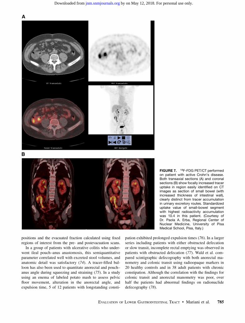
Considering that the diagnostic performance of 18F-FDGPET is excellent and that this technique avoids the cum-bersome and time-consuming preparation of radiolabeledautologous leukocytes, 18F-FDG PET may offer a definiteclinical advantage for adult patients (71). In this regard, thecombination of metabolic imaging (PET) with better ana-
tomic localization (CT) represents a further advance forincreased specificity (Fig. 7). Louis et al. have recentlydemonstrated the high prognostic value of 18F-FDG PET/CT in a group of adult patients with IBD (72). In particular,in addition to visual analysis (with 100% sensitivity fordetecting severe endoscopic lesions), they developed aglobal PET/CT score based on the maximum standardizeduptake value. Median maximum standardized uptake valuesof the affected segments were in the 4–6 range but reachedlevels as high as 12–14. This score significantly correlatedwith the endoscopic index of severity of Crohn’s disease,with a global index of severity of disease, and with theC-reactive protein serum levels. Finally, logistic regressionanalysis showed that the index of metabolic activity (ratio ofstandardized uptake value in the affected segment to stan-dardized uptake value in the liver) was significantly associ-ated with the presence of severe endoscopic lesions, incontrast to wall thickening alone (a classic CT-based marker).
Scintigraphic Defecography
Disorders of evacuation account for a significant propor-
tion of people seeking medical attention for constipation
and for a large percentage of those who fail to respond to
colectomy with ileorectal anastomosis; indeed, a consider-
able proportion of patients with severe constipation actually
have dyschezia (73).Dynamic scintigraphic defecography can quantitatively
evaluate emptying efficiency in patients with disordered evac-
uation. This technique involves considerably less radiation
than x-ray proctography (gonadal dose approximately 7%of the dose of the corresponding radiologic examination)(74). Artificial stool (rehydrated potato labeled with 99mTc-pertechnetate or methyl-cellulose paste labeled with 99mTc-ethylene diphosphonate, 180–200 MBq in either case) isinstilled into the rectum up to a volume causing impendingevacuation (75). Preevacuation images are recorded withthe patient sitting on a commode, the g-camera headagainst the left, right, and posterior pelvis (1 min perview). A postevacuation scan is then recorded in the same
FIGURE 5. Planar imagingof abdomen performed onpatient with active Crohn’sdisease approximately 45min after injection of autolo-gous 99mTc-HMPAO–labeledleukocytes. Accumulation ofradioactivity is obvious insection of small bowel thatcan be identified as preter-minal ileum.
FIGURE 6. Planar imaging (A) andSPECT/CT (B) performed on patient withactive Crohn’s disease approximately45–90 min after injection of autologous99mTc-HMPAO–labeled leukocytes. Ob-vious accumulation of radioactivity interminal ileum is seen on planar imaging.However, SPECT/CT also demonstratesIBD involvement of preterminal ileum,which was obscured on planar imagingbecause of superimposition with circu-lating blood pool and with accumulationin underlying bone marrow. (Courtesy ofDr. Paola A. Erba, Regional Center ofNuclear Medicine, University of PisaMedical School, Pisa, Italy).
784 THE JOURNAL OF NUCLEAR MEDICINE • Vol. 49 • No. 5 • May 2008
by on May 12, 2018. For personal use only. jnm.snmjournals.org Downloaded from
positions and the evacuated fraction calculated using fixedregions of interest from the pre- and postevacuation scans.
In a group of patients with ulcerative colitis who under-went ileal pouch–anus anastomosis, this semiquantitativeparameter correlated well with excreted stool volumes, andanatomic detail was satisfactory (74). A tracer-filled bal-loon has also been used to quantitate anorectal and pouch–anus angle during squeezing and straining (75). In a studyusing an enema of labeled potato mash to assess pelvicfloor movement, alteration in the anorectal angle, andexpulsion time, 5 of 12 patients with longstanding consti-
pation exhibited prolonged expulsion times (76). In a largerseries including patients with either obstructed defecationor slow transit, incomplete rectal emptying was observed inpatients with obstructed defecation (77). Wald et al. com-pared scintigraphic defecography with both anorectal ma-nometry and colonic transit using radioopaque markers in20 healthy controls and in 38 adult patients with chronicconstipation. Although the correlation with the findings forcolonic transit and anorectal manometry was poor, overhalf the patients had abnormal findings on radionuclidedefecography (78).
FIGURE 7. 18F-FDG PET/CT performedon patient with active Crohn’s disease.Both transaxial sections (A) and coronalsections (B) show focally increased traceruptake in region easily identified on CTimages as section of small bowel (withincreased thickness of intestinal wall),clearly distinct from tracer accumulationin urinary excretory routes. Standardizeduptake value of small-bowel segmentwith highest radioactivity accumulationwas 10.4 in this patient. (Courtesy ofDr. Paola A. Erba, Regional Center ofNuclear Medicine, University of PisaMedical School, Pisa, Italy.)
EVALUATION OF LOWER GASTROINTESTINAL TRACT • Mariani et al. 785
by on May 12, 2018. For personal use only. jnm.snmjournals.org Downloaded from

By administering a radiolabeled meal in divided dosesover 6 h and imaging defecation 24 and 48 h later, Lubowskyet al. demonstrated that during defecation significant emp-tying of the left colon commonly occurs, and even of theright colon (79).
Selvaggi et al. used scintigraphic defecography to evaluatethe efficiency of defecation in 16 patients with severeulcerative colitis bearing an ileal pouch (80). The patientswere classified into 2 groups according to the presence ofgood (group A) or poor (group B) pouch function, percentageemptying being higher in group A than in group B (81% 6
9% vs. 71% 6 9%, P , 0.05), although the mean maximumtolerated volume was similar in the 2 groups.
In patients with an ileal pouch performed for ulcerativecolitis, an increased frequency of defecation without anyevidence of pouch dysfunction may be correlated with analteration of emptying efficiency. Scintigraphic defecogra-phy is appropriate for investigating this condition with norelevant discomfort for the patients.
CONCLUSION
Although most studies of the gastrointestinal tract dealwith the esophagus and stomach, transit studies can also beused for the lower gastrointestinal tract. Similarly, studies ofsmall- and large-bowel inflammatory disease, detection ofMeckel’s diverticula, and identification of lower gastrointes-tinal bleeding are valuable in the appropriate clinical setting.In some countries, several practical and regulatory issueshave retarded the growth and more widespread use ofradionuclide procedures for evaluating patients with disor-ders of the lower gastrointestinal tract. Most notably in theUnited States, intestinal transit scintigraphy (including scin-tigraphic defecography) and 18F-FDG PET for imaging IBDare not reimbursed, and 99mTc-sulfur colloid is the only agentapproved for oral administration. As the population ages, it islikely that the demand for lower gastrointestinal transitstudies will increase, and longer-lived radiopharmaceuticals,such as 111In-charcoal, for the performance of this study willbe approved. Similarly, 18F-FDG PET studies of inflamma-tion, although still in their infancy, require wider validation inlarge-scale, specifically designed clinical trials.
ACKNOWLEDGMENTS
We thank Dr. Marco Anselmino (Fourth Division ofGeneral Surgery, Regional Center for Diseases of the Esoph-agus, ‘‘S. Chiara’’ University Hospital, Pisa, Italy) for helpfuldiscussions in the preparatory phase of the manuscript. Thanksare also due to Drs. Fiammetta Pesella and Alice Lorenzoni(residents in nuclear medicine at the University of PisaMedical School, Pisa, Italy) for their contribution to varioussections on radionuclide procedures. We also acknowledge thecontribution of Dr. Cristina Stasi (Gastroenterology Unit,Department of Internal Medicine, the University of PisaMedical School, Pisa, Italy) to the section on clinicalmanifestations of the lower gastrointestinal tract.
REFERENCES
1. Mariani G, Boni G, Barreca M, et al. Radionuclide gastroesophageal motor
studies. J Nucl Med. 2004;45:1004–1028.
2. Locke GR III, Pemberton JH, Phillips SF. AGA technical review on constipation.
American Gastroenterological Association. Gastroenterology. 2000;119:1766–
1778.
3. Hellard ME, Sinclair MI, Harris AH, Kirk M, Fairley CK. Cost of community
gastroenteritis. J Gastroenterol Hepatol. 2003;18:322–328.
4. Sandler RS, Everhart JE, Donowitz M, et al. The burden of selected digestive
diseases in the United States. Gastroenterology. 2002;122:1500–1511.
5. Lipsky MS, Adelman M. Chronic diarrhea: evaluation and treatment. Am Fam
Physician. 1993;48:1461–1466.
6. Wilson S, Roberts L, Roalfe A, Bridge P, Singh S. Prevalence of irritable bowel
syndrome: a community survey. Br J Gen Pract. 2004;54:495–502.
7. Bodger K. Cost of illness of Crohn’s disease. Pharmacoeconomics.
2002;20:639–652.
8. Longstreth GF, Thompson WG, Chey WD, Houghton LA, Mearin F, Spiller RC.
Functional bowel disorders. Gastroenterology. 2006;130:1480–1491.
9. Talley NJ. Management of chronic constipation. Rev Gastroenterol Disord.
2004;4:18–24.
10. Lembo A, Camilleri M. Chronic constipation. N Engl J Med. 2003;349:1360–
1368.
11. Prather CM. Subtypes of constipation: sorting out the confusion. Rev Gastro-
enterol Disord. 2004;4(suppl 2):S11–S16.
12. Longstreth GF. Epidemiology and outcome of patients hospitalized with acute
lower gastrointestinal hemorrhage: a population-based study. Am J Gastro-
enterol. 1997;92:419–424.
13. Lanas A, Sekar MC, Hirschowitz BI. Objective evidence of aspirin use in both
ulcer and nonulcer upper and lower gastrointestinal bleeding. Gastroenterology.
1992;103:862–869.
14. Elton E, Howell DA, Amberson SM, Dykes TA. Combined angiographic and
endoscopic management of bleeding pancreatic pseudoaneurysms. Gastrointest
Endosc. 1997;46:544–549.
15. Chak A, Cooper GS, Lloyd LE, Kolz CS, Barnhart BA, Wong RC. Effectiveness
of endoscopy in patients admitted to the intensive care unit with upper GI
hemorrhage. Gastrointest Endosc. 2001;53:6–13.
16. Boley SJ, DiBiase A, Brandt LJ, Sammartano RJ. Lower intestinal bleeding in
the elderly. Am J Surg. 1979;137:57–64.
17. Peura DA, Lanza FL, Gostout CJ, Foutch PG. The American College of
Gastroenterology Bleeding Registry: preliminary findings. Am J Gastroenterol.
1997;92:924–928.
18. Zuckerman GR, Prakash C. Acute lower intestinal bleeding. Part II: etiology,
therapy, and outcomes. Gastrointest Endosc. 1999;49:228–238.
19. Huguier M, Barrier A, Boelle PY, Houry S, Lacaine F. Ischemic colitis. Am J
Surg. 2006;192:679–684.
20. Meckel JF. Ueber die Divertikel am Darmkanal. Archiv Physiol (Halle). 1809;
9:421–453.
21. Blackshear JL, Baker VS, Holland A, et al. Fecal hemoglobin excretion in
elderly patients with atrial fibrillation: combined aspirin and low-dose warfarin
vs conventional warfarin therapy. Arch Intern Med. 1996;156:658–660.
22. Lin HC, Prather C, Fischer RS, et al. Measurement of gastrointestinal transit. Dig
Dis Sci. 2005;50:989–1004.
23. Lin HC, Zhao XT, Wang L. Jejunal brake: inhibition of intestinal transit by fat in
the proximal small intestine. Dig Dis Sci. 1996;41:326–329.
24. Van Citters GW, Lin HC. The ileal brake: a fifteen-year progress report. Curr
Gastroenterol Rep. 1999;1:404–409.
25. Bonapace ES, Maurer AH, Davidoff S, Krevsky B, Fisher RS, Parkman HP.
Whole gut transit scintigraphy in the clinical evaluation of patients with upper
and lower gastrointestinal symptoms. Am J Gastroenterol. 2000;95:2838–2847.
26. Proano M, Camilleri M, Phillips SF, Brown ML, Thomforde GM. Transit of
solids through the human colon: regional quantification in the unprepared bowel.
Am J Physiol. 1990;258:G856–G862.
27. Burton DD, Camilleri M, Mullan BP, Forstrom LA, Hung JC. Colonic transit
scintigraphy labeled activated charcoal compared with ion exchange pellets.
J Nucl Med. 1997;38:1807–1810.
28. Maurer AH, Parkman HP. Update on gastrointestinal scintigraphy. Semin Nucl
Med. 2006;36:110–118.
29. Malagelada JR, Robertson JS, Brown ML, et al. Intestinal transit of solid and
liquid components of a meal in health. Gastroenterology. 1984;87:1255–1263.
30. Bennink R, Peeters M, Van den Maegdenbergh V, et al. Evaluation of small-
bowel transit for solid and liquid test meal in healthy men and women. Eur J
Nucl Med. 1999;26:1560–1566.
786 THE JOURNAL OF NUCLEAR MEDICINE • Vol. 49 • No. 5 • May 2008
by on May 12, 2018. For personal use only. jnm.snmjournals.org Downloaded from

31. Camilleri M, Zinsmeister AR, Greydanus MP, Brown ML, Proano M. Towards a
less costly but accurate test of gastric emptying and small bowel transit. Dig Dis
Sci. 1991;36:609–615.
32. Madsen JL, Fuglsang S, Graff J. Reference values for the geometric centre
analysis of colonic transit measurements with 111indium-labelled diethylenetri-
amine penta-acetic acid. Clin Physiol Funct Imaging. 2003;23:204–207.
33. Mason HJ, Serrano-Ikkos E, Kamm MA. Psychological morbidity in women
with idiopathic constipation. Am J Gastroenterol. 2000;95:2852–2857.
34. Chitkara DK, Delgado-Aros S, Bredenoord AJ, et al. Functional dyspepsia, upper
gastrointestinal symptoms, and transit in children. J Pediatr. 2003;143:609–613.
35. Vassallo M, Camilleri M, Phillips SF, Brown ML, Chapman NJ, Thomforde GM.
Transit through the proximal colon influences stool weight in the irritable bowel
syndrome. Gastroenterology. 1992;102:102–108.
36. Ford PV, Bartold SP, Fink-Bennett DM, et al. Procedure guideline for
gastrointestinal bleeding and Meckel’s diverticulum scintigraphy. Society of
Nuclear Medicine. J Nucl Med. 1999;40:1226–1232.
37. O’Neill BB, Gosnell JE, Lull RJ, et al. Cinematic nuclear scintigraphy reliably
directs surgical intervention for patients with gastrointestinal bleeding. Arch
Surg. 2000;135:1076–1081.
38. Kan JH, Funaki B, O’Rourke BD, Ward MB, Appelbaum DE. Delayed 99mTc-
labeled erythrocyte scintigraphy in patients with lower gastrointestinal tract
hemorrhage: effect of positive findings on clinical management. Acad Radiol.
2003;10:497–501.
39. Smith R, Copely DJ, Bolen FH. 99mTc RBC scintigraphy: correlation of
gastrointestinal bleeding rates with scintigraphic findings. AJR. 1987;148:869–874.
40. Jacobson AF, Cerqueira MD. Prognostic significance of late imaging results in
technetium-99m-labeled red blood cell gastrointestinal bleeding studies with
early negative images. J Nucl Med. 1992;33:202–207.
41. Thorne DA, Datz FL, Remley K, Christian PE. Bleeding rates necessary for
detecting acute gastrointestinal bleeding with technetium-99m-labeled red blood
cells in an experimental model. J Nucl Med. 1987;28:514–520.
42. Bearn P, Persad R, Wilson N, Flanagan J, Williams T. 99mTechnetium-labelled
red blood cell scintigraphy as an alternative to angiography in the investigation
of gastrointestinal bleeding: clinical experience in a district general hospital. Ann
R Coll Surg Engl. 1992;74:192–199.
43. Bunker SR, Lull RJ, Tanasescu DE, et al. Scintigraphy of gastrointestinal
hemorrhage: superiority of 99mTc red blood cells over 99mTc sulfur colloid. AJR.
1984;143:543–548.
44. Siddiqui AR, Schauwecker DS, Wellman HN, Mock BH. Comparison of
technetium-99m sulfur colloid and in vitro labeled technetium-99m RBCs in the
detection of gastrointestinal bleeding. Clin Nucl Med. 1985;10:546–549.
45. Callahan RJ, Froelich JW, McKusick KA, Leppo J, Strauss HW. A modified
method for the in vivo labeling of red blood cells with Tc-99m: concise
communication. J Nucl Med. 1982;23:315–318.
46. Seldin DW, Simchon S, Jan KM, Chien S, Alderson PO. Dependence of
technetium-99m red blood cell labeling efficiency on red cell surface charge.
J Nucl Med. 1988;29:1710–1713.
47. Maurer AH, Rodman MS, Vitti RA, Revez G, Krevsky B. Gastrointestinal
bleeding: improved localization with cine scintigraphy. Radiology. 1992;185:
187–192.
48. Zettinig G, Staudenherz A, Leitha T. The importance of delayed images in
gastrointestinal bleeding scintigraphy. Nucl Med Commun. 2002;23:803–808.
49. Nwakanma L, Meyerrose G, Kennedy S, Rakvit A, Bohannon T, Silva M.
Recurrent gastrointestinal bleeding diagnosed by delayed scintigraphy with
Tc-99m-labeled red blood cells. Clin Nucl Med. 2003;28:691–693.
50. Yama N, Ezoe E, Kimura Y, et al. Localization of intestinal bleeding using a
fusion of Tc-99m-labeled RBC SPECT and x-ray CT. Clin Nucl Med. 2005;30:
488–489.
51. Schillaci O, Filippi L, Danieli R, Simonetti G. Single-photon emission computed
tomography/computed tomography in abdominal diseases. Semin Nucl Med.
2007;37:48–61.
52. Maurer AH. Gastrointestinal bleeding. In: Ell PJ, Gambhir GG, eds. Nuclear
Medicine in Clinical Diagnosis and Treatment. 3rd ed. Sydney, Australia:
Churchill Livingstone; 2004:911–917.
53. Sfakianakis GN, Conway JJ. Detection of ectopic gastric mucosa in Meckel’s
diverticulum and in other aberrations of scintigraphy: I: pathophysiology and 10
year clinical experience. J Nucl Med. 1981;22:647–654.
54. Sfakianakis GN, Haase GM. Abdominal scintigraphy for ectopic gastric mucosa:
a retrospective analysis of 143 studies. AJR. 1982;138:7–12.
55. Kong MS, Chen CY, Tzen KY, et al. Technetium-99m pertechnetate scan for
ectopic gastric mucosa in children with gastrointestinal bleeding. J Formos Med
Assoc. 1993;92:717–720.
56. Connolly LP, Treves ST, Bozorgi F, O’Connor SC. Meckel’s diverticulum:
demonstration of heterotopic gastric mucosa with technetium-99m-pertechnetate
SPECT. J Nucl Med. 1998;39:1458–1460.
57. Brown ML. Gastrointestinal bleeding. In: Wagner HN, Szabo Z, Buchanan JW,
eds. Principles of Nuclear Medicine. 2nd ed. Philadelphia, PA: W.B. Saunders
Co.; 1995:929–934.
58. Muller WA. Leukocyte-endothelial cell interactions in the inflammatory
response. Lab Invest. 2002;82:521–533.
59. Charron M, Orenstein SR, Bhargava S. Detection of inflammatory bowel disease
in pediatric patients with technetium-99m-HMPAO-labeled leukocytes. J Nucl
Med. 1994;35:451–455.
60. Li DJ, Freeman A, Miles KA, Wraight EP. Can 99Tcm HMPAO leucocyte
scintigraphy distinguish between Crohn’s disease and ulcerative colitis? Br J
Radiol. 1994;67:472–477.
61. Datz FL, Seabold JE, Brown ML, et al. Procedure guideline for technetium-99m-
HMPAO-labeled leukocyte scintigraphy for suspected infection/inflammation.
Society of Nuclear Medicine. J Nucl Med. 1997;38:987–990.
62. Seabold JE, Forstrom LA, Schauwecker DS, et al. Procedure guideline for
indium-111-leukocyte scintigraphy for suspected infection/inflammation. Soci-
ety of Nuclear Medicine. J Nucl Med. 1997;38:997–1001.
63. Martin-Comin J, Prats E. Clinical applications of radiolabeled blood elements in
inflammatory bowel disease. Q J Nucl Med. 1999;43:74–82.
64. Annovazzi A, Bagni B, Burroni L, D’Alessandria C, Signore A. Nuclear
medicine imaging of inflammatory/infective disorders of the abdomen. Nucl Med
Commun. 2005;26:657–664.
65. Yamada S, Kubota K, Kubota R, Ido T, Tamahashi N. High accumulation of
fluorine-18-fluorodeoxyglucose in turpentine-induced inflammatory tissue.
J Nucl Med. 1995;36:1301–1306.
66. Love C, Tomas MB, Tronco GG, Palestro CJ. FDG PET of infection and
inflammation. Radiographics. 2005;25:1357–1368.
67. Bicik I, Bauerfeind P, Breitbach T, von Schultess GK, Fried M. Inflammatory
bowel disease activity measured by positron-emission tomography [letter].
Lancet. 1997;350:262.
68. Skehan SJ, Issenman R, Mernagh J, Nahmias C, Jacobson K. 18F-Fluorodeoxy-
glucose positron tomography in diagnosis of paediatric inflammatory bowel
disease. Lancet. 1999;354:836–837.
69. Lemberg DA, Issenman RM, Cawdron R, et al. Positron emission tomography in
the investigation of pediatric inflammatory bowel disease. Inflamm Bowel Dis.
2005;11:733–738.
70. Loffler M, Weckesser M, Franzius C, et al. High diagnostic value of 18F-FDG-
PET in pediatric patients with chronic inflammatory bowel disease. Ann N Y
Acad Sci. 2006;1072:379–385.
71. Neurath MF, Vehling D, Schunk K, et al. Noninvasive assessment of Crohn’s
disease activity: a comparison of 18F-fluorodeoxyglucose positron emission
tomography, hydromagnetic resonance imaging, and granulocyte scintigraphy
with labeled antibodies. Am J Gastroenterol. 2002;97:1978–1985.
72. Louis E, Ancion G, Colard A, Spote V, Belaiche J, Hustinx R. Noninvasive
assessment of Crohn’s disease intestinal lesions with 18F-FDG PET/CT. J Nucl
Med. 2007;48:1053–1059.
73. Whitehead WE, Wald A, Diamant NE, Enck P, Pemberton JH, Rao SSC.
Functional disorders of the anus and rectum. Gut. 1999;45(suppl 2):1155–
1159.
74. O’Connell PR, Kelly KA, Brown ML. Scintigraphic assessment of neorectal
function. J Nucl Med. 1986;27:460–464.
75. Barkel DC, Pemberton JH, Pezim ME, et al. Scintigraphic assessment of the
anorectal angle in health and after ileal pouch-anal anastomosis. Ann Surg.
1988;208:42–49.
76. Papachrysostomou M, Griffin TM, Ferrington C, Merrick MV, Smith AN. A method
of computerised isotope dynamic proctography. Eur J Nucl Med. 1992;19:431–435.
77. Papachrysostomou M, Smith AN, Merrick MV. Obstructive defaecation and slow
transit constipation: the proctographic parameters. Int J Colorectal Dis. 1994;9:
115–120.
78. Wald A, Jafri F, Rehder J, Holeva K. Scintigraphic studies of rectal emptying in
patients with constipation and defecatory difficulty. Dig Dis Sci. 1993;38:353–
358.
79. Lubowski DZ, Meagher AP, Smart RC, Butler SP. Scintigraphic assessment of
colonic function during defaecation. Int J Colorectal Dis. 1995;10:91–93.
80. Selvaggi F, Cuocolo A, Giuliani A, et al. The role of scintigraphic defecography
in the assessment of bowel function after restorative proctocolectomy for
ulcerative colitis. Int J Colorectal Dis. 2006;21:448–452.
EVALUATION OF LOWER GASTROINTESTINAL TRACT • Mariani et al. 787
by on May 12, 2018. For personal use only. jnm.snmjournals.org Downloaded from

Doi: 10.2967/jnumed.107.040113Published online: April 15, 2008.
2008;49:776-787.J Nucl Med. StraussBellini, Mariano Grosso, Nicola de Bortoli, Gloria Mumolo, Francesco Costa, Domenico Rubello and H. William Giuliano Mariani, Ernest K.J. Pauwels, Abedallatif AlSharif, Santino Marchi, Giuseppe Boni, Marco Barreca, Massimo Radionuclide Evaluation of the Lower Gastrointestinal Tract
http://jnm.snmjournals.org/content/49/5/776This article and updated information are available at:
http://jnm.snmjournals.org/site/subscriptions/online.xhtml
Information about subscriptions to JNM can be found at:
http://jnm.snmjournals.org/site/misc/permission.xhtmlInformation about reproducing figures, tables, or other portions of this article can be found online at:
(Print ISSN: 0161-5505, Online ISSN: 2159-662X)1850 Samuel Morse Drive, Reston, VA 20190.SNMMI | Society of Nuclear Medicine and Molecular Imaging
is published monthly.The Journal of Nuclear Medicine
© Copyright 2008 SNMMI; all rights reserved.
by on May 12, 2018. For personal use only. jnm.snmjournals.org Downloaded from