radiologic management of upper gastrointestinal bleeding … managemen… · acr appropriateness...
TRANSCRIPT

ACR Appropriateness Criteria® Radiologic Management of Upper Gastrointestinal Bleeding
EVIDENCE TABLE
* See Last Page for Key 2010 Review Schenker Page 1
Reference Study Type Patients/ Events
Study Objective (Purpose of Study) Study Results Strength of
Evidence 1. Peter DJ, Dougherty JM. Evaluation of
the patient with gastrointestinal bleeding: an evidence based approach. Emerg Med Clin North Am 1999; 17(1):239-261, x.
11 N/A Review gastrointestinal (GI) bleeding in the emergency department using evidence based approach.
The first tests in lower GI are usually snoscopy/sigmoidoscopy followed by colonoscopy.
3
2. Gilbert DA, Silverstein FE, Tedesco FJ, Buenger NK, Persing J. The national ASGE survey on upper gastrointestinal bleeding. III. Endoscopy in upper gastrointestinal bleeding. Gastrointest Endosc 1981; 27(2):94-102.
15 2,225 Prospectively conducted survey to determine if there are subgroups of patients in whom outcome from a bleeding episode is predictably poor despite application of all advances in conventional therapy.
At least one diagnostic abnormality was identified in 91.9% of endoscopies. The frequency of finding an active bleeding source was observed to decrease as the admission to endoscopy interval increased. Active bleeding observed more often when endoscopy done during the first 24 hours. Patients with oozing or pumping lesion had statistically significant increase in mortality. In almost all patients with a duodenal, stomal or gastric ulcer seen on endoscopy, bleeding was attributed to this lesion. Esophagitis deemed responsible in only 50% of patients in whom it was seen. Varices listed as final diagnosis for cause of bleeding in only 66.2% of patients in whom they were observed. Complication rate of endoscopy was 0.9%.
2
3. Silverstein FE, Gilbert DA, Tedesco FJ, Buenger NK, Persing J. The national ASGE survey on upper gastrointestinal bleeding. I. Study design and baseline data. Gastrointest Endosc 1981; 27(2):73-79.
15 Study data base of 2,225
patients submitted by
269 physicians
Study design and baseline data: American Society for Gastrointestinal Endoscopy (ASGE) survey on upper GI (UGI) bleeding.
2,427 cases from 277 physicians during 18-month period were received. Number of patients per physician varied widely. Of 2,427 data forms 85% were copied and returned to the participants for correction and/or completion. Participants corrected and returned 92% of these forms.
2
4. Sugawa C, Steffes CP, Nakamura R, et al. Upper GI bleeding in an urban hospital. Etiology, recurrence, and prognosis. Ann Surg 1990; 212(4):521-526; discussion 526-527.
15 469 Retrospective study to describe the various causes of UGI bleeding and describe the various endoscopic techniques available for treatment.
Majority of UGI bleeding cases can be treated without operation, including endoscopic treatment, when diagnostic endoscopy establishes the source. Subsequent operation in selected patients can be done with low morbidity and mortality rates.
2
5. van Leerdam ME. Epidemiology of acute upper gastrointestinal bleeding. Best Pract Res Clin Gastroenterol 2008; 22(2):209-224.
15 N/A Review epidemiology of acute UGI bleeding. Most surveys focusing on peptic ulcer disease showed a significant decrease in admission and mortality of peptic ulcer disease. Recent epidemiological surveys show a decrease in incidence of all cause UGI bleeding.
4

ACR Appropriateness Criteria® Radiologic Management of Upper Gastrointestinal Bleeding
EVIDENCE TABLE
* See Last Page for Key 2010 Review Schenker Page 2
Reference Study Type Patients/ Events
Study Objective (Purpose of Study) Study Results Strength of
Evidence 6. Chak A, Cooper GS, Lloyd LE, Kolz CS,
Barnhart BA, Wong RC. Effectiveness of endoscopy in patients admitted to the intensive care unit with upper GI hemorrhage. Gastrointest Endosc 2001; 53(1):6-13.
10 214 Retrospective, multicenter study to determine the effectiveness of 3 esophagogastroduodenoscopy (EGD) factors, visualization, accurate initial diagnosis, performance within 24 hours of admission (early EGD), and appropriate intervention in the management of patients admitted to ICU with UGI hemorrhage.
Early EGD performed in 82% of patients was associated with severity-adjusted reductions in hospital (–33%) and ICU (–20%) stay.
Appropriate intervention at initial EGD, performed in 84% of patients, was associated with reductions in severity-adjusted length of ICU stay (–18%) and rate of recurrent bleeding.
Early, accurate EGD with appropriate therapeutic intervention is effective and associated with improved outcomes for patients with UGI hemorrhage admitted to the ICU.
2
7. Esrailian E, Gralnek IM. Nonvariceal upper gastrointestinal bleeding: epidemiology and diagnosis. Gastroenterol Clin North Am 2005; 34(4):589-605.
12 N/A Review article focusing on the epidemiology and diagnosis of non variceal UGI bleeding.
Summarizes recent international data on causes of UGI bleeding. Discusses role of endoscopy and risk stratification.
4
8. Adler DG, Leighton JA, Davila RE, et al. ASGE guideline: The role of endoscopy in acute non-variceal upper-GI hemorrhage. Gastrointest Endosc 2004; 60(4):497-504.
15 N/A Guideline to review role of endoscopy in acute non-variceal UGI hemorrhage.
Endoscopy is effective in diagnosing and treating most causes of UGI bleeding.
3
9. Van Dam J, Brugge WR. Endoscopy of the upper gastrointestinal tract. N Engl J Med 1999; 341(23):1738-1748.
12 N/A Review role of UGI endoscopy. Endoscopy is recommended for UGI bleeding because of its accuracy, low rate of complications, and potential for therapeutic intervention.
3
10. Spiegel BM, Ofman JJ, Woods K, Vakil NB. Minimizing recurrent peptic ulcer hemorrhage after endoscopic hemostasis: the cost-effectiveness of competing strategies. Am J Gastroenterol 2003; 98(1):86-97.
15 N/A Through decision analysis to evaluate the cost-effectiveness of four strategies: 1. Follow patients clinically after hemostasis
and repeat endoscopy only in patients with rebleeding (usual care);
2. Administer intravenous proton pump inhibitors (IV PPI) after hemostasis and repeat endoscopy only with signs of rebleeding;
3. Perform second look endoscopy at 24 h in all patients with successful endoscopic hemostasis;
4. Perform selective second look endoscopy at 24-hours only in patients at high risk for rebleeding.
Most effective and least expensive was second look endoscopy strategy. The IV PPI strategy required 50% fewer endoscopies than the competing strategies, and became the dominant strategy when the rebleed rate with IV PPI fell below 9% and when the cost of IV PPI fell below 10 dollars/day.
2

ACR Appropriateness Criteria® Radiologic Management of Upper Gastrointestinal Bleeding
EVIDENCE TABLE
* See Last Page for Key 2010 Review Schenker Page 3
Reference Study Type Patients/ Events
Study Objective (Purpose of Study) Study Results Strength of
Evidence 11. Chung SS, Lau JY, Sung JJ, et al.
Randomised comparison between adrenaline injection alone and adrenaline injection plus heat probe treatment for actively bleeding ulcers. BMJ 1997; 314(7090):1307-1311
1 276 Randomized prospective study to compare endoscopic adrenaline injection alone and adrenaline injection plus heat probe for the treatment of actively bleeding peptic ulcers.
Initial haemostasis seen in 131/134 patients (98%) who received adrenaline injection alone and 135/136 patients (99%) who had additional heat probe treatment (P=0.33). Outcome was not significantly different in the two groups. In the subgroup of patients with spurting haemorrhage 8/27 patients from the adrenaline injection alone group and 2/31 patients from the dual treatment group required operative intervention. Addition of heat probe treatment after endoscopic adrenaline injection is advantageous in ulcers with spurting haemorrhage.
1
12. Cook DJ, Guyatt GH, Salena BJ, Laine LA. Endoscopic therapy for acute nonvariceal upper gastrointestinal hemorrhage: a meta-analysis. Gastroenterology 1992; 102(1):139-148.
7 30 randomized
trials
Meta-analysis performed to examine effect of endoscopic therapy in acute nonvariceal UGI bleeding.
Endoscopic therapy significantly reduced rates of further bleeding (odds ratio, 0.38; 95% CI, 0.32-0.45), surgery (odds ratio, 0.36; 95% CI, 0.28-0.45), and mortality (odds ratio, 0.55; 95% CI, 0.40-0.76). Thermal-contact devices, (monopolar and bipolar electrocoagulation and heater probe), laser treatment, and injection therapy all significantly decreased further bleeding and surgery rates.
1
13. Rollhauser C, Fleischer DE. Nonvariceal upper gastrointestinal bleeding. Endoscopy 2004; 36(1):52-58.
7 N/A Review management of nonvariceal UGI bleeding.
Hemostatic techniques like injection and thermocoagulation, especially a combination therapy using both methods are preferable. Recent therapies like cryotherapy are interesting, but have not had widespread application. Endoscopic suturing techniques have not been adapted to the management of GI bleeding.
4
14. Zuccaro G, Jr. Bleeding peptic ulcer: pathogenesis and endoscopic therapy. Gastroenterol Clin North Am 1993; 22(4):737-750.
7 N/A Discusses pathophysiology of bleeding from peptic ulcers and reviews desired and observed tissue effects of available treatment modalities. Clinical trials assessing treatment efficacy reviewed.
Multipolar coagulation and heater probe coagulation are usually used but may give way to injection therapy as first-line therapy, because it is equally effective, cost less, and is easy to implement in different clinical settings.
3

ACR Appropriateness Criteria® Radiologic Management of Upper Gastrointestinal Bleeding
EVIDENCE TABLE
* See Last Page for Key 2010 Review Schenker Page 4
Reference Study Type Patients/ Events
Study Objective (Purpose of Study) Study Results Strength of
Evidence 15. Chamberlain CE. Acute hemorrhagic
gastritis. Gastroenterol Clin North Am 1993; 22(4):843-873.
7 N/A To describe the etiology and treatment of acute hemorrhagic gastritis.
Acute hemorrhagic gastritis accounts for one-fourth of UGI bleeding in endoscopic studies. Majority of patients with this have underlying predisposing conditions such as alcohol abuse, portal hypertension, non-steroidal anti-inflammatory drug use, and physiologic stress associated with intensive care unit hospitalization for severe disease or trauma. Treatment is the same as that for classic peptic ulcer disease. Because of the potential for diffuse mucosal bleeding, endoscopic therapy is more difficult. Surgery is an option of last resort.
4
16. Keller FS, Routh WD. Angiographic diagnosis and management. Hepatogastroenterology 1991; 38(3):207-215.
12 N/A Review role arteriography in diagnosis and treatment of various types of GI bleeding.
Diagnostic arteriography indicated when endoscopy is unsuccessful. Embolotherapy indications have broadened and are preferred over vasopressin in selected locations.
4
17. Rollins ES, Picus D, Hicks ME, Darcy MD, Bower BL, Kleinhoffer MA. Angiography is useful in detecting the source of chronic gastrointestinal bleeding of obscure origin. AJR 1991; 156(2):385-388.
10 36 Retrospective study to evaluate role of angiography in patients with chronic GI bleeding in whom findings on an extensive non-invasive workup have been normal.
The cause of bleeding was established by angiography in 44% of patients. Angiography revealed only a structural abnormality without active bleeding in 11/16 patients. 20 patients had normal angiographic findings. No false positive angiograms; 3 false negative (8%). Angiography can provide a positive diagnosis in a significant number of patients with chronic GI bleeding of obscure origin.
2
18. Aina R, Oliva VL, Therasse E, et al. Arterial embolotherapy for upper gastrointestinal hemorrhage: outcome assessment. J Vasc Interv Radiol 2001; 12(2):195-200.
15 75 consecutive
patients
To determine short- and long-term results and predictors of outcome after arterial embolization for UGI hemorrhage.
Primary clinical success with cessation of bleeding occurred in 57 patients (76%). Arterial embolotherapy for UGI hemorrhage is safe, effective, and durable. Presence of coagulopathy and use of coils as only embolic agent were associated with higher risk of rebleeding.
2
19. Miller M, Jr., Smith TP. Angiographic diagnosis and endovascular management of nonvariceal gastrointestinal hemorrhage. Gastroenterol Clin North Am 2005; 34(4):735-752.
12 N/A Review current roles of diagnostic angiography and transcatheter therapy for the patient with nonvariceal bleeding.
Although diagnostic angiography is essential in the diagnosis of GI bleeding, transcatheter therapy appears to be a viable treatment alternative. However, there has been no randomized trial to compare the two techniques. Small patient series suggest that the results of the two techniques are essentially equal, and ischemic complications appear more prevalent with embolotherapy.
4

ACR Appropriateness Criteria® Radiologic Management of Upper Gastrointestinal Bleeding
EVIDENCE TABLE
* See Last Page for Key 2010 Review Schenker Page 5
Reference Study Type Patients/ Events
Study Objective (Purpose of Study) Study Results Strength of
Evidence 20. Shapiro MJ. The role of the radiologist in
the management of gastrointestinal bleeding. Gastroenterol Clin North Am 1994; 23(1):123-181.
12 N/A To describe the role of radiologist and radiologic procedures in the management of GI bleeding.
Summary of radiologic techniques employed in diagnosis and management of GI bleeding with description of the advantages and limitations of these techniques. Diagnostic algorithm proposed.
4
21. Busch OR, van Delden OM, Gouma DJ. Therapeutic options for endoscopic haemostatic failures: the place of the surgeon and radiologist in gastrointestinal tract bleeding. Best Pract Res Clin Gastroenterol 2008; 22(2):341-354.
12 N/A Review roles of the surgeon and radiologist in the management of GI tract bleeding.
Role of radiologist has become important for diagnostic modalities and therapeutic embolization to control bleeding. Role of the surgeon is limited to the situation where both these less invasive techniques have failed to stop the bleeding.
4
22. Defreyne L, De Schrijver I, Decruyenaere J, et al. Therapeutic decision-making in endoscopically unmanageable nonvariceal upper gastrointestinal hemorrhage. Cardiovasc Intervent Radiol 2008; 31(5):897-905.
15 46 arteriographe
d and 51 operated patients
Retrospective study to identify endoscopic and clinical parameters influencing the decision-making in salvage of endoscopically unmanageable, nonvariceal UGI hemorrhage and to report the outcome of selected therapy.
Univariate analysis: higher number of patients with a coagulation disorder in the catheterization group (41.4% vs 20.4% in the laparotomy group; P=0.044).
Multivariate analysis: the identification of a bleeding peptic ulcer at endoscopy significantly steered decision-making toward surgical rescue (OR=5.2; P=0.021).
For reinterventions, hemostasis was achieved in nearly 90% of cases in both groups. Decision-making was endoscopy-based, with bleeding peptic ulcer significantly directing the choice of rescue toward surgery. Unsuccessful hemostasis and corticosteroid use, but not the choice of rescue, negatively affected outcome.
2
23. Eriksson LG, Ljungdahl M, Sundbom M, Nyman R. Transcatheter arterial embolization versus surgery in the treatment of upper gastrointestinal bleeding after therapeutic endoscopy failure. J Vasc Interv Radiol 2008; 19(10):1413-1418.
3c 91 Retrospective study to compare transcatheter arterial embolization (TAE) with surgery in the treatment of UGI bleeding after therapeutic endoscopy failure.
Patients treated with TAE were older and had slightly more comorbidities compared to patients who underwent surgery. The 30-day mortality rate in patients treated with TAE was one of 40 (3%) compared to 7/51 (14%) in patients treated with surgery (P<.07). Most repeat bleeding could be effectively treated with TAE, both in the surgical and TAE groups.
2
24. Holme JB, Nielsen DT, Funch-Jensen P, Mortensen FV. Transcatheter arterial embolization in patients with bleeding duodenal ulcer: an alternative to surgery. Acta Radiol 2006; 47(3):244-247.
3a 40 consecutive
patients
Retrospective study to evaluate the efficacy and safety of TAE in patients with bleeding/rebleeding duodenal ulcers.
26/40 patients (65%) had lasting hemostasis. Transfusion requirement was reduced from median 14 (range 3-35) units of blood before TAE to 2 (range 0-53) units after TAE. 10 patients died; 5 from continuous bleeding. TAE is effective and safe in most patients.
2

ACR Appropriateness Criteria® Radiologic Management of Upper Gastrointestinal Bleeding
EVIDENCE TABLE
* See Last Page for Key 2010 Review Schenker Page 6
Reference Study Type Patients/ Events
Study Objective (Purpose of Study) Study Results Strength of
Evidence 25. Ripoll C, Banares R, Beceiro I, et al.
Comparison of transcatheter arterial embolization and surgery for treatment of bleeding peptic ulcer after endoscopic treatment failure. J Vasc Interv Radiol 2004; 15(5):447-450.
3c 70 Retrospective study to compare the outcomes of embolotherapy and surgery as salvage therapy after therapeutic endoscopy failure in the treatment of UGI peptic ulcer bleeding.
Patients who had embolotherapy were older and had greater incidences of heart disease and previous anticoagulation treatment. No differences in the rest of the pretreatment variables. No differences were found between the embolotherapy and surgery groups in the incidence of recurrent bleeding (29% vs 23.1%), need for additional surgery (16.1% vs 30.8%), or death (25.8% vs 20.5).
2
26. Padia SA, Geisinger MA, Newman JS, Pierce G, Obuchowski NA, Sands MJ. Effectiveness of coil embolization in angiographically detectable versus non-detectable sources of upper gastrointestinal hemorrhage. J Vasc Interv Radiol 2009; 20(4):461-466.
3a 108 Retrospectively review patient charts to determine whether the effectiveness of arterial embolization in patients with acute UGI hemorrhage is related to the visualization of contrast medium extravasation at angiography.
Gastroduodenal artery (GDA) was embolized in 26/36 patients (72%) with extravasation, and the left gastric artery was embolized in 10 (28%).
GDA was embolized in 64/72 patients (89%) without extravasation, and the left gastric artery was embolized in 13 (18%).
After embolization, 23/36 patients (64%) with extravasation and 44/72 (61%) without extravasation required additional blood product transfusions. 7/36 patients (19%) with extravasation and 16/72 (22%) without extravasation required subsequent surgery secondary to bleeding.
30-day hemorrhage-related mortality was 17% (6/36 patients) in the positive extravasation group and 22% (16/72 patients) in the negative extravasation group.
Treatment success rate was 44% (16/36 patients) in the positive extravasation group and 44% (32/patients) in the negative extravasation group.
2
27. Loffroy R, Guiu B, Mezzetta L, et al. Short- and long-term results of transcatheter embolization for massive arterial hemorrhage from gastroduodenal ulcers not controlled by endoscopic hemostasis. Can J Gastroenterol 2009; 23(2):115-120.
3a 60 Retrospective review to examine the efficacy and long-term outcomes of transcatheter embolization after failed endoscopic treatments was assessed in high operative-risk patients.
Embolization was feasible and successful in 57 patients. Selective angiographic embolization is safe and effective for controlling life-threatening bleeding from gastroduodenal ulcers.
2

ACR Appropriateness Criteria® Radiologic Management of Upper Gastrointestinal Bleeding
EVIDENCE TABLE
* See Last Page for Key 2010 Review Schenker Page 7
Reference Study Type Patients/ Events
Study Objective (Purpose of Study) Study Results Strength of
Evidence 28. Schenker MP, Duszak R, Jr., Soulen MC,
et al. Upper gastrointestinal hemorrhage and transcatheter embolotherapy: clinical and technical factors impacting success and survival. J Vasc Interv Radiol 2001; 12(11):1263-1271.
15 163 Retrospective review of patients to identify clinical and technical factors influencing the outcome of transcatheter embolotherapy for nonvariceal UGI hemorrhage and to quantify the impact of successful intervention on patient survival.
None of the procedural variables analyzed had a significant influence on clinical success. Arresting nonvariceal UGI hemorrhage with transcatheter embolotherapy has a large positive effect on patient survival, independent of clinical condition or demonstrable extravasation at intervention. Aggressive treatment with transcatheter embolotherapy is recommended in patients with acute nonvariceal UGI hemorrhage.
2
29. Chua AE, Ridley LJ. Diagnostic accuracy of CT angiography in acute gastrointestinal bleeding. J Med Imaging Radiat Oncol 2008; 52(4):333-338.
11 8 studies (129
patients)
Systematic review to determine the accuracy of CTA in the diagnosis of acute GI bleeding.
CTA showed pooled sensitivity of 86% (95% CI 78%-92%) and specificity of 95% (95% CI 76%-100%), without showing significant heterogeneity (chi(2) = 3.5, P=0.6) and (chi(2) = 5.4, P=0.6), respectively. CTA is accurate in the diagnosis of acute GI bleeding and can show the precise location and aetiology of bleeding. Large, prospective studies needed to make strong recommendation for CT.
3
30. Jaeckle T, Stuber G, Hoffmann MH, Freund W, Schmitz BL, Aschoff AJ. Acute gastrointestinal bleeding: value of MDCT. Abdom Imaging 2008; 33(3):285-293.
12 N/A Review current diagnostic modalities in assessing GI tract hemorrhage, with emphasis on new MDCT technology.
Contrast-enhanced MDCT is becoming a rapid, noninvasive and accurate diagnostic method in suspected acute GI bleeding.
4
31. Jaeckle T, Stuber G, Hoffmann MH, Jeltsch M, Schmitz BL, Aschoff AJ. Detection and localization of acute upper and lower gastrointestinal (GI) bleeding with arterial phase multi-detector row helical CT. Eur Radiol 2008; 18(7):1406-1413.
10 36 consecutive
patients
To evaluate the accuracy of MDCT for detection and localization of acute upper and lower GI hemorrhage or intraperitoneal bleeding.
Correct site of bleeding was identifiable on MDCT in 24/26 patients with GI bleeding. In 20/24 patients, active CM extravasation was apparent during the exam. On 10 patients with intraperitoneal hemorrhage, MDCT correctly identified the bleeding source in 9 patients. Findings suggest fast and accurate localization of acute GI and intraperitoneal bleeding is achievable on MDCT.
2

ACR Appropriateness Criteria® Radiologic Management of Upper Gastrointestinal Bleeding
EVIDENCE TABLE
* See Last Page for Key 2010 Review Schenker Page 8
Reference Study Type Patients/ Events
Study Objective (Purpose of Study) Study Results Strength of
Evidence 32. Scheffel H, Pfammatter T, Wildi S,
Bauerfeind P, Marincek B, Alkadhi H. Acute gastrointestinal bleeding: detection of source and etiology with multi-detector-row CT. Eur Radiol 2007; 17(6):1555-1565.
10 18 To determine the ability of MDCT to identify the source and etiology of acute GI bleeding. CT scans were reviewed to determine conspicuity of bleeding source, underlying etiology, and for potential causes of false-negative prospective interpretations. Bleeding sources were prospectively identified with CT in 15 (83%) patients, and three (17%) bleeding sources were visualized in retrospect, allowing the characterization of all sources of bleeding with CT.
Contrast extravasation was shown with CT in all 11 patients with severe bleeding, but only in 1/7 patients with mild bleeding. The etiology could not be identified on unenhanced CT scans in any patient, whereas arterial-phase and portal venous-phase CT depicted etiology in 15 (83%) patients. Underlying etiology was correctly identified in all 8 patients with mild GI bleeding. MDCT enables the identification of bleeding source and precise etiology in patients with acute GI bleeding.
3
33. Yoon W, Jeong YY, Shin SS, et al. Acute massive gastrointestinal bleeding: detection and localization with arterial phase multi-detector row helical CT. Radiology 2006; 239(1):160-167.
10 26 consecutive
patients
To prospectively evaluate accuracy of arterial phase MDCT for detection and localization of acute massive GI bleeding, with angiography as reference standard.
Overall location-based sensitivity, specificity, accuracy, PPV and NPV values of MD CT for detection of GI bleeding were 90.9% (20/22), 99% (107/108), 97.6% (127/130), 95% (20/21), and 98% (107/109), respectively.
Overall patient-based accuracy of MDCT for detection of acute GI bleeding was 88.5% (23/26).
Arterial phase MDCT is accurate for detection and localization of bleeding sites in patients with acute massive GI bleeding.
2
34. Ettorre GC, Francioso G, Garribba AP, Fracella MR, Greco A, Farchi G. Helical CT angiography in gastrointestinal bleeding of obscure origin. AJR 1997; 168(3):727-731.
9 18 consecutive
patients
Comparative study to verify the usefulness of helical CTA for diagnosis of GI hemorrhage of obscure origin compared to conventional angiography.
Helical CTA showed the site of hemorrhage in 13/18 (72%) patients. Diagnosis of bleeding site was confirmed, and the cause was established at surgery in 11/13 patients. Compared to conventional angiography, helical CTA is easier and faster for localizing GI bleeding of obscure origin and useful as a guide for subsequent selective conventional angiography.
2
35. Hara AK, Leighton JA, Sharma VK, Heigh RI, Fleischer DE. Imaging of small bowel disease: comparison of capsule endoscopy, standard endoscopy, barium examination, and CT. Radiographics 2005; 25(3):697-711; discussion 711-698.
12 N/A Review capsule endoscopy compared to standard endoscopy, barium exam and CT in the evaluation of small bowel disease.
Capsule endoscopy is easy to perform, well tolerated by patients, and, allows noninvasive endoscopic evaluation of the entire small bowel.
3

ACR Appropriateness Criteria® Radiologic Management of Upper Gastrointestinal Bleeding
EVIDENCE TABLE
* See Last Page for Key 2010 Review Schenker Page 9
Reference Study Type Patients/ Events
Study Objective (Purpose of Study) Study Results Strength of
Evidence 36. Huprich JE, Fletcher JG, Alexander JA,
Fidler JL, Burton SS, McCullough CH. Obscure gastrointestinal bleeding: evaluation with 64-section multiphase CT enterography--initial experience. Radiology 2008; 246(2):562-571.
9 22 To retrospectively evaluate the findings depicted at CT enterography performed with a 64-section CT system and by using neutral enteric contrast material and a three-phase acquisition in patients with obscure GI bleeding (OGIB). Findings compared with capsule and traditional endoscopic, surgical, and angiographic findings.
CT enterographic findings were positive for a bleeding source in 10/22 (45%) patients. 8/10 positive findings at CT enterography were also positive at capsule endoscopy or subsequent clinical diagnosis. CT enterography helped correctly identify 3 lesions undetected at capsule endoscopy. Results suggest multiphase, multiplanar CT enterography may have a role in the evaluation of OGIB. Larger study needed.
3
37. Laing CJ, Tobias T, Rosenblum DI, Banker WL, Tseng L, Tamarkin SW. Acute gastrointestinal bleeding: emerging role of multidetector CT angiography and review of current imaging techniques. Radiographics 2007; 27(4):1055-1070.
12 N/A Review the role of CTA in the evaluation and localization of acute, active GI hemorrhage and examine the role of other imaging modalities (radionuclide imaging, catheter-directed angiography, and endoscopy) that are usually used for the diagnosis and treatment of acute GI bleeding.
MDCTA is a promising first-line modality for the time-efficient, sensitive, and accurate diagnosis or exclusion of active GI hemorrhage and may have a profound impact on the evaluation and subsequent treatment of patients with acute GI bleeding.
3
38. Stunell H, Buckley O, Lyburn ID, McGann G, Farrell M, Torreggiani WC. The role of computerized tomography in the evaluation of gastrointestinal bleeding following negative or failed endoscopy: a review of current status. J Postgrad Med 2008; 54(2):126-134.
12 N/A Review current literature and discuss the current status of CT as a modality in investigating the patient with GI bleeding.
In many centers, CT has become the ‘next step’ technique in identifying a bleeding source within the GI bleeding following negative or failed endoscopy in the acute setting.
3
39. Tew K, Davies RP, Jadun CK, Kew J. MDCT of acute lower gastrointestinal bleeding. AJR 2004; 182(2):427-430.
10 13 patients (14 MDCT
exams)
Retrospective study to evaluate the use of MDCT in the diagnosis and management of lower GI bleeding (hematochezia).
MDCT is proposed as an alternative first-line investigation to locate lower GI bleeding before placing the patient under observation or performing embolization or surgery.
3
40. Kuhle WG, Sheiman RG. Detection of active colonic hemorrhage with use of helical CT: findings in a swine model. Radiology 2003; 228(3):743-752.
13 16 exams in 14 swine
To evaluate the feasibility of helical CT as an imaging modality for depicting active colonic hemorrhage in a swine model.
16 swine exams yielded 16 contrast material-enhanced blood dilution curves. An excellent fit of the model to each dilution curve was achieved (mean R2 value of 0.8402). Helical CT has the potential to depict active colonic hemorrhage at rates of 0.5 mL/min or less.
3

ACR Appropriateness Criteria® Radiologic Management of Upper Gastrointestinal Bleeding
EVIDENCE TABLE
* See Last Page for Key 2010 Review Schenker Page 10
Reference Study Type Patients/ Events
Study Objective (Purpose of Study) Study Results Strength of
Evidence 41. Roy-Choudhury SH, Gallacher DJ, Pilmer
J, et al. Relative threshold of detection of active arterial bleeding: in vitro comparison of MDCT and digital subtraction angiography. AJR 2007; 189(5):W238-246.
9 N/A To determine the relative sensitivity and the lowest threshold of bleeding detectable with DSA and with MDCT using an in vitro physiologic system.
Threshold to detect bleeding : Study 1: For IV contrast-enhanced MDCT,
it was 0.35 mL/min; Study 2: DSA with a catheter 10 cm
proximal to the holes, 0.96 mL/min; Study 3: DSA with a catheter at the holes,
0.05 mL/min [corrected] or lower; Study 4: intra-arterial selective MDCT, 0.05
mL/min [corrected] or lower. Ease of detection improved with increasing mean arterial pressure and larger volumes of leakage. In vitro, IV contrast-enhanced MDCT is more sensitive than first-order aortic branch-selective DSA in detecting active hemorrhage unless the catheter position is highly superselective and is close to the bleeding artery.
3
42. Alavi A, Dann RW, Baum S, Biery DN. Scintigraphic detection of acute gastrointestinal bleeding. Radiology 1977; 124(3):753-756.
10 21 dogs To determine the rate of acute GI bleeding which could be detected using Tc-99m sulfur colloid in an animal model of GI bleeding.
In induced bleeding experiments in dogs Tc-99m sulfur colloid was a suitable agent for detecting the bleeding site in the small intestine. Bleeding sites were detectable at rates as low as 0.1ml/min. When induced in the sigmoid or descending colon, the site was demonstrated on scintigraphy with Tc-99m sulfur colloid.
4
43. Bunker SR, Lull RJ, Tanasescu DE, et al. Scintigraphy of gastrointestinal hemorrhage: superiority of 99mTc red blood cells over 99mTc sulfur colloid. AJR 1984; 143(3):543-548.
9 100 Multicenter study. Prospective evaluation of Tc-99m sulfur colloid and in vitro-labeled Tc-99m red blood cells (RBC) in localization GI hemorrhage.
38 true positive scintigrams were obtained with Tc-99m RBC, whereas, Tc-99m sulfur colloid detected only 5-sites of hemorrhage. Scintigraphic findings were corroborated by clinical, endoscopic, arteriographic and surgical findings. Tc-99m RBC was superior, with sensitivity 93%, specificity 95% and overall accuracy 95% in detecting and localizing GI hemorrhage. The anatomic distribution of true positive scintigraphic findings showed a predominance of ascending colonic and rectosigmoid sites of hemorrhage.
1

ACR Appropriateness Criteria® Radiologic Management of Upper Gastrointestinal Bleeding
EVIDENCE TABLE
* See Last Page for Key 2010 Review Schenker Page 11
Reference Study Type Patients/ Events
Study Objective (Purpose of Study) Study Results Strength of
Evidence 44. Bentley DE, Richardson JD. The role of
tagged red blood cell imaging in the localization of gastrointestinal bleeding. Arch Surg 1991; 126(7):821-824.
10 162 Retrospectively review records of patients with Tc-99m-tagged RBC scans for localization GI hemorrhage.
Tagged scans accurately localized site of bleeding in 52% of cases. Arteriography diagnostic in 14/26 (54%) with positive scans. 5/9 (56%) patients with negative scans had positive angiogram. Positive RBC scans no more likely than negative scan to yield positive angiogram.
2
45. McKusick KA, Froelich J, Callahan RJ, Winzelberg GG, Strauss HW. 99mTc red blood cells for detection of gastrointestinal bleeding: experience with 80 patients. AJR 1981; 137(6):1113-1118.
10 80 Review and analyze experience imaging with in vivo Tc-99m labeled RBC.
Bleeding detected 65% of 40 patients with bright red blood per rectum and in 71% of 35 patients with anemia. Labeled RBC imaging more sensitive than angiography. It corresponded to angiographic findings in 23/31 cases and in 17/18 with demonstrable extravasation.
2
46. Winzelberg GG, McKusick KA, Froelich JW, Callahan RJ, Strauss HW. Detection of gastrointestinal bleeding with 99mTc-labeled red blood cells. Semin Nucl Med 1982; 12(2):139-146.
9 100 Comparative study to determine the efficacy of Tc-99m RBC for detection of recurrent or active GI hemorrhage. Scintiscans were compared to angiography, endoscopy, and contrast radiography and at surgery.
Of 62 patients with melena or bright red blood per rectum, 62 had positive scintiscans. In comparison with results of angiography, endoscopy, surgery and contrast radiography, scintigraphy correctly identified the site of bleeding in 83%. 85% of scans were positive at 1-hour or greater after onset of imaging and would therefore, not have been detected with Tc-99m sulfur colloid. The Tc-99m RBC scintiscan was more sensitive than angiography in identifying the site of bleeding.
2
47. Howarth DM. The role of nuclear medicine in the detection of acute gastrointestinal bleeding. Semin Nucl Med 2006; 36(2):133-146.
12 N/A Review role of nuclear medicine in the detection of acute GI bleeding.
Tc-99m-labeled erythrocytes and Tc-99m sulfur colloid are usually used to detect active bleeding. Both tests are useful.
4
48. Robinson P. The role of nuclear medicine in acute gastrointestinal bleeding. Nucl Med Commun 1993; 14(10):849-855.
12 N/A To define the current role and limitations of nuclear medicine in the evaluation of acute GI bleeding.
Endoscopy will localize most bleeding sites. Radionuclide methods using transient labeling of blood (eg, Tc-99m sulfur colloid) and techniques using stable blood pool labeling (eg, Tc-99m labeled) RBC are preferred agents. Most useful application of radionuclide studies is in patients with recurrent or prolonged bleeding, those with inconclusive endoscopic or barium studies and those who are high-risk surgical candidates.
4
49. Zuckier LS, Freeman LM. Selective role of nuclear medicine in evaluating the acute abdomen. Radiol Clin North Am 2003; 41(6):1275-1288.
12 N/A Review role of nuclear medicine in evaluating acute abdomen.
Scintigraphic exams are important, though selective, role based on their noninvasive, physiologic, and functional nature.
4

ACR Appropriateness Criteria® Radiologic Management of Upper Gastrointestinal Bleeding
EVIDENCE TABLE
* See Last Page for Key 2010 Review Schenker Page 12
Reference Study Type Patients/ Events
Study Objective (Purpose of Study) Study Results Strength of
Evidence 50. Cello JP, Thoeni RF. Gastrointestinal
hemorrhage. Comparative values of double-contrast upper gastrointestinal radiology and endoscopy. JAMA 1980; 243(7):685-688.
9 100 consecutive
patients
Prospective, comparative study to evaluate accuracy and use of double contrast UGI series in patients with substantial UGI hemorrhage.
Endoscopy should be used as the primary method to determine the site of bleeding in patients with substantial hemorrhage and should be performed as soon as possible after hemodynamic stabilization. Double contrast UGI series should be reserved for a follow-up examination when patient has stabilized to confirm lesions seen on endoscopy or to provide information about other lesions that may have been missed on endoscopy such as lesions arising in areas that could not be completely studied endoscopically.
1
51. Appleyard M, Glukhovsky A, Swain P. Wireless-capsule diagnostic endoscopy for recurrent small-bowel bleeding. N Engl J Med 2001; 344(3):232-233.
14 4 Present the first images of pathologic conditions in the human small bowel obtained using a new endoscopic system.
Capsule endoscopy provided good views from mouth to colon and successfully imaged small-bowel pathologic features. Capsule was described as easy to swallow, painless, and preferable to conventional endoscopy by all patients.
4
52. Cellier C, Tkoub M, Gaudric M, et al. [Comparison of push-type endoscopy and barium transit study of the small intestine in digestive bleeding and unexplained iron-deficiency anemia]. Gastroenterol Clin Biol 1998; 22(5):491-494.
9 40 To compare the diagnosis efficiency of push-type endoscopy and barium transit study of the small intestine in digestive bleeding and unexplained iron-deficiency anemia.
Small bowel follow-through revealed only one lesion potentially responsible for blood loss (2.5%), corresponding to a jejunal leiomyoma. Push-type enteroscopy detected small bowel lesions potentially responsible for blood loss in 6 patients (15%). Efficiency of push-type enteroscopy for the detection of a small bowel lesion was of 22% in case of macroscopic bleeding and of 6% in case of iron-deficiency anemia. Push-type enteroscopy also revealed lesions previously undetected by gastroscopy or colonoscopy in 8 patients (20%).
2
53. Descamps C, Schmit A, Van Gossum A. "Missed" upper gastrointestinal tract lesions may explain "occult" bleeding. Endoscopy 1999; 31(6):452-455.
10 233 Retrospective review of enteroscopic exams in patients with suspected GI bleeding or overt GI bleeding treated in an institution between 1993 and 1997. Emphasis of study on “missed" UGI lesions.
Suspected bleeding lesion observed in 53 % of cases. Arteriovenous malformations (AVMs) represented 63% of the detected lesions. “Missed” UGI lesions were described in 25 patients (10.2 %). In half of these cases, the lesion was located in the upper part of the fundus. Wirsungorrhagia was the cause of bleeding in two cases. Study confirmed that push enteroscopy is an effective method of detecting lesions responsible for occult GI bleeding.
2

ACR Appropriateness Criteria® Radiologic Management of Upper Gastrointestinal Bleeding
EVIDENCE TABLE
* See Last Page for Key 2010 Review Schenker Page 13
Reference Study Type Patients/ Events
Study Objective (Purpose of Study) Study Results Strength of
Evidence 54. Iddan G, Meron G, Glukhovsky A, Swain
P. Wireless capsule endoscopy. Nature 2000; 405(6785):417.
15 N/A Describe the successful testing of a procedure involving a wireless capsule endoscope in humans.
This new type of endoscopy allows painless endoscopic imaging of the whole small bowel.
4
55. Waye JD. Enteroscopy. Gastrointest Endosc 1997; 46(3):247-256.
7 N/A Review literature on small bowel endoscopy with emphasis on the indications, findings, and the choice of each type of examination.
Majority of patients with transfusion-dependent OGIB will be best served by a combination of push enteroscopy with treatment of visible abnormalities.
4
56. Yamamoto H, Sekine Y, Sato Y, et al. Total enteroscopy with a nonsurgical steerable double-balloon method. Gastrointest Endosc 2001; 53(2):216-220
5 4 Describe a new method of enteroscopy- double-balloon method, to improve access to the small intestine. New method uses 2 balloons, one attached to the tip of the endoscope and another at the distal end of an overtube. Method was tried with a standard upper endoscope in 3 patients and a longer enteroscope in 1 patient.
Upper endoscope was successfully inserted as far as 30-50 cm beyond the ligament of Treitz in the 3 patients. In the fourth patient the longer enteroscope was successfully inserted beyond the ileo-cecal valve. The double-balloon method facilitates endoscopic access to the small intestine.
4
57. Zaman A, Katon RM. Push enteroscopy for obscure gastrointestinal bleeding yields a high incidence of proximal lesions within reach of a standard endoscope. Gastrointest Endosc 1998; 47(5):372-376.
10 95 To assess diagnostic yield and patient outcome in patients who underwent push enteroscopy for OGIB.
Suspected source of bleeding was found in 39/95 patients (16 of these patients had endoscopic treatment of their lesions).
Proximal lesions (at or above the main duodenal papilla) accounted for 25/39 sources (64%), including Cameron ulcers and AVMs of the stomach/proximal duodenum.
Distal lesions accounted for 14/39 sources (36%) with AVMs (n=0) being most common.
Patients who underwent some form of treatment (medical, surgical, or endoscopic) because of an enteroscopic finding had a statistically better outcome than patients without a lesion (73% vs 47%, P<0.05).
2
58. Sclerotherapy after first variceal hemorrhage in cirrhosis. A randomized multicenter trial. The Copenhagen Esophageal Varices Sclerotherapy Project. N Engl J Med 1984; 311(25):1594-1600.
1 187 Trial in which all patients admitted with first variceal hemorrhage were randomized to assess the effect of intensive sclerotherapy as a supplement to the medical regimen on hemostasis, survival, and bleeding recurrence.
Overall mortality in sclerotherapy group was 76% of that in the medical regimen group. Relative mortality when stratifying according to degree of encephalopathy and ascites was 63%. Main effect of sclerotherapy may be reduction of long term mortality, which after day 40 was only 43% of that in medical regimen group. Rebleeding episodes were less in the sclerotherapy group. Sclerotherapy is recommended for patients with cirrhosis who have bleeding from esophageal varices.
1

ACR Appropriateness Criteria® Radiologic Management of Upper Gastrointestinal Bleeding
EVIDENCE TABLE
* See Last Page for Key 2010 Review Schenker Page 14
Reference Study Type Patients/ Events
Study Objective (Purpose of Study) Study Results Strength of
Evidence 59. Goff JS. Gastroesophageal varices:
pathogenesis and therapy of acute bleeding. Gastroenterol Clin North Am 1993; 22(4):779-800.
7 N/A To describe pathogenesis of esophageal varices and therapy of acute bleeding.
Initial treatment of patients with acute bleeding includes vasopressin, somatostatin, or balloon tamponade with a Sengstaken-Blakemore tube. The next step is treatment with sclerotherapy, variceal ligation, or a combination of both. Continued bleeding is managed by more invasive measures, which may include radiologic embolization or shunting, esophageal transection, distal splenorenal shunt (DSRS), or liver transplantation.
4
60. Ryan BM, Stockbrugger RW, Ryan JM. A pathophysiologic, gastroenterologic, and radiologic approach to the management of gastric varices. Gastroenterology 2004; 126(4):1175-1189.
7 N/A To describe the pathophysiology, diagnosis, natural history, endoscopic, and interventional radiologic treatment options for gastric varices.
Gastroesophageal varices are classified as esophageal varices extending down to cardia or lesser curve should be treated as for esophageal varices. First-line treatment of bleeding fundal varices is endoscopic variceal obturation. Transjugular intrahepatic portal shunt (TIPS) is currently second-line acute treatment and is used for prevention of rebleeding.
4
61. Chikamori F, Kuniyoshi N, Kawashima T, Shibuya S, Takase Y. Percutaneous transhepatic obliteration for isolated gastric varices with gastropericardiac shunt: case report. Abdom Imaging 2006; 31(2):249-252.
5 1 Case report to determine whether percutaneous transhepatic obliteration (PTO) using a microtheter is effective for isolated gastric varices with a gastropericardiac shunt (GPS).
Percutaneous transhepatic splenic venography revealed that the gastric varices came from the posterior gastric vein and the main drainage route was a GPS. CT 7-days after PTO revealed that the gastric varices were completely obliterated by the thrombi. Plasma ammonia level, arterial ketone body ratio, and indocyanine green retention rate at 15 minutes were improved. Study concludes that PTO using a microcatheter is a rational, effective, and safe therapy for isolated gastric varices with a GPS.
4
62. Fukuda T, Hirota S, Sugimura K. Long-term results of balloon-occluded retrograde transvenous obliteration for the treatment of gastric varices and hepatic encephalopathy. J Vasc Interv Radiol 2001; 12(3):327-336.
3a 43 To evaluate the long-term results of balloon-occluded retrograde transvenous obliteration (B-RTO) for the treatment of gastric varices and hepatic encephalopathy.
Gastric varices disappeared or decreased markedly in size, and hepatic encephalopathy was completely cured in all patients. Improvement in Child-Pugh score was observed in 21 patients (50%) 6 months after B-RTO, but in only 11 patients (25.6%) 1 year after B-RTO. Cumulative relapse-free survival rate was 90.8% at 1 year and 87.4% at 3 years after B-RTO. B-RTO is a safe and effective treatment for patients with gastric varices and hepatic encephalopathy. Most important prognostic factors are the extent of Child-Pugh classification.
2

ACR Appropriateness Criteria® Radiologic Management of Upper Gastrointestinal Bleeding
EVIDENCE TABLE
* See Last Page for Key 2010 Review Schenker Page 15
Reference Study Type Patients/ Events
Study Objective (Purpose of Study) Study Results Strength of
Evidence 63. Kitamoto M, Imamura M, Kamada K, et
al. Balloon-occluded retrograde transvenous obliteration of gastric fundal varices with hemorrhage. AJR 2002; 178(5):1167-1174.
4 24 consecutive
patients
To evaluate the clinical efficacy, feasibility, and complications of B-RTO obliteration for patients with hemorrhage from gastric fundal varices.
Complete success was obtained in 88% (21/24) of patients, and partial success was obtained in 2 patients. In 9/11 patients with acute bleeding, complete success was achieved. B-RTO obliteration followed by any hemostatic procedure might be effective for both prophylaxis of rebleeding and eradication of gastric fundal varices, even in urgent cases.
3
64. Lunderquist A, Vang J. Transhepatic catheterization and obliteration of the coronary vein in patients with portal hypertension and esophageal varices. N Engl J Med 1974; 291(13):646-649.
5 4 patients treated; 2
with variceal bleeding
To examine role of transhepatic catherization and obliteration of the coronary vein in patients with portal hypertension and esophageal varices.
Obliteration of the coronary vein was successful in all four patients. Two patients were treated because of bleeding esophageal varices and two had not bled.
4
65. Ninoi T, Nakamura K, Kaminou T, et al. TIPS versus transcatheter sclerotherapy for gastric varices. AJR 2004; 183(2):369-376.
3c 104 Retrospective study to compare the efficacy and long-term results of TIPS with those of transcatheter sclerotherapy for the treatment of gastric varices.
The cumulative gastric variceal bleeding rate at 1 year was 20% in the TIPS group and 2% in the transcatheter sclerotherapy group (P<0.01).
Cumulative survival rates at 1, 3, and 5 years were, respectively, 81%, 64%, and 40% in the TIPS group and 96%, 83%, and 76% in the transcatheter sclerotherapy group (P<0.01).
For patients categorized in Child-Pugh class A, the survival rate was higher in the transcatheter sclerotherapy group than in the TIPS group (P<0.01).
For patients in Child-Pugh classes B and C, no significant difference was seen between the two groups.
Transcatheter sclerotherapy may provide better control of gastric variceal bleeding than TIPS. Transcatheter sclerotherapy may contribute to a higher survival rate than TIPS in patients with Child-Pugh class A disease.
2
66. Ninoi T, Nishida N, Kaminou T, et al. Balloon-occluded retrograde transvenous obliteration of gastric varices with gastrorenal shunt: long-term follow-up in 78 patients. AJR 2005; 184(4):1340-1346.
3a 78 Retrospective cohort study to evaluate the long-term clinical results after B-RTO for gastric varices with spontaneous gastrorenal shunt.
Recurrence of gastric varices found in 2 patients; the 5-year recurrence rate was 2.7%. Bleeding of gastric varices occurred in only one patient after B-RTO; the 5-year bleeding rate was 1.5%. B-RTO is an effective method for gastric varices with gastrorenal shunt and provides lower recurrence and bleeding rates.
2

ACR Appropriateness Criteria® Radiologic Management of Upper Gastrointestinal Bleeding
EVIDENCE TABLE
* See Last Page for Key 2010 Review Schenker Page 16
Reference Study Type Patients/ Events
Study Objective (Purpose of Study) Study Results Strength of
Evidence 67. Karsan HA, Morton SC, Shekelle PG, et
al. Combination endoscopic band ligation and sclerotherapy compared with endoscopic band ligation alone for the secondary prophylaxis of esophageal variceal hemorrhage: a meta-analysis. Dig Dis Sci 2005; 50(2):399-406.
7 8 studies Meta-analysis to compare the efficacy and safety of endoscopic band ligation (EBL) and sclerotherapy vs EBL alone for the secondary prophylaxis of esophageal variceal hemorrhage.
No significant differences between EBL and sclerotherapy vs EBL alone in the risk of esophageal variceal rebleeding. However, the incidence of esophageal stricture formation was significantly higher in the EBL group than in the sclerotherapy group. No evidence that the addition of sclerotherapy to EBL changes clinically relevant outcomes (variceal rebleeding, death, time to variceal obliteration) in the secondary prophylaxis of esophageal variceal hemorrhage. Combination EBL and sclerotherapy had more esophageal stricture formation than EBL alone.
2
68. Kravetz D. Prevention of recurrent esophageal variceal hemorrhage: review and current recommendations. J Clin Gastroenterol 2007; 41 Suppl 3:S318-322.
7 N/A A review and recommendations on prevention of recurrent esophageal variceal hemorrhage.
Comparison of portacaval shunts with TIPS showed that TIPS patients presented higher rebleeding, treatment failure, and transplantation. Another randomized controlled trial comparing DSRS with TIPS shows that variceal rebleeding was similar in both groups without differences in encephalopathy and mortality. The only difference observed was the higher rate of reintervention observed in the TIPS group to maintain his patency.
3
69. Stiegmann GV. Evolution of endoscopic therapy for esophageal varices. Surg Endosc 2006; 20 Suppl 2:S467-470.
7 N/A Review evolution of endoscopic treatment, summarize current outcomes data, and speculate on future development.
Endoscopic therapy is currently the primary treatment for patients with bleeding esophageal varices at most centers.
4
70. Stiegmann GV, Goff JS, Michaletz-Onody PA, et al. Endoscopic sclerotherapy as compared with endoscopic ligation for bleeding esophageal varices. N Engl J Med 1992; 326(23):1527-1532.
1 129 Randomized trial to compare effectiveness of endoscopic sclerotherapy and endoscopic ligation in patients with cirrhosis who had proved bleeding from esophageal varices.
Active bleeding at the first treatment was controlled by sclerotherapy in 10/13 patients (77%) and by ligation in 12/14 patients (86%). The mortality rate was significantly higher in the sclerotherapy group (45% vs 28%, P=0.041), as was the rate of complications (22% vs 2%). Patients with cirrhosis with bleeding esophageal varices have fewer treatment-related complications and better survival rates when treated by esophageal ligation than when treated by sclerotherapy.
1

ACR Appropriateness Criteria® Radiologic Management of Upper Gastrointestinal Bleeding
EVIDENCE TABLE
* See Last Page for Key 2010 Review Schenker Page 17
Reference Study Type Patients/ Events
Study Objective (Purpose of Study) Study Results Strength of
Evidence 71. Cello JP, Grendell JH, Crass RA, Trunkey
DD, Cobb EE, Heilbron DC. Endoscopic sclerotherapy versus portacaval shunt in patients with severe cirrhosis and variceal hemorrhage. N Engl J Med 1984; 311(25):1589-1594.
1 52 Prospective randomized trial comparing endoscopic therapy vs portacaval shunt in patients with cirrhosis and variceal hemorrhage.
No differences in short-term survival. Sclerotherapy group required more days of hospitalization for rebleeding, but no significant difference in long-term survival between sclerotherapy and shunt groups. Health care costs significantly higher for shunt group. Sclerotherapy is less costly and as effective for treatment of esophageal varices associated with cirrhosis.
1
72. Henderson JM. Surgery versus transjugular intrahepatic portal systemic shunt in the treatment of severe variceal bleeding. Clin Liver Dis 2006; 10(3):599-612, ix.
7 N/A Review surgery vs TIPS in the treatment of severe variceal bleeding.
Significantly better control of variceal bleeding in the group that received surgical shunts, with significantly lower reintervention rates in that group. Overall “failure rate” with TIPS was significantly greater than was the overall failure rate with surgical shunts. Investigators conclude that the surgical shunts were preferable to TIPS in the population studied.
4
73. Luketic VA, Sanyal AJ. Esophageal varices. II. TIPS (transjugular intrahepatic portosystemic shunt) and surgical therapy. Gastroenterol Clin North Am 2000; 29(2):387-421, vi.
7 N/A To review the role of surgery and TIPS in the prevention and treatment of variceal hemorrhage.
The types of surgery, used for variceal bleeding include total shunts (portasystemic shunt), partial shunts (side-side portal vein to vena cava), selective shunts (distal splenorenal) and devascularization procedures. The TIPS procedure also has a role in the management of variceal hemorrhage.
4
74. Cavaluzzi JA, Sheff R, Harrington DP, et al. Hepatic venography and wedge hepatic vein pressure measurements in diffuse liver disease. AJR 1977; 129(3):441-446.
9 90 To examine the use of free hepatic venography in patients with several forms of chronic diffuse liver disease. Study compared free hepatic venograms, wedge hepatic venograms, and hepatic vein wedge pressure measurements with the results of liver biopsies. A subgroup of patients with alcoholic hepatitis was studied before and after 30 days of medical therapy.
Free hepatic venogram is a reliable predictor of the degree of hepatic fibrosis in patients with alcoholic liver disease and postnecrotic cirrhosis; it also correlates well with the corrected hepatic wedge pressure in patients with alcoholic hepatitis.
2
75. Heeney DJ, Bookstein JJ, Bell RH, Orloff MJ, Miyai K. Correlation of hepatic and portal wedged venography and manometry with histology in alcoholic cirrhosis and periportal fibrosis. Radiology 1982; 142(3):591-597.
9 4 patients with portal
fibrosis 10 patients
with alcoholic cirrhosis
To compare histologic, manometric, and direct magnification venographic studies of four patients with portal fibrosis with 10 patients with alcoholic cirrhosis before and for variable periods after portacaval shunt procedures.
In periportal fibrosis (presinusoidal obstruction), portal studies demonstrated portal venular atrophy and occlusions, with egress of contrast material from hepatic sinusoids via hepatic veins; hepatic studies were near normal. Results support classical concepts of pre-and postsinusoidal obstructive disease and confirm the utility of wedge hepatic and portal venography in differentiating these two entities.
3

ACR Appropriateness Criteria® Radiologic Management of Upper Gastrointestinal Bleeding
EVIDENCE TABLE
* See Last Page for Key 2010 Review Schenker Page 18
Reference Study Type Patients/ Events
Study Objective (Purpose of Study) Study Results Strength of
Evidence 76. Smith GW, Westgaard T, Bjorn-Hansen
R. Hepatic venous angiography in the evaluation of cirrhosis of the liver. Ann Surg 1971; 173(4):469-480.
13 84 To examine the use of hepatic venous angiography in the evaluation of cirrhosis of the liver.
Hepatic venous angiogram assists in assessing the stage of the cirrhotic process and is of value in predicting both surgical risk and long-term prognosis.
2
77. Coldwell DM, Ring EJ, Rees CR, et al. Multicenter investigation of the role of transjugular intrahepatic portosystemic shunt in management of portal hypertension. Radiology 1995; 196(2):335-340.
3c 96 Prospective, multicenter trial to determine the safety and efficacy of TIPS in management of portal hypertension.
TIPS placement successful in all patients. Mean decrease in portal pressure 12.8 mmHg. Complications included liver capsule puncture in 12, hepatic artery puncture in 3, main portal vein puncture in 1 and increased encephalopathy in 28. 30 day mortality = 0% for Child’s class A, 18% for Class B, and 40% for class C. Primary patency at 6 months = 88%, assisted patency 94%. TIPS is an effective treatment for portal hypertension.
2
78. LaBerge JM, Ring EJ, Gordon RL, et al. Creation of transjugular intrahepatic portosystemic shunts with the wallstent endoprosthesis: results in 100 patients. Radiology 1993; 187(2):413-420.
3a 100 To describe clinical and procedural results of first 100 consecutive TIPS procedures.
Shunts completed in 96 patients. Acute variceal bleeding controlled in 29/30 patients. Shunt stenosis occurred in 6, occlusion in 9. 17 patients developed new or worsening encephalopathy. TIPS procedure effective method of reducing portal pressure and controlling bleeding. Wallstent used.
3
79. Rossle M, Haag K, Ochs A, et al. The transjugular intrahepatic portosystemic stent-shunt procedure for variceal bleeding. N Engl J Med 1994; 330(3):165-171.
3a 100 To describe results in their first 100 patients to undergo the TIPS procedure for variceal bleeding for variceal bleeding.
Technical success in 93%, major complications in 15 patients, shunt stenosis in 21, and occlusion in 10 patients; hepatic encephalopathy in 25% of patients. 92% of patients free of bleeding at 6 months and 82% at one year. 30-day mortality was 3% and cumulative 1 year survival was 85%. Palmaz stents were used.
3

ACR Appropriateness Criteria® Radiologic Management of Upper Gastrointestinal Bleeding
EVIDENCE TABLE
* See Last Page for Key 2010 Review Schenker Page 19
Reference Study Type Patients/ Events
Study Objective (Purpose of Study) Study Results Strength of
Evidence 80. Tesdal IK, Filser T, Weiss C, Holm E,
Dueber C, Jaschke W. Transjugular intrahepatic portosystemic shunts: adjunctive embolotherapy of gastroesophageal collateral vessels in the prevention of variceal rebleeding. Radiology 2005; 236(1):360-367.
2 95 consecutive
patients
To prospectively compare rebleeding rates in patients treated with TIPS creation alone and those treated with TIPS creation combined with adjunctive embolotherapy of gastroesophageal collateral vessels.
Proportion of patients (Kaplan-Meier estimation) with TIPS who remained free of bleeding was 61% after 2 years and 53% after 4 years. Patients who underwent both the TIPS procedure and embolotherapy remained free of bleeding in 84% of cases after 2 years and in 81% of cases after 4 years. For rebleeding rate, the difference between the groups was statistically significant (log-rank test, P=.02). Results of multiple logistic regression analysis showed that variceal embolotherapy significantly reduced the risk of rebleeding (Wald test, P<.001). Results suggest TIPS and adjunctive embolotherapy of gastroesophageal collateral vessels significantly lower the rebleeding rate in comparison to TIPS alone.
2
81. Boyer TD, Haskal ZJ. American Association for the Study of Liver Diseases Practice Guidelines: the role of transjugular intrahepatic portosystemic shunt creation in the management of portal hypertension. J Vasc Interv Radiol 2005; 16(5):615-629.
15 N/A To provide practice guideline for role of TIPS creation in the management of portal hypertension.
TIPS has important role in treatment of complications of portal hypertension. TIPS should be created when patient has failed other forms of medical therapy including pharmacological or endoscopic therapy. Surveillance necessary post TIPS.
2
82. Vidal V, Joly L, Perreault P, Bouchard L, Lafortune M, Pomier-Layrargues G. Usefulness of transjugular intrahepatic portosystemic shunt in the management of bleeding ectopic varices in cirrhotic patients. Cardiovasc Intervent Radiol 2006; 29(2):216-219.
3a 24 cirrhotic patients
To evaluate the safety and efficacy of TIPS in the control of bleeding from ectopic varices.
TIPS controlled the bleeding in all patients and induced a decrease in the portacaval gradient (PCG) from 19.7 +/- 5.4 to 6.4 +/- 3.1 mmHg. Study shows bleeding from ectopic varices can be managed safely by TIPS placement with low rebleeding and good survival rates.
3
83. Boyer TD, Henderson JM, Heerey AM, et al. Cost of preventing variceal rebleeding with transjugular intrahepatic portal systemic shunt and distal splenorenal shunt. J Hepatol 2008; 48(3):407-414.
1 73 patients – DSRS
67 patients - TIPS
Randomized controlled trial comparing DSRS and TIPS to examine their cost-effectiveness in the prevention of variceal rebleeding. Cost utility analysis was performed using TreeAge DATA.
The average yearly costs of managing patients after TIPS and DSRS over 5 years were similar, $16,363 and $13,492, respectively. Cost of TIPS for surviving patients exceeded the cost of DSRS at years 3 and 5 but not significantly. TIPS is as effective as DSRS in preventing variceal rebleeding and may be more cost effective.
2

ACR Appropriateness Criteria® Radiologic Management of Upper Gastrointestinal Bleeding
EVIDENCE TABLE
* See Last Page for Key 2010 Review Schenker Page 20
Reference Study Type Patients/ Events
Study Objective (Purpose of Study) Study Results Strength of
Evidence 84. Henderson JM, Boyer TD, Kutner MH, et
al. Distal splenorenal shunt versus transjugular intrahepatic portal systematic shunt for variceal bleeding: a randomized trial. Gastroenterology 2006; 130(6):1643-1651.
1 140 Prospective randomized controlled clinical trial at 5 centers to test the hypothesis that patients receiving DSRS would have significantly lower rebleeding and encephalopathy rates than TIPS in management of refractory variceal bleeding.
No significant difference in rebleeding (DSRS 5.5%; TIPS 10.5%; P=.29) or first encephalopathy event (DSRS 50%; TIPS 50%). Survival at 2 and 5 years (DSRS 81% and 62% TIPS, 88% and 61%, respectively) were not significantly different (P=.87). Thrombosis, stenosis, and reintervention rates (DSRS 11%; TIPS 82%) were significantly (P<.001) higher in the TIPS group. Ascites, need for transplant, quality of life, and costs were not significantly different.
1
85. Khan S, Tudur Smith C, Williamson P, Sutton R. Portosystemic shunts versus endoscopic therapy for variceal rebleeding in patients with cirrhosis. Cochrane Database Syst Rev 2006; (4):CD000553.
7 22 trials (1,409
patients)
Meta-analysis to compare the effects of shunts (total surgical shunt; DSRS or TIPS with endoscopic therapy, sclerotherapy and/or banding) for prevention of variceal rebleeding in patients with cirrhosis.
All shunts resulted in a significantly lower rebleeding rate at the expense of a higher incidence of encephalopathy. TIPS was complicated by a high incidence of shunt dysfunction. No survival advantage was demonstrated with any shunt.
2
86. Hauenstein KH, Haag K, Ochs A, Langer M, Rossle M. The reducing stent: treatment for transjugular intrahepatic portosystemic shunt-induced refractory hepatic encephalopathy and liver failure. Radiology 1995; 194(1):175-179.
5 7 To examine the efficacy of a stent device in reducing the diameter of TIPS in patients with progressive liver failure or with shunt-induced hepatic encephalopathy.
Duplex sonography showed that stent flow decreased by 41% +/- 18 (mean +/- standard deviation). The four patients with hepatic encephalopathy showed substantial improvement, however functional impairment progressed in the three patients treated for liver failure. Authors suggest that shunt-induced hepatic encephalopathy can be effectively treated with implantation of a reducing stent. Hepatic failure, however, is a deleterious complication that seems to be irreversible.
4
87. Madoff DC, Wallace MJ, Ahrar K, Saxon RR. TIPS-related hepatic encephalopathy: management options with novel endovascular techniques. Radiographics 2004; 24(1):21-36; discussion 36-27.
7 N/A Review management options for TIPS- related hepatic encephalopathy.
Further research needed to improve understanding of TIPS-related hepatic encephalopathy so that newer, less invasive and safer procedures can be developed.
4
88. Maleux G, Verslype C, Heye S, Wilms G, Marchal G, Nevens F. Endovascular shunt reduction in the management of transjugular portosystemic shunt-induced hepatic encephalopathy: preliminary experience with reduction stents and stent-grafts. AJR 2007; 188(3):659-664.
3c 266 Retrospectively evaluate the safety, feasibility, and midterm clinical outcome of the use of three types of stents (constraining bare stents, constraining polyurethane-covered stent-grafts, and constraining expanded polytetrafluoroethylene (e-PTFE)-covered stent-grafts) inserted to manage TIPS-induced hepatic encephalopathy refractory to medical treatment.
16/266 patients presented with TIPS-induced hepatic encephalopathy. Use of covered reduction stent-graft results in a greater increase in portosystemic gradient immediately after reduction than use of a bare reduction stent. Relief of TIPS-induced hepatic encephalopathy tends to be greater in patients with reduction stent-grafts than in those with bare reduction stents.
3

ACR Appropriateness Criteria® Radiologic Management of Upper Gastrointestinal Bleeding
EVIDENCE TABLE
* See Last Page for Key 2010 Review Schenker Page 21
Reference Study Type Patients/ Events
Study Objective (Purpose of Study) Study Results Strength of
Evidence 89. Saket RR, Sze DY, Razavi MK, et al.
TIPS reduction with use of stents or stent-grafts. J Vasc Interv Radiol 2004; 15(7):745-751.
5 5 Retrospective study to describe new techniques to perform TIPS reduction in patients with post-TIPS complications. Methods included hourglass-shaped stents and stent-grafts, and parallel stents and stent-grafts.
All procedures were technically successful, resulting in increased portosystemic gradients and decreased symptoms, although patient outcomes were mixed. Stent-grafts have the advantage of immediate exclusion of blood flow outside the reducing stent, resulting in an immediate reduction of the caliber of the shunt. Techniques that allow fine adjustment of shunt diameters may have further advantages.
4
90. Sze DY, Hwang GL, Kao JS, et al. Bidirectionally adjustable TIPS reduction by parallel stent and stent-graft deployment. J Vasc Interv Radiol 2008; 19(11):1653-1658.
5 6 Retrospective study to describe refinements of the parallel stent/stent-graft technique of flow reduction that is adjustable in either direction. Patients had TIPS reduction with varying stent positioning and a variety of commercial products.
All cases were technically successful, but 1-year survival was seen in only the patient who underwent liver transplantation.
4
91. Kamath PS, Lacerda M, Ahlquist DA, McKusick MA, Andrews JC, Nagorney DA. Gastric mucosal responses to intrahepatic portosystemic shunting in patients with cirrhosis. Gastroenterology 2000; 118(5):905-911.
3c 54 Prospective study to determine the response of gastric mucosal lesions in cirrhotic patients with portal hypertension, namely, portal hypertensive gastropathy (PHG) and gastric vascular ectasia (GVE), to TIPS.
Mild and severe PHG respond to TIPS. Because GVE does not respond to TIPS, recommend that TIPS be avoided for the treatment of GI bleeding associated with GVE.
2
92. Cura M, Cura A, Suri R, El-Merhi F, Lopera J, Kroma G. Causes of TIPS dysfunction. AJR 2008; 191(6):1751-1757.
7 N/A Review the biologic and technical factors that predispose to TIPS failure and how the use of an e-PTFE-covered-stent has significantly improved TIPS patency.
Biologic and technical factors may predispose to shunt failure. The combination of improved technique and e-PTFE has significantly improved TIPS patency. The need for follow-up venography and secondary interventions has been reduced significantly as a result of improved shunt patency.
4
93. LaBerge JM, Somberg KA, Lake JR, et al. Two-year outcome following transjugular intrahepatic portosystemic shunt for variceal bleeding: results in 90 patients. Gastroenterology 1995; 108(4):1143-1151.
3a 90 To evaluate the mid-term outcome after TIPS in patients who successfully underwent the procedure for variceal bleeding. Patients were followed up prospectively by clinical exam and radiological shunt evaluation.
Average follow-up in surviving patients was 2.2 years. Cumulative survival was 60% at 1 year and 51% at 2 years. Cumulative rate of rebleeding was 26% at 1 year and 32% at 2 years. A shunt abnormality was noted in all rebleeding patients. Cumulative detection of stenosis or occlusion was 31% at 1 year and 47% at 2 years. Shunt revision was successful in 21 of 22 patients in whom it was attempted. Although mid-term primary patency is limited in many patients, shunt function can be maintained by surveillance and percutaneous intervention.
2

ACR Appropriateness Criteria® Radiologic Management of Upper Gastrointestinal Bleeding
EVIDENCE TABLE
* See Last Page for Key 2010 Review Schenker Page 22
Reference Study Type Patients/ Events
Study Objective (Purpose of Study) Study Results Strength of
Evidence 94. Rosemurgy AS, Serafini FM, Zweibel
BR, et al. Transjugular intrahepatic portosystemic shunt vs. small-diameter prosthetic H-graft portacaval shunt: extended follow-up of an expanded randomized prospective trial. J Gastrointest Surg 2000; 4(6):589-597.
1 132 To report results of extended follow-up of an expanded prospective randomized clinical trial comparing TIPS to 8 mm prosthetic H-graft portacaval shunt as definitive treatment for variceal bleeding due to portal hypertension.
Median follow-up after each shunt was 4 years; minimum follow-up 1 year. Shunt stenosis/occlusion was more frequent after TIPS. After TIPS, 42 patients failed (64%), whereas after H-graft portacaval shunt 23 failed (35%). Major variceal rehemorrhage, hepatic transplantation, and late death were significantly more frequent after TIPS. Rebleeding frequency higher with TIPS 16% vs 3% TIPS requires more interventions and leads to more major rehemorrhage, irreversible occlusion, transplantation, and death. TIPS provide less optimal outcomes than H-graft portacaval shunt for patients with portal hypertension and variceal bleeding.
1
95. Angermayr B, Cejna M, Koenig F, et al. Survival in patients undergoing transjugular intrahepatic portosystemic shunt: ePTFE-covered stentgrafts versus bare stents. Hepatology 2003; 38(4):1043-1050.
3c Bare TIPS (419/466) e-PTFE
endoprostheses (89/100)
Retrospective analysis of patients receiving either bare TIPS or undergoing implantation of e-PTFE endoprostheses in several centers in Austria.
Patients having e-PTFE stentgraft implantation had higher survival rates in all analyses. The 3-month, 1-year, and 2-year survival rates were 93%, 88%, and 76% for the e-PTFE-group and 83%, 73%, and 62% for conventional TIPS patients, respectively. Prospective study needed for validation of data.
2
96. Barrio J, Ripoll C, Banares R, et al. Comparison of transjugular intrahepatic portosystemic shunt dysfunction in PTFE-covered stent-grafts versus bare stents. Eur J Radiol 2005; 55(1):120-124.
3c 70 Retrospective analysis to compare the clinical and hemodynamic outcome between PTFE-coated stent-grafts and bare stents in patients who required both elective and emergency TIPS placement due to portal hypertension related complications.
Baseline characteristics, PCG after TIPS placement and at 1 month angiographic revision were similar in both groups. At 6 month follow-up, PCG was significantly lower in patients with stent-grafts (14.2 mmHg (5.6 mmHg) vs 7 mmHg (1 mmHg), P<0.001). Overall, there were no cases of clinically relevant TIPS dysfunction in the coated stent group while 22% of patients in the bare stent group had recurrence of portal hypertension related complications (P=0.085). Actuarial probability of TIPS dysfunction in bare stents was 82% at 12 months compared to no episode in covered stent-grafts (P=0.03). Mean increase in total serum bilirubin was higher in the PTFE-coated stent group (6.7 mg/dl (14.4 mg/dl) vs 0.5 mg/dl (2.4 mg/dl), P=0.01) without differences in neither encephalopathy nor mortality rate.
2

ACR Appropriateness Criteria® Radiologic Management of Upper Gastrointestinal Bleeding
EVIDENCE TABLE
* See Last Page for Key 2010 Review Schenker Page 23
Reference Study Type Patients/ Events
Study Objective (Purpose of Study) Study Results Strength of
Evidence 97. Biecker E, Roth F, Heller J, Schild HH,
Sauerbruch T, Schepke M. Prognostic role of the initial portal pressure gradient reduction after TIPS in patients with cirrhosis. Eur J Gastroenterol Hepatol 2007; 19(10):846-852.
15 118 cirrhotic patients
To determine the prognostic relevance of the portal pressure gradient (PPG) before and after TIPS insertion in patients with liver cirrhosis and recurrent oesophageal variceal bleeding.
TIPS insertion reduced the PPG by 53.2 +/-17.7%. During follow-up 21 patients suffered significant rebleeding (17.8%); bleeding-related mortality was 3.4% (4 patients). The initial decrease in the PPG after TIPS is a predictor for the risk of rebleeding but not for survival after TIPS. For that reason, in patients undergoing TIPS placement for the prevention of recurrent bleeding from oesophageal varices, an initial reduction of the PPG of 30%-50% should be attempted.
2
98. Bureau C, Pagan JC, Layrargues GP, et al. Patency of stents covered with polytetrafluoroethylene in patients treated by transjugular intrahepatic portosystemic shunts: long-term results of a randomized multicentre study. Liver Int 2007; 27(6):742-747.
1 80 Multicenter study. Patients randomized to be treated by TIPS either with a covered stent (Group 1) or an uncovered prosthesis (Group 2) were followed-up for 2 years.
Actuarial rates of primary patency in Groups 1 and 2 were 76% and 36%, respectively (P=0.001). Actuarial rates of being free of encephalopathy were 67% in Group 1 and 51% in Group 2 (P<0.05). Probability of survival was 58% and 45% at 2 years, respectively, in Groups 1 and 2. Improvement in TIPS patency by using covered prostheses is maintained over time with a decreased risk of encephalopathy, while the risk of death was not increased.
1
99. Charon JP, Alaeddin FH, Pimpalwar SA, et al. Results of a retrospective multicenter trial of the Viatorr expanded polytetrafluoroethylene-covered stent-graft for transjugular intrahepatic portosystemic shunt creation. J Vasc Interv Radiol 2004; 15(11):1219-1230.
3a 100 consecutive
patients
To report the results of a retrospective multicenter experience with the Viatorr e-PTFE-covered stent-graft for TIPS creation in which patency and clinical outcome were evaluated.
Technical success rate was 100%. TIPS creation resulted in an immediate decrease in mean portosystemic pressure gradient (+/- SD) from 21 mm Hg +/- 6 to 7 mm Hg +/- 3. Acute repeat intervention (within 30 days) was required for portal vein thrombosis (n=1), continued bleeding (n=3), and encephalopathy (n=1). The all-cause 30-day mortality rate was 12%. Study confirms preliminary findings of other investigators of good technical results and improved patency compared with bare stents.
2

ACR Appropriateness Criteria® Radiologic Management of Upper Gastrointestinal Bleeding
EVIDENCE TABLE
* See Last Page for Key 2010 Review Schenker Page 24
Reference Study Type Patients/ Events
Study Objective (Purpose of Study) Study Results Strength of
Evidence 100. Jung HS, Kalva SP, Greenfield AJ, et al.
TIPS: comparison of shunt patency and clinical outcomes between bare stents and expanded polytetrafluoroethylene stent-grafts. J Vasc Interv Radiol 2009; 20(2):180-185.
3c 81 Retrospective study to compare shunt patency and clinical outcomes in two groups of patients who received TIPS: one group with bare stents and one with e-PTFE stent-grafts.
Bare stent group: primary shunt patency rates were 63%, 48%, and 24% at 3, 6, and 12 months, respectively. Secondary patency rates were 75% and 62% at 3 and 6 months, respectively.
Stent-graft group: primary patency rates were 94%, 67%, and 38% at 3, 6, and 12 months, respectively. Secondary patency rates were 100% and 92% at 3 and 6 months, respectively.
TIPS created with stent-grafts had better 3-month primary patency rates and better 3-month and 12-month clinical success rates compared with those created with bare stents.
2
101. Vignali C, Bargellini I, Grosso M, et al. TIPS with expanded polytetrafluoroethylene-covered stent: results of an Italian multicenter study. AJR 2005; 185(2):472-480.
3a 114 To describe the results of a multicenter prospective trial on the safety and efficacy of TIPS using the Viatorr stent-graft.
Procedure was successful in 113 (99.1%) of 114 patients. Viatorr stent-graft is safe and effective in TIPS creation, with high primary patency rates. Covering the entire track up to the inferior vena cava can increase patency.
2
102. Bureau C, Garcia-Pagan JC, Otal P, et al. Improved clinical outcome using polytetrafluoroethylene-coated stents for TIPS: results of a randomized study. Gastroenterology 2004; 126(2):469-475.
1 80 Randomized trail to compare patency rates of PTFE-coated prostheses and uncoated stents in patients treated by TIPS.
After 300 days of follow-up, 5 patients (13%) in group 1 and 18 (44%) in group 2 had shunt dysfunction (P<0.001). Clinical relapse occurred in 3 patients (8%) in group 1 and 12 (29%) in group 2 (P<0.05). Actuarial rates of encephalopathy were 21% in group 1 and 41% in group 2 at 1 year (not significant). Estimated probabilities of survival were 71% and 60% at 1 year and 65% and 41% at 2 years in groups 1 and 2, respectively (not significant). Study concludes that the use of PTFE-covered prostheses improves TIPS patency and decreases the number of clinical relapses and reinterventions without increasing the risk of encephalopathy.
1

ACR Appropriateness Criteria® Radiologic Management of Upper Gastrointestinal Bleeding
EVIDENCE TABLE
* See Last Page for Key 2010 Review Schenker Page 25
Reference Study Type Patients/ Events
Study Objective (Purpose of Study) Study Results Strength of
Evidence 103. Green MH, Duell RM, Johnson CD,
Jamieson NV. Haemobilia. Br J Surg 2001; 88(6):773-786.
11 222 cases Medline search performed to establish whether increased use of invasive procedures and the trend toward conservative management of major trauma has resulted in an increased incidence of haemobilia.
Two-thirds of cases were iatrogenic while accidental trauma accounted for 5%. Diagnosis is most commonly confirmed by angiography. Management is aimed at stopping bleeding and relieving biliary obstruction; 43% of cases were managed conservatively and 36% were managed by TAE. Surgery was indicated when laparotomy was performed for other reasons and for failed TAE. Mortality rate was 5%.
3
104. Ammori BJ, Madan M, Alexander DJ. Haemorrhagic complications of pancreatitis: presentation, diagnosis and management. Ann R Coll Surg Engl 1998; 80(5):316-325.
14 5 Present patients with alcohol-induced pancreatitis complicated either by haematemesis, intraperitoneal haemorrhage or both haematemesis and intraperitoneal haemorrhage.
Rebleeding occurred in two patients and required subsequent distal pancreatectomy and splenectomy in one; the other patient died of splenic rupture. After resection, no rebleeding and no mortality occurred. Primary pancreatic resection is recommended. Other management options include embolization and ligation.
4
105. Etienne S, Pessaux P, Tuech JJ, et al. Hemosuccus pancreaticus: a rare cause of gastrointestinal bleeding. Gastroenterol Clin Biol 2005; 29(3):237-242.
14 9 Retrospective study to analyze the diagnostic and therapeutic features of hemosuccus pancreaticus.
Embolization was effective in 3 patients. Therapeutic abstention proved successful in one patient. There were no deaths. Diagnosis can be provided by upper digestive endoscopy and selective digestive angiography during active bleeding. Endovascular treatment can control an unstable hemodynamic situation before elective surgery to prevent recurrence, which can be more severe than the first event.
4
106. Suter M, Doenz F, Chapuis G, Gillet M, Sandblom P. Haemorrhage into the pancreatic duct (Hemosuccus pancreaticus): recognition and management. Eur J Surg 1995; 161(12):887-892.
14 4 Retrospective study and review of publications to highlight the clinical presentation, investigation and treatment of haemorrhage into the pancreatic duct.
Diagnosis was made preoperatively in 3 cases by gastroduodenoscopy and arteriography. Operation was the primary treatment in all patients and was effective with low morbidity and no mortality in 3.
4
107. Akpinar H, Dicle O, Ellidokuz E, et al. Hemosuccus pancreaticus treated by transvascular selective arterial embolization. Endoscopy 1999; 31(2):213-214.
14 1 Case report on a patient with chronic pancreatitis and severe UGI bleeding attacks caused by hemosuccus pancreaticus.
Angiography can confirm diagnosis. The definitive treatment is usually surgery, but an alternative is successful selective embolization of the arteries. Transvascular superselective embolization of the pancreaticoduodenal artery was effective in patient.
4

ACR Appropriateness Criteria® Radiologic Management of Upper Gastrointestinal Bleeding
EVIDENCE TABLE
* See Last Page for Key 2010 Review Schenker Page 26
Reference Study Type Patients/ Events
Study Objective (Purpose of Study) Study Results Strength of
Evidence 108. Lygidakis NJ, Okazaki M, Damtsios G.
Iatrogenic hemobilia: how to approach it. Hepatogastroenterology 1991; 38(5):454-457.
3c 19 To retrospectively evaluate superselective embolization and surgical management of hemobilia occurring after surgical or non-surgical interventions on the liver.
Superselective embolization showed a lower mortality and morbidity than did surgery and resulted in a higher success rate in terms of control of bleeding and identification of the origin of the bleed. More than one embolization may be needed for control of bleeding.
3
109. Sakorafas GH, Sarr MG, Farley DR, Que FG, Andrews JC, Farnell MB. Hemosuccus pancreaticus complicating chronic pancreatitis: an obscure cause of upper gastrointestinal bleeding. Langenbecks Arch Surg 2000; 385(2):124-128.
14 8 Review experience with management (operative and angiographic embolization) of patients with hemosuccus pancreaticus complicating histologically documented chronic pancreatitis between 1976 and 1997.
Angiography (n=6) was diagnostic of a pseudoaneurysm; CT (n=7) showed a pseudoaneurysm in two and a pseudocyst in five. Endoscopy (n=8) revealed blood issuing from the ampullary papilla in two patients. Operative management (n=6) involved distal pancreatectomy, pancreatoduodenectomy, or total pancreatectomy in two patients each. Angiographic embolization was successful in one patient, but the other died from uncontrollable hemorrhage.
4
110. Balthazar EJ, Fisher LA. Hemorrhagic complications of pancreatitis: radiologic evaluation with emphasis on CT imaging. Pancreatology 2001; 1(4):306-313.
15 26 had hemorrhagic complication
s
To analyze and describe the incidence, pathophysiology, radiographic diagnosis and the initial management of hemorrhagic complications associated with pancreatitis.
10 patients had CT evidence of pancreatic necrosis, 12 patients chronic pancreatitis, and 17 patients’ pancreatic pseudocysts. The cause of hemorrhage was bleeding pseudoaneurysm in 16 patients (61%), diffuse bleeding with pancreatic necrosis in 5 patients (19.5%) and hemorrhagic pseudocysts in 5 patients (19.5%). Intra-abdominal hemorrhage developed in 21 patients and gastro-intestinal bleeding in 5 patients. Arterial embolization was attempted in 12 patients and was successful in 9 patients (75%). Surgery was used in 16 patients and the overall mortality rate was 11%.
3
111. Koizumi J, Inoue S, Yonekawa H, Kunieda T. Hemosuccus pancreaticus: diagnosis with CT and MRI and treatment with transcatheter embolization. Abdom Imaging 2002; 27(1):77-81.
14 1 Present the case of a 56-year-old woman with a presyncopal episode followed by melena.
Sentinel clot sign in the pancreatic duct on precontrast CT and the presence of a splenic artery aneurysm on postcontrast CT strongly suggested a fistula between the aneurysm and the duct, as visualized by MRI. The patient was treated successfully by complete embolization of the splenic artery aneurysm.
4

ACR Appropriateness Criteria® Radiologic Management of Upper Gastrointestinal Bleeding
EVIDENCE TABLE
* See Last Page for Key 2010 Review Schenker Page 27
Reference Study Type Patients/ Events
Study Objective (Purpose of Study) Study Results Strength of
Evidence 112. van Rooyen W, van Blankenstein M,
Eeftinck Schattenkerk M, et al. Haemorrhage from the pancreatic duct: a rare form of upper gastrointestinal bleeding. Br J Surg 1984; 71(2):137-140.
14 5 (3 had
ruptured aneurysm of the splenic artery and 2 had chronic pancreatitis)
To examine recurrent UGI haemorrhage arising from the pancreatic duct.
If routine endoscopy does not reveal the site of the haemorrhage and there are no signs of cholestasis, endoscopic retrograde pancreatography and selective coeliac arteriography should be performed to evaluate the possibility of haemorrhage from the pancreatic duct. Surgical management depends on the site of the causative lesion.
4
113. Dasgupta R, Davies NJ, Williamson RC, Jackson JE. Haemosuccus pancreaticus: treatment by arterial embolization. Clin Radiol 2002; 57(11):1021-1027.
14 5 To retrospectively review imaging, laboratory results and clinical notes of patients with GI bleeding subsequently found to be haemosuccus pancreaticus.
Immediate technical success in all cases without major complication. No patient required operative surgery for the pseudoaneurysm. Follow-up from 18 months to 7 years. One patient died 4-years after embolization due to hepatic failure but the other four remain well without further GI bleeding. Endovascular embolization is recommended for haemosuccus pancreaticus.
3
114. Moodley J, Singh B, Lalloo S, Pershad S, Robbs JV. Non-operative management of haemobilia. Br J Surg 2001; 88(8):1073-1076.
13 23 patients with liver
trauma and 6 with
inflammatory conditions
Retrospective study to evaluate a non-operative approach to the management of haemobilia.
All patients required resuscitation with fluids and blood transfusion, and had the haemobilia controlled successfully by angiographic embolization. Study shows efficacy of a non-operative approach to haemobilia using radiological diagnosis and intervention.
2
115. Koren M, Kinova S, Bedeova J, Javorka V, Kovacova E, Kekenak L. Hemosuccus pancreaticus. Bratisl Lek Listy 2008; 109(1):37-41.
14 1 Describe a case of a 40-year old patient with a history of chronic pancreatitis due to ethanol abuse, repeatedly hospitalized for intermittent bleeding into GI tract.
Diagnosis was established using endoscope, Doppler US and CTA. During therapeutic process, while stopping bleeding, two different radiological interventions were used. In pseudoaneurysm of GDA, hemostasis was achieved using selective TAE with steel coils. In the second intervention, a stent was inserted into vascular lesion.
4
116. Massani M, Bridda A, Caratozzolo E, Bonariol L, Antoniutti M, Bassi N. Hemosuccus pancreaticus due to primary splenic artery aneurysm: a diagnostic and therapeutic challenge. Jop 2009; 10(1):48-52.
14 1 Present a case of true splenic artery aneurysm fistulized in the main pancreatic duct and misdiagnosed as a bleeding pancreatic pseudocyst on preoperative examination which included CT and MRCP.
Study confirms that diagnosis of bleeding from the main pancreatic duct is very difficult and requires careful and repeated evaluation by a team of specialists.
4

ACR Appropriateness Criteria® Radiologic Management of Upper Gastrointestinal Bleeding
EVIDENCE TABLE
* See Last Page for Key 2010 Review Schenker Page 28
Reference Study Type Patients/ Events
Study Objective (Purpose of Study) Study Results Strength of
Evidence 117. Sugiki T, Hatori T, Imaizumi T, et al.
Two cases of hemosuccus pancreaticus in which hemostasis was achieved by transcatheter arterial embolization. J Hepatobiliary Pancreat Surg 2003; 10(6):450-454.
5 2 Present two cases of hemosuccus pancreaticus in which hemostasis was achieved by TAE.
1st patient - UGI endoscopy failed to detect the source of bleeding, but CT showed a hypervascular area about 3 cm in diameter in a pseudocyst at the pancreatic tail. Angiography revealed a pseudoaneurysm in the caudal pancreatic artery. TAE successfully stopped the bleeding.
2nd patient- UGI endoscopy detected bleeding from the papilla of Vater. CT showed hemorrhage in a pseudocyst at the pancreatic body. Angiography revealed angiogenesis around the pseudocyst. TAE was performed and hemostasis was achieved.
Study concludes that TAE is a minimally invasive and highly effective treatment for hemosuccus pancreaticus.
4
118. Boudghene F, L'Hermine C, Bigot JM. Arterial complications of pancreatitis: diagnostic and therapeutic aspects in 104 cases. J Vasc Interv Radiol 1993; 4(4):551-558.
9 104 cases of arterial lesions
Retrospective study to examine the diagnostic and therapeutic benefits of arteriography compared with US and CT.
Arteriography was positive in 90/93 patients, Of 32 cases (embolization performed), immediate success was achieved in all cases but bleeding recurred in 12 cases (37%). Treatment in 25 patients was a definitive success (78%), and 5 patients died of hemorrhage (16%). Arteriography is important for diagnosis of arterial lesions. Embolization may be indicated as a stabilizing preoperative procedure and also for stopping hemorrhage immediately.
2
119. Dossa CD, Pipinos, II, Shepard AD, Ernst CB. Primary aortoenteric fistula: Part II. Primary aortoesophageal fistula. Ann Vasc Surg 1994; 8(2):207-211.
11 N/A Review primary aortoenteric fistula (AEF) — variant of aortoesophageal fistula.
15-year review and analysis of all English language reports of this lesion yielded 79 patients with primary AEF.
3
120. Saers SJ, Scheltinga MR. Primary aortoenteric fistula. Br J Surg 2005; 92(2):143-152.
11 N/A Medline search conducted from January 1994 to December 2003 to determine whether management and survival have changed over time.
GI bleeding, pain and a pulsating mass were present in 11% of 81 patients. Majority of primary AEF were caused by an aneurysmal aorta. CT provides images superior to those of other diagnostic modalities, like gastroduodenoscopy or conventional angiography. Study concludes that GI bleeding combined with a negative endoscopy in the presence of an aneurysmal aorta suggests primary AEF and requires urgent evaluation by CT.
3
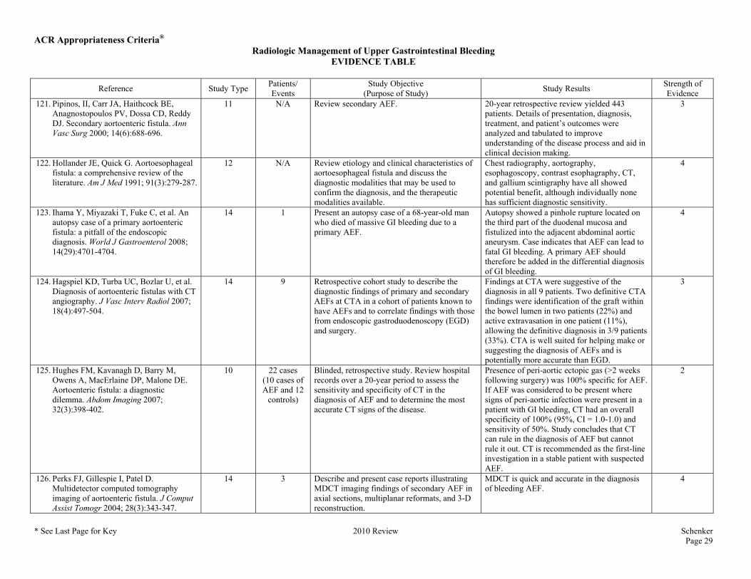
ACR Appropriateness Criteria® Radiologic Management of Upper Gastrointestinal Bleeding
EVIDENCE TABLE
* See Last Page for Key 2010 Review Schenker Page 29
Reference Study Type Patients/ Events
Study Objective (Purpose of Study) Study Results Strength of
Evidence 121. Pipinos, II, Carr JA, Haithcock BE,
Anagnostopoulos PV, Dossa CD, Reddy DJ. Secondary aortoenteric fistula. Ann Vasc Surg 2000; 14(6):688-696.
11 N/A Review secondary AEF. 20-year retrospective review yielded 443 patients. Details of presentation, diagnosis, treatment, and patient’s outcomes were analyzed and tabulated to improve understanding of the disease process and aid in clinical decision making.
3
122. Hollander JE, Quick G. Aortoesophageal fistula: a comprehensive review of the literature. Am J Med 1991; 91(3):279-287.
12 N/A Review etiology and clinical characteristics of aortoesophageal fistula and discuss the diagnostic modalities that may be used to confirm the diagnosis, and the therapeutic modalities available.
Chest radiography, aortography, esophagoscopy, contrast esophagraphy, CT, and gallium scintigraphy have all showed potential benefit, although individually none has sufficient diagnostic sensitivity.
4
123. Ihama Y, Miyazaki T, Fuke C, et al. An autopsy case of a primary aortoenteric fistula: a pitfall of the endoscopic diagnosis. World J Gastroenterol 2008; 14(29):4701-4704.
14 1 Present an autopsy case of a 68-year-old man who died of massive GI bleeding due to a primary AEF.
Autopsy showed a pinhole rupture located on the third part of the duodenal mucosa and fistulized into the adjacent abdominal aortic aneurysm. Case indicates that AEF can lead to fatal GI bleeding. A primary AEF should therefore be added in the differential diagnosis of GI bleeding.
4
124. Hagspiel KD, Turba UC, Bozlar U, et al. Diagnosis of aortoenteric fistulas with CT angiography. J Vasc Interv Radiol 2007; 18(4):497-504.
14 9 Retrospective cohort study to describe the diagnostic findings of primary and secondary AEFs at CTA in a cohort of patients known to have AEFs and to correlate findings with those from endoscopic gastroduodenoscopy (EGD) and surgery.
Findings at CTA were suggestive of the diagnosis in all 9 patients. Two definitive CTA findings were identification of the graft within the bowel lumen in two patients (22%) and active extravasation in one patient (11%), allowing the definitive diagnosis in 3/9 patients (33%). CTA is well suited for helping make or suggesting the diagnosis of AEFs and is potentially more accurate than EGD.
3
125. Hughes FM, Kavanagh D, Barry M, Owens A, MacErlaine DP, Malone DE. Aortoenteric fistula: a diagnostic dilemma. Abdom Imaging 2007; 32(3):398-402.
10 22 cases (10 cases of AEF and 12
controls)
Blinded, retrospective study. Review hospital records over a 20-year period to assess the sensitivity and specificity of CT in the diagnosis of AEF and to determine the most accurate CT signs of the disease.
Presence of peri-aortic ectopic gas (>2 weeks following surgery) was 100% specific for AEF. If AEF was considered to be present where signs of peri-aortic infection were present in a patient with GI bleeding, CT had an overall specificity of 100% (95%, CI = 1.0-1.0) and sensitivity of 50%. Study concludes that CT can rule in the diagnosis of AEF but cannot rule it out. CT is recommended as the first-line investigation in a stable patient with suspected AEF.
2
126. Perks FJ, Gillespie I, Patel D. Multidetector computed tomography imaging of aortoenteric fistula. J Comput Assist Tomogr 2004; 28(3):343-347.
14 3 Describe and present case reports illustrating MDCT imaging findings of secondary AEF in axial sections, multiplanar reformats, and 3-D reconstruction.
MDCT is quick and accurate in the diagnosis of bleeding AEF.
4

ACR Appropriateness Criteria® Radiologic Management of Upper Gastrointestinal Bleeding
EVIDENCE TABLE
* See Last Page for Key 2010 Review Schenker Page 30
Reference Study Type Patients/ Events
Study Objective (Purpose of Study) Study Results Strength of
Evidence 127. Roos JE, Willmann JK, Hilfiker PR.
Secondary aortoenteric fistula: active bleeding detected with multi-detector-row CT. Eur Radiol 2002; 12 Suppl 3:S196-200.
14 1 Report a case of active bleeding of a secondary AEF, in which CTA with MDCT was diagnostic after negative catheter angiography and unsatisfactory endoscopy.
MDCT angiography showed the fistulous tract between the abdominal aortic graft and the duodenum. MDCT angiography with its excellent 3D image quality is recommended.
4
128. Vu QD, Menias CO, Bhalla S, Peterson C, Wang LL, Balfe DM. Aortoenteric fistulas: CT features and potential mimics. Radiographics 2009; 29(1):197-209.
12 N/A Review diagnosis of AEFs with CT. CT has become the imaging modality of choice for evaluations in the emergency setting. CT has widely variable sensitivity (40%-90%) and specificity (33%-100%).
4
129. Mylona S, Ntai S, Pomoni M, Kokkinaki A, Lepida N, Thanos L. Aorto-enteric fistula: CT findings. Abdom Imaging 2007; 32(3):393-397.
12 N/A Pictorial essay presenting the various CT findings seen in patients with aorto-enteric fistulas.
CT provides fast and effective evaluation in hemodynamically stable patients suspected of having an aorto-enteric fistula.
4
130. Low RN, Wall SD, Jeffrey RB, Jr., Sollitto RA, Reilly LM, Tierney LM, Jr. Aortoenteric fistula and perigraft infection: evaluation with CT. Radiology 1990; 175(1):157-162.
10 55 cases AEF (n=23); PGI (n=12); and normal, noninfected
grafts (n=20).
Blinded retrospective study to determine the sensitivity and specificity of CT in detecting perigraft infection (PGI) and AEF.
Two observers reviewed CT scans. Each observer correctly identified as abnormal 33 of 35 cases of PGI either with or without AEF (sensitivity, 94%) and that results were falsely positive in three cases (specificity, 85%). Authors conclude CT will continue to be valuable for diagnosing PGI and AEF.
2
131. Orton DF, LeVeen RF, Saigh JA, et al. Aortic prosthetic graft infections: radiologic manifestations and implications for management. Radiographics 2000; 20(4):977-993.
12 N/A Discuss CT and MRI of aortic prosthetic graft infections and review the radiologic findings, adjunctive studies, and associated conditions.
CT combined with interventional sinography, US, and nuclear medicine allows detection and staging of graft infections and is important in the surgical management of aortic prosthetic graft infections.
4
132. Baril DT, Carroccio A, Ellozy SH, et al. Evolving strategies for the treatment of aortoenteric fistulas. J Vasc Surg 2006; 44(2):250-257.
3c 16 Reviewed experience of open surgical and endovascular treatment of AEFs to compare outcomes over a time period.
Overall 30-day mortality rate was 18.8%. Overall perioperative complication rate was 50.0%. The mean in-hospital length of stay was significantly longer for patients undergoing open repair compared with endovascular repair (44.0 vs 19.4 days; P=.04). The median overall survival after hospital discharge was 23.1 months. Endovascular therapy is alternative to open surgical repair; is associated with decreased perioperative morbidity and mortality and shorter in-hospital stay, and allows for acceptable survival. Endovascular repair provides a therapeutic option to control bleeding and allow for continued intervention in a stabilized setting.
3

ACR Appropriateness Criteria® Radiologic Management of Upper Gastrointestinal Bleeding
EVIDENCE TABLE
* See Last Page for Key 2010 Review Schenker Page 31
Reference Study Type Patients/ Events
Study Objective (Purpose of Study) Study Results Strength of
Evidence 133. Bergqvist D, Bjorck M, Nyman R.
Secondary aortoenteric fistula after endovascular aortic interventions: a systematic literature review. J Vasc Interv Radiol 2008; 19(2 Pt 1):163-165.
7 16 cases of aortoenteric fistulation in 14 patients
with abdominal
aortic aneurysm
Systematic literature review to identify cases of aortoenteric fistulation after aortic stent-graft procedures or stent implantation. Collective incidence of, and experience with, AEF after endovascular aortoiliac therapy was evaluated.
Aortoenteric fistulation occurs after endovascular implantation of stents and stent-grafts. The incidence is unknown but is probably low. Follow-up time in most publications was <1 year, which is considered short to assess potential graft infection.
3
134. Brountzos EN, Vasdekis S, Kostopanagiotou G, et al. Endovascular treatment of a bleeding secondary aorto-enteric fistula. A case report with 1-year follow-up. Cardiovasc Intervent Radiol 2007; 30(5):1037-1041.
5 1 Case report of a patient with life-threatening GI bleeding caused by a secondary aorto-enteric fistula.
Bleeding was successfully stopped by endovascular placement of bifurcated aortic stent-graft. The patient developed periaortic infection 4 months later, but he was managed with antibiotics. The patient is well 1 year after the procedure.
4
135. Busuttil SJ, Goldstone J. Diagnosis and management of aortoenteric fistulas. Semin Vasc Surg 2001; 14(4):302-311.
12 N/A Review diagnosis and management of AEFs. EGD and CT are the most useful tests to diagnose AEF.
4
136. Karkos CD, Vlachou PA, Hayes PD, Fishwick G, Bolia A, Naylor AR. Temporary endovascular control of a bleeding aortoenteric fistula by transcatheter coil embolization. J Vasc Interv Radiol 2005; 16(6):867-871.
5 1 Case report of a 67-year-old man presented with GI bleeding resulting from a fistula between the aortic stump and the fourth part of the duodenum 14 months after an axillobifemoral bypass, graft excision, and aortic stump closure for aortic graft infection.
Coil embolization may be a helpful addition in the management of such problems. This approach may buy time and allow careful planning of a definitive treatment or may serve as a palliative procedure in inoperable patients.
4
137. Leonhardt H, Mellander S, Snygg J, Lonn L. Endovascular management of acute bleeding arterioenteric fistulas. Cardiovasc Intervent Radiol 2008; 31(3):542-549.
5 5 Retrospective study to examine the outcome of endovascular transcatheter repair of emergent arterioenteric fistulas.
4 fistulas were successfully sealed with stent-grafts — technical success rate of 80%. Endovascular repair is efficient and safe — stabilizes patients with life-threatening bleeding arterioenteric fistulas in the emergent episode. But in patients with severe comorbidities, the risk of rebleeding is high and further intervention must be considered. Patients with cancer may only need treatment for the acute bleeding episode, and an endovascular approach has the advantage of low morbidity.
4

ACR Appropriateness Criteria® Radiologic Management of Upper Gastrointestinal Bleeding
EVIDENCE TABLE
* See Last Page for Key 2010 Review Schenker Page 32
Reference Study Type Patients/ Events
Study Objective (Purpose of Study) Study Results Strength of
Evidence 138. Papacharalambous G, Skourtis G,
Saliveros A, et al. Endovascular treatment of a primary aortoduodenal fistula: 2-year follow-up of a case report. Vasc Endovascular Surg 2007; 41(3):265-270.
5 1 To evaluate the role of endovascular management for primary aortoduodenal fistula in poor surgical risk patients. 2-year follow-up study.
At 21-month follow-up, the patient had no symptoms and signs of graft infection, and radiologic studies confirmed decreasing aneurysm size without associated signs of local sepsis. Endovascular stent grafts can efficiently arrest massive exsanguination in critically ill patients with primary AEF. Risk of graft infection remains the most serious problem associated with this approach.
4
139. Suzuki S, Imoto K, Uchida K, Hashiyama N, Takanashi Y. Endovascular repair of a presumed aortoduodenal fistula. Ann Thorac Cardiovasc Surg 2005; 11(6):424-428.
5 1 To describe the use of transluminal endovascular grafting for the treatment of a presumed aortoduodenal fistula.
Endovascular stent-grafts may be beneficial in the management of aortoduodenal fistula.
4
140. Verhey P, Best A, Lakin P, Nachiondo J, Petersen B. Successful endovascular treatment of aortoenteric fistula secondary to eroding duodenal stent. J Vasc Interv Radiol 2006; 17(8):1345-1348.
5 1 To describe the successful endovascular repair of a primary AEF caused by eroding duodenal stent.
Medical history and previous medical and surgical interventions must be considered when evaluating patients. Maintaining a high clinical suspicion for the possibility of an AEF in patients with enteric stents and GI bleeding is important and can help in the diagnosis and successful treatment of an AEF.
4
141. Finch L, Heathcock RB, Quigley T, Jiranek G, Robinson D. Emergent treatment of a primary aortoenteric fistula with N-butyl 2-cyanoacrylate and endovascular stent. J Vasc Interv Radiol 2002; 13(8):841-843.
5 1 To describe treatment involving cyanoacrylate injection followed by endovascular stent placement in the setting of acute exsanguination through a primary AEF.
N-butyl 2-cyanoacrylate was injected into the fistula tract of patient. Rapid polymerization first provided hemostasis and allowed the patient to be resuscitated and stabilized for several hours, and then an AneuRx endovascular stent-graft was deployed to reinforce the aortic wall and permanently occlude the fistula. Approach can benefit patients whose condition would otherwise be too unstable for open surgery.
4

ACR Appropriateness Criteria®
ACR Appropriateness Criteria® Evidence Table Key
Evidence Table Key
Study Type Key Numbers 1-7 are for studies of therapies while numbers 8-15 are used to describe studies of diagnostics.
1. Randomized Controlled Trial — Treatment 2. Controlled Trial 3. Observation Study
a. Cohort b. Cross-sectional c. Case-control
4. Clinical Series 5. Case reviews 6. Anecdotes 7. Reviews
8. Randomized Controlled Trial — Diagnostic 9. Comparative Assessment 10. Clinical Assessment 11. Quantitative Review 12. Qualitative Review 13. Descriptive Study 14. Case Report 15. Other (Described in text)
Strength of Evidence Key Category 1 - The conclusions of the study are valid and strongly supported by study design, analysis and results. Category 2 - The conclusions of the study are likely valid, but study design does not permit certainty. Category 3 - The conclusions of the study may be valid but the evidence supporting the conclusions is inconclusive or equivocal. Category 4 - The conclusions of the study may not be valid because the evidence may not be reliable given the study design or analysis.