radiobiological basics of biodosimetry - nucleus · radiobiological basics of biodosimetry oleg...
TRANSCRIPT

IAEA International Atomic Energy Agency
Radiobiological basics of biodosimetry
Oleg Belyakov Radiation Biologist
Applied Radiation Biology and Radiotherapy Section Division of Human Health

IAEA
Contents
10 June 2013 OvB BIODOSE-21, Hiroshima, Japan 2
• IAEA Cytogenetic biodosimetry publications • Application of dose concepts in biological dosimetry • Biophysical background to chromosome damage • Human lymphocytes • Chromosomal structure • Radiation induced chromosomal alterations • Blood sampling • Production of in vitro dose–response curve • Dicentric analysis • Translocation analysis • Premature chromosome condensation (PCC) analysis • The cytokinesis-block micronucleus (CBMN) assay • Automation of chromosomal assays • Conclusions and acknowledgements

IAEA
Cytogenetic Dosimetry, IAEA 1986
10 June 2013 OvB BIODOSE-21, Hiroshima, Japan 3
IAEA
1986 – IAEA publication: “Biological Dosimetry: Chromosomal Aberration Analysis for Dose Assessment”, Technical report series No 260.

IAEA
Cytogenetic Dosimetry, IAEA 2001
10 June 2013 OvB BIODOSE-21, Hiroshima, Japan 4
IAEA
2001 – IAEA, the second edition manual “Cytogenetic Analysis for Radiation Dose Assessment”, Technical report series No 405.

IAEA
Cytogenetic Dosimetry, IAEA 2011
10 June 2013 OvB BIODOSE-21, Hiroshima, Japan 5
IAEA WHO • 2011 – the third edition was extensively
updated to reflect progress made in cytogenetic biological dosimetry during the past decade and co-sponsored by the Pan American Health Organisation and the World Health Organisation.
• PDF copy can be found on the USB memory stick, which will be distributed among all participants at the end of the meeting.

IAEA
Application of dose concepts in biological dosimetry
10 June 2013 OvB BIODOSE-21, Hiroshima, Japan 6
• Chromosome aberrations (ChA) in lymphocytes are used to estimate absorbed dose.
• Number of ChA is interpreted in relation to absorbed dose by reference to a dose response calibration curve.
• The calibration curve should be established by exposure of blood in vitro to a range of doses of the corresponding quality of radiation.
• Those doses should be measured by physical dosimetry and traceable to a primary or secondary standard.

IAEA
Biophysical background to chromosome damage
10 June 2013 OvB BIODOSE-21, Hiroshima, Japan 7
• Ionising radiation induces primary events, ionisations and excitations by deposition of energy.
• Linear Energy Transfer (LET) reflects amount of energy deposited along the track.
• The effectiveness of different types of radiation endpoint is reflected by Relative Biological Effectiveness (RBE).
• It is equal to the ratio of the dose of the reference radiation (X-rays) to the dose of the particular radiation being studied that produces the same biological effect.
IAEA
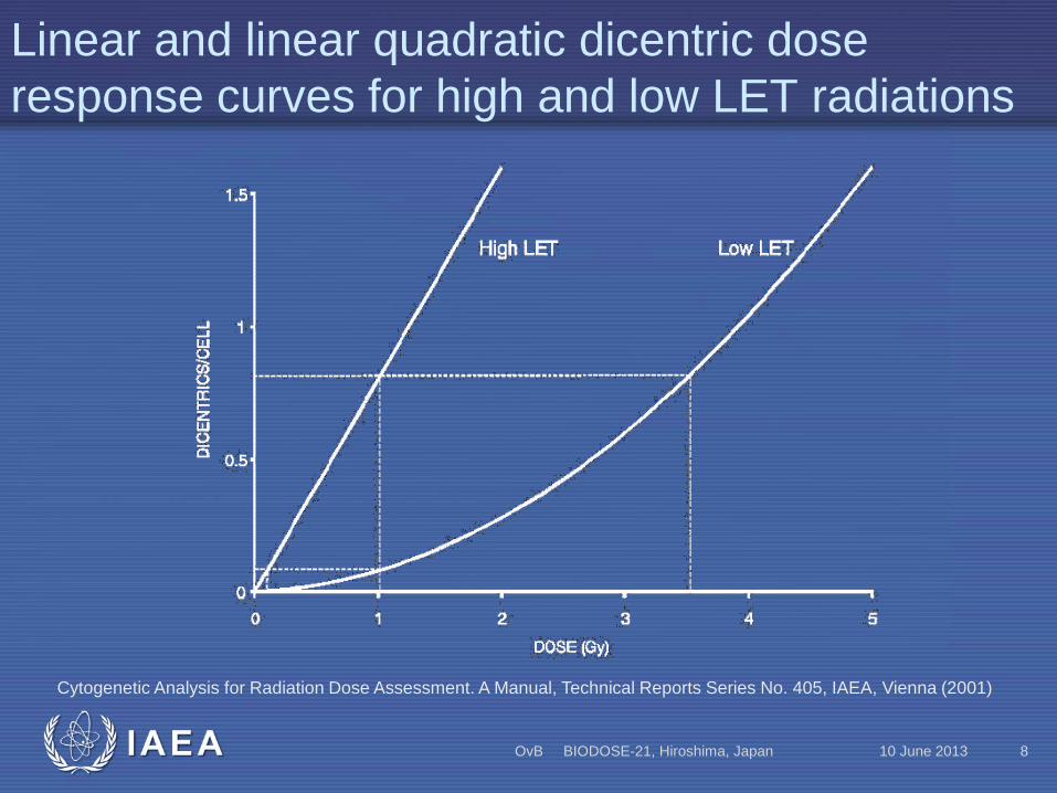
Linear and linear quadratic dicentric dose response curves for high and low LET radiations
10 June 2013 OvB BIODOSE-21, Hiroshima, Japan 8
Cytogenetic Analysis for Radiation Dose Assessment. A Manual, Technical Reports Series No. 405, IAEA, Vienna (2001)

IAEA
Generalized relationship between RBE and LET
10 June 2013 OvB BIODOSE-21, Hiroshima, Japan 9
Cytogenetic Analysis for Radiation Dose Assessment. A Manual, Technical Reports Series No. 405, IAEA, Vienna (2001)

IAEA
Linear Quadratic Model
10 June 2013 OvB BIODOSE-21, Hiroshima, Japan 10
The dose–response curve for low LET induced dicentrics will be a combination of one- and two-track events.
Y = C +αD + βD2
Y - is the yield of dicentrics, D is the dose, C is the control (background frequency), α is the linear coefficient, and β is the dose squared coefficient
The ratio of α/β is equal to the dose at which the linear and the quadratic components contribute equally to the formation of dicentrics.

IAEA
Human peripheral lymphocytes
10 June 2013 OvB BIODOSE-21, Hiroshima, Japan 11
Cytogenetic Dosimetry: Applications in Preparedness for and Response to Radiation Emergencies, EPR-Biodosimetry, IAEA, Vienna (2011)

IAEA
Chromosomal structure and Chromatin Packing
10 June 2013 OvB BIODOSE-21, Hiroshima, Japan 12
Courtesy REAC/TS, USA. Cytogenetic Dosimetry: Applications in Preparedness for and Response to Radiation Emergencies, EPR-Biodosimetry, IAEA, Vienna (2011)

IAEA
Human Karyotype and DNA Content of Chromosomes
10 June 2013 OvB BIODOSE-21, Hiroshima, Japan 13
A banded chromosome/karyotype preparation from a normal male, 46, XY (left) and a normal female 46, XX (right, courtesy Mayo Clinic, USA. Cytogenetic Dosimetry: Applications in Preparedness for and Response to Radiation Emergencies, EPR-Biodosimetry,
IAEA, Vienna (2011)

IAEA
Cell Cycle
10 June 2013 OvB BIODOSE-21, Hiroshima, Japan 14
Courtesy REAC/TS, USA. Cytogenetic Dosimetry: Applications in Preparedness for and Response to Radiation Emergencies, EPR-Biodosimetry, IAEA, Vienna (2011)

IAEA
Radiation induced chromosomal alterations
10 June 2013 OvB BIODOSE-21, Hiroshima, Japan 15
• Ionizing radiation deposits discrete energy events (i.e. spurs, blobs, and tracks) in time and space.
• It damage DNA directly and indirectly by the generation of reactive species mainly produced by the radiolysis of water.
• Low LET radiation can produce localised clusters of ionizations within a single electron track. High-LET radiation produces a larger number of ionisations that are close to each other.

IAEA
Radiation Induced DNA Lesions
10 June 2013 OvB BIODOSE-21, Hiroshima, Japan 16
• DNA including base damage (BD), single strand breaks (SSB), abasic sites (AS), DNA-protein cross-links (DPC), and double strand breaks (DSB).
• Ionization pattern for low- and high-LET radiation and radiation induced DNA lesions is different.
Cytogenetic Dosimetry: Applications in Preparedness for and Response to Radiation Emergencies, EPR-Biodosimetry, IAEA, Vienna (2011)
IAEA
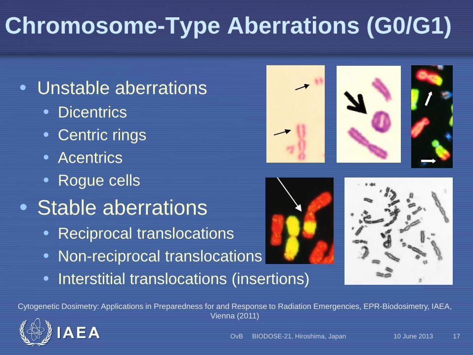
Chromosome-Type Aberrations (G0/G1)
10 June 2013 OvB BIODOSE-21, Hiroshima, Japan 17
• Unstable aberrations • Dicentrics • Centric rings • Acentrics • Rogue cells
• Stable aberrations • Reciprocal translocations • Non-reciprocal translocations • Interstitial translocations (insertions)
Cytogenetic Dosimetry: Applications in Preparedness for and Response to Radiation Emergencies, EPR-Biodosimetry, IAEA, Vienna (2011)

IAEA
Chromatid-Type Aberrations (G2/S)
10 June 2013 OvB BIODOSE-21, Hiroshima, Japan 18
• Terminal and interstitial deletions
• Achromatic lesions • Isochromatid deletions • Asymmetrical interchanges • Symmetrical interchanges • Asymmetrical and
symmetrical interchanges • Triradials Cytogenetic Dosimetry: Applications in Preparedness for and Response to Radiation Emergencies, EPR-Biodosimetry, IAEA,
Vienna (2011)
IAEA
Premature Chromosome Condensation PCC
10 June 2013 OvB BIODOSE-21, Hiroshima, Japan 19
• The PCC technique is a very useful research tool to probe the immediate post-irradiation processes and kinetics of chromosomal break restitution and/or misrepair to form aberrations.
• Fusion-PCC assay is a method when lymphocytes are fused with mitotic cells to induce premature condensation of the human chromosomes.
• Rapid Interphase Chromosome Assay (RICA) allows the visualisation of radiation-induced damage using FISH probes.
• Dic-PCC and Ring-PCC assays.

IAEA
Micronuclei
10 June 2013 OvB BIODOSE-21, Hiroshima, Japan 20
• Micronuclei (MN) are formed from acentric chromosomal fragments or whole chromosomes at anaphase which are not included in the nuclei of daughter cells.
• Cytokinesis block technique by adding cytochalasin B improved test’s sensitivity. John R K Savage Micronuclei : Pitfalls and Problems, July
2000, Atlas of Genetics and Cytogenetics in Oncology and Haematology,
http://atlasgeneticsoncology.org/Deep/MicronucleiID20016.html

IAEA
Blood sampling, timing
10 June 2013 OvB BIODOSE-21, Hiroshima, Japan 21
• A venipuncture blood sample, preferably 10 mL, could be taken within a few hours after a whole body radiation exposure.
• In the case of a partial-body or non-uniform exposure the lymphocytes in the circulating and extravascular pools will not reach equilibrium until about 24 hours.
• In the event of a serious overexposure, a “time window” of possibly only a few hours or days exists before the lymphocyte count drops.

IAEA
Anticoagulant, containers and transport
10 June 2013 OvB BIODOSE-21, Hiroshima, Japan 22
• Preservative free lithium heparin is the most commonly used anticoagulant for lymphocyte cultures, although it is possible to use sodium or ammonium heparin.
• Disposable glass or plastic specimen tubes containing the correct amounts of lithium heparin are available from several manufacturers.
• Blood specimens should be maintained ideally between 18 and 24°C during transportation.
• Non-pathogenic samples are labelled as UN 3373. BIOLOGICAL SUBSTANCE, CATEGORY B.

IAEA
Production of an in vitro dose–response curve, General Considerations
10 June 2013 OvB BIODOSE-21, Hiroshima, Japan 23
• There are differences between laboratories’ calibration curves, so intercomparison exercises are useful.
• Most accidental overexposures involve gamma radiation sources but there are also an appreciable number of events involving X-rays.
• Lymphocytes should be irradiated in vitro to approximate as closely as possible the in vivo situation.

IAEA
Physical Considerations
10 June 2013 OvB BIODOSE-21, Hiroshima, Japan 24
• The preparation of a dose–response curve must be supported by reliable and accurate physical dosimetry.
• During irradiation important to avoid the complications of scattered radiation.
• The exposure set up should be calibrated by physical measurements.
• The usual method of determining doses is to convert the measured air kerma into absorbed dose and then correct as necessary for distance and absorption.

IAEA
Statistical Considerations
10 June 2013 OvB BIODOSE-21, Hiroshima, Japan 25
• Yields of chromosome aberrations or micronuclei (Y) are related to dose (D) by the linear quadratic equation: Y = C + αD + βD2 for low LET radiation.
• For high LET radiation dose response is approximated by the linear equation Y = C + αD.
• Since curve fitting methods are based on Poisson statistics, the dicentric cell distribution should be tested for compliance with the Poisson distribution for each dose used to construct the calibration curve (u test).

IAEA
Culturing
10 June 2013 OvB BIODOSE-21, Hiroshima, Japan 26
• Dicentric assay requires two day and CBMN 3 days cultures.
• Fusion-PCN requires no cell culturing but chemically induced methods generally do.
• Dicentric and other assays on metaphases require mitotic arrest with Colcemid.
• CBMN assay requires cytokinesis blocking with cytochalasin B.

IAEA
Fixation Procedure
10 June 2013 OvB BIODOSE-21, Hiroshima, Japan 27
• Lymphocyte cultures are conventionally incubated for 48-52 hours.
• The cultures should be centrifuged and the supernatant removed and replaced by a hypotonic solution.
• After ~15 min at 37°C cells should be spun again, the hypotonic solution removed and the cell pellet resuspended in freshly prepared fixative (3:1 methanol/acetic acid).
• To make metaphase spreads 2-3 drops of pellet dispensed onto the slide and air dried.

IAEA
Staining
10 June 2013 OvB BIODOSE-21, Hiroshima, Japan 28
• Fluorescence plus Giemsa (FPG) staining is recommended as this permits the analysis to be confined to the first in vitro division metaphases (M1).
• Conventional Giemsa (2%) staining is common. • It is possible to modify the staining specifically to
highlight centromeres by FISH, using a pancentromeric probe or with Giemsa stain using the C-banding method.
IAEA
Analysis of Slides
10 June 2013 OvB BIODOSE-21, Hiroshima, Japan 29
• With conventional microscopy, first, the scanning should be done at low magnification (~x100-x200).
• After that, the scorer analyse aberrant metaphase spreads found, using higher magnification (~x1000-x2000).
• Computer assisted microscopy can be used, these systems include semi-automated analysis of digitised images that assist with locating aberrant chromosomes, however, no system is fully automatic.

IAEA
Recording of Data
10 June 2013 OvB BIODOSE-21, Hiroshima, Japan 30
• Good laboratory practice requires that a unique identifier code or labelling system should be used for specimens, slides and associated paperwork.
• The receipt and processing of specimens, whether for experiments or for overdose investigations, should be recorded in a laboratory diary.
• Electronic systems for data storage and handling are available.

IAEA
Storage of Information and Slides
10 June 2013 OvB BIODOSE-21, Hiroshima, Japan 31
• Research data have to be filed and stored for future reference.
• Most laboratories store the microscope slides in a box in a dry place at room temperature.
• Faded slides can be retrieved by carefully soaking off the coverslip and restaining.
IAEA
Dose Assessment
10 June 2013 OvB BIODOSE-21, Hiroshima, Japan 32
• Appropriate calibration curve have to be chosen. • It is suggested that 500 cells or 100 dicentrics
should be scored in order to give a reasonably accurate estimate of dose.
• Uncertainty on dose estimates should be taken in consideration.
• Special procedures are required in case of criticality accidents, low dose overexposure cases, partial body exposures, delayed blood sampling, protracted and fractionated exposure, internal incorporation of radionuclides etc.

IAEA
Translocation analysis
10 June 2013 OvB BIODOSE-21, Hiroshima, Japan 33
• Known drawback of the dicentric and CBMN assays is that the damage is unstable and therefore is eliminated from the peripheral blood lymphocyte pool at the rate that cell renewal occurs.
• The introduction of Florescent In Situ Hybridisation (FISH) technique has opened the possibility to detect translocations.
Cytogenetic Dosimetry: Applications in Preparedness for and Response to Radiation Emergencies, EPR-Biodosimetry, IAEA, Vienna (2011)

IAEA
Cell Culture and Fixing Procedures
10 June 2013 OvB BIODOSE-21, Hiroshima, Japan 34
• The procedures for obtaining blood, culturing lymphocytes and harvesting fixed cells are similar to those described for the dicentric assay.
• Generally, for biological dosimetry, only a part of the genome is painted. This leads to the requirement to score more metaphases than would be scored with the dicentric assay.
• It is therefore helpful and more cost effective to produce slides, each with a large number of scorable quality metaphases.

IAEA
Painting the Chromosomes
10 June 2013 OvB BIODOSE-21, Hiroshima, Japan 35
• Whole chromosome painting. • Use of pancetromeric probe. • Painting all chromosomes by the method known as
multicolour FISH (mFISH). • Intrachromosomal exchanges such as pericentric
inversions may be detected by selectively painting the p and q arms of a chromosome in different colours.
• Rearrangements within a single arm may be detected by mBAND where multi-coloured banding is produced along a chromosome.

IAEA
Scoring Criteria
10 June 2013 OvB BIODOSE-21, Hiroshima, Japan 36
• Selection of scorable cells involves a certain degree of judgement because the limits of resolution with current FISH technology are about 11–15 Mbp.
• Nomenclature and recording data: • PAINT was developed to be purely descriptive of each
aberrant painted object in the metaphase. • Savage and Simpson (S&S) proposed a terminology
comprising numerals and letters describing each exchange in its entirety.
• Nowadays the most widely used method is modified PAINT.

IAEA
Data Handling
10 June 2013 OvB BIODOSE-21, Hiroshima, Japan 37
Lucas et al., 1992 derived the equations for calculating genome equivalence, and these have been further summarised by Lucas and Deng in 2000.
FG is the full genome aberration frequency, Fp is the translocation frequency detected by FISH, and
fp is the fraction of genome hybridised, taking into account the gender of the subjects.

IAEA
The Control Level of Translocations
10 June 2013 OvB BIODOSE-21, Hiroshima, Japan 38
Control levels of translocations are higher than for dicentrics, and to some extent this is due to the higher stability of this type of aberration (Sigurdson et al. 2008).

IAEA
Persistence of Translocations
10 June 2013 OvB BIODOSE-21, Hiroshima, Japan 39
• The greatest disadvantage of the dicentric method is that the aberration yield in exposed people decreases with time after exposure.
• Stable ChA background level is higher than in case of unstable ChA and tend to increase with age.
• Therefore sensitivity of translocation assay is generally lower than for dicentric one.
• Good control and follow up is required.

IAEA
Calibration Curves
10 June 2013 OvB BIODOSE-21, Hiroshima, Japan 40
• Each laboratory needs to establish its own curves for dose estimations with FISH translocation method.
• FISH dose estimations generally will be undertaken for cases where doses are high, but protracted or after low radiation exposure a long time ago, revealing no medical symptoms.
• In contrast to acute exposure dosimetry, where the linear quadratic curve will be used, here the linear α term of the dose response curve is crucially important.

IAEA
Examples of Fish Being Used for Retrospective Biological Dosimetry
10 June 2013 OvB BIODOSE-21, Hiroshima, Japan 41
• In populations with no prior biological and physical dosimetry investigation.
• In populations with known physical dosimetry estimates.
• In populations with known biological dosimetry estimates using conventional dicentric analysis immediately following exposure (like “tritium”, Goiânia, German, Estonian, Istanbul and Georgian accidents.

IAEA
Premature chromosome condensation (PCC) analysis
10 June 2013 OvB BIODOSE-21, Hiroshima, Japan 42
• Biological dosimetry is generally performed by analysing dicentrics and/or translocations at the first mitosis.
• These assays have several problems, namely radiation induced mitotic delay and cell death, especially after high doses, which can cause considerable underestimation of the dose.
• PCC performed some time before the first mitosis and reduces or eliminates aforementioned issues.

IAEA
PCC
10 June 2013 OvB BIODOSE-21, Hiroshima, Japan 43
• CHO mitotic cells can be used to induce PCC. • Standard chromosome breaks analysis. • Dicentric analysis using C banding. • Translocation and dicentric analysis using the
chromosome painting assay. • PCC by chemical induction.
• The rapid interphase chromosome assay (RICA). • The PCC ring assay.

IAEA
RICA assay with FISH painted human chromosomes in interphase lymphocytes
10 June 2013 OvB BIODOSE-21, Hiroshima, Japan 44
Courtesy Pathak and Prasanna, AFRRI, USA Cytogenetic Dosimetry: Applications in Preparedness for and Response to Radiation Emergencies, EPR-Biodosimetry, IAEA,
Vienna (2011)

IAEA
The cytokinesis-block micronucleus (CBMN) assay
10 June 2013 OvB BIODOSE-21, Hiroshima, Japan 45
• Acentric chromosome fragments or whole chromosome can form into a small separate nucleus; hence the term micronucleus.
• In the 1990s the CBMN–centromere assay was developed, using FISH and a pancentromeric probe to visualise centromeres.
• More recently an advanced version of the CBMN assay (known as the Cytokinesis-Block Micronucleus Cytome - CBMN Cyt) assay has been developed and validated.
IAEA
CBMN Assay Scoring Criteria
10 June 2013 OvB BIODOSE-21, Hiroshima, Japan 46
• The cells should be binucleated (BN). • Nuclei in should have intact membranes and be in the
same cell. • Nuclei in should be equal in size, staining pattern and
intensity. • Nuclei may be connected by fine nucleoplasmic bridges. • Nuclei in may touch but ideally should not overlap. • A cell with overlapping nuclei can be scored if
boundaries are distinguishable. • The cytoplasmic boundary should be intact and
distinguishable from other cells.

IAEA
Criteria for scoring micronuclei
10 June 2013 OvB BIODOSE-21, Hiroshima, Japan 47
• The diameter of MN should varies between 1/16th and 1/3rd of the mean diameter of the main nuclei.
• MN are non-refractile. • MN are not linked or connected to the main nuclei. • MN may touch but not overlap the main nuclei,
boundary should be distinguishable from nuclear one.
• MN usually have the same staining intensity as the main nuclei but occasionally staining may be more intense.

IAEA
Application of the CBMN Assay for Biological Dosimetry
10 June 2013 OvB BIODOSE-21, Hiroshima, Japan 48
CBMN assay is sensitive enough to detect the genetic damage in circulating lymphocytes from exposure to low averaged whole body dose from internal and external exposures:
• Patient studies (i.e. radioiodine case study); • Biomonitoring studies; • Accident studies such as Chernobyl and Istanbul
accidents, Semipalatinsk Nuclear Test Site, accident with a 50 kV contact radiotherapy X ray device.
• Large scale radiation accidents, when hundreds of people may be exposed and triage is needed.

IAEA
Automation of chromosomal assays
10 June 2013 OvB BIODOSE-21, Hiroshima, Japan 49
Cytogenetic laboratory automation involves: • Automation of sample preparation; • Automation of analysis; • Laboratory information management system for
sample tracking and data handling.

IAEA
Automated Sample Processing
10 June 2013 OvB BIODOSE-21, Hiroshima, Japan 50
Automated sample-processing in a cytogenetic laboratory may consist of any or all of the following equipment stations:
• A robotic blood handler; • Metaphase harvester; • Metaphase spreader; • Slide stainer.

IAEA
Automated Image Analysis
10 June 2013 OvB BIODOSE-21, Hiroshima, Japan 51
• Automated analysis of images captured in a microscope is not used routinely for biological dosimetry yet.
• Metaphase finding and image capture systems. • Automation of the dicentric assay. • Automated scoring of micronuclei. • PCC assay automation is similar to that for
dicentric analysis. • FISH automated translocation assay is under
development.
IAEA
Laboratory Information Management System (LIMS)
10 June 2013 OvB BIODOSE-21, Hiroshima, Japan 52
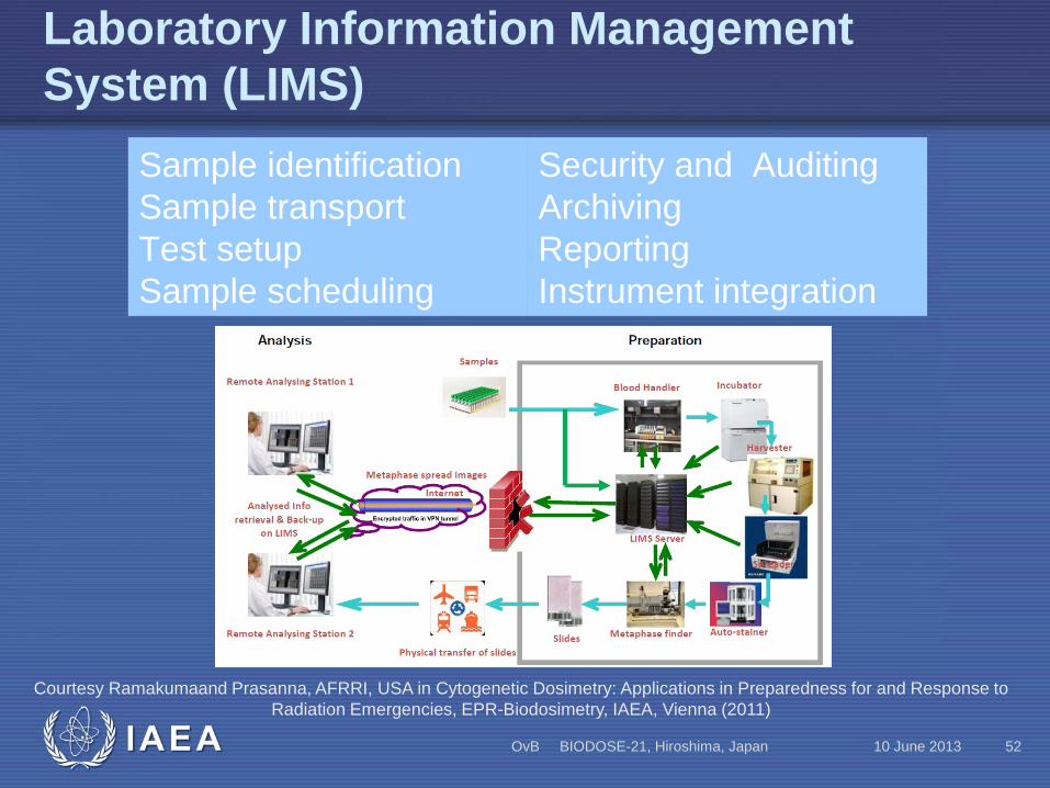
Sample identification Sample transport Test setup Sample scheduling
Security and Auditing Archiving Reporting Instrument integration
Courtesy Ramakumaand Prasanna, AFRRI, USA in Cytogenetic Dosimetry: Applications in Preparedness for and Response to Radiation Emergencies, EPR-Biodosimetry, IAEA, Vienna (2011)

IAEA
Conclusions
10 June 2013 OvB BIODOSE-21, Hiroshima, Japan 53
• Chromosome based retrospective biological dosimetry became an important tool for validation physical measurements and reconstructed doses.
• A variety of methods have been developed and more are pending validation.
• Conventional “Dicentric analysis” in stimulated blood lymphocytes remains the “golden standard” of biodosimetry.
• FISH based translocation analysis, PCC and CBMN assays also became mature enough to be used for accurate estimation of doses.
IAEA
Acknowledgments
10 June 2013 OvB BIODOSE-21, Hiroshima, Japan 54
Contributors to drafting and review Ainsbury, E. Health Protection Agency, United Kingdom Barquinero, J.F. Universidad Autónoma de Barcelona, Spain Beinke, C. Bundeswehr Institute of Radiobiology, Germany Blakely, W.F. Armed Forces Radiobiology Research Institute, United States of America Braselmann, H. Helmholz Zentrum, Germany Buglova, E. International Atomic Energy Agency (IAEA) Carr, Z. World Health Organization (WHO) Di Giorgio, M. Autoridad Regulatoria Nuclear, Argentina Fenech, M. Commonwealth Scientific and Industrial Research Organisation (CSIRO), Australia Garcia Lima, O. Center for Hygiene and Radiation Protection, Cuba Kodama, Y. Radiation Effects Research Foundation, Japan Lindholm, C. Radiation and Nuclear Safety Authority (STUK), Finland Livingston, G. Oak Ridge Associated Universities (ORAU), United States of America Lloyd, D.C. Consultant, United Kingdom Maznyk, N.A. Institute for Medical Radiology of ANSU, Ukraine Prasanna, P.G.S. National Institutes of Health, United States of America Previsani, N. World Health Organization (WHO) Romm, H. Bundesamt für Strahlenschutz (BfS), Germany Roy, L. Institut de Radioprotection et de Sûreté Nucléaire (IRSN), France Voisin, P.J. Institut de Radioprotection et de Sûreté Nucléaire (IRSN), France Vral, A. Ghent University, Belgium Wilkins, R.C. Health Canada, Canada Yoshida, M. Hirosaki University, Japan
Comments received Azizova, T. Southern Urals Biophysics Institute, Russian Federation Barrios, L. Autonomous University of Barcelona, Spain Bognár, G. National Research Institute for Radiobilogy and Radiohygiene, Hungary Darroudi, F. Leiden University Medical Centre, Netherlands Devantier, Y, Chalk River Laboratories, Canada Espinoza Zevallos, M. Instituto Peruano de Energía Nuclear, Peru Guerrero Carbajal, Y.C. Instituto Nacional de Investigaciones Nucleares, Mexico Güçlü, I. Çekmece Nuclear Research and Training Centre (ÇNAEM), Turkey Hayata, I. Consultant, Japan Martínez-López , W. Instituto de Investigaciones Biológicas Clemente Estable, Uruguay Natarajan, A.T. University of Tuscia, Italy Oliveira, M. Instituto de Radioproteção e Dosimetria, Brasil Palitti, F. University of Tuscia, Italy Pantelias, G. Institute of Radioisotopes and Radiodiagnostics, National Centre for Scientific Research “Demokritos”, Greece Sasaki, M.S. Kyoto University, Japan Sotnik, N. Southern Urals Biophysics Institute, Russian Federation Turai, I. National Research Institute for Radiobiology and Radiohygiene, Hungary Valdivia Pottstock, P. Chilean Nuclear Energy Commission, Chile Vozilova, A. Urals Research Centre for Radiation Medicine (URCRM), Russian Federation Wilkinson, D. Defence and Research Development Canada, Canada